synaptic targets of thalamic reticular nucleus terminals...
TRANSCRIPT

Synaptic Targets of Thalamic ReticularNucleus Terminals in the Visual
Thalamus of the Cat
SITING WANG,1 MARTHA E. BICKFORD,1* SUSAN C. VAN HORN,2 ALEV ERISIR,3
DWAYNE W. GODWIN,4AND S. MURRAY SHERMAN2
1Department of Anatomical Sciences and Neurobiology, University of Louisville, School ofMedicine, Louisville, Kentucky 40292
2Department of Neurobiology, State University of New York, Stony Brook,New York 11794-5230
3Department of Psychology, University of Virginia, Charlottesville, Virginia 29904-44004Department of Neurobiology and Anatomy, Wake Forest University, School of Medicine,
Winston-Salem, North Carolina 27157-1010
ABSTRACTA major inhibitory input to the dorsal thalamus arises from neurons in the thalamic reticular
nucleus (TRN), which use gamma-aminobutyric acid (GABA) as a neurotransmitter. We exam-ined the synaptic targets of TRN terminals in the visual thalamus, including the A lamina of thedorsal lateral geniculate nucleus (LGN), the medial interlaminar nucleus (MIN), the lateralposterior nucleus (LP), and the pulvinar nucleus (PUL). To identify TRN terminals, we injectedbiocytin into the visual sector of the TRN to label terminals by anterograde transport. We thenused postembedding immunocytochemical staining for GABA to distinguish TRN terminals asbiocytin-labeled GABA-positive terminals and to distinguish the postsynaptic targets of TRNterminals as GABA-negative thalamocortical cells or GABA-positive interneurons. We foundthat, in all nuclei, the TRN provides GABAergic input primarily to thalamocortical relay cells(93–100%). Most of this input seems targeted to peripheral dendrites outside of glomeruli. TheTRN does not appear to be a significant source of GABAergic input to interneurons in the visualthalamus. We also examined the synaptic targets of the overall population of GABAergic axonterminals (F1 profiles) within these same regions of the visual thalamus and found that the TRNcontacts cannot account for all F1 profiles. In addition to F1 contacts on the dendrites ofthalamocortical cells, which presumably include TRN terminals, another population of F1 pro-files, most likely interneuron axons, provides input to GABAergic interneuron dendrites. Ourresults suggest that the TRN terminals are ideally situated to modulate thalamocortical trans-mission by controlling the response mode of thalamocortical cells. J. Comp. Neurol. 440:321–341,2001. © 2001 Wiley-Liss, Inc.
Indexing terms: gamma amino butyric acid; lateral geniculate nucleus; lateral posterior nucleus;
medial interlaminar nucleus; pulvinar nucleus; perigeniculate nucleus
A major inhibitory input to the dorsal thalamus arisesfrom the thalamic reticular nucleus (TRN), a sheet-likestructure that surrounds the rostral and lateral bordersof the thalamus. All cells in the TRN use gamma-aminobutyric acid (GABA) as a neurotransmitter(Houser et al., 1980; Oertel et al., 1983; Fitzpatrick etal., 1984; Yen et al., 1985; de Biasi et al., 1986; Rinviket al., 1987; Rinvik and Ottersen, 1988; Spreafico et al.,1991) and are thought to function in the inhibition ofthe thalamocortical signals. In particular, reciprocalprojections between the TRN and thalamocortical cellsare thought to underlie a rhythmic firing of the thala-
mus, which may block the transfer of sensory signalsduring slow wave sleep (Livingstone and Hubel, 1981;
Grant sponsor: NIH; Grant number: NS35377; Grant number: NE12138;Grant number: EY11695; Grant number: EY03038; Grant number:EY11409; Grant sponsor: NSF; Grant number: 9728089.
*Correspondence to: Martha E. Bickford, Department of AnatomicalSciences and Neurobiology, University of Louisville School of Medicine,500 S. Preston Street, Louisville, KY 40292.E-mail: [email protected]
Received 15 February 2001; Revised 26 July 2001; Accepted 28 August2001
THE JOURNAL OF COMPARATIVE NEUROLOGY 440:321–341 (2001)
© 2001 WILEY-LISS, INC.

McCormick and Feeser, 1990; Steriade and McCarley,1990; McCormick and Bal, 1997).
More modality-specific functions of the TRN are sug-gested by the topographic projections of the TRN to indi-vidual thalamic relay nuclei. Based on the precision ofthese projections, the TRN can be divided into modality-specific sectors (Minderhoud, 1971; Jones, 1975; Monteroet al., 1977; Steriade et al., 1984; Crabtree and Killackey,1989; Conley and Diamond, 1990; Conley et al., 1991;Harting et al., 1991; Crabtree, 1992, 1996, 1998; Pinaultand Deschenes, 1998). For example, the primary visualsector of the TRN, which includes the perigeniculate nu-cleus (PGN; a part of the TRN), projects topographically tothe dorsal lateral geniculate nucleus (LGN), which relaysvisual signals from the retina to the visual cortex. Projec-tions from the visual TRN to the LGN are also alignedwith topographic projections from the retina and visualcortex (Montero et al., 1977; Crabtree and Killackey, 1989;Conley and Diamond, 1990; Harting et al., 1991; Colemanand Mitrofanis, 1996).
The visual sector of the TRN also projects to other visualthalamic nuclei, such as the pulvinar and lateral posterior(LP) nuclei. These nuclei receive visual information frommore indirect sources such as the superior colliculus, pre-tectum, and a variety of visual cortical areas (Jones andPowell, 1971; Niimi et al., 1971; Kawamura et al., 1974;Berman, 1977; Berson and Graybiel, 1978, 1983; Graybieland Berson, 1980; Robertson and Cunningham, 1981; Up-dyke, 1981; Raczkowski and Rosenquist, 1983; Rodrigo-Angulo and Reinoso-Suarez, 1995). The projections fromthe TRN to the pulvinar/LP complex appear to be morediffusely organized than the projections to the LGN(Rodrigo-Angulo and Reinoso-Suarez, 1988; FitzGibbon,1994; FitzGibbon et al., 1995; Guillery et al., 1998) andmay arise from distinct cells within the TRN (Sumitomo etal., 1988; Conley and Diamond, 1990).
To understand more fully the functions of the projec-tions from the TRN to the visual thalamus, a detaileddescription of the synaptic organization of TRN terminalsis needed. Several previous studies have reported thatTRN terminals in the LGN contain flattened or pleomor-phic vesicles and form symmetric synapses on dendritesand somata (Montero and Scott, 1981; Harting et al., 1991,Cucchiaro et al., 1991a). However, it has not been deter-mined whether TRN terminals primarily contact thalamo-cortical cells or interneurons in visual relay nuclei of the
thalamus. It is also not known whether the projectionsfrom the TRN to the various nuclei of the visual thalamustarget different regions of the neuropil. To address thisissue, we injected the visual sector of the TRN with bio-cytin to label TRN terminals by anterograde transport. Byusing the electron microscope, we examined the synaptictargets of TRN terminals in the A lamina of the LGN, themedial interlaminar nucleus (MIN), the lateral subdivi-sion of the lateral posterior (LP) nucleus, and the pulvi-nar. Postembedding immunocytochemical staining forGABA was used to further characterize biocytin-labeledTRN terminals and to determine whether the postsynap-tic targets were GABAergic (i.e., interneurons) or non-GABAergic (i.e., thalamocortical relay cells). Some ofthese results were previously published in abstract form(Bickford et al., 1994; Wang et al., 1999).
MATERIALS AND METHODS
Tracer injections
A total of 10 cats were used in this study. All procedureswere conducted in accordance with NIH guidelines for thecare and use of laboratory animals and were approved bythe State University of New York at Stony Brook AnimalCare and Use Committee and the University of LouisvilleAnimal Care and Use Committee. Eight cats receivedbilateral injections of biocytin in the vicinity of the TRN.One cat received a biocytin injection in the right LGN anda biocytin injection in the left TRN. To inject the biocytin,cats were deeply anesthetized with an intravenous injec-tion of sodium phenobarbital (initially 15 mg/kg, with 5- to10-mg supplements when needed) and placed in a stereo-taxic apparatus. The heart rate was monitored, and therectal temperature was maintained at 38°C by means of afeedback-controlled heating blanket. A craniotomy wasperformed, and the dura was reflected. By using stereo-taxic coordinates, a glass pipette (5- to 10-mm tip diame-ter) containing a solution of 5% biocytin (Sigma ChemicalCompany, St. Louis, MO) in saline was lowered verticallyinto the TRN overlying the LGN on each side. Iontophore-sis was achieved by means of DC current (1.5 mA for 15minutes), and the pipette was removed.
One cat received a unilateral injection of fluorescein-labeled latex microspheres in the pulvinar nucleus. Thecat was initially anesthetized with an intramuscular in-jection of ketamine (10 mg/kg) and intubated for gas an-esthesia (0.5–1% nitrous oxide and 1–2% halothane). Thecat was then placed in a stereotaxic apparatus and pre-pared for sterile surgery. A small area of the skull over-lying the pulvinar nucleus was removed, and the dura wasreflected. A Hamilton syringe containing an undilutedsolution of green fluorescent latex microspheres (Luma-Fluor, Naples, FL) was lowered vertically into the pulvi-nar nucleus, and a volume of 0.1 ml was injected.
Histology
Three hours after the biocytin injections, the cats wereperfused through the aorta with saline followed by 2 litersof fixative solution (2% paraformaldehyde and 2% glutar-aldehyde in 0.1 M phosphate buffer). One week after themicrosphere injection, the cat was perfused by using afixative solution of 4% paraformaldehyde. The brains wereremoved and immersed in the fixative solution overnight.The following day, the thalamus was cut into 50-mm-thick
Abbreviations
A, AI, C laminae of the LGNF1 GABAergic profile with densely packed vesiclesF2 GABAergic profile with loosely packed vesiclesGABA gamma-aminobutyric acidIC inferior colliculusLGN dorsal lateral geniculate nucleusLP lateral posterior nucleusMGN medial geniculate nucleusMIN medial interlaminar nucleusOT optic tractPGN perigeniculate nucleusPUL pulvinar nucleusRL large profile with round vesiclesRLP large profile with round vesicles and pale mitochondriaRS small profile with round vesiclesTRN thalamic reticular nucleusvLGN ventral lateral geniculate nucleus
322 S. WANG ET AL.

sections by using a Vibratome and collected in 0.1 Mphosphate buffer (PB; pH 7.4). Sections containing fluo-rescent microspheres were mounted on slides for exami-nation under blue light.
Sections that contained biocytin were incubated over-night at 4°C in a 1:200 dilution of avidin and biotinylatedhorseradish peroxidase (Vector, Burlingame, CA) inphosphate-buffered saline (PBS; 0.01 M PB with 0.9%NaCl, pH 7.4), with 1% normal goat serum and 0.5%Triton added to sections used only for light level analysis.The next day, the sections were rinsed three times in PB(10 minutes each) and reacted with nickel intensified dia-minobenzidine (DAB) for 30 minutes. After buffer washes,sections were either mounted on slides for light level ex-amination or prepared for electron microscopy as de-scribed below.
Electron microscopy
Selected sections were post-fixed in osmium, dehy-drated in an alcohol series, and embedded in Durcupanresin (Ted Pella, Redding, CA). Ultra-thin sections werecut, and every tenth section was collected on Formvar-coated nickel slot grids. Sections were stained for thepresence of GABA by using previously reported postem-bedding immunocytochemical techniques (Patel and Bick-ford, 1997; Patel et al., 1999; Carden and Bickford, 1999;Datskovskaia et al., 2001). We used a rabbit polyclonalanti-GABA antibody (Sigma) at a dilution of 1:2,500. As inour previous studies, the GABA antibody was tagged witha goat anti-rabbit antibody conjugated to 15-nm gold par-ticles (Amersham, Arlington Heights, IL). GABA-stainedsections were examined by using an electron microscope,and biocytin-labeled terminals that also contained a highdensity of gold particles were photographed when sec-tioned through a synaptic contact. For comparison, wealso photographed synaptic contacts made by terminals inthe surrounding neuropil that contained a high density ofgold particles but were not biocytin labeled.
Quantification of GABA labeling
We quantified the GABA labeling by using previousmethods (Patel and Bickford, 1997; Carden and Bickford,1999; Patel et al., 1999; Datskovskaia et al., 2001). Briefly,for presynaptic profiles, and profiles postsynaptic to them,we calculated the gold density by counting the overlyinggold particles and measuring the profile areas by using adigitizing tablet. The density of gold particles within theseprofiles was then compared with the density of gold par-ticles within small profiles with round vesicles (RS pro-files), which are always GABA-negative in the LGN(Montero and Singer, 1985; Beaulieu and Cynader, 1992;Godwin et al., 1996; Erisir et al., 1997).
Computer-generated figures
The distribution of cells in the TRN labeled by theretrograde transport of biocytin or fluorescent latex micro-spheres was plotted by using a Minnesota Datametricsplotting system (Shoreview, MN), and the figure was com-posed by using Freelance Graphics (Cambridge, MA). Toillustrate TRN injection sites, and the resulting cell andterminal distributions, sections were drawn by using acamera lucida attachment. The sketches were digitized byusing an Arcus II flatbed scanner (AGFA, Woburn, MA)and composed by using Freelance Graphics. Light levelphotographs were taken by using a digitizing camera
(Spot RT, Diagnostic Instruments Incorporated, SterlingHeights, MI). We used Photoshop software (Adobe Sys-tems Incorporated, San Jose, CA) to adjust the brightnessand contrast of the images.
Identification of terminal types
We identified five terminal types: three GABA-negative(RS, RL, and RLP) and two GABA-positive (F1 and F2).RS profiles are small; contain densely packed round vesi-cles; and form short, highly asymmetric, contacts. Theseterminals originate from brainstem sources (de Lima etal., 1985; Raczkowski and Fitzpatrick, 1989; Beaulieu andCynader, 1992; Erisir et al., 1997; Patel and Bickford,1997; Patel et al., 1999) or cortical layer VI (Jones andPowell, 1969; Vidnyanszky and Hamori, 1994; Erisir et al.1997; Bourassa and Deschenes, 1995; Ojima et al., 1996;Erisir et al., 1997). RL profiles are large; contain moder-ately packed round vesicles; and form multiple, short,slightly asymmetric contacts. These terminals originatefrom cells in cortical layer V and are found in thepulvinar/LP complex (Mathers, 1972; Robson and Hall,1977; Hoogland et al., 1991; Pare and Smith, 1996; Vid-nyanszky et al., 1996). RLP profiles are large, containmoderately packed round vesicles, and pale mitochondria.Similar to RL profiles, they form multiple, short, slightlyasymmetric contacts. These terminals originate from theretina (Guillery, 1969; Robson and Mason, 1979; Hamos etal., 1987) and are found in the LGN and MIN. It has beensuggested that RL and RLP terminals are the same typefound in the pulvinar/LP complex and LGN, respectively(Guillery, 1969; Hajdu et al., 1974).
F1 profiles contain densely packed, pleomorphic vesi-cles, and form symmetric contacts. F2 profiles containmore scattered, pleomorphic vesicles and form symmetriccontacts. Based on studies of the LGN, these profiles likelyrepresent the dendritic terminals of interneurons(Montero and Scott, 1981; Montero and Singer, 1984;Hamos et al., 1985). The main difference between the F1and F2 terminals is the packing density of vesicles; thisgives the F1 profiles a darker appearance when comparedwith the lighter F2 profiles. In addition, F2 profiles can bemuch larger than F1 profiles.
Sampling methods
The purpose of this study was to determine whetherTRN terminals contact thalamocortical cells or interneu-rons. In addition, we determined the size of the profilestargeted by TRN terminals. To accomplish this, we exam-ined tissue from the visual thalamus that contained la-beled TRN terminals. We photographed the labeled TRNterminals if they were involved in a synaptic connection. Ifthe size of the synaptic zones of TRN terminals variedwith their location on the postsynaptic dendritic arbors,our sample would be biased in favor of the larger synapticzones. To determine whether the synaptic zones of TRNterminals varied with location, we measured the length ofthe synaptic contacts and the diameter of the postsynapticprofiles and tested for statistically significant correlationsby using the Pearson test.
This study did not attempt to determine the density ofTRN terminals within a volume of tissue. Therefore, ste-reologic methods that have been developed to correct forsampling biases to determine an accurate count of neu-rons or terminals within a given volume of tissue were notappropriate for this study. Instead, we sampled the TRN
323TRN TERMINALS IN THE VISUAL THALAMUS

terminals that were labeled and determined their relativedistribution on the dendritic arbors of thalamocorticalcells and interneurons within the same blocks of tissue.Therefore, any shrinkage effects were applied equally toall postsynaptic profiles.
Similarly, we did not correct for the anatomic distribu-tion of dendritic profiles within the visual thalamus. Al-though distal dendrites are more numerous than proximaldendrites and, therefore, will be encountered more oftenwithin a block of tissue, this finding simply reflects themorphology of thalamic neurons, and it is inappropriate to“correct” for this nonrandom distribution (Benes andLange, 2001). Finally, it should be pointed out that ouranalysis of postsynaptic profiles was secondary to thesampling of TRN terminals. We did not sample a variety ofdendrites to determine whether they were contacted byTRN terminals. Rather, we sampled TRN terminal syn-aptic zones and subsequently determined what type ofprofile was contacted. Therefore, any biases in our sam-pling methods would be introduced by variations in thesize of the synaptic zones of the labeled TRN terminalsand not by variations in the size of the postsynaptic pro-files.
RESULTS
Location of TRN cells that project to theLGN and pulvinar nucleus
As illustrated in Figure 1, injections in the LGN orpulvinar label cells within the TRN overlying the LGN.Injections in the LGN label a restricted subset of TRNcells that, for the most part, directly overlie the injectionsite. In contrast, injections in the pulvinar (as previouslyreported by FitzGibbon, 1994) label cells throughout therostrocaudal and mediolateral extent of the visual TRN.Thus, injections placed within restricted regions of thevisual TRN should label terminals within a restrictedregion of the LGN, whereas labeled terminals in the pulv-inar nucleus should be more widely distributed.
Morphology and distribution of labelresulting from injections in the visual TRN
To examine the synaptic targets of TRN terminals in thevisual thalamus, small injections of biocytin were placedin the TRN overlying the LGN. The injection sites weredorsal to the LGN and included both the outer tier of theTRN as well as the PGN. Figure 2 illustrates an exampleof an injection site and the resulting label in the visualthalamus in plots through a series of parasagittal sec-tions. TRN injections resulted in dense columns of over-lapping fibers labeled by anterograde transport and cellslabeled by retrograde transport within the LGN, MIN, andthe LP nucleus. The pulvinar and the ventral LGN con-tained sparser label.
Figure 3 shows examples of the anterograde and retro-grade labeling that resulted from the biocytin injections inthe TRN. Many thalamocortical cells were labeled in aGolgi-like manner (Fig. 3A), and two types of fibers wereidentified: fine fibers with short side branches ending interminal boutons (Fig. 3B; type I; Guillery, 1966), mostlikely of cortical origin (Jones and Powell, 1969), andthicker fibers with beaded swellings (Fig. 3C–E), whichlikely arise from the TRN (Uhlrich et al., 1991).
Ultrastructure of labeled structures
At the ultrastructural level, three types of labeled pro-files could be distinguished by their vesicle morphologyand GABA staining. GABA-negative, biocytin-labeled ter-minals that contained round vesicles and made asymmet-ric synaptic contacts (RS or RL profiles; Fig. 4B) werepresumed to be cortical in origin and are not consideredfurther. GABA-positive, biocytin-labeled terminals thatcontained densely packed pleomorphic vesicles and madesymmetric synaptic contacts (F1 profiles; Fig. 4A) werepresumed to be TRN terminals and are the focus of thefollowing results. Labeled cells and dendrites were alsoobserved and were always GABA-negative. These struc-tures are presumed to be labeled thalamocortical relaycells and are not considered further.
Synaptic targets of TRN terminalsin the LGN
Lamina A. We limited our more detailed analysis ofthe TRN terminals in the LGN to the region of lamina Aimmediately ventral to the injection sites. We examinedthe synaptic targets of a total of 166 biocytin-labeled,GABA-positive terminals (111 from case 1 and 55 fromcase 2). In both cases, we found that the majority of TRNterminals contacted dendrites outside of glomeruli (Fig.5). These findings included primary dendrites, distal den-drites, and dendritic appendages. However, the majorityof dendrites postsynaptic to TRN terminals were of smallcaliber, and most unlabeled terminals that were found tomake synaptic contacts with profiles that were alsopostsynaptic to TRN terminals were small GABA-negative terminals that contained round vesicles (RS pro-files; Fig. 6).
We found that the overwhelming majority of the TRNcontacts were made with GABA-negative profiles (Fig.5A). In the first case, 103 of 111 (93%) of the postsynaptictargets were GABA-negative and, thus, originate fromthalamocortical cells. Of these contacts, 14 of 103 (14%) ofthe targets were located on somata, and the remaining 89(86%) were located on dendrites. In addition, 4 of 111(3.5%) of the TRN terminals contacted GABA-positivedendritic shafts (Fig. 5B), and the remaining 4 (3.5%)contacted glomerular dendritic terminals of interneurons.The distribution of these contacts within the examinedblock of tissue is illustrated in Figure 7. Although thereappeared to be some tendency for TRN terminals to con-tact more GABA-positive profiles toward the periphery ofthe column of label, this was not found in the second case.In the second case, we examined the synaptic contacts of55 TRN terminals in both the center and the periphery ofthe column of label and found that all of them contactedGABA-negative extraglomerular dendrites.
Fig. 1. Cells in the visual sector of the TRN are labeled by retro-grade transport after injections in the PUL or LGN. A: Plots of a seriesof coronal sections, arranged from rostral (top) to caudal (bottom),illustrate the distribution of cells labeled after an injection in thePUL. B: Plots of a series of parasagittal sections, arranged fromlateral (top) to medial (bottom), illustrate the distribution of cellslabeled after an injection in the LGN. Injections in the LGN label arestricted subset of TRN cells, whereas injections in the PUL labelcells throughout the TRN. D, dorsal; L, lateral; C, caudal. For otherabbreviations, see list. Scale bar 5 5 mm.
324 S. WANG ET AL.

Figure 1
MIN. We photographed a total of 108 TRN terminalsmaking synaptic contacts in the MIN (53 in case 1 and 55in case 2). As in lamina A, the majority of TRN terminalsin the MIN contacted dendrites outside of glomeruli (Fig.8). We observed two contacts (2%) on somata; the remain-der were located on dendrites. Most terminals adjacent toTRN dendritic contacts were RS profiles (Fig. 6). Almostall (106 of 108 or 98%) of the TRN profiles contacted theGABA-negative dendrites or somata of thalamocorticalcells. The remaining TRN terminals contacted the GABA-positive dendritic terminals of interneurons (2 of 108 or2%).
Synaptic targets of F1 profiles in the LGN
Lamina A. To compare the synaptic targets of TRNterminals with the synaptic targets of all GABAergic axonterminals in lamina A, we also photographed synapticcontacts made by F1 profiles in the surrounding neuropilthat contained a high density of gold particles but thatwere not biocytin-labeled (Fig. 9A). We examined the syn-aptic targets of 121 F1 profiles in case 1. As illustrated inFigure 10, in contrast to the TRN contacts, we found thatonly 50 (41%) of F1 profiles contacted GABA-negative
thalamocortical cells: 46 contacts on dendrites and 4 con-tacts on somata. The remainder contacted GABA-positivedendritic shafts (41 of 121 or 34%), GABA-positive den-dritic terminals (29 of 121 or 24%), or GABAergic somata(1 of 121 or 1%).
MIN. Within the surrounding neuropil of the MIN, wealso photographed the synaptic contacts made by F1 pro-files that were not biocytin-labeled. We examined the syn-aptic targets of 169 F1 terminals in the MIN (95 synapticcontacts in case 1 and 74 in case 2). Similar to the TRNcontacts, most GABAergic terminals contacted GABA-negative dendrites that were also contacted by RS profiles(Fig. 9B). Contacts on somata were present, but rare (1contact on a GABA-negative soma observed).
As illustrated in Figure 10, in contrast to the postsyn-aptic targets of TRN terminals, many of the postsynaptictargets of F1 terminals in the MIN are GABAergic profiles(20 of 95 or 21% in case 1 and 13 of 74 or 18% in case 2).Most of these postsynaptic GABAergic profiles containedvesicles (13 of 20 or 65% in case 1 and 11 of 13 or 85% incase 2). These vesicle-containing profiles are likely to beF2 terminals.
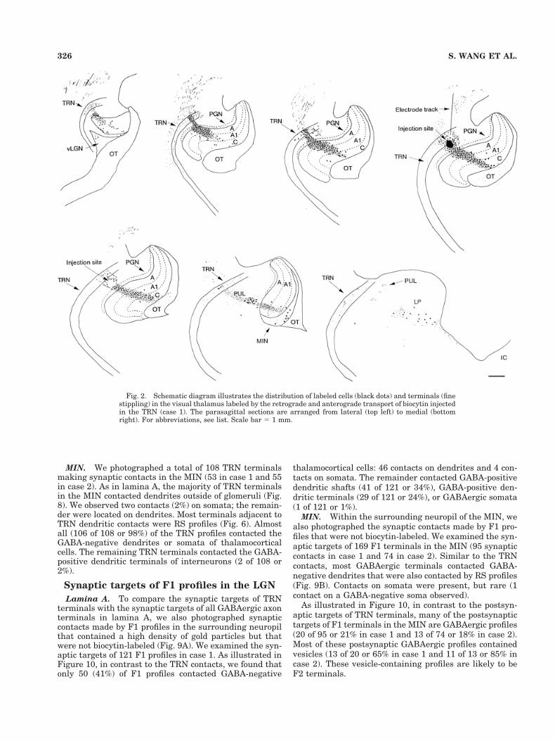
Fig. 2. Schematic diagram illustrates the distribution of labeled cells (black dots) and terminals (finestippling) in the visual thalamus labeled by the retrograde and anterograde transport of biocytin injectedin the TRN (case 1). The parasagittal sections are arranged from lateral (top left) to medial (bottomright). For abbreviations, see list. Scale bar 5 1 mm.
326 S. WANG ET AL.

Fig. 3. Photomicrographs show examples of cells and fibers in thevisual thalamus labeled by retrograde or anterograde transport afterbiocytin injections in TRN. A: A thalamocortical cell labeled by retro-grade transport. B: Presumed corticothalamic axons labeled by an-
terograde transport. The fibers are thin and have “drumstick-like”terminal boutons. C–E: Presumed TRN fibers labeled by anterogradetransport. The fibers are thicker and heavily beaded. For abbrevia-tions, see list. Scale bar 5 30 mm in A (applies to A–E).
327TRN TERMINALS IN THE VISUAL THALAMUS

Fig. 4. Electron photomicrographs show examples of profiles inthe visual thalamus labeled by anterograde transport after biocytininjections in the TRN. A: TRN terminals are identified as biocytin-positive (dark reaction product) and GABA-positive (high density ofgold particles). This terminal contacts (white arrowhead) a GABA-
negative dendrite. B: Small profiles that contain round vesicles (RSprofiles) are presumed to be cortical in origin. They are biocytin-positive but GABA-negative (low density of gold particles). One ofthese profiles contacts (white arrowhead) a GABA-negative dendrite.For abbreviations, see list. Scale bar 5 1 mm in A,B.

Fig. 5. Electron photomicrographs show examples of TRN terminals in the A lamina of the LGN. A: ATRN terminal contacts (white arrowheads) a thalamocortical cell dendrite. B: A TRN terminal contacts(white arrowheads) an interneuron dendrite. This terminal is adjacent to a biocytin-labeled thalamocor-tical cell dendrite (asterisk). For abbreviations, see list. Scale bar 5 1 mm in A,B.

Synaptic targets of TRN terminals in thelateral LP nucleus
We photographed a total of 112 TRN terminals makingsynaptic contacts in the lateral LP nucleus. As illustratedin Figure 11, almost all of these TRN terminals contactedextraglomerular, GABA-negative dendrites (case 1, 56 of60 or 93%; case 2, 52 of 52 or 100%). The majority ofterminals adjacent to TRN dendritic contacts were RSprofiles (Fig. 6). We observed one contact on a GABA-negative soma. The remaining four terminals contactedGABA-positive dendritic terminals, three of which con-tained vesicles.
Synaptic targets of F1 profiles in the lateralLP nucleus
As illustrated in Figures 10 and 12, similar to the TRNterminals, the majority of F1 terminals in the surroundingneuropil of the lateral LP nucleus also contacted extraglo-merular GABA-negative dendrites (case 1, 55 of 59 or93%; case 2, 60 of 60 or 100%). Most of these dendriteswere also postsynaptic to RS profiles. One GABAergiccontact was observed on a GABA-negative soma, andthree GABAergic contacts were made with GABA-positive, vesicle-containing dendrites.
Fig. 6. Most terminals adjacent to TRN terminals are RS profiles.The histogram illustrates the number of RS, RL/RLP, F1, or F2terminals that were found to make synaptic contacts on profiles thatwere also postsynaptic to TRN terminals. For abbreviations, see list.
Fig. 7. The schematic diagram illustrates the distribution of postsynaptic targets of TRN terminalsobserved in case 1. For abbreviations, see list.
330 S. WANG ET AL.

Synaptic targets of TRN terminals in thepulvinar nucleus
Our TRN injections labeled a very sparse population ofGABAergic terminals in the pulvinar nucleus. After ex-amination of 48 sections in each case, we photographed atotal of 24 synaptic contacts (9 in case 1 and 15 in case 2).All the synaptic targets were dendritic shafts outside ofglomeruli (Fig. 13). None of these dendrites were GABA-
positive, and none contained vesicles. Approximately halfof these postsynaptic dendrites were also contacted by RSprofiles (Fig. 8).
Evaluation of sampling methods
If size of the synaptic zones of TRN terminals variedwith their presynaptic location, our sample would be bi-ased in favor of the larger synaptic zones. To determine
Fig. 8. A TRN terminal in the MIN contacts (white arrowheads) a thalamocortical cell dendrite.Adjacent contacts are RS profiles (asterisks). For abbreviations, see list. Scale bar 5 1 mm.
331TRN TERMINALS IN THE VISUAL THALAMUS

Fig. 9. Electron photomicrographs show examples of F1 terminals in the LGN. A: An F1 terminal inthe A lamina contacts (black arrowhead) a thalamocortical cell dendrite. This dendrite is also contactedby an F2 profile (asterisk). B: An F1 terminal in the MIN contacts two (black arrowheads) thalamocor-tical cell dendrites. For abbreviations, see list. Scale bar 5 1 mm in A,B.

whether the size of the synaptic zones of TRN terminalswas correlated with the size of the postsynaptic dendrites,we measured both of these parameters and tested forstatistically significant correlations. As shown in Figure14, in each nucleus, there was no correlation between the
size of the synaptic zone and the size of the postsynapticprofile (Pearson test; LGN A lamina: r 5 0.002, P 5 0.976,n 5 164; MIN: r 5 0.048, P 5 0.621, n 5 109; LP: r 50.036, P 5 0.704, n 5 112; PUL: r 5 0.199, P 5 0.351, n 524). Therefore, with the assumption that the orientation ofTRN terminal synaptic zones is random, our data suggestthat the length of TRN terminal synaptic zones does notvary with presynaptic location. Therefore, our samplingmethods did not bias our data in favor of small or largedendrites.
DISCUSSION
We have identified the synaptic targets of GABAergicTRN terminals throughout the visual thalamus. We ex-amined the synaptic arrangements of TRN terminals inlamina A and MIN of the LGN, the lateral LP nucleus, andthe pulvinar, and we found that, in all nuclei, the TRNprovides GABAergic input primarily to thalamocorticalrelay cells (93–100%). The majority of this input appearstargeted to peripheral dendrites outside of glomeruli, andthis finding is summarized schematically in Figure 15.The TRN does not seem to be a significant source ofGABAergic input to interneurons in the visual thalamus.
We also examined the synaptic targets of the overallpopulation of GABAergic axon terminals (F1 profiles)within these same regions of the visual thalamus andfound that the TRN contacts cannot account for all F1profiles. In addition to F1 contacts on the dendrites ofthalamocortical cells, which likely arise from the TRN,another population of F1 terminals provides input toGABAergic interneuron dendrites (either dendritic shaftsor F2 profiles) and to glomeruli. The relative number ofthese contacts varies across nuclei, but it appears to behighest in the LGN. As discussed below, interneuron ax-ons are a likely source of F1 contacts onto interneurondendrites and within glomeruli.
Comparison with previous anatomicstudies of the LGN
Previous studies have examined the synaptic targets ofTRN terminals in the LGN of the rat (Ohara et al., 1980;Montero and Scott; 1981) and cat (Cucchiaro et al., 1991a),and, like the present study, these studies reported thatthe majority of postsynaptic targets of TRN terminals aredendrites. These dendrites were presumed to originatefrom thalamocortical cells, but definitive identificationwas not possible, because the tissue was not stained forGABA. By using postembedding staining for GABA, ourstudy confirms that the vast majority of TRN terminalscontact thalamocortical cells. In addition, the postembed-ding staining for GABA allowed us to examine onlyGABAergic axon terminals labeled from our injectionsites. A preliminary study of the monkey LGN also usedthis technique and obtained similar results (Feig et al.,1998).
A previous study by Cucchiaro et al. (1991a) showedthat the projections from the PGN, a subdivision of theTRN that is immediately dorsal to the LGN, terminatespecifically in a column within the A laminae of the LGNwhere the majority contact small diameter, presumablydistal dendrites. Our injection sites included both thePGN and TRN. The most significant difference betweenthe present study and that of Cucchiaro et al. is our
Fig. 10. TRN contacts do not account for all F1 contacts. Thehistograms compare the postsynaptic targets of TRN terminals andF1 terminals in the LGN A lamina, MIN, and LP. Most TRN termi-nals contact thalamocortical cell dendrites. F1 terminals contact moreinterneurons. For abbreviations, see list.
333TRN TERMINALS IN THE VISUAL THALAMUS

Fig. 11. A TRN terminal in the LP nucleus contacts (white arrowheads) a thalamocortical celldendrite. An adjacent contact is an RS profile (asterisk). For abbreviations, see list. Scale bar 5 1 mm.
334 S. WANG ET AL.

Fig. 12. Electron photomicrograph shows an example of an F1 terminal in the lateral posteriornucleus (LP) nucleus that contacts (black arrowheads) two thalamocortical cell dendrites. Adjacentcontacts are RS profiles (asterisks). For abbreviations, see list. Scale bar 5 1 mm.

identification of axosomatic contacts. We also found some-what more contacts from our injections on dendritic pro-files in the retinal recipient zone, suggesting perhaps thatthe TRN terminals may occupy a more proximal positionon the dendrites of thalamocortical relay cells than do thePGN terminals.
Our results also indicate that neither the PGN nor theouter tiers of the TRN are a significant source of GABAer-gic input to interneurons. In the LGN, other extrinsicGABAergic inputs to interneurons arise from the pretec-tum (Cucchiaro et al., 1991b). These terminals have beenshown to contact interneuron dendritic terminals, whichcontact relay cell dendrites outside glomeruli (Cucchiaroet al., 1993). By process of elimination, we suggest thatGABAergic inputs to interneurons within glomeruli mayarise from interneuron axons. A similar conclusion wasreached by Takacs et al. (1991) based on a comparison ofvesicle size within subpopulations of F1 terminals. This
conclusion predicts that interneurons may form a networkover which the TRN has little influence.
In contrast to F1 profiles, 48% of which contact inter-neurons, we noted that F2 profiles generally contactthalamocortical cell dendrites and that contacts betweenF2 profiles are quite rare. In fact, Van Horn et al. (2000)found that none of the synaptic targets of F2 profiles wereinterneurons. Accordingly, when F1 and F2 profiles areconsidered together, a much lower percentage is found tocontact interneurons (13%; Erisir et al., 1998). Thus, it islikely that interneurons are primarily interconnectedthrough axodendritic connections and not through dendro-dendritic connections.
Comparison with previous studies of TRNsectors related to other sensory modalities
Data from other sensory systems suggest that the syn-aptic targets of TRN terminals are similar throughout the
Fig. 13. Electron photomicrograph shows an example of a TRN terminal in the PUL that contacts(white arrowheads) a thalamocortical cell dendrite. For abbreviations, see list. Scale bar 5 1 mm.
336 S. WANG ET AL.

dorsal thalamus. For example, Liu et al. (1995) examinedthe synaptic targets of TRN terminals within the cat ven-troposterior nucleus labeled by the anterograde transportof phaseolus vulgaris leucoagglutinin injected into the so-matosensory sector of the TRN. As in the present study,postembedding immunocytochemical labeling for GABAwas used to identify thalamocortical cells and interneu-rons. With these techniques, they found that 82% of TRNterminals contacted thalamocortical cell dendrites, 9.3%contacted thalamocortical cell and interneuron somata,and 8.5% contacted the dendrites of interneurons. Thus,the somatosensory sector of the TRN also primarily tar-gets thalamocortical cells. In addition, TRN terminalsoriginating in the auditory sector of the rat TRN contactsomata and dendrites of presumed thalamocortical cells inthe medial geniculate nucleus (Montero, 1983). Thus, itseems likely that terminals originating from all the sen-sory sectors of TRN primarily contact the dendrites ofthalamocortical cells.
In contrast, a much greater number of TRN terminals inthe anterior, mediodorsal, and ventral anterior nuclei of
the rhesus monkey contact interneurons (Kultas-Ilinskyet al., 1995; Tai et al., 1995; Ilinsky et al., 1999). By usingmethods similar to those in the current study, it was foundthat at least 50% of TRN terminals in these thalamicnuclei contact GABAergic interneurons. Thus, the projec-tions of the TRN to the sensory thalamus are not repre-sentative of TRN projections to all dorsal thalamic nuclei.
Topography of TRN projections to thevisual thalamus
Our results indicate that the visual TRN projects in atopographic manner to the LGN (including both theA-laminae and MIN) and LP nucleus. However, the pro-jections from the TRN to the pulvinar nucleus appear to beorganized differently than projections to the other visualthalamic nuclei we studied. As previously shown byFitzGibbon (1994), we found that injections in the pulvi-nar nucleus labeled cells throughout the rostrocaudal andmediolateral extent of the TRN. In addition, after injec-tions within restricted regions of the TRN, we labeled onlya small number of TRN terminals in the pulvinar nucleus.
Fig. 14. The length of the synaptic contacts made by TRN terminals is plotted against the diameterof the postsynaptic dendrites in the LGN lamina A (A), MIN (B), LP nucleus (C), and PUL (D). Nocorrelation was found between these two parameters (Pearson test). For abbreviations, see list.
337TRN TERMINALS IN THE VISUAL THALAMUS

FitzGibbon et al. (1995) also noted that TRN terminationsin the pulvinar nucleus are not as prominent as those inthe lateral posterior nucleus. This finding suggests thatthe axons of individual TRN cells project throughout thepulvinar nucleus, but that the terminations of each axonare sparse. This finding contrasts with the focused denseprojections that have been identified after intracellularlabeling of TRN cells that project to the LGN and LPnucleus (Uhlrich et al., 1991; Sanchez-Vives et al., 1996;Pinault and Deschenes 1998).
Data in the Galago suggests that the TRN cells thatproject to the LGN are distinct from those that project tothe pulvinar nucleus and occupy distinct tiers within theTRN (Conley and Diamond, 1990). Although our datacould not reveal an organization related to TRN tiers, itdoes suggest that cells in the TRN that project to the LGNare distinct from cells that project to the pulvinar nucleus.Thus, activity in one region of the TRN may have a wide-spread, but weak, influence on thalamocortical cells in thepulvinar nucleus and a more restricted, but strong, influ-ence on thalamocortical cells of the LGN, MIN, and LPnuclei.
Functional implications
Role during sleep. During slow wave sleep, boththalamocortical relay cells and TRN cells are relativelyhyperpolarized. In this state, both cell types tend to fire insynchronized, rhythmic bursts. A key factor in this pat-tern of activity is the activation of T channels (Jahnsenand Llinas, 1984a,b; Mulle et al., 1986; Avanzini et al.,1989; Bal and McCormick, 1993; Contreras et al., 1993).Burst firing of TRN cells produces large and long-lastinginhibitory postsynaptic potentials (IPSPs) in relay cells(Kim and McCormick, 1998). Because of the voltage de-pendency of the T channels, such IPSPs effectively de-
inactivate these channels, promoting burst firing as wellin the relay cells. Interestingly, several studies have con-cluded that these T channels may be concentrated withinthe dendrites of thalamocortical cells (Zhou et al., 1997;Destexhe et al., 1998; Williams and Stuart, 2000; Zhan etal., 2000). Because we found that TRN terminals are pri-marily distributed on the dendrites of thalamocorticalcells (see Fig. 15), it appears that TRN inputs are ideallyarranged to control the inactivation state of T channels.
Additional evidence for a role of the TRN in synchroniz-ing the activity of the thalamus during sleep comes fromstudies of connections between TRN cells. Dendroden-dritic and/or axodendritic connections between neighbor-ing and distant TRN cells have been identified in the cat(Deschenes et al., 1985) and rat (Pinault et al., 1997).Preliminary results also indicate that TRN cells may beelectrically coupled (Landisman et al., 2000). These con-nections are thought to link the activity of cells in allsectors of the TRN and, in turn, synchronize the activity ofall dorsal thalamic nuclei.
Role during wakefulness. The topographic nature ofthe projections from the TRN to the dorsal thalamus arethought to underlie an additional function that has beenattributed to the TRN. That is, the TRN is thought tomodulate the activity of the dorsal thalamus to maintainselective attention. Several different lines of evidence sup-port this concept of TRN function. First, lesions of theTRN, either experimentally induced or resulting from car-diac arrest or head injuries, seem to impair the ability toattend to stimuli (Friedberg and Ross, 1993; Ross andGraham, 1993; Ross et al., 1993). Second, it has beenfound that C-FOS expression in the TRN is related toattention. Specifically, C-FOS expression is induced onlyin the sector of the TRN related to the sensory modalityinvolved in attentional demands (Montero, 1997; McAlo-nan et al., 2000).
How the TRN might influence attention has yet to bedetermined. However, the pattern of terminations of TRNsynapses offers a suggestion for the function of this path-way (see Fig. 15). It is interesting both that these termi-nals are prevalent on relay cells rather than interneurons,and that they synapse mostly on small caliber (presum-ably peripheral) dendrites outside of glomeruli. In addi-tion, if we consider the LGN as an example, most termi-nals that are adjacent to TRN terminals are RS profilesand a smaller number are RLP profiles. This pattern issimilar to the general distribution of terminals encoun-tered in a random sampling of contacts on relay cells; evenwithout correction for terminal size, relay cells are foundto be contacted by more RS profiles (56%) than RLP pro-files (15%; Van Horn et al., 2000). In addition, if oneconsiders the dendritic arbors of relay cells, the volumeoccupied by small caliber, peripheral dendrites is muchgreater than that occupied by proximal dendrites. Thus, ifTRN terminals are distributed fairly evenly across thedendritic arbors of relay cells, one would expect to encoun-ter most of these terminals on distal dendrites adjacent toRS profiles.
This distribution suggests that, unlike GABAergic in-puts from interneurons or cholinergic inputs from thebrainstem that specifically target the retinorecipientzones of relay cell dendritic arbors within glomeruli(Hamos et al., 1985; Erisir et al., 1997), the TRN terminalsdo not appear to be distributed to influence the transfer ofone type of input. Instead, the distribution of TRN termi-
Fig. 15. Schematic diagram illustrates the distribution of termi-nals on thalamocortical cells within the LGN A lamina, MIN, LP, andPUL. TRN terminals are primarily distributed on distal dendritesadjacent to RS profiles. For abbreviations, see list.
338 S. WANG ET AL.

nals might be better situated to affect response modebased on the inactivation state of T channels, which, asnoted above, are concentrated on dendrites, including pe-ripheral dendrites (Zhou et al., 1997; Destexhe et al.,1998; Williams and Stuart, 2000; Zhan et al., 2000). Re-cent evidence suggests that response mode is an impor-tant feature of thalamic relays in normal, waking function(Guido and Weyand, 1995; Ramcharan et al., 2000; Sher-man, 2001). The TRN may function in the maintenance ofselective attention by modulating the response mode ofthalamocortical cells during the waking state.
ACKNOWLEDGMENTS
We thank Martin Boyce for his skillful assistance withthe histology and text editing, and Michael Eisenback andCathie Caple for their expert technical assistance with theelectron microscopy. S.W. received a Sigma Xi Grant inAid; M.E.B., A.E., D.W.G., and S.M.S. received supportfrom the NIH; and M.E.B received support from the NSF.
LITERATURE CITED
Avanzini G, de Curtis M, Panzica F, Spreafico R. 1989. Intrinsic propertiesof nucleus reticularis thalami neurones of the rat studied in vitro.J Physiol (Lond) 416:111–122.
Bal T, McCormick DA. 1993. Mechanisms of oscillatory activity in guinea-pig nucleus reticularis thalami in vitro: a mammalian pacemaker.J Physiol (Lond) 468:669–691.
Beaulieu C, Cynader M. 1992. Preferential innervation of immunoreactivecholine acetyltransferase synapses on relay cells of the cat’s lateralgeniculate nucleus: a double-labeling study. Neuroscience 47:33–44.
Benes FM, Lange N. 2001. Two-dimensional versus three-dimensional cellcounting: a practical perspective. Trends Neurosci 24:11–17.
Berman N. 1977. Connections of the pretectum in the cat. J Comp Neurol174:227–254.
Berson DM, Graybiel AM. 1978. Parallel thalamic zones in the LP-pulvinarcomplex of the cat identified by their afferent and efferent connections.Brain Res 147:139–148.
Berson DM, Graybiel AM. 1983. Organization of the striate-recipient zoneof the cat’s lateralis posterior-pulvinar complex and its relations withthe geniculostriate system. Neuroscience 9:337–372.
Bickford ME, Gunluk AE, Van Horn SC, Vaughan JW, Godwin DW, Sher-man SM. 1994. Thalamic reticular nucleus synaptic targets in the catLGN. Soc Neurosci Abstr 20:8.
Bourassa J, Deschenes M. 1995. Corticothalamic projections from theprimary visual cortex in rats: a single fiber study using biocytin as ananterograde tracer. Neuroscience 66:253–263.
Carden WB, Bickford ME. 1999. Location of muscarinic type 2 receptorswithin the synaptic circuitry of the cat visual thalamus. J Comp Neurol410:431–443.
Coleman KA, Mitrofanis J. 1996. Organization of the visual reticularthalamic nucleus of the rat. Eur J Neurosci 8:388–404.
Conley M, Diamond IT. 1990. Organization of the visual sector of thethalamic reticular nucleus in Galago: evidence that the dorsal lateralgeniculate and pulvinar nuclei occupy separate parallel tiers. EurJ Neurosci 2:211–226.
Conley M, Kupersmith AC, Diamond IT. 1991. The organization of projec-tions from subdivisions of the auditory cortex and thalamus to theauditory sector of the thalamic reticular nucleus in Galago. Eur J Neu-rosci 3:1089–1103.
Contreras D, Curro Dossi R, Steriade M. 1993. Electrophysiological prop-erties of cat reticular thalamic neurones in vivo. J Physiol (Lond)470:273–294.
Crabtree JW. 1992. The somatotopic organization within the cat’s thalamicreticular nucleus. Eur J Neurosci 4:1352–1361.
Crabtree JW. 1996. Organization in the somatosensory sector of the cat’sthalamic reticular nucleus. J Comp Neurol 366:207–222.
Crabtree JW. 1998. Organization in the auditory sector of the cat’s tha-lamic reticular nucleus. J Comp Neurol 390:167–182.
Crabtree JW, Killackey HP. 1989. The topographic organization and axis ofprojection within the visual sector of the rabbit’s thalamic reticularnucleus. Eur J Neurosci 1:94–109.
Cucchiaro JB, Uhlrich DJ, Sherman SM. 1991a. Electron-microscopic anal-ysis of synaptic input from the perigeniculate nucleus to the A-laminaeof the lateral geniculate nucleus in cats. J Comp Neurol 310:316–336.
Cucchiaro JB, Bickford ME, Sherman SM. 1991b. A GABAergic projectionfrom the pretectum to the dorsal lateral geniculate nucleus in the cat.Neuroscience 41:213–226.
Cucchiaro JB, Uhlrich DJ, Sherman SM. 1993. Ultrastructure of synapsesfrom the pretectum in the A-laminae of the cat’s lateral geniculatenucleus. J Comp Neurol 334:618–630.
Datskovskaia A, Carden WB, Bickford ME. 2001. Y retinal terminalscontact interneurons in the cat dorsal lateral geniculate nucleus.J Comp Neurol 430:85–100.
de Biasi S, Frassoni C, Spreafico R. 1986. GABA immunoreactivity in thethalamic reticular nucleus of the rat. A light and electron microscopicalstudy. Brain Res 399:143–147.
de Lima AD, Montero VM, Singer W. 1985. The cholinergic innervation ofthe visual thalamus: an EM immunocytochemical study. Exp Brain Res59:206–212.
Deschenes M, Madariaga-Domich A, Steriade M. 1985. Dendrodendriticsynapses in the cat reticularis thalami nucleus: a structural basis forthalamic spindle synchronization. Brain Res 334:165–168.
Destexhe A, Neubig M, Ulrich D, Huguenard J. 1998. Dendritic low-threshold calcium currents in thalamic relay cells. J Neurosci 18:3574–3588.
Erisir A, Van Horn SC, Bickford ME, Sherman SM. 1997. Immunocyto-chemistry and distribution of parabrachial terminals in the lateralgeniculate nucleus of the cat: a comparison with corticogeniculateterminals. J Comp Neurol 377:535–549.
Erisir A, Van Horn SC, Sherman SM. 1998. Distribution of synapses in thelateral geniculate nucleus of the cat: differences between laminae Aand A1 and between relay cells and interneurons. J Comp Neurol390:247–255.
Feig SL, Manning KA, Uhlrich DJ. 1998. Axon projections from the tha-lamic reticular nucleus (TRN) to the lateral geniculate nucleus (LGN)in the prosimian primate Galago. Soc Neurosci Abstr 24:140.
FitzGibbon T. 1994. Rostral reticular nucleus of the thalamus sends apatchy projection to the pulvinar lateralis-posterior complex of the cat.Exp Neurol 129:266–278.
FitzGibbon T, Tevah LV, Sefton AJ. 1995. Connections between the retic-ular nucleus of the thalamus and pulvinar-lateralis posterior complex:a WGA-HRP study. J Comp Neurol 363:489–504.
Fitzpatrick D, Penny GR, Schmechel DE. 1984. Glutamic aciddecarboxylase-immunoreactive neurons and terminals in the lateralgeniculate nucleus of the cat. J Neurosci 4:1809–1829.
Friedberg EB, Ross DT. 1993. Degeneration of rat thalamic reticular neu-rons following intrathalamic domoic acid injection. Neurosci Lett 151:115–119.
Godwin DW, Van Horn SC, Erisir A, Sesma M, Romano C, Sherman SM.1996. Ultrastructural localization suggests that retinal and corticalinputs access different metabotropic glutamate receptors in the lateralgeniculate nucleus. J Neurosci 16:8181–8192.
Graybiel AM, Berson DM. 1980. Autoradiographic evidence for a projectionfrom the pretectal nucleus of the optic tract to the dorsal lateralgeniculate complex in the cat. Brain Res 195:1–12.
Guido W, Weyand T. 1995. Burst responses in thalamic relay cells of theawake behaving cat. J Neurophysiol 74:1782–1786.
Guillery RW. 1966. A study of Golgi preparations from the dorsal lateralgeniculate nucleus of the adult cat. J Comp Neurol 128:21–50.
Guillery RW. 1969. The organization of synaptic interconnections in thelaminae of the dorsal lateral geniculate nucleus of the cat. Z Zellforsch96:1–38.
Guillery RW, Feig SL, Lozsadi DA. 1998. Paying attention to the thalamicreticular nucleus. Trends Neurosci 21:28–32.
Hajdu F, Somogyi G, Tombol T. 1974. Neuronal and synaptic arrangementin the lateralis posterior-pulvinar complex of the thalamus in the cat.Brain Res 73:89–104.
Hamos JE, Van Horn SC, Raczkowski D, Uhlrich DJ, Sherman SM. 1985.Synaptic connectivity of a local circuit neuron in lateral geniculatenucleus of the cat. Nature 317:618–621.
Hamos JE, Van Horn SC, Raczkowski D, Sherman SM. 1987. Synapticcircuits involving an individual retinogeniculate axon in the cat.J Comp Neurol 259:165–192.
339TRN TERMINALS IN THE VISUAL THALAMUS

Harting JK, Van Lieshout DP, Feig S. 1991. Connectional studies of theprimate lateral geniculate nucleus: distribution of axons arising fromthe thalamic reticular nucleus of Galago crassicaudatus. J Comp Neu-rol 310:411–427.
Hoogland PV, Wouterlood FG, Welker E, Van der Loos H. 1991. Ultra-structure of giant and small thalamic terminals of cortical origin: astudy of the projections from the barrel cortex in mice using Phaseolusvulgaris leuco-agglutinin (PHA-L). Exp Brain Res 87:159–172.
Houser CR, Vaughn JE, Barber RP, Roberts E. 1980. GABA neurons arethe major cell type of the nucleus reticularis thalami. Brain Res 200:341–354.
Ilinsky IA, Ambardekar AV, Kultas-Ilinsky K. 1999. Organization of pro-jections from the anterior pole of the nucleus reticularis thalami (NRT)to subdivisions of the motor thalamus: light and electron microscopicstudies in the Rhesus monkey. J Comp Neurol 409:369–384.
Jahnsen H, Llinas R. 1984a. Electrophysiological properties of guinea-pigthalamic neurones: an in vitro study. J Physiol (Lond) 349:205–226.
Jahnsen H, Llinas R. 1984b. Ionic basis for the electro-responsiveness andoscillatory properties of guinea-pig thalamic neurones in vitro.J Physiol (Lond) 349:227–247.
Jones EG. 1975. Some aspects of the organization of the thalamic reticularcomplex. J Comp Neurol 162:285–308.
Jones EG, Powell TP. 1969. An electron microscopic study of the mode oftermination of cortico-thalamic fibres within the sensory relay nuclei ofthe thalamus. Proc R Soc Lond B Biol Sci 172:173–185.
Jones EG, Powell TPS. 1971. An analysis of the posterior group of thalamicnuclei on the basis of its afferent connections. J Comp Neurol 143:185–216.
Kawamura S, Sprague JM, Niimi K. 1974. Corticofugal projections fromthe visual cortices to the thalamus, pretectum and superior colliculusin the cat. J Comp Neurol 158:339–362.
Kim U, McCormick DA. 1998. The functional influence of burst and tonicfiring mode on synaptic interactions in the thalamus. J Neurosci 18:9500–9516.
Kultas-Ilinsky K, Yi H, Ilinsky IA. 1995. Nucleus reticularis thalami inputto the anterior thalamic nuclei in the monkey: a light and electronmicroscopic study. Neurosci Lett 186:25–28.
Landisman CE, Beierlein M, Connors BW. 2000. Electrical synapses be-tween thalamic reticular neurons. Soc Neurosci Abstr 26:819.
Liu XB, Warren RA, Jones EG. 1995. Synaptic distribution of afferentsfrom the reticular nucleus in ventroposterior nucleus of cat thalamus.J Comp Neurol 352:187–202.
Livingstone MS, Hubel DH. 1981. Effects of sleep and arousal on theprocessing of visual information in the cat. Nature 291:554–561.
Mathers LH. 1972. The synaptic organization of the cortical projection tothe pulvinar of the squirrel monkey. J Comp Neurol 146:43–60.
McAlonan K, Brown VJ, Bowman EM. 2000. Thalamic reticular nucleusactivation reflects attentional gating during classical conditioning.J Neurosci 20:8897–8901.
McCormick DA, Bal T. 1997. Sleep and arousal: thalamocortical mecha-nisms. Annu Rev Neurosci 20:185–215.
McCormick DA, Feeser HR. 1990. Functional implications of burst firingand single spike activity in lateral geniculate relay neurons. Neuro-science 39:103–113.
Minderhoud JM. 1971. An anatomical study of the efferent connections ofthe thalamic reticular nucleus. Exp Brain Res 112:435–446.
Montero VM. 1983. Ultrastructural identification of axon terminals fromthe thalamic reticular nucleus in the medial geniculate body in the rat:an EM autoradiographic study. Exp Brain Res 51:338–342.
Montero VM. 1997. C-FOS induction in sensory pathways of rats exploringa novel complex environment: shifts of active thalamic reticular sectorsby predominant sensory cues. Neuroscience 76:1069–1081.
Montero VM, Scott GL. 1981. Synaptic terminals in the dorsal lateralgeniculate nucleus from neurons of the thalamic reticular nucleus: alight and electron microscope autoradiographic study. Neuroscience6:2561–2577.
Montero VM, Singer W. 1984. Ultrastructure and synaptic relations ofneural elements containing glutamic acid decarboxylase (GAD) in theperigeniculate nucleus of the cat. A light and electron microscopicimmunocytochemical study. Exp Brain Res 56:115–125.
Montero VM, Singer W. 1985. Ultrastructural identification of somata andneural processes immunoreactive to antibodies against glutamic aciddecarboxylase (GAD) in the dorsal lateral geniculate nucleus of the cat.Exp Brain Res 59:151–165.
Montero VM, Guillery RW, Woolsey CN. 1977. Retinotopic organizationwithin the thalamic reticular nucleus demonstrated by a double labelautoradiographic technique. Brain Res 138:407–421.
Mulle C, Madariaga A, Deschenes M. 1986. Morphology and electrophysi-ological properties of reticularis thalami neurons in cat: in vivo study ofa thalamic pacemaker. J Neurosci 6:2134–2145.
Niimi K, Kawamura S, Ishimaru S. 1971. Projections of the visual cortex tothe lateral geniculate and posterior thalamic nuclei in the cat. J CompNeurol 143:279–312.
Oertel WH, Graybiel AM, Mugnaini E, Elde RP, Schmechel DE, Kopin IJ.1983. Coexistence of glutamic acid decarboxylase- and somatostatin-like immunoreactivity in neurons of the feline nucleus reticularis thal-ami. J Neurosci 3:1322–1332.
Ohara PT, Sefton AJ, Lieberman AR. 1980. Mode of termination of affer-ents from the thalamic reticular nucleus in the dorsal lateral genicu-late nucleus of the rat. Brain Res 197:503–506.
Ojima H, Murakami K, Kishi K. 1996. Dual termination modes of cortico-thalamic fibers originating from pyramids of layers 5 and 6 in cat visualcortical area 17. Neurosci Lett 208:57–60.
Pare Dl, Smith Y. 1996. Thalamic collaterals of corticostriatal axons: theirtermination field and synaptic targets in cats. J Comp Neurol 372:551–567.
Patel NC, Bickford ME. 1997. Synaptic targets of cholinergic terminals inthe pulvinar nucleus of the cat. J Comp Neurol 387:266–278.
Patel NC, Carden WB, Bickford ME. 1999. Synaptic targets of cholinergicterminals in the cat lateral posterior nucleus. J Comp Neurol 410:31–41.
Pinault D, Deschenes M. 1998. Projection and innervation patterns ofindividual thalamic reticular axons in the thalamus of the adult rat: athree-dimensional, graphic, and morphometric analysis. J Comp Neu-rol 391:180–203.
Pinault D, Smith Y, Deschenes M. 1997. Dendrodendritic and axoaxonicsynapses in the thalamic reticular nucleus of the adult rat. J Neurosci17:3215–3233.
Raczkowski D, Fitzpatrick D. 1989. Organization of cholinergic synapses inthe cat’s dorsal lateral geniculate and perigeniculate nuclei. J CompNeurol 288:676–690.
Raczkowski D, Rosenquist AC. 1983. Connections of the multiple visualcortical areas with the lateral posterior-pulvinar complex and adjacentthalamic nuclei in the cat. J Neurosci 3:1912–1942.
Ramcharan EJ, Gnadt JW, Sherman SM. 2000. Burst and tonic firing inthalamic cells of unanesthetized, behaving monkeys. Vis Neurosci 17:55–62.
Rinvik E, Ottersen OP. 1988. Demonstration of GABA and glutamate inthe nucleus reticularis thalami: a postembedding immunogold labelinginvestigation in the cat and baboon. In: Bentivoglio M, Spreafico R,editors. Cellular thalamic mechanisms. New York: Elsevier Science. p321–337.
Rinvik E, Ottersen OP, Storm-Mathisen J. 1987. Gamma-aminobutyrate-like immunoreactivity in the thalamus of the cat. Neuroscience 21:781–805.
Robertson RT, Cunningham TJ. 1981. Organization of corticothalamicprojections from parietal cortex in cat. J Comp Neurol 199:569–585.
Robson JA, Hall WC. 1977. The organization of the pulvinar in the greysquirrel (Sciurus carolinensis). II. Synaptic organization and compar-isons with the dorsal lateral geniculate nucleus. J Comp Neurol 173:389–416.
Robson JA, Mason CA. 1979. The synaptic organization of terminals tracedfrom individual labeled retino-geniculate axons in the cat. Neuro-science 4:99–111.
Rodrigo-Angulo ML, Reinoso-Suarez F. 1988. Connections to the lateralposterior-pulvinar thalamic complex from the reticular and ventrallateral geniculate thalamic nuclei: a topographical study in the cat.Neuroscience 26:449–459.
Rodrigo-Angulo ML, Reinoso-Suarez F. 1995. Afferent connections of thelateralis medialis thalamic nucleus in the cat. Brain Res Bull 38:53–67.
Ross DT, Graham DI. 1993. Selective loss and selective sparing of neuronsin the thalamic reticular nucleus following human cardiac arrest.J Cereb Blood Flow Metab 13:558–567.
Ross DT, Graham DI, Adams JH. 1993. Selective loss of neurons from thethalamic reticular nucleus following severe human head injury. J Neu-rotrauma 10:151–165.
Sanchez-Vives MV, Bal T, Kim U, von Krosigk M, McCormick DA. 1996.Are the interlaminar zones of the ferret dorsal lateral geniculate nu-
340 S. WANG ET AL.

cleus actually part of the perigeniculate nucleus? J Neurosci 16:5923–5941.
Sherman SM. 2001. Tonic and burst firing: dual modes of thalamocorticalrelay. Trends Neurosci 24:122–126.
Spreafico R, Battaglia G, Frassoni C. 1991. The reticular thalamic nucleus(RTN) of the rat: cytoarchitectural, Golgi, immunocytochemical, andhorseradish peroxidase study. J Comp Neurol 304:478–490.
Steriade M, McCarley RW. 1990. Brainstem control of wakefulness andsleep. New York: Plenum Press.
Steriade M, Parent A, Hada J. 1984. Thalamic projections of nucleusreticularis thalami of cat: a study using retrograde transport of horse-radish peroxidase and fluorescent tracers. J Comp Neurol 229:531–547.
Sumitomo I, Hsiao CF, Fukuda Y. 1988. Two types of thalamic reticularcells in relation to the two visual thalamocortical systems in the rat.Brain Res 446:354–362.
Tai Y, Yi H, Ilinsky IA, Kultas-Ilinsky K. 1995. Nucleus reticularis thalamiconnections with the mediodorsal thalamic nucleus: a light and elec-tron microscopic study in the monkey. Brain Res Bull 38:475–488.
Takacs J, Hamori J, Silakov V. 1991. GABA-containing neuronal processesin normal and cortically deafferented dorsal lateral geniculate nucleusof the cat: an immunogold and quantitative EM study. Exp Brain Res83:562–574.
Uhlrich DJ, Cucchiaro JB, Humphrey AL, Sherman SM. 1991. Morphologyand axonal projection patterns of individual neurons in the cat peri-geniculate nucleus. J Neurophysiol 65:1528–1541.
Updyke BV. 1981. Projections from visual areas of the middle suprasylvian
sulcus onto the lateral posterior complex and adjacent thalamic nucleiin cat. J Comp Neurol 201:477–506.
Van Horn SC, Erisir A, Sherman SM. 2000. Relative distribution of syn-apses in the A-laminae of the lateral geniculate nucleus of the cat.J Comp Neurol 416:509–520.
Vidnyanszky Z, Hamori J. 1994. Quantitative electron microscopic analy-sis of synaptic input from cortical areas 17 and 18 to the dorsal lateralgeniculate nucleus in cats. J Comp Neurol 349:259–268.
Vidnyanszky Z, Borostyankoi Z, Gorcs TJ, Hamori J. 1996. Light andelectron microscopic analysis of synaptic input from cortical area 17 tothe lateral posterior nucleus in cats. Exp Brain Res 109:63–70.
Wang S, Erisir A, Sherman SM, Bickford ME. 1999. Thalamic reticularnucleus synaptic targets in the cat lateral posterior nucleus. Soc Neu-rosci Abstr 25:1426.
Williams SR, Stuart GJ. 2000. Action potential backpropagation andsomato-dendritic distribution of ion channels in thalamocortical neu-rons. J Neurosci 20:1307–1317.
Yen CT, Conley M, Hendry SH, Jones EG. 1985. The morphology of phys-iologically identified GABAergic neurons in the somatic sensory part ofthe thalamic reticular nucleus in the cat. J Neurosci 5:2254–2268.
Zhan XJ, Cox CL, Sherman SM. 2000. Dendritic depolarization efficientlyattenuates low-threshold calcium spikes in thalamic relay cells. J Neu-rosci 20:3909–3914.
Zhou Q, Godwin DW, O’Malley DM, Adams PR. 1997. Visualization ofcalcium influx through channels that shape the burst and tonic firingmodes of thalamic relay cells. J Neurophysiol 77:2816–2825.
341TRN TERMINALS IN THE VISUAL THALAMUS