synaptotoxicityinalzheimer’sdiseaseinvolveda ... · cellular/molecular...
TRANSCRIPT
Cellular/Molecular
Synaptotoxicity in Alzheimer’s Disease Involved aDysregulation of Actin Cytoskeleton Dynamics throughCofilin 1 Phosphorylation
Travis Rush,1,2* X Jose Martinez-Hernandez,1,2* Marc Dollmeyer,1,2 Marie Lise Frandemiche,1,2 XEve Borel,1,2
X Sylvie Boisseau,1,2 X Muriel Jacquier-Sarlin,1,2 and X Alain Buisson1,2
1Institut National de la Sante et de la Recherche Medicale, U1226, BP 170, and 2Universite Grenoble Alpes, Grenoble Institut des Neurosciences, BP 170,Grenoble Cedex 9, F-38042, France
Amyloid-� (A�) drives the synaptic impairment and dendritic spine loss characteristic of Alzheimer’s disease (AD), but how A� affectsthe actin cytoskeleton remains unknown and contentious. The actin-binding protein, cofilin-1 (cof1), is a major regulator of actindynamics in dendritic spines, and is subject to phospho-regulation by multiple pathways, including the Rho-associated protein kinase(ROCK) pathway. While cof1 is implicated as a driver of the synaptotoxicity characteristic of the early phases of AD pathophysiology,questions remain about the molecular mechanisms involved. Cofilin-actin rods are observed in neurons exposed to A� oligomers (A�o)and in tissue from AD patients, and others have described an increased cofilin phosphorylation (p-cof1) in AD patients. Here, we reportelevated p-cof1 of the postsynaptic enriched fraction of synaptosomes from cortical samples of male APP/PS1 mice and human AD casesof either sex. In primary cortical neurons, A�o induced rapid actin stabilization and increased p-cof1 in the postsynaptic compartmentof excitatory synapses within 30 min. Fluorescence recovery after photobleaching of actin-GFP and calcium imaging in live neuronsexpressing active or inactive cof1 mutants suggest that cof1 phosphorylation is necessary and sufficient for A�o-induced synapticimpairment via actin stabilization before the reported formation of cofilin-actin rods. Moreover, the clinically available and well-tolerated ROCK inhibitor, fasudil, prevented A�o-induced actin stabilization, synaptic impairment, and synaptic loss by blocking cofilinphosphorylation. A�o also blocked the LTP-induced insertion of the AMPAR subunit, GluA1, at the postsynaptic density, in a fasudil-sensitive manner. These data support an important role for ROCKs and cofilin in mediating A�-induced synaptic impairment.
Key words: actin; Alzheimer’s disease; amyloid-� oligomers; cofilin; fasudil; Rho-associated protein
IntroductionAlzheimer’s disease (AD) is the leading cause of dementia, yetcurrent treatments provide only modest clinical benefit. Inci-
dence of AD and its societal costs are expected to balloon incoming decades, as the world population ages (Alzheimer’s
Received May 7, 2018; revised Sept. 18, 2018; accepted Oct. 4, 2018.Author contributions: T.R., J.M.-H., M.D., S.B., and M.J.-S. wrote the first draft of the paper; T.R., J.M.-H., M.D.,
M.L.F., E.B., S.B., M.J.-S., and A.B. edited the paper; T.R., J.M.-H., M.D., M.L.F., S.B., M.J.-S., and A.B. designedresearch; T.R., J.M.-H., M.D., M.L.F., E.B., S.B., and M.J.-S. performed research; E.B. contributed unpublished re-agents/analytic tools; T.R., J.M.-H., M.D., M.L.F., E.B., S.B., M.J.-S., and A.B. analyzed data; T.R., J.M.-H., and A.B.wrote the paper.
This work was supported by the Fondation Neurodis, Fondation Bettencourt, Institut National de la Sante et de laRecherche Medicale, and Agence Nationale de la Recherche (ANR MALAAD program). Human postmortem tissue
was obtained from the Banco de Tejidos, suppress fundation CIEN, Madrid, Spain. All experiments were performedin accordance with local animal welfare committee (Comite Local Grenoble Institute Neurosciences, C2EA-04) andEuropean Union guidelines (Directive 2010/63/EU). Every precaution was taken to minimize stress and the numberof animals used in each series of experiments.
The authors declare no competing financial interests.*T.R. and J.M.-H. contributed equally to this work.Correspondence should be addressed to Dr. Alain Buisson, Institut National de la Sante et de la Recherche
Medicale U1216, Institut des Neurosciences de Grenoble, BP 170, Grenoble Cedex 9, 38042 France. E-mail:[email protected].
Significance Statement
We report that amyloid-� oligomers rapidly induce aberrant stabilization of F-actin within dendritic spines, which impairssynaptic strength and plasticity. Activation of the Rho-associated protein kinase (ROCK) pathway results in phosphorylation ofcof1 and is sufficient to mediate A�o-induced actin stabilization synaptic impairment and synaptic loss. Further, the ROCKinhibitor, fasudil, prevents cofilin phosphorylation, acute synaptic disruption, and synaptotoxicity in primary cortical neurons.Together, the herein presented data provide strong support for further study of the ROCK pathway as a therapeutic target for thecognitive decline and synaptotoxicity in Alzheimer’s disease.
The Journal of Neuroscience, November 28, 2018 • 38(48):10349 –10361 • 10349

Association, 2017). AD is characterized by accumulation ofamyloid-� (A�) plaques and microtubule-associated protein tauinclusions. A� aggregates, and in particular low molecular weightoligomers (A�o), are considered triggers of disease through atau-dependent mechanism (Rapoport et al., 2002; Oddo et al.,2004; Tanzi and Bertram, 2005). Appearances of A�o and tau-dependent dendritic spine loss are the features that correlate mostdirectly with cognitive decline in AD (DeKosky and Scheff, 1990;Terry et al., 1991). Moreover, novel findings suggest that den-dritic spine plasticity can provide cognitive resilience against de-mentia among the elderly with AD pathology (Boros et al., 2017).Last, acute exposure to A�o impairs LTP in rodent tissue, pre-dominantly by acting at the postsynaptic compartment (Kervernet al., 2012).
The actin cytoskeleton (CSK), enriched in dendritic spines, iscentral to the regulation of spine morphology, stability, and plas-ticity (Hotulainen and Hoogenraad, 2010). The neuronal actin-binding protein, cofilin-1 (cof1), is a major regulator of spineactin dynamics by severing filamentous actin (F-actin), leading toactin depolymerization (Rust, 2015). cof1 activity is regulated byphosphorylation of serine 3, rendering cof1 inactive. Synapticactivity-driven cof1 phosphorylation contributes to modifica-tions of spine morphology (Rust et al., 2010; Noguchi et al.,2016). Further, functional loss of cof1 causes aberrant spine mor-phogenesis and impairs synaptic plasticity (Zhou et al., 2009; Guet al., 2010). Constitutively active or inactive mutants of cof1influence spine morphology and maturity in neurons (Yang et al.,1998; Niwa et al., 2002). Similarly, inactivation or ablation of theupstream regulators, Rho-associated protein kinase (ROCK) orLIM-kinase (LIMK1), also leads to increased cof1 activity, de-creased spine F-actin, and immature spines or spine collapse(Niwa et al., 2002; Zhou et al., 2009; Swanger et al., 2015). cof1 isalso necessary for the trafficking of the AMPARs at the postsyn-aptic density (PSD) during synaptic plasticity (Gu et al., 2010).
While several research groups have described the implicationof cof1 in the pathophysiology of AD, the pathological modifica-tion of cof1 activity remains controversial. Indeed, cof1 was dis-covered to accumulate in senile plaques in AD tissue and ADmouse models (Bamburg and Bernstein, 2016). Additionally, re-cent publications suggest that active (i.e., dephosphorylated) cof1forms aberrant cofilin-actin rods, which block axonal traffickingand may contribute to deficits in synaptic plasticity (Davis et al.,2011; Mendoza-Naranjo et al., 2012; Barone et al., 2014). BothA� fibrils and A�o (i.e., dimers and trimers) promote cofilin-actin rod formation with prolonged exposure (Davis et al., 2011;Mendoza-Naranjo et al., 2012). However, brain tissue from ADpatients and APP-expressing mouse models exhibit elevatedROCK levels and corresponding elevated levels of inactive p-cof1(Herskowitz et al., 2013; Barone et al., 2014; Henderson et al.,2016; Han et al., 2017). Furthermore, in other neurodegenerativediseases characterized by an early synaptic loss, such as sporadicCreutzfeld-Jacob disease, upregulation of p-cof1 has been de-scribed (Zafar et al., 2018). These data highlight that cof1 exerts apivotal role in the synaptotoxic process of neurodegenerative dis-eases (Heredia et al., 2006; Lacor et al., 2007; Herskowitz et al.,2013).
Here, we tested the hypothesis that phosphorylation-in-activation of cof1 induced by exposure to A�o promotes synapticdeficits and impairs plasticity. We showed that there are elevatedp-cof1 levels in PSD-enriched synaptosomes of APP/PS1 miceand human AD cases, consistent with overactivation of theROCK pathway. In primary cortical cultures, while a 24 h expo-sure to A�o decreases spine density (Lacor et al., 2007), we reportthat acute exposure to A�o increases p-cof1 at the postsynapticcompartment and favors its distribution in the Triton-insoluble(TI) fractions, leading to a subsequent stabilization of spineF-actin and impairment of synaptic signaling and plasticity. Fur-ther, the clinically available and well-tolerated ROCK inhibitor,fasudil, can prevent these cof1-mediated synaptic deficits andsynaptic loss.
Materials and MethodsAnimals. hAPP/PS1–21 (line 21) mice (APP/PS1) were used at 3 monthsof age for this study. APP/PS1 mice coexpress human APP carrying theK670N/M671L “Swedish” double mutation and HPS1 L166P with athreefold overexpression of human APP over endogenous mouse APP.Mice express the transgene under the control of a neuron-specificmThy-1 promoter element and were generated on a C57bl6 background.For immunohistochemical studies, Thy1-YFP-H (The Jackson Labora-tory, B6 Cg-Tgn 2Jrs) mice were crossed with heterozygote APP/PS1mice to generate C57BL/6Thy1-eYFP APP/PS1–21 mouse colony. All ex-periments involving animals were conducted in accordance with thepolicy of Institut des Neurosciences de Grenoble and French legislation,in compliance with the European Community Council Directive ofNovember 24, 1986 (86/609/EEC). The research involving animals wasauthorized by the Direction Departementale de la protection des popu-lations–Prefecture de l’Isere France and by the ethics committee of Insti-tut des Neurosciences de Grenoble accredited by the French Ministry ofResearch.
Immunohistochemistry. Male mice were deeply anesthetized and per-fused intracardially with 0.9% NaCl followed by 35 ml 4% PFA in 0.1 M
PBS, pH 7.3. Brains were rapidly removed, postfixed overnight at 4°C in4% PFA, immersed in 20% sucrose in 0.1 M PBS at pH 7.5 overnight,frozen in isopentane, and stored at �30°C. Serial frontal sections (30�m) were obtained with a cryostat microtome. Sections were blocked byincubation with 3% BSA in TBS-Tween-Triton (TBSTT) (0.1 M Tris base,0.15 M NaCl, 0.1% Tween, 0.1% Triton X-100) for 30 min (dilution/blocking buffer) and incubated overnight at 4°C with anti-phospho-cofilin 1 antibody (Santa Cruz Biotechnology, rabbit polyclonal; 1:200).Tissue sections were washed in TBSTT and incubated for 2 h at roomtemperature with Cyanin 3-conjugated secondary antibodies. Sectionswere washed in TBSTT and mounted in fluorescent mounting medium.Sections were examined on an LSM 710 confocal laser-scanning micro-scope (Carl Zeiss), and images were acquired with an Airyscan module(Carl Zeiss) with an oil-immersion Plan Apochromat 63� objective (NA1.46) to improve lateral resolution (�140 nm) and signal-to-noise ratios.For illustration, images were merged with ImageJ software (RRID:SCR_003070). For quantitative analysis, ROIs were drawn to delimitatedendritic spine and the fluorescence intensity of p-cof1 staining mea-sured using Analyze/Measure/Mean gray value functions of ImageJ.
Primary cultures of cortical neurons. Mouse cortical neurons were cul-tured from 14- to 15-d-old OF1 embryos of either sex as previouslydescribed (Leveille et al., 2008). Cerebral cortices were dissected, disso-ciated, and cultured in DMEM containing 5% FBS, 5% horse serum, and2 mM glutamine (all from Sigma-Aldrich) on 24-well plates for biochem-ical experiments. Neurons were seeded on 35 mm glass-bottom dishes(MatTek) at a final concentration of two cortical hemispheres per dishfor confocal experiments. All plates and dishes were coated with 0.1mg/ml poly-D-lysine and 0.02 mg/ml laminin (Sigma-Aldrich). Cultureswere maintained at 37°C in a humidified atmosphere containing 5%CO2/95% air. After 3– 4 DIV, cytosine arabinoside (AraC, 10 �M; Sigma-Aldrich) was added to inhibit proliferation of non-neuronal cells incultures used for biochemistry experiments; 98% of the cells were con-
T. Rush’s present address: Disease Biology and Cellular Pharmacology, Recursion Pharmaceuticals, Salt Lake City,UT 84108.
J. Martinez-Hernandez’s present address: IKERBasque, Department of Biochemistry and Molecular Biology, Uni-versity of the Basque Country, 48940 Leiola, Spain.
https://doi.org/10.1523/JNEUROSCI.1409-18.2018Copyright © 2018 the authors 0270-6474/18/3810350-13$15.00/0
10350 • J. Neurosci., November 28, 2018 • 38(48):10349 –10361 Rush, Martinez-Hernandez et al. • cof1 Mediates A�-Induced Synaptic Impairment in AD
sidered as neuronal. The day before the experiments, cells were washed inDMEM. Treatments were performed on neuronal cultures at 14 –15 DIV.
A� oligomerization. Amyloid � oligomers were prepared as previouslydescribed (Frandemiche et al., 2014). Briefly, human A� 1– 42 peptides(Bachem) was resuspended in 1,1,1,3,3,3-hexafluoro-2-propanol (Sigma-Aldrich) to 1 mM until complete resuspension as previously described(Stine et al., 2003). A�o was prepared by diluting A� to 1 mM in anhy-drous DMSO (Sigma-Aldrich) and then to 100 �M in ice-cold, phenolred-free HEPES and bicarbonate-buffered saline solution (HBBSS) withimmediate vortexing and ice bath sonication for 15 min, and then incu-bated at 4°C for 24 h with mild agitation.
Site-directed mutagenesis. Constitutively inactive phosphomimetic(S3E) and active nonphosphorylatable (S3A, active) cof1-mCherry con-structs were generated using quick change site-directed mutagenesis kit(Agilent Technologies), according to the instructions of the manufac-turer, with the following primers: S3A (forward: 5�-atggccgccggtgtggct-gtctcctg-3�; reverse: 5�-cagagacagccacaccctcggccat-3�), S3E (forward:5�-atggccgagggtgtggctgtctcctg-3�; reverse: 5�-cagagacagccacaccctcggccat-3�).
Neuronal transfection. Transfections were performed on cortical neu-ron cultures after 12–13 DIV with calcium phosphate precipitation tech-nique. Growth medium (DMEM and sera) was removed and kept untilthe last transfection step. Cells were washed for 1–1.5 h in DMKY buffercontaining the following (in mM): 1 kynurenic acid, 0.9 NaOH, 0.5HEPES, 10 MgCl2, plus phenol red 0.05%, pH 7.4; 3 �g of the followingplasmids: GCaMP6 fast, Lifeact-eGFP, actin-GFP, cof1-S3E-mCherry,and cof1-S3A-mCherry, mixed with CaCl2 (120 mM) in HBS containingthe following (in mM): 25 HEPES, 140 NaCl, and 0.750 Na2HPO4, pH7.06, and left for 15 min for DNA precipitation. Plasmids were appliedfor 30 min. Transfection medium was replaced with conditioned growthmedium, and cultures were returned to the incubator until use at DIV14 –15.
Fluorescence recovery after photobleaching (FRAP) experiments. FRAPwas performed on cultured neurons (DIV 14 –15) 48 h after transfectionwith actin-GFP. Images were acquired with an inverted Nikon Eclipse TiC2 confocal microscope with a Nikon 60� water objective with a 1.33numerical aperture. Actin-GFP was imaged for at least 20 s before beingbleached at 405 nm. The subsequent fluorescence recovery was measuredfor 120 s (at 1 frame/s). Fluorescent signal analysis was performed withthe Nikon software NIS Elements. Fluorescent signals from the dendriticspine heads were normalized to prebleach values. Average recovery pla-teau values were calculated using nonlinear regression (two-factor decaymodel) from individual spine recovery curves with GraphPad Prism 6.0(RRID:SCR_002798).
Neuronal live calcium imaging. Primary neurons were transfected atDIV 13 using the calcium phosphate technique. Imaging experimentswere performed at DIV 14. Neurons were washed into HBBSS (contain-ing the following in mM: 116 NaCl, 5.4 KCl, 1.8 CaCl2, 0.8 MgSO4 1.3NaH2PO4, 12 HEPES, 5.5 glucose, 25 bicarbonate, and 10 �M glycine atpH 7.45) and moved to the stage of a Nikon Ti Eclipse C2 confocalmicroscope driven by NIS Elements software and equipped with a 60�1.2 NA water-immersion lens. Cultures were continuously perfused withHBBSS in a closed-loop (for baseline and 100 nM A�o application; totalsystem volume of 12.5 ml) at a rate of 2 ml/min, and maintaining aconstant culture volume of �2.5 ml. For synaptic activation with bicuc-ulline (Bic) and 4-aminopyridine (4AP), the inlet tube was moved to astock of 50 �M Bic � 2.5 mM 4AP in HBBSS for 15 s three times beforeand three times after incubation with the A�o and/or fasudil treatment,and then to a tube of fresh HBBSS. The outlet tube of the perfusion pumpwas immediately directed to a waste container to avoid pharmacologicalcontamination of the HBBSS stock. Event markers were applied to theimaging files as the Bic � 4AP solution reached the culture and when theapplication was finished. The A�o and/or fasudil (30 �M) in HBBSS wereapplied by perfusion pump in a closed-loop for 15 min. For subsequentsynaptic activation, the perfusion loop was opened to fresh HBBSS for 5min before the subsequent series of three Bic � 4AP applications.Twelve-bit images of GCaMP6 fast fluorescence brought on by excitationwith a 488 nm argon laser were acquired using a pinhole of 2.2 AU at 1frames/s with a resolution of 512 � 512 to optimize scan speed and
signal-to-noise ratio. Images were denoised and filtered before ROI ex-traction of the signal data using the Time Measurement function withinNIS Elements software, and the data subsequently exported to MS Excelfor calculation of the �F/F values, where F was the average baselinefluorescence value from within the ROI before experimentation. �F/Ftraces from each cell were entered into GraphPad Prism, after baselinesubtraction, where area under curve values for each peak were normal-ized to the first Bic � 4AP (B4AP)-induced peak area. The three pretreat-ment and post-treatment responses were averaged by neuron andcombined by group for statistical comparison.
Chemically induced LTP. Chemically induced LTP (cLTP) was inducedin primary cortical neurons as described previously (Frandemiche et al.,2014). Briefly, primary neurons were exposed to 50 �M Bic (a GABAaantagonist) with 2.5 mM 4AP (a weak potassium channel blocker) for15–30 min, with or without 15 min pretreatment with A�o (100 nM)and/or fasudil (30 �M). cLTP was verified by detecting increased GluA1in the PSD-enriched synaptosomal fraction.
Subcellular fractionation and Western blotting. PSD fractions. Culturedneurons, cortical samples from APP/PS1 male mice, or dorsolateral PFCsamples from humans of either sex (Banco de Tejidos CIEN; for casedetails, see Table 1) were homogenized in cold buffer containing 0.32 M
sucrose and 10 mM HEPES, pH 7.4. Samples were maintained at 4°Cduring all steps of the experiment. Homogenates were cleared at 1000 �g for 10 min to remove nuclei and large debris. The resulting superna-tants were concentrated at 12,000 � g for 20 min to obtain a crudemembrane fraction, which was then resuspended twice (4 mM HEPES, 1mM EDTA, pH 7.4, 20 min at 12,000 � g). Then, the pellet was incubated(20 mM HEPES, 100 mM NaCl, 1% Triton X-100, pH 7.2) for 1 h at 4°Cwith mild agitation, and centrifuged at 12,000 � g for 20 min to pellet thesynaptosomal membrane fraction. The supernatant was collected as thenon-PSD fraction or Triton-soluble (TS) fraction. The pellet was thensolubilized (20 mM HEPES, 0.15 M NaCl, 1% Triton X-100, 1% deoxy-cholic acid, 1% SDS, pH 7.5) for 1 h at 4°C and centrifuged at 10,000 � gfor 15 min. The supernatant contained the PSD or TI fraction. The in-tegrity of non-PSD fraction was verified by synaptophysin immunoblot-ting, and the integrity of the PSD-enriched fraction was verified byPSD-95 immunoblotting.
Soluble and cytoskeletal fractions. The cellular fractions were preparedas described by Hinck et al. (1994). Primary cortical neurons (DIV 14)exposed to A�o (100 nM for 15 min) were rinsed in HBBSS and homog-enized in “cytoskeletal preserving” buffer (50 mM NaCl, 10 mM PIPES,pH 6.8, 3 mM MgCl2, 0.5% Triton X-100, 300 mM sucrose) supplementedwith protease (Roche Diagnostics) and phosphatase (Sigma-Aldrich) in-hibitor cocktails for 10 min at 4°C with gentle rocking. After centrifuga-tion for 10 min at 14 000 rpm and 4°C, the supernatant constituted the TSfraction. The pellet was triturated in SDS buffer (20 mM Tris, pH 7.5, 5mM EDTA, 2.5 mM EGTA, 1% SDS and boiled at 100°C for 10 min) andcentrifuged for 10 min at 14,000 rpm at 4°C; the supernatant constitutedthe TI fraction. This fraction usually contained 5- to 8-fold less proteinthan the TS fraction, as determined by micro-BCA assay (Pierce Chem-ical). Equal amounts of proteins were routinely analyzed.
Immunoblotting. Samples in loading buffer were boiled for 5 min (10min for tissue samples), and equal amounts of proteins (10 –25 �g, quan-tified by micro-BCA assay in duplicate for cell extracts, in triplicate fortissues) were resolved on 4%–20% gradient Bis-Tris polyacrylamide pre-
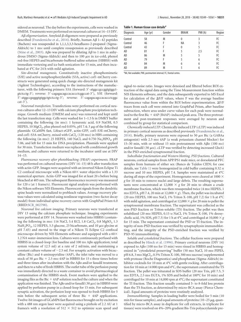
Table 1. Human tissue case detailsa
Diagnosis Age (yr) Gender BRAAK PMI (h) Region
Control 58 M 0 3 FCControl 46 F NA 3 FCControl 43 F 0 5 FCAD 81 F 5 2.5 FCAD 86 M 5 5 FCAD 79 F 5 6 FCAD 84 M 6 6 FCAD 89 M 5 6 FCaNA, Not available; PMI, postmortem interval; FC, frontal cortex.
Rush, Martinez-Hernandez et al. • cof1 Mediates A�-Induced Synaptic Impairment in AD J. Neurosci., November 28, 2018 • 38(48):10349 –10361 • 10351
cast gels (Bio-Rad) � stain-free system for tissues (Bio-Rad) in denatur-ing conditions. Proteins were transferred to a PVDF membrane (EMDMillipore) for 35 min at 4°C. Membranes were blocked with Tris-buffered saline (10 mM Tris, 150 mM NaCl, pH 7.4) containing 0.02%Tween 20 and 3% BSA for 1 h at room temperature. Membranes werethen incubated overnight at 4°C with the following primary antibodies.
Membranes were incubated with the HRP-conjugated secondary an-tibodies rabbit (1:40,000) and mouse (1:5000) (both from Sigma-Aldrich) for 45 min at room temperature. Specific proteins werevisualized with an enhanced chemiluminescence ECL Detection System(Bio-Rad). Chemiluminescence detection was performed in a dark roomwith hyperfilms (GE Healthcare) and analyzed with ImageJ software(RRID:SCR_003070). For tissue analysis, chemiluminescence signalswere normalized to protein loading acquired using Stain-free precast gels(Bio-Rad) illumination by the chemidoc system and analyzed with Im-ageLab (Table 2).
Experimental design and statistical analysis. Results are expressed as themean � SEM from independent biological samples. Statistical analyses
Figure 1. Cortical samples from APP/PS1 mice and AD cases have elevated p-cof1. A, Western blot images of PSD-enriched (top) and non-PSD fraction (bottom) of cortical synaptosomes ofAPP/PS1 mice (left) and humans (right) showing the enrichment of presynaptic and postsynaptic marker in each fraction. B, Western blot images (top) along with associated quantification (bottom)showing elevated phospho-cofilin1 in APP/PS1 mice (left) compared with their nontransgenic littermates (Western blots n 4; p-cof1 Mann–Whitney U 6.0 (72,138) p 0.0011; cof1Mann–Whitney U 43.0 (102,88) p 0.9025) and in AD cases (right) compared with healthy controls (WB, n 4; p-cof1 Mann–Whitney U 0.0 (6,30) p 0.0357; cof1 Mann–Whitney U 7.0 (13,23) p 1.000). *p 0.05. ***p 0.001. For case details, see Table 1.
Table 2. Antibody information
Antibody Species Dilution Supplier
Anti-A�17-24 4G8 Mouse (monoclonal) 1/500 Covance (SIG-39220)PSD-95 Mouse (monoclonal) 1/1000 Abcam (ab2723)Synaptophysin Mouse (monoclonal) 1/1000 Abcam (ab8049)p-Erk1/2 (Thr202/Tyr204) Rabbit (polyclonal) 1/500 Santa Cruz Biotechnology
(sc-16982)Erk1/2 Mouse (monoclonal) 1/500 Santa Cruz Biotechnology
(sc-135900)p-cof1 (Ser3) Rabbit (polyclonal) 1/500 Santa Cruz Biotechnology
(sc-21867)Cof1 Rabbit (polyclonal) 1/500 Santa Cruz Biotechnology
(sc-33779)GluA1 Rabbit (polyclonal) 1/1000 EMD Millipore (AB1504)
10352 • J. Neurosci., November 28, 2018 • 38(48):10349 –10361 Rush, Martinez-Hernandez et al. • cof1 Mediates A�-Induced Synaptic Impairment in AD
were performed with GraphPad Prism 6.0 (RRID:SCR_002798). West-ern blots were analyzed by Mann–Whitney U test, ANOVA with Tukey’spost hoc test with correction for multiple comparisons, or two-wayANOVA with Tukey’s post hoc test with correction for multiple compar-isons; histological analysis by unpaired t test. Spine density quantifica-tion was analyzed by paired t test. FRAP data and calcium imaging wereanalyzed by two-way ANOVA using A� and drug treatment (e.g., fasudilor Bic � 4AP) or cof1 genotype as factors, followed by Tukey’s multiplecomparison tests with corrected p values, or as specified in the corre-sponding figure legend.
ResultsPhospho-cofilin1 is elevated in AD model mice andhuman ADIn neurons, actin and actin binding proteins are enriched in thepostsynaptic compartment of excitatory synapses. Thus, we per-formed brain tissue fractionation to isolate the postsynaptic com-partment (PSD-enriched fraction). Then, we probed thesefractions obtained from APP/PS1 model mice and human ADcases (Table 1) for PSD 95 and synaptophysin to verify the integ-
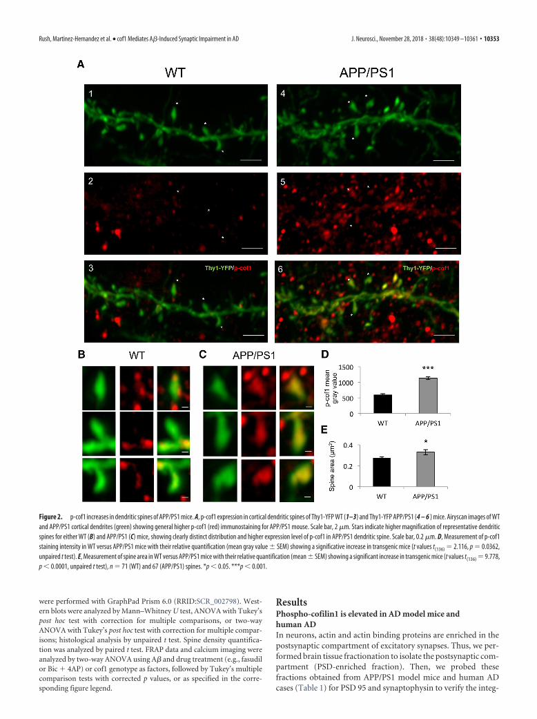
Figure 2. p-cof1 increases in dendritic spines of APP/PS1 mice. A, p-cof1 expression in cortical dendritic spines of Thy1-YFP WT (1–3) and Thy1-YFP APP/PS1 (4 – 6 ) mice. Airyscan images of WTand APP/PS1 cortical dendrites (green) showing general higher p-cof1 (red) immunostaining for APP/PS1 mouse. Scale bar, 2 �m. Stars indicate higher magnification of representative dendriticspines for either WT (B) and APP/PS1 (C) mice, showing clearly distinct distribution and higher expression level of p-cof1 in APP/PS1 dendritic spine. Scale bar, 0.2 �m. D, Measurement of p-cof1staining intensity in WT versus APP/PS1 mice with their relative quantification (mean gray value � SEM) showing a significative increase in transgenic mice (t values t(136) 2.116, p 0.0362,unpaired t test). E, Measurement of spine area in WT versus APP/PS1 mice with their relative quantification (mean � SEM) showing a significant increase in transgenic mice (t values t(136) 9.778,p 0.0001, unpaired t test), n 71 (WT) and 67 (APP/PS1) spines. *p 0.05. ***p 0.001.
Rush, Martinez-Hernandez et al. • cof1 Mediates A�-Induced Synaptic Impairment in AD J. Neurosci., November 28, 2018 • 38(48):10349 –10361 • 10353
rity of PSD-enriched and non-PSD fraction (Fig. 1A). Then, wecompared the levels of cof1 and p-cof1 protein expression on thePSD-enriched fraction (Fig. 1B). Although total cof1 levels weresimilar in both APP/PS1 AD mice and human AD cases, we founda twofold increase of p-cof1 levels in transgenic APP/PS1 ADmice and threefold increase in human AD cases, compared withcontrols, consistent with previous reports (Barone et al., 2014).However, the increased in p-cof-1 observed in the human tissuePSD fraction might reflect the general increase in whole AD brainlysate compared with non-AD brains.
To strengthen this result, we performed immunohistologicalstaining against p-cof1 in 3-month-old Thy1-YFP � APP/PS1mice, when senile plaques are sparse in the cortex (Radde et al.,2006). We observed a twofold increase in p-cof1 immunostainingin cortical dendritic spines of Thy-1-YFP � APP/PS1 animalscompared with Thy-1-YFP mice (Fig. 2A–D). In addition, theThy-1-YFP � APP/PS1 mice showed a significantly increaseddendritic spine area (Fig. 2E). Together, these results highlightthat AD patients and AD animal models have elevated p-cof1located in the postsynaptic sites of excitatory synapses.
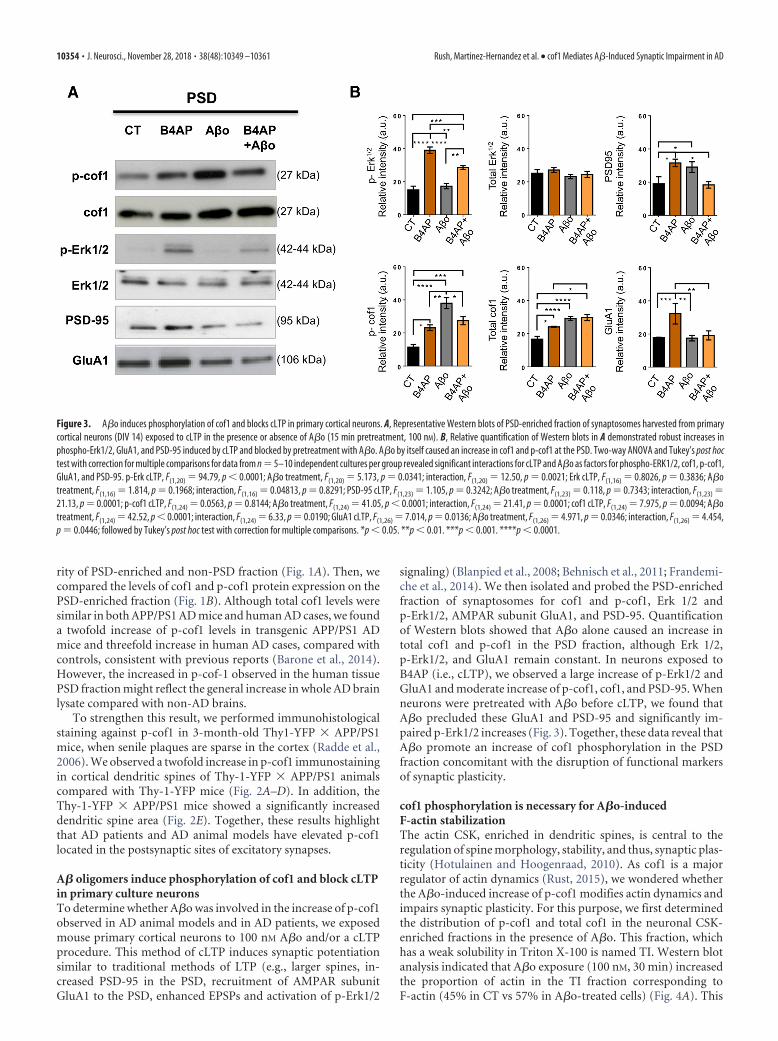
A� oligomers induce phosphorylation of cof1 and block cLTPin primary culture neuronsTo determine whether A�o was involved in the increase of p-cof1observed in AD animal models and in AD patients, we exposedmouse primary cortical neurons to 100 nM A�o and/or a cLTPprocedure. This method of cLTP induces synaptic potentiationsimilar to traditional methods of LTP (e.g., larger spines, in-creased PSD-95 in the PSD, recruitment of AMPAR subunitGluA1 to the PSD, enhanced EPSPs and activation of p-Erk1/2
signaling) (Blanpied et al., 2008; Behnisch et al., 2011; Frandemi-che et al., 2014). We then isolated and probed the PSD-enrichedfraction of synaptosomes for cof1 and p-cof1, Erk 1/2 andp-Erk1/2, AMPAR subunit GluA1, and PSD-95. Quantificationof Western blots showed that A�o alone caused an increase intotal cof1 and p-cof1 in the PSD fraction, although Erk 1/2,p-Erk1/2, and GluA1 remain constant. In neurons exposed toB4AP (i.e., cLTP), we observed a large increase of p-Erk1/2 andGluA1 and moderate increase of p-cof1, cof1, and PSD-95. Whenneurons were pretreated with A�o before cLTP, we found thatA�o precluded these GluA1 and PSD-95 and significantly im-paired p-Erk1/2 increases (Fig. 3). Together, these data reveal thatA�o promote an increase of cof1 phosphorylation in the PSDfraction concomitant with the disruption of functional markersof synaptic plasticity.
cof1 phosphorylation is necessary for A�o-inducedF-actin stabilizationThe actin CSK, enriched in dendritic spines, is central to theregulation of spine morphology, stability, and thus, synaptic plas-ticity (Hotulainen and Hoogenraad, 2010). As cof1 is a majorregulator of actin dynamics (Rust, 2015), we wondered whetherthe A�o-induced increase of p-cof1 modifies actin dynamics andimpairs synaptic plasticity. For this purpose, we first determinedthe distribution of p-cof1 and total cof1 in the neuronal CSK-enriched fractions in the presence of A�o. This fraction, whichhas a weak solubility in Triton X-100 is named TI. Western blotanalysis indicated that A�o exposure (100 nM, 30 min) increasedthe proportion of actin in the TI fraction corresponding toF-actin (45% in CT vs 57% in A�o-treated cells) (Fig. 4A). This
Figure 3. A�o induces phosphorylation of cof1 and blocks cLTP in primary cortical neurons. A, Representative Western blots of PSD-enriched fraction of synaptosomes harvested from primarycortical neurons (DIV 14) exposed to cLTP in the presence or absence of A�o (15 min pretreatment, 100 nM). B, Relative quantification of Western blots in A demonstrated robust increases inphospho-Erk1/2, GluA1, and PSD-95 induced by cLTP and blocked by pretreatment with A�o. A�o by itself caused an increase in cof1 and p-cof1 at the PSD. Two-way ANOVA and Tukey’s post hoctest with correction for multiple comparisons for data from n 5–10 independent cultures per group revealed significant interactions for cLTP and A�o as factors for phospho-ERK1/2, cof1, p-cof1,GluA1, and PSD-95. p-Erk cLTP, F(1,20) 94.79, p 0.0001; A�o treatment, F(1,20) 5.173, p 0.0341; interaction, F(1,20) 12.50, p 0.0021; Erk cLTP, F(1,16) 0.8026, p 0.3836; A�otreatment, F(1,16) 1.814, p 0.1968; interaction, F(1,16) 0.04813, p 0.8291; PSD-95 cLTP, F(1,23) 1.105, p 0.3242; A�o treatment, F(1,23) 0.118, p 0.7343; interaction, F(1,23) 21.13, p 0.0001; p-cof1 cLTP, F(1,24) 0.0563, p 0.8144; A�o treatment, F(1,24) 41.05, p 0.0001; interaction, F(1,24) 21.41, p 0.0001; cof1 cLTP, F(1,24) 7.975, p 0.0094; A�otreatment, F(1,24) 42.52, p 0.0001; interaction, F(1,24) 6.33, p 0.0190; GluA1 cLTP, F(1,26) 7.014, p 0.0136; A�o treatment, F(1,26) 4.971, p 0.0346; interaction, F(1,26) 4.454,p 0.0446; followed by Tukey’s post hoc test with correction for multiple comparisons. *p 0.05. **p 0.01. ***p 0.001. ****p 0.0001.
10354 • J. Neurosci., November 28, 2018 • 38(48):10349 –10361 Rush, Martinez-Hernandez et al. • cof1 Mediates A�-Induced Synaptic Impairment in AD
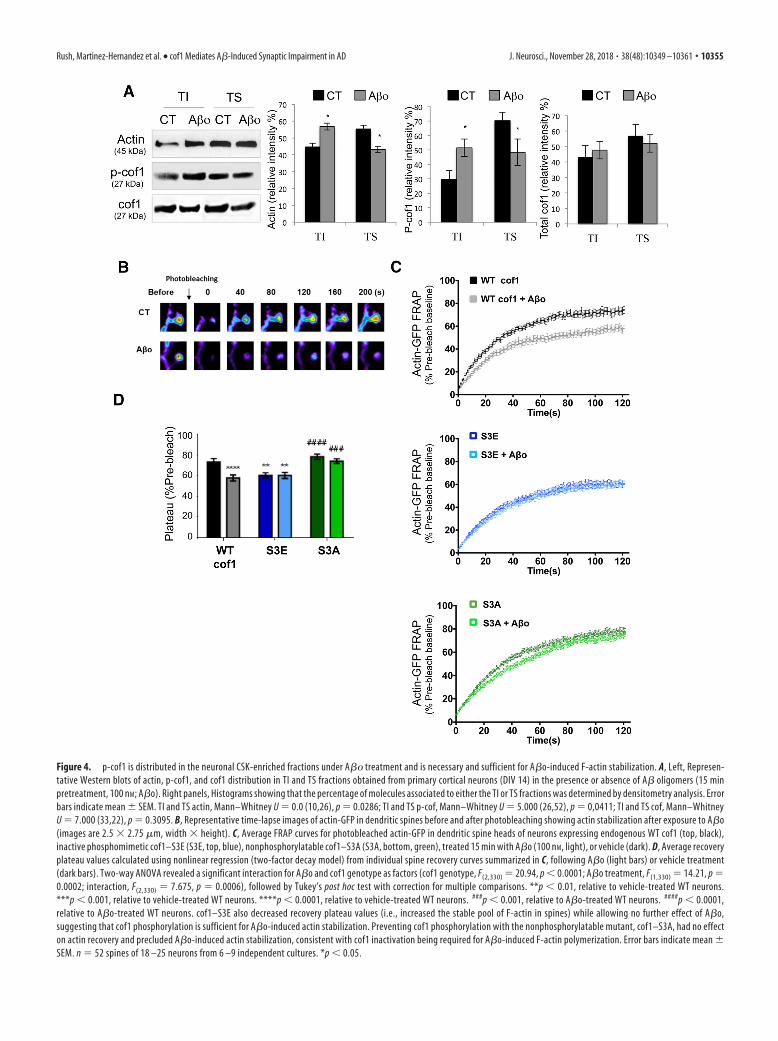
Figure 4. p-cof1 is distributed in the neuronal CSK-enriched fractions under A�� treatment and is necessary and sufficient for A�o-induced F-actin stabilization. A, Left, Represen-tative Western blots of actin, p-cof1, and cof1 distribution in TI and TS fractions obtained from primary cortical neurons (DIV 14) in the presence or absence of A� oligomers (15 minpretreatment, 100 nM; A�o). Right panels, Histograms showing that the percentage of molecules associated to either the TI or TS fractions was determined by densitometry analysis. Errorbars indicate mean � SEM. TI and TS actin, Mann–Whitney U 0.0 (10,26), p 0.0286; TI and TS p-cof, Mann–Whitney U 5.000 (26,52), p 0,0411; TI and TS cof, Mann–WhitneyU 7.000 (33,22), p 0.3095. B, Representative time-lapse images of actin-GFP in dendritic spines before and after photobleaching showing actin stabilization after exposure to A�o(images are 2.5 � 2.75 �m, width � height). C, Average FRAP curves for photobleached actin-GFP in dendritic spine heads of neurons expressing endogenous WT cof1 (top, black),inactive phosphomimetic cof1–S3E (S3E, top, blue), nonphosphorylatable cof1–S3A (S3A, bottom, green), treated 15 min with A�o (100 nM, light), or vehicle (dark). D, Average recoveryplateau values calculated using nonlinear regression (two-factor decay model) from individual spine recovery curves summarized in C, following A�o (light bars) or vehicle treatment(dark bars). Two-way ANOVA revealed a significant interaction for A�o and cof1 genotype as factors (cof1 genotype, F(2,330) 20.94, p 0.0001; A�o treatment, F(1,330) 14.21, p 0.0002; interaction, F(2,330) 7.675, p 0.0006), followed by Tukey’s post hoc test with correction for multiple comparisons. **p 0.01, relative to vehicle-treated WT neurons.***p 0.001, relative to vehicle-treated WT neurons. ****p 0.0001, relative to vehicle-treated WT neurons. ###p 0.001, relative to A�o-treated WT neurons. ####p 0.0001,relative to A�o-treated WT neurons. cof1–S3E also decreased recovery plateau values (i.e., increased the stable pool of F-actin in spines) while allowing no further effect of A�o,suggesting that cof1 phosphorylation is sufficient for A�o-induced actin stabilization. Preventing cof1 phosphorylation with the nonphosphorylatable mutant, cof1–S3A, had no effecton actin recovery and precluded A�o-induced actin stabilization, consistent with cof1 inactivation being required for A�o-induced F-actin polymerization. Error bars indicate mean �SEM. n 52 spines of 18 –25 neurons from 6 –9 independent cultures. *p 0.05.
Rush, Martinez-Hernandez et al. • cof1 Mediates A�-Induced Synaptic Impairment in AD J. Neurosci., November 28, 2018 • 38(48):10349 –10361 • 10355
process is correlated with recruitment of p-cof1 in the TI frac-tions (almost twofold increase), whereas no increase is observedfor total cof1 (Fig. 4A); it suggests that A�o exposure promotes animportant change in the ratio p-cof1/cof1 leading to increasedactin content in the CSK enriched fraction.
We next used FRAP imaging of neurons expressing actin-GFPto determine whether actin dynamics were altered in dendriticspines as a result of A�o-induced cof1 phosphorylation. In neu-rons coexpressing mCherry-tagged cof1, A�o decreased theFRAP plateau reached by 20% (Fig. 4B–D), consistent with alarger pool of stable F-actin in the dendritic spine. This was fur-ther confirmed performing FRAP of actin-GFP in neurons coex-pressing cof1 phosphomimetic and nonphosphomimetic mutants,referred to as S3E and S3A, respectively. The phosphomimetic S3Ecof1 mutant is constitutively inactive (i.e., this cof1 does not severactin filaments) and mirrors the effects of A�o on fluorescencerecovery of actin in dendritic spines with no additive effectupon A�o exposure (Fig. 4C,D). The nonphosphorylatablecof1-S3A mutant had no effect on actin stabilization, and pre-cluded A�o-induced actin stabilization, suggesting thatA�o-induced stabilization of spine actin required cof1 phos-phorylation (Fig. 4C,D).
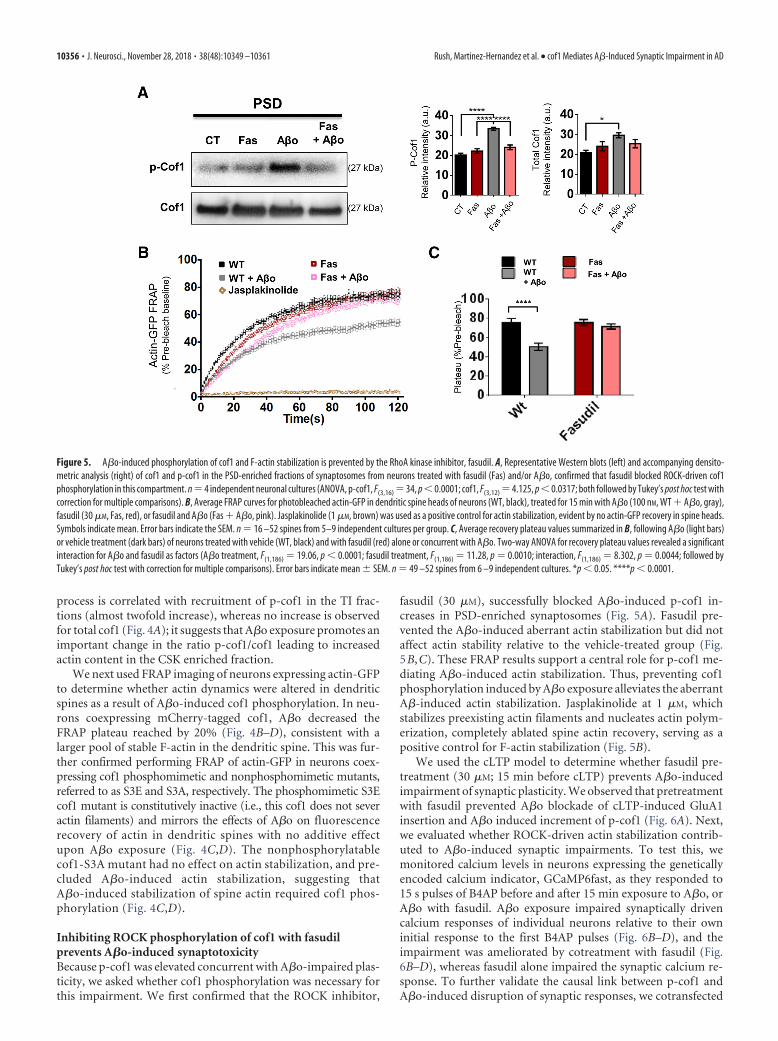
Inhibiting ROCK phosphorylation of cof1 with fasudilprevents A�o-induced synaptotoxicityBecause p-cof1 was elevated concurrent with A�o-impaired plas-ticity, we asked whether cof1 phosphorylation was necessary forthis impairment. We first confirmed that the ROCK inhibitor,
fasudil (30 �M), successfully blocked A�o-induced p-cof1 in-creases in PSD-enriched synaptosomes (Fig. 5A). Fasudil pre-vented the A�o-induced aberrant actin stabilization but did notaffect actin stability relative to the vehicle-treated group (Fig.5B,C). These FRAP results support a central role for p-cof1 me-diating A�o-induced actin stabilization. Thus, preventing cof1phosphorylation induced by A�o exposure alleviates the aberrantA�-induced actin stabilization. Jasplakinolide at 1 �M, whichstabilizes preexisting actin filaments and nucleates actin polym-erization, completely ablated spine actin recovery, serving as apositive control for F-actin stabilization (Fig. 5B).
We used the cLTP model to determine whether fasudil pre-treatment (30 �M; 15 min before cLTP) prevents A�o-inducedimpairment of synaptic plasticity. We observed that pretreatmentwith fasudil prevented A�o blockade of cLTP-induced GluA1insertion and A�o induced increment of p-cof1 (Fig. 6A). Next,we evaluated whether ROCK-driven actin stabilization contrib-uted to A�o-induced synaptic impairments. To test this, wemonitored calcium levels in neurons expressing the geneticallyencoded calcium indicator, GCaMP6fast, as they responded to15 s pulses of B4AP before and after 15 min exposure to A�o, orA�o with fasudil. A�o exposure impaired synaptically drivencalcium responses of individual neurons relative to their owninitial response to the first B4AP pulses (Fig. 6B–D), and theimpairment was ameliorated by cotreatment with fasudil (Fig.6B–D), whereas fasudil alone impaired the synaptic calcium re-sponse. To further validate the causal link between p-cof1 andA�o-induced disruption of synaptic responses, we cotransfected
Figure 5. A�o-induced phosphorylation of cof1 and F-actin stabilization is prevented by the RhoA kinase inhibitor, fasudil. A, Representative Western blots (left) and accompanying densito-metric analysis (right) of cof1 and p-cof1 in the PSD-enriched fractions of synaptosomes from neurons treated with fasudil (Fas) and/or A�o, confirmed that fasudil blocked ROCK-driven cof1phosphorylation in this compartment. n 4 independent neuronal cultures (ANOVA, p-cof1, F(3,16) 34, p 0.0001; cof1, F(3,12) 4.125, p 0.0317; both followed by Tukey’s post hoc test withcorrection for multiple comparisons). B, Average FRAP curves for photobleached actin-GFP in dendritic spine heads of neurons (WT, black), treated for 15 min with A�o (100 nM, WT � A�o, gray),fasudil (30 �M, Fas, red), or fasudil and A�o (Fas � A�o, pink). Jasplakinolide (1 �M, brown) was used as a positive control for actin stabilization, evident by no actin-GFP recovery in spine heads.Symbols indicate mean. Error bars indicate the SEM. n 16 –52 spines from 5–9 independent cultures per group. C, Average recovery plateau values summarized in B, following A�o (light bars)or vehicle treatment (dark bars) of neurons treated with vehicle (WT, black) and with fasudil (red) alone or concurrent with A�o. Two-way ANOVA for recovery plateau values revealed a significantinteraction for A�o and fasudil as factors (A�o treatment, F(1,186) 19.06, p 0.0001; fasudil treatment, F(1,186) 11.28, p 0.0010; interaction, F(1,186) 8.302, p 0.0044; followed byTukey’s post hoc test with correction for multiple comparisons). Error bars indicate mean � SEM. n 49 –52 spines from 6 –9 independent cultures. *p 0.05. ****p 0.0001.
10356 • J. Neurosci., November 28, 2018 • 38(48):10349 –10361 Rush, Martinez-Hernandez et al. • cof1 Mediates A�-Induced Synaptic Impairment in AD

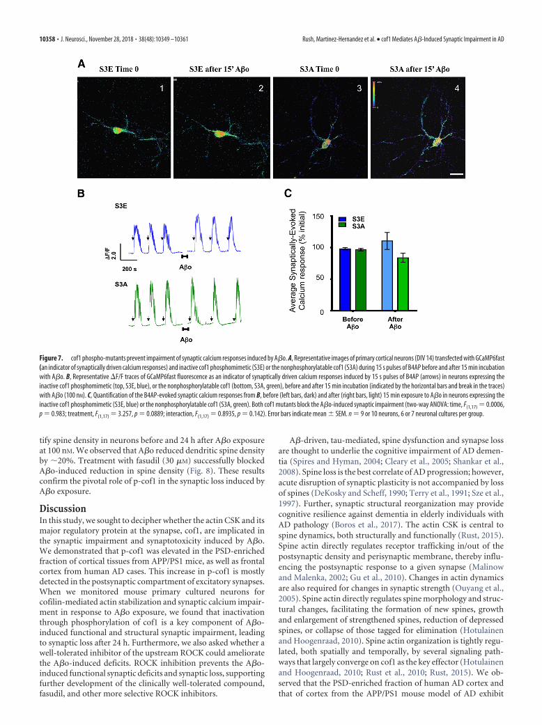
neurons with the phosphomimetic cof1-S3E mutant or nonphos-phorylatable cof1-S3A mutant and GcAMP6f in neurons sub-jected to B4AP stimulation. In these experimental conditions, weobserved that expression of both cof1 mutants abolished the A�oeffect on B4AP-driven synaptic responses (Fig. 7A–C), validatingthe importance of cof1 phosphorylation to A�o-induced disrup-tion of synaptic responses.
Another cardinal feature of A�o-induced synaptotoxicity isthe decrease of excitatory synapse density. To address this issue,we further investigated whether a treatment with fasudil alteredthe effect of A�o exposure on synaptic density. For this purpose,we monitored spine density in neurons transfected with LifeAct-GFP, a 16 amino acid peptide that binds selectively to F-actin.This approach allowed us to visualize dendritic spines and quan-
Figure 6. Fasudil inhibition of RhoA kinase prevents A�o-induced phosphorylation of cof1 and A�o-impaired cLTP. A, Representative Western blot images (left) and quantification (right) ofGluA1 and p-cof in the PSD-enriched fraction of synaptosomes isolated from neurons exposed to vehicle (CT), cLTP (B4AP, 15 min, brown) alone, or cLTP after pretreatment (15 min) with A�o(orange) or fasudil � A�o (pink). Fasudil prevented the A�o-induced GluA1 trafficking impairment and restored the p-cof values as determined by one-way ANOVA (GluA1, F(3,20) 36.26, p 0.0001; p-cof1, F(3,9) 20.33, p 0.0002) followed by Tukey’s post hoc test with correction for multiple comparisons. n 3–11 cultures per group. B, Representative images of primary corticalneurons (DIV 14) transfected with GCaMP6fast (an indicator of synaptically driven calcium responses) activated by 15 s pulses of B4AP before and after 15 min incubation with A�o or fasudil � A�o.C, Representative �F/F traces of GCaMP6fast fluorescence as an indicator of synaptically driven calcium responses induced by 15 s pulses of B4AP (arrows) in neurons expressing endogenous WT cof1(WT, black), before and after 15 min incubation (indicated by the horizontal bars and break in the traces) with vehicle (dark) or A�o (light), or coincubated with or without fasudil (red).D, Quantification of the B4AP-evoked synaptic calcium responses from C before (left bars, dark), and after (right bars, light) 15 min treatment (Tx) with vehicle (black), A�o (gray), fasudil (red), orfasudil � A�o (pink). Fasudil blocks the A�o-induced synaptic impairment, as revealed by two-way ANOVA using time and treatment as factors (time, F(1,30) 16.84, p 0.0003; treatment,F(3,30) 8.297, p 0.0004; interaction, F(3,30) 6.742, p 0.0013; followed by Tukey’s post hoc test with correction for multiple comparisons). n 9 –11 neurons, 5–7 neuronal cultures pergroup. *p 0.05. **p 0.01. ***p 0.001. ****p 0.0001.
Rush, Martinez-Hernandez et al. • cof1 Mediates A�-Induced Synaptic Impairment in AD J. Neurosci., November 28, 2018 • 38(48):10349 –10361 • 10357
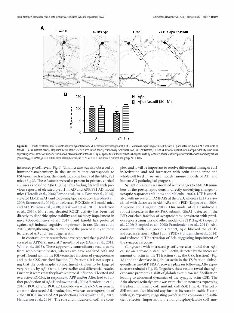
tify spine density in neurons before and 24 h after A�o exposureat 100 nM. We observed that A�o reduced dendritic spine densityby �20%. Treatment with fasudil (30 �M) successfully blockedA�o-induced reduction in spine density (Fig. 8). These resultsconfirm the pivotal role of p-cof1 in the synaptic loss induced byA�o exposure.
DiscussionIn this study, we sought to decipher whether the actin CSK and itsmajor regulatory protein at the synapse, cof1, are implicated inthe synaptic impairment and synaptotoxicity induced by A�o.We demonstrated that p-cof1 was elevated in the PSD-enrichedfraction of cortical tissues from APP/PS1 mice, as well as frontalcortex from human AD cases. This increase in p-cof1 is mostlydetected in the postsynaptic compartment of excitatory synapses.When we monitored mouse primary cultured neurons forcofilin-mediated actin stabilization and synaptic calcium impair-ment in response to A�o exposure, we found that inactivationthrough phosphorylation of cof1 is a key component of A�o-induced functional and structural synaptic impairment, leadingto synaptic loss after 24 h. Furthermore, we also asked whether awell-tolerated inhibitor of the upstream ROCK could amelioratethe A�o-induced deficits. ROCK inhibition prevents the A�o-induced functional synaptic deficits and synaptic loss, supportingfurther development of the clinically well-tolerated compound,fasudil, and other more selective ROCK inhibitors.
A�-driven, tau-mediated, spine dysfunction and synapse lossare thought to underlie the cognitive impairment of AD demen-tia (Spires and Hyman, 2004; Cleary et al., 2005; Shankar et al.,2008). Spine loss is the best correlate of AD progression; however,acute disruption of synaptic plasticity is not accompanied by lossof spines (DeKosky and Scheff, 1990; Terry et al., 1991; Sze et al.,1997). Further, synaptic structural reorganization may providecognitive resilience against dementia in elderly individuals withAD pathology (Boros et al., 2017). The actin CSK is central tospine dynamics, both structurally and functionally (Rust, 2015).Spine actin directly regulates receptor trafficking in/out of thepostsynaptic density and perisynaptic membrane, thereby influ-encing the postsynaptic response to a given synapse (Malinowand Malenka, 2002; Gu et al., 2010). Changes in actin dynamicsare also required for changes in synaptic strength (Ouyang et al.,2005). Spine actin directly regulates spine morphology and struc-tural changes, facilitating the formation of new spines, growthand enlargement of strengthened spines, reduction of depressedspines, or collapse of those tagged for elimination (Hotulainenand Hoogenraad, 2010). Spine actin organization is tightly regu-lated, both spatially and temporally, by several signaling path-ways that largely converge on cof1 as the key effector (Hotulainenand Hoogenraad, 2010; Rust et al., 2010; Rust, 2015). We ob-served that the PSD-enriched fraction of human AD cortex andthat of cortex from the APP/PS1 mouse model of AD exhibit
Figure 7. cof1 phospho-mutants prevent impairment of synaptic calcium responses induced by A�o. A, Representative images of primary cortical neurons (DIV 14) transfected with GCaMP6fast(an indicator of synaptically driven calcium responses) and inactive cof1 phosphomimetic (S3E) or the nonphosphorylatable cof1 (S3A) during 15 s pulses of B4AP before and after 15 min incubationwith A�o. B, Representative �F/F traces of GCaMP6fast fluorescence as an indicator of synaptically driven calcium responses induced by 15 s pulses of B4AP (arrows) in neurons expressing theinactive cof1 phosphomimetic (top, S3E, blue), or the nonphosphorylatable cof1 (bottom, S3A, green), before and after 15 min incubation (indicated by the horizontal bars and break in the traces)with A�o (100 nM). C, Quantification of the B4AP-evoked synaptic calcium responses from B, before (left bars, dark) and after (right bars, light) 15 min exposure to A�o in neurons expressing theinactive cof1 phosphomimetic (S3E, blue) or the nonphosphorylatable cof1 (S3A, green). Both cof1 mutants block the A�o-induced synaptic impairment (two-way ANOVA: time, F(1,17) 0.0006,p 0.983; treatment, F(1,17) 3.257, p 0.0889; interaction, F(1,17) 0.8935, p 0.142). Error bars indicate mean � SEM. n 9 or 10 neurons, 6 or 7 neuronal cultures per group.
10358 • J. Neurosci., November 28, 2018 • 38(48):10349 –10361 Rush, Martinez-Hernandez et al. • cof1 Mediates A�-Induced Synaptic Impairment in AD
increased p-cof1 levels (Fig 1). This increase was also observed byimmunohistochemistry in the structure that corresponds toPSD-positive fraction: the dendritic spine heads of the APP/PS1mice (Fig 2). These features were also present in primary corticalcultures exposed to A�o (Fig. 3). This finding fits well with pre-vious reports of elevated p-cof1 in AD and APP/PS1 AD modelmice (Heredia et al., 2006; Barone et al., 2014; Fowler et al., 2014),elevated LIMK in AD and following A�o exposure (Heredia et al.,2006; Barone et al., 2014), and elevated ROCKs in AD model miceand AD (Petratos et al., 2008; Herskowitz et al., 2013; Hendersonet al., 2016). Moreover, elevated ROCK activity has been tieddirectly to dendritic spine stability and memory impairment inmice (Bobo-Jimenez et al., 2017), and fasudil has protectedagainst A�-induced cognitive impairment in rats (Sellers et al.,2018), strengthening the relevance of the present study to thesefeatures of AD and neurodegeneration.
In contrast, other researchers have reported that p-cof is de-creased in APP/PS1 mice at 7 months of age (Davis et al., 2011;Woo et al., 2015). These apparently contradictory results camefrom whole-tissue lysates, whereas we have analyzed cof1 andp-cof1 found within the PSD-enriched fraction of synaptosomesand in the CSK-enriched fraction (TI fraction). It is not surpris-ing that the postsynaptic compartment (known to be targetedvery rapidly by A�o) would have earlier and differential results.Further, it seems that they have reciprocal influence. Elevated andoveractive ROCKs, in response to APP and/or A�o, lead to fur-ther production of A� (Herskowitz et al., 2013; Henderson et al.,2016). ROCK1 and ROCK2 knockdown with siRNA or geneticablation decreased A� production, whereas overexpression ofeither ROCK increased A� production (Herskowitz et al., 2013;Henderson et al., 2016). The role and influence of cof1 are com-
plex, and it will be important to resolve differential timing of cof1in/activation and rod formation with actin at the spine andwhole-cell level in in vitro models, mouse models of AD, andhuman AD pathological progression.
Synaptic plasticity is associated with changes to AMPAR num-bers at the postsynaptic density directly underlying changes tosynaptic responses (Malinow and Malenka, 2002). LTP is associ-ated with increases in AMPARs at the PSD, whereas LTD is asso-ciated with decreases in AMPARs at the PSD (Kopec et al., 2006;Anggono and Huganir, 2012). Our model of cLTP induced arobust increase in the AMPAR subunit, GluA1, detected in thePSD-enriched fraction of synaptosomes, consistent with previ-ous reports using this and other models of cLTP (Fig. 4) (Kopec etal., 2006; Blanpied et al., 2008; Frandemiche et al., 2014). Alsoconsistent with our previous report, A�o blocked the cLTP-induced insertion of GluA1 at the PSD (Frandemiche et al., 2014)and reduced cLTP activation of Erk, suggesting impairment ofthe synaptic response.
Congruent with increased p-cof1, we also found that A�ocaused an increase in stabilized F-actin, detected by the increasedamount of actin in the TI fraction (i.e., the CSK fraction) (Fig.4A) and the decrease in globular actin in the TS fraction. Subse-quently, actin-GFP FRAP recovery plateaus following A�o expo-sure are reduced (Fig. 5). Together, these results reveal that A�oexposure promotes a shift of globular actin toward fibrilizationleading to abnormal dynamics of the synaptic actin CSK. TheA�o-altered actin dynamic was mimicked in neurons expressingthe phosphomimetic cof1 mutant, cof1-S3E (Fig. 4). The cof1-S3E mutant also blocked any further increase in stable F-actinwith A�o exposure, suggesting p-cof1 as the common and suffi-cient effector. Importantly, the nonphosphorylatable cof1 mu-
Figure 8. Fasudil treatment reverses A�o-induced synaptotoxicity. A, Representative images of DIV 14 –15 neurons expressing actin-GFP before (t 0) and after incubation 24 h with A�o orfasudil � A�o. Bottom panels, Magnified detail of the selected area in top panels, respectively. Scale bars: Top, 30 �m; Bottom, 10 �m. B, Relative quantification of spine density in neuronsexpressing actin-GFP before and after incubation 24 h with A�o or fasudil � A�o. A paired t test showed that 24 h exposition to A�o caused decrease in the spine density that was blocked by fasudil(t values t(10) 8.931, p 0.0001). Error bars indicate mean � SEM. n 11 neurons, 3 cultures per group. *p 0.05.
Rush, Martinez-Hernandez et al. • cof1 Mediates A�-Induced Synaptic Impairment in AD J. Neurosci., November 28, 2018 • 38(48):10349 –10361 • 10359
tant, cof1-S3A, had no effect on synaptic actin dynamics,suggesting that maintaining stable spine structure does not re-quire cof1 inactivation. Changes to spine structure and function,however, require a temporal sequence of cof1 dephosphoryla-tion/phosphorylation (Gu et al., 2010; Calabrese et al., 2014;Noguchi et al., 2016). Expression of cofilin-S3A also precludedA�o-increased stability of F-actin, confirming that inactivationof cof1 is required. Finally, fasudil reversed the A�o-inducedstabilization of actin and subsequent synaptic loss observed aftera 24 h exposure. This finding strengthens the conclusion thatphosphorylation of cof1 mediates A�o-induced actin stabiliza-tion and the subsequent synaptic loss.
Monitoring somatic calcium responses driven by brief boutsof synaptic stimulations brought on by Bic4AP exposure revealedthat A�o acutely disrupts the postsynaptic response. cLTP fol-lowing A�o exposure did not show an activity-driven increase inp-cof1 relative to cLTP alone (Fig. 3), underscoring the impor-tance of tight temporal regulation of cof1 and the actin CSK.Indeed, A�o blocked both GluA1 insertion at the PSD and syn-aptic calcium responses. Fasudil treatment reversed both A�o-induced synaptic dysfunctions. These results suggest that, byrestoring a pool of active cofilin-1, fasudil treatment promoted arecovery of the synaptic plasticity potential in the presence ofA�o and abolished the synaptic loss observed in neurons exposedto A�o for 24 h. The fact that fasudil inhibition of the ROCKpathway alone reduces the postsynaptic calcium responses is inaccordance with a previous report (Gonzalez-Forero et al., 2012).It may reflect the consequences of the alteration of the physiolog-ical balance between of active cof1/p-cof1 on excitatory neu-rotransmission and synaptic plasticity. Together, these resultsunderscore the importance of balanced cytoskeletal regulation inthe postsynaptic compartment and unveil the therapeutic poten-tial of targeting rho kinase in AD.
Collectively, our results support further study of actin CSKdynamics, and drugs targeting ROCK regulation thereof, as atherapeutic target for neuronal dysfunction in AD. Fasudil, aclinically approved drug, is a timely candidate for this purpose,although studies of prolonged fasudil treatment have not beenperformed in AD. According to our results, the development andstudy of more selective ROCK inhibitors are warranted.
ReferencesAnggono V, Huganir RL (2012) Regulation of AMPA receptor trafficking
and synaptic plasticity. Curr Opin Neurobiol 22:461– 469. CrossRefMedline
Alzheimer’s Association (2017) Alzheimer’s disease facts and figures. Alz-heimers Dement 13:325–373. CrossRef
Bamburg JR, Bernstein BW (2016) Actin dynamics and cofilin-actin rods inAlzheimer disease. Cytoskeleton 73:477– 497. CrossRef Medline
Barone E, Mosser S, Fraering PC (2014) Inactivation of brain cofilin-1 byage, Alzheimer’s disease and gamma-secretase. Biochim Biophys Acta1842:2500 –2509. CrossRef Medline
Behnisch T, Yuanxiang P, Bethge P, Parvez S, Chen Y, Yu J, Karpova A, FreyJU, Mikhaylova M, Kreutz MR (2011) Nuclear translocation of jacob inhippocampal neurons after stimuli inducing long-term potentiation butnot long-term depression. PLoS One 6:e17276. CrossRef Medline
Blanpied TA, Kerr JM, Ehlers MD (2008) Structural plasticity with pre-served topology in the postsynaptic protein network. Proc Natl Acad SciU S A 105:12587–12592. CrossRef Medline
Bobo-Jimenez V, Delgado-Esteban M, Angibaud J, Sanchez-Moran I, de laFuente A, Yajeya J, Nagerl UV, Castillo J, Bolanos JP, Almeida A (2017)APC/CCdh1-Rock2 pathway controls dendritic integrity and memory.Proc Natl Acad Sci U S A 114:4513– 4518. CrossRef Medline
Boros BD, Greathouse KM, Gentry EG, Curtis KA, Birchall EL, Gearing M,Herskowitz JH (2017) Dendritic spines provide cognitive resilienceagainst Alzheimer’s disease. Ann Neurol 82: 602– 614. CrossRef Medline
Calabrese B, Saffin JM, Halpain S (2014) Activity-dependent dendritic spineshrinkage and growth involve downregulation of cofilin via distinctmechanisms. PLoS One 9:e94787. CrossRef Medline
Cleary JP, Walsh DM, Hofmeister JJ, Shankar GM, Kuskowski MA, Selkoe DJ,Ashe KH (2005) Natural oligomers of the amyloid-beta protein specifi-cally disrupt cognitive function. Nat Neurosci 8:79 – 84. CrossRef Medline
Davis RC, Marsden IT, Maloney MT, Minamide LS, Podlisny M, Selkoe DJ,Bamburg JR (2011) Amyloid beta dimers/trimers potently induce cofilin-actin rods that are inhibited by maintaining cofilin-phosphorylation. MolNeurodegener 6:10. CrossRef Medline
DeKosky ST, Scheff SW (1990) Synapse loss in frontal cortex biopsies inAlzheimer’s disease: correlation with cognitive severity. Ann Neurol 27:457– 464. CrossRef Medline
Fowler SW, Chiang AC, Savjani RR, Larson ME, Sherman MA, Schuler DR,Cirrito JR, Lesne SE, Jankowsky JL (2014) Genetic modulation of solu-ble abeta rescues cognitive and synaptic impairment in a mouse model ofAlzheimer’s disease. J Neurosci 34:7871–7885. CrossRef Medline
Frandemiche ML, De Seranno S, Rush T, Borel E, Elie A, Arnal I, Lante F,Buisson A (2014) Activity-dependent tau protein translocation to excit-atory synapse is disrupted by exposure to amyloid-beta oligomers. J Neu-rosci 34:6084 – 6097. CrossRef Medline
Gonzalez-Forero D, Montero F, García-Morales V, Domínguez G, Gomez-Perez L, García-Verdugo JM, Moreno-Lopez B (2012) Endogenous rho-kinase signaling maintains synaptic strength by stabilizing the size of thereadily releasable pool of synaptic vesicles. J Neurosci 32:68 – 84. CrossRefMedline
Gu J, Lee CW, Fan Y, Komlos D, Tang X, Sun C, Yu K, Hartzell HC, Chen G,Bamburg JR, Zheng JQ (2010) ADF/cofilin-mediated actin dynamicsregulate AMPA receptor trafficking during synaptic plasticity. Nat Neu-rosci 13:1208 –1215. CrossRef Medline
Han F, Zhuang TT, Chen JJ, Zhu XL, Cai YF, Lu YP (2017) Novel derivativeof Paeonol, Paeononlsilatie sodium, alleviates behavioral damage andhippocampal dendritic injury in Alzheimer’s disease concurrent withcofilin1/phosphorylated-cofilin1 and RAC1/CDC42 alterations in rats.PLoS One 12:e0185102. CrossRef Medline
Henderson BW, Gentry EG, Rush T, Troncoso JC, Thambisetty M, MontineTJ, Herskowitz JH (2016) Rho-associated protein kinase 1 (ROCK1) isincreased in Alzheimer’s disease and ROCK1 depletion reduces amyloid-beta levels in brain. J Neurochem 138:525–531. CrossRef Medline
Heredia L, Helguera P, de Olmos S, Kedikian G, Sola Vigo F, LaFerla F,Staufenbiel M, de Olmos J, Busciglio J, Caceres A, Lorenzo A (2006)Phosphorylation of actin-depolymerizing factor/cofilin by LIM-kinasemediates amyloid beta-induced degeneration: a potential mechanism ofneuronal dystrophy in Alzheimer’s disease. J Neurosci 26:6533– 6542.CrossRef Medline
Herskowitz JH, Feng Y, Mattheyses AL, Hales CM, Higginbotham LA, DuongDM, Montine TJ, Troncoso JC, Thambisetty M, Seyfried NT, Levey AI,Lah JJ (2013) Pharmacologic inhibition of ROCK2 suppresses amyloid-beta production in an Alzheimer’s disease mouse model. J Neurosci 33:19086 –19098. CrossRef Medline
Hinck L1, Näthke IS, Papkoff J, Nelson WJ (1994) Dynamics of cadherin/catenin complex formation: novel protein interactions and pathways ofcomplex assembly. J Cell Biol 125:1327–1340. CrossRef Medline
Hotulainen P, Hoogenraad CC (2010) Actin in dendritic spines: connectingdynamics to function. J Cell Biol 189:619 – 629. CrossRef Medline
Kervern M, Angeli A, Nicole O, Leveille F, Parent B, Villette V, Buisson A,Dutar P (2012) Selective impairment of some forms of synaptic plastic-ity by oligomeric amyloid-beta peptide in the mouse hippocampus: im-plication of extrasynaptic NMDA receptors. J Alzheimers Dis 32:183–196.CrossRef Medline
Kopec CD, Li B, Wei W, Boehm J, Malinow R (2006) Glutamate receptorexocytosis and spine enlargement during chemically induced long-termpotentiation. J Neurosci 26:2000 –2009. CrossRef Medline
Lacor PN, Buniel MC, Furlow PW, Clemente AS, Velasco PT, Wood M, ViolaKL, Klein WL (2007) Abeta oligomer-induced aberrations in synapsecomposition, shape, and density provide a molecular basis for loss ofconnectivity in Alzheimer’s disease. J Neurosci 27:796 – 807. CrossRefMedline
Leveille F, El Gaamouch F, Gouix E, Lecocq M, Lobner D, Nicole O, BuissonA (2008) Neuronal viability is controlled by a functional relation be-tween synaptic and extrasynaptic NMDA receptors. FASEB J 22:4258 –4271. CrossRef Medline
10360 • J. Neurosci., November 28, 2018 • 38(48):10349 –10361 Rush, Martinez-Hernandez et al. • cof1 Mediates A�-Induced Synaptic Impairment in AD
Malinow R, Malenka RC (2002) AMPA receptor trafficking and synapticplasticity. Annu Rev Neurosci 25:103–126. CrossRef Medline
Mendoza-Naranjo A, Contreras-Vallejos E, Henriquez DR, Otth C, BamburgJR, Maccioni RB, Gonzalez-Billault C (2012) Fibrillar amyloid-be-ta1– 42 modifies actin organization affecting the cofilin phosphorylationstate: a role for Rac1/cdc42 effector proteins and the slingshot phospha-tase. J Alzheimers Dis 29:63–77. CrossRef Medline
Niwa R, Nagata-Ohashi K, Takeichi M, Mizuno K, Uemura T (2002) Con-trol of actin reorganization by slingshot, a family of phosphatases thatdephosphorylate ADF/cofilin. Cell 108:233–246. CrossRef Medline
Noguchi J, Hayama T, Watanabe S, Ucar H, Yagishita S, Takahashi N, Kasai H(2016) State-dependent diffusion of actin-depolymerizing factor/cofilinunderlies the enlargement and shrinkage of dendritic spines. Sci Rep6:32897. CrossRef Medline
Oddo S, Billings L, Kesslak JP, Cribbs DH, LaFerla FM (2004) Abeta immu-notherapy leads to clearance of early, but not late, hyperphosphorylatedtau aggregates via the proteasome. Neuron 43:321–332. CrossRef Medline
Ouyang Y, Wong M, Capani F, Rensing N, Lee CS, Liu Q, Neusch C, MartoneME, Wu JY, Yamada K, Ellisman MH, Choi DW (2005) Transient de-crease in F-actin may be necessary for translocation of proteins into den-dritic spines. Eur J Neurosci 22:2995–3005. CrossRef Medline
Petratos S, Li QX, George AJ, Hou X, Kerr ML, Unabia SE, Hatzinisiriou I,Maksel D, Aguilar MI, Small DH (2008) The beta-amyloid protein ofAlzheimer’s disease increases neuronal CRMP-2 phosphorylation by arho-GTP mechanism. Brain 131:90 –108. CrossRef Medline
Radde R, Bolmont T, Kaeser SA, Coomaraswamy J, Lindau D, Stoltze L,Calhoun ME, Jaggi F, Wolburg H, Gengler S, Haass C, Ghetti B, Czech C,Holscher C, Mathews PM, Jucker M (2006) Abeta42-driven cerebralamyloidosis in transgenic mice reveals early and robust pathology. EMBORep 7:940 –946. CrossRef Medline
Rapoport M, Dawson HN, Binder LI, Vitek MP, Ferreira A (2002) Tau isessential to beta-amyloid-induced neurotoxicity. Proc Natl Acad SciU S A 99:6364 – 6369. CrossRef Medline
Rust MB (2015) ADF/cofilin: a crucial regulator of synapse physiology andbehavior. Cell Mol Life Sci 72:3521–3529. CrossRef Medline
Rust MB, Gurniak CB, Renner M, Vara H, Morando L, Gorlich A, Sassoe-Pognetto M, Banchaabouchi MA, Giustetto M, Triller A, Choquet D,Witke W (2010) Learning, AMPA receptor mobility and synaptic plas-ticity depend on n-cofilin-mediated actin dynamics. EMBO J 29:1889 –1902. CrossRef Medline
Sellers KJ, Elliott C, Jackson J, Ghosh A, Ribe E, Rojo AI, Jarosz-Griffiths HH,Watson IA, Xia W, Semenov M, Morin P, Hooper NM, Porter R, Preston
J, Al-Shawi R, Baillie G, Lovestone S, Cuadrado A, Harte M, Simons P,et al. (2018) Amyloid � synaptotoxicity is wnt-PCP dependent andblocked by fasudil. Alzheimers Dement 14:306 –317. CrossRef Medline
Shankar GM, Li S, Mehta TH, Garcia-Munoz A, Shepardson NE, Smith I,Brett FM, Farrell MA, Rowan MJ, Lemere CA, Regan CM, Walsh DM,Sabatini BL, Selkoe DJ (2008) Amyloid-beta protein dimers isolated di-rectly from Alzheimer’s brains impair synaptic plasticity and memory.Nat Med 14:837– 842. CrossRef Medline
Spires TL, Hyman BT (2004) Neuronal structure is altered by amyloidplaques. Rev Neurosci 15:267–278. CrossRef Medline
Stine WB, Dahlgren KN, Krafft GA, LaDu MJ (2003) In vitro characteriza-tion of conditions for amyloid-beta peptide oligomerization and fibrillo-genesis. J Biol Chem 278:11612–11622. CrossRef Medline
Swanger SA, Mattheyses AL, Gentry EG, Herskowitz JH (2015) ROCK1 andROCK2 inhibition alters dendritic spine morphology in hippocampalneurons. Cell Logist 5:e1133266. CrossRef Medline
Sze CI, Troncoso JC, Kawas C, Mouton P, Price DL, Martin LJ (1997) Lossof the presynaptic vesicle protein synaptophysin in hippocampus corre-lates with cognitive decline in Alzheimer disease. J Neuropathol Exp Neu-rol 56:933–944. CrossRef Medline
Tanzi RE, Bertram L (2005) Twenty years of the Alzheimer’s disease amy-loid hypothesis: a genetic perspective. Cell 120:545–555. CrossRefMedline
Terry RD, Masliah E, Salmon DP, Butters N, DeTeresa R, Hill R, Hansen LA,Katzman R (1991) Physical basis of cognitive alterations in Alzheimer’sdisease: synapse loss is the major correlate of cognitive impairment. AnnNeurol 30:572–580. CrossRef Medline
Woo JA, Zhao X, Khan H, Penn C, Wang X, Joly-Amado A, Weeber E,Morgan D, Kang DE (2015) Slingshot-cofilin activation mediates mito-chondrial and synaptic dysfunction via abeta ligation to beta1-integrinconformers. Cell Death Differ 22:921–934. CrossRef Medline
Yang N, Higuchi O, Ohashi K, Nagata K, Wada A, Kangawa K, Nishida E,Mizuno K (1998) Cofilin phosphorylation by LIM-kinase 1 and its rolein rac-mediated actin reorganization. Nature 393:809 – 812. CrossRefMedline
Zafar S, Younas N, Sheikh N, Tahir W, Shafiq M, Schmitz M, Ferrer I,Andreoletti O, Zerr I (2018) Cytoskeleton-associated risk modifiers in-volved in early and rapid progression of sporadic Creutzfeldt-Jakob dis-ease. Mol Neurobiol 55:4009 – 4029. CrossRef Medline
Zhou Z, Meng Y, Asrar S, Todorovski Z, Jia Z (2009) A critical role of rho-kinase ROCK2 in the regulation of spine and synaptic function. Neuro-pharmacology 56:81– 89. CrossRef Medline
Rush, Martinez-Hernandez et al. • cof1 Mediates A�-Induced Synaptic Impairment in AD J. Neurosci., November 28, 2018 • 38(48):10349 –10361 • 10361