synthesis and applications of thermosensitive hydrogels ... · characterization of thermosensitive...
TRANSCRIPT

Aus dem Institut für Biomaterialforschung, Helmholtz-Zentrum Geesthacht
Synthesis and Characterization of
Thermosensitive Hydrogels Derived
From Polysaccharides
Dissertation zur Erlangung des akademischen Grades
"doctor rerum naturalium"
(Dr. rer. nat.)
in der Wissenschaftsdisziplin
"Materialien in den Lebenswissenschaften"
eingereicht an der
Mathematisch-Naturwissenschaftlichen Fakultät
der Universität Potsdam
von
Harshal Diliprao Santan
aus Nanded (Maharashtra), Indien
Potsdam, den 26.06.2013

This work is licensed under a Creative Commons License: Attribution - Noncommercial - Share Alike 3.0 Germany To view a copy of this license visit http://creativecommons.org/licenses/by-nc-sa/3.0/de/ Published online at the Institutional Repository of the University of Potsdam: URL http://opus.kobv.de/ubp/volltexte/2014/6979/ URN urn:nbn:de:kobv:517-opus-69793 http://nbn-resolving.de/urn:nbn:de:kobv:517-opus-69793


Statement of Originality
I, Harshal Santan, formally submit the dissertation entitled “Synthesis and
Characterization of Thermosensitive Hydrogels Derived from Polysaccharides” to the
Department of Mathematics and Natural Sciences University of Potsdam, Germany,
for the acquirement of the academic degree of Doctor of Natural Sciences (Dr. rer.
nat.) in Materials for Life Science.
I hereby certify that this submission is entirely my own original work and that, to the
best of my knowledge and belief, it contains no material previously published or
written by another person, except where due reference is made in the thesis itself.
Neither the dissertation, nor any sections thereof, has been previously submitted for
a degree or other qualification to any other University or Institution. Any contribution
made to the research by other, with whom I worked at HZG or elsewhere, is explicitly
acknowledged in the thesis.

Contents
I
Contents Abstract......................................................................................................................................V
Zusammenfassung..................................................................................................................VIII
List of Abbreviations...............................................................................................................XII
List of Figures........................................................................................................................XIII
List of Schemes and Tables..................................................................................................XVII
Acknowledgments...............................................................................................................XVIII
1. Introduction............................................................................................................................1
1.1 Introduction to Hydrogels................................................................................................2
1.2 Hydrogels Derived From Natural Polymers.....................................................................4
1.3 Temperature Sensitive Hydrogels....................................................................................5
1.4 Introduction to CMC Based Thermosensitive Hydrogels................................................7
1.5 Thermosensitive Architectured Hydrogels.......................................................................9
1.6 The Vitreous Body and Vitreous Body Substitutes........................................................10
2. Aim of the Thesis.................................................................................................................13
3. Strategies and Concepts........................................................................................................15
4. Synthesis of Hyaluronic Acid based Thermosensitive Hydrogels with Tailorable Properties
4.1 Synthesis of Amine Terminated PEPE.........................................................................20
4.1.1 Thermal Properties of Amine Functionalized PEPE..............................................23
4.1.2 Rheological Investigation of Amine Functionalized PEPE....................................24
4.2 Thermosensitive Hydrogels From Blends of Hyaluronic Acid and mono-PEPE.........26
4.2.1 Investigation of Thermo-mechanical Properties of HA·mono-PEPE Hydrogels...26
4.2.2 Determination of CMC and CMT..........................................................................30
4.2.3 Micelle Formation by 1H-NMR Analysis...............................................................34
4.2.4 Cryo-TEM measurement to determine the superstructure formed by micelles......35

Contents
II
4.2.5 Small angle scattering measurements for determination of micelle.......................36
4.2.6 Change of mechanical properties during enzymatic degradation of HA·mono-
PEPE hydrogels...............................................................................................................38
4.3 Thermosensitive hydrogels derived from di-PEPE and hyaluronic acid......................40
4.3.1 Synthesis of thermosensitive hydroges...................................................................41
4.3.2 Thermo-mechanical properties...............................................................................42
4.3.3 Stability of hydrogels at 37 °C...…...…......……...……...…….............................43
4.3.4 Degradation study of HA·di-PEPE hydrogels........................................................45
4.3.5 Biological evaluation of hydrogels…............……...…...…...…......…..................46
4.4 Summary.......................................................................................................................46
5. Synthesis of covalently coupled polysaccharide and PEPE hydrogels with tunable
properties.
5.1 Synthesis of Pectin and Chondroitin sulfate grafted PEPE hydrogels using EDC as a
coupling agent...........................................................................................................................48
5.2 Characterization of hydrogels.......................................................................................50
5.2.1 Thermogravimetric analysis of CGP and PGP Hydrogels.....................................50
5.2.2 Determination thermo-mechanical properties of hydrogels...................................51
5.2.3 Determination of CMC and CMT properties..........................................................53
5.2.4 Determination of molecular weight by gel electrophoresis....................................54
5.2.5 Effect of enzymatic degradation on thermo-mechanical properties of hydrogels..56
5.3 Summary.......................................................................................................................58
5.4 Synthesis of HA covalently coupled with PEPE using DMTMM................................59
5.4.1 Synthesis of hydrogels............................................................................................59
5.4.2 ART-FTIT spectroscopy.........................................................................................61
5.4.3 Thermal properties and determination of composition of hydrogels......................62

Contents
III
5.4.4 Determination of thermo-mechanical properties induced by temperature and
concentration of hydrogels……………………………………………………………..65
5.5 Summary……………………………………………………………………………...69
6. Synthesis of GA-g-PEPE thermosensitive architecture hydrogels (T-ArcGel)
6.1 Synthesis of GA-g-PEPE hydrogels.............................................................................71
6.2 Characterization of GA-g-PEPE hydrogels..................................................................72
6.2.1 ART-FTIT and 1H-NMR analysis..........................................................................72
6.2.2 Thermo-mechanical properties of GA-g-PEPE hydrogels.....................................74
6.3 Synthesis of GA-g-PEPE T-ArcGel..............................................................................79
6.3.1 Investigation of water uptake by T-ArcGel............................................................80
6.3.2 Analysis of porous structure of GA-g-PEPE T-ArcGel.........................................82
6.3.3 Thermo-mechanical properties of T-ArcGel..........................................................84
6.4 Hydrolytic degradation of T-ArcGel.............................................................................97
6.5 Summary.....................................................................................................................100
7. Summary and Outlook........................................................................................................102
8. Materials and Methods.......................................................................................................106
8.1 ART-FTIR analysis.....................................................................................................106
8.2 Nuclear Magnetic Resonance (NMR) spectroscopy...................................................106
8.3 Wide angle X-ray scattering........................................................................................106
8.4 Small angle X-ray scattering.......................................................................................107
8.5 Cryo-TEM analysis.....................................................................................................107
8.6 Thermo-mechanical analysis.......................................................................................108
8.7 Temperature modulated DSC (TMDSC)....................................................................108
8.8 Thermo gravimetric analysis (TGA)...........................................................................109
8.9 Imaging with confocal laser scanning microscope (CLMS).......................................109
8.10 Gel electrophoresis.....................................................................................................109
Contents
IV
8.11 Mass Spectrometry......................................................................................................110 8.12 TNBS assay procedure...............................................................................................110
8.13 Critical Micelle Concentration (CMC) and Critical Micelle Temperature (CMT)....111
8.14 Water uptake test........................................................................................................111
8.15 Endotoxin test (Quanti-Blue assay)...........................................................................111
8.16 LAL test.....................................................................................................................112
8.17 Synthesis of mono-amine terminated PEPE..............................................................112
8.18 Synthesis of di-amine terminated PEPE....................................................................113
8.19 Synthesis of Pectin-g-PEPE hydrogels......................................................................113
8.20 Synthesis of Chondroitin sulfate-g-PEPE hydrogels.................................................114
8.21 Preparation of hydrogels via physical mixing of amine terminated PEPE and HA...114
8.22 Synthesis of HA-g-PEPE hydrogels..........................................................................115
8.23 Synthesis of HA-x-PEPE hydrogels...........................................................................115
8.24 Synthesis of p-nitrophenylformate substituted PEPE................................................115
8.25 Synthesis of GA-g-PEPE hydrogels...........................................................................116
8.26 Synthesis of GA-g-PEPE T-ArcGel...........................................................................116
9. References..........................................................................................................................118

Abstract
V
Abstract In this work, thermosensitive hydrogels having tunable thermo-mechanical properties
were synthesized. Generally the thermal transition of thermosensitive hydrogels is
based on either a lower critical solution temperature (LCST) or critical micelle
concentration/ temperature (CMC/ CMT). The temperature dependent transition from
sol to gel with large volume change may be seen in the former type of
thermosensitive hydrogels and is negligible in CMC/ CMT dependent systems. The
change in volume leads to exclusion of water molecules, resulting in shrinking and
stiffening of system above the transition temperature. The volume change can be
undesired when cells are to be incorporated in the system. The gelation in the latter
case is mainly driven by micelle formation above the transition temperature and
further colloidal packing of micelles around the gelation temperature. As the gelation
mainly depends on concentration of polymer, such a system could undergo fast
dissolution upon addition of solvent. Here, it was envisioned to realize a
thermosensitive gel based on two components, one responsible for a change in
mechanical properties by formation of reversible netpoints upon heating without
volume change, and second component conferring degradability on demand. As first
component, an ABA triblockcopolymer (here: Poly(ethylene glycol)-b-poly(propylene
glycol)-b-poly(ethylene glycol) (PEPE) with thermosensitive properties, whose sol-gel
transition on the molecular level is based on micellization and colloidal jamming of
the formed micelles was chosen, while for the additional macromolecular component
crosslinking the formed micelles biopolymers were employed.
The synthesis of the hydrogels was performed in two ways, either by physical mixing
of compounds showing electrostatic interactions, or by covalent coupling of the
components. Biopolymers (here: the polysaccharides hyaluronic acid, chondroitin
sulphate, or pectin, as well as the protein gelatin) were employed as additional
macromolecular crosslinker to simultaneously incorporate an enzyme
responsiveness into the systems. In order to have strong ionic/electrostatic
interactions between PEPE and polysaccharides, PEPE was aminated to yield
predominantly mono- or di-substituted PEPEs. The systems based on aminated
PEPE physically mixed with HA showed an enhancement in the mechanical
properties such as, elastic modulus (G′) and viscous modulus (G′′) and a decrease of

Abstract
VI
the gelation temperature (Tgel) compared to the PEPE at same concentration.
Furthermore, by varying the amount of aminated PEPE in the composition, the Tgel of
the system could be tailored to 27-36 °C. The physical mixtures of HA with di-amino
PEPE (HA·di-PEPE) showed higher elastic moduli G′ and stability towards
dissolution compared to the physical mixtures of HA with mono-amino PEPE
(HA·mono-PEPE). This indicates a strong influence of electrostatic interaction
between –COOH groups of HA and –NH2 groups of PEPE.
The physical properties of HA with di-amino PEPE (HA·di-PEPE) compare
beneficially with the physical properties of the human vitreous body, the systems are
highly transparent, and have a comparable refractive index and viscosity. Therefore,
this material was tested for a potential biological application and was shown to be
non-cytotoxic in eluate and direct contact tests. The materials will in the future be
investigated in further studies as vitreous body substitutes. In addition, enzymatic
degradation of these hydrogels was performed using hyaluronidase to specifically
degrade the HA. During the degradation of these hydrogels, increase in the Tgel was
observed along with decrease in the mechanical properties. The aminated PEPE
were further utilised in the covalent coupling to Pectin and chondroitin sulphate by
using EDC as a coupling agent. Here, it was possible to adjust the Tgel (28-33 °C) by
varying the grafting density of PEPE to the biopolymer. The grafting of PEPE to
Pectin enhanced the thermal stability of the hydrogel. The Pec-g-PEPE hydrogels
were degradable by enzymes with slight increase in Tgel and decrease in G′ during
the degradation time.
The covalent coupling of aminated PEPE to HA was performed by DMTMM as a
coupling agent. This method of coupling was observed to be more efficient compared
to EDC mediated coupling. Moreover, the purification of the final product was
performed by ultrafiltration technique, which efficiently removed the unreacted PEPE
from the final product, which was not sufficiently achieved by dialysis. Interestingly,
the final products of these reaction were in a gel state and showed enhancement in
the mechanical properties at very low concentrations (2.5 wt%) near body
temperature. In these hydrogels the resulting increase in mechanical properties was
due to the combined effect of micelle packing (physical interactions) by PEPE and
covalent netpoints between PEPE and HA. PEPE alone or the physical mixtures of
the same components were not able to show thermosensitive behavior at
concentrations below 16 wt%. These thermosensitive hydrogels also showed on-

Abstract
VII
demand solubilisation by enzymatic degradation. The concept of thermosensitivity
was introduced to 3D architectured porous hydrogels, by covalently grafting the
PEPE to gelatin and crosslinking with LDI as a crosslinker. Here, the grafted PEPE
resulted in a decrease in the helix formation in gelatin chains and after fixing the
gelatin chains by crosslinking, the system showed an enhancement in the
mechanical properties upon heating (34-42 °C) which was reversible upon cooling. A
possible explanation of the reversible changes in mechanical properties is the strong
physical interactions between micelles formed by PEPE being covalently linked to
gelatin. Above the transition temperature, the local properties were evaluated by AFM
indentation of pore walls in which an increase in elastic modulus (E) at higher
temperature (37 °C) was observed. The water uptake of these thermosensitive
architectured porous hydrogels was also influenced by PEPE and temperature (25 °C
and 37 °C), showing lower water up take at higher temperature and vice versa. In
addition, due to the lower water uptake at high temperature, the rate of hydrolytic
degradation of these systems was found to be decreased when compared to pure
gelatin architectured porous hydrogels. Such temperature sensitive architectured
porous hydrogels could be important for e.g. stem cell culturing, cell differentiation
and guided cell migration, etc.
Altogether, it was possible to demonstrate that the crosslinking of micelles by a
macromolecular crosslinker increased the shear moduli, viscosity, and stability
towards dissolution of CMC-based gels. This effect could be likewise be realized by
covalent or non-covalent mechanisms such as, micelle interactions, physical
interactions of gelatin chains and physical interactions between gelatin chains and
micelles. Moreover, the covalent grafting of PEPE will create additional net-points
which also influence the mechanical properties of thermosensitive architectured
porous hydrogels. Overall, the physical and chemical interactions and reversible
physical interactions in such thermosensitive architectured porous hydrogels gave a
control over the mechanical properties of such complex system. The hydrogels
showing change of mechanical properties without a sol-gel transition or volume
change are especially interesting for further study with cell proliferation and
differentiation.

Zusammenfassung
VIII
Zusammenfassung In der vorliegenden Arbeit wurden thermosensitive Hydrogele mit einstellbaren
thermo-mechanischen Eigenschaften synthetisiert. Im Allgemeinen basiert der
thermische Übergang thermosensitiver Gele auf einer niedrigsten kritischen
Löslichkeitstemperatur (LCST) oder kritischer Mizellkonzentration bzw. –temperatur
(CMC/ CMT). Der temperaturabhängige Übergang von Sol zu Gel mit großer
Volumenänderung wurde im ersten Fall bei thermosensitiven Hydrogelen beobachtet
und ist vernachlässigbar für CMC/ CMT abhängige Systeme. Die Änderung des
Volumens führt zum Ausschluss von Wassermolekülen, was zum Schrumpfen und
Versteifen des Systems oberhalb der Übergangstemperatur führt. Die
Volumenänderung kann unerwünscht sein, wenn Zellen in das Gel eingeschlossen
werden sollen. Die Gelierung im zweiten Fall beruht hauptsächlich auf der
Mizellbildung oberhalb der Übergangstemperatur und weiterem kolloidalem Packen
von Mizellen im Bereich der Gelierungstemperatur. Weil die Gelierung hauptsächlich
von der Polymerkonzentration abhängt, kann sich das Gel bei Zugabe von
Lösungsmittel leicht wieder lösen. Hier sollten thermosensitive Gele entwickelt
werden, die auf zwei Komponenten beruhen. Eine Komponente sollte aus einem
ABA-Triblockcopolymer mit thermosensitiven Eigenschaften bestehen, dem
Poly(ethylen glycol)-b-Poly(propylenglycol)-b-Poly(ethylen glycol) (PEPE), dessen
Sol-Gel-Übergang auf Mizellierung und kolloidalem Jamming der gebildeten Mizellen
basiert, und einer weiteren makromolekularen Komponente, einem Biopolymer, dass
die Mizellen vernetzt. Auf diese Weise sollten thermosensitive Gele realisiert werden,
die keine oder nur eine kleine Volumenänderung während der Änderung der
mechanischen Eigenschaften zeigen, die stabiler gegenüber Verdünnung sein sollten
als klassische Hydrogele mit einem CMC-basierten Übergang und die jedoch gezielt
abgebaut werden können.
Die Hydrogele wurden auf zwei Arten vernetzt, entweder durch physikalisches
Vermischen, bei dem die Vernetzung durch elektrostatische Wechselwirkungen
erfolgte, oder durch kovalente Kopplung der beiden Komponenten. Als
makromolekulare Komponente zur Vernetzung der Mizellen wurden Biopolymere
(hier: die Polysaccharide Hyaluronsäure (HA), Chondroitinsulfat oder Pektin oder das
Protein Gelatin) verwendet, um die Hydrogele enzymatisch abbaubar zu gestalten.
Um eine starke ionische/elektrostatische Wechselwirkung zwischen dem PEPE und

Zusammenfassung
IX
den Polysachariden zu erzielen, wurde PEPE aminiert, um hauptsächlich
monoaminiertes bzw. diaminiertes PEPE einsetzen zu können. Die Gele, die auf der
physikalischen Mischung von aminierten PEPE mit HA bestehen, zeigten im
Vergleich zu PEPE bei gleicher Konzentration eine Zunahme der mechanischen
Eigenschaften, wie beispielsweise dem elastischem Modulus (G′) und dem
Viskositätsmodulus (G′′) bei gleichzeitiger Abnahme der Gelierungstemperatur (Tgel).
Durch Variation des Gehalts an aminierten PEPE-, konnte die Tgel in einem Bereich
von 27-36 °C eingestellt werden. Interessanterweise zeigten die physikalischen
Mischungen mit diaminierten PEPE (HA·di-PEPE) höhere mechanische
Eigenschaften (elastischer Modulus G′) und eine höhere Stabilität gegenüber
Verdünnungseffekten als Mischungen mit monoaminiertem PEPE (HA·mono-PEPE).
Dies zeigt den starken Einfluss der elektrostatischen Wechselwirkungen zwischen
der Carboxylgruppe der HA und der Amingruppe von PEPE. Die physikalischen
Eigenschaften HA·di-PEPE sind vergleichbar mit den physikalischen Eigenschaften
des Glaskörpers im Auge hinsichtlich Transparenz, Brechungsindex und Viskosität.
Deswegen wurde das Material hinsichtlich seiner biologischen Anwendung getestet
und zeigte sich sowohl im Überstand als auch im direkten Kontakt als nicht-
zytotoxisch. Zukünftig wird dieses Material in weiteren Untersuchungen bezüglich
seiner Eignung als Glaskörperersatz geprüft werden. Zusätzlich konnte der
enzymatische Abbau der Hydrogele mit Hyaluronidase gezeigt werden, die spezifisch
HA abbaut. Beim Abbau der Hydrogele stieg Tgel bei gleichzeitiger Abnahme der
mechanischen Eigenschaften.
Aminiertes PEPE wurde zusätzlich zur kovalenten Bindung unter Verwendung von
EDC als Aktivator an Pektin und Chondroitinsulfat eingesetzt. Tgel konnte auf 28 – 33
°C eingestellt werden durch Variation der Pfropfungsdichte am Biopolymer bei
gleichzeitiger Zunahme der thermischen Stabilität. Die Pec-g-PEPE Hydrogele waren
enzymatisch abbaubar, was zu einer leichten Erhöhung von Tgel und zu einer
Abnahme von G′ führte.
Die kovalente Bindung der aminierten PEPE an HA erfolgte unter Verwendung von
DMTMM als Aktivator, der sich in diesem Fall als effektiver als EDC herausstellte.
Die Reinigung mittels Ultrafiltration führte zu einer deutlich besseren Aufreinigung
des Produkts als mittels Dialyse. Die gegrafteten Systeme waren in Nähe der
Körpertemperatur bereits im Gelstadium und zeigten eine Erhöhung der
mechanischen Eigenschaften bereits bei sehr geringen Konzentrationen von 2.5
Zusammenfassung
X
Gew.%. Die höheren mechanischen Eigenschaften dieser Hydrogele erklären sich
durch die Kombination der Mizellbildung (physikalische Wechselwirkung) des PEPE
und der Bildung kovalenter Netzpunkte zwischen PEPE und HA. PEPE bzw.
entsprechende physikalische Mischungen derselben Komponenten zeigten kein
thermosensitives Verhalten bei einer Konzentration unterhalb von 16 Gew%. Diese
thermosensitiven Hydrogele zeigten auch eine Löslichkeit auf Abruf durch
enzymatischen Abbau.
Das Konzept der Thermosensitivität wurde in 3D strukturierte, poröse Hydrogele (T-
ArcGel) eingeführt, bei dem PEPE kovalent an Gelatin gebunden wurde und mit LDI
vernetzt wurde. Das gepfropfte PEPE führte zu einer Erniedrigung der Helixbildung
der Gelatinketten. Nach Fixierung der Gelatinketten durch Vernetzung zeigte das
System eine Erhöhung der mechanischen Eigenschaften bei Erwärmung (34-42 °C).
Dieses Phänomen war reversibel beim Abkühlen. Eine mögliche Erklärung der
reversiblen Änderungen bezüglich der mechanischen Eigenschaften sind die starken
physikalischen Wechselwirkungen zwischen den Mizellen des PEPE, die kovalent an
Gelatin gebunden wurden. Ferner wurde durch AFM Untersuchungen festgestellt,
dass bei Temperaturerhöhung (37 °C) die örtlichen elastischen Moduli (E) der
Zellwände zugenommen haben. Zusätzlich wurde die Wasseraufnahme der T-
ArcGele durch PEPE und die Temperatur (25 °C und 37 °C) beeinflusst und zeigte
eine niedrigere Wasseraufnahme bei höherer Temperatur und umgekehrt. Durch die
niedrigere Wasseraufnahme bei hohen Temperaturen erniedrigte sich die
Geschwindigkeit des hydrolytischen Abbaus im Vergleich zu dem strukturierten
Hydrogel aus reiner Gelatin. Diese temperatursensitiven ArcGele könnten bedeutsam
sein für Anwendungen im Bereich Stammzellkultivierung, Zelldifferenzierung und
gerichteter Zellmigration.
Zusammenfassend konnte bei den thermosensitiven Hydrogelen gezeigt werden,
dass die Vernetzung von Mizellen mit einem makromolekularen Vernetzer die
Schermoduli, Viskosität und Löslichkeitsstabilität im Vergleich zu reinen ABA-
Triblockcopolymeren mit CMC-Übergang erhöht. Dieser Effekt konnte durch
kovalente und nichtkovalente Mechanismen, wie beispielsweise Mizell-
Wechselwirkungen, physikalische Interaktionen von Gelatinketten und physikalische
Interaktionen von Gelatinketten und Mizellen, realisiert werden. Das Pfropfen von
PEPE führte zu zusätzlichen Netzpunkten, die die mechanischen Eigenschaften der
thermosensitiven architekturisierten, porösen Hydrogele beeinflussten. Insgesamt

Zusammenfassung
XI
ermöglichten die physikalischen und chemischen Bindungen und die reversiblen
physikalischen Wechselwirkungen in den strukturierten, porösen Hydrogelen eine
Kontrolle der mechanischen Eigenschaften in diesem sehr komplexen System. Die
Hydrogele, die eine Veränderung ihrer mechanischen Eigenschaften ohne
Volumenänderung oder Sol-Gel-Übergang zeigen sind besonders interessant für
Untersuchungen bezüglich Zellproliferation und –differenzierung.

List of Abbreviations
XII
List of Abbreviations AFM Atomic Force Microscopy CLSM Confocal Laser Scanning Microscopy CS Chondroitin Sulfate °C Degree Celsius DMF N,N-Dimethylformamide DMSO Dimethylsulfoxide DMTMM 4-(4,6-dimethoxy-1,3,5-triazin-2-yl)-4-methylmorpholinium
chloride DSC Differential Scanning Calorimetry E Young’s modulus Ec Compression modulus ECM Extracellular matrix EDC 1-Ethyl-3-(3-dimethylaminopropyl)carbodiimide EtOH Ethanol FT-IR Fourier Transform Infrared Spectroscopy G′ Elastic modulus G′′ Viscous modulus GPC Gel Permeation Chromatography HA Hyaluronic acid LAL test Limulus Amebocyte Lysate Test MALDI-TOF Matrix-assisted Laser Desorption/Ionization Time of Flight
Mass Spectrometry Mn Number average molecular weight Mp Peak average molecular weight NMR Nuclear Magnetic Resonance Spectroscopy PBS Phosphate buffered saline PEG Poly(ethylene glycol) PEC Pectin PPG Poly(propylene glycol) SAXS Small Angle X-ray Scattering SEM Scanning Electron Microscope Tgel Gelation Temperature Tg Glass transition temperature Tm Melting temperature Tsw Switching temperature TGA Thermogravimetric Analysis TMDSC Temperature modulated DSC TNBS Trinitrobenzenesulfonic acid WAXS Wide Angle X-ray Scattering σmax Compressive stress

List of Figures
XIII
List of figures Figure 1: A general representation of hydrophilic Poly(ethylene oxide).
Figure 2: The phase transition in stimuli sensitive polymers based on, A. LCST, B. CMC, C.
cross-linked system of LCST.
Figure 3: An eye model with an injectable themosensitive hydrogel.
Figure 4: Potential electrostatic interactions resulting after physically mixing bi-functional
PEPE with polysaccharide (HA).
Figure 5: Synthesis of thermosensitive hydrogel by coupling PEPE to the polysaccharide.
Figure 6: The CMC based micelle formation in GA-g-PEPE architectured hydrogel (-
ArcGel). A. The -ArcGel system of GA-g-PEPE at swollen state in water below Tsw
(25 °C), B. -T-ArcGel system of GA-g-PEPE at swollen state in water above Tsw at
which micelles are formed and enhance the viscoelastic properties of system.
Figure 7: ART-FTIR spectroscopy of PEPE and amine functionalized PEPE indicating
appearance of new peak at 1735 cm-1 (C=O stretch) referred to coupling of propane
di-amine to PEPE via carbamate linkage. Where, PEPE (─), mono-PEPE (─), and
di-PEPE (─).
Figure 8: MALDI-TOF spectra of di-PEPE (a), mono-PEPE (b), and PEPE (c).
Figure 9: Thermal analysis of PEPE (─), mono-PEPE (─) and di-PEPE (─) A: DSC
measurement and B: TGA measurement.
Figure 10: Evaluation of effect of functionalization on Tgel and mechanical properties of PEPE,
determined by rheology as a function of temperature (A: G′ Vs T and B: G′′ Vs T).
Where, di-PEPE(▲), PEPE (■), and mono-PEPE (●).
Figure 11: A temperature ramp measurement of HA·mono-PEPE hydrogel with different wt%
of PEPE in composition. A: G′ vs Temperature and B: G′′ vs Temperature.
{HA:PEPE composition, 2.6:97.4 (■), 3.7:96.3 (●), 5.1:94.9 (▲), and 8.2:91.8
(▲)}.
Figure 12: Second heating scans of, A. HA (─), mono-PEPE (─), and HA·mono-PEPE (─), B.
wet DSC of PEPE (─), mono-PEPE (─), and HA·PEPE (─) (sample conc. 20 wt%).
Figure 13: TGA of HA (─), mono-PEPE (─), and HA·mono-PEPE (─) samples in dry state.
Figure 14: CLSM images of HA·mono-PEPE micellar aggregates.
Figure 15: Determination of CMC and CMT of HA·mono-PEPE sample.
Figure 16: The 1H-NMR spectra of HA·mono-PEPE (4 wt% solution in D2O) showing
decrease in the intensity of methylene proton of PPG with increase in temperature.

List of Figures
XIV
Figure 17: Cryo-TEM analysis of HA·mono-PEPE (2.7:97.3) solution at two level of
magnifications, indicating the micelles and superstructure formed due to packing of
micelles above Tgel.
Figure 18: Fourier Transformation of Cryo-TEM images showing packing of micelles, A.
Body centered cubic (BCC) packing of micelles and B. Hexagonal packing of
micelles.
Figure 19: Small angle scattering measurements of PEPE (A ), HA·mono-PEPE (B), and
HA·di-PEPE (C) shows packing of micelles upon heating above CMC/CMT
indicated by formation of rings at 45 °C.
Figure 20: Comparison of the small-angle X-ray scattering signal obtained from, A. PEPE (─),
HA·mono-PEPE (─), and HA·di-PEPE (─) hydrogel (concentration 20 wt%) and
B. A closer look at PEPE and HA·mono-PEPE samples.
Figure 21: Temperature ramp of HA·mono-PEPE (2.6:97.4 wt%) hydrogels during
degradation at different time points.{ 0h (■), 4h (●), 8h (▲), 10h (▲), and 48h
(■)}.
Figure 22: Frequency sweep test of HA·mono-PEPE (2.6:97.4 wt%) hydrogels at different
time points. {0h (■), 1h (▲), 2h (▲), and 4h (●)}.
Figure 23: Rheological temperature ramp of HA·mono-PEPE (■), Tgel = 29 °C and HA·di-
PEPE (●), Tgel= 26 °C. (concentration = 20 wt%).
Figure 24: Stability test of HA·di-PEPE at 37 °C.
Figure 25: Enzymatic degradation of HA·di-PEPE showing the remaining mass of the sample
{Performed in, Intraocular solution (●), vitreous extract (▲), and hyaluronidase
(■)}.
Figure 26: Endotoxin test of di-PEPE (□) and HA·di-PEPE (■).
Figure 27: Thermo gravimetric analysis of pectin, PEPE, and PGP40 in an N2 atmosphere
(solid line: pectin, dot line: PEPE, and dash line: PGP 40).
Figure 28: Rheological temperature ramp of, a. Pectin and b. Chondroitin sulfate showing a
non-thermosensitive behavior (sample conc. 5 wt%)
Figure 29: A reversible sol-gel transition of PGP40 sample at 20 wt% concentration.
Figure 30: Tunable Tgel (from 25 – 34 °C) by varying the grafting ratio of PEPE. a. Pectin-g-
PEPE (■:PGP40, ●: PGP 28, ▲: PGP20, and ■: PGP12) and b. Chondroitin
sulfate-g-PEPE hydrogels (where, ■: CGP 39, ●: CGP28, ▲: CGP20, and ■:
CGP11) (conc. for all sample = 20 wt% solutions).

List of Figures
XV
Figure 31: Electrophoresis of CS-g-PEPE and PEC-g-PEPE on 0.5% agarose (for each sample
the concentration was 3 mg/ml).
Figure 32: Rf values of DNA ladder and corresponding molecular weight.
Figure 33: Thermo-mechanical properties of PEC-g-PEPE40 hydrogels during degradation at
different time points. A. Temperature ramp and B. Frequency sweep at 37 °C {0h
(■), 1h (●), 2h (▲), 4h (▲), and 6h (■)}.
Figure 34: Temperature ramp of Pec-g-PEPE11 hydrogel during degradation. {0h (■), 1h (●),
2h (▲), 4h (▲), and 6h (■)}.
Figure 35: ATR-FTIR spectra: A. mono-PEPE (a), HA·mono-PEPE (b), HGP88 (c) and HA
(d). B. di-PEPE (a), HA·di-PEPE (b), HXP93 (c), and HA (d). The amide bond
formation in HGP88 (A(c)) or HXP93 (B(c)) can be seen by the absorption peak at
1740 cm-1 (C=O stretching), 1660 cm-1 (N-H bending), and 1555 cm-1 (N-H
bending) which are well separated from amide peak of HA (1550 cm-1 and 1600
cm-1).
Figure 36: Thermograms of Differential Scanning Calorimetry (DSC): A. Grafted hydrogels
HGP73 (─) and HGP58 (─) compared with HA (─), PEPE 1 (─), and HA·PEPE1
(─). B. Partially crosslinked hydrogels HXP93 (─) and HXP88 (─) compared with
HA (─), PEPE 2 (─), and HA·PEPE2 (─). The melting temperature (Tm) and �H
increased with degree of functionalization, likely because of the increase in
crystallinity and crystal size.
Figure 37: Thermogravimetric analysis (TGA): A. Grafted hydrogel HGP88 (…) compared
with its educts (HA (▬), PEPE 1 (---) and HA·PEPE1 (▬) and the mathematical fit
of HGP88 (-·-). B. Crosslinked hydrogel HXP93 (…) compared with its educts (HA
(▬), PEPE 2 (---), and HA·PEPE2 (▬) and the mathematical fit of HXP93 (-·-).
Figure 38: Increase in complex shear storage moduli (G′) of HXP93 hydrogel with different
concentration measured as a function of temperature (A) and as a function of
frequency (B) by rheology. (a: 2.5 wt% HXP93, b: 5 wt% HXP93, and c: 10 wt%
HXP93).
Figure 39: IR of GA 200 (▬) and GA200-g-PEPE24 (▬).
Figure 40: 1H-NMR spectra of hydrogels in D2O, pure Gelatin and GA200-g-PEPE34.
Figure 41: DSC spectra of GA200 (▬), GA200-g-PEPE28 (▬), and GA200-g-PEPE34 (▬).
Figure 42: TGA spectra of GA200 (▬), GA200-g-PEPE28 (▬), and GA200-g-PEPE34 (▬).
List of Figures
XVI
Figure 43: WAXS spectra of pure gelatin and GA-g-PEPE hydrogels. GA90 (▬), GA90-g-
PEPE (▬), GA200 (▬), GA200-g-PEPE (▬), GA300 (▬), and GA300-g-PEPE
(▬).
Figure 44: Temperature ramp of GA200 (■), GA200-g-PEPE30 (▲), and GA200-g-PEPE37
(▲). (Concentration = 10 wt% each).
Figure 45: Water uptake (H) of, A. GA200-g-PEPE24 T-ArcGel and B. GA200 ArcGel at 25
and 37 °C (●: 25 °C and ●: 37 °C).
Figure 46: The µCT measurements of A. GA200-g-PEPE24 and B. GA90-g-PEPE43.
Figure 47: Porous morphology of T-ArcGel investigated by SEM. A. GA200-g-PEPE24 and
B. GA90-g-PEPE43 at different magnification (at 50, 100, and 500x magnification
levels).
Figure 48: DSC spectra of, A. GA200 (▬), GA200-g-PEPE34 (▬), and GA200-g-PEPE34 T-
ArcGel (▬) and B. GA200 (▬), GA200-g-PEPE28 (▬), and GA200-g-PEPE28
TArcGel (▬).
Figure 49: TGA analysis of, A. GA200 (▬), GA200-g-PEPE34 (▬), and GA200-g-PEPE34
T-ArcGel (▬) and B. GA200 (▬), GA200-g-PEPE28 (▬), and GA200-g-PEPE28
TArcGel (▬).
Figure 50: Compression of T-ArcGel under dry and wet condition (at 25 °C).
Figure 51: Mechanical properties of pure gelatin ArcGel as a function of temperature. G′ (■)
and G′′ (●).
Figure 52: Mechanical properties of GA-g-PEPE T-ArcGel as a function of temperature, A.
GA200-g-PEPE28 and B. GA200-g-PEPE34. Where, G′ (■), G′′ (●).
Figure 53: Measurement of force as a function of temperature by rheology. A. GA200_8x (■).
B. GA200-g-PEPE28_3X (■) and GA200-g-PEPE28_8X (■). C. GA200-g-
PEPE34_3X (■) and GA200-g-PEPE34_8X (■).
Figure 54: Mechanical properties of T-ArcGel in wet condition, Young′s modulus E
determined by AFM by varying the temperature and LDI content. A. GA200-g-
PEPE28_3X_25 °C (≡), B. GA200-g-PEPE28_3X_37 °C (≡), C. GA200-g-
PEPE28_8X_25 °C (≡), D. GA200-g-PEPE28_8X_37 °C (≡).
Figure 55: Mechanical properties of T-ArcGel in wet condition, Young′s modulus E
determined by AFM by varying the temperature and LDI content. A. GA200-g-
PEPE34_3X_25 °C (≡), B. GA200-g-PEPE34_3X_37 °C (≡), C. GA200-g-
PEPE34_8X_25 °C (≡), D. GA200-g-PEPE34_8X_37 °C (≡).
Figure 56: GA-g-PEPE T-ArcGel reversible shape memory experiment.

List of Figures
XVII
Figure 57: The mass loss during the hydrolytic degradation of GA-g-PEPE T-ArcGel in PBS
(7.4 pH). A. GA200-g-PEPE24 (25 °C =■ and 37 °C =●) and B. GA90-g-PEPE37
(25 °C =■ and 37 °C =●).
Figure 58: SEM morphology of degradation studies of GA90-g-PEPE3 T-ArcGel at, A. 25 °C
and B. 37 °C.
Figure 59: SEM morphology of degradation studies of GA200-g-PEPE24 T-ArcGel at, A. 25
°C and B. 37 °C.

List of Schemes and Tables
XVIII
List of Schemes Scheme 1 Synthesis of mono-PEPE and di-PEPE. Scheme 2 Synthesis route of HA-g-PEPE (HGP) and HA-x-PEPE (HXP) by using DMTMM
in water at room temperature. Scheme 3 Synthesis of GA-g-PEPE hydrogels. Scheme 4 Synthesis of GA-g-PEPE T-ArcGel.
List of Tables Table 1 Thermo-mechanical properties of HA·mono-PEPE hydrogels.
Table 2 The CMC values of HA·mono-PEPE.
Table 3 The CMT values of HA·mono-PEPE.
Table 4 Comparison of CMT values by UV and Rheology.
Table 5 Composition of thermosensitive hydrogels of HA·di-PEPE.
Table 6 Composition and grafting density (determined by eq.3) of the PGP and CGP.
Table 7 The Rf values and corresponding molecular weights of CS-g-PEPE and PEC-g-PEPE
hydrogels.
Table 8 Nomenclature and composition of HA: PEPE in HXP and HGP systems synthesized
in MES buffer (pH 6.5) or water.
Table 9 Rheological properties of physical mixtures of HA·aminated-PEPE, HGP, and HXP
hydrogels.
Table 10 Degree of grafting of PEPE to gelatin.
Table 11 Assignment of specific peaks in the 1H-NMR spectrum of gelatin solution.
Table 12 Switching temperature of different GA-g-PEPE hydrogels.
Table 13 The pore size for different T-ArcGel determined by �CT and SEM analysis.
Table 14 The compression data for dry scaffold.
Table 15 The compression data for wet scaffold at 25 °C.

Acknowledgements
XIX
Acknowledgements I would like to express my sincere gratitude to my supervisor, Prof. Andreas
Lendlein, for giving me the opportunity to carry out my PhD work at the HZG and for
his valuable guidance and support throughout these years. I would also like to thank
my group leader, Dr. Axel Neffe, for supporting me during my work, for his
suggestions, advices, and for helping me with my thesis.
I would like to express my gratitude to Dr. Stefan Kamlage, Dr. Benjamin
Pierce, Dr. Karola Luetzow, and Dr. Giuseppe Tripodo for supporting me and for their
special kindness and availability to discuss my work with them.
It is a pleasure for me to thank all the people without whom this thesis would
have not been completed: thank you to Dr. H. Kosmella for the mechanical tests,
Susanne Schwanz for the thermal analyses, Dr. Ulrich Noechel for the WAXS and
SAXS measurements and also for his helpful suggestions, Dr. T. Weigel for the µCT
analysis and Frau Heike Schmidt for the NMR measurements. I would also like to
thank Karoline Trescher for the CLSM images, and Dr. Toralf Roch for the
cytotoxicity tests, Oliver Frank for the AFM measurements, and Dr. Michael
Schroeter for his support regarding the thesis submission. I wish to extent my thanks
to all members of BMM and BMF.
I would like to express my sincere gratitude to Prof. Stefan Mundlos, for being
my second supervisor and for his valuable suggestions and support during each
mentoring committee meetings.
A special thanks goes to Simi and Lucile with whom I started my PhD and also
to Maik, with whom, I shared the office and enjoyed sharing this experienced with you
all. I would also like to thank my friends (Stefania, Maik, Konstanze, Tilman, Tim,
Susanna, Candy) for the nice time and for all the fun during these years. I wish to
thank all the PhD students for having a great time with you all.
A special thank goes to my parents, my wife Shweta, and my loving son Ojas
for their support, endless love, and understanding throughout these years.


Introduction
1
1. Introduction Polymeric biomaterials are intended for in vivo applications, either as an implant or in
extracorporeal systems. They substitute, temporarily or permanently, a part of the
living system or at least function in intimate contact with cells, tissues or body fluids,
without negatively interfering with the biological processes 1–3. In addition, smart
materials which can response to the small change in their environment have attracted
research to develop new materials which can be used in different biomedical
applications. These materials require chemical and mechanical properties based on
the targeted applications. Synthetic polymers have been used in this context, as their
properties can be easily adjusted by changing their copolymer composition,
molecular weight, or architecture. However, even such widely applied polymers such
as poly(lactic-co-glycolic acid) (PLGA) have shown drawbacks, such as change of pH
of their environment during degradation4. Biopolymers such as polysaccharides or
proteins give pH neutral degradation and can provide further functionalities such as
cell adhesion5, but suffer from batch to batch variation, poor mechanical properties,
and their properties are more difficult to control than of synthetic polymers. Therefore,
recently the development of materials combining biopolymers and synthetic polymers
has attracted the interest of researchers6–9. A further approach is the incorporation of
stimuli sensitive “smart” structures in these materials to increase the applicability
and/or display on-demand functionality. Stimuli sensitive or smart materials are one
of the examples of such system which respond with a large change in the properties
to a small change in their physical or chemical environment10–17. In this context, the
combination and composition of polymeric system is very important, which on further

Introduction
2
physical or chemical functionalization may lead to a material with tunable/controlled
properties.
1.1 Introduction to Hydrogels
In the recent studies, stimuli sensitive systems have been reported for various
biomedical applications such as, drug delivery, cell encapsulation, and tissue
engineering18–21. For these applications, often soft materials are of interest and very
often hydrogels were studied. Fundamentally, hydrogels are three-dimensional
polymeric networks, which are insoluble in water but have the capacity to take up
large amounts of water. Their ability to absorb water (swelling of hydrogels) is due to
the presence of hydrophilic groups (e.g. -OH, -COOH, -CONH2, -SO3H, -NH2, -
CONH-), which also affect the chemical potential of the hydrogels. Due to the
hydrophilic nature, high water content, soft and rubbery consistency, permeability to
small molecules, and resemblance to tissues, hydrogels can be exploited for
biomedical and biotechnological applications22,23. Hydrogels can be physically
crosslinked or chemically crosslinked systems21,24. Physically crosslinked hydrogels
may dissolve by addition of excess of water, while the chemically crosslinked one
remains unaffected. This can be explained by the difference between a permanent
and temporary hydrogel system, which is based on type of bonding within the
polymeric molecules. In both cases, a wide range of polymer compositions has been
used to synthesize different types of hydrogels. The amount of water in the hydrogel
influences the properties of hydrogel such as swelling, permeability of other small
molecules, and stability (degradation). Specifically in chemically crosslinked
hydrogels, the polymer chains are covalently coupled to each other via crosslinking
agents. As the chemical crosslinking process is often irreversible, it makes the
resulting hydrogel insoluble in solvents; this limits their usage in many

Introduction
3
applications25,26. Moreover, the crosslinking agents used are mainly toxic and are
hard to remove completely from the final product. In contrast, the physically
crosslinked hydrogels have physical netpoints associated with intra or intermolecular
interactions such as chains entanglement, hydrophobic/hydrophilic interactions,
crystallinity, or hydrogen bonding. The physical netpoints could be induced by an
external stimuli like, pH, temperature, light, ionic strength, and electric field; and show
reversible effect upon removal of external stimuli16,27,28. Hence, the physically
crosslinked hydrogels have wide uses in biomedical applications.
In general, hydrogels are treated as neither a solution nor a solid, but display
properties of both, which resembles naturally occurring systems such as vitreous
humour, cartilage, blood clots, etc. The key success of hydrogels as a biomaterial is
their viscoelastic behavior, and permeability of small molecules (e.g. proteins) while
maintaining their shape for the required time of application.
In the field of synthetic polymers forming hydrogels, Poly(ethylene glycol) (PEG)
(Figure 1) based hydrogels have attracted much attention because of their
biocompatibility and hydrophilic properties29–36. Their chain length can be modified
and biological molecules can be chemically attached. PEG or poly(ethylene oxide) is
chemically synonymous, but historically PEG has tended to refer to shorter polymers
and PEO to high-molecular adducts.
Figure 1. A general representation of hydrophilic Poly(ethylene oxide)
PEG or PEO is a neutral, non-toxic, synthetic polymer. Its water solubility at room
temperature is caused by its strong tendency to form hydrogen bonds with water. In
recent development, thermosensitive hydrogels derived from PEG system have been
extensively used for structurally mimicking the ECM30,34,37. In some cases, the
Introduction
4
disulfide crosslinking method was used for the preparation of blended HA–gelatin
hydrogels to form a synthetic, covalently linked mimic of the ECM9,38.
1.2 Hydrogels derived from natural polymers
Polysaccharides and proteins, especially the ones present in or derived from
the extracellular matrix (ECM) such as, hyaluronic acid, chondroitin sulphate, gelatin
etc. are examples of naturally occurring hydrogel forming polymers39–44. Hyaluronic
acid (HA), for example is a major component of ECM, vitreous humor, and of synovial
fluid in joints. It has attractive physical properties such as viscoelastic behavior and
unique rheological properties which created a special interest in development of
hydrogels derived from HA. Naturally occurring HA is typically a very high molecular
weight polysaccharide with molecular weight up to 10-20 MDa. Structurally, the HA is
a polysaccharide with a repeating disaccharide unit of D-glucuronic acid and D-N-
acetyl glucosamine connected with β (1→3) and β (1→4) linkages. HA is a very
hydrophilic molecule which forms hydrogen bonding with water molecules and also
have intra and inter molecular hydrogen bonding in presence of water. This property
allows it to hold very large amounts of water, which is used by nature to regulate the
water content in tissues. Very high molecular weight HA in solution is very viscous
even at low concentrations (1-2 wt%). Due to this viscoelastic behavior HA is widely
used in different biomedical applications. As HA is a polyelectrolyte, the solution
properties are affected by ionic strength. The pure HA is water soluble, but has a
short residence time in situ which limits the use of HA in biomedical applications.
Other naturally occurring polymers like chondroitin sulphate, pectin, and gelatin are
mainly in a gel phase at low temperature and form solution at high temperature. The
chemical functionalization of these polymers with synthetic polymers can be useful in

Introduction
5
producing hydrogels with tunable mechanical properties and specifically, by physical
mixing and covalent coupling with thermosensitive polymers may be useful in
producing thermosensitve hydrogels with natural polymer as an backbone.
1.3 Temperature sensitive hydrogels
In recent studies, special types of hydrogels, which undergo (reversible) phase
or volume transition upon very minute changes in their external environment, have
attracted interest for biomedical applications. Such hydrogels are referred as stimuli
sensitive hydrogels, and they can respond to pH, temperature, ionic strength, UV,
etc. Figure 2 gives the schematic representation of the phase transition in stimuli
sensitive hydrogels. Their response to a stimulus is demonstrated as a dramatic
change in shape, surface characteristics, or mechanical properties. Such
environmental sensitive hydrogels are ideal candidates for developing self-regulated
drug delivery systems20,45 and also have promising applications in controlled drug
delivery applications by changing to a gel structure in response to environmental
stimuli, which can be exploited e.g. for fixation of an implant in the body as to
influence material-cell interactions.
Generally, temperature sensitive hydrogels showing a reversible sol-gel transition
upon increase in temperature are based on two main types, hydrogels having a lower
critical solution temperature (LCST) or hydrogels having a critical micelle temperature
(CMC). The LCST based hydrogels show shrinking when the temperature increases
above the LCST. Generally, water solubility of polymers increases with an increase in
temperature and certain hydrogels formed by interpenetrating polymer networks
composed of poly[acrylamide–co-(butyl methacrylate)] and poly(acrylic acid) exhibit
swelling at high temperature and shrinking at low temperature9. However, polymers

Introduction
6
exhibiting an LCST, show inverse solubility behavior with an increase in temperature.
These hydrogels are made-up of polymer chains that either have moderately
hydrophobic groups or a mixture of hydrophilic and hydrophobic segments. Poly(N-
isopropylacrylamide) is the most extensively studied thermosensitive polymer
exhibiting LCST in the range of 31-33 °C46,47. Thermosensitive polymers having a
wide range of LCST based on N-alkyl acrylamide homopolymers and copolymers
have been investigated. Hydrogels made of polymers having an LCST are highly
swollen below LCST and collapse completely above LCST. Below the LCST,
hydrogels swell as a result of hydrogen bonding between hydrophilic groups of
polymer and water. However, hydrogen bonding weakens above LCST and
hydrophobic interactions dominate which results in shrinking of the hydrogel.
Figure 2. The phase transition in stimuli sensitive polymers based on, A. LCST, B. CMC, C. cross-
linked system with LCST.
.
The main cause of thermally induced phase separation is the breaking of polymer-
water hydrogen bonding and formation of polymer-polymer bonding at the critical

Introduction
7
temperature. PEG systems show also a LCST48. The hydrophilic character with
surface-wetting properties is especially important for prevention of protein adsorption
and induction of blood compatibility in biomedical applications.
1.4 Introduction to CMC Based Thermosensitive Hydrogels
Certain polymers form polymeric micelles in aqueous solution when the
concentration of the polymer increases above a certain concentration, named the
critical micelle concentration (CMC). At the CMC, hydrophobic segments of block
copolymers start to associate to minimize the contact with water molecules, leading
to the formation of a core-shell micelle structure. The formation of polymeric micelles
in aqueous solution occurs at a temperature called the critical micelle temperature
(CMT). The CMC and CMT are two interdependent terms which can be varied by
change in composition of the hydrophilic and hydrophobic blocks. Generally, A-B-A
triblock copolymer with A-hydrophilic segment, and B-hydrophobic segments can
show a CMC/CMT based sol-gel transition e.g. poly[(ethylene oxide)-b-(propylene
oxide)-b-(ethylene oxide)] (PEG-PPG-PEG)49.
At low temperatures or concentrations, PEO-PPO-PEO copolymers dissolve in water
as individual monomers. Temperature dependent micellization and gel formation are
the most remarkable properties of aqueous A-B-A triblock polymer solution50,51. The
micellization of block copolymers with a PPO core and a corona are dominated by
hydrated PEO segments (hydrophilic group) in aqueous solution, and it is initiated at
a given concentration by increasing the temperature up to its CMT. However, there is
no information about whether the degree of rolling up is a function of temperature or
concentration. There are discrepancies in CMC data simply due to impurities and
different molecular weight distributions presented in these commercial products52–54.

Introduction
8
However ABA type block copolymers are only one of the example of CMC
based hydrogels in recent studies, PEG based diblock copolymers, which has an
hydrophilic segment (PEG) and a hydrophobic segment (lipid or short aliphatic
hydrocarbon chains or hydrophobic polymer) were also studied as a CMC based
hydrogel systems55,56. In addition, ABC type copolymers, grafted systems of
hydrophilic/hydrophobic polymers also showed a micelle driven gel formation57–59.
Moreover, by coupling two different homopolymers (PEG and PPG) or ABA triblock
copolymers (PEG-PPG-PEG) via an degradable linkage to have multiblock
copolymers, they showed much higher mechanical properties compared to pure ABA
triblock copolymers or homo-polymers when heated near body temperature with
same concentration60–62.
Therefore, it was possible to get different size and structures of micelles
(spherical, cylindrical, lamellar etc.) to show a gelation based on CMC. But, it is not
necessary that every CMC based system should show a thermally induced
gelation63,64.
However, if the system gets more complicated, it is more difficult to evaluate
the micelle formation. The CMC of polymer is frequently determined with fluorescent
probes. Pyrene, a nonpolar polyaromatic molecule, preferentially partition from a
hydrophilic to more hydrophobic environment (e.g. the core of the polymeric micelles)
with a concurrent change in its fluorescent properties such as a red shift in the
excitation spectrum and vibrational structure changes in the emission spectrum. The
CMC of polymeric micelles can be determined as the onset of these spectral changes
as a function of the polymer concentration. The advantages of the CMT gels over
LSCT gels comprise low volume change (shrinking or swelling) during gel formation
(Figure 2) and wide range of switching temperatures (by varying either the molecular
weight or the composition of the polymer).

Introduction
9
1.5 Thermosensitive architecture hydrogels
3D architectured hydrogels with interconnected pores could provide
reasonable structural, morphological, physical and biological support which is
necessary to act as temporary substitutes or mimic the extracellular matrix. These
porous materials also provide control over its functions such as mechanical
properties. Recent development in such materials was focused on designing
biodegradable materials which can interact with and facilitate the regeneration of
native tissue during their stay in the body. A general approach was to utilize 3D
architectured hydrogels which provide sufficient structural and functional support
during the regeneration process. The degradation process of such material should
also be controlled in a way that it resembles the rate of regeneration. To accomplish
such a system with controlled mechanical properties and degradability, a combination
of synthetic and biopolymers seem to be the ultimate choice. The system derived
from such combinations could provide desire structural support for the biomaterials to
promote and maintain biological environment (due to presence of biopolymers) which
enables cellular growth, adhesion, and differentiation which could simulate the
process of regeneration as similar as extracellular matrix34,65–69. Very few studies
have been made in the area of thermosensitive architectured hydrogels because it
was difficult to have a combination of rigidity (mechanical properties) in the system as
well as tunable functionality, specifically a thermosensitivity70. Various methods have
been used to form porous hydrogel network such as, salt leaching, electrospinning
fibres, different molds (to direct the pores), and foaming. However, the degradability
is an additional factor playing an important role in terms of stability of these
hydrogels, which mainly depends upon the amount of water in the system. So, it

Introduction
10
would be a potential point of interest, if the water content could be regulated in the
system depending upon the temperature.
Some recent approaches to thermosensitive scaffolds are based on N-
isopropylacrylamide (NIPAAm), PEG-PCL-PEG (triblock copolymer), and poly(DL-
lactic acid) (PDLLA) in conjugation with dextran or collagen71–74. These scaffolds
have several disadvantages such as, large volume change (shrinking-swelling),
dramatic decrease in water uptake capability near body temperature (37 °C), fast
degradation rate (1 week) or small pore size (50-70 µm). This may restrict the use of
such scaffolds in cell and nutrition transport and less stability in vitro which may be an
important requirement for their use in biomedical applications.
Hence, taking all the challenges in to account, a 3D porous hydrogel system having a
combination of biopolymer (biofunctionality and biodegradability) and synthetic
polymer (thermosensitivity, and tunable mechanical properties) would be of potential
interest.
1.6 The vitreous body and vitreous body substitutes
The vitreous body is a highly hydrated polymer matrix which behaves like a
gel. It consists of almost 98% of water and the remaining 2% is a physical mixture of
hyaluronic acid and collagen75. Although it contains only small amounts of
biopolymers, their physical interactions within the vitreous body results in sufficient
mechanical strength and elasticity to function as a gel, which simultaneously display
a very high optical transparency. Main functions of the vitreous body are to provide
support to retina, protect surrounding ocular tissues, allows light to reach to the
sensory part at the back of eye, and also permits the circulation of metabolic solutes
and nutrients76–78. Thus the vitreous body is playing an important role in maintaining

Introduction
11
the function of an eye. There are some crucial situations where it becomes necessary
to repair or replace the vitreous body with an artificial substitute79,80. Foremost, in
many cases the vitreous itself get dysfunctional due to liquefaction, opacification or
by physical collapse, which mainly results in poor vision or blindness. The main
causes of dysfunctional vitreous are inflammation due to infection or injury or retinal
diseases, systemic diseases like diabetes, and degenerative process due to aging. In
all of these situations, replacement of damaged vitreous became necessary with
biomaterial which is transparent, biocompatible, and similar mechanical properties
like natural vitreous. The main purpose of replacement of vitreous is either to retain
the original structure of eye prior to surgery or to replace the damaged vitreous in
total. Until now many materials ranging from gases to liquids and polymers to oils
have been applied. Although there were many synthetic materials tested as a
vitreous substitute, only few of them such as perfluorocarbon gases, silicon oil,
perfluoro carbon liquids, hyaluronic acid-collagen mixture, methylated collagen,
poly(1-vinyl-2-pyrrolidinone), polyvinyl alcohol, etc. have been tested clinically or
experimentally75,77.
Since 1962, silicon oil and silicon oil derivatives have been proposed and
investigated as vitreous substitute, due to suitable physical properties like
transparency, stability, and high interfacial surface energy with aqueous humor and
retina. However, most of recent studies have reported that due to hydrophobic nature
of silicon oil, it leads to poor contact with retina and surrounding aqueous fluids.
Potential emulsification within the eye due to the infiltration of humor aqueous into the
vitreous cavity can lead to of silicon oil as vitreous substitute. It also prevents the
passage of solutes and electrolytes, triggering metabolic changes in the cornea that

Introduction
12
Figure 3. An eye model with an injectable themosensitive hydrogel
lead to the precipitation of calcium salts. Long term presence (6 months) of silicon oil
leads to complicated situations such as keratopathy, cataract, glaucoma, and corneal
decompensation which necessitate its removal. Unfortunately, it is very difficult to
remove silicon oil completely. Thus to overcome these challenges, hydrogels are
introduced as a vitreous substitutes. In this category many biopolymers, synthetic
polymers and semisynthetic polymers have been tested80,81. Especially high
molecular weight water soluble polymers like, sodium hyaluronate, dextran, alginic
acid, chondroitin sulfate, guar gum, methylcellulose, carboxymethylcellulose, and
hyroxypropylmethylcellulose, were studied as vitreous substitutes75. However, due to
short time of residence in vitreous body and biodegradation of these polymers made
them inconvenient for long term use a vitreous substitute. Thus, it is challenging and
necessary to overcome certain critical material properties to develop a synthetic
substitute for vitreous humor (Figure 3). In this regard, there is enduring demand to
develop a hydrogel which could behave as a temporary substitute, which may be
achieved by combination of biopolymers and synthetic, biocompatible/biodegradable
polymers which can produce hydrogel mimicking structure or properties of vitreous
body.

Aim of the Thesis
13
2. Aim of the Thesis The aim of this thesis was to develop thermosensitive hydrogel systems with tunable
thermo-mechanical properties, which exhibit higher elastic moduli (G′) and viscosities
as well as a lower Tsw compared to typical ABA triblockcopolymers at the same
concentration. Furthermore, other than ABA triblockcopolymers, the systems should
not be destabilized upon the addition of water, but should rather show a Gel-Sol
transition on demand, even without change of temperature. Similar to ABA
triblockcopoylmers, no or only a small volume change upon switching should be
observed.
It is hypothesized that such hydrogels can be prepared through a combination of a
synthetic thermosensitive polymer forming micelles upon temperature increase and
an additional macromolecular component providing additional netpoints to the system
by crosslinking the formed micelles. The macromolecular component should be
enzymatically degradable to provide dissolution of the systems on demand.
This hypothesis is tested in three different approaches.
1. Physical mixing of the two components, which can interact through
electrostatic interactions, e.g. a thermosensitive polymer with positively
charged end-groups, while the additional macromolecular component contains
negatively charged groups. Such systems should display a thermoreversible
sol-gel-transition, which might be exploitable for applications such as
substituting the vitreous body of the eye.
2. Covalent coupling of the two components, which can result in systems
showing a sol-gel or gel-gel transition with enhancement of the mechanical

Aim of the Thesis
14
properties (such as elastic modulus (G′) and viscosity) upon heating and could
undergo on-demand solubilisation upon enzymatic degradation.
3. Transfer of the concept to functionalized architectured hydrogels, in which a
three dimensionally structured, porous system under swollen condition could
display an increase in mechanical properties upon heating and thus showing a
control on the mechanical properties by temperature.

Strategies and Concepts
15
3. Strategies and Concepts The three approaches to the thermosensitive systems detailed in the aims section
require proper selection of materials and methods. As thermosensitive component, a
polymer showing a CMC will be the right choice, as these systems typically do not
show (large) volume change during the gelation. A typical and well investigated
representative is poly(ethylene glycol)-b-poly(propylene glycol)-b-poly(ethylene
glycol) (PEPE). These compounds furthermore offer the possibility of end group
functionalization with amino groups to enable electrostatic interactions with a second
component bearing carboxylic acid groups as well as easy covalent coupling to such
groups. An additional advantage can in the longer run be the known applicability of
PEPEs in vivo.
As a macromolecular component, polysaccharides will be a smart choice. Many
polysaccharides bear carboxylic acid groups in their repeating units, which are
required for the electrostatic or covalent coupling to the aminated PEPEs.
Furthermore, these polysaccharides often form gels and are biodegradable through
hydrolysis and especially by enzymatic action. To enable an efficient crosslinking of
micelles by the second macromolecular component, high molecular weights of the
biopolymer component will be beneficial. Therefore, HA will be the first candidate. In
a following step, the results can be compared to other polysaccharide components
with lower molecular weight, such as CS or pectin to evaluate the effect of type of
biopolymer and molecular weight of biopolymer on thermo-mechanical properties of
the resulting hydrogels.
The physical mixing of amine terminated PEPE will be performed with HA as a
biopolymer, with different ratios of the two component to yield tailorable physico-

Strategies and Concepts
16
chemical properties such as Tsw and elastic/viscous modulus. Furthermore, it will be
investigated, whether differences between systems based on mono-amine PEPE
(mono-PEPE) or di-amine PEPE (di-PEPE) will be observed (figure 4). The systems
are planned to show a sol-gel transition. A possible application of such a system are
injectable systems solidifying at body temperature. If transparent, they might be
applicable as vitreous body substitutes, so optical transparency, biocompatibility, and
refractive index of these systems will be investigated in view of this potential
application.
Figure 4. Potential electrostatic interactions resulting after physically mixing bi-functional PEPE with
polysaccharide (HA)
Though the described systems might show a better stability towards dilution in water
than the PEPE alone, covalent coupling of the components should show a better
performance in this respect. Thus efficient chemical coupling of synthetic and natural
polymers via an amide bond formation between carboxylic groups of the biopolymer
and amine functionalized PEPE could be achieved e.g. by using 1-ethyl-3-(3-
dimethylaminopropyl) carbodiimide (EDC) or 4-(4,6-Dimethoxy-1,3,5-triazin-2-yl)-4-
methylmorpholinium chloride (DMTMM) as coupling agents. The mono-PEPE and di-
PEPE will be selectively coupled to different biopolymers via carbodiimide chemistry
Positively charged PEPE Negatively charged HA

Strategies and Concepts
17
(figure 5). For removing unreacted PEPE, dialysis as well as ultrafiltration will be
investigated.
Figure 5. Synthesis of thermosensitive hydrogel by coupling PEPE to the polysaccharide
Again, it was planned to vary the ratio between the two components to yield tailorable
switching temperatures and viscoelastic properties of the systems. In the recent
studies, such systems with covalent coupling of polysaccharides and thermosensitive
polymers have been described, but the characterization were not done in a way to
understand the gelation mechanism, tailorable gelation temperature and mechanical
properties, and more importantly to determine whether these two components are
actually coupled (by means of covalent bonding). Therefore to understand these
challenging and missing concepts, it was necessary to characterized these system in
depth. Hence, a strategy to elucidate the molecular structure of these hydrogels
could be the key to evaluate the mechanism of gelation and the viscoelastic
properties, which could be determined by CMC/CMT determination of micelles or
rheological measurements. The structure or micelles arrangement could be
evaluated by using small angle x-ray scattering analysis and the Cryo-TEM analysis
HydrophilicH ydrophobic Polysaccharide

Strategies and Concepts
18
will be used for the direct visualization of micelles and the thermal properties will be
determined by DSC and TGA analysis. These characterization methods will allow to
understand the gelation mechanism as well as thermo-mechanical properties of
these thermosensitive hydrogels.
The biopolymer component of the system potentially enables an enzyme
responsiveness of the materials, e.g. dissolution on demand by a stimulus other than
temperature. This will be investigated by incubating the materials with an enzyme
selectively hydrolysing the biopolymer component (e.g. hyaluronidase in the HA
containing compounds) and observing the macroscopic properties of gels as well as
measure the rheological properties of the systems as a function of time of
degradation. Furthermore, the concept of thermosensitivity will be transferred to a
more complex system, such as 3D architecture hydrogels (ArcGel). As previously
studied, a well-defined ArcGel was derived from crosslinking gelatin by LDI having
tailorable properties, shown biocompatibility, and in vivo regenerative properties. In
addition to these known properties, thermosensitivity in such well-defined system
might add another tailorable concept. This would show enhancement in mechanical
properties upon heating near body temperature, which might be very useful in stem
cells culturing to guide stem cells differentiation.
Such a ArcGel having thermosensitivity was planned to be synthesized by grafting
PEPE to amino groups of gelatin, to achieve gelatin-g-PEPE thermosensitive
architectured hydrogels (figure 6). The amount of grafting will be determined by
TNBS assay and the remaining amino groups of gelatin will be utilized in crosslinking
by lysine diisocyanate as a crosslinker. Before crosslinking, the effect of grafting
PEPE to gelatin helix formation will be analysed by WAXS measurements, and
further thermo-mechanical properties will be analysed by TGA, DSC, and most
interestingly, the rheological properties will be evaluated to evaluate the effect of

Strategies and Concepts
19
grafting PEPE on Tsw of the hydrogels. The crosslinking will be performed by LDI,
with the PEPE itself acting as a surfactant in the crosslinking process. These ArcGel
will be more difficult to be characterized, but, as a first step, thermal stability, and
helix content will be determined by TGA and WAXS measurements to see the effect
of crosslinking on thermal stability and helical content in the resulting ArcGel.
Furthermore, in order to check the effect of PEPE micellization on water uptake and
degradation to the T-ArcGels, the water uptake water uptake at 25 °C and 37 °C will
be determined. Finally, the most interesting thermo-mechanical analysis will be
performed by rheology (bulk properties), aiming at an enhancement of the
viscoelastic properties upon heating. The Tsw in such system could be adjusted by
changing the grafting density of PEPE to gelatin. Further, by using AFM, local
mechanical properties will be measured at temperature above Tsw and below Tsw of
the T- ArcGel system.
Figure 6. The CMC based micelle formation in GA-g-PEPE architectured hydrogel (-ArcGel). A. The -
ArcGel system of GA-g-PEPE at swollen state in water below Tsw (25 °C), B. T-ArcGel system of GA-
g-PEPE at swollen state in water above Tsw, at which micelles are formed and enhance the
viscoelastic properties of system.
A B

Chapter 4
20
4. Synthesis of Hyaluronic Acid based
Thermosensitive Hydrogels with Tailorable
Properties
In this chapter, synthesis and characterization of HA based thermosensitive hydrogels
prepared via physical crosslinking with amine terminated PEPE are discussed. The
hydrogels are characterized by rheology, MALDI-TOF, CMC/CMT analysis, DSC, TGA, Cryo-
TEM, and cytotoxicity test.
4.1 Synthesis of amine terminated PEPE The amino groups were introduced by activation of the hydroxyl end groups of
PEPE (by 4-nitrophenyl chloroformate) and followed by the addition of
diaminopropane in the next step (scheme 1)82. As, there are two hydroxyl groups
available on each chain of PEPE, the degree of functionalization to yield
predominantly mono-aminated PEPE (mono-PEPE) or di-aminated PEPE (di-PEPE)
was controlled by the molar ratios of the reactants. However, the synthesized
compounds (mono-PEPE and di-PEPE) will likely be a mixture of unreacted PEPE
and mono or di-PEPE. The amount of amino groups after the functionalization were
determined with the Trinitrobenzenesulfonic acid (TNBS) assay and was about 80-95
mol% in both cases, based on either one (mono-PEPE) or two (di-PEPE) reactive
groups.
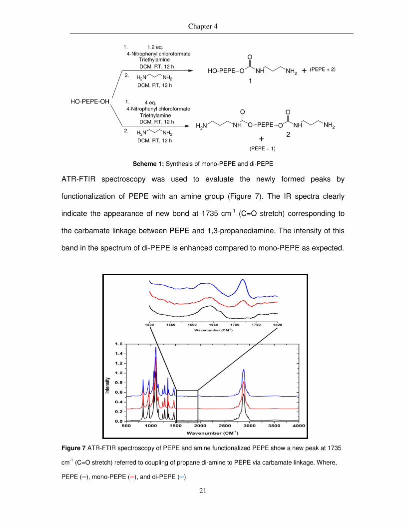
Chapter 4
21
TriethylamineDCM, RT, 12 h
NH2 NH2
DCM, RT, 12 h
HO-PEPE-OH
2.
1.
O
NHNH2O
O
PEPE O NH NH2
4-Nitrophenyl chloroformate
TriethylamineDCM, RT, 12 h
NH2 NH2
DCM, RT, 12 h
2.
1.4-Nitrophenyl chloroformate
O
HO-PEPE O NH NH2
1
1.2 eq.
4 eq.
+
+ 2
(PEPE + 2)
(PEPE + 1)
Scheme 1: Synthesis of mono-PEPE and di-PEPE
ATR-FTIR spectroscopy was used to evaluate the newly formed peaks by
functionalization of PEPE with an amine group (Figure 7). The IR spectra clearly
indicate the appearance of new bond at 1735 cm-1 (C=O stretch) corresponding to
the carbamate linkage between PEPE and 1,3-propanediamine. The intensity of this
band in the spectrum of di-PEPE is enhanced compared to mono-PEPE as expected.
Figure 7 ATR-FTIR spectroscopy of PEPE and amine functionalized PEPE show a new peak at 1735
cm-1 (C=O stretch) referred to coupling of propane di-amine to PEPE via carbamate linkage. Where,
PEPE (─), mono-PEPE (─), and di-PEPE (─).

Chapter 4
22
In the 1H-NMR spectra two new peaks for amine terminated PEPE appeared, δ= 9.76
(m,O=C-NH-) and 2.55 (m,-CH2- of di-amine) ppm. Further, the molecular weight of
PEPE, mono and di-PEPE was determined by MALDI-TOF-MS. The desired
molecular weight of mono-PEPE was expected to show an increase of 150 g·mol-1
and di-amine with 300 g·mol-1 compared to non-functionalized PEPE. The non-
functionalized PEPE gave a peak average molecular weight (Mp) of 16,175 g·mol-1.
The spectrum showed high polydispersity with no individual masses identifiable, as is
typical for PEPE. As shown in figure 8, an increased Mp of around 200 g·mol-1 in
mono-PEPE (16,378 g·mol-1) and 300 g·mol-1 in di-amine PEPE (16,460 g·mol-1).
Though the spectra of the functionalized PEPE showed the expected increase in the
final molecular weight, because of the broad distribution of molecular weight of
starting polymer (PEPE) and presence of mixture of PEPE, mono, and di-PEPE in
amine functionalized PEPE, the spectra do not give a clear results due to presence of
small amounts of the unreacted PEPE and aminated PEPE in it. The incorporation of
amino groups to PEPE led to change its physical and thermo-mechanical properties
compared to non-functionalized PEPE.
Figure 8. MALDI-TOF spectra of di-PEPE (a), mono-PEPE (b), and PEPE (c).
a
b
c
c
b
a

Chapter 4
23
4.1.1 Thermal properties of amine functionalized PEPE
To evaluate the influence of functionalization of PEPE on the thermal properties such
as melting temperature (Tm) and thermal degradation, amine functionalized and non-
functionalized PEPE are analysed by DSC and TGA. As PEPE is semi-crystalline, it
shows a Tm, which is around 62 °C for non-functionalized PEPE. After mono and di-
amine functionalization, a decrease of Tm was observed as mono-PEPE has a Tm
around 60 °C and di-PEPE of 58 °C (Figure 9 A) this corresponds to a decrease in
crystallite size. Furthermore, a decrease of ∆Hm in the aminated compounds
corresponds to pure PEPE was observed, which is related to the overall crystallinity
of the sample.
A thermo gravimetric analysis (TGA) was performed to analyse the thermal stability
of PEPE before and after functionalization (Figure 9 B). The major mass loss for
PEPE started at around 285 °C and further at around 410 °C the remaining mass
was close to zero. The initial mass loss for amine terminated PEPE seems to be
shifted slightly to higher temperature which is 314 °C for mono-PEPE and 327 °C for
di-PEPE as shown in figure 9 B. Moreover, both amine functionalized PEPEs have
shown a similar trend of thermal degradation as of PEPE with slight shift in the
starting point of the thermal degradation.

Chapter 4
24
Figure 9. Thermal analysis of PEPE (─), mono-PEPE (─) and di-PEPE (─) A: DSC measurement and
B: TGA measurement.
4.1.2 Rheological investigation of amine functionalized PEPE
The thermo-mechanical properties of PEPE and amine terminated PEPE were
determined by dynamic shear oscillation measurements to specifically evaluate
viscoelastic properties. The gel formation in the PEPE is driven by the formation of
micelles and further packing of micelles at higher temperature to form a gel. This
temperature induced gelation and changes in mechanical properties during the
gelation process could be determined by rheological analysis. The micelle formation
depended on the critical micelle concentration (CMC); the concentration required to
form micelles and critical micelle temperature (CMT); the temperature required to
form micelles. The gelation temperature (Tgel) of PEPE and functionalized PEPE
solutions (16.8 wt%, prepared in phosphate buffer pH 7.4) were determined by
thermo-mechanical observation of elastic (G′) and viscous modulus (G′′) as a function
of temperature. For the measurements, solutions were placed between rheometer
plates and heated at the rate of 0.5 °C·min-1 from 15 to 45 °C under constant stress
A B

Chapter 4
25
of 1 Pa and frequency of 0.65 Hz for each sample. Before running the temperature
ramp, each solution was equilibrated at 15 °C.
The temperature ramp of these solutions show that after a certain degree of
temperature (30 °C), the elastic modulus (G′) and viscous modulus (G′′) shows a
sharp increase in the magnitude within a small temperature interval (Figure 10). The
temperature at which both moduli increased rapidly is stated as a gelation
temperature (Tgel). Interestingly, the sol-gel transition of amine functionalized PEPE
was shifted compared to PEPE (34 °C), particularly showing a higher Tgel of mono-
PEPE (37 °C) and showing a lower Tgel (31 °C) of di-PEPE. This shift in Tgel may be
related to the change in the end groups of PEPE, which influences the (ionic)
physical interactions between PEPE chains in solution. Additionally, the PEPE chains
with similar functional group (either -OH or -NH2) have higher mechanical properties
(PEPE: G′ at T>Tgel = 16460 Pa (±3%) and di-PEPE: G′ at T>Tgel =16110 Pa (±3%))
compared to mono-PEPE which shows a value of G′ at T>Tgel = 11750 Pa (±3%).
Moreover, the hydrogels with higher elastic modules (G′) showed low viscous
modules (G′′), meaning, due to higher elasticity, the mobility of polymer
chains/micelles or viscosity decreased in such hydrogels. This analysis was very well
supported by the values of viscous modulus as, PEPE: G′′ at T>Tgel = 2072 Pa (±3%),
di-PEPE: G′′ at T>Tgel = 987 Pa (±3%), and mono-PEPE: G′′ at T>Tgel = 2318 Pa
(±3%). Hence the rheological analysis of PEPE and amine functionalized PEPE
demonstrate a reversible sol-gel transition with a slight shift in Tgel. Moreover during
the cooling of samples the dissolution occurred at temperature lower then Tgel
(observed around 8 °C lower than Tgel), because of strong physical interactions, the
disintegration of micelles takes place at lower temperature.
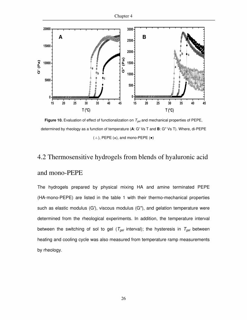
Chapter 4
26
Figure 10. Evaluation of effect of functionalization on Tgel and mechanical properties of PEPE,
determined by rheology as a function of temperature (A: G′ Vs T and B: G′′ Vs T). Where, di-PEPE
(▲), PEPE (■), and mono-PEPE (●)
4.2 Thermosensitive hydrogels from blends of hyaluronic acid
and mono-PEPE
The hydrogels prepared by physical mixing HA and amine terminated PEPE
(HA·mono-PEPE) are listed in the table 1 with their thermo-mechanical properties
such as elastic modulus (G′), viscous modulus (G′′), and gelation temperature were
determined from the rheological experiments. In addition, the temperature interval
between the switching of sol to gel (Tgel interval); the hysteresis in Tgel between
heating and cooling cycle was also measured from temperature ramp measurements
by rheology.
A B

Chapter 4
27
Table 1. Thermo-mechanical properties of HA·mono-PEPE hydrogels
Mn (HA)
[MDa]
HA: PEPE
[wt.-%]
Tgel
-
[°C]
Tgel Interval
[°C]
Hys. [°C]
G′a
[Pa] G′'
a
[Pa] |ƞƞƞƞ*|
a
[kPas]
G′b
[Pa] G′'
b
[Pa] |ƞƞƞƞ*|
b
[kPas]
Conc. [wt.-%]
3 2.8 97.2 36 5 n.d. 0.722 0.824 0.291 9594 675 2551 16.8
3 3.5 96.5 37 4 n.d. 1,13 0.408 0.319 7641 1551 2068 16.8
2.33 2.6 97.4 26 3 7 0.755 1.27 0.336 15600 812 3824 20
2.33 3.7 96.3 27 3 8 0.931 1.63 0.46 15570 716 3816 20
2.33 5.1 94.9 29 4 10 1.268 1.265 0.439 12440 1370 3063 20
2.33 8.2 91.8 29 4 12 1.61 1.844 0.599 9297 565 2281 20
1.6 4.6 95.4 31 3 n.d. 1.39 0.915 0.441 12330 584 3275 20
1.6 3.4 96.4 31 3 n.d. 1.054 0.851 0,359 13370 642 3552 20
1.13 2.7 97.3 31 5 n.d. 0.856 0.869 0.323 10920 701 2903 16.8
1.13 3.4 96.6 30 4 n.d. 1.2 0.935 0.406 15490 2678 4169 16.8
1.13 4.4 95.6 37 7 n.d. 0.849 0.53 0.265 10630 851 2830 16.8
0.9 2.6 97.4 33 3 n.d. 1.192 0.785 0.379 10250 285 2720 16.8
0.9 5.7 94.3 30 3 n.d. 0.57 1.106 0.33 11440 1430 3059 16.8
0.9 4.4 95.6 31 4 n.d. 0.863 1.184 0.389 12130 589 3221 16.8
2.6* 5.1 94.9 32 3 4 33.61 26.06 10.41 10640 1033 2619 20
Where, a: below Tgel, b:above Tgel, n.d. not determined, *: HO-PEPE-OH, Hys: hysteresis in Tgel (error
for G′, G′′ and |ƞ*|= ±3 % and for Tgel=± 1.5 °C )
4.2.1 Investigation of thermo-mechanical properties of
HA·mono-PEPE hydrogels
In case of temperature responsive hydrogels, the change in their physical state of
polymer below and above the transition temperature is due to organization of chains
in a stable physical state. Generally, they are in solution state below the gelation
temperature and form a gel at higher temperature. This transition temperature is
often called as an gelation temperature (Tgel). Here, the phase transition of HA·mono-
PEPE solution is from sol to gel is mainly driven by micelle formation by PEPE
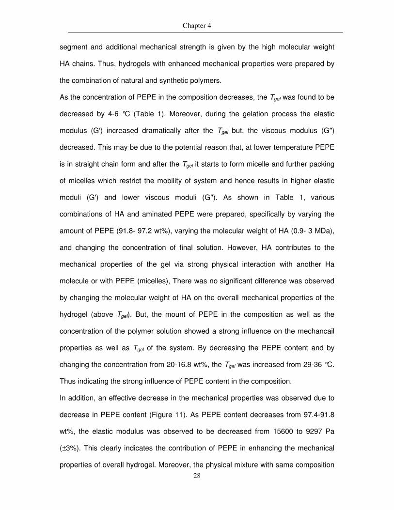
Chapter 4
28
segment and additional mechanical strength is given by the high molecular weight
HA chains. Thus, hydrogels with enhanced mechanical properties were prepared by
the combination of natural and synthetic polymers.
As the concentration of PEPE in the composition decreases, the Tgel was found to be
decreased by 4-6 °C (Table 1). Moreover, during the gelation process the elastic
modulus (G′) increased dramatically after the Tgel but, the viscous modulus (G′′)
decreased. This may be due to the potential reason that, at lower temperature PEPE
is in straight chain form and after the Tgel it starts to form micelle and further packing
of micelles which restrict the mobility of system and hence results in higher elastic
moduli (G′) and lower viscous moduli (G′′). As shown in Table 1, various
combinations of HA and aminated PEPE were prepared, specifically by varying the
amount of PEPE (91.8- 97.2 wt%), varying the molecular weight of HA (0.9- 3 MDa),
and changing the concentration of final solution. However, HA contributes to the
mechanical properties of the gel via strong physical interaction with another Ha
molecule or with PEPE (micelles), There was no significant difference was observed
by changing the molecular weight of HA on the overall mechanical properties of the
hydrogel (above Tgel). But, the mount of PEPE in the composition as well as the
concentration of the polymer solution showed a strong influence on the mechancail
properties as well as Tgel of the system. By decreasing the PEPE content and by
changing the concentration from 20-16.8 wt%, the Tgel was increased from 29-36 °C.
Thus indicating the strong influence of PEPE content in the composition.
In addition, an effective decrease in the mechanical properties was observed due to
decrease in PEPE content (Figure 11). As PEPE content decreases from 97.4-91.8
wt%, the elastic modulus was observed to be decreased from 15600 to 9297 Pa
(±3%). This clearly indicates the contribution of PEPE in enhancing the mechanical
properties of overall hydrogel. Moreover, the physical mixture with same composition
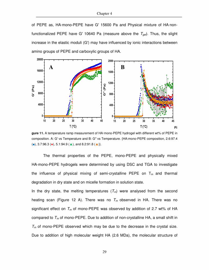
Chapter 4
29
of PEPE as, HA·mono-PEPE have G′ 15600 Pa and Physical mixture of HA·non-
functionalized PEPE have G′ 10640 Pa (measure above the Tgel). Thus, the slight
increase in the elastic moduli (G′) may have influenced by ionic interactions between
amino groups of PEPE and carboxylic groups of HA.
Fi
gure 11. A temperature ramp measurement of HA·mono-PEPE hydrogel with different wt% of PEPE in
composition. A: G′ vs Temperature and B: G′′ vs Temperature. {HA:mono-PEPE composition, 2.6:97.4
(■), 3.7:96.3 (●), 5.1:94.9 (▲), and 8.2:91.8 (▲)}.
The thermal properties of the PEPE, mono-PEPE and physically mixed
HA·mono-PEPE hydrogels were determined by using DSC and TGA to investigate
the influence of physical mixing of semi-crystalline PEPE on Tm and thermal
degradation in dry state and on micelle formation in solution state.
In the dry state, the melting temperatures (Tm) were analysed from the second
heating scan (Figure 12 A). There was no Tm observed in HA. There was no
significant effect on Tm of mono-PEPE was observed by addition of 2.7 wt% of HA
compared to Tm of mono-PEPE. Due to addition of non-crystalline HA, a small shift in
Tm of mono-PEPE observed which may be due to the decrease in the crystal size.
Due to addition of high molecular weight HA (2.6 MDa), the molecular structure of
A B

Chapter 4
30
composition and simultaneously the crystallization of mono-PEPE got hampered,
resulting in to broadening of crystallization peak of mono-PEPE
Figure 12. Second heating scans of, A. HA (─), mono-PEPE (─), and HA·mono-PEPE (─), B. DSC in solution state of PEPE (─), mono-PEPE (─), and HA·PEPE (─) (sample conc. 20 wt%).
In DSC measurements, an interesting point of evaluation was determination of
micelles. As shown in figure 12B, PEPE, mono-PEPE, and HA·mono-PEPE shows a
small transition at around 21 °C, this transition is due to the change in the
arrangements of PEPE chains into micelles, showing an endothermic transition in the
DSC measurement83. In addition, the endothermic peak referring to the micellization
process, may contribute from the change in the enthalpy of the polymer solution.
Overall, there was no significant difference in the micelle formation temperature due
to the amine functionalization or addition of HA was observed by DSC
measurements.
TGA was performed in a N2 atmosphere to determine the thermal stability potentially
ascribed by physical crosslinking between HA and mono-PEPE (figure 13). A TGA
plot of pure HA show a two-step degradation process which starts at around 70 °C
with loss of moisture in the sample, and further at 250 °C due to thermal degradation
B A

Chapter 4
31
with nearly 60 wt% mass loss and finally combustion of remaining mass between 250
and 500 °C. While the mono-PEPE is quite thermally stable and show only one major
transition at around 400 °C with major mass loss of around 95 wt% within further 50
°C. Interestingly, the physical mixture of HA and mono-PEPE has shown a two-step
thermal transition, in which the first transition at 250 °C with around 10 wt% mass
loss can be attributed to the HA degradation with the second transition with major
mass loss of 85 wt% at around 380 °C was due to the degradation of mono-PEPE.
Figure 13. TGA of HA (─), mono-PEPE (─), and HA·mono-PEPE (─) samples in dry state
4.2.2 Determination of CMC and CMT
The concentration of polymer at and above which micelles are formed is known as
critical micellization concentration for that polymer solution. The determination of
micellization of amphiphilic block copolymers is more complex compared to small
molecules or surfactants. Here, CMC and CMT values of physical mixtures of HA and
mono-PEPE were determined by using Diphenyl hexatriene (DHP)49. DHP gives
fluorescence only in a hydrophobic environment (such as core of a micelle) providing

Chapter 4
32
a sensitive method of formation of micelles. The PEPE solution with concentration
above CMC and temperature above CMT forms micelles having a hydrophobic core
and a hydrophilic corona. So when the micelles are formed in the solution, the dye
will give fluorescence and can be detected by UV spectroscopy by determining the
absorbance at 356 nm.
Trans-trans-trans-1,6-Diphenyl-1,3,5-hexatriene (DPH)
Figure 14. CLSM images of HA·mono-PEPE (sr. no.1 in table 2) micelles aggregates
Generally, with increasing concentration of the polymer in sample, the CMC value
was observed at lower temperature and vice versa (Figure 15). The micelles were
also visualised under confocal light scanning microscope (CLSM) in the form of small
aggregates as shown in figure 14.
Figure 15. Determination of CMT and CMC of HA·mono-PEPE (4.4:95.6) sample

Chapter 4
33
Table 2. The CMC values of HA·mono-PEPE
Temp. 30°C 35 °C 40 °C 45 °C Sr. no.
Weight composition
of HA: mono-PEPE
Molecular weight of HA (MDa)
Critical Micelle Concentration (% w/v)
1 2.7:97.3 1.13 3.89 0.93 0.53 0.21 2 3.4:96.5 1.13 3.97 1.2 0.38 0.24
3 4.4:95.6 1.13 4.44 1.23 0.34 0.16
4 2.6:97.4 0.9 5.34 1.79 0.84 0.53 5 5.8:94.2 0.9 3.8 1.09 0.51 0.26
6 4.4:95.6 0.9 4.23 1.25 0.35 0.16
7 2.8:97.2 3 1.97 1.31 0.23 0.18
(error for all CMC values = ±5%) The CMC and CMT values depend on temperature and concentration respectively.
As the amount of PEPE was increased in the composition, lower concentration was
required to show CMC and CMT (table 2 and Table 3). There was an absolute
difference observed between CMT and Tgel as, the CMT is the temperature at which
the micelle formation process starts and the Tgel is the process at which the micelles
(at certain concentration) comes together to form a gel (two-step process). The
formation of micelles does not lead to formation of gel as to form a gel it requires a
certain amount of micelles to be present nearby to interact.
Moreover, the CMT values obtained from UV measurements were compared to the
CMT values obtained by rheology as shown in Table 4.
Table 3. The CMT values of HA·mono-PEPE
Conc.0.1 %-w/v
0.5 1 2.5 5 7.5 10 20 Sr. no.
Weight composition of HA: PEPE
Molecular weight of
HA (MDa) Critical Micelle Temperature (°C)
1 2.7:97.3 1.13 40 35 33 29 28 25 24 23 2 3.4:96.5 1.13 - 35 33 29 37 25 24 23 3 4.4:95.6 1.13 39 36 35 30 29 25 24 22 4 2.6:97.4 0.9 39 36 35 33 32 26 25 24 5 5.8:94.2 0.9 41 35 33 31 29 27 25 24 6 4.4:95.6 0.9 - 36 35 31 30 25 24 22 7 2.8:97.2 3 42 36 34 31 28 24 23 22
(error for all CMT values = ±5%)

Chapter 4
34
Table 4. Comparison of CMT values by UV and Rheology
Sr. no.
Weight composition of HA: PEPE
Molecular weight of HA (MDa)
CMT (°C) by UV
CMT (°C) by Rheology
Tgel (°C)
1 0:100 0 21 22 30
2 2.8:97.2 3 22 23 33
3 2.7:97.3 1.13 23 23 31 4 3.4:96.5 1.13 23 23 30 5 4.4:95.6 1.13 22 22 32 6 2.6:97.4 0.9 24 23 32 7 5.8:94.2 0.9 24 24 31 8 4.4:95.6 0.9 22 22 31
(error for all CMT values = ±5%)
The CMT values for all measured samples was determined from the temperature
ramp. The temperature above which the elastic modulus (G′) starts to increase
constantly, the temperature was taken as CMT. Because, as the temperature
reaches to CMT for the polymer solution, the viscosity decreases due to, micelle
formation and packing of micelles at higher temperature. This study was quite
interesting as it was showing the values of CMT close to the values obtained by UV
measurements.
4.2.3 Micelle formation by 1H-NMR analysis
During the micelle formation in PEPE, the hydrophobic segment i.e. polypropylene
glycol (PPG) will form the core while the hydrophilic segment i.e. polyethylene glycol
(PEG) will form the corona, which is likely to happen due to the hydrophilic
interactions of PEG segment with water molecules. Such a micelle with hydrophobic
core and hydrophilic corona may hinder the interaction of PPG segment with
surrounding water molecules. This could be observed by line broadening of the peak
corresponding to the CH3- group of PPG in temperature dependent 1H-NMR
measurements84 (Figure 16). Thus, an experiment was performed in D2O at
temperature range of 25-40 °C with interval of 5 °C. The sample concentration of 4
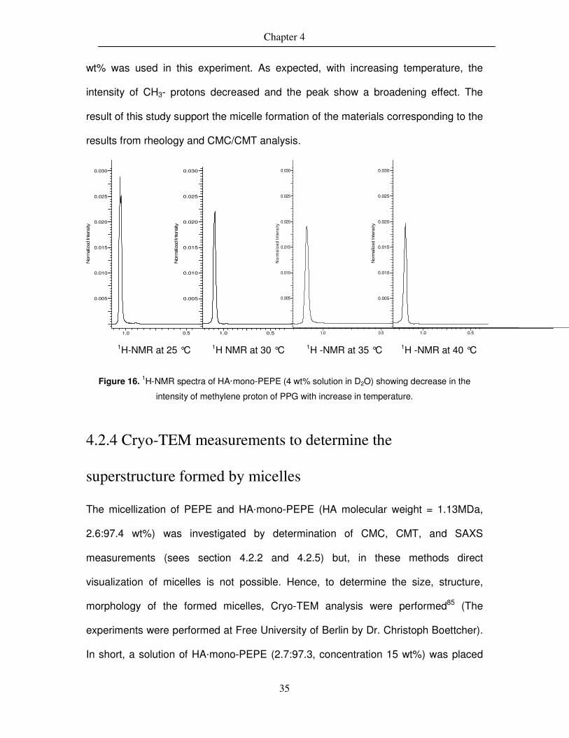
Chapter 4
35
wt% was used in this experiment. As expected, with increasing temperature, the
intensity of CH3- protons decreased and the peak show a broadening effect. The
result of this study support the micelle formation of the materials corresponding to the
results from rheology and CMC/CMT analysis.
1H-NMR at 25 °C 1H NMR at 30 °C 1H -NMR at 35 °C 1H -NMR at 40 °C
Figure 16. 1H-NMR spectra of HA·mono-PEPE (4 wt% solution in D2O) showing decrease in the
intensity of methylene proton of PPG with increase in temperature.
4.2.4 Cryo-TEM measurements to determine the
superstructure formed by micelles
The micellization of PEPE and HA·mono-PEPE (HA molecular weight = 1.13MDa,
2.6:97.4 wt%) was investigated by determination of CMC, CMT, and SAXS
measurements (sees section 4.2.2 and 4.2.5) but, in these methods direct
visualization of micelles is not possible. Hence, to determine the size, structure,
morphology of the formed micelles, Cryo-TEM analysis were performed85 (The
experiments were performed at Free University of Berlin by Dr. Christoph Boettcher).
In short, a solution of HA·mono-PEPE (2.7:97.3, concentration 15 wt%) was placed
1.0 0.5
0.005
0.010
0.015
0.020
0.025
0.030
Nor
mal
ized
Inte
nsity
1.0 0.5
0.005
0.010
0.015
0.020
0.025
0.030
Nor
mal
ized
Inte
nsity
1.0 0.5
0.005
0.010
0.015
0.020
0.025
0.030Nor
mal
ized
Inte
nsity
1.0 0.5
0.005
0.010
0.015
0.020
0.025
0.030
Nor
mal
ized
Inte
nsity

Chapter 4
36
on a TEM grid which was kept under humid conditions at 37 °C (T> Tgel) in a closed
chamber for 20 mins. allowing the formation of micelles. Then, the sample was
suddenly immersed in a liquid ethane (-190 °C) to freeze the micelles.
Figure 17. Cryo-TEM analysis of HA·mono-PEPE (2.7:97.3) solution at two level of magnifications,
indicating the micelles and superstructure formed due to packing of micelles above Tgel.
As shown in figure 17, the average size of micelle around 20-30 nm. The alignment
of micelles in the superstructure is non-uniform and can be supported by the Fourier
transformation image, which indicates two different types of packing of micelles as,
Figure 18. Fourier Transformation of Cryo-TEM images showing packing of micelles, A. Body centred
cubic (BCC) packing of micelles and B. Hexagonal packing of micelles.
A B

Chapter 4
37
BCC and hexagonal packing (Figure 18). However, the Cryo-TEM analysis clearly
showed the formation of micelles and different types packing of micelles upon
heating near body temperature.
4.2.5 Small angle x-ray scattering measurement for
determination of micelles
The small angle measurements were performed to investigate the influence of HA on
micelles formation and its arrangements in the gel state. The small angle x-ray
scattering measurements were performed at 20 °C (below Tgel) and 45 °C (above
Tgel). As shown in figure 19, in all samples there was no micelle formation at 20 °C
and hence showing an amorphous circle. But, when heated at 45 °C, the micelles
were formed and got packed together to form a gel. At this situation, the ordering of
micelles was appeared as a ring around the amorphous region and further rings are
appeared as micelles were got packed in to second and third order of structure. The
intensity of amorphous region decreased more micelle were arranged to form highly
ordered structure86. The micelle formation was slightly hindered by the addition of
HA, which can be seen from the intensity of amorphous region in the physical
mixtures and the appearance of rings was not clear especially in physical mixture of
HA and di-PEPE (HA·di-PEPE) as compared to PEPE.
Moreover, the appearance of crystalline peaks due to packing and ordering of
micelles were also detected by small angle x-ray scattering measurements (Figure
20). The PEPE resulted in sharp and narrow peaks than physical mixture of HA and
amine terminated PEE which might happen due to the increase in amorphous HA.
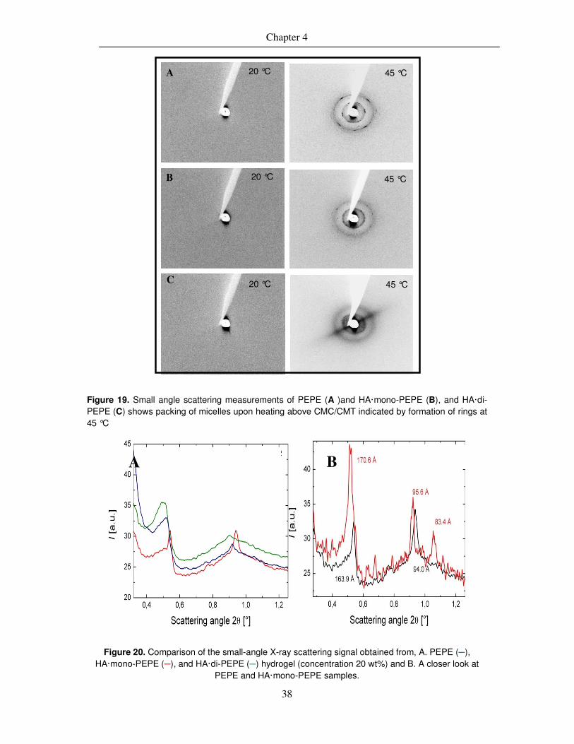
Chapter 4
38
Figure 19. Small angle scattering measurements of PEPE (A )and HA·mono-PEPE (B), and HA·di-PEPE (C) shows packing of micelles upon heating above CMC/CMT indicated by formation of rings at 45 °C
Figure 20. Comparison of the small-angle X-ray scattering signal obtained from, A. PEPE (─), HA·mono-PEPE (─), and HA·di-PEPE (─) hydrogel (concentration 20 wt%) and B. A closer look at
PEPE and HA·mono-PEPE samples.
A B
A
B
C
20 °C 45 °C
20 °C
20 °C
45 °C
45 °C

Chapter 4
39
4.2.6 Change of mechanical properties during enzymatic
degradation of HA·mono-PEPE hydrogels.
As the HA component contributes to the mechanical properties of the gel, the
enzymatic degradation of HA may influence the mechanical properties of these
hydrogels. Although there was no chemical crosslinking between HA and mono-
PEPE (as it was seen in the rheological analysis) the enzymatic degradation study
was performed to evaluate the changes in mechanical properties and Tgel during the
degradation process. To study the degradation of physical mixtures of HA with
different composition of mono-PEPE, the hydrogels prepared are reported in Table 1.
For this study HA (2.33 MDa) with different composition of mono-PEPE were
selected. During the degradation of the hydrogel, the hyaluronidase will randomly
cleave β-N-acetylhexosamine-[1→4] glycosidic bonds in HA87. However, it is known
that the physical and mechanical properties of hydrogels are strongly dependent on
polymer chain length and polymer backbone and chain-chain interactions. Thus, by
the degradation of long chain HA may influence the mechanical properties of
hydrogel. Moreover, due to the interactions of small fragments of HA with PEPE the
gelation process was also delayed, resulting in shift of Tgel to higher temperature as
degradation progresses (Figure 21). The Tgel of initial composition was 26 °C, which
was shifted to 29 °C after 48 h. of degradation time and clearly indicating the
influence of HA degradation on gelation mechanism of PEPE. A similar kind of
behavior was observed in all other hydrogels of HA·mono-PEPE composition.

Chapter 4
40
Figure 21. Temperature ramp of HA·mono-PEPE (2.6:97.4 wt%) hydrogels during degradation at
different time points.{ 0h (■), 4h (●), 8h (▲), 10h (▲), and 48h (■)}.
The frequency sweep test at 37 °C (T>Tgel) was performed in order to investigate to
detect the changes in the mechanical properties during the degradation process. As
shown in Figure 22, the initial elastic modulus (G′) of hydrogel was around 18000 Pa,
which was decreased to 15000 Pa after 1h degradation time. Further it decreased
down to 12500 Pa within 4h of degradation time. This clearly indicate that the
degradation of high molecular weight HA into small fragments with which it loses the
chain entanglement and intra molecular chain interactions, resulted in decrease in
the mechanical properties of hydrogel. Nevertheless, the hydrogel does not lose the
ability to show a thermal gelation but, was shifted to higher temperature. Thus, the
study clearly indicate that the HA does contribute to the mechanical properties of
hydrogel and further degradation shows decrease in mechanical properties with time
of degradation, even the final wt% composition oh HA is too low compare to PEPE

Chapter 4
41
Figure 22. Frequency sweep test of HA·mono-PEPE (2.6:97.4 wt%) hydrogels at different time points.
{0h (■), 1h (▲), 2h (▲), and 4h (●)}.
4.3 Thermosensitive hydrogels derived from di-PEPE and
hyaluronic acid
In this section, the development of cytocompatible thermosensitive hydrogels is
presented. The mechanical properties, endotoxin test, cytotoxicity test, and direct cell
test of hyaluronic acid based hydrogels are discussed. (Cooperation project funded
by BMBF: Biocompatible and thermosensitive polymers as vitreous replacement).
4.3.1 Synthesis of thermosensitive hydrogels
Regarding the background outlined above, here it is necessary not to have an
absolute replacement but possibly mimic the viscoelastic properties and structure of
as vitreous body substitute. A vitreous substitute is defined as the treatment agent,
which helps to incorporate the functional characteristics of the glass body while
exerting an additional stabilizing effect in the diseased eye, and to be treated. This

Chapter 4
42
general consideration suggests that there will not be an ideal vitreous replacement,
but a temporary vitreous replacement prevent the dysfunction of eye. The goal of this
research study was to develop a polymer system as a vitreous replacement or as
vitreous humor tamponade by using biocompatible polymer system, which basically
derived from combination of biopolymers and synthetic polymers with
thermosensitive properties resulting an injectable hydrogel which can form a gel at
body temperature.
As reported in section 4.2, the ionic interaction of mono-PEPE and HA leads to form
a thermosensitive hydrogel and it was also observed that, the ionic interactions of
mono-PEPE was stronger than ionic interactions of non-functionalized PEPE with HA
which was proved from rheological investigations. In this study, initially combination
of mono-PEPE and HA was used to be evaluated as an artificial vitreous but, due to
lack of sufficient mechanical properties, stability of hydrogel, and osmolarity, the
combination of di-PEPE and HA (HA·di-PEPE) was chosen as it seems more
promising material combination. The hydrogel was prepared by using different
molecular weight of HA and different composition of di-PEPE as shown in the Table
5. Preparation of hydrogels was performed in clean room environment using
endotoxin free materials.
Table 5. Composition, Tgel, and viscoelastic properties of thermosensitive hydrogels of HA·di-PEPE
Mn (HA)
[MDa]
HA: PEPE
Tgel
[°C] Tgel
Interval [°C]
Hys. [°C]
G′a
[Pa] G′′
a
[Pa] |ƞ*|
a
[kPas]
G′b
[Pa] G′'
b
[Pa] |ƞ*|
b
[kPas]
Conc.
[wt%]
2.33 4.5 95.5 34 2 7 2.395 0.507 0.599 15580 1176 3825 17
2.33 4.5 95.5 37 5 8 2.082 1.062 0.572 1648 243 407 13
1.2 5.5 94.5 29 3 8 1.202 0.997 0.382 21280 940 5216 20
1.2 5.5 94.5 32 3 7 1.189 1.065 0.391 15560 1376 3824 17
1.2 5.5 94.5 39 5 5 1,732 0.737 0.461 2972 590 741 13
Where, a: below Tgel, b:above Tgel (error for G′, G′′ and |ƞ*|= ±3%, for Tgel and Tgel (interval) = ± 1.5 °C)

Chapter 4
43
4.3.2 Thermo-mechanical properties
The Thermo-mechanical properties and effect of additional ionic interaction
(due to amino groups) on mechanical properties was investigated by rheological
temperature ramp shown in Figure 23. The enhancement in the elastic modulus (G′)
from 16330 Pa to 22450 Pa (±3%,) and decrease in the Tgel from 29 to 26 °C
indicates the strong interaction between HA·di-PEPE is stronger compare to
HA·mono-PEPE. The reason for enhancement in mechanical properties could be the
increase in number of amino groups on PEPE, which then has more effective ionic
interactions with carboxylic group of HA. Moreover, the mechanical properties and
Tgel could be controlled by changing the concentration of polymer. Interestingly, the
combination of high molecular weight HA and strong ionic interaction (due to di-
PEPE) shows a thermosensitive gelation even at 13 wt% concentration.
The transition tended to be a little sharper, and the transition was also observed at
lower concentrations compared to HA·mono-PEPE. Furthermore, the systems
studied here showed a higher stability against dilution experiments. The material with
the composition of HA (2.33 MDa) + di-PEPE (4.5:95.5 wt%) showed all the physical
parameters that were set to start as specifications of target application.

Chapter 4
44
Figure 23. Rheological temperature ramp of HA·mono-PEPE (■), Tgel = 29 °C and HA·di-PEPE (●),
Tgel= 26 °C. (Concentration = 20 wt%)
4.3.3 Stability of hydrogels at 37 °C
Stability test was performed by using 20 wt% solution of HA·di-PEPE hydrogel and
Tryptan blue (30 %) dye in water at 37 °C (Figure 24). The study shows an
interesting behavior of hydrogels as the dye was did not diffuse into the gel (even
after 2 weeks); a very important factor considering a hydrogel as a vitreous substitute
that should not allow other solvent to dilute it which will destroy the structure and also
the function of the system. The reason could be explain by the strong micelle packing
through ionic interactions in BCC and hexagonal packing structure (already shown in
Cryo-TEM analysis in Figure 18 and SAXS analysis Figure 19).
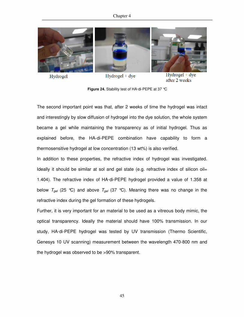
Chapter 4
45
Figure 24. Stability test of HA·di-PEPE at 37 °C
The second important point was that, after 2 weeks of time the hydrogel was intact
and interestingly by slow diffusion of hydrogel into the dye solution, the whole system
became a gel while maintaining the transparency as of initial hydrogel. Thus as
explained before, the HA·di-PEPE combination have capability to form a
thermosensitive hydrogel at low concentration (13 wt%) is also verified.
In addition to these properties, the refractive index of hydrogel was investigated.
Ideally it should be similar at sol and gel state (e.g. refractive index of silicon oil=
1.404). The refractive index of HA·di-PEPE hydrogel provided a value of 1.358 at
below Tgel (25 °C) and above Tgel (37 °C). Meaning there was no change in the
refractive index during the gel formation of these hydrogels.
Further, it is very important for an material to be used as a vitreous body mimic, the
optical transparency. Ideally the material should have 100% transmission. In our
study, HA·di-PEPE hydrogel was tested by UV transmission (Thermo Scientific,
Genesys 10 UV scanning) measurement between the wavelength 470-800 nm and
the hydrogel was observed to be >90% transparent.

Chapter 4
46
4.3.4 Degradation study of HA·di-PEPE hydrogels
The enzymatic degradation of HA·di-PEPE was performed in order to investigate the
mass loss of hydrogel using different solutions. Degradation studies were performed
to evaluate the long-term behavior of the materials. There are two important aspects
to be considered. On the one hand, the mass loss, which gives information about
whether soluble fragments are formed, this can no longer contribute to the
functionality of the material. On the other hand, material properties may change on a
weight loss and material composition. These changes are, therefore very essential to
be determined during the degradation processes. However, are also important for the
functionality of the materials during application. As shown in figure 25, significant
initial mass loss occurred within the first week in all the samples but, afterwards the
gel was stable for many weeks. Moreover it was observed that there was no
significant difference in behavior of the materials in different mediums used.
0 10 20 30 40 500
20
40
60
80
100
120
Hyaluronidase Intraokular-Spülflüssigkeit Glaskörper-Extrakt
Figure 25. Enzymatic degradation of HA·di-PEPE showing the remaining mass of the sample
{Performed in, Intraocular solution (●), vitreous extract (▲), and hyaluronidase (■)}
Days

Chapter 4
47
4.3.5 Biological evaluation of hydrogel
In order to evaluate the biological properties of first the materials have been tested by
the endotoxin test and cytotoxicity test (These test were performed by Dr. Toralf
Roch). The material bound microbial products were analysed by a cell based assay
called Quanti-Blue assay as shown in Figure 26. LPS 10 ng·ml-1 was used as a
positive control and elutes were prepared according to ISO10993-12.
25 12,5
6,25
3,125
1,562
5
0,781
25
0,390
60,1
95
0,097
6
0,048
8
0,024
4
0,012
2RAW
LPS 10 ng/m
l 25 12,5
6,25
3,125
1,562
5
0,781
25
0,390
60,1
95
0,097
6
0,048
8
0,024
4
0,012
2RAW
0 . 0
0 . 5
1 . 0
1 . 5
2 . 0
H S -
S A -
Abs
620
nm
Figure 26. Endotoxin test of di-PEPE ( ) and HA·di-PEPE ( )
There was some cell activated by HA·di-PEPE which indicate presence of some
endotoxin in the material. In order to evaluate the cell behavior when coming in
contact with these hydrogels a cell test with direct contact of hydrogel was performed
(This test were performed by Dr. K. Kobuch) and variable results were obtained
during the cell tests, change of pH and osmolarity was detected during the tests.
4.4 Summary
Thermosensitive hydrogels prepared via physical mixing of aminated PEPE and HA
showed enhancement in the mechanical properties, especially elastic modulus (G )
and also decreased the gelation temperature. This effect was observed due to strong
electrostatic interactions of carboxylic group of HA and amino groups of PEPE. In
addition, it was also observed that with increase in number of amino group (mono-

Chapter 4
48
PEPE to di-PEPE) these physical interactions became stronger and resulted in
further increase in mechanical properties and decrease in Tgel. Further, it was
possible to control the Tgel by varying the PEPE content in the composition. But,
overall no effect was observed by varying the molecular weight of HA on the
viscoelastic properties of these hydrogels.
Thermosensitive hydrogel system, which shows an alternative concept and provides
better biocompatibility and could be used as a potential artificial vitreous substitute.
The hydrogel was prepared via physical mixing of hyaluronic acid with amine
terminated thermosensitive poly(ethylene glycol-b-propylene glycol-b-ethylene
glycol)s (PEPEs). The hydrogel exhibits enhancement of shear viscosity and also
form a transparent gel near body temperature. The ionic interaction between amino
groups of PEPE (especially di-amino PEPE) and carboxylic group of HA show an
increase in mechanical properties and lower gelation temperature compare to the
mono-amine PEPE and non-functionalized PEPE samples with same concentration.
In addition the hydrogel was able to closely fit the required properties such as, pH
(7.1-7.3), optical transparency of >90% at 37 °C (in gel state) and at wavelength of
430-800 nm, a refractive index of 1.358, injectability through a 27G needle, steam
sterilizability, which was close to the properties required by an synthetic vitreous
substitute. The in vitro studies with mouse fibroblast cells show a promising
biocompatibility of the hydrogel. Histological examination of the gel shows that all
layers of retina were intact and no loss of tissues was evident.
Chapter 5
48
5. Synthesis of Covalently Coupled
Polysaccharide and PEPE Hydrogels with
Tunable Properties
The data and part of the text presented in this chapter which is related to Pectin-g-PEPE
hydrogels have been published as: Thermal Gelation and Stability of Pectin grafted with
PEPE, Harshal D. Santan, Axel T. Neffe, Stefan Kamlage, and Andreas Lendlein, Mater.
Res. Soc. Symp. Proc. / Volume 1403 / mrsf11-1403-v17-05 2012.
In addition, the data presented related to covalent coupling of HA formed the basis for a
manuscript with the title, Thermosensitive Hydrogels Derived From Covalently Coupled HA
and PEPE With Tunable Thermo-Mechanical Properties, Harshal D. Santan, Axel T. Neffe,
Karola Luetzow, Ulrich Noechel, and Andreas Lendlein.
In this chapter, pectin, chondroitin sulphate, and hyaluronic acid based
covalently coupled thermosensitive hydrogels with tunable properties were
synthesized via EDC or DMTMM mediated coupling. The resulting hydrogels
properties were characterized by IR, NMR, rheology, DSC, TGA, gel electrophoresis,
and enzymatic degradation.
5.1 Synthesis of Pectin and Chondroitin sulphate grafted PEPE
hydrogels using EDC as a coupling agent.
The hydrogels were synthesized via EDC mediated coupling of carboxylic group of
polysaccharides with amino group of PEPE. The grafting of a thermosensitive PEPE
onto pectin with different grafting ratios was achieved in two steps. PEPE was

Chapter 5
49
aminated88 by adding sub-equivalent amount of 4-Nitrophenyl chloroformate,
followed by addition of 1,3-diaminopropane in the next step (Scheme 1).
Subsequently, the free carboxylic groups of pectin were activated with EDC and the
aminated PEPE was added to give the Pectin-g-PEPE (PGP). An analogues
synthesis route was used for the synthesis of chondroitin sulphate-g-PEPE (CGP)
hydrogels (Table 6). The synthesized hydrogels were characterized by IR showing
the formation of new amide bond by appearance of new peaks at 1672 cm-1 (amide I)
and 1595 cm-1 (amide II) hence, giving the first proof of coupling.
The grafting yield was calculated using equation 1:
Grafting yield = MAP
PSPSGP
W
WW − (eq. 5.1),
where, WPSGP is the weight of the freeze-dried grafted copolymer, and WPS and WMAP
are the weights of pectin and aminated PEPEs in the feed, respectively.
Further, The number of free carboxylic acid groups in the pectin was determined with
equation 2:
Free carboxylic acid groups (Pectin) =
mol
g93.176
)95.0*(WPec (eq. 5.2)
For pectin, the galacturonic acid content (74 wt%) and degree of methylation (6.7
mol%) in the pectin. The grafting density, i.e. the percentage of free carboxylic acid
groups of pectin and chondroitin sulfate functionalized with PEPE, was calculated
with equation 3:
Grafting density = PS
n
PEPEPSGP
W
PEPEM
CW
)(
*
(eq. 5.3)
with CPEPE = PEPE weight content in the PGP.

Chapter 5
50
Table 6: Composition and grafting density (determined by eq.5.3) of the PGP and CGP
Sample
ID
Composition of
polysaccharide :
PEPE (wt%)
Experimental
yielda (wt%)
Grafting density
(mol%)b
PGP 40
PGP 28
PGP 20
PGP 12
CGP 39
CGP 28
CGP 20
CGP 11
3.9 : 96.1
5.4 : 94.6
7.5 : 92.5
12 : 88
3: 97
4.2 : 95.8
5.6 : 94.4
9.3 : 90.7
93
94
93
91
91
93
94
92
40
28
20
12
39
28
20
11
a: determined from final weight of product after dialysis, b: from eq. 5.3
5.2 Characterization of hydrogels
5.2.1 Thermogravimetric Analysis of CGP and PGP
Hydrogels
The thermogravimetric analysis is often a useful method for determining the
composition of graft polymers. The thermograms of pectin, PEPE, and the PGP 40
are shown in Figure 27. Weight loss of pectin starts in the range of 60−75 °C, which
is likely to be associated with loss of absorbed moisture in the sample.
Decomposition was observed between 110−135 °C. In the next step between
225−250 °C, the sample rapidly lost around 55 wt%. Weight loss of PEPE starts at
250 °C, with a rapid mass loss between 350 °C and 380 °C. The expected curve of
the grafted system would be the mathematical combination of the curves described
above, and this is what is observed for physical mixtures of the two components
(data not shown). However, interestingly the onset and major part of the mass loss of
the grafted systems was observed at higher temperatures, i.e. the grafting process
increased the thermal stability of the system. This observation might be explained by

Chapter 5
51
a degradation mechanism of the polysaccharide in which the free carboxylic acid
groups play an important role. These are now (at least partially) carboxamides, and
hydrogen bonds between the two components are formed, which increased the
thermal stability of the grafted system.
Figure 27. Thermo gravimetric analysis of pectin, PEPE, and PGP 40 in an N2 atmosphere (solid line:
pectin, dot line: PEPE, and dash line: PGP 40)
5.2.2 Determination thermo-mechanical properties of
hydrogels
The rheological properties of pectin, chondroitin sulfate, PEPE, and the grafted
hydrogels at different temperatures for Pectin (Figure 28 A.) and chondroitin sulphate
(Figure 28 B) were observed to be non-thermosensitive in the given temperature
range. The 20 wt% solution of PEPE has shown sol-gel transitions (Tgel) at around 34
°C, while after grafting to pectin (20 wt% solution) the Tgel was observed near 30 °C
(Figure 29). This clearly shows the effect of grafting, which allows to form gels at
lower temperature. By variation of the grafting density of the PEPE on the pectin, the
Tgel of the system could be adjusted from 25 °C to 33 °C, whereby increasing the

Chapter 5
52
grafting resulted in a decreasing Tgel (Figure 30). However, also the temperature
interval for the transition is larger for systems with lower Tgel. Interestingly, the elastic
moduli G′, the viscous moduli G′’ and the viscosities of the systems were found to be
similar and in all cases G′ and the viscosity increased during the gelation by four
orders of magnitude.
Figure 28. Rheological temperature ramp of, a. Pectin and b. Chondroitin sulfate showing a non-
thermosensitive behavior (sample conc. 5 wt%)
Figure 29. A reversible sol-gel transition of PGP40 sample at 20 wt% concentration
Heating
Cooling 25 °C 37 °C
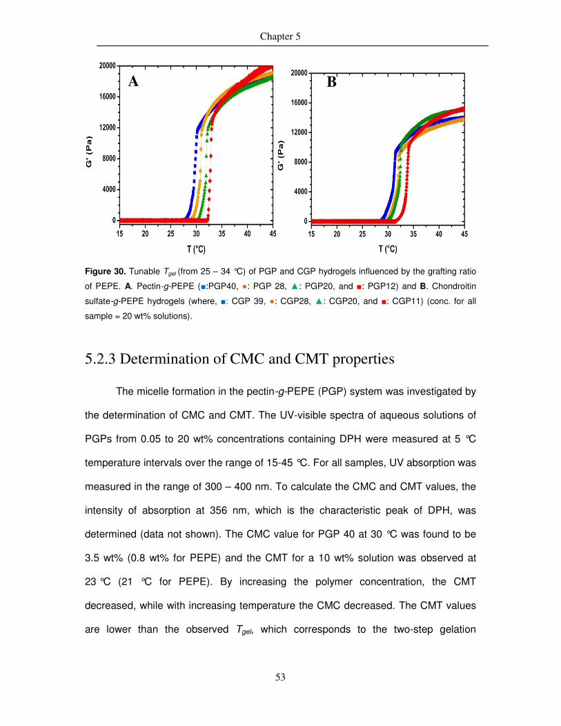
Chapter 5
53
Figure 30. Tunable Tgel (from 25 – 34 °C) of PGP and CGP hydrogels influenced by the grafting ratio
of PEPE. A. Pectin-g-PEPE (■:PGP40, ●: PGP 28, ▲: PGP20, and ■: PGP12) and B. Chondroitin
sulfate-g-PEPE hydrogels (where, ■: CGP 39, ●: CGP28, ▲: CGP20, and ■: CGP11) (conc. for all
sample = 20 wt% solutions).
5.2.3 Determination of CMC and CMT properties
The micelle formation in the pectin-g-PEPE (PGP) system was investigated by
the determination of CMC and CMT. The UV-visible spectra of aqueous solutions of
PGPs from 0.05 to 20 wt% concentrations containing DPH were measured at 5 °C
temperature intervals over the range of 15-45 °C. For all samples, UV absorption was
measured in the range of 300 – 400 nm. To calculate the CMC and CMT values, the
intensity of absorption at 356 nm, which is the characteristic peak of DPH, was
determined (data not shown). The CMC value for PGP 40 at 30 °C was found to be
3.5 wt% (0.8 wt% for PEPE) and the CMT for a 10 wt% solution was observed at
23 °C (21 °C for PEPE). By increasing the polymer concentration, the CMT
decreased, while with increasing temperature the CMC decreased. The CMT values
are lower than the observed Tgel, which corresponds to the two-step gelation
A B

Chapter 5
54
mechanism (first, formation of micelles, second, gelation due to micelle-micelle
interactions).
5.2.4 Determination of molecular weight by gel
electrophoresis
The determination of the molecular weight of the polysaccharide-g-PEPE systems is
important to i) confirm the covalent attachment of the PEPEs, b) estimate the degree
of grafting and c) investigate, if actually crosslinking took place. On the other hand,
because of the extreme high molecular weight and self-ordering behavior of the
systems in aqueous solution, it is also a challenging task.
DN
A
HA
-g-P
EP
E(1
3)
CS
-g-P
EP
E(4
0)
CS
-g-P
EP
E(2
8)
CS
-g-P
EP
E(2
0)
CS
-g-P
EP
E(1
2)
PE
C-g
-PE
PE
(39
)
PE
C-g
-PE
PE
(28
)
PE
C-g
-PE
PE
(20
)
PE
C-g
-PE
PE
(11
)
Figure 31. Electrophoresis of CS-g-PEPE and PEC-g-PEPE on 0.5% agarose (for each sample the
concentration was 3 mg/ml)
As standard, DNA fragments of known molecular weights were used, as these were
the only available standards in a suitable weight range (Figure 31). In the table 7, the
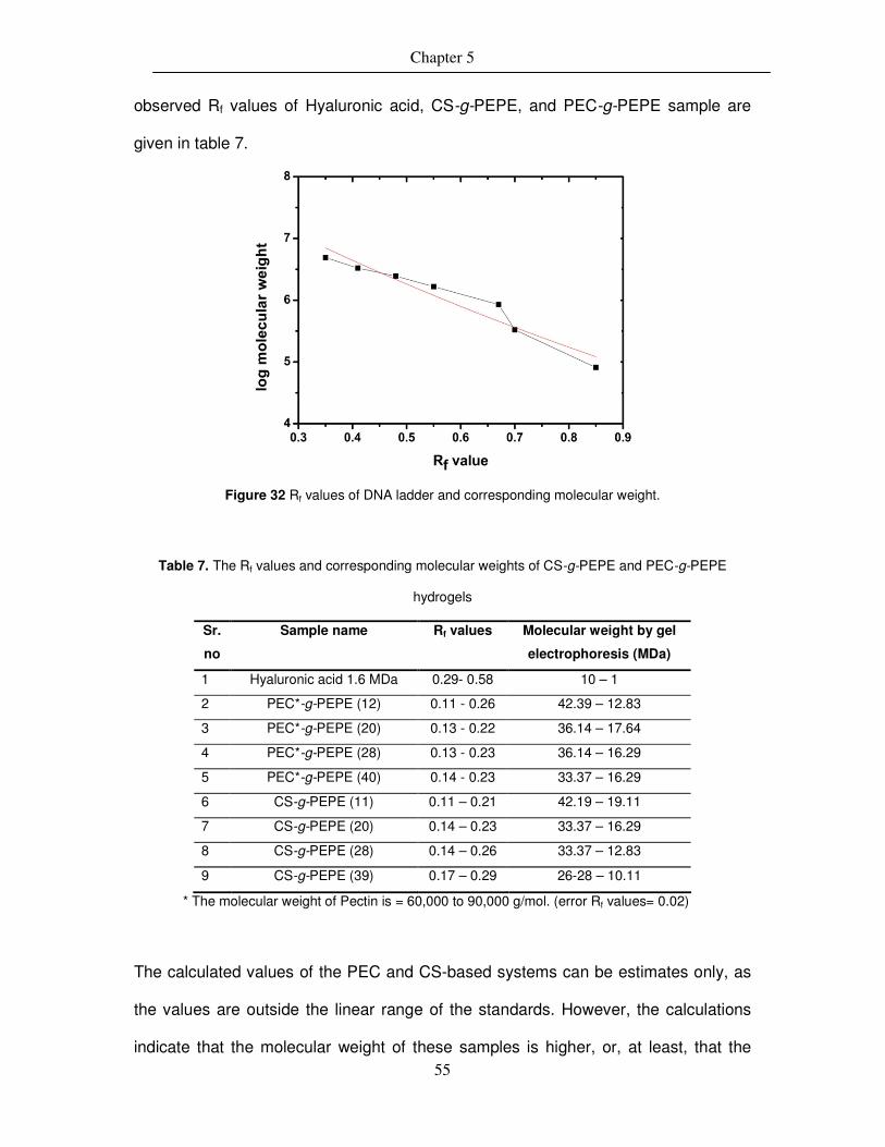
Chapter 5
55
observed Rf values of Hyaluronic acid, CS-g-PEPE, and PEC-g-PEPE sample are
given in table 7.
Figure 32 Rf values of DNA ladder and corresponding molecular weight.
Table 7. The Rf values and corresponding molecular weights of CS-g-PEPE and PEC-g-PEPE
hydrogels
Sr.
no
Sample name Rf values
Molecular weight by gel
electrophoresis (MDa)
1 Hyaluronic acid 1.6 MDa 0.29- 0.58 10 – 1
2 PEC*-g-PEPE (12) 0.11 - 0.26 42.39 – 12.83
3 PEC*-g-PEPE (20) 0.13 - 0.22 36.14 – 17.64
4 PEC*-g-PEPE (28) 0.13 - 0.23 36.14 – 16.29
5 PEC*-g-PEPE (40) 0.14 - 0.23 33.37 – 16.29
6 CS-g-PEPE (11) 0.11 – 0.21 42.19 – 19.11
7 CS-g-PEPE (20) 0.14 – 0.23 33.37 – 16.29
8 CS-g-PEPE (28) 0.14 – 0.26 33.37 – 12.83
9 CS-g-PEPE (39) 0.17 – 0.29 26-28 – 10.11
* The molecular weight of Pectin is = 60,000 to 90,000 g/mol. (error Rf values= 0.02)
The calculated values of the PEC and CS-based systems can be estimates only, as
the values are outside the linear range of the standards. However, the calculations
indicate that the molecular weight of these samples is higher, or, at least, that the
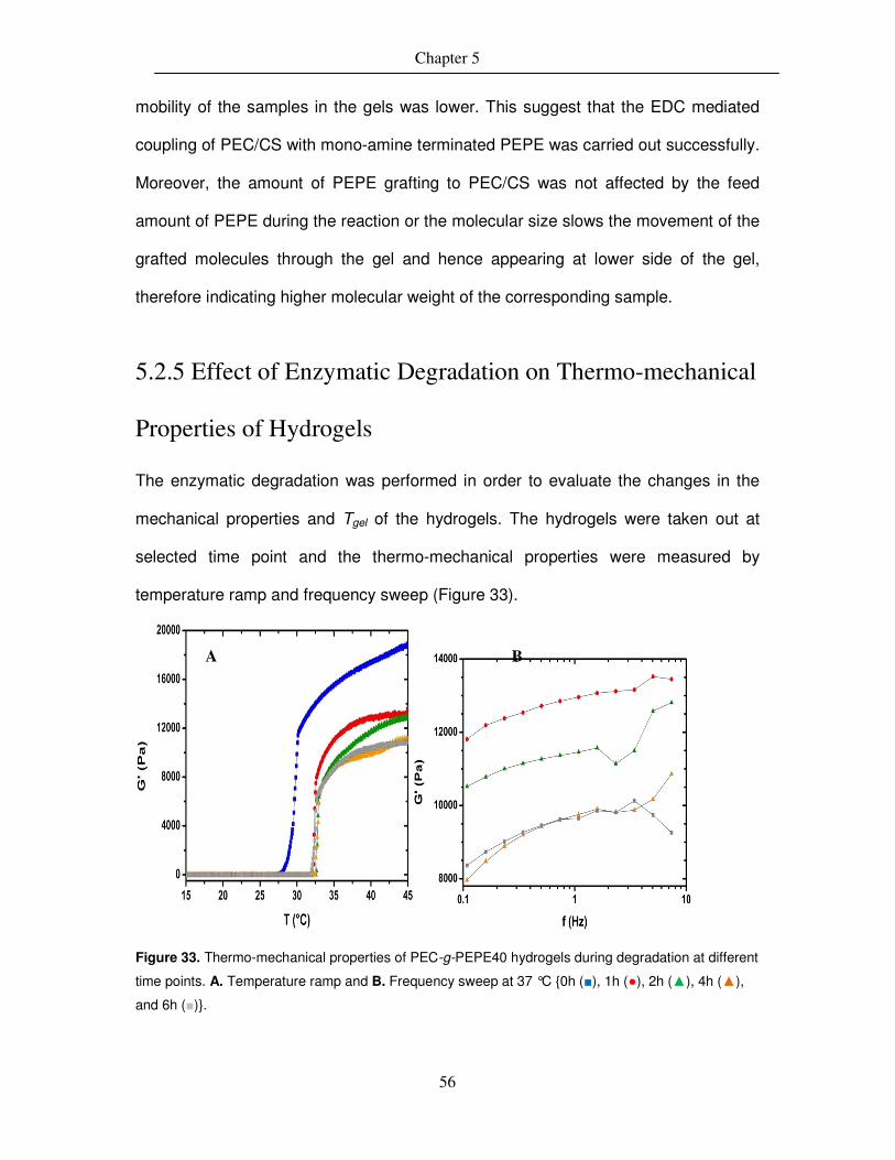
Chapter 5
56
mobility of the samples in the gels was lower. This suggest that the EDC mediated
coupling of PEC/CS with mono-amine terminated PEPE was carried out successfully.
Moreover, the amount of PEPE grafting to PEC/CS was not affected by the feed
amount of PEPE during the reaction or the molecular size slows the movement of the
grafted molecules through the gel and hence appearing at lower side of the gel,
therefore indicating higher molecular weight of the corresponding sample.
5.2.5 Effect of Enzymatic Degradation on Thermo-mechanical
Properties of Hydrogels
The enzymatic degradation was performed in order to evaluate the changes in the
mechanical properties and Tgel of the hydrogels. The hydrogels were taken out at
selected time point and the thermo-mechanical properties were measured by
temperature ramp and frequency sweep (Figure 33).
Figure 33. Thermo-mechanical properties of PEC-g-PEPE40 hydrogels during degradation at different
time points. A. Temperature ramp and B. Frequency sweep at 37 °C {0h (■), 1h (●), 2h (▲), 4h (▲),
and 6h (■)}.
A B

Chapter 5
57
As shown in figure 33 A, during the degradation of pectin-g-PEPE hydrogels, the Tgel
was slightly shifted from (28- 33 °C) within 6h of time, due to enzymatic degradation
of α-1,4-glycoside bonds between galacturonic acid. Moreover, the G′ of the hydrogel
was decreased from around 17500-9000 Pa (±3%) during the degradation process.
This effect in decrease in G′ was also clearly shown in frequency sweep test at 37
°C. The fragments formed due to degradation of pectin could interfere on the
micellelization process of PEPE, thus delaying the process of gelation after
degradation. The degradation process not only affected the gelation process but also
the mechanical properties. As shown in figure 33 B, due to the degradation of pectin
chain, it loses the chain entanglement and intra molecular chain interactions, resulted
in decrease in the mechanical properties of overall hydrogel. Overall it should be
noted that due to the random cleavage of Pectin during the enzymatic degradation
process could results in slight different mechanical properties e.g. elastic modulus
(G′) and Tgel. The similar effect of decrease in mechanical properties with decrease in
Tgel was observed in all other composition of pectin-g-PEPE hydrogel, one example
of temperature ramp of pectin-g-PEPE11 is shown in Figure 34.
Figure 34. Temperature ramp of Pec-g-PEPE11 hydrogel during degradation. {0h (■), 1h (●), 2h (▲), 4h (▲), and 6h (■)}.

Chapter 5
58
5.3 Summary
The polysaccharides pectin and chondroitin sulfate were functionalized with
thermosensitive PEPE to have a thermosensitive, degradable hydrogel. Grafting of
amine terminated PEPE to pectin and chondroitin sulfate was performed in order to
investigate if tailoring of the sol-gel-transition temperature can be achieved by
adjusting the grafting ratio. The sol-gel transition of the pectin, chondroitin sulphate,
PEPE, and the grafted system (PGP and CGP) was investigated by rheology, where
it was clearly shown that pectin and chondroitin sulfate was unable to show
thermosensitive gelation. The gelation temperature (Tgel) of the system could be
adjusted by varying the grafting density of PEPE onto pectin and chondroitin sulfate
as well as by the concentration of the thermosensitive polymer in aqueous solution. A
concentration of 15 – 20 wt% of the grafted system in water led to gelation
temperatures in the range of 25 – 33 °C and the critical micelle concentration (CMC)
and critical micelle temperature (CMT) of the Pec-g-PEPE systems were determined
by UV spectroscopy. The viscosity and the G′ increased by four orders of magnitudes
at Tgel, which is comparable to PEPEs alone, but could be reached at lower PEPE
concentrations.
Moreover, the enzymatic degradation of Pec-g-PEPE hydrogels show that the
viscoelastic properties (G′ and G′′) of hydrogel was decreased by degradation of
pectin and the gelation of hydrogel was shifted to higher temperature. However, the
hydrogel still hold the ability to show a sol-gel transition after the degradation of its
biopolymer backbone, such property of these hydrogel could be very useful in
different biomedical application, e.g. drug delivery where a long term presence of the
implant is necessary.

Chapter 5
59
5.4 Synthesis of HA covalently coupled with PEPE using
DMTMM.
5.4.1 Synthesis of hydrogels
The hydrogels were synthesized via amide bond formation between carboxylic group
of HA and amine terminated PEPE using DMTMM as a coupling agent. This coupling
method was found to be efficient over the EDC mediated coupling method due to, a.
it does not required a specific pH (4.5 - 4.7) to carry out the coupling and b. the by-
products of this reaction are easily removable by washing with water. Moreover, the
ultrafiltration (filtration using a membrane with pore size 0.025 µm) provided a reliable
removal of unreacted PEPE from the reaction mixture to give pure covalently coupled
product.
PE
PE
OO
OH
O
NH
OH
OH
O
O
OH O
NH
n
OO
OH
O
NH
OH
OH
O
OHO
NH
O n
OO
OH
O
NH
OH
OH
O
O
OH O
OH
n
HO
-PE
PE
OO
OH
O
NH
OH
OH
O
O
OH O
NH
n
DMTMM
Water, RT, 48 h
Water, RT, 48 hDMTMM1
2
3
4
Scheme 2. Synthesis route of HA-g-PEPE (HGP), and HA-x-PEPE (HXP) by using DMTMM
in water at room temperature

Chapter 5
60
By coupling mono-PEPE and di-PEPE to HA, the hydrogels with two different
architecture (grafted and partially crosslinking) was expected which, could show a
distinguishable variance in there thermo-mechanical properties due to the type of
covalent bonding (Scheme 2 ). Thus, the hydrogel was synthesized and purified by
systematic methods, and further characterized by different methods to evaluate the
thermo-mechanical properties depending upon either concentration or temperature.
The covalent coupling between HA and amine terminated PEPE was confirm by
ATR-FTIR, the degree of functionalization and expected molecular weight of
hydrogels was calculated as,
Free –COOH groups in HA (MCOOH)=
mol
g383
)(WHA (eq. 5.4)
Degree of functionalization (D. F.) = 100M PEPE ×
HAM (eq. 5.5)
Mx = )(
%
xMW
wt x
Molecular weight of HGP or HXP = [ ]HAPEPEPEPE MWWMFD +*)*.).*((MWHA (eq. 5.6)
Where, MCOOH = mols of free carboxylic groups in HA, WHA = amount of HA, MPEPE = mols of PEPE in
HGP or HXP, MHA = mols of HA in HGP or HXP, MWHA = Molecular weight of HA, x = HA or PEPE,
MW = 383 g·mol-1 (HA) or 14500 g·mol-1 (Mn of PEPE).
The hydrogels obtained by this method are listed below in table 8.

Chapter 5
61
Table 8. Nomenclature and composition of HA:PEPE in HXP and HGP systems synthesized in MES
buffer (6.5 pH) or water
Determined by weighing Determined by TGA
Sample ID* HA:PEPE
(wt%)
D. F.
(mol%)
Molecular
weight
calculated
(MDa)
HA:PEPE
(wt%)
D. F.
(mol%)
Molecular weight
calculated (MDa)
HXP93 27:73 7 9.3 7:93 36 37.1
HXP88# 21:79 11 13.2 12:88 20 21.8
HXP83 25:75 8 10.3 17:83 13 15.1
HGP88 28:72 6 8.3 12:88 19 21.4
HGP73 32:68 6 8.3 27:73 7 9.3
HGP58# 52:48 3 5.47 42:58 4 6.4
D. F.: Degree of functionalization, *: Nomenclature according to composition determined by TGA, #: In
MES buffer (6.5 pH)
5.4.2 ATR-FTIR spectroscopy
The successful coupling between HA and PEPE could be demonstrated by new
bands in the IR spectroscopy corresponding to amide bonds which indicates the
formation of new functional group within the molecules (Figure 35). The amide bond
formation between HA and mono-PEPE or di-PEPE was confirmed with ATR-FTIR by
the fairly strong increase of the absorption peaks at 1660 cm-1 (C=O stretching) and
1555 cm-1 (N-H bending, Amide II) . The additional peak at 1740 cm-1 (compared to
HA) due to the urethane bond, which is present in the aminated PEPE (C=O
stretching) as shown in figure 7. In addition, the presence of PEPE was investigated
by the strong absorption peak at 2883 cm-1 which shows C-H stretching vibrations
and a peak at 1097 cm-1 which was attributed to the characteristic C-O-C stretching
vibration from PEPE segment.

Chapter 5
62
Figure 35. ATR-FTIR spectra: A. mono-PEPE (a), physical mixture HA·mono-PEPE (b), HGP88 (c)
and HA (d). B. di-PEPE (a), physical mixture HA·di-PEPE (b), HXP93 (c), and HA (d). The amide bond
formation in HGP88 (A(c)) or HXP93 (B(c)) can be seen by the absorption peak at 1740 cm-1 (C=O
stretching), 1660 cm-1 (N-H bending), and 1555 cm-1 (N-H bending) which are well separated from
amide peak of HA (1550 cm-1 and 1600 cm-1).
5.4.3 Thermal properties and determination of composition of
hydrogels
The thermal properties of HGP and HXP copolymers were investigated by DSC and
TGA. The DSC thermograms show a Tm for the PEPE educts 1 and 2 of 55 °C and
53 °C while HA showed no Tm. While the physical mixtures of HA and 1 or HA and 2
showed a similar Tm as the corresponding aminated PEPE, the HGP and HXP
copolymers showed lower melting temperatures (Tm) (Figure 36 A) than the
corresponding PEPEs 1 or 2. The lowest Tm of 42 °C was found for the grafted
hydrogel HGP58, which also has the lowest PEPE content, while the partially
crosslinked hydrogel with the lowest Tm content was HXP88 with a Tm of 45 °C
(Figure 36 B). In both cases, the increase in PEPE content led to a rise in Tm
revealing the resulting higher crystal size with increase of PEPE content in the

Chapter 5
63
corresponding HGP and HXP gels. The increase in crystallinity was also seen in x-
ray scattering investigations of the partially crosslinked gels (data not shown).
The exact HA:PEPE ratio of the synthesized hydrogels was calculated from the TGA
measurements which was based on the TGA analysis of the educts HA and the
aminated PEPE 1 or 2. In case of HA, the thermal weight loss started at 70-90 °C
with an initial mass loss of 15-18 wt%, which includes loss of moisture. A major mass
loss of around 40 wt% was observed at 240 °C. The mass loss of the aminated
PEPE started at a much higher temperature. In case of the mono-amino PEPE 1, the
major mass loss started at around 352 °C and after another 50 ° (at 405 °C) 95 wt%
of the initial mass was lost. In HGP and HXP hydrogels, the peak at 237 °C indicates
the first step of thermal degradation presumably due to the HA segment which is
Figure 36. Thermograms from Differential Scanning Calorimetry (DSC): A. Grafted hydrogels HGP73
(─) and HGP58 (─) compared with HA (─), PEPE 1 (─), and physical mixture HA·PEPE1 (─). B.
Partially crosslinked hydrogels HXP93 (─) and HXP88 (─) compared with HA (─), PEPE 2 (─), and
physical mixture HA·PEPE2 (─). The melting temperature (Tm) and ∆H increased with degree of
functionalization, likely because of the increase in crystallinity and crystal size.
A B

Chapter 5
64
around 5-10 wt% and another peak observed at 345 °C with mass loss of 80 wt%,
which is presumably due to the PEPE segment (Figure 37 A and B). However, in
case of HGP and HXP systems, the mass loss at 405 °C was only approximately 85
wt% and thus lower, which is expected to be due to the covalent coupling of HA and
amino PEPE. Thus, it was possible to estimate the amount of HA and PEPE
incorporated in HGP and HXP copolymer system from TGA analysis. The
quantitative amount (in wt%) of each segment was calculated by plotting a line with a
quantitative combination of HA and the corresponding amino PEPE, which was fitted
to the HGP or HXP line obtained by TGA (Table 8). By calculating the amount of
amino PEPE coupled to HA in HGP and HXP hydrogels, it was possible to calculate
the estimated molecular weight of copolymers by using eq. 5.6.
However, in case of physical mixtures no significant difference was observed in
comparison to the corresponding amino PEPE, which is assumed to be due to
absence of covalent bonding between HA and amino PEPE. Thus, this study
demonstrates that the covalent coupling and degree of functionalization in HGP and
HXP copolymer system have a strong effect on the thermal properties. The results
from the TGA were compared to the final weight of the synthesized hydrogel.
However, it showed that the calculations based on the weight difference between the
quantity of the HA used for the reaction and the weight of the final hydrogel is too un-
exact to draw any further conclusions presumably because of weight loss during the
work-up.

Chapter 5
65
Figure 37. Thermogravimetric analysis (TGA): A. Grafted hydrogel HGP88 (…) compared with its
educts (HA (___), PEPE 1 (---) and the physical mixture HA·PEPE1 (___) and the mathematical fit of
HGP88 (-·-). B. Crosslinked hydrogel HXP93 (…) compared with its educts (HA (___), PEPE 2 (---), and
physical mixture HA·PEPE2 (___) and the mathematical fit of HXP93 (-·-).
5.4.4 Determination of Thermo-Mechanical Properties
Induced by Temperature and Concentration of Hydrogels
In order to better understand and evaluate the influence of covalent linkage on the
mechanical properties during the gelation process, a rheological analysis was
performed. The elastic modulus (G′) and viscous modulus (G′′) were monitored as a
function of temperature and frequency. Gel formation of PEPE is triggered by micelle
formation and takes place above CMC and CMT at a concentration of >16 wt% which
is reversible upon cooling. In case of HA, no thermosensitive behavior is observed,
but it has a very high viscosity at low concentration (1-3 wt%). The physical mixture
of the HA and the aminated PEPE 1 or 2 respectively (weight ratio 5:95) show a Tsw
of 28 °C or 25 °C respectively (close to the Tsw of PEPE 1 and PEPE 2 using the
same concentration) and an enhancement in elastic modulus from 0.892 Pa to 14660
Pa upon heating HA·mono-PEPE at a concentration of 20 wt%, while no effects were

Chapter 5
66
observed at a concentration of 10 wt%. Similar results were found for the physical
mixture HA·diamino-PEPE95. The grafted and crosslinked hydrogels showed already
at a concentration as low as 2.5 wt% an increase in thermo-mechanical properties,
while previously studied systems required a polymer concentration of 18-25 wt% to
show gelation near body temperature88–91. This gelation at a much lower
concentration was possible by creating covalent net points by covalently coupling
amino-PEPE to the HA backbone in addition to the physical netpoints, which are
created by micelles of PEPE above CMC/CMT (Table 9).
In case of the grafted hydrogels, it could be observed that an increase in PEPE
content of the hydrogel leads to an increase in the elastic modulus. However, no
significant effect on Tsw in relation to PEPE content was observed which was at 32/33
°C. At T > Tsw, the 5 wt% hydrogels prepared from HGP58, HGP73, and HGP73 88
showed an increase in elastic modulus G′ from 18 Pa to 703 Pa and finally to 1367
Pa. The difference between the mechanical properties below Tsw and above Tsw
varied only by the factor 1.6 to 4.5, which is much lower than for the physical
mixtures.
In case of the HXP copolymer systems, the thermo-mechanical properties varied in
dependence of the hydrogel concentration (2.5 – 10 wt%) (Figure 38). The Tsw of 30
°C was also much higher than for the physical mixtures. In the temperature ramp of
HXP93 above Tsw, the 2.5 wt% sample revealed a G′ of 1150 Pa which increased to
4326 Pa (±5%) at concentration of 5 wt % and up to 13080 Pa (±5%) at concentration
of 10 wt%. It is also noticeable that, in case of HXP93 at 2.5 -10 wt% concentration
the G′ after Tsw increased by factor 2.3-3 compared to G′ below Tsw.

Chapter 5
67
Table 9. Rheological properties of physical mixtures of HA·aminated-PEPE, HGP, and HXP
hydrogels.
Sample Concentration
(wt%)
Tsw
(°C)
G′ before
Tsw (Pa)
G′ after
Tsw (Pa)
Tint
(°C)
Factor
(fold)
HA·PEPE1* 20 28 0.892 14660 3 1.6.104
HA·PEPE1* 10 NO 0.956 0.972 NO 1
HA·PEPE2* 20 25 1.116 16670 3 1.4.104
HA·PEPE2* 10 NO 1.245 1.342 NO 1.1
HXP93 2.5 30 514 1150 10 2.3
HXP93 5 30 1286 4326 10 3
HXP93 10 30 5000 13080 10 2.6
HGP88 5 33 1367 2416 12 1.7
HGP88 10 32 10810 19130 12 1.7
HGP73 2.5 32 250 400 12 1.6
HGP73 5 32 306 703 11 2.3
HGP58 5 33 4 18 12 4.5
Tsw: Switching temperature, Tint: Temperature interval, NO: Not observed. * HA:PEPE ratio 5:95 wt%
(Error for G′ and G′′ = ±5%, Error for Tsw and Tint = ±1.5 °C)
This study demonstrates that the covalent coupling of amino PEPE to HA leads to
thermosensitive hydrogels which have a tunable Tsw and tunable mechanical
properties. While earlier reports on physical mixtures required high concentrations of
18-27 wt%15,23,92 of HA and the thermosensitive PEPE, the now presented approach
results in hydrogels that have enhanced mechanical properties at physiological
temperature using only 2.5 wt% of polymer concentration. The physical mixtures
required a concentration of 25-27 wt% of HA and PEPE to demonstrate a Tsw of 33-
37 °C at which G′ and G′′ increased; the addition of only 1-2 wt% of HA to 25-27 wt%
PEPE solution, which would be comparable concentration to the now reported HGP

Chapter 5
68
or HXP hydrogels, does not have a significant influence on the gelation temperature
of physical mixtures.
Figure 38. Increase in complex shear storage moduli (G′) of HXP93 hydrogel with different
concentration measured as a function of temperature (A) and as a function of frequency (B) by
rheology. (a: 2.5 wt% HXP93, b: 5 wt% HXP93, and c: 10 wt% HXP93).
The gelation in physical mixture results from the hydrophilic/hydrophobic association
of polypropylene glycol (PPG) and polyethylene glycol (PEG) segments in the PEPE
and occurs within a definite range of concentration as well as temperature. The gel
formed by such mechanism, easily disintegrates to a solution by further reduction in
concentration.
Hydrogels resulted from EDC mediated coupling and purified by dialysis, seem to
have a large quantity of non-reacted educts89,93–95. The observation, that PEPE with a
molecular weight of approximately 14.6 kDa does not pass during dialysis the
membrane, and the described gelation and the corresponding mechanical properties
lead to this assumption. The thermal gelation of these hydrogels required a
concentration of 18-25 wt% gel in water to show a gelation temperature in the range
of 26-34 °C. The reported minimal concentration for gelation of HA-g-PEPE was 18

Chapter 5
69
wt%94,95 Even in case of the highest grafting degree, no significant effect in shift of
the gelation temperature due to grafting was observed. Thus, covalent grafting and
cross-linking using DMTMM as a coupling mediator and the work-up using
ultrafiltration to remove unreacted PEPE quantitatively, lead to hydrogels, which
show a strong increase in the thermo-mechanical properties compared to the
physical mixtures; an increase in the elastic modulus (G′) was observed already at a
concentration as low as 2.5 wt% gel in water. The current results show that (1)
varying the coupling strategy, i.e. choosing the grafting or the cross-linking approach
and variation of coupling density, (2) varying the PEPE content in the copolymer, and
(3) varying the dilution of the hydrogel in water, and (4) varying the applied
temperature, i.e. working below or above Tsw, allows the final tuning of the
mechanical properties.
5.5 Summary
This study shows that covalent coupling of the thermosensitive unit PEPE to HA
leads to a phase transition at lower concentrations (2.5 -10 wt%) compared to the
physical mixtures (16.8-20 wt%) of the same components, which was achieved by an
efficient covalent coupling of amine terminated PEPE to HA to form a thermosensitive
hybrid. DMTMM showed to be a much more effective coupling agent than EDC, and
purification by ultrafiltration was necessary to remove unreacted PEPE quantitatively
to obtain covalently coupled hydrogels. All synthesized compounds were in the gel
state at low concentrations at room temperature, in contrast to physical mixtures of
the same components. The covalent coupling of HA and amine terminated PEPE
was shown by FT-IR, and TGA was a suitable method for the exact determination of
the composition. The covalently coupled hydrogels showed enhanced elastic

Chapter 5
70
modulus (G′) when heated near to body temperature. The viscoelastic properties
were tailorable by the covalent coupling strategy, PEPE content, concentration of the
hydrogel, and the degree of functionalization. Interestingly, the thermosensitive
behavior could be demonstrated even at a very low concentration of these hydrogels
in water (2.5-10 wt%), in contrast to physical mixtures of the same components,
which need a concentration of more than 16 wt%. furthermore, when these hydrogels
were treated with the enzyme hyaluronidase, it turned into a solution due to the
degradation of HA chains and thereby showed an on-demand solubilization of these
hydrogels. Moreover, the rheological experiment of these degraded hydrogels
showed a sol-gel transition with slight shift in Tsw to 40 °C, indicating the presence of
thermosensitivity even after degradation. Thus, unlike the CMC based hydrogels,
these hydrogels were not solubilized after addition of excess solvent but, showed an
on-demand solubilization by enzymatic degradation.
Thus, covalent coupling of amine terminated PEPE to HA with DMTMM provides a
sufficiently high local concentration of PEPE segments, which show thermosensitivity
near body temperature as well as tunable mechanical properties. Future study on
these promising hydrogels concerning their hydrolytic and enzymatic degradation and
the corresponding effect on the viscoelastic properties (G′, G′′, and |ƞ*|) and
thermosensitivity of these hydrogels is needed to draw a conclusion concerning their
application in Regenerative Medicine.

Chapter 6
71
6. Synthesis of Gelatin-g-PEPE
thermosensitive architectured hydrogels
(T-ArcGel)
In this chapter, the synthesis and characterization of Gelatin-g-PEPE based
architectured hydrogels were described. Further, the evaluation of swelling, water
uptake, thermo-mechanical properties, and hydrolytic degradation of the hydrogels is
discussed.
6.1 Synthesis of GA-g-PEPE hydrogels
The hydrogels of GA-g-PEPE were synthesized by covalent coupling of PEPE and
amino groups of gelatin. First PEPE were reacted with 4-nitophenyl chloroformate
and further reacted with amino groups of gelatin as shown in scheme 3.
Scheme 3. Synthesis of GA-g-PEPE hydrogels

Chapter 6
72
Further by controlling the initial amount of PEPE added during the reaction, the
grafting density of PEPE in gelatin-g-PEPE was varied. The TNBS test revealed
degrees of functionalization of 22 to 54 mol% of amino groups of gelatin by PEPE.
Here, different amount of PEPE functionalization on different blooms of gelatin (see
table 10) was studied. In this study gelatin with 300, 200, and 90-120 bloom was
used. The strategy behind using different blooms of gelatin and different grafting
density was to evaluate the effect on mechanical properties of the resulting hydrogel.
The non-functionalized amino groups of gelatin were used for crosslinking in the next
step of the synthesis
Table 10. Degree of grafting of PEPE to gelatin
Sample. no.
Gelatin bloom Functionalization of -NH2 groups of gelatin (mol%)
1 300 22 2 300 54 3 200 24 4 200 28 5 200 30 6 200 34 7 90-120 37 8 90-120 43
6.2 Characterization of GA-g-PEPE hydrogels
6.2.1 ATR-FTIR and 1H-NMR analysis
The GA-g-PEPE hydrogels were characterized by ATR-FTIR and NMR spectroscopy
to detect the changes in the peaks due to formation of new bonds by grafting PEPE
to gelatin and hence identifying the product. As shown in figure 39, decrease in the
intensity of -NH2 (3400 cm-1) group due to a chemical modification by PEPE was
observed. Other peaks at 2950 cm-1 (C-H stretching) and 1020 cm-1 and 950 cm-1 (C-
O-C, ether linkage in PEPE) reveal the presence of PEPE in the sample.

Chapter 6
73
Figure 39. IR of GA200 bloom (▬) and GA200-g-PEPE24 (▬).
As gelatin is a natural polymer derived from collagen i.e. consist of amino acids, there
are several signals appeared in the spectra the 1H-NMR spectra and was
complicated to understand. So, the proton signal of our particular interest was -NH2
groups of lysine which were functionalized by PEPE. The list of different signals is
given in Table 11. The NMR was measured at 45 °C for better resolution of proton
signals (Figure 40).
7.5 7.0 6.5 6.0 5.5 5.0 4.5 4.0 3.5 3.0 2.5 2.0 1.5 1.0 0.5Chemical Shift (ppm)
Figure 40. 1H-NMR spectra of hydrogels in D2O, A: pure Gelatin and B: GA200-g-PEPE34.
A
B

Chapter 6
74
Table 11. Assignment of specific peaks in the 1H-NMR spectrum of gelatin solution
Peak
No.
Chemical Shift (ppm)
of gelatin at 45 °C
Chemical Shift (ppm)
of GA-g-PEPE at 45 °C
Assigned to
different protons
1 1.06 1.04 Leucine, Valine, and
Isoleucine
2 1.39 1.40 Threonine
3 -- 1.34 -CH3 of PPG
4 1.58 1.58 Alanine
5 1.86 1.86 Lysine and arginine
6 1.98 1.97 Lysine and arginine
7 2.89 2.90 Aspartic acid
8 3.18 3.19 Lysine
9 3.40 3.40 Arginine
10 -- 3.74 -O-CH2 of PEG
11 3.82 3.89 Proline
12 7.43-7.56 (m) 7.44-7.55 (m) Tyrosine
Due to the chemical binding of PEPE:4NCP to –NH2 group of lysine, mainly the
protons of the carbon adjacent to amino group in lysine (-CH2-NH2) shows shift from
3.18 to 3.19 ppm. The appearance of peak at 1.34 ppm (-CH3 of PPG), 3.74 ppm (-
CH2-O protons of PEG and PPG), and 3.82 ppm (-CH-O protons of PPG) indicate the
presence of PEPE in the GA-g-PEPE hydrogel.
6.2.2 Thermo-mechanical properties
The GA-g-PEPE systems were characterized by DSC (TMDSC), TGA, and rheology
to evaluate the thermo-mechanical properties. First, the DSC analysis was carried
out in order to identify thermal transition in the GA-g-PEPE hydrogels in dry state
(Figure 41). However, the PEPE is known to have a sharp Tm at around 58 °C, the
thermal properties of gelatin were complicated. By using temperature modulated
DSC (TMDSC) the glass transition of dry gelatin hydrogels was determined. The pure
gelatin generally shows an endothermic peak mainly associated to helix-coil

Chapter 6
75
transition, which mainly depend on the sample history (source, storage condition,
etc.). For anhydrous solid gelatin the Tg = 217 °C and Tm = 230 °C and both values
are strongly affected by the moisture content*. Here one main endothermic peak at
170-190 °C was observed which was seemed to be reduced due to moisture in the
sample. But for GA-g-PEPE hydrogel samples, two peaks are observed, one peak at
50 °C is likely from the PEPE melting and another peak from 200 to 230 °C is mainly
due to the melting of triple helices of gelatin.
Figure 41. DSC spectra of GA200 (▬), GA200-g-PEPE28 (▬), and GA200-g-PEPE34 (▬).
In addition, the TGA was performed on dry samples of GA-g-PEPE hydrogels
in order to characterize the thermal properties of PEPE functionalized gelatin. TGA
was performed in N2 atmosphere (Figure 42). The TGA of pure gelatin shows three
thermal degradation steps as, loss of moisture from 50-100 °C, gelatin decomposition
between 250-350 °C, and finally combustion of remaining mass between 400-700 °C.
The GA-g-PEPE hydrogels also shows three step degradation at 80, 150-300, and
350-700 °C. The former is attributed to moisture loss; mass loss at 150-300 °C can
be related to the gelatin decomposition not as sharp as pure gelatin but, due to the

Chapter 6
76
resistance created by grafted PEPE chains and the third mass loss from 350-700 °C
is due to the combustion of gelatin and PEPE. In both GA-g-PEPE samples the
degradation starts earlier then compare to pure gelatin, the reason could be the
grafting of PEPE to gelatin restricts the gelatin chains to from triple helices and thus
losing the strength of the network. Thus, it seems that GA-g-PEPE hydrogels
required low energy to disintegrate the intermolecular and partial breaking of the
molecular structure. From the figure 42, it seems that higher the PEPE content in the
composition lower is the temperature required to show thermal degradation process.
Hence, PEPE-grafting to gelatin reduced the formation of triple helices and therefore
the thermal degradation of GA-g-PEPE hydrogels is occurred at low temperature
(200 °C) compared to pure gelatin (290 °C).
Figure 42. TGA spectra of GA200 (▬), GA200-g-PEPE28 (▬), and GA200-g-PEPE34 (▬).
The WAXS analysis was carried out to elucidate the details of chain
organization and inherent molecular organization of GA-g-PEPE hydrogels. Here, it
was interesting to evaluate the effective suppression of formation of triple helices
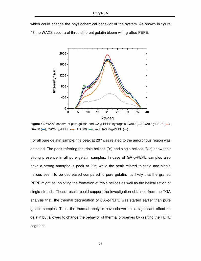
Chapter 6
77
which could change the physiochemical behavior of the system. As shown in figure
43 the WAXS spectra of three different gelatin bloom with grafted PEPE.
Figure 43. WAXS spectra of pure gelatin and GA-g-PEPE hydrogels. GA90 (▬), GA90-g-PEPE (▬),
GA200 (▬), GA200-g-PEPE (▬), GA300 (▬), and GA300-g-PEPE (▬).
For all pure gelatin sample, the peak at 20° was related to the amorphous region was
detected. The peak referring the triple helices (9°) and single helices (31°) show their
strong presence in all pure gelatin samples. In case of GA-g-PEPE samples also
have a strong amorphous peak at 20°, while the peak related to triple and single
helices seem to be decreased compared to pure gelatin. It’s likely that the grafted
PEPE might be inhibiting the formation of triple helices as well as the helicalization of
single strands. These results could support the investigation obtained from the TGA
analysis that, the thermal degradation of GA-g-PEPE was started earlier than pure
gelatin samples. Thus, the thermal analysis have shown not a significant effect on
gelatin but allowed to change the behavior of thermal properties by grafting the PEPE
segment.

Chapter 6
78
Figure 44. The temperature ramp of GA200 (■), GA200-g-PEPE30 (▲), and GA200-g-PEPE37 (▲).
(concentration = 10 wt% each).
The thermo-mechanical properties of GA-g-PEPE hydrogels were investigated by the
rheological measurements. The hydrogels were pre-dissolved in water and analysed
by rheometer from 15-45 °C temperature ramp (figure 44). The hydrogels with
different bloom of gelatin and different grafting density of PEPE have been analysed
to see the effect of grafting PEPE on thermo-mechanical properties, especially the
elastic modulus (G′). As shown in table 12, no significant difference was observed by
changing the bloom value of gelatin on the gelation behavior except, gelatin with 300
bloom have shown higher Tsw compare to gelatin with 90 and 200 bloom. The
interesting point here is, with increasing PEPE content in the gelatin hydrogels it
seems that the mechanical properties of hydrogels (G′) found to be decreased. At 20
°C the elastic modulus (G′) of GA200 = 9270 Pa (±3%), which was reduced to 4338
Pa (±3%) by 30 mol% PEPE grafting which further brought down to 2145 Pa (±3%)
by 37 mol% grafting of PEPE to gelatin. Moreover, the micelle formation by PEPE
chains during the temperature ramp was not observed, which may be dominated by
the gelatin chains. However, the PEPE show an influence on aggregation of gelatin
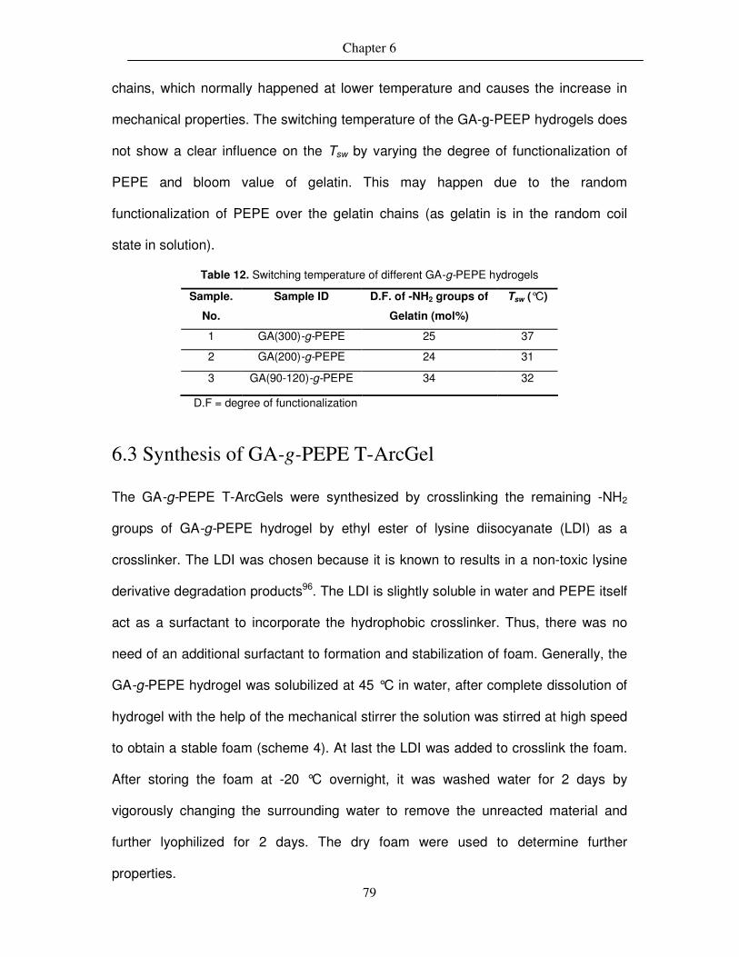
Chapter 6
79
chains, which normally happened at lower temperature and causes the increase in
mechanical properties. The switching temperature of the GA-g-PEEP hydrogels does
not show a clear influence on the Tsw by varying the degree of functionalization of
PEPE and bloom value of gelatin. This may happen due to the random
functionalization of PEPE over the gelatin chains (as gelatin is in the random coil
state in solution).
Table 12. Switching temperature of different GA-g-PEPE hydrogels
Sample.
No.
Sample ID D.F. of -NH2 groups of
Gelatin (mol%)
Tsw (°C)
1 GA(300)-g-PEPE 25 37
2 GA(200)-g-PEPE 24 31
3 GA(90-120)-g-PEPE 34 32
D.F = degree of functionalization
6.3 Synthesis of GA-g-PEPE T-ArcGel
The GA-g-PEPE T-ArcGels were synthesized by crosslinking the remaining -NH2
groups of GA-g-PEPE hydrogel by ethyl ester of lysine diisocyanate (LDI) as a
crosslinker. The LDI was chosen because it is known to results in a non-toxic lysine
derivative degradation products96. The LDI is slightly soluble in water and PEPE itself
act as a surfactant to incorporate the hydrophobic crosslinker. Thus, there was no
need of an additional surfactant to formation and stabilization of foam. Generally, the
GA-g-PEPE hydrogel was solubilized at 45 °C in water, after complete dissolution of
hydrogel with the help of the mechanical stirrer the solution was stirred at high speed
to obtain a stable foam (scheme 4). At last the LDI was added to crosslink the foam.
After storing the foam at -20 °C overnight, it was washed water for 2 days by
vigorously changing the surrounding water to remove the unreacted material and
further lyophilized for 2 days. The dry foam were used to determine further
properties.

Chapter 6
80
Moreover, it is also important to know the reactivity of LDI as, it mainly reacts with
nucleophilic groups of gelatin (mainly amino groups of lysine). However, it may reacts
with other nucleophilic groups in gelatin such as, aromatic NH-groups (histidine),
alcohols (serine, threonine and also in PEPE), phenols (tyrosine), thiols (cysteine),
carboxylic acids and amide functional groups97–99. Generally, gelatin has
approximately 3 mol % of amino groups100 (lysine/hydroxylysine) which was also
verified in the starting by using TNBS colorimetric assay. Generally, during the
crosslinking, isocyanates group of LDI reacts with lysine ε-amino groups along two
opposing gelatin chains. On the other hand, in aqueous condition, decarboxylation of
ioscyanates may occur, which forms free amino groups that can react with other LDI
molecules. Thus, the crosslinking reaction of LDI is quite complex and may have side
reactions and may influence the properties of gelatin after crosslinking.
EtONCO
O
NCO
45 °C, water, 1500 rpm
Scheme 4. Synthesis of GA-g-PEPE T-ArcGel
6.3.1 Investigation of water uptake by T-ArcGel
The GA-g-PEPE T-ArcGel of known weight were used to investigate the water uptake
studies of these hydrogels. The incorporation of crosslinker with a rigid structure
between gelatin chains may reduce the movements of individual chains and may
results in a more rigid network with low degree of swelling. The water absorption
capacity of these hydrogels was determined by gravimetrically and volumetrically by
using following equation of water uptake (H),

Chapter 6
81
d
ds
W
WWH
−= (Equation 6.1)
Generally in crosslinked hydrogels, with increasing crosslinking density have a
tendency to show decreasing water uptake. But here, the influence of PEPE on water
uptake capacity was investigated. The measurements were performed at two
different temperatures as 25 °C to observe the effect of linear PEPE and at 37 °C to
observe the effect of micellar PEPE on water uptake. As shown in figure 45, the
water uptake of GA200-g-PEPE24 has lower water uptake at 37 °C and slightly
higher water uptake at 25 °C. At higher temperature (37 °C), there may be micelle
formation of PEPE and less mobility of gelatin chains (due to crosslinking) which
could have reduced the capacity of hydrogel to absorb water.
Whereas, at lower temperature (25 °C) PEPE do not have any micelles and thus the
water uptake was slightly increased. But, when crosschecked with ArcGel without
PEPE (grafted), the effect of temperature on water uptake was found to be similar.
This may be due to the fact that, overall there are only 3 mol% amino groups (from
lysine) and if out of that, 20-40 mol% are functionalized with PEPE, still the total
amount of PEPE was very less compare to amount of gelatin in the T-ArcGel.
Thus, water uptake did not show a clear trend which could be influenced either by
temperature or by grafting PEPE to gelatin. This may be due to the total amount of
PEPE in the crosslinked system compare to amount of gelatin which may influence
more on water uptake capacity of overall system.

Chapter 6
82
Figure 45. Water uptake (H) of, A. GA200-g-PEPE24 T-ArcGel and B. GA200 ArcGel at 25 and 37 °C
(●: 25 °C and ●: 37 °C).
6.3.2 Analysis of porous structure of GA-g-PEPE T-ArcGel
The porous structure was analysed by SEM and µCT measurements in the dry state,
and gelatin of two different bloom (90-120 and 200) was used for the analysis. As
shown in the figure 47, the SEM images at 50, 100, 500-fold magnification show the
open porous structure of the T-ArcGel as well as the interconnectivity of the pores in
the matrix which might be necessary for promoting cell proliferation and transport of
nutrition. The pore size was calculated from both SEM and µCT measurements, the
average pore size for GA200-g-PEPE24 T-ArcGel was 272 µm and 180 µm by µCT
(Figure 46) and SEM analysis respectively (See Table 13). Generally, there was no
effect of bloom on porosity of material was observed, but in case of the GA90-g-
PEPE43 sample a large pore distribution was observed compared to GA200-g-
PEPE24 sample. Moreover, the pore size remained unaffected by the PEPE grafting.
Finally, the SEM and µCT analysis show a difference in the pore size, which might
occurred due to the limitations of methods to analyse the pore distribution in the T-
ArcGel.
A B

Chapter 6
83
Figure 46. The �CT measurements of A. GA200-g-PEPE24 and B. GA90-g-PEPE43.
Table 13 The pore size for different T-ArcGel determined by �CT and SEM analysis
Pore size (�m) Sample name
By �CT By SEM
GA90-g-PEPE37 207 (± 5) NC GA200-g-PEPE24 272 (± 12) 180 (±71) GA90-g-PEPE43 283 (± 10) 116 (±43)
NC: not calculated
Figure 47. Porous morphology of T-ArcGel investigated by SEM. A. GA200-g-PEPE24 and B. GA90-
g-PEPE43 at different magnification (at 50, 100, and 500x magnification levels)
B A
A
B

Chapter 6
84
6.3.3 Thermo-mechanical properties of T-ArcGel
DSC, TGA, rheology, and AFM analysis were performed on T-ArcGel in order to
evaluate the thermo-mechanical properties. The DSC (TMDSC) and TGA were
performed at dry state and rheology and AFM was performed in swollen state of the
sample. As already discussed in section 6.2.2, the thermal properties of gelatin were
complicated to evaluate and mainly depend upon the history of sample. Here, it was
interesting to see the effect of crosslinking gelatin chains on thermo-mechanical
properties of T-ArcGel.
The pure gelatin and GA-g-PEPE (figure 48 A and 48 B) shows a endothermic peak
associated with the helix-coil transition in the temperature range of 180-230 °C and a
peak at around 50 °C due to the melting of PEPE segment. But after crosslinking with
LDI, the helix-coil transition disappeared, meaning the gelatin chains were majorly
crosslinked by LDI and hence the mobility of the chains is blocked. However, in figure
48 A, peak at 50 °C indicate the melting of PEPE segment which is grafted to gelatin,
which still have mobility to show the effect. There is decrease in the melting
temperature of PEPE from 58 °C to 50 °C, which is may be due to the crystal size
and amount of PEPE in the system.
As previously explained in section 5.4.3, the Tm of PEPE was found to be decreased
with decreasing PEPE content in the hydrogel. Moreover, TGA analysis of T-ArcGel
was compare with pure gelatin and GA-g-PEPE hydrogel in a temperature range of
25-800 °C to evaluate the effect of crosslinking on the thermal stability of the system.

Chapter 6
85
Figure 48. DSC spectra of, A. GA200 (▬), GA200-g-PEPE34 (▬), and GA200-g-PEPE34 ArcGel (▬)
and B. GA 00(▬), GA200-g-PEPE28 (▬), and GA200-g-PEPE28 T-ArcGel (▬).
As discussed in section 6.2.2, the grafting reduces the formation of triple helix and
the thermal degradation occurs at lower temperature compared to pure gelatin. But,
further crosslinking of GA-g-PEPE hydrogel by LDI enhanced the thermal stability of
hydrogel compared to the grafted system. As shown in figure 49, irrespective of the
amount of PEPE in the hydrogel, the crosslinking has increased the rigidity in the
hydrogel by fixing the gelatin chains which gave a similar trend as the triple helix
formation in pure gelatin in case of thermal stability. Though the triple helices are
formed due to physical crosslinking and the actual chemical crosslinking of GA-g-
PEPE by LDI show similar behavior, which may be due to the grafted PEPE segment
which creates soft segments in the system and showed dominance over gelatin
chains after crosslinking by LDI.
B A

Chapter 6
86
Figure 49. TGA analysis of, A. GA200 (▬), GA200-g-PEPE34 (▬), and GA200-g-PEPE34 ArcGel
(▬) and B. GA200 (▬), GA200-g-PEPE28 (▬), and GA200-g-PEPE28 T-ArcGel (▬).
Thus, even chemically crosslinked GA-g-PEPE hydrogel have shown similar thermal
behavior. The compression test of GA-g-PEPE T-ArcGel was performed in order to
investigate the mechanical properties (compressive modulus (Ec) and compressive
stress (σmax)) in dry and wet conditions. The initial shape of material was not
recovered after 50% compression (Figure 50) due to compression of porous structure
at dry state (and recovered the original shape after swelling in water). But, in dry
state the mechanical properties of material was very high (Table 14). In case of wet
compression, the material was equilibrated at 25 °C in water for 24 h before actual
measurement. The shape of material was recovered even after 50% compression;
the recovery of shape at 25 °C was possible due to the macroscopic elasticity of
porous structure which may be related to the lowering in the Tg of gelatin in wet state
which also reduced the mechanical properties in wet condition (Table 14).
B A

Chapter 6
87
before compression 50% compression Recovery after compression
dry
wet
Figure 50. Compression of scaffold under dry and wet condition (at 25 °C).
The thermo-mechanical properties of GA-g-PEPE T-ArcGel were determined by
rheological measurements. The evaluation was performed under constant stress and
temperature and frequency as a function for measuring changes in the elastic
modulus (G′) and viscous modulus (G′′). Gelatin ArcGel was used a control sample.
Table 14 The compression data for dry scaffold
Sample Name Ec/kPa σmax/kPa
GA90-g-PEPE37 1440 (±110) 526 (±84)
GA200-g-PEPE24 1517 (±125) 381 (±28)
GA90-g-PEPE43 2323 (±139) 476 (±38)
Table 15 The compression data for wet scaffold at 25 °C
Sample Name Ec/kPa σmax/kPa
GA90-g-PEPE37 6 (±3) 4 (±2)
GA200-g-PEPE24 7 (±2) 4 (±2)
GA90-g-PEPE43 4 (±2) 3 (±2)
Where, K: compressive elastic modulus, σmax: stress at 50% compression
As shown in figure 51, a 10wt% sample of pure gelatin was run through a
temperature ramp measurement from 15 to 45 °C. The G′ decreases from 25 to 45

Chapter 6
88
°C from 1100 to 850 Pa (±3%) and the G′′ remains constant (100 Pa (±3%)) during
the measurement, showing no thermosensitive effect.
Figure 51. Mechanical properties of pure gelatin ArcGel as a function of temperature. G′ (■), G′′ (●).
However, in GA-g-PEPE ArcGel, the Tsw was decreased from 37 to 32 °C due to
grafting PEPE to gelatin; here the thermal gelation of PEPE was dominated by
gelatin. So, here it was interesting to see the effect of fixing the mobility of gelatin
chains (crosslinking with LDI) on thermo-mechanical properties of T-ArcGel.
Two samples of T-ArcGels with similar gelatin bloom (200) and different PEPE
grafting density (28 and 34 mole %) were measured. As shown in the figure, the first
interesting thing was these hydrogels have shown a reversible thermosensitive
behavior i.e. increase in the mechanical properties upon heating and coming back to
the initial mechanical properties upon cooling below Tsw, which strongly contradicts
the normal gelatin behavior (gelatin chains in water form triple helix at room
temperature which makes it solid and upon heating the solution above Tsw, from a
solution due to disintegration of triple helix in to single helix). The fact here was, upon
crosslinking the gelatin chain in solution state, it was possible to hinder the formation
of triple helix upon and vice versa. So, by fixing the chain structure, it was possible to

Chapter 6
89
suppress the gelatin behavior and thus allowing the grafted PEPE to demonstrate its
effect of thermal gelation, which shows an enhancement in viscoelastic properties
(G′, G′′, and |ƞ*|) upon heating above Tsw. The phenomenon could be explained by
formation of micelles by grafted PEPE upon heating and deformation of micelles in
chains upon cooling, thus having a reversible effect on viscoelastic properties.
However the determination of micelle formation in such a system was a challenging
task and may not be possible to detect due to the overall low concentration (28-34
mol%) of PEPE in the sample, which may limit the minimum concentration required to
detect the micelle formation in the structured hydrogel. Nevertheless, it was shown
before in the section 5.4.4 that, using lower concentration of PEPE (2.5-10 wt %) it
show a thermo-sensitive behavior with enhancement in mechanical properties upon
heating near body temperature (30-32 °C). However, for pure PEPE minimum
concentration of 16 wt% is required to show the thermosensitve behavior.
Moreover, the amount of PEPE grafted to gelatin was having an influence on the
mechanical properties as well as Tsw of T-ArcGel. As shown in figure 52, in the
sample GA200-g-PEPE28 the observed Tsw was 42 °C with G′ above Tsw was in the
range of 50,000-65,000 Pa (± 3%) and for the sample GA200-g-PEPE34 the
observed Tsw was 37 °C with G′ above Tsw was in the range of 90,000-150,000 Pa (±
3%). This values gave an indication that may be due to increase in the PEPE content
in the system, the number of micelle formation was increased which contributed in
decreasing the Tsw and enhancing the mechanical properties of overall system. In
addition, the viscous modulus (G′′) was also increased from around 100-15,000 Pa (±
3%) in GA200-g-PEPE28 and from 50-30,000 Pa (± 3%) in GA200-g-PEPE34
respectively.
In addition, fully water swollen GA-g-PEPE T-ArcGel upon heating (up to 50 °C) or
cooling (up to 5 °C) did not show any change in the physical structure, which was

Chapter 6
90
contradictory to the previously stated polyNIPAM based crosslinked thermosensitive
porous hydrogel, which have a large volume change (shrinking) when heated from 4 -
37 °C under swollen condition71. Moreover, the actual enhancement in the
mechanical properties (elastic modulus G’) was only 10 fold, whereas in the GA-g-
PEPE it was 108 fold (GA200-g-PEPE28) and 170 fold (GA200-g-PEPE34) higher
compare to initial values and thus providing a wide range of tunability in GA-g-PEPE
T-ArcGel system. The swelling or shrinking of ArcGel may cause additional forces
(during swelling) or may not fit properly in the defect (during shrinking) while using
such materials as an implant. Thus, on demand changes in the mechanical
properties without changing size of material could be an advantage.
These evaluation of GA-g-PEPE T-ArcGel shows a successful incorporation of
thermosensitive functionality, which allow the rigid system to show an enhancement
in the mechanical properties upon heating without any volume change, which was not
shown by pure gelatin ArcGel under similar conditions.
In addition to the temperature dependent rheological measurements, temperature
dependent force measurements were also performed to study the force applied by
the material when deforming above the Tsw. For this study four T-ArcGel with 28 and
34 mole % PEPE content and two different crosslinking densities i.e. 3x and 8x of LDI
were measured. As shown in figure 53A, GA200 crosslinked using 8X LDI was used
as a control sample (without PEPE grafting) which show overall constant force during
the given temperature range. This may be due to the fact that once the gelatin chains
are crosslinked, the major deformation upon heating is blocked (i.e. triple helix ↔
single helix formation) and the mechanical properties of the material remains
constant within the given range of temperature. These results also correlate with the
rheological temperature ramp measurements which also show similar behavior. But,
when the gelatin chains are grafted by PEPE (with different grafting densities) before

Chapter 6
91
crosslinking with LDI, it has shown increasing mechanical properties upon heating
the sample. Thus, it was interesting to crosscheck the property of the T-ArcGel by
performing a compression test with temperature as a function. As shown in figure 53
B and 53 C, The T-ArcGel crosslinked by 3X and 8X LDI shown similar behavior as in
temperature ramp. In addition, it was possible to detect the early stage of deformation
changes and corresponding mechanical properties of the ArcGel. The Tsw show a
slight shift to lower temperature range compare to the previous results by
temperature ramp measurements. Moreover the Tsw was not affected by the amount
of crosslinker (3x or 8x LDI) but, the amount of PEPE grafted to gelatin. Thus GA200-
g-PEPE28_3x and GA200-g-PEPE28_8x showing a Tsw = 30 °C and GA200-g-
PEPE34_3x and GA200-g-PEPE34_8x showing a Tsw = 27 °C approximately.
Nevertheless, the sample crosslinked with 8x LDI show higher force of 0.28 - 0.45 N
while sample crosslinked with 3x show 0.07- 0.3 N, indicating the initial strength of
material depended on the amount of crosslinker used. Moreover, it was also seen
that samples crosslinked with 3x LDI have wide range of deformation compared with
highly crosslinked samples.
Thus, the compression measurement by rheology with temperature as function could
also support the thermosensitive behavior of T-ArcGel by showing the increase in the
value of force above the Tsw. The rheological measurements evaluated the bulk
thermo-mechanical properties of GA-g-PEPE T-ArcGel, however it was also an
interesting task to evaluate further the microscopic properties of such a system to
demonstrate the thermally induced mechanical changes happening at micro level.
Thus, a method which accurately characterize the local mechanical properties of
porous material is required. Hence, atomic force microscopy (AFM) technique was
used to evaluate the local mechanical properties of T-ArcGel.

Chapter 6
92
Figure 52. Mechanical properties of GA-g-PEPE T-ArcGel as a function of temperature, A. GA200-g-
PEPE28 and B. GA200-g-PEPE34. Where, G′ (■),G′′ (●).
As the mechanical properties of porous material mainly depends upon the bulk
properties as well as geometry and porosity of the T-ArcGel, the evaluation of local
mechanical properties of such complex system was not easy. The AFM
measurements were mainly performed on the single pore walls of swollen T-ArcGel
at two different temperatures (25 °C and 37 °C) one below the Tsw and other above
the Tsw (The AFM measurements were performed by Oliver Frank). The isolation of
single pore wall under swollen condition was a difficult job and previously measured
(section 6.3.3, page 104) compression under swollen condition gave very low
compressive moduli (Ec = 4-8 kPa) which make the AFM measurement extremely
challenging.
The AFM results in the figure 54 and 55 show variation of data compared to the
rheological measurements, which is probably due to the difference between areas of
measurement and may be due to the technical limitation of the method. Taking this
into account, the Young’s moduli are compared between the samples with high and
low crosslinker (LDI) and temperature (25 and 37 °C). The overall results show that
Tsw= 42 °C
A
Tsw= 37 °C
B

Chapter 6
93
in the sample GA200-g-PEPE28_3x at both temperatures the local E moduli are in
the same range. But, in case of GA200-g-PEPE28_8x sample the local E moduli at
25 °C was in the range of 5-10 kPa which further increased to 25-35 kPa at 37 °C.
The reason to have increase in mechanical properties might be influenced by two
factors, the higher concentration of crosslinker (8x LDI) and temperature. On the
other hand, changing the PEPE grafting also demonstrate similar behavior for the T-
ArcGel crosslinked by 3x and 8x LDI as, there was no significant difference in the E
at 25 and 37 °C for GA200-g-PEPE34_3x samples whereas, in case of same
composition with 8X LDI content show a dramatic increase in the E from 10 to 30 kPa
(average values).
The results obtained from AFM measurements show that there might be an increase
in Young’s moduli (E) due to the amount of crosslinker or the PEPE content in the
porous material. But, the values of E for all sample containing 3x LDI (irrespective of
amount of PEPE) remained unchanged even after at higher temperature.
Nevertheless, the E values for all sample with 8X LDI was shifted to higher values
upon heating to 37 °C.
The overall results obtained by AFM for the local mechanical properties of T-ArcGel
showed a change in the mechanical properties however, may not be as convincing
as the bulk mechanical properties obtained from rheology hence, defining local
mechanical properties of a porous material was crucial compare to the bulk
mechanical properties.

Chapter 6
94
Figure 53. Measurement of force as a function of temperature by rheology. A. GA200_8x (■). B.
GA200-g-EPE28_3X (■) and GA200-g-PEPE28_8X (■). C. GA200-g-PEPE34_3X (■) and GA200-g-
PEPE34_8X (■).
A
B C
Tsw = 30 °C Tsw = 27 °C
Tsw = ?

Chapter 6
95
Figure 54. Mechanical properties of T-ArcGel in wet condition, Young′s modulus E determined by
AFM by varying the temperature and LDI content. A. GA200-g-PEPE28_3X_25 °C (≡), B. GA200-g-
PEPE28_3X_37 °C (≡), C. GA200-g-PEPE28_8X_25 °C (≡), D. GA200-g-PEPE28_8X_37 °C (≡).
Furthermore, shape-memory experiments were conducted with GA-g-PEPE T-ArcGel
samples to utilise the effect of thermo-reversible enhancement of mechanical
properties, e.g. elastic modulus (G′), which was induced by grafting PEPE to gelatin
and further crosslinking of gelatin to fix the structure of hydrogel, which retrieve the
effect from grafted PEPE (Figure 56). Following set of experiments were conducted,
B A
D C

Chapter 6
96
Figure 55. Mechanical properties of T-ArcGel in wet condition, Young′s modulus E determined by
AFM by varying the temperature and LDI content. A. GA200-g-PEPE34_3X_25 °C (≡), B. GA200-g-
PEPE34_3X_37 °C (≡), C. GA200-g-PEPE34_8X_25 °C (≡), D. GA200-g-PEPE34_8X_37 °C (≡).
Experiment 1: Sample was equilibrated at 25 °C and compressed at 25 °C. The
compressed sample was then equilibrated at 50 °C. When stress was released, the
sample swelled. (The temporary compressed shape is not fixed at 50 °C, although
micellization occurred and the mechanical properties increased).
Experiment 2: Sample was equilibrated at 25 °C, placed in 50 °C water for 5 min
and compressed. The compressed sample was then equilibrated in 25 °C water.
When stressed is released, the sample is fixed, but only for ~1h. This was in contrast
B
C D
A

Chapter 6
97
to non-grafted systems, however, which immediately recovered when the
compression stress was released.
Experiment 1:
Initial shape
Compression
at 25°C
Wetted
and soaked
@ 25 °C
Fixing in 50 °C
water
(Wet, 25 °C)
(Wet, 50 °C)
Shape recovery at 50 °C
Experiment 2:
Initial shape
Place in 50°C
water
Wetted
and soaked
@ 25 °C
Compressed at
50 °C; placed in
25 °C water
Shape Recovery
After 1 h in 25 °C
Water
This was not observed
for ungrafted systems
(Wet, 25 °C) (Wet, 50 °C)
Shape Fixation
(temporary)
Wet 25 °C Release stress
OR
Shape Recovery
in 1 min in 50 °C
water
Figure 56. GA-g-PEPE T-ArcGel reversible shape memory experiment.
25 °C water

Chapter 6
98
Experiment 3: Sample was equilibrated at 25 °C and compressed at 25 °C. When
stress is released, the sample recovered after 1 hr.
Crosslinked GA-g-PEPE T-ArcGel did not show inverse shape-memory effect
(Experiment 1). However, a shape could temporarily (time = 1 h) be fixed at 25 °C
when deformed at 50 °C, which was not possible for only gelatin systems
(Experiment 2). This temporary shape could only be realized by fixing at 50 °C and
cooling to 25 °C (Experiment 3). The temporary fixation may be due to the micelle-
linear chain transition of the PEPE chains, which may cause an increase in entropy of
the system. Hence, the desire inverse shape memory effect was not observed in GA-
g-PEPE T-ArcGel though, the material was possible to fix the temporary shape which
was not seen in pure gelatin ArcGel.
6.4 Hydrolytic degradation of T-ArcGel
The GA-g-PEPE T-ArcGel were also studied by the hydrolytic degradation process.
For any material with future in vivo application, it could be very important that the
material is degradable either by hydrolytic or enzymatic process. Here the hydrolytic
degradation of GA-g-PEPE T-ArcGel with different gelatin bloom and PEPE content
was investigated at 25 and 37 °C. As these porous materials are based on
chemically crosslinked gelatin, the hydrolytic cleavage of gelatin backbone could
occur. But, compared to pure gelatin, PEPE grafted to gelatin could influence the
degradation behavior. As, PEPE is known to form micelles at higher temperature, at
this temperature it may influence the water uptake by the T-ArcGel and hence slower
the rate of degradation of the material.

Chapter 6
99
As shown in figure 57, GA200-g-PEPE24 and GA90-g-PEPE37 were tested for
hydrolytic degradation over the period of 8 weeks. Both samples show higher
degradation at 25 °C in case of total mass loss at different time-point. The reason
might be, at 25 °C the gelatin and GA-g-PEPE ArcGel were found to have more
water uptake then at 37 °C. Thus, the higher water content in the sample may trigger
the hydrolytic degradation process faster. It is also to be noted that at 25 °C the
PEPE is not in the form of micelles thus, more water is regulated through the pores of
the T-ArcGel leading the higher degradation rate and mass loss.
Figure 57. The mass loss during the hydrolytic degradation of GA-g-PEPE T-ArcGel in PBS (7.4 pH).
A. GA200-g-PEPE24 (25 °C =■ and 37 °C = ●) and B. GA90-g-PEPE37 (25 °C =■ and 37 °C = ●).
In contrast, at 37 °C the PEPE form micelles, this could prevent the diffusion
of water in to the porous matrix. Hence by controlling the water diffusion the
hydrolytic degradation of the T-ArcGel could be controlled. Moreover, no significant
difference was observed in degradation of different bloom of gelatin so, the
degradation controlling factor could be the PEPE. However, the grafting density of
PEPE in GA-g-EPEP T-ArcGel does not show influence on the degradation behavior
and hence showing similar rate of degradation in both cases (figure 58). Moreover
A B

Chapter 6
100
the SEM investigation showed that there was no significant difference in the
morphology of samples before and after degradation (Figure 59).
The ArcGel without PEPE have shown dramatic mass loss up to 80-90 wt% in 5-6
weeks’ time and as previously reported71, thermosensitive T-ArcGel of polyNIPAM
show a mass loss of 60 wt% at 25 C within 30 h. and around 30 wt% within 10 h in
alkaline solution of 0.7mmol NaOH.
SA-10-035-1W-37C
SA-10-035-2W-25C
SA-10-035-2W-37C
SA-10-035-4W-25C
SA-10-035-4W-37C
SA-10-035-1W-25C
Figure 58. SEM morphology of degradation studies of GA90-g-PEPE37 T-ArcGel at, A. 25 °C and B.
37 °C.
However, the GA-g-PEEP T-ArcGel shows a maximum mass loss of around 6 wt% in
8 weeks. Thus, by grafting PEPE to gelatin not only the thermosensitivity but also
controlled degradability was induced in the ArcGel.
The grafting of PEPE to gelatin chain resulted as an additional parameter, which
could control the water uptake of the system by formation and deformation of PEPE
micelles depending upon the temperature of the environment. Thus, the structural
4W, 37 °C 2W, 37 °C 1W, 37 °C
1W, 25 °C 2W, 25 °C 4W, 25 °C

Chapter 6
101
changes in such system show a better control over the water uptake, degradation
and importantly, the temperature dependent tunability of viscoelastic properties.
SA-10-039-1W-25C
SA-10-039-1W-37C
SA-10-039-2W-25C
SA-10-039-2W-37C
SA-10-039-4W-25C
SA-10-039-4W-37C
Figure 59. SEM morphology of degradation studies of GA200-g-PEPE24 T-ArcGel at, A. 25 °C and B.
37 °C.
6.5 Summary
A gelatin based T-ArcGel system with tunable mechanical properties was
successfully synthesized and characterized. The thermosensitivity was successfully
introduced in the rigid porous system by grafting PEPE to gelatin before covalent
crosslinking by LDI. The thermo-mechanical properties before grafting was
dominated by gelatin which normally became sol at higher temperature (> 37 °C) but
slightly shifted the Tsw after PEPE grafting. But even at this stage, the triple helix
formation of gelatin was affected by PEPE which was shown by WAXS analysis.
Further, the thermosensitivity of the system was realized only after covalent
crosslinking of gelatin by LDI and further the Tsw was tunable by the amount of PEPE
grafting to the gelatin. Moreover, the grafting density of PEPE was an important
factor in the water uptake and hydrolytic degradation of T-ArcGel. As with
1W, 25 °C 2W, 25 °C 4W, 25 °C
4W, 37 °C 2W, 37 °C 1W, 37 °C

Chapter 6
102
incorporation of PEPE in the rigid structure controlled the diffusion of water at higher
temperature by formation of micelles and hence gave longer stability compare to pure
gelatin porous material. Thus porous, thermosensitive, tunable mechanical and
degradation properties made this system a promising material for future investigation
as an biomaterial.

Summary and Outlook
102
7. Summary and Outlook
Biopolymer based thermosensitive hydrogels with tunable thermo-mechanical
properties were prepared by either physical or covalent coupling. In the first part,
mono and di-amine PEPE were synthesized by controlling the mole ratio of reactants
to have predominately the mono or di-PEPE (even though the final product was
having small amount of unreacted PEPE, mono/di-PEPE, which was unable to
remove from the product). The amine functionalization of PEPE has slightly
influenced the thermo-mechanical properties. A shift in Tm (from 62 to 58 °C) and a
decrease in Tgel (36 to 30 °C) were determined. Furthermore, these aminated PEPEs
were used in physical mixing as well as covalent coupling with polysaccharides.
The hydrogels obtained by physical mixing of hyaluronic acid with amine terminated
PEPE showed an enhancement of shear viscosity and also form a transparent gel
near body temperature. The ionic interaction between amino groups of PEPE
(especially di-PEPE) and carboxylic group of HA show an increase in mechanical
properties (elastic modulus; G′, viscosity, and Tgel) and lower Tgel compared to the
mono-PEPE and non-functionalized PEPE hydrogels of same concentration.
Moreover, it was possible to tune Tgel of these hydrogels by varying the amount of
PEPE in the composition. The overall mechanical properties of these hydrogels were
observed to be not largely affected by varying the molecular weight of HA. In
addition, the gelation mechanism of these hydrogels was determined by SAXS and
the micelles were directly visualised by performing Cryo-TEM analysis (average
micelle size = 20-30 nm), which gave an additional understanding of the system by
knowing the gelation mechanism.

Summary and Outlook
103
In addition, HA·di-PEPE hydrogels were demonstrated to closely fit the required
properties such as, pH (7.1-7.3), optical transparency of >90% at 37 °C (in gel state)
and at wavelength of 430-800 nm, a refractive index of 1.358, injectability through a
27G needle, steam sterilizability for a potential application as vitreous body
substitute. The cytotoxicity studies (direct cell test) show that these hydrogels are
slightly toxic but in vitro studies with mouse fibroblast cells show a promising
biocompatibility of the hydrogel. Thus the physical mixing strategy was not only used
successfully in these hydrogels but also investigated in detail to understand the
physical interactions and thermo-mechanical properties of these thermosensitive
hydrogels.
The covalent coupling of the thermosensitive unit PEPE to HA leads to materials,
which show an increase in the viscoelastic modulus at much lower concentrations
compared to the physical mixtures. This increase was achieved by an efficient
covalent coupling of amine terminated PEPE to HA to form a thermosensitive hybrid.
DMTMM showed to be a much more effective coupling agent than EDC. Purification
by ultrafiltration was necessary to remove unreacted PEPE quantitatively from the
final hydrogel. In contrast to physical mixtures of the same components, all
synthesized compounds were in the gel state already in low concentration at room
temperature, which is likely due to a small degree of crosslinking. The covalent
coupling of HA and amine terminated PEPE was shown by FT-IR, and TGA was a
suitable method for the determination of the composition of HA and PEPE in the
system. The covalently coupled hydrogels showed enhanced mechanical properties
when heated near body temperature (37 °C), with the mechanical properties (elastic
modulus; G′) being tailorable by the covalent coupling strategy, PEPE content,
concentration of the hydrogel, and the degree of functionalization. Interestingly, the
thermosensitive behavior could be demonstrated even at a very low concentration of

Summary and Outlook
104
these hydrogels in water (2.5-10 wt%), in contrast to physical mixtures of the same
components, which need a concentration of more than 16 wt%. Thus, covalent
coupling of amine terminated PEPE to HA with DMTMM provides a sufficiently high
local concentration of PEPE segments, which show thermosensitivity near body
temperature. These tunable and reversible mechanical properties (e.g. elastic
modulus; G′) make the hydrogels as a potential candidate for drug release systems
due to the expected control on the rate of diffusion.
Finally, a gelatin based T-ArcGel system with tunable viscoelastic properties
was successfully synthesized and characterized. The thermosensitivity was
successfully introduced in the porous system by grafting PEPE to gelatin before
covalent crosslinking by LDI. The thermo-mechanical properties before grafting was
dominated by gelatin, which normally became a sol at higher temperature (> 37 °C)
but slightly shifted the Tsw after PEPE grafting. But even at this stage, the triplex helix
formation of gelatin was affected by PEPE, which was shown by WAXS analysis.
Furthermore, the thermosensitivity of the system was realized only after covalent
crosslinking of gelatin by LDI and further the Tsw was tunable by the amount of PEPE
grafting to the gelatin. Moreover, the grafting density of PEPE was an important
factor in the water uptake and hydrolytic degradation of T-ArcGel. As with
incorporation of PEPE in the rigid structure controlled the diffusion of water at higher
temperature by formation of micelles and hence gave longer stability compare to pure
gelatin porous material.
The thermosensitive hydrogels were synthesized by using three different
approaches, which can show the thermosensitive effect by physically mixing of
aminated PEPE with 1. polysaccharide (HA), 2. covalent coupling with Pectin and
chondroitin sulphate (using EDC method) and covalent coupling with HA (using
DMTMM) and 3. covalent coupling with gelatin and further crosslinking with LDI to

Summary and Outlook
105
obtain a 3D T-ArcGel. In all different polymer architectures, the thermosensitive effect
was observed via enhancement in the mechanical properties especially in the elastic
modulus (G′), Tsw, and viscosity, when the samples were heated near body
temperature.

Materials and Methods
106
8. Materials and Methods
Materials: Pectin (from citrus peel, galacturonic acid content ≥ 74.0 wt%, methoxy
content 6.7 mol%, molecular weight 60,000 - 90,000 g/mol, N-(3-
Dimethylaminopropyl)-N′-ethylcarbodiimide hydrochloride (EDC· HCl), Poly(ethylene
glycol)-block-poly(propylene glycol)-block-poly(ethylene glycol) (PEPE) average Mn
~14,600 g/mol (ca. PEO132PPO50-PEO132), 4-Nitrophenyl chloroformate (4NCP,
96%), 1,3-Diaminopropane (DAP) (> 99%), Hexane (≥ 95%), Dichloromethane
(DCM, ≥ 99.5%), 1,3-Diphenyl-1,3,5-hexatriene(DPH) (98%), 2,4,6-trinitro-
benzensulfonic acid (TNBS), and dimethylsulfoxide (DMSO). All chemicals were
purchased from Sigma Aldrich (Schnelldorf, Germany) and all the reagents and
solvents were of analytical grade and were used without further purification
8.1 ATR-FTIR analysis
ATR-FTIR measurements were performed by using [Bruker Optics Alpha-P
(Ettlingen, Germany) and Shimadzu FTIR-8400S (Duisburg, Germany)] in the range
400 - 4000 cm-1. The samples were dried under vacuum to remove residual moisture
prior to measurement.
8.2 Nuclear Magnetic Resonance (NMR) spectroscopy
Nuclear Magnetic Resonance spectra were recorded on a NMR Bruker system
(DRX 500 Avance, Bruker Biospin GmbH, Rheinstetten, Germany) and analyzed with
Topspin (version 1.3). D2O or CDCl3 was used as a solvent for the dissolution of
samples.
8.3 Wide-angle X-ray Scattering
WAXS measurements were carried out using the X-ray diffractometer Bruker
D8 Discover with a two dimensional detector (HIStar) operating in 1024*1024 pixel

Materials and Methods
107
mode. The X-ray generator was operated at a voltage of 40kV and a current of 20
mA. A copper anode and a graphite monochromator produced Cu Kα radiation with a
wavelength (λ) of 0.154 nm. WAXS images were collected from all samples in
transmission geometry with a collimator-opening of 0.8 mm at a sample-to-detector
distance of 15 cm. The exposure time employed was 600 s. Integration of the two-
dimensional scattering data gave the intensity as a function of the scattering angle
2θ. A beam stop was placed immediately after the sample to avoid air scattering.
Two procedures were tested for the extraction of the individual peak areas. A peak
fitting program (TOPAS, from Bruker) inserted peaks at the desired positions and fit
them to the total scattering curve, and then the peak areas were calculated. A second
method consisted of cutting the helix-peaks from the curve and fit the residual data
points with a spline function (leading in the pure amorphous scattering), after
subtraction from the total scattering, the helix-peaks were obtained and the areas
were calculated by integration.
8.4 Small-angle X-ray Scattering
SAXS measurements were performed using a Bruker AXS Nanostar
diffractometer equipped with a two dimensional VANTEC-2000 detector (68 µm pixel
size) at a wavelength of 0.154 nm. The distance sample to detector was 1070 mm,
the beam size 400 µm. The minimum s value determined by the beam stop was s =
0.015 nm-1 and the maximum resolution in real space was 65 nm. In addition SAXS
data with short exposure times were recorded at the µSpot beam line at BESSYII
(Berlin, Germany) with a sample to detector distance of 810 mm and 0.15418 nm
wavelength on a MAR-CCD detector (73.242 µm pixel size). Machine background
weighted by the absorption factor was subtracted from the raw data. Invalid pixels
(e.g. beam stop) were masked.
8.5 Cryo-TEM analysis
For Cryo-TEM analysis, aqueous polymer solutions with a concentration of 15
wt% are used. Droplets of the sample solution (5 µL) were applied to perforated (1µm
hole diameter) carbon film covered 200 mesh grids (R1/4batch of Quantifoil Micro

Materials and Methods
108
Tools GmbH, Jena, Germany), which had been hydrophilized prior to use by a 60 s
plasma treatment at 8 W in a BALTEC MED 020 (Leica Micro-systems, Wetzlar,
Germany) device. The supernatant fluid was removed with a filter paper until an ultra-
thin layer (100 – 200 nm) was obtained spanning the holes of the carbon film. The
samples were immediately vitrified by propelling the grids into liquid ethane (-190 °C)
operating a guillotine-like plunging device. The vitrified samples were transferred
under liquid nitrogen into a Tecnai F20 FEG transmission electron microscope (FEI
Company, Oregon, USA) using the Gatan (Gatan Inc., California, USA) cryo-holder
and -stage (Model 626). Microscopy was carried out at -180 °C sample temperature
using the microscopes low dose protocol at a calibrated primary magnification of
62,000x and an accelerating voltage of 160 kV (FEG-illumination). Images were
recorded using an EAGLE 2k-CCD de-vice (FEI Company, Oregon, USA) at 2048 by
2048 pixel size.
8.6 Thermo-mechanical Tests
Rheological behavior was investigated with a stress controlled rheometer
MARS (Thermo Haake, Germany) in parallel plate-plate geometry (20 mm), equipped
with a solvent trap to minimize solvent evaporation. Preliminary stress-sweep
experiments were carried out to determine the linear viscoelastic range. All
experiments were performed in oscillation mode at 3 Hz by applying a constant
stress of 3 Pa in a frequency range from 0.1-100 Hz. The temperature was varied
from 0 to 45 °C during the temperature ramp measurements of hydrogels.
8.7 Temperature modulated DSC (TMDSC)
The thermal properties of gelatin films were measured using a Phönix DSC
204 F1 (Netzsch) apparatus. Dry gelatin-network samples were sealed in a hermetic
pan during TM-DSC measurements. Two consecutive heating runs from -20 to 150
°C in the first heating run and up to 250 °C in the second heating run were performed
with a modulated heating rate with a period of 60 s, amplitude of 0.5 °C, and a
heating rate of 5 °C·min-1. Between heating runs, the samples were quickly cooled at
a rate of 10 °C·min-1. The melting temperature (Tm) was measured during the first

Materials and Methods
109
heating, while the glass transition temperature (Tg) was determined during the
second heating run.
8.8 Thermo gravimetric analysis (TGA)
Thermal stability of all polymers and starting materials was carried out by
using thermo microbalance [Netzsch GmbH TG 209C (Selb, Germany)] under
nitrogen atmosphere within the temperature range of 0 to 700 °C with the heating
rate of 10 K·min-1.
8.9 Imaging with confocal laser scanning microscope
(CLSM)
The critical micelle temperature and critical micelle concentration (CMT and
CMC) values of amine terminated PEPE and HA hydrogels were characterized by
using a confocal laser scanning microscope (CLSM 510, Carl Zeiss). The solution of
all samples were placed between two glass slides and excited by using 405 nm laser
and imaged in the blue channel.
8.10 Gel electrophoresis
The 0.5% agarose gel was prepared by dissolving 0.2 g of agarose in 40 ml
1% TAE buffer (Tris-Acetate-EDTA buffer) and the gel was kept overnight in the
chamber containing 1% TEA buffer at room temperature. On the next day, the
samples were loaded (13 µl sample + 2 µl tracking dye = 15 µl each) in the gel with
the DNA standard and Hyaluronic acid as a reference sample. The electrophoresis
was initially run for 30 min. at 20 V and afterwards at 40 V for 3 h. After that, the gel
was kept in the staining medium (0.05% stains all solution in 50% EtOH) for 12 h to
stain the gel. After staining the gel was destained by using 10% EtOH for one day
(the 10% EtOH was changed once). On the next day the gel was kept under normal
light for 2 h to do the final destaining. After finishing the destaining the gel pictures
was taken by computer operated camera system in house.

Materials and Methods
110
8.11 Mass Spectrometry
The Mass spectra reported in the chapter 4 were acquired in positive ion mode
using an integrated ESI-Q-TOFmicro quadruple time-of-flight mass spectrometer
(Micromass, Manchester, UK) equipped with an ESI source. The MALDI-TOF
spectrum of PEPE, PEPE1, and PEPE2 reported in figure 8 (chapter 4) were
measured using a MALDI-TOF/TOF (Ultraflextreme MALDI-TOF/TOF, Bruker
Daltonics).
8.12 TNBS assay procedure
11 mg of solid sample were placed in a 50 mL centrifuge tube, followed by
addition of 1 mL of 4% NaHCO3 (pH 8.5) and 1 mL of 0.5 % TNBS. The reaction
mixture was kept 40 °C for 4 hours with mild shaking. Then HCl (6M, 3 mL) was
added and the mixture was heated at 110 °C for 1 h to hydrolysed and dissolve any
insoluble material. The samples were cooled down to room temperature and diluted
with 5 mL of water. The hydrolysed dilution was extracted with 20 mL (3x) of
anhydrous ethyl ether to remove excess of unreacted TNBS. A 5 mL aliquot of the
aqueous phase was removed and heated for 20 min in a hot water bath to evaporate
residual ether. The aliquot was diluted with 15 mL of water and the absorbance
measured at 346 nm. All samples were read against a blank, prepared by the same
procedure as the samples. However the HCl was added before the addition of TNBS
to avoid any reaction of TNBS with the protein.
In this spectrophotometric method TNBS (as a UV-chromophore) reacts with primary
amino groups of PEPE at an alkaline pH to form a trinitrophenyl derivative and a
sulfate ion. By determination of the yellow absorbance of the trinitrophenyl derivative,
the number of amino groups in PEPE can be calculated, by using an equation,
)()()/104.1(
)020.0()(24
2
xbcmmoleliters
litersabsorbance
PEPEg
NHmoles
⋅×= (8.1)
Where, 1.4·104 is the absorption coefficient for 2,4,6-trinitrophenyl derivative, b is the
cell path length in cm, and x is the sample weight in g.

Materials and Methods
111
8.13 Critical Micelle Concentration (CMC) and Critical
Micelle Temperature (CMT)
The critical micelle concentration and critical micelle temperature was
determined by UV spectrophotometer [uvikon 931 UV/VIS spectrophotometer (Leeds,
GB)]. The dye used in these measurements was diphenyl hexatriene (DPH). All
measurements were performed at 15-45 °C at 5 °C intervals. The sample were
prepared in the range of 0.05 – 20 w/v%. To each sample 25 µL 0.004 mM DHP
solution (in methanol) was added and the samples were stirred at room temperature
(for 2-3 h.) in a dark room. Further, the samples were measured in a temperature
range of 15 – 45 °C with 5 °C interval and corresponding UV absorption was
recorded to evaluate the CMC and CMT values.
8.14 Water uptake
Freeze dried T-ArcGel samples (of size 1cm X 1 cm X 0.8 cm) were used
for this study. The samples were immersed in PBS buffer (7.4 pH) of 25 °C and 37 °C
temperature. The water uptake (H) was measured by using eq. 6.1. The water uptake
was determined by using 4 replicas of each samples. The data points described the
median.
8.15 Endotoxin tests (Quanti-Blue Assay)
The material bound microbial products were analysed by a cell based assay.
In order to determine the endotoxin content, 5x10⁶ RAW-Blue™ cells (InvivoGen,
San Diego, USA) were cultured 1 mL VLE-RPMI (Biochrom®) supplemented with
200 ìg/mL Zeocin (InvivoGen, San Diego, USA) and100 ìg/mL in a 24-well flat-bottom
plate in the presence of PEI membranes for 24 hours. As positive control, 10 ìg/mL
LPS (O111:B4) was added to the culture. Directly after incubation, 40 ìl of cell culture
supernatants were added to 160 ìl Quanti-BlueTM solution (InvivoGen, San Diego,
USA) in a 96-well plate and incubated for 3 hours at 37 °C. Secreted embryonic

Materials and Methods
112
alkaline phosphatase (SEAP) catalyzed color change was measured at620 nm using
a TECAN-InfiniteM200pro (Tecan, Männedorf, Switzerland).
8.16 LAL test
A Limulus amebocyte lysate test (LAL-test) was performed. The here used
LAL is a standard test to determine several bacterial endotoxins or lipopolysaccarides
(LPS; compounds of the outer membranes) of gram negative bacteria. The test is
based on an aqueous extract of lysated blood cells (amoebocytes) from the
horseshoe crab (Limulus polyphemus) which contains a clotting factor. If endotoxins
like LPS are present in the test solution, coagulation will occur and the extent of
coagulation can be related to the endotoxin contamination of the sample. The
material eluates were prepared according to ISO10993–12. The eluates were
analyzed for lipopolysaccharide contamination using the LAL-Test (Lonza, Cologne,
Germany), which was performed according to manufacture instructions.The values
are given in endotoxin units (EU) per milliliter.
8.17 Synthesis of Mono-amine terminated PEPE
12 g (0.816 mmol) of PEPE was dissolved in 200 ml dichloromethane; 4-
nitrophenyl chloroformate (198 mg, 0.979 mmol) was added in the presence of
triethylamine (0.113 ml, 0.979 mmol) at room temperature. The reaction was stirred
overnight and precipitated in 8 fold hexane. The vacuum dried intermediate was
dissolved in 200 ml dichloromethane and reacted with excess of diaminopropane
(0.206 ml, 2.44 mmol) overnight under stirring at room temperature, and the mixture
was precipitated in 8 fold hexane. The precipitated product was dried under vacuum
at 40 °C for 24 h. It was re-dissolved in dichloromethane and passed through an
alumina (Al2O3) column to remove the side-product (p-nitrophenol), re-precipitated in
8 fold hexane and vacuum dried before use. The degree of functionalization was
determined by TNBS colorimetric assay (80-85 mol%). IR (υ max/cm-1), 3300-3600 (-
OH), 2900 (C-H stretch), 1720 (C=O stretching), 1640 (N-H bending), and 1050-1150
(C-O-C stretching). 1H-NMR (500 MHz, CDCl3) δ = 9.76 (m, O=C–NH- (amide), 2.55

Materials and Methods
113
(m, -CH2- of biaminopropane), 1.1 (s, 3H, -CH3 of PPG), and 3.55-3.74 (m, 27H, CH2-
O of PEG and PPG, -CH-O- of PPG) ppm.
8.18 Synthesis of di-amine terminated PEPE
After dissolving 12 g (0.816 mmol) PEPE in 200 ml dichloromethane, an
excess of 4-nitrophenyl chloroformate (493 mg, 2.448 mmol) was added in the
presence of triethylamine (0.283 ml, 2.448 mmol) at room temperature. The reaction
mixture was stirred overnight and precipitated in 8 fold hexane. The vacuum dried
intermediate was dissolved in 200 ml dichloromethane and reacted with excess of
biaminopropane (0.274 ml, 3.26 mmol) overnight at room temperature. Further
workup was identical with workup of mono-amine terminated PEPE. The degree of
functionalization was determined by TNBS colorimetric assay (85-90 mol%). IR
(υmax/cm-1), 3300-3600 (-OH), 2910 (C-H stretch), 1715 (C=O stretching), 1646 (N-H
bending), and 1040-1150 (C-O-C stretching). 1H-NMR (500 MHz, CDCl3) δ = 9.72 (m,
O=C–NH- (amide), 2.5 (m, -CH2- of di-amine), 1.13 (s, 3H, -CH3 of PPG), and 3.52-
3.78 (m, 27H, -CH2-O- of PEG and PPG, -CH-O- of PPG) ppm.
8.19 Synthesis of Pectin-g-PEPE hydrogels
Pectin (453 mg, corresponding to 1.9 mmol of free carboxylic acid groups) was
dissolved in 300 ml of distilled water with continuous stirring for 4 h to obtain a
homogeneous solution, then EDC.HCl (633 mg, 4.08 mmol) was added and the pH
was adjusted to 4.6 - 4.7 with 0.1 M HCl. Then, the aminated PEPE (12 g, ca. 0.367
mmole amino groups) was added. The reaction was stirred overnight at room
temperature (pH = 4.6 – 4.7). The crude solution was dialyzed against water by using
cellulose membrane (Spectra/Por® MWCO 25000 Da) with water changes each hour
for the first 12 h and then dialyzed for 2 days. Lyophilization gave a white solid.

Materials and Methods
114
8.20 Synthesis of Chondroitin sulfate-g-PEPE hydrogels
Chondroitin sulfate (337 mg, corresponding to 0.816 mmol of free carboxylic
acid groups) was dissolved in 300 ml of distilled water with continuous stirring for 4 h
to obtain a homogeneous solution, then EDC.HCl (633 mg, 4.08 mmol) was added
and the pH was adjusted to 4.6 - 4.7 with 0.1 M HCl. Then, the aminated PEPE (12 g,
ca. 0.367 mmole amino groups) was added. The reaction was stirred overnight at
room temperature (pH = 4.6 – 4.7). The crude solution was dialyzed against water by
using cellulose membrane (Spectra/Por® MWCO 25000 Da) with water changes each
hour for the first 12 h and then dialyzed for 2 days. Lyophilization gave a white solid.
8.21 Preparation of physical mixtures of amine terminated
PEPE and HA hydrogels
The physical mixtures of HA and mono/di-amine terminated PEPE were
prepared by using different wt% of PEPE. The general procedure for physical mixture
of mono-PEPE is, 322 mg (0.816 mmol) of HA was pre dissolved in distilled water by
stirring at room temperature for 6 h. and followed by addition of EDC 633 mg (4.08
mmol) then, 12 g (0.816 mmol) of mono-PEPE were added to the solution. The
mixing was performed for 12 h. at room temperature and dialysed by using cellulose
membrane of MWCO of 25,000 for 2 days with changing the surrounding water for 7-
8 times. Further the product was freeze dried for 2 days to obtain a dry polymer.
The physical mixture of di-amino PEPE and HA was prepared by dissolving
640 mg (1.62 mmol) of HA in 350 ml of distilled water at room temperature for 6 h.
and 1.26g of EDC and then 12 g of di-amino PEPE were added. The solution was
stirred at room temperature for 12 h. and dialysed by using cellulose membrane of
MWCO of 25,000 for 2 days with changing the surrounding water for 7-8 times.
Further the product was freeze dried for 2 days to obtain a dry polymer.

Materials and Methods
115
8.22 Synthesis of HA-g-PEPE hydrogels
HA (320 mg, 0.81 mmol) was dissolved in 190 ml of distilled water with
continuous stirring for 6 h. to obtain a homogeneous solution, then DMTMM (672 mg,
2.43 mmol) was added and stirred for 2 h. After addition of mono-amino PEPE ( 12 g,
0.81 mmol), the reaction was stirred overnight at room temperature. The crude
product was purified by ultrafiltration and then freeze-dried for 2 days to obtain a
white product. IR (υ max/cm-1) 3300-3600 (-OH), 2910 (C-H stretch), 1740 (C=O
stretching), 1660 (N-H bending, amide I), 1555 (amide II), and 1030-1145 (C-O-C
stretching).
8.23 Synthesis of HA-x-PEPE hydrogels
HA (320 mg, 0.81 mmole) was dissolved in 190 ml of distilled water with
continuous stirring for 6 h. to obtain a homogeneous solution, then DMTMM (672 mg,
2.43 mmole) was added and stirred for 2 h. After addition of bi-amino PEPE (6 g,
0.405 mmole), the reaction was stirred overnight at room temperature. The crude
product was purified by ultrafiltration and then freeze-dried for 2 days to obtain a
white product. IR (υmax/cm-1) 3300-3600 (-OH), 2915 (C-H stretch), 1740 (C=O
stretching), 1658 (N-H bending, amide I), 1558 (amide II), and 1040-1150 (C-O-C
stretching).
8.24 Synthesis of p-nitrophenylformate substituted PEPE
12 g (0.816 mmol) of PEPE was dissolved in 200 ml dichloromethane; 4-
nitrophenyl chloroformate (198 mg, 0.979 mmol) was added in the presence of
triethylamine (0.131 ml, 0.979 mmol) at room temperature. The reaction was stirred
overnight and precipitated in 8 fold hexane followed by vacuum drying before use. By
this method we can achieve predominantly the mono-substituted PEPE. Moreover,
this approach may produce a certain amount of di-substituted PEPE. The content of
bi-substituted could not be determined, one of the reasons being the high
polydispersity of the starting materials.

Materials and Methods
116
8.25 Synthesis of GA-g-PEPE hydrogels
15 g of Gelatin was dissolved in 350 ml of DMSO at 45 °C in an oil bath. Dry
PEPE-4NCP (9 g) was added to gelatin solution slowly, the colour of solution
changed from colourless to light yellow (Due to the elimination of 4-nitrophenol as a
side product). The reaction mixture was stirred at 45 °C for 3-4 h, then at room
temperature overnight. The reaction mixture was precipitated in 10 fold ethanol to
remove unreacted PEPE-4NCP. The compound was dried under high vacuum at
40 °C for 2 days and used for further analysis and crosslinking reaction.
8.26 Synthesis of GA-g-PEPE Scaffolds
11.11 g of Gelatin functionalized with PEPE was dissolved in 100 ml water (to
get 10 wt% concentration) at 45 °C by using a mechanical stirrer at 500 rpm. After
complete dissolution, the solution was stirred at 1500 rpm for 20 minutes to get stable
foam and then 8 fold L-Lysine Diisocyanate ethyl ether (LDI) was added and stirring
was continued for 90 sec. to crosslink the foam. Afterwards, the foam was collected
in small portions and kept immediately in the freeze at -20 °C to allow the remaining
crosslinking and also to stabilize the foam. The next day the scaffolds were washed
several times with water to remove unreacted PEPE functionalized gelatin and
lyophilised for 3 days to obtain dry T-ArcGel.

Materials and Methods
117

References
117
9. References
(1) Ifkovits, J. L.; Burdick, J. Tissue Engineering 2007, 13, 2369–2385.
(2) Seeberger, P. H.; Werz, D. B. Nature 2007, 446, 1046–1051.
(3) Stuart, M. C.; Huck, W. T. S.; Genzer, J.; Müller, M.; Ober, C.; Stamm, M.; Sukhorukov, G. B.; Szleifer, I.; Tsukruk, V. V; Urban, M.; Winnik, F.; Zauscher, S.; Luzinov, I.; Minko, S. Nature Materials 2010, 9, 101–113.
(4) Makadia, H. K.; Siegel, S. J. Polymers 2011, 3, 1377–1397.
(5) Nair, L. S.; Laurencin, C. T. Progress in Polymer Science 2007, 32, 762–798.
(6) Huebsch, N.; Mooney, D. J. Nature 2009, 462, 426–432.
(7) Wang, C.; Stewart, R. J.; Kopecek, J. Nature 1999, 397, 417–420.
(8) Kim, T. G.; Chung, H. J.; Park, T. G. Acta Biomaterialia 2008, 4, 1611–1619.
(9) Tibbitt, M. W.; Anseth, K. S. Biotechnology and Bioengineering 2009, 103, 655–663.
(10) Jeong, B.; Kim, S. W.; Bae, Y. H. Advanced Drug Delivery Reviews 2002, 54, 37–51.
(11) He, C.; Kim, S. W.; Lee, D. S. Journal of Controlled Release 2008, 127, 189–207.
(12) Namgung, R.; Nam, S.; Kim, S. K.; Son, S.; Singha, K.; Kwon, J.S.; Ahn, Y.; Jeong, M. H.; Park, I.-K.; Garripelli, V. K.; Jo, S.; Kim, W. J. Biomaterials 2009, 30, 5225–5233.
(13) Jeong, B.; Gutowska, A. Trends in Biotechnology 2002, 20, 305–311.
(14) Chitosan, F.; Zhang, W.; Gilstrap, K.; Wu, L.; C, R. B. K.; Moss, M. A.; Wang, Q.; Lu, X.; He, X. 2010, 4, 6747–6759.
(15) Park, K. M.; Lee, S. Y.; Joung, Y. K.; Na, J. S.; Lee, M. C.; Park, K. D. Acta
Biomaterialia 2009, 5, 1956–1965.
(16) Ahn, S.; Kasi, R. M.; Kim, S.-C.; Sharma, N.; Zhou, Y. Soft Matter 2008, 4, 1151-1157.
(17) Janmey, P.; McCulloch, C. Annual review of biomedical Engineering 2007, 9, 1–34.
(18) Yu, L.; Ding, J. Chemical Society Reviews 2008, 37, 1473–1481.
(19) Ifkovits, J. L.; Burdick, J. Tissue Engineering 2007, 13, 2369–2385.

References
118
(20) Miyata, T.; Uragami, T.; Nakamae, K. Advanced Drug Delivery Reviews 2002, 54, 79–98.
(21) Oh, E. J.; Park, K.; Kim, K. S.; Kim, J.; Yang, J.-A.; Kong, J.-H.; Lee, M. Y.; Hoffman, A. S.; Hahn, S. K. Journal of Controlled Release 2010, 141, 2–12.
(22) Habas, J.-P.; Pavie, E.; Lapp, A.; Peyrelasse, J. Journal of Rheology 2004, 48, 1–21.
(23) Katono, H.; Maruyama, A.; Sanui, K.; Ogata, N.; Okano, T.; Sakurai, Y. Journal of
Controlled Release 1991, 16, 215–227.
(24) Chung, H. J.; Lee, Y.; Park, T. G. Journal of controlled release 2008, 127, 22–30.
(25) Collins, M. N.; Birkinshaw, C. Journal of Applied Polymer Science 2008, 109, 923–931.
(26) Hennink, W. E.; Van Nostrum, C. F. Advanced Drug Delivery Reviews 2002, 54, 13–36.
(27) Jeong, B.; Bae, Y. H.; Lee, D. S.; Kim, S. W. Nature 1997, 388, 860–862.
(28) Dayananda, K.; He, C.; Park, D. K.; Park, T. G.; Lee, D. S. Polymer 2008, 49, 4968–4973.
(29) Choi, K. Y.; Min, K. H.; Yoon, H. Y.; Kim, K.; Park, J. H.; Kwon, I. C.; Choi, K.; Jeong, S. Y. Biomaterials 2011, 32, 1880–1889.
(30) Kraehenbuehl, T. P.; Zammaretti, P.; Van der Vlies, A. J.; Schoenmakers, R. G.; Lutolf, M. P.; Jaconi, M. E.; Hubbell, J. A. Biomaterials 2008, 29, 2757–2766.
(31) Jin, R.; Moreira Teixeira, L. S.; Krouwels,; Dijkstra, P. J.; Van Blitterswijk, C.; Karperien, M.; Feijen, J. Acta Biomaterialia 2010, 6, 1968–1977.
(32) Lin, C.-H.; Lin, W.-C.; Yang, M.-C. Colloids and surfaces. B, Biointerfaces 2009, 71, 36–44.
(33) Seo, B.-B.; Park, M.-R.; Chun, C.; Lee, J.-Y.; Song, S.-C. Biomaterials 2011, 32, 8271–8280.
(34) Zhu, J.; Beamish, J. A.; Tang, C.; Kottke-Marchant, K.; Marchant, R. E. Macromolecules 2006, 39, 1305–1307.
(35) Pritchard, C. D.; Crafoord, S.; Andréasson, S.; Arnér, K. M.; O’Shea, T. M.; Langer, R.; Ghosh, F. K. Acta Biomaterialia 2011, 7, 936–43.
(36) Segura, T.; Anderson, B. C.; Chung, P. H.; Webber, R. E.; Shull, K. R.; Shea, L. D. Biomaterials 2005, 26, 359–371.
(37) Ehrbar, M.; Rizzi, S. C.; Schoenmakers, R. G.; Miguel, B. S.; Hubbell, J. A.; Weber, F. E.; Lutolf, M. P. Biomacromolecules 2007, 8, 3000–3007.

References
119
(38) Shu, X. Z.; Liu, Y.; Palumbo, F.; Prestwich, G. D. Biomaterials 2003, 24, 3825–3834.
(39) Pouyani, T.; Harbison, G. S.; Prestwich, G. D. 1994, 7515–7522.
(40) Joshi, H. N.; Stella, V. J.; Topp, E. M. Journal of Controlled Release 1992, 20, 109–121.
(41) Van Beek, M.; Jones, L.; Sheardown, H. Biomaterials 2008, 29, 780–789.
(42) Homma, A.; Sato, H.; Okamachi, A.; Emura, T.; Ishizawa, T.; Kato, T.; Matsuura, T.; Sato, S.; Tamura, T.; Higuchi, Y.; Watanabe, T.; Kitamura, H.; Asanuma, K.; Yamazaki, T.; Ikemi, M.; Kitagawa, H.; Morikawa, T.; Ikeya, H.; Maeda, K.; Takahashi, K.; Nohmi, K.; Izutani, N.; Kanda, M.; Suzuki, R. Bioorganic & Medicinal
Chemistry 2009, 17, 4647–4656.
(43) Ohya, S.; Nakayama, Y.; Matsuda, T. Biomacromolecules 2001, 2, 856–863.
(44) Kuo, J. Hyaluronic Acid; Elsevier Ltd., 2011; pp. 239–259.
(45) Wang, B.; Zhu, W.; Zhang, Y.; Yang, Z.; Ding, J. Reactive and Functional Polymers 2006, 66, 509–518.
(46) Heskins, M.; Guillet, J. E. Journal of Macromolecular Science: Part A - Chemistry 1968, 2, 1441–1455.
(47) Schild, H. G. Progress in Polymer Science 1992, 17, 163–249.
(48) Neugebauer, D. Polymer International 2007, 56, 1469–1498.
(49) Alexandridis, P.; Holzwarth, J. F.; Hatton, T. A. Macromolecules 1994, 27, 2414–2425.
(50) Alexandridis, P.; Alan Hatton, T. Colloids and Surfaces A: Physicochemical and
Engineering Aspects 1995, 96, 1–46.
(51) Booth, C.; Attwood, D. Macromolecular Rapid Communications 2000, 21, 501–527.
(52) Paterson, I. F.; Chowdhry, B. Z.; Leharne, S. A. Langmuir 1999, 15, 6187–6194.
(53) Holland, R. J.; Parker, E. J.; Guiney, K.; Zeld, F. R. The Journal of Physical Chemistry 1995, 99, 11981–11988.
(54) Lopes, J. R.; Loh, W. Langmuir 1998, 14, 750–756.
(55) Haque, M. A.; Kurokawa, T.; Gong, J. P. Soft Matter 2013, 9, 5223–5230.
(56) Emoto, K.; Iijima, M.; Nagasaki, Y.; Kataoka, K. Journal of the American Chemical
Society 2000, 122, 2653–2654.
(57) Lu, C.; Yoganathan, R. B.; Kociolek, M.; Allen, C. Journal of Pharmaceutical
Sciences 2013, 102, 627–637.

References
120
(58) Reinicke, S.; Döhler, S.; Tea, S.; Krekhova, M.; Messing, R.; Schmidt, A. M.; Schmalz, H. Soft Matter 2010, 6, 2760–2773.
(59) Li, F.; Danquah, M.; Mahato, R. I. Biomacromolecules 2010, 11, 2610–2620.
(60) Cohn, D.; Sosnik, A.; Levy, A. Biomaterials 2003, 24, 3707–3714.
(61) Cohn, D.; Lando, G.; Sosnik, A.; Garty, S.; Levi, A. Biomaterials 2006, 27, 1718–1727.
(62) Sosnik, A.; Cohn, D. 2004, 25, 2851–2858.
(63) Li, T.; Lin, J.; Chen, T.; Zhang, S. Polymer 2006, 47, 4485–4489.
(64) Ma, Y.; Cao, T.; Webber, S. E. Macromolecules 1998, 31, 1773–1778.
(65) De Cock, L. J.; De Wever, O.; Hammad, H.; Lambrecht, B. N.; Vanderleyden, E.; Dubruel, P.; De Vos, F.; Vervaet, C.; Remon, J. P.; De Geest, B. G. Chemical
Communications 2012, 48, 3512–3514.
(66) Wolf, M. T.; Daly, K. a; Reing, J. E.; Badylak, S. F. Biomaterials 2012, 33, 2916–2925.
(67) Weigel, T.; Schinkel, G.; Lendlein, A. Expert Review of Medical Devices 2006, 3, 835–851.
(68) Frisman, I.; Seliktar, D.; Bianco-Peled, H. Acta Biomaterialia 2012, 8, 51–60.
(69) Velema, J.; Kaplan, D. 2006, 187–238.
(70) Huang, X.; Zhang, Y.; Donahue, H. J.; Lowe, T. L. Tissue Engineering 2007, 13, 2645–2652.
(71) Galperin, A.; Long, T. J.; Ratner, B. D. Biomacromolecules 2010, 11, 2583–2592.
(72) Duarte, A. R. C.; Mano, J. F.; Reis, R. L. Acta Biomaterialia 2011, 7, 526–529.
(73) Fu, S.; Ni, P.; Wang, B.; Chu, B.; Zheng, L.; Luo, F.; Luo, J.; Qian, Z. Biomaterials 2012, 33, 4801–4819.
(74) Kim, D. H.; Heo, S.-J.; Shin, J.-W.; Mun, C. W.; Park, K. M.; Park, K. D.; Jee, K. S. Macromolecular Research 2010, 18, 387–391.
(75) Baino, F. Acta Biomaterialia 2011, 7, 921–35.
(76) Gao, Q.; Mou, S.; Ge, J.; To, C.-H.; Hui, Y.; Liu, A.; Wang, Z.; Long, C.; Tan, J. Eye 2008, 22, 461–468.
(77) Soman, N.; Banerjee, R. Bio-medical Materials and Engineering 2003, 13, 59–74.

References
121
(78) Nickerson, C. S.; Park, J.; Kornfield, J. a; Karageozian, H. Journal of Biomechanics 2008, 41, 1840–1846.
(79) Gao, Q.; Mou, S.; Ge, J.; To, C.; Hui, Y.; Liu, A.; Wang, Z.; Long, C.; Tan, J. Eye
2008, 22, 461–468.
(80) Annaka, M.; Mortensen, K.; Vigild, M. E.; Matsuura, T.; Tsuji, S.; Ueda, T.; Tsujinaka, H. Biomacromolecules 2011, 12, 4011–4021.
(81) Chirila, T. V.; Hong, Y. Polymer International 1998, 46, 183–195.
(82) Hsu, S.-H.; Leu, Y.-L.; Hu, J.-W.; Fang, J.-Y. Chemical & Pharmaceutical Bulletin 2009, 57, 453–458.
(83) Michels, B.; Waton, G.; Zana, R. Colloids and Surfaces A: Physicochemical and
Engineering Aspects 2001, 183-185, 55–65.
(84) Ma, J.-H.; Guo, C.; Tang, Y.-L.; Wang, J.; Zheng, L.; Liang, X.-F.; Chen, S.; Liu, H.-
Z. Langmuir 2007, 23, 3075–3083.
(85) Lam, Y.; Grigorieff, N.; Goldbeck-Wood, G. Physical Chemistry Chemical Physics 1999, 1, 3331–3334.
(86) Wu, G.; Liu, L.; Buu, V.-B.; Chu, B.; Schneider, D. K. Physica A: Statistical
Mechanics and its Applications 1996, 231, 73–81.
(87) Burdick, J. a; Chung, C.; Jia, X.; Randolph, M. a; Langer, R. Biomacromolecules 2005, 6, 386–391.
(88) Su, S. H.; Eu, Y. L.; U, J. H.; Ang, J. F. Chemical & Pharmaceutical Bulletin 2009, 57, 453–458.
(89) K.Y. Choa, T.W. Chungb, B.C. Kimb, M.K. Kim, J.H. Leec, W.R.Weec, C. S. C. International Journal of Pharmaceutics 2003, 260, 83–91.
(90) Santan, H. D.; Neffe, A. T.; Kamlage, S.; Lendlein, A. MRS Proceedings 2012, 1403, mrsf11–1403–v17–05.
(91) Ho, E.; Lowman, A.; Marcolongo, M. Biomacromolecules 2006, 7, 3223–3228.
(92) Chen, C.-C.; Fang, C.-L.; Al-Suwayeh, S. A.; Leu, Y.-L.; Fang, J.-Y. International
Journal of Pharmaceutics 2011, 415, 119–128.
(93) Gajewiak, J.; Cai, S.; Shu, X. Z.; Prestwich, G. D. Biomacromolecules 2006, 7, 1781–1789.

References
122
(94) Lee, H.; Park, T. G. Journal of Biomedical Materials Research. Part A 2009, 88, 797–806.
(95) Su, S. H.; Eu, Y. L.; U, J. H.; Ang, J. F. Chemical & Pharmaceutical Bulletin 2009, 57, 453–458.
(96) Harley, B. A.; Leung, J. H.; Silva, E. C. C. M.; Gibson, L. J. Acta biomaterialia 2007, 3, 463–474.
(97) Mráz, J.; Boušková, Š. Chemico-Biological Interactions 1999, 117, 173–186.
(98) Annabi, N.; Mithieux, S. M.; Boughton, E. A.; Ruys, A. J.; Weiss, A. S.; Dehghani, F. Biomaterials 2009, 30, 4550–4557.
(99) Nowatzki, P. J.; Tirrell, D. A. Biomaterials 2004, 25, 1261–1267.
(100) EASTOE, J. E. The Biochemical Journal 1955, 61, 589–600.