synthesis and electrochemical properties of single-walled carbon nanotube–gold nanoparticle...
TRANSCRIPT
Sn
Ha
b
c
a
ARR1A
KSGCDE
1
Idnrhisp
eoifMbc
CT
a
0d
Materials Chemistry and Physics 114 (2009) 879–883
Contents lists available at ScienceDirect
Materials Chemistry and Physics
journa l homepage: www.e lsev ier .com/ locate /matchemphys
ynthesis and electrochemical properties of single-walled carbonanotube–gold nanoparticle composites
ui-Jun Jianga,b,∗, Yu Zhaoa, Hui Yangc, D.L. Akinsa,∗
CASI and the Department of Chemistry, The City College of The City University of New York, New York 10031, USASchool of Pharmacy, Nanjing Medical University, Nanjing 210029, PR ChinaShanghai Institute of Microsystem and Information Technology, Chinese Academy of Sciences, Shanghai 200050, PR China
r t i c l e i n f o
rticle history:eceived 23 June 2008eceived in revised form4 September 2008
a b s t r a c t
We report a facile and environmentally friendly, one-step procedure that allows the aqueous synthesis ofa nanocomposite composed of single-walled carbon nanotubes (SWNTs) and gold nanoparticles. In thisprocedure, chitosan (a nontoxic natural polysaccharide) served as a polymer for wrapping SWNTs, allow-ing dispersion of SWNTs in aqueous solution, and also functions both as a reducing agent for gold cations
ccepted 24 October 2008
eywords:ingle-walled carbon nanotubesold nanoparticleshitosan
and a stabilizing agent for the resulting gold nanoparticles (GNPs). The synthesized GNPs are shown byTEM to coat the side walls of the carbon nanotubes (CNTs). We report UV–vis, Raman, and electrochemicalmeasurements that provide information concerning properties of the resulting nanocomposite. A discus-sion is provided regarding the use of a film composed of the nanocomposite on a gold electrode to promotethe direct electron transfer of immobilized microperoxidase-11 (MP-11). Additionally, our studies indicatethat the immobilized MP-11 retains its bioelectrocatalytic activity for the reduction of oxygen.
(saiuoRal
ndcac
irect electrochemistrylectrocatalysis
. Introduction
Since the recognized discovery of carbon nanotubes (CNTs) byijima in 1991 [1], CNTs have been the focus of intense researchue to their unique structural, mechanical, electrical, thermal, mag-etic and chemical properties; making them candidates for a wideange of promising applications [2–5]. Practical applications thatave been proposed for CNTs include their use as active elements
n nanoelectronic and nanomechanical devices, catalyst supports,upercapacitors, energy storage materials, reinforcements in com-osites, sensors and probes [6–16].
In order to optimize the use of carbon nanotubes for a vari-ty of applications, the principal approach has been the activationf the surface of carbon nanotubes by covalent or noncovalentnteractions. However, recently, interest in functionalizing the sur-
ace of carbon nanotubes with metal nanocomposites has evolved.etal nanoparticles (MNPs), such as Au, Ag, Pt, Pd, Ni and Cu, haveeen coated or deposited onto the side walls of both single-walledarbon nanotubes (SWNTs) and multi-walled carbon nanotubes
∗ Corresponding authors at: CASI and the Department of Chemistry, The Cityollege of The City University of New York, New York 10031, USA.el.: +1 212 650 6953 fax: +1 212 650 6848.
E-mail addresses: huijun [email protected] (H.-J. Jiang),[email protected] (D.L. Akins).
ati
dcwabit
254-0584/$ – see front matter © 2008 Elsevier B.V. All rights reserved.oi:10.1016/j.matchemphys.2008.10.075
© 2008 Elsevier B.V. All rights reserved.
MWNTs) [17–23]. These new hybrid nanomaterials have beenhown to exhibit excellent catalytic activity, electrical conductivity,nd photonic properties. Of particular interest are nanocompositesnvolving gold nanoparticles (GNPs), due to the combination of thenique electronic properties of carbon nanotubes and their easef surface modification, along with the biocompatibility of GNPs.esultant composite materials have provided physicists, chemistsnd biologists vast opportunities to develop unique catalysis andight harvesting and sensing systems [22,24–34].
Different methods have been utilized to coat or deposit goldanoparticles on carbon nanotubes. These techniques can beivided into chemical and physical approaches. Usually, chemi-al approaches involve environmentally toxic chemicals. Physicalpproaches, including UV, IR and microwave radiation, and sono-hemical technology, are in large measure environmentally benign,lthough they may require the use of chemical stabilizers to pro-ect Au nanoparticles against agglomeration. Both approaches oftennvolve tedious processes and usually require multi-step reactions.
Since prior studies from our laboratory have shown thatiameter-selective dispersion of single-walled carbon nanotubesan be accomplished through noncovalent complexation with the
ater-soluble, biocompatible polymer chitosan at room temper-ture [35], and, recently, it has been reported that chitosan haseen employed as green agents to synthesize “green” GNPs through
n situ reduction [36–43], in the present paper, we combine thewo observed phenomena. We describe a facile one-step, envi-
880 H.-J. Jiang et al. / Materials Chemistry and Physics 114 (2009) 879–883
F s shows
rauns
mtt1
2
2
I1o(F
2
1pdt2af
2
tRF6ufo
ccttctmt
3
tnto1
b2
now(nroFtii(1TbonSiS
asaaand GNPs adsorbed onto SWNTs (i.e., the composite material). Thered shift of the SPR band of the nanocomposite may be attributedto lowering of energy states of the GNPs due to interaction withthe substrate and/or aggregation of GNPs resulting from interpar-
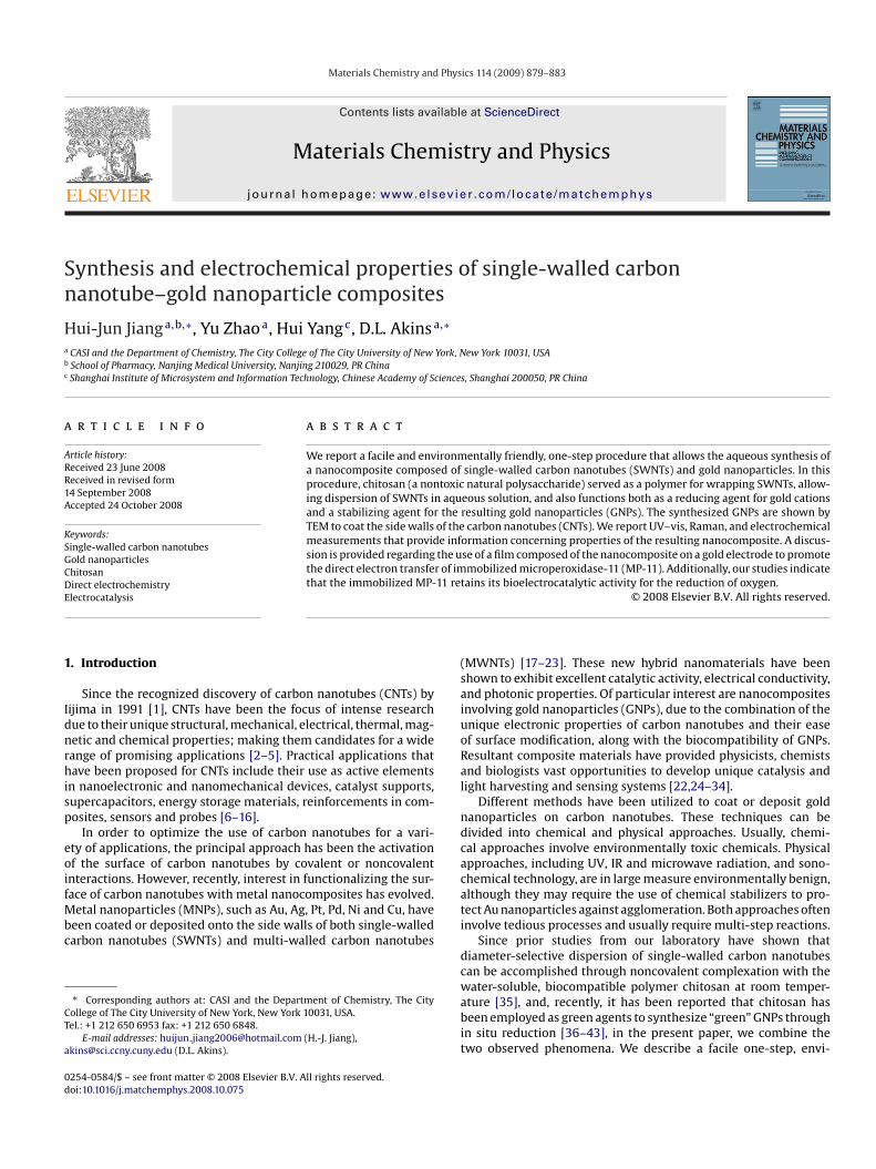
ig. 1. TEM images of (A) SWNTs dispersed in chitosan solution, where the particleynthesized in chitosan solution. The scale bars represent 100 nm.
onmentally friendly approach for formation of gold nanoparticlesttached onto the walls of single-walled carbon nanotubes, throughse of chitosan. The resulting single-walled carbon nanotube-goldanocomposites are stabilized and dispersed in aqueous chitosanolution.
UV–vis and Raman spectroscopy and transmission electronicroscopy (TEM) have been employed in the characterization of
he nanocomposites. Nanocomposite films were prepared and usedo study electrochemistry and electrocatalysis of microperoxidase-1 (MP-11).
. Experimental
.1. Chemicals and reagents
Purified HiPco SWNTs were purchased from Carbon Nanotechnologiesnc. (Houston, TX). Chitosan (85% deacetylation, average molecular weight of× 106 g mol−1) and microperoxidase-11 from horse heart cytochrome c werebtained from Sigma–Aldrich Corp., without further purification. Chloroauric acidHAuCl4·3H2O; 49.0% minimum Au) and all other chemicals were purchased fromisher Scientific. All solutions were made using deionized water.
.2. Synthesis of SWNT-gold nanocomposites
Initially, chitosan solution (1 wt%) was prepared by dissolving chitosan power in.0% (v/v) acetic acid solution with stirring for 1 h at room temperature until com-letely dispersed. In a typical composite material preparation, 10 mg SWNTs wereispersed in 20 mL of chitosan solution, with 2 h sonication and 10 min centrifuga-ion, to prepare a homogeneous dispersion of SWNT-chitosan. Subsequently, 1 mL of5 mM HAuCl4 was added to the SWNT-chitosan dispersion under vigorous stirringt room temperature for 10 min. The mixture was then heated to 80 ◦C while stirringor ca. 1 h until the color of the solutions did not change.
.3. Characterization
UV–vis absorption spectra were obtained using a Perkin-Elmer Lambda 18 spec-rophotometer over the spectral range 350–900 nm; plastic cuvettes were used.aman spectra were obtained using an HR800 Horiba Jobin Yvon Raman Microprobe.or Raman measurements, samples were placed in quartz cuvettes and excited with32.8 nm laser radiation. The morphology of the nanocomposites was observednder a Zeiss EM 902 TEM operating at an accelerating voltage of 80 kV, and samplesor inspection were prepared by slow evaporation of one drop of aqueous dispersionf the nanocomposites on a copper mesh grid coated with a carbon film.
Electrochemical measurements were performed using a CHI 630B electrochemi-al workstation (CH Instruments, Austin, TX) and a conventional three-electrode cellomprised of a platinum wire as the auxiliary electrode, a saturated calomel elec-rode as the reference electrode, and a modified gold electrode (2 mm diameter) ashe working electrode. The SWNT–GNPs/Au modified electrode was fabricated byasting 5 �L of the synthesized nanocomposite onto a freshly polished gold elec-rode. Water was then removed by evaporation, and the resulting SWNT–GNPs/Au
odified electrode was immersed overnight in 1 mmol L−1 of MP-11 solution at 4 ◦Co obtain the MP-11/SWNT–GNPs/Au modified electrode.
. Results and discussion
The mixing of chloroauric acid with the dispersed SWNTs in chi-
osan solution and the heating of the mixture to 80 ◦C initially didot result in any obvious color change. After ca. 15 min, however,he mixture changed color to wine-red, indicative of the formationf GNPs. The dispersion was cooled to room temperature after ca.h of heating. The resulting SWNT–GNPs nanocomposite solutionn are aggregated chitosan nanoparticles; (B) and (C) SWNT–GNP nanocomposites
ecame completely homogeneous and remained stable for at leastmonths when stored at 4 ◦C.
The morphologies and microstructures of the as-preparedanocomposites were investigated using TEM. Typical TEM imagesf SWNTs and SWNT–GNPs are shown in Fig. 1. When comparedith the TEM image of SWNTs dispersed in chitosan solution
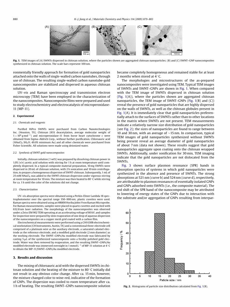
Fig. 1(A)), where the particles shown are aggregated chitosananoparticles, the TEM image of SWNT–GNPs (Fig. 1(B) and (C))eveal the presence of gold nanoparticles that are highly dispersedn the walls of SWNTs, as well as the chitosan globules present inig. 1(A). It is immediately clear that gold nanoparticles preferen-ially attach to the surfaces of SWNTs rather than to other locationsn the matrix where SWNTs are not present. TEM measurementsndicate a relatively narrow size distribution of gold nanoparticlessee Fig. 2); the sizes of nanoparticles are found to range between0 and 30 nm, with an average of ∼15 nm. In comparison, typicalEM images of gold nanoparticles synthesized without SWNTseing present reveal an average diameter of gold nanoparticlesf about 7 nm (data not shown). These results suggest that goldanoparticles aggregate upon coating onto the chitosan wrappedWNTs. Additionally, under sonification for 30 min, TEM imagingndicate that the gold nanoparticles are not dislocated from theWNTs.
Fig. 3 shows surface plasmon resonance (SPR) bands inbsorption spectra of systems in which gold nanoparticles wereynthesized in the absence and presence of SWNTs. The strongbsorptions at 521 nm (curve b) and 524 nm (curve d), respectively,re attributable to plasmon resonances of essentially isolated GNPs
Fig. 2. Histograms of particle size distribution calculated from Fig. 1(B).

H.-J. Jiang et al. / Materials Chemistry and Physics 114 (2009) 879–883 881
F solus he secr
to
turStc
STrraoAbbtn
FSpi
satRetHo
ts1rctbenzymes [50]. Fig. 5 shows cyclic voltammograms (CVs) of differ-
ig. 3. UV–vis absorption spectra for (a) the chitosan solution, (b) the colloid goldolution, and (d) the colloid gold solution synthesized in the presence of SWNTs. Tegion of 600–900 nm.
icle interactions; [44] the latter conjecture is consistent with TEMbservations.
The inset of Fig. 3, shows characteristic van Hove singulari-ies (vHSs) for dispersed SWNTs [45–47]. The vHSs are essentiallynchanged whether GNPs are absent or present (curves c and d,espectively). We can thus conclude that the electronic structures ofWNTs are preserved whether or not GNPs coat them. We can addi-ionally conclude that the presence of GNPs does not measurablyhange the dispersed character of the SWNT samples.
Fig. 4 shows the Raman spectrum of SWNTs and theWNT–GNPs nanocomposites using laser excitation at 632.8 nm.hree prominent characteristic vibrational bands are found, cor-esponding to the radial breathing mode (RBM) in the frequencyange of 200–300 cm−1, a disorder-induced D band near 1300 cm−1,nd the G band near 1600 cm−1 originating from the tangentialscillation involving the carbon atoms of the walls of SWNTs [48].s revealed in Fig. 4, no distinctive difference in Raman spectra
etween SWNTs and the composite are found (see curves a and). And, especially of note, is the fact that the ratio of intensi-ies of D and G bands is unperturbed, despite the presence of Auanoparticles in the composite. We can conclude that our synthe-ig. 4. Raman spectra for samples dispersed in chitosan solution: (a) SWNTs; (b)WNT–Au nanocomposites. The insets show spectral regions that have been multi-lied by a factor for ease of viewing; the top and bottom curves, respectively, in the
nsets correspond to spectral regions in (a) and (b).
epp
F1a
tion synthesized in the absence of SWNTs, (c) the SWNTs suspension in chitosanond panel shows magnified UV–vis absorption spectra of curves (c) and (d) in the
is procedure causes little or no damage to the structure of SWNTsnd does not significantly alter the basic electronic properties ofhe SWNTs in the composites [49]. The absence of any enhancedaman signal that might be attributable to a surface-enhancementffect (i.e., surface-enhanced Raman scattering (SERS)) results fromhe lack of a near resonance between the 632.8 nm frequency of theeNe excitation source and the peak plasmon resonance frequencyf 512 nm for the gold nanoparticles.
Our next aim in the present study was to explore the use ofhe composite as a substrate for enzymatic reactions at electrodeurfaces. As a first example, we immobilized microperoxidase-1 to assess its electrochemical properties. MP-11 is a small-sizeedox enzyme obtained by proteolytic digestion of horse heartytochrome c, and because of the relative simplicity of its struc-ure, it has been widely used as a good model to mimic theehavior of macrobiomolecules such as hemoproteins and some
nt modified electrodes in 0.1 M N2-saturated and O2-saturatedhosphate buffer (pH 7.0) at a scan rate of 100 mV s−1. Com-ared with curve a (the SWNT–GNPs/Au electrode), a pair of
ig. 5. Cyclic voltammograms of (a and c) SWNT–GNPs/Au and (b and d) MP-1/SWNT–GNPs/Au electrodes in 0.1 M N2-saturated (a and b) and O2-saturated (cnd d) phosphate buffer (pH 7.0) at scan rate of 100 mV s−1.
8 istry a
wM0rtp−ppeit
tcagOSitd
fSSrewfoilSmiMd
msnnSfinsaioehmS
TEp
M
MMM
tf
A
iguCDNKf
R
[
[[[
[[[[
[[[[[
[
[[
[
[[
[[[
[
[
82 H.-J. Jiang et al. / Materials Chem
ell-defined and nearly symmetrical redox peaks appeared at theP-11/SWNT–GNPs/Au electrode in the potential range of −0.7 to
.2 V (curve b). The electrochemical response, attributable to theedox center of the immobilized MP-11, indicates a direct elec-ron transfer (DET) between MP-11 and the electrode. The anodiceak potential (Epa) and cathodic peak potential (Epc) are located at0.304 and −0.356 V at a scan rate of 100 mV s−1, respectively. Theeak potential separation (�Ep) is about 52 mV, with the formalotential (E◦′) equal to −0.330 V. The anodic peak current is almostqual to that of the cathodic peak, indicating that MP-11 moleculesmmobilized at the modified electrode undergo a reversible elec-rochemical reaction.
The electrocatalytic reduction of oxygen at the modified elec-rode was also investigated. As also shown in Fig. 5, a markedatalytic reduction current occurred at both the SWNT–GNPs/Aund the MP-11/SWNT–GNPs/Au electrodes in the presence of oxy-en (curves c and d, respectively) as compared to that without2 (see curves a and b, respectively). One notes that although theWNT–GNPs/Au electrode catalyzes the reduction of O2 (curve c)n phosphate buffer, the catalytic current is significantly less thanhat for the MP-11/SWNT–GNPs/Au electrode (as shown in curve).
Table 1 compares the electrochemical parameters obtainedrom the CV responses of MP-11 at GNPs/Au, SWNT/Au andWNT–GNPs/Au electrodes. For MP-11 immobilized on theWNT–GNPs nanocomposite electrode, the peak potential sepa-ation (�Ep) was greatly decreased compared to the other twolectrodes and the formal potential shifted positively ca. 60 mV,hich implied that the immobilized MP-11 may require less energy
or direct electron transfer with the electrode compared to thether cases. Our results suggest that the modified nanocompos-te electrode possesses extremely good electrochemical properties,ikely because of a synergistic effects attributed to its constituents.pecifically, we might expect that the GNPs provide a naturalicroenvironment for protein immobilization and reduces the
nsulating effect of peptide shell on the direct electron transfer ofP-11; also, SWNTs may act as tiny conduction centers to facilitate
irect electron transfer [51,52].In summary, this investigation reports a facile and environ-
entally friendly, one-step procedure that allows the aqueousynthesis of a nanocomposite composed of single-walled carbonanotubes and gold nanoparticles. In this procedure, chitosan (aontoxic natural polysaccharide) served as a polymer for wrappingWNTs, allowing dispersion of SWNTs in aqueous solution, and alsounctions both as a reducing agent for gold cations and a stabiliz-ng agent for the resulting gold nanoparticles. Importantly, goldanoparticle-coated SWNTs were obtained, without additionalteps involving, for example, oxidizing the SWNTs with mixedcids, or introducing other chemical reducing agents and protect-ng agents for GNPs. Our approach leads to the “green” synthesisf a nanocomposite, which may significantly improve the direct
lectron transfer and electrocatalysis of enzymes. In particular, weave examined electron transfer involving microperoxidase–11 onodified gold electrodes, and found significant improvement forWNT–GNP nanocomposite electrodes. Experiments are underway
able 1lectrochemical parameters of MP-11 modified electrodes in 0.1 M N2-saturatedhosphate buffer (pH 7.0).
odified electrode Epa (V)a Epc (V) �Ep (mV) E◦′(V)
P-11/SWNT/Au −0.450 −0.354 100 −0.402P-11/GNPs/Au −0.453 −0.318 135 −0.386P-11/SWNT–GNPs/Au −0.356 −0.304 52 −0.330
a Scan rate: 100 mV s−1.
[[[[[[
[
[[[[[
[
[
nd Physics 114 (2009) 879–883
o extend the potential applications of the nanocomposites in bio-uel cells and biosensors.
cknowledgments
DLA thanks the NSF and DoD-ARO for support of this work,n part, through the following awards: (1) the NSF-IGERT pro-ram under grant DGE-9972892; (2) the NSF-MRSEC programnder grant DMR-0213574; the (3) NSF-NSEC program under grantHE-0641523; and (4) DoD-ARO under Cooperative AgreementAAD19-01-1-0759 and grant W911NF-04-1-0029. HY thanks theational Natural Science Foundation of China (20673136) andnowledge Innovation Engineering of Chinese Academy of Sciences
or support of this work.
eferences
[1] S. Iijima, Nature 354 (1991) 56.[2] S.B. Sinnott, R. Andrews, Crit. Rev. Solid State Mater. Sci. 26 (2001) 145.[3] G. Mamalis, L.O.G. Vogtlander, A. Markopoulos, Precis. Eng. 28 (2004) 16.[4] N. Grobert, Mater. Today 10 (2007) 28.[5] M. Paradise, T. Goswami, Mater. Des. 28 (2007) 1477.[6] R.H. Baughman, A.A. Zakhidov, W.A. de Heer, Science 297 (2002) 787.[7] P. Avouris, Z.H. Chen, V. Perebeinos, Nat. Nanotechnol. 2 (2007) 605.[8] P. Serp, M. Corrias, P. Kalck, Appl. Catal. A: Gen. 253 (2003) 337.[9] G. Lota, K. Lota, E. Frackowiak, Electrochem. Commun. 9 (2007) 1828.10] M. Jorda-Beneyto, F. Suarez-Garcia, D. Lozano-Castello, D. Cazorla-Amoros, A.
Linares-Solano, Carbon 45 (2007) 293.11] B. Mahar, C. Laslau, R. Yip, Y. Su, IEEE Sens. J. 7 (2007) 266.12] N. Chopra, V.G. Gavalas, L.G. Bachas, B.J. Hinds, Anal. Lett. 40 (2007) 2067.13] G.A. Rivas, M.D. Rubianes, M.C. Rodriguez, F.N.F. Erreyra, G.L. Luque, M.L.
Pedano, S.A. Miscoria, C. Parrado, Talanta 74 (2007) 291.14] M. Moniruzzaman, K.I. Winey, Macromolecules 39 (2006) 5194.15] B.S. Harrison, A. Atala, Biomaterials 28 (2007) 344.16] A.M.K. Esawi, M.M. Farag, Mater. Des. 28 (2007) 2394.17] V. Georgakilas, D. Gournis, V. Tzitzios, L. Pasquato, D.M. Guldie, M.J. Pratodf,
Mater. Chem. 17 (2007) 2679.18] D. Wang, Z.C. Li, L.W. Chen, J. Am. Chem. Soc. 128 (2006) 15078.19] S. Hrapovic, Y.L. Liu, K.B. Male, J.H.T. Luong, Anal. Chem. 76 (2004 1083).20] B. Yoon, C.M. Wai, J. Am. Chem. Soc. 127 (2005) 17174.21] Y.T. Kim, T. Mitani, J. Catal. 238 (2006) 394.22] Z.J. Wang, M.Y. Li, Y.J. Zhang, J.H. Yuan, Y.F. Shen, L. Niu, A. Ivaska, Carbon 45
(2007) 2111.23] T.M. Day, P.R. Unwin, N.R. Wilson, J.V. Macpherson, J. Am. Chem. Soc. 127 (2005)
10639.24] B.S. Kong, D.H. Jung, S.K. Oh, C.S. Han, H.T. Jung, J. Phys. Chem. C 111 (2007) 8377.25] A. Star, V. Joshi, S. Skarupo, D. Thomas, J.C.P. Gabriel, J. Phys. Chem. B 110 (2006)
21014.26] A.V. Ellis, K. Vijayamohanan, R. Goswami, N. Chakrapani, L.S. Ramanathan, P.M.
Ajayan, G. Ramanath, Nano Lett. 3 (2003) 279.27] L.Q. Jiang, L. Gao, Carbon 41 (2003) 2923.28] K. Kim, S.H. Lee, W. Yi, J. Kim, J.W. Choi, Y. Park, J.I. Jin, Adv. Mater. 15 (2003)
1618.29] R.Y. Zhang, X.M. Wang, Chem. Mater. 19 (2007) 976.30] J.X. Li, H. Grennberg, Chem. Eur. J. 12 (2006) 3869.31] M.S. Raghuveer, S. Agrawal, N. Bishop, G. Ramanath, Chem. Mater. 18 (2006)
1390.32] K.N. Lin, T.Y. Yang, H.M. Lin, Y.K. Hwu, S.H. Wu, C.K. Lin, Chin. Particuol. 5 (2007)
237.33] J. Shi, Z. Wang, H.L. Li, J. Nanopart. Res. 8 (2006) 743.34] X.G. Hu, T. Wang, X.H. Qu, S.J. Dong, J. Phys. Chem. B 110 (2006) 853.35] H. Yang, S.C. Wang, P. Mercier, D.L. Akins, Chem. Commun. (2006) 1425.36] S.J. Guo, E.K. Wang, Anal. Chim. Acta 598 (2007) 181.37] K. Esumi, N. Takei, T. Yoshimura, Colloids Surf. B: Biointerf. 32 (2003) 117.38] H.Z. Huang, X.R. Yang, Biomacromolecule 5 (2004) 2340.39] B. Wang, K. Chen, S. Jiang, F. Reincke, W.J. Tong, D.Y. Wang, C.Y. Gao, Biomacro-
molecules 7 (2006) 1203.40] D.S. dos Santos, P.J.G. Goulet, N.P.W. Pieczonka, O.N. Oliveira, R.F. Aroca, Lang-
muir 20 (2004) 10273.41] T. Miyama, Y. Yonezawa, Langmuir 20 (2004) 5918.42] D.W. Wei, W.P. Qian, Acta Chim. Sinica 65 (2007) 379.43] H.Z. Huang, X.R. Yang, Carbohyd. Res. 339 (2004) 2627.44] T. Wang, X.G. Hu, X.H. Qu, S.J. Dong, J. Phys. Chem. B 110 (2006) 6631.
45] M.J. O’Connell, S.M. Bachilo, C.B. Huffman, V.C. Moore, M.S. Strano, E.H. Haroz,K.L. Rialon, P.J. Boul, W.H. Noon, C. Kittrell, J.P. Ma, R.H. Hauge, R.B. Weisman,R.E. Smalley, Science 297 (2002) 593.
46] S.M. Bachilo, M.S. Strano, C. Kittrell, R.H. Hauge, R.E. Smalley, R.B. Weisman,Science 298 (2002) 2361.
47] C.A. Mitchell, R. Krishnamoorti, Macromolecules 40 (2007) 1538.

[[[
H.-J. Jiang et al. / Materials Chemistry a
48] M.S. Dresselhaus, G. Dresselhaus, A. Jorio, J. Phys. Chem. C 111 (2007) 17887.49] R.J. Graupner, Raman Spectrosc. 38 (2007) 673.50] H.J. Jiang, X.H. Huang, X.F. Wang, X. Li, W. Xing, X.L. Ding, T.H. Lu, J. Electroanal.
Chem. 545 (2003) 83.
[
[
nd Physics 114 (2009) 879–883 883
51] J. Manso, M.L. Mena, P. Yanez-Sedeno, J. Pingarron, J. Electroanal. Chem. 603(2007) 1.
52] Y. Liu, M.K. Wang, F. Zhao, Z.H. Guo, H.J. Chen, S.J. Dong, J. Electroanal. Chem.581 (2005) 1.