synthesis and functionalization of nanoparticles with
TRANSCRIPT

Synthesis and Functionalization of Nanoparticles
with Biogenic Amines and
their Biological Application
Synthese und Funktionalisierung von Nanopartikeln mit
biogenen Aminen und deren biologische Anwendung
Dissertation zur Erlangung des Doktorgrades
der Naturwissenschaftlichen Fachbereiche
im Fachgebiet Anorganische und Analytische Chemie
der Justus-Liebig-Universität Gießen
vorgelegt von
Friederike Britta Gasiorek
aus
Wehrheim
Gießen 2016


Diese Arbeit wurde im Zeitraum von Januar 2013 bis Februar 2016 am Institut für Anorganische
und Analytische Chemie der Justus-Liebig-Universität Gießen angefertigt, unter der Betreuung von
Prof. Dr. Sabine Schlecht begonnen und unter Herr Prof. Dr. Siegfried Schindler und Herr Prof. Dr.
Mathias S. Wickleder beendet.
Erstgutachter: Prof. Dr. Siegfried Schindler
Zweitgutachter: Prof. Dr. Mathias S. Wickleder


Für meinen Vater


„Our greatest weakness lies in giving up. The most certain way to succeed is always to try just one
more time.”
Thomas A. Edison


Table of Content
Table of Content
1. Introduction ................................................................................................................................. 1
2. Basic Knowledge ........................................................................................................................ 3
2.1 Nanomaterials ..................................................................................................................... 3
2.2 Metal Nanoparticles ............................................................................................................ 8
2.1.1 Synthesis of Coinage Metal Nanoparticles ................................................................. 8
2.1.2 Optical Properties of Gold Nanoparticles ................................................................... 9
2.3 Semiconductor Nanoparticles ........................................................................................... 11
2.4 Surface Functionalization .................................................................................................. 16
2.5 Characterization ................................................................................................................ 20
2.6 Nanoparticles in Biological Systems ................................................................................ 22
2.7 Multivalent Ligands .......................................................................................................... 24
3. Motivation and Goals ................................................................................................................ 27
4. Gold Nanoparticles.................................................................................................................... 29
4.1 Syntheses of Gold Nanoparticles ...................................................................................... 29
4.2 Optical Properties .............................................................................................................. 33
4.3 Alloyed Metal Nanoparticles ............................................................................................ 34
4.4 Ligand Exchange Reactions at Gold Nanoparticles .......................................................... 35
4.5 Gold Nanoparticles with Positively Charged Ligands ...................................................... 42
4.6 Mixed Ligand Shells on Gold Nanoparticles .................................................................... 45
4.7 Functionalization at the Ligand Periphery ........................................................................ 47
4.8 Functionalization with Bioactive Amines ......................................................................... 49
4.9 Biological Functionality .................................................................................................... 57
4.10 Effects of Multivalent Histamine Supported on Gold Nanoparticles: Activation of
Histamine Receptors by Derivatized Histamine at Subnanomolar Concentrations ...................... 62
5. Quantum Dots ........................................................................................................................... 79
5.1 Quantum Dots for Cellular Imaging ................................................................................. 79
Table of Content
5.2 The Ternary System CdTexSe1-x-MPA .............................................................................. 88
6. Summary/Zusammenfassung .................................................................................................... 93
6.1 Summary ........................................................................................................................... 93
6.2 Zusammenfassung ............................................................................................................. 95
7. Outlook ...................................................................................................................................... 99
8. Experimental Section .............................................................................................................. 101
8.1 Chemicals, Solvents and Consumables ........................................................................... 101
8.2 Analytics .......................................................................................................................... 101
8.3 Synthesis of the Nanoparticles ........................................................................................ 104
8.4 Ligand Exchange Reactions ............................................................................................ 107
8.5 Dithiocarbamate Functionalized Gold Nanoparticles ...................................................... 109
8.6 Positively Charged Gold Nanoparticles .......................................................................... 110
8.7 Mixed Ligand Shell ......................................................................................................... 111
8.8 Functionalization of the Particles Shell ........................................................................... 112
8.9 Synthesis of the Ligands .................................................................................................. 115
8.10 Synthesis of Quantum Dots ............................................................................................. 118
9. References ............................................................................................................................... 123
10. Appendix ............................................................................................................................. 135
10.1 Abbreviations and Symbols ............................................................................................. 135
10.2 Calculation of core diameters .......................................................................................... 136
10.3 List of Figures ................................................................................................................. 137
10.4 List of Tables ................................................................................................................... 142
11. Publications and Presentations ............................................................................................ 143
12. Lebenslauf ........................................................................................................................... 144
13. Acknowledgement ............................................................................................................... 145
14. Erklärung ............................................................................................................................. 147

Introduction
1
1. Introduction
Nowadays nanotechnology is omnipresent: from nanodevices in technology to nanorobots for
medicine, a lot of materials have found their way into our daily life. Sunscreens based on
nanoparticles made of titanium dioxide or zinc oxide or clothes with nanosilver, which are able to
kill bacteria and eradicate unwanted odor are only two examples of nanomaterials making our daily
life more comfortable. A special feature of nanotechnology is its cross section character. It contains
pulses from almost every scientific-technical disciplines and in many branches this promising
technology is indispensable. Accordingly the importance of nanotechnology increases steadily.
While nanomaterials already enhance the properties of many products in present life, future
techniques are under development. What Ostwald described in 1914 as “The world of neglected
dimensions” is today known as nanochemistry. [1] He saw the opportunities, new properties and
interesting application of these materials.
Even though it is a relatively young topic, nanotechnology has become a widely inquired branch of
science. The number of publications with “nanoparticles” as buzzword exponentially increased
during the last decades. In 2014 a special issue for Nanotechnology & Nanomaterials,
Nanotoxicology & Nanomedicine appeared in Angewandte Chemie Int. Ed. summarizing the
versatile applications. [2]
The term nano emanates from the Greek word for dwarf and is on prefix at the metric scale for the
factor 10-9
(1 nm = 10-9
m). For comparison, the diameter of a human hair is about a thousand times
larger but still visible to the naked eye, red blood cells are even smaller and the size of a virus is
about 20 nm to 100 nm (Figure 1). Objects with a size less than 100 nm are typically called
nanomaterials.
Figure 1: Length scale for classification of nanomaterials. Adapted from [3]

Introduction
2
Besides the size, their large surface area is characteristic for these materials compared to the bulk
material and the physicochemical properties are directly dependent. The surface properties are
characteristic for nanomaterials and due to the increased surface-to-volume ratio they are different
compared to their bulk material. Nanoparticles resemble both bulk material and molecules, they
combine the ability to move (molecule) with specific properties of solids, e. g. catalytic activity,
magnetism, surface.
The interaction of nanoscale materials with biological systems is currently the focus of a fast-
growing area of investigation and progress in nanomedicine increased rapidly in the last years. As
these interactions are mostly governed by the surface of the particles, modifications of those can
tune the properties. Nanoparticulate materials are in a comparable size range relative to proteins or
enzymes, they can selectively intervene in cellular processes in a way that small molecules cannot.
Engineered nanoparticles have shown versatile applications for e. g. drug delivery. Compared to
conventional therapeutics nanoparticles can overcome problems like poor solubility, lack or
targeting or non-specific distribution. Remarkable progress has been made in the development of
new nanomaterials with enhanced water-solubility, bioavailability and reduced toxicity. Although
nanomaterials are currently widely used, there is a lack of information concerning environmental
implications and it is necessary to spend investigations on the behavior. Gold nanoparticles haven
been used for biomedical applications since their colloidal synthesis. Because of their high
biocompatibility they have versatile applications. A lot of synthetic approaches have been
developed to prepare size- and shape-controlled particles. Future challenges will be to find new
methods of functionalizing gold nanoparticles with compounds that involve sufficient
biocompatibility and are efficient pharmaceutical agents.

Basic Knowledge
3
2. Basic Knowledge
2.1 Nanomaterials
The development of colloidal chemistry is closely related to the advances in methods and
techniques and colloidal gold for example is no invention of modern times. It was Michael Faraday
who reported on the earliest experimental studies on noble metals and their interactions with light
giving a first description of colloidal solutions. [4] He reduced tetrachloroaurate (AuCl4-) with
phosphorous in CS2 and obtained a deep red solution. He described it as finely disperse gold, not
knowing that the optical phenomenon was caused by nanoparticles. Scientists assumed much earlier
that gold must be present in such a degree that it is not visible to the human eye. [5] The term
colloid, from the French word colle (glue), was introduced by Thomas Graham in 1861. [6] Some
colloidal phenomena are known unconsciously since applications in ancient times, e. g. inks or
cosmetics and manufacturing of colored glass based on dispersions of pigments. The Lycurgus cup
is a fascinating example for nanomaterials from ancient times (Figure 2). Dated back in the 4th
century B.C. it is a masterpiece of handcraft for this time. It appears in different colors depending of
the incident of light; if it is reflected it appears jade green, if it is transmitted it appears ruby red.
The material consists of metal nanoparticles of gold and silver. [7] Another example for
nanomaterials in daily use is the Purple of Cassius. It was popular in the 17th century as colorant for
glasses consisting of gold particles and tin oxide. [8]
Figure 2: The Lycurgus cup contains nanoparticles of gold and silver (British Museum). [9]

Basic Knowledge
4
At the nano-scale not only miniaturization but also changes in properties are possible. The TiO2,
which is applied as UV/Vis protector in sunscreens, is only about 50 nm in diameter. Absorption of
solar radiation is much higher in materials composed of nanoparticles than it is in thin films. At this
size it appears no longer white but transparent and can be applied to the skin.
Nanoparticles with a diameter roughly between 10 nm and 100 nm are natural bridges between
molecules and extended solids and are also called advanced molecules. They combine the molecular
ability to move with properties of bulk material, like fluorescence. Besides their size, the vast
surface area is also characteristic. Nanoparticles possess a high surface-to-volume ratio. For
example 10 nm nanoparticles have about 20% of the atoms at the surface, whereas for 2 nm
nanoparticles this value increases up to 80% surface atoms. These surface atoms are different in
energy and mainly govern the properties of the material. [3] At the nanoscale the electronic energy
states become discrete to unique optical, electronical and mechanical properties of the materials.
Thus miniaturization has drastic effects for the physical and chemical properties. Not only for metal
nanoparticles, but for all nanomaterials the characteristics at the submicroscopic scale are different.
Gold as bulk material appears with a metallic glance, gold at the nanosize appears as a purple
solution (Figure 3). In addition gold nanoparticles melt at much lower temperatures (~300 °C for
2.5 nm size) than the gold slabs (1064 °C). This is due to the increasing number of surface atoms.
Solid gold is inert against oxygen and is used for jewelry or tooth crowns. When presented in
nanostructures it is very affine to oxygen and therefore applied as catalyst. The optical appearance
at the nanoscale is also no longer metallic but red caused by quantum effects.
Figure 3: Gold as bulk material and dispersed in water as colloids.
Quantum effects are based on the fact that electrons behave both as particle and as wave resulting in
the wave-particle-duality. When electrons are trapped in a space similar to their wavelength their
energy levels are no longer continuous but discrete. The values depend on the size and shape of the
particle. Other size-dependent property changes include quantum confinement in semiconductor

Basic Knowledge
5
particles (CdSe, CdTe), surface plasmon resonance in some metal nanoparticles (Au, Ag) and
superparamagnetism in magnetic materials (Fe2O3, Fe3O4).
Within a colloidal system, nanoparticles are uniformly dispersed in an agent. Classification is made
in dispersion, molecular and association colloids. Dispersion colloids are thermodynamically
instable systems consisting of crystalline or amorphous solids in a liquid agent. Micelles and
biocolloids belong to the category of associative colloids. The stabilization force of dispersion
colloids basically derives from the repulsive interactions of the charge distribution around the
particles or a stabilizing shell. [10] In this context colloid describes the characteristics and nano
gives the indication for size and quantum effects.
Nanoparticles can be understood as a small unit of a solid, accordingly they can be synthesized by
two different routes. While in the top-down method a macrocrystalline material is crushed down to
nanoscale under mechanical action, in the bottom-up method nanoscopic structures are built from
atomic components (Figure 4).
Figure 4: Top-down and bottom-up approach for the synthesis of nanoparticles.
In a top-down approach usually the nanoparticulate material is generated from the bulk material by
physical methods, e. g. lithography. Contrastingly, bottom-up methods have a chemical approach.
The bottom up synthesis starts with the nucleation step, and then the surface is saturated with
further atoms (surface growth). The growth mechanism has to be stopped at the nanoscale and the
Bottom-Up
Top-Down
Micrometer
10-6
m
Nanometer
10-9
m
Atomic
Bulk
Powder
Nanoparticle
Cluster
Atom
Crushing down of
macroscopic structures e. g. lithography, ball-milling physical
Synthesis of nanostructures
from molecular compounds, e. g. colloidal synthesis
chemical
Basic Knowledge
6
resulting nanoparticles have to be stabilized to avoid further particle growth. Nanomaterials are
synthesized from molecular/atomic precursors usually in solution by self-organization. The growth
mechanism can be controlled during the synthesis. The nanoparticles need to be stabilized at the
surface to prevent coalescence.
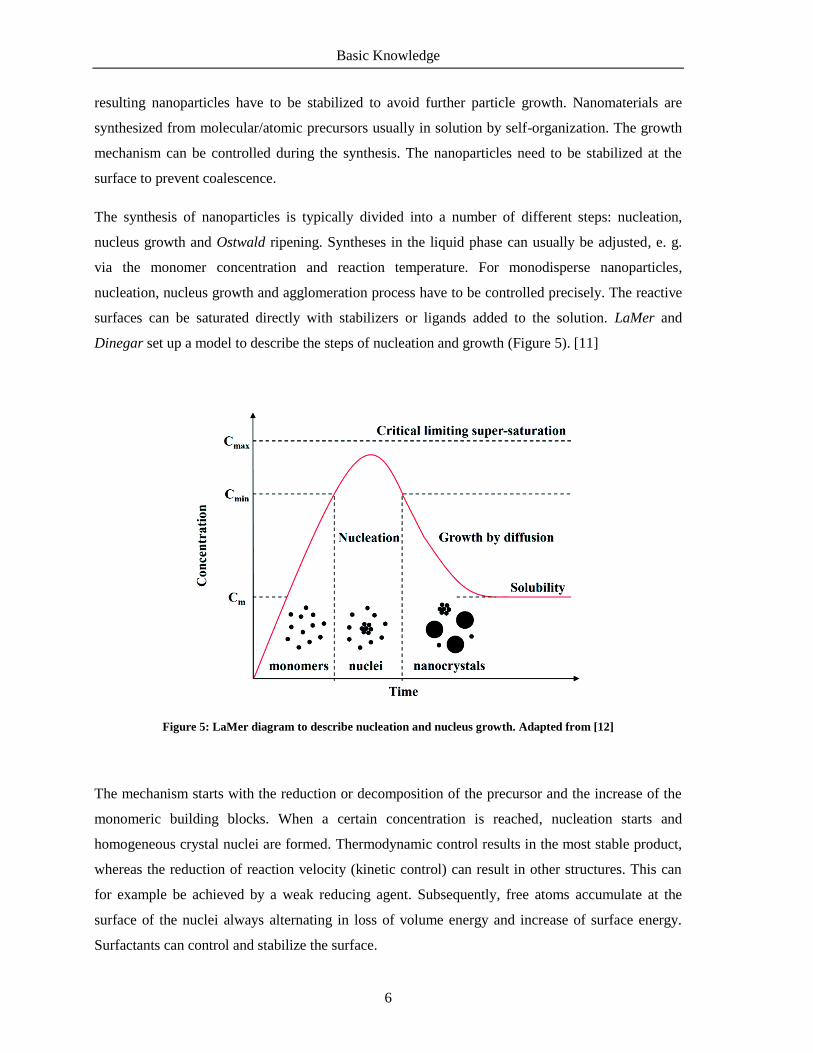
The synthesis of nanoparticles is typically divided into a number of different steps: nucleation,
nucleus growth and Ostwald ripening. Syntheses in the liquid phase can usually be adjusted, e. g.
via the monomer concentration and reaction temperature. For monodisperse nanoparticles,
nucleation, nucleus growth and agglomeration process have to be controlled precisely. The reactive
surfaces can be saturated directly with stabilizers or ligands added to the solution. LaMer and
Dinegar set up a model to describe the steps of nucleation and growth (Figure 5). [11]
Figure 5: LaMer diagram to describe nucleation and nucleus growth. Adapted from [12]
The mechanism starts with the reduction or decomposition of the precursor and the increase of the
monomeric building blocks. When a certain concentration is reached, nucleation starts and
homogeneous crystal nuclei are formed. Thermodynamic control results in the most stable product,
whereas the reduction of reaction velocity (kinetic control) can result in other structures. This can
for example be achieved by a weak reducing agent. Subsequently, free atoms accumulate at the
surface of the nuclei always alternating in loss of volume energy and increase of surface energy.
Surfactants can control and stabilize the surface.

Basic Knowledge
7
Figure 6: Growth and stabilization of nanoparticles. Adapted from [3]
As colloidal solutions are thermodynamically instable and aim for a stable, state the surface must be
immediately saturated. This process can be suppressed by stabilization. This can either be achieved
sterically or electrostatically (Figure 6). Within a colloidal system there are repulsive and attractive
forces. Attractive actions result from dipole-dipole interactions and are also called van-der-Waals-
forces. Repulsion results from electrostatic interactions from charged particles. Dispersed particles
in a medium have a charge at their surface called charge cloud. It is symmetrically placed around
the particle. When the particles move through a homogeneous phase the static and a part of the
diffuse layer move along. Within this motion a potential barrier occurs between the layer and the
surrounding medium. It increases linearly in the Stern-layer, a rigid layer around the particle, and
falls exponentially in the diffuse layer. The resulting difference is called zeta-potential, which is a
characteristic value for the stability of colloidal solutions and can be measured by electrophoresis or
streaming potentials. It depends on the properties of the surface as well as of the solvent. Ligands
surrounding the nanoparticles must be able to stabilize the colloidal solution either electrostatically
or sterically. This can be achieved by surface tailoring. The stabilization of nanoparticles is
described in the literature with various methods, mostly using polymers. A proper monolayer is
often needed to shield the surface. The surface energy is decreased, which reduces the formation of
aggregates.

Basic Knowledge
8
2.2 Metal Nanoparticles
Compared to bulk metals, metallic nanoparticles have different properties due to their size and thus
the resulting high surface-to-volume ratio. Nanostructured materials of coinage metals such as gold
and silver have unique optical properties due to strong surface plasmon absorption in the visible
region of light. [13] They have versatile applications in modern research and technology and are no
longer indispensable. They are applied in catalysis, electronic compounds, sensors or medicinal
devices. [14, 15] Their properties are based on physical parameters and can easily be tuned. Size,
shape and structure are the mainly contributing parameters.
2.1.1 Synthesis of Coinage Metal Nanoparticles
Various synthetic procedures have been published. In the majority of reports, HAuCl4 proved to be
the favored starting material due to its ready availability, despite the disadvantage of limited control
over nucleation process and possible chloride contamination in the resulting particles. This method
was first described by Turkevich and enhanced by Frens. [16, 17] It is based on the chemical
reduction of tetrachloroaurate with citrate in aqueous solution. Citrate is both reducing agent and
ligand for the stabilization of the formed nanoparticles. Nanoparticles with a diameter between
10 nm and 100 nm can be prepared. Frens also showed that the colloidal stability strongly depends
on the size of the nanoparticles. [18] Mechanistic studies on the formation of the nanoparticles
showed that in the synthesis acetone is formed and stabilizes the surface. If NaBH4 is used as
reducing agent different particle sizes could be obtained and the surface can be covered with
different ligands added during the synthesis. The reduction of AuCl4- with NaBH4 in a two-phase
system was described by Brust and Schiffrin. [19, 20] Nanoparticles of Au, Ag, Pt, and Ir in toluene
can be readily synthesized using a biphasic reduction procedure. [21–23] A noble metal salt (e. g.,
HAuCl4) dissolved in water is first extracted into an organic phase with the help of a phase transfer
reagent, tetraoctylammonium bromide (TOAB). Upon reduction with NaBH4 it is possible to obtain
uniform-sized metal nanoparticles. Small gold nanoparticles with a diameter of <10 nm can be
synthesized in organic solvents. 4-Dimethylaminopyridine (DMAP) is often used for the
stabilization of the resulting nanoparticles in aqueous solution. [24, 25]
Stucky and co-workers developed a method for the reduction of a gold salt in organic solvents using
an amine-borane complex and n-dodecanethiol for stabilization. [26] The resulting nanoparticles

Basic Knowledge
9
have a narrow size distribution and are nearly spherical. Reaction time, temperature, molar ratios,
and solvents can be varied to adjust the size of the gold nanoparticles. In addition, other metal
nanoparticles like Ag, Pd, and alloy compositions can be synthesized. Furthermore, the synthesis
can be easily upscaled to get multigrams of nanoparticles for applications. Gold nanoparticles
smaller than ~1.5 nm consist of a distinct number of atoms and the compounds are called clusters.
These gold clusters can either behave like discrete molecules or exhibit properties of nanoparticles,
depending on the number of atoms. The well-known Schmid-cluster Au55 consists of a phosphine
stabilized gold cluster Au55(PPh3)12Cl6 with a stable closed shell. It is a molecule with a well-
defined formula weight, unlike the colloidal solutions. Different to nanoparticles they have no
weakly bound ligands on the surface. In cluster structures metal-metal bonding interactions can be
found. [27] Apart from spherical nanoparticles, other shapes of gold nanostructures can be
synthesized. Nanorods can be prepared by a seeding growth mechanism. [28] Hollow gold
nanospheres can be prepared by sacrificial galvanic replacement of cobalt nanoparticles. [29]
Colloidal stability of the nanoparticles is the key challenge in synthesis and can be controlled by the
ligands at the surface. Weakly capped nanoparticles agglomerate faster than those with strongly
bound ligands. Citrate-capped gold nanoparticles are stabilized via weakly physisorption of citrate
ions, whereas dodecanethiol-stabilized gold nanoparticles are stabilized via strongly chemisorbed
thiolate ligands. [30] This thiol stabilization of gold nanoparticles and the self-assembly of organic
sulfur compounds on gold surfaces was first reported by Giersig and Mulvaney and gave access to
highly stable colloidal solutions. [31]
2.1.2 Optical Properties of Gold Nanoparticles
A characteristic of all gold colloids is the color, which can vary from light red via purple-red to
blueish-red. The optical properties of gold nanoparticles, first described and attributed to those by
Faraday in the 19th century, are unique and caused by quantum sized effects. The color results from
absorption and scattering of electromagnetic irradiation by local surface plasmon resonance, a
collective oscillation of the free electrons in the conduction band. The expression plasmon consists
of the physical terms for plasma and vibration. For gold nanoparticles with different core size, the
absorption maximum shifts. For larger nanoparticles it shifts to a longer wavelength, meaning lower
energies (Figure 7). The maxima are also broadened. It is also possible to determine the particle size
via the correlation with λmax of the surface plasmon. [32] This phenomenon is very characteristic for
both gold and silver nanoparticles, which appear red and yellow, respectively.

Basic Knowledge
10
Figure 7: UV/Vis spectra (normalized) for Au nanoparticles with different core sizes in aqueous solution.
Adapted from [32]
When small spherical metallic nanoparticles are irradiated by light, the oscillating electric field
causes the conduction electrons to oscillate coherently. The displaced electron cloud has a restoring
force which arises from the Coulomb attractions between negatively charged electrons and
positively charged nuclei (Figure 8). An oscillation caused by the displacement appears relative to
the nuclei framework. When incoming light enters into resonance with the vibration of the electron
cloud an amplification proceeds and the absorption is intensified.
The frequency is related to the size and shape of the resonance body and the density and effective
mass of electrons. Thus metal nanoparticles of different sizes appear in different colors. For gold
nanoparticles the maximum lies at around 520 nm, for silver nanoparticles at 420 nm with respect to
a particle size of 20 nm.
Figure 8: Plasmon resonance: scheme for oscillation of the electron cloud at a spherical nanoparticle.
Adapted from [33]
Au Au

Basic Knowledge
11
2.3 Semiconductor Nanoparticles
From a historical perspective the research on semiconductor-based nanomaterials is much younger
than those based on metals, but many principles from metal nanoparticles could be transferred. In
the beginning of the 1980s CdS colloids were synthesized and analyzed by Grätzel, Brus and
Henglein. [34–36] A few years later the quantum size-effect has been introduced by Brus. [37] It
describes the relation of size and electronic band gap and is based on a strong confinement of
electrons and holes in a small particle with a diameter below the exciton Bohr radius, the average
diameter between electron and hole. As the size is reduced the electronic excitation shifts to higher
energies. It means that the larger the core diameters of semiconductor nanoparticles the lower the
energy of the emitted light and the longer the wavelength. [38]
Semiconductor nanoparticles between 2 nm and 10 nm are quasi zero-dimensional single crystals
and are often called quantum dots (QDs). These colloidal nanocrystals exhibit strong fluorescence
with a size-dependent emission wavelength. The energy states become discrete (Figure 9). They
possess unique optical properties and are often used in electronic and optical devices. QDs are
applied for LEDs and solid-state lighting, displays, photovoltaics, transistors, quantum computing,
medical imaging, biosensors, among many others.
Figure 9: Density of states in a semiconductor crystal in dependence of the dimension. Adapted from [38]
The optical properties are a result of the quantum confinement effect and depend on size, shape and
composition of the nanocrystals. Most common quantum dots are CdTe and CdSe. When the core
Den
sity
of
Sta
tes
Energy
3 D
2 D
1 D
0 D

Basic Knowledge
12
diameter of the QDs is smaller than the exciton Bohr radius, the energy levels become discrete and
recombination on the band gap causes emission. Larger QDs emit longer wavelengths (Figure 10).
For both materials at the nanoscale the emission wavelength lies in the visible part of the
electromagnetic spectrum making these materials suitable for imaging of biological tissues. [39],
[40] The photo- and electroluminescence properties can be fine-tuned to emit any color of light by
changing the material’s crystallite size.
Figure 10: Size-dependent photoluminescence (a + b) and absorption and emission spectra (c) of CdSe.
Adapted from [41]
The size-dependent optical properties allow the synthesis of quantum dots covering the entire
visible to near-IR wavelength range. A different growth of the nanocrystals, and therefore different
optical properties, can be achieved by the use of different ligands in the synthesis. [42, 43] One
major disadvantage of this tuning method is that each maximum is related to a different core size,
which makes a direct comparison difficult. This could be avoided by the use of mixed crystal
systems, multicomponent materials with gradient composition. A ternary system of CdTe and CdSe
varies in chemical composition of the nanocrystal and the optical properties of both binary
compounds can be combined. [44] In the core-shell structure of two component materials the
external shell improves the quantum yields and gives stability to the nanocrystal.
Originally QDs were prepared by a hot-injection method, a bottom-up synthesis using dimethyl
cadmium and a selenide or telluride precursor in a coordinating solvent with trioctylphosphine
(TOP) and trioctylphosphine oxide (TOPO). [39] Besides the extremely toxic precursor the

Basic Knowledge
13
resulting nanoparticles are capped with a hydrophobic shell, which makes them unsuitable for
biological applications. Different cadmium precursors like CdO, Cd(OAc)2 or CdCO3 have been
applied in the organic approach, but for biological applications solubility in water and stability are
required. Synthesis in organic solvent must be followed by ligand exchange at the nanoparticles or
they have to be synthesized directly in aqueous solution. The direct synthesis of cadmium
chalcogenide nanoparticles in aqueous solution was achieved by thiol-capping of the nanocrystals
yielding size-controlled and highly luminescent QDs. [45] First described by Weller and further
investigated by Zhang CdTe nanocrystals were synthesized in aqueous solution. [42, 46] A freshly
prepared NaHTe precursor solution is added to a mixture of Cd2+
ions and thiol ligands under inert
conditions leading to highly luminescent CdTe quantum dots. Later different synthetic approaches
using hydrothermal synthesis, ultrasonic or microwave irradiation were developed but mainly based
on the procedure by Weller. [47–50]
To obtain water soluble quantum dots from organic synthesis the hydrophobic shell must be
replaced by ligands providing both solubility in water and stability of the colloidal solution.
Mercaptopropionic acid capped CdTe/ZnS nanocrystals could be synthesized via ligand
exchange. [51] One major disadvantage of this method is the decreasing photoluminescence upon
site exchange of the ligands. The impact of thiol ligands on the photophysical properties of quantum
dots has been studied intensively. [52] They mainly depend on the concentration and pH value in
solution.
Optical properties of quantum dots are mainly determined by the elements in the core material and
are size-dependent. For some applications tuning of optical properties without changing the size is
important, e. g. in cell uptake studies. This can be achieved by a variation of the core components.
CdTe and CdSe nanocrystals are both versatile materials for biological applications, both emit in
the visible to near infra-red spectra (Figure 11). Both compounds crystallize in cubic structures and
a combination of both can lead to combined optical properties. Mixed crystal systems with a
homogeneous internal structure allow fine tuning of optical properties. [53]
Compared to binary compounds like CdTe, CdSe or other semiconductors, ternary systems are only
scarcely explored. Until now some ternary and quaternary systems have been synthesized and
characterized. [54] Aqueous and microwave-assisted syntheses are known for the preparation of
these CdTexSe1-x quantum dots. [55, 56]

Basic Knowledge
14
Figure 11: Quantum dot core materials and their emission wavelengths. Adapted from [57]
Semiconductor quantum dots (QDs) with high photoluminescence and narrow size distribution are
versatile diagnostic and therapeutic tools for a variety of in vitro and in vivo bioapplications. [58]
Due to their bright photoluminescence and their high photostability they have major advantages
when compared to organic dyes. Nonetheless it should also be considered that upon
functionalization of the QDs the hydrodynamic diameter of the particles increases and can have an
effect the cell uptake.
Figure 12: Confocal fluorescence microscopy images of cells incubated with quantum dots. Two different kinds of
modified CdTe coloring nuclei (red) and cytoplasm (green). Adapted from [59]

Basic Knowledge
15
In vitro they can be applied for labeling of cells or biomolecules and immunostaining. In vivo they
can be used as vessels and for tumor imaging, visualization of the bio distribution of QDs and
tracing of labeled cells in a body [60] and they are also applied for cell imaging (Figure 12). In
contrast to organic dyes they are stable against photo bleaching (Figure 13). Fluorescein
isothiocyanate (FITC) luminescence for example fades in color after only a few minutes whereas
CdTe/CdS/ZnS quantum dots show luminescence for more than 30 min under the same irradiation
conditions.
Figure 13: Photostability of a traditional dye compared to quantum dots. Adapted from [61]
QDs can also be linked to biomolecules such as antibodies, peptides or small molecules, allowing a
highly sensitive and specific targeting or detection. [57]
Another important aspect for biological applications of QDs is their biocompatibility. Cadmium-
based quantum dots can leak cytotoxic Cd2+
ions and have the tendency to aggregate, which are two
major factors essentially for biocompatibility. Both can be prevented by a core-shell structure.
Passivation of the bare QDs with a polymer coating or the growth of a ZnS shell can reduce the
cytotoxicity. [62] Cytotoxic effects can not only result from the core material but also from ligands
at the surface. [63] However there are also a number of studies where no cytotoxic effects of
quantum dots were observed. [59]

Basic Knowledge
16
2.4 Surface Functionalization
Surface functionalization of nanomaterials is in important topic of current research. The interaction
of nanoscale materials with (biological) systems is mostly governed by the surface of the materials.
Modifications of these can tune properties. Nanoparticles are well suited as templates for the
immobilization of bioactive ligands, e. g. amino acids, peptides or enzymes, and their multiple
presentations to receptors for a simultaneous binding. [64–66] Figure 14 shows a general scheme
for the functionalization of nanoparticles.
Figure 14: General scheme for the functionalization of nanoparticles.
Various core materials like noble metals (Au, Ag), quantum dots (CdTe, CdSe) or metal oxides
(Fe3O4, TiO2) can be synthesized and applied as multivalent scaffolds. For both noble metal and
semiconductor nanoparticles ligands with a sulfide anchor are often used due to their excellent
stability. For iron oxide nanoparticles catechol-like ligands exhibit high stability. [67] The spacer
between the nanoparticle and the active moiety usually consists of alkyl or polyethylene glycol
chains, while the length of which may vary. The spacer must provide enough stability to the
colloidal solution. Bifunctionalized ligands with both a moiety on one end anchoring at the
nanoparticle and a free functionality at the other end offer an additional moiety for the further
attachments of bioactive ligands. Functionalization of gold nanoparticles is mainly based on the
work on self-assembled monolayers (SAMs) of molecules on planar gold surfaces. The dynamics
and conformations of these assemblies have been studied intensively. [68, 69] A variety of
functional ligands is available including thiolates, dithiolates, dithiocarbamates, amines,
carboxylates, or phosphines. These molecules can be attached to the nanoparticle’s surface via
ligand exchange reactions. For stable conjugations thiol-based anchoring groups are favored.
It could be an advantage when the ligand used in the synthesis is only weakly bound to the surface.
Citrate- or DMAP-stabilized gold nanoparticles are good precursors for these reactions, but also

Basic Knowledge
17
thiol-stabilized gold nanoparticles can undergo an exchange. [25, 64, 70, 71] Even if gold
nanoparticles synthesized with the method by Turkevich are only weakly covered by citrate,
chloride ions and acetone dicarboxylate, the oxidation product of citrate, are also present. [72, 73]
These species bind strongly to the surface and give certain stability to the colloidal solution. For a
successful exchange ligands with stronger affinity to the gold surface must be used.
Schiffrin et al. described for the first time a two-phase synthesis of gold nanoparticles. Thiol-
stabilized gold nanoparticles were synthesized in aqueous solution and transferred into an organic
phase in a single synthesis step. [19] Monolayer protected nanoparticles smaller than 5 nm can be
synthesized with this method. Thiol-gold interactions and van-der-Waals attractions stabilize the
nanoparticles. Ionic and polymeric stabilization are much weaker. Through ligand-exchange
reactions at the surface citrate-capped gold nanoparticles can be stabilized. However, upon
chemisorption of thiolate ligands desorption of charged ligands like citrate sacrifices the
electrostatic stability and can cause irreversible aggregation. [74]
Ligand exchange reactions at gold nanoparticles are carried out by adding an excess of thiolate
ligands into the aqueous solution of the colloids. Suitable are ω-functionalized thiols, which offer
both the ability to bind at the surface via the thiol moiety and stabilization via van-der-Waals
interactions. Colloidal solutions with ligands which do not shield the nanoparticles properly turn
steel blue in their optical appearance indicating particle aggregation. A two-step approach for the
functionalization can be used. [70] Negatively charged ligands provide enough electrostatic stability
whereas ligand exchange with neutral or positively charged ligands is critical and often results in
aggregation of the colloidal solution. Especially mercaptocarboxylic (Figure 15) acids turned out to
be excellent ligands. [75]
Figure 15: Mercaptocarboxylic acids for ligand exchange reactions.
Studies on the stability of gold nanoparticles showed that methylene chains from mercaptoacetic
acid (thioglycolic acid, TGA) or mercaptopropionic acid (MPA) are too short and do not establish

Basic Knowledge
18
enough steric stabilization for the colloids. [76] 6-Mercaptohexanoic acid (MHA) is still not
sufficient, however 11-mercaptocarboxylic acid (MUDA) with its long alkyl-chain protects the
surface sufficiently and stabilizes the nanoparticles, because it possesses more methylene units. [77]
Vibrations C-H bond in the IR spectrum appears at 2918 cm-1
indicating a polymethylene
monolayer of trans-zigzag chains. [78] This effect was also observed on gold surfaces with self-
assembled-monolayers (SAM).
To obtain gold nanoparticles with short-chained ligands, other synthetic strategies must be selected.
The biphasic reduction of a metal salt (HAuCl4 or AgNO3) with NaBH4 and the addition of organic
ligands give access to these colloidal solutions. [79, 80] Positively charged gold nanoparticles
cannot be synthesized via ligand exchange reaction. The opposite charges interfere with each other
and destabilize the solution. [70] This type of nanoparticles has to be synthesized directly from the
precursor salts or via a two-step modification. [81]
The dynamic and mechanism of place-exchange reactions at the surface of gold nanoparticles has
been studied intensively. Kinetic studies have revealed that either an associative SN2-like, a
dissociative SN1-like or a combination of both takes place when long alkythiolate ligands are used.
The rate depends on the concentration of the incoming ligand and decreases with the size of it. [71]
Detailed 1H NMR studies by Murray and coworkers examined the ligand exchange on monolayer
protected gold clusters in dependence of concentration and ligand chain length and concluded an
SN2-like associative mechanism. [82] Later, this was confirmed by Montalti et al., who examined
the kinetics of the release of a pyrene derivative from the surface via fluorescence
spectroscopy. [83]
Through reactions at the ligand periphery bioactive molecules can be attached to the surface of the
nanoparticles. The molecules are covalently bound to the ligand and stable against detachment.
Depending on the free functional groups of ω-functionalized thiols at the nanoparticle’s surface,
there are a variety of reaction types possible (Figure 16). [84], [85] The synthetic methods require
highly reactive species like active esters or nucleophiles but reaction conditions are always limited
to the protection of the colloidal stability. The reaction between carboxylic acids and amino groups
results in a very stable amide bond. This is a rather common method for the immobilization of
biomolecules, because many of them possess a free amino group. MUDA-stabilized nanoparticles
can be coupled directly in solution with the respective amine. Usually carbodiimide based agents
are used for the activation of the carboxylate, followed by the reaction with N-
hydroxysuccinimide. [86] Then the active ester reacts with the amine immobilizing the molecule via
a covalent bond. This method can be applied for reactions both in aqueous and organic solvents.

Basic Knowledge
19
Esterification between both alcohol and carboxylate functionality can also yield in a covalent
attachment between bioactive molecule and ligand. [87]
Nucleophilic substitution at ω-functionalized halogen alkanes is also an approved strategy for the
immobilization of bioactive molecules. As in organic synthesis the steric demand of the ligand
directs the reaction to be SN1 or SN2-like. [88] Another powerful synthetic method for incorporating
bioactive molecules on the surface of nanoparticles is the 1,3-dipolar cycloaddition between azides
and alkynes. Although these click-reactions are usually catalyzed by a Cu(I) salt, triazole formation
can also occur at room temperature and uncatalyzed. [89]
Figure 16: Different reaction types for covalent attachment of bioactive molecules.
Besides covalent linkage of active molecules to the surface of nanoparticles also physical methods
like hydrogen bonding and electrostatic or hydrophobic interactions can be used for the adsorption
of molecules. This process plays an important role in protein and enzyme adsorption. [57] For
example negatively charged DNA can be linked to gold surfaces via electrostatic interactions. [90,
91] Compared to covalently bound moieties, the stability of the resulting nanoparticles is weaker
and strongly depends on the solvent, pH value, and ionic strength. Furthermore, the number of
immobilized molecules can vary significantly.

Basic Knowledge
20
In general functionalization depends on the type of molecule which is to be immobilized and the
type of application. In some cases, for example drug delivery, a weakly bound ligand is favored. For
all synthetic methods the molecules need to possess functional groups which are either already there
or must be inserted through chemical modifications. Functionalized molecules can be qualitatively
verified by a number of characterization methods, e. g. vibrational spectroscopy, thermogravimetric
analysis, determination of hydrodynamic diameter and zeta potentials.
2.5 Characterization
There are different methods to characterize nanoparticles or colloidal solutions, generally all those
which can be used for bulk materials. For functionalized nanoparticles with an inorganic core and
an organic ligand shell, a technique can only characterize either core or shell. For a complete
characterization a combination is necessary. The different methods for characterization used in this
work are shown in Table 1. The precise determination of size, dispersity, state of aggregation and
composition of the ligand shell is crucial for applications not only in biological media. For all
techniques only small amounts in the nanomolar range are available and the method has to be
sensitive enough to analyze these quantities.
Table 1: Summary of the nanoparticle characterization methods.
Core
Shell
TEM dcore H NMR δH
EDX composition IR νbond
DLS dhydr MS m/z
UV/Vis λabs ζ-potential charge
Fluorescence λem
For the characterization of the core size transmission-electron-microscopy (TEM) is mainly used.
The mean core diameter of the nanoparticles can be determined. The sample is penetrated by a high
energy electron beam which is scattered at the atoms of the core. High accelerating voltage and the

Basic Knowledge
21
low wavelength of the electrons can lead to single atom resolution. For the measurement of
biological samples the voltage needs to be lower to avoid destruction of the material. TEM
measurements with lower resolution can give an overview of the sample and give a statistical
distribution of the nanoparticles. High-resolution TEM can visualize single nanoparticles in
structure and crystallinity. Lattice planes indicate crystalline materials and allow lattice parameter
determination. Due to complex interference patterns the determination can be very complex.
Elements with a high atomic number and therefore a heavy core like gold or silver have a high
scattering rate and appear with high contrast, whereas organic compounds, like single ligands,
mainly consisting of carbon do not appear. In addition, energy-dispersive X-ray emission
spectroscopy (EDX) can be done to obtain the elemental constitution of the sample. For alloyed
systems or mixed crystals the respective content can be determined using this technique.
Dynamic light scattering (DLS) gives the hydrodynamic diameter of the nanoparticles, where the
volume of the whole nanoparticle including the core and ligand shell is determined, compared to
TEM measurements where only the core diameter is measured. For ionic shells, coordinating
solvent molecules can also be depicted. The method is nondestructive and can be assessed directly
of the sample. DLS measurements give the average diameter of all the nanoparticles included in the
sample, for a reliable result the nanoparticles should be monodisperse. Furthermore, the colloidal
solution can be characterized by their surface potential. Streaming potential measurements indicate
the charge at the surface.
Optical properties of nanoparticles can be determined with UV/Vis spectroscopy and fluorescence
measurements. As mentioned above, the optical properties strongly depend on the shape of the
nanoparticles. For metal nanoparticles the plasmon resonance causes the color appearance, quantum
dots can either absorb or emit light with distinct wavelengths. Gold nanoparticles larger than 3 nm
develop the characteristic plasmon resonance. The maximum shifts towards longer wavelength and
therefore smaller energies for bigger nanoparticles. The position of the maximum can be used for
the determination of the nanoparticle’s size, the shape for the dispersity. Monodisperse colloidal
solutions have a narrow absorption band. By comparison with reference solutions the concentration
of the nanoparticles can be calculated. Anisotropic gold nanoparticles appear in different colors and
possess more than one maximum. The optical properties of quantum dots also strongly depend on
the size and shape. Besides absorption they also have an emission maximum when irradiated with a
distinct wavelength. With fluorescence spectroscopy the size and size distribution can also be
calculated. Another figure for high quality fluorophores is the quantum yield (QY). It is defined as
the ratio between emitted and absorbed photons. QY below 100% means that some electrons in

Basic Knowledge
22
excited states relax without radiation. It is drastically reduced by any kind of defect in the crystal
lattice.
The ligand shell of the nanoparticles can be characterized with the same methods used in organic
chemistry. NMR (nuclear magnetic resonance) and IR (infra-red) spectroscopy give information
about the composition of the shell. The signals of immobilized ligands are typically broadened
compared to the free ones. Broadening is caused by the chemical environment and the
inhomogeneity of the shifts caused by tightly bound molecules. Close to the core the signals are
even more broadened. [92] Due to only small concentrations of the particles mostly only proton
NMR is recorded, for significant signals in carbon NMR analysis the concentrations are too low.
IR spectroscopy also characterizes the organic compounds of the nanoparticle. Both NMR and IR
can monitor the surface and indicate changes after ligand exchange or coupling reactions.
Characteristic vibrations of functional groups, e. g. S-H or C-H bonds or the carbonyl group in
amide bonds, can help to identify the ligand.
Mass spectrometry can be used for the characterization of the single ligand before immobilized on
nanoparticles. Depending on the reaction way the complete ligand can be synthesized separately,
characterized and then attached to the surface. Elemental analysis also helps to characterize the
organic compound.
The number of ligands can be determined with thermogravimetric analysis (TGA). The loss of
weight depending on the temperature can be attributed to the amount of organic compound. With
the diameter from TEM measurements, the assumption of a spherical shape and the molecular mass
of the ligand, the number of ligand molecules can be calculated. This analysis needs a relatively
large amount of sample.
2.6 Nanoparticles in Biological Systems
The synthesis and controlled functionalization of nanoparticles allows their application in biological
systems. Due to the small size and large surface their properties are beyond the performance of
previously used materials and offer new opportunities but also render them to unexpected
interactions and unanticipated consequences. [93] Depending on the material, the application
possibilities are widespread. For example, gold nanoparticles can be used for labelling, delivering,

Basic Knowledge
23
heating and sensing [94] and quantum dots can be used as fluorescent markers for in vitro and in
vivo imaging.
Traditionally, gold nanoparticles were used for visualization due to their strong interactions with
light. Applied on tissue and enriched in areas of interest, they provide enough contrast.
Consideration should also be given to the toxicity of nanomaterials which is also a significant part
in drug development and has to be elucidated carefully. The term nanotoxicology originated and
describes the effects of nanomaterials on living tissues. Since to their size similar to biological
components (cells, proteins) nanoparticles can interact at a cellular level. They also possess
dynamic properties. Nanoparticles have the ability to move in the system allowing a wide
distribution, but they also have the ability to build agglomerates, which are less mobile. Through
functionalization of the ligand shell stabilization in biological media can be achieved. Liposomes,
polymeric nanoparticles and gold nanoparticles have already found their way into medicine as
powerful carrier systems. [95–98] The first FDA-approved nanodrug was Doxil®, a liposom based
nanocarrier. [99] Iron oxide nanoparticles are already approved as MR imaging agent and targeted
polymeric nanoparticles entered clinical trials. In the second generation of nanomedicine
nanoparticles were developed for specific ligand targeting of organs, tissues, or cells. The ligand is
presented at the surface of the carrier, which is accumulated at the site of action. [100] Third-
generation nanoparticles act at the subcellular and organelle-level. [101] Ligand-functionalized
nanoparticles are the focus of current research and have a huge potential application.
Biofunctionalized nanoparticles can overcome natural barriers in organisms, e. g. biological
membranes, skin, lung, or the blood-brain barrier. Like small molecules they can diffuse through
the lipid bilayer depending on size, shape, and charge. This can be used in therapeutic applications
and medical diagnostics, but the nanoparticles need to have efficient binding, biocompatibility, and
long-time safety. [102] Transport across a membrane can either proceed in vesicles, mostly found
for oxidic nanoparticles, or directly without modification. The charge and zeta-potential of the
nanoparticles have a vast effect on the cell uptake. Solubility of the nanoparticles in the biological
medium is decisive for average residence time in the organism. Depending on the size and
composition nanoparticles can enter the cell by different endocytic pathways. There are several
internalization mechanisms like phagocytosis, macropinocytosis, and clathrin- and caveolae-
mediated endocytosis. [103] The processes are triggered by cell-surface receptors either targeted
directly by the nanoparticle or indirectly by effective molecules (e. g. growth factors). When the
nanoparticle is incorporated in a vesicle, the natural digestion mechanism starts. Phagocytosis
engulfs foreign particles and disables the pathogen. This process is a natural defense mechanism in

Basic Knowledge
24
organisms. Furthermore pinocytosis is also a pathway for materials entering eukaryotic cells. [103]
Through lysosomes and phagocytosis the nanoparticle is infiltrated into the cell, where the pH value
is lower. Depending on their chemical properties, oxidic and metallic nanoparticles are not stable
under acidic conditions and toxic ions can be released. Heavy metal ions in the cytosol cause stress
and have an influence on production and activation of reaction oxygen species (ROS). [104]
Through passivation of the core with polymers or zinc oxide, this can be prevented. For the
development of nanocarrier systems an in-depth understanding of cellular uptake is of great
importance.
2.7 Multivalent Ligands
Multivalency is a key interaction in biological systems. [105] It means polyvalent interactions in
(biological) systems and often results in an amplification of binding strength. Multivalency is the
simultaneous interaction of multiple ligands on one entity with multiple receptors on another. The
valency of a particle is determined by the number of separate active moieties. This concept plays a
central role in recognition processes, self-assembly of matter and in signal transduction pathways.
The recognition element on the central scaffold can be a carbohydrate, peptide, protein or small
molecule, any moiety binding to the receptor. The scaffold defines the geometric features of the
multivalent ligand, e. g. shape, size, orientation, flexibility and valency. Nanomaterials provide a
versatile platform for immobilization of bioactive molecules. Due to the high surface-to-volume
ratio a multitude of molecules can be assembled on them. For the immobilization of effector
molecules, a central particle is needed. Several inorganic and organic scaffolds belong to the class
of nanomaterials: dendrimers, fullerenes, liposomes, SPIONS, Au NP, polymer NP, QDs, nano
hollow spheres, or self-assembled peptides.
Multivalent ligands can act as effectors or inhibitors of biological processes. The terms are different
to bioactive monovalent molecules which are often referred to as agonists (activating) or
antagonists (inhibiting). High functional affinities and binding part density can result in an
amplification of the effect. The potency of a multivalent ligand can depend on the mechanism of
interaction (Figure 17). For monovalent ligands single site receptor binding or hetero dimerization
are typically found. In multivalent presentation there are different interaction modes between ligand
and receptor. For example, natural occurring multivalent vaccines are able to cluster cell surface
receptors and have a high effectiveness. The inhibitory activity of pentameric Shiga-like toxins
derives from the ability to occupy multiple binding sites. Steric stabilization is given when the size

Basic Knowledge
25
ligand prevents further interactions. Subsite binding and chelating effects are secondary binding
effects in regions of the receptor different to the primary binding site. Statistical effects can also
occur when rebinding of the multivalent ligand is favored through high local
concentration. [106, 107] Different binding modes can result because of the structural complexity of
multivalent ligands. There are many biological systems in which multivalency plays a crucial role.
Understanding the molecular features that influence the binding can help to develop new systems.
The scaffold structure mainly determines the effects on activity. Small multivalent scaffolds may
not be capable of spanning a large distance between several receptors but may effectively occupy
multiple subsites.
Receptors are usually proteins embedded in cell surfaces and transmit information from outside into
the inside of a cell. Signal transduction is predominantly mediated by ligand-gated ion channels.
Many receptors act as part of oligomeric complexes and the interaction with multiple ligands may
have an effect on signal transduction. Multivalent ligands provide new opportunities for the
activation or inhibition of receptor interactions. [108] The valency and orientation of recognition
sites and the stability of the resulting complex can influence the interactions. [109]
Figure 17: Different mechanisms of binding with monovalent and multivalent ligands.

Basic Knowledge
26
Nanoparticles offer a versatile platform for multivalent action. When functionalized with ligands
including both a bioactive moiety and an anchor for the surface they are a highly multivalent
system. Size, shape and physical properties can be selectively adjusted. For example multivalent
presentation of carbohydrate mimetics on gold nanoparticles for selectin inhibition exhibited IC50
values in picomolar range with high selectivity. [110]
Although nanoparticles with terminally functionalized ligand shells are used for a large number of
applications in the life sciences gold nanoparticles have only been used as multivalent ligands for
the presentation of bioactive amines in receptor binding interaction. [111]

Motivation and Goals
27
3. Motivation and Goals
Due to their similar size to biological systems, e. g. enzymes or receptors, nanoparticles are
interesting tools for studying the interaction with each other. Functionalized nanomaterials might
help to answer fundamental questions in biochemical and cell biology to understand molecular
mechanisms and might also find their way into medicinal applications. The behavior of
nanomaterials in biological systems is still not completely understood and currently under
investigation.
These nanomaterials, especially nanoparticles, can be functionalized with simplified binding
moieties of biologically active compounds. This type of functionalization implies a multivalent
presentation allowing polyvalent interactions between ligand and receptor. The concept of
multivalency proved to have special effects in biological interactions. The newly developed
conjugates possess different properties compared to monovalent analogues. Besides studies on the
multivalent interactions of natural molecules, e. g. carbohydrates or selectin inhibitors, only little is
known about the interactions of neurotransmitters multivalently presented to their receptors.
Neurotransmitters belong to an essential class of natural products and represent an appealing class
of small molecules for immobilization on nanomaterials. These molecules are endogenous
chemicals which enable transfer of information in biological systems. They are chemical
messengers transmitting a signal. Their biosynthesis is rather simple and they are readily available
in living organisms. There are different types of neurotransmitters, e. g. amino acids, peptides or
amines. Investigations are to be focused on biogenic amines. Histamine, dopamine and serotonin
are only three examples with important functions in the central nervous system. All
neurotransmitters have distinct receptors for signal transductions. These native amines act as
agonists activating a receptor response. Their monovalent ligand-receptor interactions are well
studied and understood.
Gold nanoparticles proved to be versatile tools for the application in biological systems due to their
high biocompatibility. The surface can easily be modified and furthermore, size and shape of the
nanomaterials can be adjusted during synthesis. By the determination of the size, the number of free
active moieties can be varied.
In this work gold nanoparticles are to be synthesized, functionalized and fully characterized.
Different synthetic approaches can be used to obtain various core sizes, yet water solubility and
stability must be ensured. Subsequently these nanoparticles should be functionalized with the

Motivation and Goals
28
biogenic amines mentioned above. It has to be tested, which core sizes are suitable for application.
The effect of these newly developed conjugates on the ligand-receptor interactions should be
studied. Therefore a cooperation project between chemistry and veterinary medicine will be
established, where the biological functionality of the gold nanoparticles can be tested. These results
can be compared to the effects of the native amines. Immobilization of the amines also involves a
loss of recognition attribute as the amino group will be bound to the nanoparticles. But
simplification of binding moieties is also common in multivalent presentation. As there is no
information about multivalent contribution in these interactions, these investigations can give new
insights in this phenomenon. Depending on the results the scaffold and geometry of the components
can be improved. The particular aim is the development of new nanoscaled materials for medical
application.
Besides the interaction of gold nanoparticles in biological systems another class of nanomaterials is
to be investigated. Quantum dots of low nanometer scale are able to penetrate biological barriers
and can be used for cell imaging. The physical properties of these materials are size- and shape-
dependent, which can be precisely adjusted during the synthesis. Optical characteristics like
emission and absorption ability of different quantum dots should be investigated and improved, as
these are important for upcoming use. The quantum dots can be applied as cell dyes and the cellular
uptake can be studied. Additionally, the synthesis should be followed by surface functionalization
with bioactive amines. Fluorescence microscopy can be used for a visualization of the behavior in
biological systems.

Gold Nanoparticles
29
4. Gold Nanoparticles
4.1 Syntheses of Gold Nanoparticles
In this work various gold nanoparticles with a nearly spherical shape were synthesized using
different gold precursor materials. The size of the nanoparticles varies between 4 nm and 25 nm
and they are either already water soluble or dispersed in an organic solvent. Three different
synthetic approaches were chosen to obtain nanoparticles with different diameters. For
biomedical applications a uniform size is required. Thus the size distribution of the resulting
particles should be very narrow. Additionally, the colloidal solution must be stable in water and
under physiological conditions. Therefore ligand exchange reactions have to be performed. The
size of the colloids can be adjusted from smaller to larger spheres depending on the reaction
conditions. Figure 18 gives an overview of the synthesized gold nanoparticles from 4 nm to
25 nm obtained by different synthetic approaches.
Figure 18: TEM images of colloidal gold nanoparticles with different sizes synthesized via the Stucky approach
(A, B), the Brust-Schiffrin method (C) and the Turkevich method (D, E).
The most popular and frequently used concept for the synthesis of gold nanoparticles is the
reduction of tetrachloroaurate AuCl4- in aqueous solution using NaBH4 or trisodium citrate. This
method provides gold nanoparticles which are already water soluble. First published by
Turkevich in 1951 and improved by Panigrahi et al. these gold nanoparticles were synthesized
size-selective. [16], [112]
A B C D E

Gold Nanoparticles
30
Figure 19: Reaction scheme for the synthesis of citrate-stabilized gold nanoparticles.
For the synthesis HAuCl4∙3 H2O was dissolved in ultrapure water and heated up to 110 °C
(Figure 19). Under vigorous stirring a solution of trisodium citrate was added at once and the
mixture was stirred at 80 °C. The reaction time and the molar ratios of Au(III) and trisodium
citrate determined the size of the resulting particles, less reduction agent gave bigger
nanoparticles. The reaction volume was kept at 50 mL for each synthesis. Trisodium citrate
serves not only as a reducing agent but also as the stabilizing ligand. The formation of the
nanoparticles in solution can be monitored by the change of the color from pale yellow over
greyish after a few minutes to final red. After a certain time the solution was cooled down with
an ice bath to stop the reaction. Three different reactions were performed via this approach
yielding three different sizes (Table 2).
Table 2: Reaction conditions for the synthesis of Au-Citrate nanoparticles in aqueous solution. [112]
dexpected n(HAuCl4) n(Na3Cit) Au:Lig T t
8 nm 12.5 µmol 76 µmol 1:6 110 °C 30 min
14 nm 12.5 µmol 50 µmol 1:4 110 °C 30 min
25 nm 12.5 µmol 28 µmol 1:2 110 °C 30 min
The size of the gold nanoparticles was characterized by TEM and DLS and summarized in
Table 3. All synthesized gold nanoparticles are mostly of a spherical shape with an average size
distribution. The larger particles of 25 nm and 14 nm diameter were reproducible with respect to
the literature; only the smaller particles with a diameter of 8 nm could not be synthesized
successfully. To obtain gold nanoparticles from the citrate reduction a reversed addition of the
compounds is needed. [113] The hydrodynamic diameter is slightly larger than the core
diameter determined by TEM due to the ligand shell and coordinated solvent molecules.

Gold Nanoparticles
31
Table 3: Three different gold nanoparticles synthesized through reduction of HAuCl4 with different amounts
of Na3Citrate. TEM and DLS values are given for the determination of the core size. Scale bar is 20 nm.
dexpected = 8 nm dexpected = 14 nm dexpected = 25 nm
TEM: d = 17.4 ± 1.2 nm TEM: d = 14.0 ± 0.9 nm TEM: d = 25.3 ± 2.4 nm
DLS: d = 19 ± 6 nm DLS: d = 16 ± 3 nm DLS: d = 26 ± 10 nm
Smaller gold nanoparticles can be synthesized via the Stucky method. Au(PPh3)Cl and
1-dodecanethiol were used in different ratios and different solvents (Figure 20). [26] This
method gives access to nanoparticles smaller than 10 nm dispersed in organic media. Two
different sizes were prepared and the substitution of benzene through toluene was investigated.
Figure 20: Reaction scheme for the synthesis of dodecanethiol-stabilized gold nanoparticles in organic solution.
All syntheses were carried out under atmospheric conditions. The gold precursor was dissolved
with the capping ligand in chloroform or benzene, heated up to 55 °C and then the amine-borane
complex was added. The resulting gold nanoparticles are summarized in Table 4, A-C. All three
types are monodisperse with a narrow size distribution and nearly perfectly spherical. This can
be explained by the use of the amine-borane complex. Compared to commonly used reducing
agents like NaBH4 this is a milder alternative. The reducing ability is weaker and the formation
process is slowed down resulting in a constant growth. [26] This process is visible in the color
change of the reaction mixture and is much slower than in the citrate reduction. At lower
temperatures small gold nanoparticles of 4 nm can be synthesized. The slightly bigger 6 nm
gold nanoparticles were synthesized in benzene. The substitution of toxic benzene by toluene

Gold Nanoparticles
32
results in monodisperse 7 nm gold nanoparticles. Characterization was done with TEM
microscopy and DLS measurements. All gold nanoparticles synthesized via this method are
dispersed in organic solvents and a phase transfer into aqueous solution must be done
afterwards.
Table 4: Different gold nanoparticles were synthesized in different organic solvents. TEM and DLS values are
given for the determination of the core size.
dexpected = 4 nm dexpected = 6 nm dexpected = 7 nm dexpected = 6 nm
TEM: d=3.8 ± 1.3 nm TEM: d=6.4 ± 0.4 nm TEM: d=7.2 ± 0.4 nm TEM: d=6.0 ± 1.5 nm
DLS: d = 5 ± 0 nm DLS: d = 13 ± 2 nm DLS: d = 12 ± 2 nm DLS: d = 35 ± 10 nm
Chloroform Benzene Toluene Toluene/Water
A third method for the preparation of gold nanoparticles was performed in a two phase liquid-
liquid system via the Brust-Schiffrin method. [19] Using the phase transfer agent TOAB AuCl4--
ions where transferred into an organic phase were they were reduced with NaBH4 and stabilized
(Figure 21). With DMAP the gold nanoparticles were transferred back into the aqueous phase.
[114] The resulting gold nanoparticles were about 6 nm in diameter, spherical but rather
disperse in size and prone to agglomeration (Table 4, D).
Figure 21: Reaction scheme for the biphasic synthesis of DMAP-stabilized gold nanoparticles
A
A
B C D

Gold Nanoparticles
33
Nevertheless these gold nanoparticles are good precursors for ligand exchange reactions due to
the weak interactions of ligand and surface.
4.2 Optical Properties
Figure 22 gives an overview of the UV/Vis absorption spectra of the colloids, measured in
quartz cuvettes. Spherical gold nanoparticles exhibit a characteristic surface plasmon band
around 520 nm induced by the collective oscillation of electrons in the conducting band.
Depending on the size of the nanoparticles the absorption maximum can shift. The characteristic
plasmon resonance bond can be seen in all synthesized sizes. The gold nanoparticles of 6 nm
and 7 nm size cannot be distinguished in the spectra; they show no difference in the position of
the maximum.
Figure 22: UV/Vis absorption spectra of all gold nanoparticles synthesized. For bigger nanoparticles the
maximum of the plasmon resonance shifts to longer wavelengths (inset).
Only for the 4 nm gold nanoparticles is the shape of the absorption spectrum different (Figure
22). The maximum is not as distinct as for the other particles. For small nanoparticles no
plasmon resonance can be observed.
0
0,1
0,2
0,3
0,4
0,5
0,6
0,7
0,8
0,9
1
400 500 600 700 800
Ab
sorb
ance
/ a
.u.
Wavelength / nm
Au-DT (4 nm)
Au-DT (6 nm)
Au-DT (7 nm)
Au-DMAP (6 nm)
Au-Citrate (8 nm)
Au-Citrate (14 nm)
Au-Citrate (25 nm) 500
510
520
530
0 5 10 15 20 25
wav
ele
ngt
h /
nm
d / nm
Absorbance maxima

Gold Nanoparticles
34
4.3 Alloyed Metal Nanoparticles
Besides these gold colloids also silver nanoparticles were synthesized in aqueous solution.
Again citrate reduction of the metal salt was performed (Figure 23). To prevent the formation of
the insoluble AgCl precipitate all educts have to be added quickly. Alloyed nanoparticles of Au
and Ag can also be synthesized via this method by co-reduction of both the gold and silver
precursor salts. [115] [116, 117] Co-reduction of the precursor salts gives simple access to
different compositions and the elements are equally distributed.
Figure 23: Reaction scheme for the synthesis of alloyed nanoparticles with the citrate reduction route.
Optical properties of the alloyed nanoparticles are plotted in Figure 24. The absorption
maximum shifts from 521 nm for pure gold nanoparticles to 413 nm for pure silver
nanoparticles corresponding to the surface plasmon resonance respectively; the particle size is
nearly the same for all different particles. The maximum shifts linearly in dependence of the
molar ratios of Au and Ag indicating a constant distribution of both elements.

Gold Nanoparticles
35
Figure 24: UV/Vis absorption spectra of Au/Ag alloy nanoparticles. The photograph shows the colloidal
solutions. The maxima of the plasmon resonance shift linearly between both Au and Ag nanoparticles. The
spectra are normalized for a better comparison between the different alloys.
4.4 Ligand Exchange Reactions at Gold Nanoparticles
After the synthesis of the gold nanoparticles ligand exchange reactions were performed. Ligand
exchange reactions offer an effective way to modify the properties of nanoparticles such as
solubility and stability. For biological applications they need to be stable at physiological
conditions meaning water-solubility and pH stability. As the ligands used in the syntheses
cannot be functionalized with biomolecules they must be exchanged by different ligands giving
both stability and an anchor for further linkage to biomolecules.
Water soluble gold nanoparticles coated with mercaptocarboxylic acids are well-known and
versatile for the preparation of multivalent species. [110] The particles can either be obtained by
simple ligand exchange starting with Au-Citrate nanoparticles or can be synthesized directly in
solution by the reduction of gold(I) or gold(III) salts with suitable ligands. Using the second
approach the core size of the nanoparticles can be tuned during the synthesis, but also the size of
Au-Citrate nanoparticles can be varied between 3 nm and 60 nm and the size distribution is
excellent. [112] Au-Citrate nanoparticles are known to be excellent precursors for ligand
exchange reactions but also Au-DMAP and Au-DT exhibit those characteristics. [118]
The gold colloids were stirred at room temperature under atmospheric conditions and the new
ligand was added in large amount, while the pH was controlled and held constantly over 8. The
0
0,2
0,4
0,6
0,8
1
300 400 500 600 700
Ab
sorb
ance
/ a
. u.
Wavelength / nm
Au
Au-Ag 3:1
Au-Ag 1:1
Au-Ag 1:3
Ag
Au-Citrate 521 nm Au0.75Ag0.25-Citrate 500 nm Au0.5Ag0.5-Citrate 465 nm Au0.25Ag0.75-Citrate 532 nm Ag-Citrate 413 nm

Gold Nanoparticles
36
stability of the nanoparticles in solution is extremely affected by the ligands on the surface.
There is a risk of aggregation during ligand exchange reactions. A typical reaction is shown in
Figure 25. The ligand is added to the nanoparticles solution in excess and the exchange is
performed for at least 16 hours at room temperature. The progress of the reaction can be
monitored by UV/Vis spectroscopy. Within a ligand exchange the maximum slightly shifts
between 2 nm and 5 nm. This is due to the change in dielectric permittivity of the gold
nanoparticles caused by the formation of the thiolate monolayer. [119]
Figure 25: Reaction scheme for the ligand exchange at citrate-stabilized gold nanoparticles with
mercaptocarboxylic acids.
The exchange of the citrate ligand by the respective mercaptocarboxylic acids proceeds
completely due to the stronger affinity of the thiol moiety to the gold surface and the huge
reactant excess. The binding strength between O and Au is more like an electrostatic interaction
whereas S and Au form covalent-type bonds. The thiol ligand is bound very tightly on the
surface. [75, 120] The stability of the nanoparticles strongly depends on the spacer length. 11-
Mercaptoundecanoic acid (MUDA) appeared as a very suitable ligand due to its capability of
shielding the core with its flexible chain. The fatty acid chain presumably surrounds the surface,
forms a monolayer assembly and stabilizes the nanoparticles. This ability decreases with shorter
chain length. With 6-mercaptohexanoic acid (MHA) the stability in aqueous solution is still
given but the shortest ligand 3-mercaptopropionic acid (MPA) does not stabilize the particles
enough under the reaction conditions of the ligand exchange.

Gold Nanoparticles
37
Figure 26: UV/Vis absorption spectra of gold nanoparticles stabilized different ligands compared to Au-
Citrate are shown. For short chained ligands MHA and LA no stable solution could be obtained.
In Figure 26 the UV/Vis spectra of both Au-Citrate 14 nm and the gold nanoparticles with the
respective ligands are shown. For Au-Citrate the maximum appears clearly at 522 nm. It shifts
to 524 nm after the ligand exchange with MUDA. The resulting gold nanoparticles are stable
under atmospheric conditions. For the shorter ligands with a chain length of three and six
carbon atoms no stable dispersion in water could be achieved with the in situ exchange. In both
spectra no maximum of the plasmon resonance is observable indicating aggregation of the
nanoparticles. A black precipitate is formed which is also visible with the naked eye. Only for
MHA-stabilized nanoparticles is a slight absorbance indicated but no stabilization of the
particles could be achieved. In addition a ligand with a disulfide anchor was also tested. Lipoic
acid (LA) contains two thiol moieties which can be attached at the surface giving more stability
for the nanoparticles. A stable colloidal solution with a maximum at 531 nm could be
synthesized via the ligand exchange protocol.
Ligand exchange reactions with MUDA at gold nanoparticles with different core sizes were
performed. The smaller 4 nm and 7 nm gold nanoparticles were transferred into aqueous
solution via two-phase ligand exchange reaction, the bigger 14 nm and 25 nm gold
nanoparticles were already dispersed in water and only modified at the surface. All kinds of
nanoparticles are stable in aqueous solution and no agglomeration is observable indicated by the
plasmon resonance maxima (Figure 27).
0
0,2
0,4
0,6
0,8
1
1,2
1,4
1,6
400 500 600 700 800
Ab
sorb
ance
/ a
.u.
Wavelength / nm
Au-Citrate
Au-MPA
Au-MHA
Au-MUDA
Au-LA
Au- Citrate 522 nm Au-MUDA 524 nm Au-MHA xx Au-MPA xx Au-LA 531 nm

Gold Nanoparticles
38
Figure 27: UV/Vis absorption spectra of gold nanoparticles with different sizes after the ligand exchange
reaction with MUDA. All colloidal solutions are stable.
Silver nanoparticles and the alloyed compounds were also tested in the ligand exchange with
MUDA. Again a red shift for the absorption maxima can be observed. The maxima shift linearly
between both pure gold and silver nanoparticles.
Figure 28: UV/Vis absorption spectra of Au/Ag alloy nanoparticles after the ligand exchange with MUDA. All
colloidal solutions are stable. The spectra are normalized for better comparison between the different alloys.
0
0,2
0,4
0,6
0,8
1
400 500 600 700 800
Ab
sorb
ance
/ a
.u.
Wavelength / nm
Au-MUDA 4 nm
Au-MUDA 7 nm
Au-MUDA 14 nm
Au-MUDA 25 nm
Au-MUDA 4 nm 509 nm Au-MUDA 7 nm 524 nm Au-MUDA 14 nm 525 nm Au-MUDA 25 nm 532 nm
0
0,2
0,4
0,6
0,8
1
300 400 500 600 700
Ab
sorb
ance
/ a
.u.
Wavelength / nm
Au-MUDA
Au-Ag 2:1 MUDA
Au-Ag 1:1 MUDA
Au-Ag 1:2 MUDA
Ag-MUDA
Au-MUDA 524 nm Au0.75Ag0.25-MUDA 499 nm Au0.75Ag0.25-MUDA 473 nm Au0.75Ag0.25-MUDA 444 nm Ag-MUDA 424 nm

Gold Nanoparticles
39
Since MUDA-stabilized gold nanoparticles show excellent stability, a ligand with similar chain
length but a terminal alcohol group was also tested. Unfortunately, 11-mercaptoundecanol
(MUDOL) is only barely soluble in water even under basic conditions and the resulting gold
nanoparticles agglomerate immediately. The absorption spectra show the differences at the
plasmon resonance maximum (Figure 29). However with a sulfated hydroxyl group the
solubility and stability of the gold nanoparticles in water is good. Within two steps
11-mercaptoundecanol can be sulfated (MUDOLS) with SO3∙DMF in good yields and coupled
to the surface of gold nanoparticles (see publication chapter 4.10).
Figure 29: UV/Vis absorption spectra of gold nanoparticles after the ligand exchange with MUDA, MUDOL
and MUDOLS. No stable solution of alcohol-terminated nanoparticles could be obtained.
Three different sizes of gold nanoparticles were synthesized with MUDOLS at the surface.
After the ligand exchange the plasmon resonance of all three types is still visible and red shifted
compared to the precursor (Figure 30). The colloidal solution is stable in water.
0
0,2
0,4
0,6
0,8
1
1,2
1,4
400 500 600 700 800
Ab
sorb
ance
/ a
.u.
Wavelength / nm
Au-Citrate
Au-MUDA
Au-MUDOL
Au-MUDOLS
Au-Citrate 524 nm Au-MUDA 525 nm Au-MUDOL xx Au-MUDOLS 525 nm

Gold Nanoparticles
40
Figure 30: UV/Vis absorption spectra of gold nanoparticles with different core sizes after the ligand exchange
with MUDOLS. All colloidal solutions are stable.
Besides MUDA also LA anchoring with two sulfur atoms at the surface showed good colloidal
stability. Following the idea to stabilize the nanoparticles with a bidentate anchor the
dithiocarbamate (DTC) linker is a useful ligand. Due to its simple synthesis it gained attention
for the functionalization of metal nanoparticles. It is formed in situ from CS2 and an amine and
can be directly coupled to the gold nanoparticles. [121] DTCs are unstable in their acidic form
and are often prepared as metal salts under strong basic conditions. [122] Primary as well as
secondary amines can condense with CS2. The ligands have a stronger affinity to gold surfaces
and form stable conjugates. [123] Thus this method allows for a wide variety of conjugation
with biomolecules containing an amine moiety. The method has been used for dopamine
functionalization of gold nanoparticles which can be applied in sensoring, detection and
quantification of small molecules. [124], [125] Not only gold nanoparticles but also quantum
dots can be functionalized in the same way with this method. [126] Compared to the previously
used alkyl chained linker this can be seen as a zero-length-linker which binds the biomolecules
close to the surface. For some interactions this might be too short. With γ-aminobutyric acid
(GABA) a spacer can be placed between surface and biomolecule. It can be linked to CS2 via its
amino functionality and the carboxylic acid is free for further coupling steps (Figure 31).
0
0,1
0,2
0,3
0,4
0,5
0,6
0,7
0,8
0,9
1
400 500 600 700 800
Ab
sorb
ance
/ a
.u.
wavelength / nm
Au-MUDOLS 7 nm
Au-MUDOLS 14 nm
Au-MUDOLS 25 nm
Au-MUDOLS 7 nm 522 nm Au-MUDOLS 14 nm 525 nm Au-MUDOLS 25 nm 528 nm

Gold Nanoparticles
41
Figure 31: Reaction scheme of the formation of DTC-anchors followed by the ligand exchange at citrate-
stabilized gold nanoparticles.
The intermediate product of GABA and CS2 was formed in situ in aqueous solution with
ultrasonic treatment. It was not characterized but added directly to gold nanoparticles
functionalized with citrate, which were synthesized separately via the Turkevich method. The
ligand can be exchanged easily using this method and the newly formed conjugates are stable in
aqueous solution due to the free carboxylic acid moiety. UV/Vis spectra before and after the
reaction are shown in Figure 32.
Figure 32: UV/Vis absorption spectra of gold nanoparticles after the exchange with DTC-ligands.
The maximum shifts slightly indicating the modification at the ligand shell. The nanoparticles
are still dispersed and did not suffer in size. The DTC linker was also tested with dopamine as
0
0,2
0,4
0,6
0,8
1
1,2
1,4
400 450 500 550 600
Ab
sorb
ance
/ a
.u.
Wavelength / nm
Au-Citrate
Au-DTC-GABA
Au-DTC-DA
Au-Citrate 522 nm Au-DTC-DA 543 nm Au-DTC-GABA 526 nm

Gold Nanoparticles
42
representative for an amine reaction. The UV/Vis spectrum shows a larger shift of the maximum
indicating a growth of the particles. Due to the sensivity of dopamine towards oxidation, the
reaction was done under argon atmosphere in degassed water. Nevertheless autooxidation might
occur. This problem will be discussed in detail in chapter 4.8 where gold nanoparticles
functionalized with biogenic amines are presented. In general multidentate ligands enhance the
longtime colloidal stability of nanoparticles in comparison to monodentate ligands. [127] The
bidentate lipoic acid (LA) ligand can be compared to DTC-GABA ligands due to nearly the
same chain length and the free carboxylic acid giving similar water solubility properties.
4.5 Gold Nanoparticles with Positively Charged Ligands
Besides the already mentioned mercaptocarboxylic acid terminated ligand shells also gold
nanoparticles with a free amine moiety were synthesized. Free amino groups can also serve as
an anchor for ligands via peptide bond formation for multivalent presentation.
The first attempt for the preparation of amine functionalized gold nanoparticles was a ligand
exchange reaction with N-acetylcysteamine (NAc-CA) at citrate-stabilized gold nanoparticles
followed by deprotection, which was done directly after the coupling reaction (Figure 33). The
major advantage of this synthetic strategy is the size control of the particles. This can be set in
the synthesis route after Turkevich. Moreover, ligand exchange reactions with functionalized
thiols are favored and rather efficient. In Figure 33 the reaction way is shown.
Figure 33: Reaction scheme for the ligand exchange with NAc-CA at Au-Citrate followed by deprotection of
the amino moiety.
The first step of ligand exchange proceeds completely and the resulting N-acetylcysteamine-
functionalized gold nanoparticles are stable in aqueous solution. However the deprotection of
the amine group could not be realized, even if different methods were tested (Table 5).

Gold Nanoparticles
43
Table 5: Reaction conditions for the deprotection of the amino moiety at gold nanoparticles.
Reagent pH value result
NaOH, RT >10 No deprotection NaOH, reflux, 24 h >10 Slight deprotection NaOH, reflux, 3 d >10 Slight deprotection 4 M HCl, RT <4 NP not stable
Reaction under basic conditions gave only slight deprotection after three days; reaction in acidic
media destroyed the stability of the nanoparticles. Any other deprotection methods in organic
solvents were not possible due to the aqueous media needed.
Ligand exchange reaction with cysteamine (CA), 2-aminoethanthiol, gave immediate
agglomeration of the nanoparticles. Nevertheless, there are some synthetic routes to gold
nanoparticles with cysteamine known in the literature. [90, 128, 129] Most of them refer to the
work by Niidome et al. about the preparation of amine modified gold nanoparticles. [81] They
report the synthesis using HAuCl4, CA and NaBH4 under atmospheric conditions yielding
30 nm nanoparticles (Figure 34). A reproduction of these results under the given conditions was
not possible. The resulting nanoparticles were relatively monodisperse but squared, the mean
diameter was about 50 nm.
Figure 34: Reaction scheme for the synthesis of gold nanoparticles with free amino moieties.
In the following reaction conditions were varied to obtain smaller nanoparticles. The influence
of reaction temperature and molar ratios of the reactants were tested. Heating to 40 °C gave no
improvement but at 80 °C and 110 °C nearly monodisperse but still squared gold nanoparticles
were formed (Figure 35).The molar ratios of gold precursor and the ligand given in the paper
were 1:1.5 with only a ratio of 0.002 for the reducing agent NaBH4. This resulted in star-like
squared nanoparticles but rather polydisperse.

Gold Nanoparticles
44
RT 40 °C 80 °C 110 °C
Figure 35: Variation of the reaction temperature for the synthesis of amino-stabilized gold nanoparticles.
The variation of molar ratios did not result in any monodisperse nanoparticles. The size
distribution was very broad and a mixture of both small and bigger nanoparticles was achieved
when the amount of the reducing agent was increased (Figure 36). The high diversity in sizes
can also be observed when NaBH4 is used as reducing agent in the Brust-Shiffrin method. [19]
Also cysteamine does not seem to be the best ligand for the stabilization of the nanoparticles as
can be concluded from these results. The system is also highly pH-dependent due to the free
amino moiety.
1:1.5:0.5 1:1.5:1 1:1.5:10 1:1:0.002
Figure 36: Variation of the molar ratios for the synthesis of amino-stabilized gold nanoparticles.
Even if nearly monodisperse gold nanoparticles could be obtained at higher reaction
temperatures the synthesis itself needs to be further optimized. Further parameters have to be
found to adjust the diameter of the gold nanoparticles. Nevertheless cysteamine is an promising
ligand for functionalization of gold surfaces. The linker itself can be built separately and then
immobilized at the surface.

Gold Nanoparticles
45
4.6 Mixed Ligand Shells on Gold Nanoparticles
Interactions of nanoparticles with their environment are mainly influenced by the properties of
their surface and the functional groups of the ligand shell. These properties can be tuned by the
choice of the ligands. A particle shell with (two) different ligands may thus combine different
properties, e. g. hydrophobic and hydrophilic properties. [130] Portilla et al. presented a simple
synthesis approach for mixed ligand monolayers by adding different concentrations of precursor
ligands to the nanoparticle solution and solubility and stability in different solvents has thus
been achieved in this way. For multivalent presentation of active molecules a surface with only
partial functionalization and therefore a reduced density is interesting. It could be shown that the
density of active moieties is an important parameter and does have an influence on the potency
of multivalent interactions. [107] This concept can be applied for gold nanoparticles with citrate
ions at the surface and the ligand exchange with thiolate ligands. The idea was to modify the
surface with less than 100% coverage with carboxylic acid anchors but still ensuring water
solubility, to investigate whether the degree of coverage has an effect on the interactions.
Figure 37: Surface structure of gold nanoparticles with two different ligand containing either carboxylic acid
(MUDA) or sulfated alcohol (MUDOLS) termination.
Both MUDA and MUDOLS functionalized gold nanoparticles exhibit excellent stability in
aqueous solution. For coupling with biomolecules only free carboxylic acid moieties are
relevant and the active surface can be thinned out. Ligand exchange reaction was performed
with two different ligands. Stoichiometric mixtures of both MUDA and MUDOLS were added
to citrate-stabilized gold nanoparticles and stirred at room temperature. Five different gold
nanoparticles were obtained (Figure 37). The solutions were dialyzed against water and
characterized with UV/Vis, DLS and zeta-potential analysis. As the diameter of the

Gold Nanoparticles
46
nanoparticles did not change no characteristic shifts are observable in the UV/Vis spectra
(Figure 38).
Figure 38: UV/Vis absorption spectra of gold nanoparticles with a mixed ligand shell of both MUDA and
MUDOLS are shown. All colloidal solutions are stable.
The composition of the ligand shell was characterized via zeta-potential analysis because the
surface modifications can be monitored via the change of this value. The zeta-potential
describes the electrostatic potential near the surface of particles in solution. It depends on the
ligands terminating the shell. Compared to citrate-stabilized gold nanoparticles the values for
the potential of MUDA and MUDOLS are higher. For both Au-MUDA and Au-MUDOLS it
differs significantly. The potentials of the mixed ligand shell nanoparticles correlate linearly
(Table 6). With increasing content of negatively charged MUDOLS the value rises. DLS
measurements show slightly different diameters of the nanoparticles in solution.
0
0,1
0,2
0,3
0,4
0,5
0,6
0,7
0,8
400 450 500 550 600 650 700
Ab
sorb
ance
/ a
.u.
Wavelength / nm
Au-MUDA
Au-MUDA/OLS-2:1
Au-MUDA/OLS-1:1
Au-MUDA/OLS-1:2
Au-MUDOLS
Au-Citrate
Au-MUDA 526 nm Au-MUDA/OLS-2:1 526 nm Au-MUDA/OLS-1:1 529 nm Au-MUDA/OLS-1:2 526 nm Au-MUDOLS 525 nm Au-Citrate 521 nm
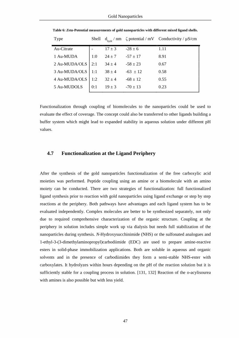
Gold Nanoparticles
47
Table 6: Zeta-Potential measurements of gold nanoparticles with different mixed ligand shells.
Type Shell dhydr
/ nm ζ potential / mV Conductivity / µS/cm
Au-Citrate - 17 ± 3 -28 ± 6 1.11
1 Au-MUDA 1:0 24 ± 7 -57 ± 17 8.91
2 Au-MUDA/OLS 2:1 34 ± 4 -58 ± 23 0.67
3 Au-MUDA/OLS 1:1 38 ± 4 -63 ± 12 0.58
4 Au-MUDA/OLS 1:2 32 ± 4 -68 ± 12 0.55
5 Au-MUDOLS 0:1 19 ± 3 -70 ± 13 0.23
Functionalization through coupling of biomolecules to the nanoparticles could be used to
evaluate the effect of coverage. The concept could also be transferred to other ligands building a
buffer system which might lead to expanded stability in aqueous solution under different pH
values.
4.7 Functionalization at the Ligand Periphery
After the synthesis of the gold nanoparticles functionalization of the free carboxylic acid
moieties was performed. Peptide coupling using an amine or a biomolecule with an amino
moiety can be conducted. There are two strategies of functionalization: full functionalized
ligand synthesis prior to reaction with gold nanoparticles using ligand exchange or step by step
reactions at the periphery. Both pathways have advantages and each ligand system has to be
evaluated independently. Complex molecules are better to be synthesized separately, not only
due to required comprehensive characterization of the organic structure. Coupling at the
periphery in solution includes simple work up via dialysis but needs full stabilization of the
nanoparticles during synthesis. N-Hydroxysucchinimide (NHS) or the sulfonated analogues and
1-ethyl-3-(3-dimethylaminopropyl)carbodiimide (EDC) are used to prepare amine-reactive
esters in solid-phase immobilization applications. Both are soluble in aqueous and organic
solvents and in the presence of carbodiimides they form a semi-stable NHS-ester with
carboxylates. It hydrolyzes within hours depending on the pH of the reaction solution but it is
sufficiently stable for a coupling process in solution. [131, 132] Reaction of the o-acylisourea
with amines is also possible but with less yield.

Gold Nanoparticles
48
Figure 39: Reaction pathway of the formation of the stable amide bond between carboxylate and amine
mediated by EDC and NHS. A: unstable reactive o-acylisourea ester, B: semi-stable amine-reactive NHS ester,
C: stable amide bond.
Au-MUDA nanoparticles were mixed together with NHS and EDC under the addition of NaOH
and the active ester was formed (Figure 39). Purification and isolation of the product was not
possible. Au-MUDA-NHS is not stable enough in aqueous solution. To obtain active-ester
coordinated nanoparticles the linker has to be synthesized and then coupled onto the surface of
nanoparticles dispersed in organic solvents like DMF. These particles can be coupled with an
amine and then transferred into aqueous solution. [133] Dodecanethiol-stabilized gold
nanoparticles can serve as precursor material.
Figure 40: Formation of Au-MUDA-NHS active ester in solution. The product is not stable and hydrolyzed
within hours.
As the active ester is stable for only hours coupling with the respective amine has to be done in
situ. An equivalent of amine can be added to the reaction solution and the amide bond forms
immediately (Figure 40 & Figure 41). To ensure full reaction another equivalent of NHS, EDC
and amine is added after two hours. After that the solution is purified via dialysis against water.
For the stability of the resulting nanoparticles a basic pH is needed, so TMAH or NaOH are
added.

Gold Nanoparticles
49
Figure 41: Coupling of amines to MUDA-functionalized gold nanoparticles by a NHS/EDC mediated reaction.
The resulting nanoparticles are stable in aqueous solution.
4.8 Functionalization with Bioactive Amines
Gold nanoparticles with a free carboxylate group were functionalized with four different
neurotransmitters, bioactive amines responsible for communication between cells (Figure 42).
[134] Histamine, dopamine and serotonin are biogenic amines derived from amino acids by
decarboxylation. Carbachol is structurally similar to acetylcholine and a stable derivate. It binds
to acetlycholinic receptors and acts as a cholinergic agonist. Because of its amino moiety it can
be coupled to the gold nanoparticles in the same way as the biogenic amines. All four types are
frequently found in organisms. By binding onto the surface the free amino group gets coupled
and can no longer participate in receptor recognition. Only a simplified binding moiety is
available for the interaction, but several studies proved this to be sufficient for specific
interactions. [110]
Immobilization of neurotransmitters on gold nanoparticles is little explored and the biological
functionality of the conjugates even less. The amines were coupled onto 14 nm gold
nanoparticles with a carboxylic acid terminated shell via the EDC/NHS method (Figure 39).
Figure 42: Bioactive amines histamine, dopamine, serotonin (5-hydroxytryptamine) and carbachol.
Immobilization of the amines was accomplished in aqueous solution. MUDA-functionalized
gold nanoparticles were mixed with EDC/NHS and NaOH forming the active ester.

Gold Nanoparticles
50
Subsequently the bioactive amine was added. All chemicals were added in large amount to
ensure complete coupling of the free carboxylic acid.
Figure 43: UV/Vis spectra of gold nanoparticles (14 nm) functionalized with histamine (HA), dopamine (DA),
serotonin (5HT) and carbachol (CB). Au-MUDA is plotted for comparison with the precursor solution.
Coupling with the molecules histamine and serotonin gave stable nanoparticles; the plasmon
resonance maxima are clearly visible in the UV/Vis spectra. Compared to Au-MUDA the
maxima shift only slightly to longer wavelengths. Only the maximum of dopamine
functionalized gold nanoparticles lost its shape indicating a change of particle dispersity (Figure
43). Also Au-MUDA-CB were stable despite a broadened maximum. This can be explained
because of the positively charged ligand shell responsible for electrostatic interactions between
the nanoparticles. The colloidal solution is prone to aggregation and the pH value has to be
adjusted at values between 8 and 10. Additionally, gold nanoparticles of 7 nm and 25 nm size
were functionalized with histamine and dopamine. Histamine functionalized gold nanoparticles
were fully characterized in the publication chapter 4.10.
0
0,1
0,2
0,3
0,4
0,5
0,6
0,7
0,8
0,9
1
400 500 600 700 800
Ab
sorb
ance
/ a
.u.
Wavelength / nm
Au-MUDA
Au-MUDA-HA
Au-MUDA-DA
Au-MUDA-5HT
Au-MUDA-CB
Au-MUDA 526 nm Au-MUDA-HA 528 nm Au-MUDA-DA 527 nm (flat) Au-MUDA-5HT 528 nm Au-MUDA-CB 532 nm

Gold Nanoparticles
51
Histamine functionalized gold nanoparticles
Figure 44: Histamine functionalized gold nanoparticles with three different core sizes: reaction scheme and
TEM images. A dTEM = 7.3 ± 0.9 nm, dhydr = 11 ± 2 nm; B dTEM = 14.7 ± 1.2 nm, dhydr = 26 ± 6 nm;
C dTEM = 25.7 ± 1.2 nm, dhydr = 32 ± 8 nm.
Histamine functionalized gold nanoparticles were synthesized with three different core sizes
(Figure 44). All three core sizes are suitable for biological applications. In the tests of biological
functionality the effect of the mean diameter shall be investigated. The relative number of active
epitopes depends on the surface provided for immobilization and should effect the potency of
the nanoparticles. The histamine functionalized gold nanoparticles are stable at a neutral pH
value in contrast to the other gold nanoparticles which need basic conditions. This might be
explained by to the buffering capacity of the imidazole moiety.
A B C

Gold Nanoparticles
52
Dopamine functionalized gold nanoparticles
Figure 45: Dopamine functionalized gold nanoparticles with two different core sizes: reaction scheme and
TEM images. A dTEM = 7.8 ± 1.2 nm, dhydr = 9 ± 2 nm; B dTEM = 15 ± 1 nm, dhydr = 26 ± 6 nm.
Only for dopamine functionalization transformation of the nanoparticles was observed. After
some hours the nanoparticles agglomerate and the solution turned brown, but is still clear.
Figure 45 shows a TEM image after the functionalization. NMR analysis confirms immobilized
dopamine molecules at the gold nanoparticles (see Experimental Section). The brown color
arises from the formation of polydopamine (Figure 46). The catechol moiety of dopamine is
oxidized to a quinone under aerobic conditions and intramolecular cyclization takes place.
Another oxidative step and isomerization leads to a 5,6-dihydroxylindole, which can then be
oxidized to the analogue quinone.
A B

Gold Nanoparticles
53
Figure 46: Mechanism of the formation of polydopamine under aerobic conditions. [135]
The surface of the nanoparticles is coated with the polymer but they are still water-soluble. This
is formed when dopamine is exposed to an aerobic environment in solution. Oxidation to
quinone and intramolecular cyclization leads to 5,6-indolequinone which appears brownish. It
forms a non-covalent polymer with supramolecular aggregation. [135] NMR analysis of the
ligand shell did not indicate a polymeric layer but only signals from immobilized dopamine. No
further investigations on this phenomenon were performed. The synthesized gold nanoparticles
with dopamine at the surface were tested for their biological functionality.
There are some applications for dopamine-coated inorganic and organic materials. [136] Noble
metal nanoparticles can be used as optical sensing agents for the detection of dopamine. [137]
Dopamine can also serve as a reducing agent. [138] Besides the amino group dopamine also
contains a catechol moiety. This functional group can also interact with surfaces; the reversed
anchor has a free amino moiety where biomolecule can be attached. Iron oxide nanoparticles
can be synthesized or functionalized via this method. [139–141] Gold nanoparticles were used
as synthesized for the biological applications.

Gold Nanoparticles
54
Serotonin functionalized gold nanoparticles
Figure 47: Serotonin functionalized gold nanoparticles: reaction scheme and TEM image. Diameter
dTEM = 14.1 ± 0.9 nm, dhydr = 16 ± 4 nm.
Gold nanoparticles with serotonin were also synthesized. Figure 47 shows a TEM image of the
nanoparticles after functionalization. The hydroxylated indolamine backbone is also prone for
oxidation and all reaction steps were performed in degassed water and under argon atmosphere.
No change in color of the solution was observable. The plasmon resonance maximum clearly
appears at 528 nm (Figure 43). With NMR analysis immobilization of serotonin at the gold
nanoparticles surface can be confirmed.

Gold Nanoparticles
55
Carbachol functionalized gold nanoparticles
Figure 48: Carbachol functionalized gold nanoparticles: reaction scheme and TEM image. Diameter
dTEM = 14.5 ± 0.7 nm, dhydr = 17 ± 6 nm.
Gold nanoparticles were also functionalized with the small molecule carbachol. It is a derivate
of acetylcholine and can be applied as a drug for the activation of acetylcholine receptors. The
molecule possesses a positive charge at the quaternary amine. Immobilization is difficult due to
repulsion between the nanoparticles. Figure 48 shows a TEM image of the gold nanoparticles
after the functionalization indicating no difference in particle size and dispersity. So the
immobilization on gold nanoparticles was indeed successful. NMR analysis confirms
immobilized carbachol at the gold nanoparticles (see Experimental Section). Due to interaction
with the material of the dialysis tube purification must be done carefully. Time for dialysis
should not be longer than 1 hour after which the sample has to be transferred into a new tube.
The functionalized gold nanoparticles should be stored in an Eppendorf cup. This phenomenon
could also be observed with the amine-modified nanoparticles also inheriting a positive charge.
Interactions with both the dialysis material and glass vessel could be observed.

Gold Nanoparticles
56
Functionalization of amine-modified gold nanoparticles
Figure 49: L-Dopa functionalized gold nanoparticles: reaction scheme and TEM image. Diameter
dTEM = 30.7 ± 7.2nm, dhydr = 45 ± 9 nm.
Coupling partners for gold nanoparticles with free carboxylate moieties need to possess either
an amino group for peptide coupling or an alcohol group for esterification. Vice versa, bioactive
molecules with a free carboxylic acid moiety can be coupled on gold nanoparticles, but
requirement for the nanoparticles is a free amino moiety. As mentioned previously nanoparticles
with a positively charged ligand shell are relatively instable and particle growth is difficult to
control. Yet the synthesis of 30 nm gold nanoparticles with cysteamine at the surface was
successful. Structurally amino acids are very similar to the biogenic amines, because they
possess both a carboxylic group and an amino group. Subsequent reaction of amino acids results
in peptides which are coupled via peptide (amide) bonds. The immobilization of such small
peptides on gold nanoparticles is also an interesting field of research. [142] Here L-Dopa was
coupled successfully to the amine-free gold nanoparticles successfully. Figure 49 shows a TEM
image after functionalization and NMR analysis confirmed the immobilization (See
Experimental Section). Once again a brown coloring of the solution was observed resulting
from the autooxidation of the catechol moiety.

Gold Nanoparticles
57
4.9 Biological Functionality
The biological functionality of the four synthesized gold nanoparticles was tested. Like many
other bioactive substances the neurotransmitters exert their effects by binding to receptors
located at the cell surface. The native ligands, molecular histamine, dopamine, serotonin and
carbachol, address their particular receptors and subunits. The newly developed conjugates of
ligand and gold nanoparticle were tested for the same effects and compared to the native
analogues where applicable. Investigations on the ability to induce chloride secretion at
receptors on a tissue were done in the research group of Prof. Marin Diener, Institute for
Veterinary Physiology at the Justus-Liebig-University Giessen. Gold nanoparticles were
administered to rat mucosa mounted in an Ussing Chamber. When receptors are stimulated an
increase in chloride secretions takes place and a rise in the short circuit (Isc) is observed.
Histamine functionalized gold nanoparticles were tested with three different core sizes. All
types were able to stimulate the receptors but for the bigger 25 nm nanoparticles the signals
were unstable and too low for evaluation. Both 7 nm and 14 nm gold nanoparticles clearly
induce a chloride secretion. After the addition of the gold nanoparticles the tissue is treated with
the secretagogue forskolin. The cAMP-dependent stimulator is a messenger for proper
biological functionality. As it induced a prompt rise in Isc the tissue is not harmed by the
nanoparticles. The results from 14 nm gold nanoparticles with histamine were published in Org.
Biomol. Chem. (see chapter 4.10). The dose-effect curve of 7 nm gold nanoparticles is shown in
Figure 50.
Figure 50: Au-MUDA-HA 7 nm at the serosal side induced a time-dependent increase in the Isc, forskolin was
added for the control of proper biological functionality; each peak is an addition of gold nanoparticle samples.
Five different samples were tested.
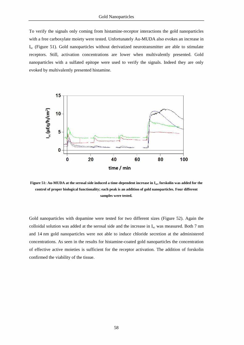
Gold Nanoparticles
58
To verify the signals only coming from histamine-receptor interactions the gold nanoparticles
with a free carboxylate moiety were tested. Unfortunately Au-MUDA also evokes an increase in
Isc (Figure 51). Gold nanoparticles without derivatized neurotransmitter are able to stimulate
receptors. Still, activation concentrations are lower when multivalently presented. Gold
nanoparticles with a sulfated epitope were used to verify the signals. Indeed they are only
evoked by multivalently presented histamine.
Figure 51: Au-MUDA at the serosal side induced a time-dependent increase in Isc, forskolin was added for the
control of proper biological functionality; each peak is an addition of gold nanoparticles. Four different
samples were tested.
Gold nanoparticles with dopamine were tested for two different sizes (Figure 52). Again the
colloidal solution was added at the serosal side and the increase in Isc was measured. Both 7 nm
and 14 nm gold nanoparticles were not able to induce chloride secretion at the administered
concentrations. As seen in the results for histamine-coated gold nanoparticles the concentration
of effective active moieties is sufficient for the receptor activation. The addition of forskolin
confirmed the viability of the tissue.

Gold Nanoparticles
59
2
4
6
8
10
12
1430 min
50 nM 10 nM
Au-MUDA-DOPA
100 nM forskolin
n = 2
Isc (
µE
q h
c
m )
-1-2
0
2
4
6
8
10
12
14
n = 3
30 min
50 pM 10 pM
Au-MUDA-DOPA
100 pM forskolin
Figure 52: Au-MUDA-DA 7 nm (left) and 14 nm (right) at the serosal side induced a time dependent increase
in Isc, forskolin was added for the control of biological functionality.
One possible explanation for the absent signals might be the autooxidation of the catechol
moiety. Both colloidal solutions turned brown after exposure to air which indicates the
formation of the quinone. Nevertheless there was no cyclization visible in the NMR spectra, up
to now no further experiments were done to prove this assumption.
-1
0
1
2
3
4
Isc (
µE
q h
c
m
)-1
-2
1 pM 100 fM 500 fM 5 pM 10 pM
Au-MUDA-5-HT
30 min
Figure 53: Au-MUDA-5HT at the serosal side induced a time-dependent increase in Isc, forskolin was added
for the control of proper biological functionality; each peak is an addition of gold nanoparticles. Four different
samples were tested; n = 7.
Serotonin-coated 14 nm gold nanoparticles were also tested for the activation of receptors
(Figure 53). They induce a concentration-dependent increase in the Isc as did the histamine-
coated. The signals correspond more likely to chloride secretion. Since native serotonin

Gold Nanoparticles
60
activates different receptor types no definite classification can be made from results. Compared
to native serotonin the concentrations for activation are much lower at nanomolar
concentrations. Again multivalent presentation of the active moiety enhances the interactions.
Serotonin receptors belong to the group of G protein-coupled receptors and ligand-gated ion
channels and found in a variety of organisms. Serotonin plays diverse roles in normal
physiology, including for example developmental, cardiovascular, gastrointestinal, and
endocrinal functions. [143] Studies on multivalent effects on the activation of the 5-HT3-
receptor showed enhanced activity for bivalent ligands. [144] Dendrimeric ligands also showed
enhanced affinity at the 5-HT3 receptor. [145] These are examples for multivalency enhancing
activities of natural processes.
Last but not least carbachol-capped 14 nm gold nanoparticles were tested. The stable
actelycholine derivative showed excellent affinity to the receptors. The results were compared
directly to native carbachol (Figure 54). Both Au-MUDA-CB and native CB induce a
concentration-dependent increase in Isc corresponding to chloride secretion.
Figure 54: Au-MUDA CB induced an increase in the Isc, native carbachol induced an increase in the Isc at
different concentrations; n = 6.
The observed activation concentrations are in picomolar, nearly half as much as for the native
analogue. A nearly equivalent response is evoked by 1 pM potency between Au-MUDA-CB in
comparison to 500 nM native CB indicating a strong increase in potency (Figure 55). The effect
of these gold nanoparticles is again supported by the multivalent presentation. The high local
density and the special arrangement allow simultaneous binding which enhances the effects.
Au-MUDA-CB (x 10-12
mol/L) Native CB (x 10-6
mol/L)

Gold Nanoparticles
61
Figure 55: Potency of Au-MUDA-CB versus native carbachol; an equivalent potency between Au-MUDA-CB
and carbachol was obtained at 1 pM Au-MUDA-CA and 500 nM CB.
Au-MUDA-CB 1 pM Native CB 500 nM

Gold Nanoparticles
62
4.10 Effects of Multivalent Histamine Supported on Gold
Nanoparticles: Activation of Histamine Receptors by Derivatized
Histamine at Subnanomolar Concentrations
This work was published in Organic and Biomolecular Chemistry, 2015, 13, 9984-9992
Received 3rd July 2015, Accepted 5th August 2015
First published on the web 20th August 2015
Friederike Gasiorek a, Ervice Pouokam
b, Martin Diener *
b, Sabine Schlecht
a and Mathias S.
Wickleder *a
aInstitute of Inorganic and Analytical Chemistry, Justus-Liebig-University, Heinrich-Buff-Ring
58, 35392 Giessen, Germany. E-mail: [email protected]
bInstitute for Veterinary Physiology and Biochemistry, Justus-Liebig-University, Frankfurter
Straße 100, 35392 Giessen, Germany. E-mail: [email protected]

Gold Nanoparticles
63
Abstract
Colloidal gold nanoparticles with a functionalized ligand shell were synthesized and used as
new histamine receptor agonists. Mercaptoundecanoic acid moieties were attached to the
surface of the nanoparticles and derivatized with native histamine. The multivalent presentation
of the immobilized ligands carried by the gold nanoparticles resulted in extremely low
activation concentrations for histamine receptors on rat colonic epithelium. As a functional
read-out system, chloride secretion resulting from stimulation of neuronal and epithelial
histamine H1 and H2 receptors was measured in Ussing chamber experiments. These responses
were strictly attributed to the histamine entities as histamine-free particles Au-MUDOLS or the
monovalent ligand AcS-MUDA-HA proved to be ineffective. The vitality of the tissues used
was not impaired by the nanoparticles.
Introduction
During the past 20 years, the number of applications of nano- and microdevices in the fields of
biotechnology and biomedicine has been increased drastically. [3, 146] Most promising tools
are nanoparticles. [147] They represent nanoscale materials with unique chemical and physical
properties due to their size. These are for example optical absorption (metal nanoparticles),
photoluminescence (semiconductor quantum dots) or magnetic behavior (iron oxides). Their
applicability derives not only from the fact that their dimension range is in the size of some
biomolecules like proteins or receptors, but also that through simple surface modification a huge
variety of interactions can be achieved. The basic requirement for every application is the
proper surface functionalization of the particles, which determines their interaction with the
environment. In this study we chose gold nanoparticles as a template because of their well-
known synthesis and functionalization with small molecules. [14, 112] Moreover they are
biocompatible. We took these particles and applied the concept of multivalency for the
interaction with biological tissue. Multivalent presentation of immobilized ligands is a common
concept in ligand–receptor interactions and can result in high affinities of ligands that possess
only moderate binding constants in a similar concentration range when they are presented in
their monovalent form. [105, 106, 148] Simplifying binding structures reduces the synthetic
complexity of the molecules immobilized on the nanoparticles and has the advantage of being
synthetically well accessible. Multivalent carriers can either inhibit or activate biological
processes in a different way than the monovalent analogue. Usually the effective concentration
is much lower. [110] There are only few examples where this concept is applied on receptor–
ligand interactions with neurotransmitters. Paolino et al. synthesized dendrimeric tetravalent
ligands for the activation of ligand-gated ion channels. [145] Indeed they found activation

Gold Nanoparticles
64
concentrations in the low picomolar range. Saada et al. synthesized gold nanoparticles coated
with histamine and applied the conjugates for enzyme activation. [111] They also found an
enhancement of the activation properties for these conjugates. Mediators such as histamine are
key regulators of physiological processes such as ion transport across epithelial barriers.
Chloride secretion is one of these processes occurring for example at the colonic epithelium.
[149, 150] Such a secretion can be activated via stimulation of histamine H1 and/or H2 receptors
as previously shown. [151–153] The receptor activity can also be blocked by inhibitors, which
are well-known for the histaminic receptors. Mediators or neurotransmitters represent an
interesting class of bioactive molecules and yet there is no study on multivalent ligand
presentation on biological systems. In this paper we report the immobilization and multivalence
of the biogenic amine histamine on gold nanoparticles as well as its biological function in
receptor activation.
Results and Discussion
Synthesis of the gold nanoparticles
By attaching histamine derivatives on gold nanoparticles we developed a new conjugate for the
interaction with compatible receptors. Water soluble gold nanoparticles with a terminally
functionalized thiol shell were prepared and used as a scaffold for the multivalent presentation
of histamine. Gold nanoparticles with an average diameter of 14 nm were synthesized by a
modification of a well-known method developed by Turkevich et al. (Figure 56). [16, 116] In an
aqueous solution Au(III) was reduced by sodium citrate which serves not only as reducing agent
but also as a ligand to prevent aggregation of the formed particles. Even if the solvent is limited
to water or polar equivalents like DMSO, the resulting size of the particles can be defined by the
molar ratios of the starting materials, the reaction temperature and time. By this method, gold
nanoparticles with a diameter of 14 nm (NP 1, Figure 56) could be obtained. Depending on the
size of the particles, the surface is coated with several thousand ligands with respect to the steric
demand of a single ligand. The resulting nanoparticles are stabilized by electrostatic repulsion of
the negatively charged citrate ions adsorbed on their surface. This layer can be easily replaced
by ligands with a stronger binding affinity, such as for example thiols. [70, 118] Thus, in a
second step the ligand shell was substituted by a bivalent ligand. Bivalent ligands give access to
further functionalization steps at the ligand periphery. One end binds to the surface and gives
stability to the particle whereas the other end is exposed to the solution. Depending on the free
moiety, the particles possess different stabilities at different pH values.

Gold Nanoparticles
65
Figure 56: Synthetic procedure for gold nanoparticles with a mean diameter of 14 nm (NP 1) followed by
ligand exchange with 11-mercaptoundecanoic acid (NP 2) and functionalization with histamine (NP 3, Au–
MUDA–HA). Double arrows at the Au core represent the ligand.
There is a wide variety of ligands suitable for exchange still providing stability in aqueous
media. Often poly(ethylene glycol) (PEG) is used as a ligand as it provides enough colloidal
stability and reduces nonspecific adsorption. Besides “PEGylation”, ω-functionalized thiol
surfactants with free carboxylic acid groups are a versatile type of ligand and are often used in
aqueous solution. They offer an additional anchor point for further attachments of biological
molecules. Herein we chose 11-mercaptoundecanoic acid (MUDA). On the one hand, a new
bond between Au and S with high affinity is built upon transfer, ensuring a complete
replacement of citrate due to strong binding affinities. On the other hand, the free carboxylic
acid can serve as an anchor for further coupling reactions with amines. In addition, the long
alkyl chain provides enough flexibility for the active moiety to interact multivalently with the
receptor. The ligand exchange with MUDA giving NP 2 proceeded completely as it is indicated
in the infra-red (IR) spectra (Figure 57). The final step towards the fully functionalized
nanoparticle was accomplished by peptide coupling with EDC (1-ethyl-3-(3-
dimethylaminopropyl)carbodiimide) and NHS (N-hydroxysuccinimide). The free carboxylic
acid is activated by EDC and forms with NHS in situ a so called ‘active ester’. This reacts easier
with amine moieties of e. g. amino acids or peptides.

Gold Nanoparticles
66
Figure 57: IR spectra of (a) Au–Citrate NP 1, (b) Au–MUDA NP 2 and (c) Au–MUDA–HA NP 3; vibration for
the amide bond is found at 1641 cm−1.
Although the prepared NHS ester is sufficiently stable for the reaction, it hydrolyses within
hours depending on the pH of the reaction solution. To ensure the coupling, another equivalent
of histamine, EDC and NHS were added after two hours. The excess of coupling agents and
byproducts of the reaction were removed by purification with dialysis against water (MWCO
3500, three times). The characterization of the synthesized and functionalized nanoparticles NP
1–3 was performed with different methods. For the characterization of the core transmission
electron microscope (TEM) images, dynamic light scattering (DLS) measurements and UV/Vis
spectroscopy were used. The images shown in Figure 58 indicate that the synthesized
nanoparticles have mostly a spherical shape; the average diameter determined by TEM is
14.0 ± 0.9 nm. The hydrodynamic diameter of the nanoparticles in solution obtained by DLS
measurements was determined by 16.7 ± 3.0 nm. The absorption maximum was found at
524 nm. The position and form of the maximum is also a hint for the size of the nanoparticles,
as it derives from a distinctive plasmon resonance at the gold surface depending on the shape of
the material. The obtained data confirmed that the average diameter of the particles cores and
the maxima of absorption remained unchanged after ligand exchange reaction and

Gold Nanoparticles
67
functionalization. The slight shift can be explained due to the new ligand. The affinity to a gold
surface is higher for thiolated ligands than for citrate and therefore effects on the plasmon
resonance of the particle. Nonetheless, neither the exchange of the ligands nor the purification
via dialysis had an effect on the agglomeration state. Also the functionalization at the ligand
periphery did not alter the particles significantly. In addition to these methods, the success of the
ligand exchange was verified by IR spectroscopy (Figure 57). Identification of the newly
formed amide bond at 1641 cm−1 gives evidence for the successful coupling. Again the colloidal
solution was purified by dialysis. Unlike the particles with MUDA as ligand shell, the final
functionalization with histamine provides a good stability at physiological pH value and in a
standard buffer solution for the Ussing chamber experiments (see below). The number of the
ligands at the surface determined by TGA is about 10.000 for 14 nm gold nanoparticles,
however determination is difficult due to small amounts of product. In order to find out whether
the size of the nanoparticles could be a limiting factor for their properties, two other sizes were
investigated. Following the same synthetic procedure as described above but with different
reaction conditions, 25 nm nanoparticles were obtained. Also, 7 nm nanoparticles were prepared
using dodecanethiol-protected particles from the Stucky method as basic material. [26]
Figure 58: (A) UV/Vis spectra of the stepwise functionalization of Au–Citrate NP 1, Au–MUDA NP 2 and Au–
MUDA–HA NP 3 nanoparticles. (B) TEM images and DLS measurements with values for the distribution of
the hydrodynamic diameters of (i) Au–Citrate NP 1 d = 14.0 ± 0.9 nm, dhydr = 16.7 ± 3.0 nm, (ii) Au–MUDA NP
2 d = 14.5 ± 1.2 nm, dhydr = 24.1 ± 6.7 nm and (iii) Au–MUDA–HA NP 3 d = 14.3 ± 0.7 nm,
dhydr = 19.3 ± 2.9 nm; d = diameter.
As a negative control for the biochemical application, gold nanoparticles NP 4 (Figure 59) with
a sulfated ligand shell were synthesized. We already reported from sulfated ligands for the
inhibition of selectin binding. [133] A structurally similar and simple ligand is sulfated 11-
mercaptoundecanol (MUDOLS), which can be immobilized on gold nanoparticles in the same

Gold Nanoparticles
68
way as mentioned before. Again citrate-stabilized gold nanoparticles NP 1 served as basic
material.
Figure 59: (A) Synthetic procedure for the preparation of Au–MUDOLS NP 4, (B) UV/Vis spectra of Au–
Citrate NP 1 and Au–MUDOLS NP 4, (C) TEM images and DLS measurements with values for the
distribution of the hydrodynamic diameters of (i) Au–Citrate NP 1 d = 14.0 ± 0.9 nm, dhydr = 16.7 ± 3.0 nm, (ii)
Au–MUDOLS NP 4 d = 14.3 ± 1.3 nm, dhydr = 18.6 ± 2.5 nm; d = diameter.
The sulfated ligand was synthesized separately and then attached to the surface via ligand
exchange reaction. TEM images, DLS measurements and UV/Vis spectra (Figure 59) indicated
that the colloidal solution was intact and the nanoparticles were stable. Sulfated nanoparticles
showed excellent stability over a wide range of pH value due to interparticle repulsion through a
negatively charged ligand shell. Unlike the unsulfated Au-MUDA particles they are perfectly
stable at physiological pH.
Figure 60: Synthesis of the monovalent analogue AcS–MUDA–HA (3) starting from 11-bromoundecanoic acid
1. Reaction conditions: (a) KSOAc, DMF, 2 h, rt, (b) NHS, DCM, 18 h, rt, (c) EDC, HA·2HCl, NEt3, DCM, 6 h,
rt.
When looking at multivalent effects, the monovalent ligand 3 (Figure 60) also has to be
investigated. Thus the complete ligand was synthesized separately. To prevent the formation of
a disulfide bond between two molecules, the thiol moiety was protected with an acetyl group.

Gold Nanoparticles
69
Functionality of the histamine-nanoparticles
From all the synthesized nanoparticles of different sizes, only the 14 nm nanoparticles yielded
consistent effects on short-circuit current (Isc) when they were administered to rat colonic
mucosa mounted in Ussing chambers. Therefore, this type of particles was used for all
subsequent experiments. To investigate the functionality of the Au-MUDA-HA nanoparticles,
their ability to induce chloride secretion was compared with that of native histamine, which
induces a strong chloride secretion across rat colonic epithelium. [151, 152] Indeed, the
histamine-conjugated nanoparticles induced a concentration-dependent increase in Isc, which is a
measure for net ion transport across the epithelium (Figure 61).
Figure 61: Au–MUDA–HA (10−11 mol l−1 at the serosal side, arrow) induced an increase in Isc (A), which was
almost abolished in the absence of chloride (0 Cl−; black bar, (B). At the end of the experiments, the
secretagogues carbachol (5 × 10⁻⁵ mol l−1 at the serosal side, white bars) and forskolin (forsk; 5 × 10⁻⁶ mol l−1 at
the mucosal and the serosal side, grey bars, (A & B), known to induce chloride secretion, were administered.
Line interruptions are caused by omission of time intervals in order to synchronize the tracings of individual
records to the administration of drugs. Values are means (symbols) ± SEM (parallel continuous lines), n = 6–8.
These changes in Isc reflect a secretion of chloride ions as they were abolished in Cl−-free buffer
solution (Figure 61). However, repetitive administration of these particles led to a
desensitization of the tissue (Table 7). Thus in all subsequent experiments, each tissue was only
exposed once to a single concentration of the Au-MUDA-HA nanoparticles (10−11 mol l−1 at the
serosal side). The tissue conductance (Gt) was not significantly changed by the nanoparticles
(10−11 mol l−1 at the serosal side); Gt amounted to 14.4 ± 1.87 mS cm−2 before and
14.7 ± 1.96 mS cm−2 after administration of the particles (n = 6; difference not significant).
Viability of the tissue was also not impaired by the Au-MUDA-HA nanoparticles as both the

Gold Nanoparticles
70
Ca2+-dependent secretagogue carbachol (5 × 10⁻⁵ mol l−1 at the serosal side), i.e. a stable
derivative of acetylcholine, [154], [151] as well as the cAMP-dependent secretagogue forskolin
(5 × 10⁻⁶ mol l−1 at the mucosal and the serosal side), which stimulates the intracellular
production of cAMP, [155] induced a prompt rise in Isc in all tissues (Figure 61). As the
response to histamine evokes chloride secretion in part via stimulation of histamine receptors
located on submucosal neurons, the effect of the inhibitory neurotoxin tetrodotoxin, which
inhibits the propagation of action potentials via blockade of voltage-dependent Na+ channels, on
the Isc evoked by the Au-MUDA-HA nanoparticles was investigated. [151, 156] Indeed, in the
presence of tetrodotoxin (10−6 mol l−1 at the serosal side), the Isc evoked by Au-MUDA-HA
(10−11 mol l−1 at the serosal side) only amounted to 0.69 ± 0.20 μEq h−1 cm−2 (n = 6) in
comparison to 2.92 ± 0.64 μEq h−1 cm−2 in untreated control tissues (P < 0.05, n = 6).
Table 7: Effects of additive concentrations of Au–MUDA–HA 14 nm on Isc and H1 and H2 receptors.
Increase in short-circuit current ΔIsc (μEq h−1
cm−2
)
Au-MUDA-HA 14 nm
No inhibitor 1.09 ± 0.43b 0.61 ± 0.11a,b 0.42 ± 0.11a,b
Mepyramine + cimetidine 0.083 ± 0.041a 0.13 ± 0.033a 0.13 ± 0.02a
Cumulative administration of gradually increased concentrations (10−11
, 5 × 10⁻¹¹
, 10−10
mol l−1
,
at the serosal side) of Au–MUDA–HA induced an increase in Isc concomitant with an obvious
desensitization of the tissue as indicated by the reduced Isc response after repeated
administration. Given is the control response in the absence of any inhibitors and the response
to Au–MUDA–HA in the combined presence of mepyramine (10−4
mol l−1
at the serosal side)
and cimetidine (10−4
mol l−1
at the serosal side). Values are given as difference to the baseline in
Isc just before administration of the corresponding agonist (ΔIsc) and are means ± SEM, n = 6.
Different letters (a, b, c) indicate statistically homogenous groups (analysis of variances
followed by post hoc test of Tukey).
The response to native histamine is mediated by stimulation of histamine H1 and histamine H2
receptors. [155] In accordance with that observation, the increase in Isc evoked by
Au-MUDA-HA (10−11
mol l−1
at the serosal side) was strongly inhibited by a combination of the
histamine H1 receptor blocker, mepyramine (10−4
mol l−1
at the serosal side), and the histamine
H2 receptor blocker, cimetidine (10−4
mol l−1
at the serosal side, Table 7). Mepyramine is a
substituted ethylamine derivative, whereas cimetidine has an imidazole ring system similar to
native histamine. [157] Both drugs act as competitive inhibitors of the G-protein coupled
histamine receptors type 1 [157] and type 2, [158] respectively. Viability of the tissue was not
altered, as forskolin, the activator of adenylate cyclase(s), [155] evoked an increase in Isc of

Gold Nanoparticles
71
11.90 ± 1.10 μEq h−1
cm−2
(n = 6) in the absence and of 10.72 ± 1.18 μEq h−1
cm−2
(n = 6,
difference not significant) in the presence of the histamine receptors antagonists.
Controls with histamine-free nanoparticles
In order to make sure that the effects of the nanoparticles are not unspecific artefacts resulting
from the assembly components of the particles, controls were performed either with the
conjugator AcS-MUDA-HA or with the sulfated moiety Au-MUDOLS. As shown in Figure 62,
these components failed to induce a rise in Isc. Furthermore, Au-MUDOLS impaired the
viability of the epithelium as it prevented the secretion induced by a subsequent administration
of forskolin (5 × 10⁻⁶ mol l−1 at the mucosal and the serosal side).
Figure 62: Au–MUDA–HA (10−11, 5 × 10⁻¹¹, 10−10 mol l−1, administered cumulatively at the serosal side,
arrows) induced an increase in Isc. (B) AcS–MUDA–HA (10−7, 5 × 10⁻⁷, 10−6 mol l−1, at the serosal side, arrows)
was ineffective. (C) Au–MUDOLS (10−12, 5 × 10⁻¹², 10−11 mol l−1, at the serosal side, arrows) failed to change
the Isc. All particles except Au–MUDOLS preserved the secretory function of the tissues as they did not impair
the effect of the secretagogue forskolin (forsk; 5 × 10⁻⁶ mol l−1 at the mucosal and the serosal side, grey bars).
Values are means (symbols) ± SEM (parallel continuous lines), n = 4–8.
Potency of the Au–MUDA–HA nanoparticles
The highest changes in the Isc or the Gt induced by the nanoparticles were obtained at the
concentration 10−11 mol l−1. For the Isc, the change amounted to 1.09 ± 0.42 μEq h−1 cm−2. When
comparing this response with a concentration-response curve of native histamine (Figure 63) it
turned out that a comparable increase in Isc (1.05 ± 0.24 μEq h−1 cm−2) was obtained with
histamine in a concentration of 10−5 mol l−1 (Figure 63). Assuming a maximum of 10 000
histaminic units at the surface determined by TGA, the relative potency is still some magnitudes
higher. In other words, the conjugation to the 14 nm nanoparticles strongly enhanced the
potency of histamine to evoke anion secretion.

Gold Nanoparticles
72
Figure 63: (A) Histamine induced a concentration-dependent increase in Isc. (B) shows the potency of Au–
MUDA–HA versus histamine; an equivalent potency between Au–MUDA–HA and histamine was obtained
with 10−11 mol l−1 and 10−5 mol l−1, respectively. Values are given as increase in Isc above baseline in short-
circuit current (ΔIsc) just before administration of the corresponding drug and are means ± SEM, n = 6–8.
Conclusion
The gold nanoparticles with an average diameter of 14 nm were loaded with the histamine
derivative and showed excellent activation properties in the Ussing chamber experiments.
Compared to native histamine, the nanoparticles were more effective at extremely low
concentrations. It was found that the newly developed conjugate was able to induce a
concentration-dependent chloride secretion across rat colonic epithelium via stimulation of
histamine H1 and H2 receptors. These receptors, which are composed of 487 or 359 amino acids,
respectively, belong to the class of G-protein coupled receptors with seven transmembrane
spanning regions. Upon binding of the agonist, these receptors induce the exchange of
guanosine diphosphate (GDP) against guanosine triphosphate (GTP) at the a-subunit of trimeric
GTP-binding proteins. The consequence is the dissociation of the G-protein in the GTP-binding
a-subunit and a b/g-complex and the activation of intracellular enzymes such as phospholipase
C or adenylate cyclase, which then activate intracellular second messenger pathways. [159] An
explanation for this stimulation might be the high local density of the immobilized ligand. The
molecular recognition between the receptors on the surface and ligands bound to the
nanoparticles is strengthened through the geometric arrangement given by the spherical shape.
The density of the binding moieties allows simultaneous binding resulting in a lower activation
concentration. [107] The interaction can be blocked with same inhibitors used for native
histamine. To ensure the response originated only from multivalent histamine supported on gold
nanoparticles and not from any unspecific interactions with the biological tissue, the
monovalent ligand AcS-MUDA-HA or the gold nanoparticles with a simple sulfated moiety
from 11-mercaptoundecanol were synthesized and tested in parallel. Indeed, no chloride
secretion could be observed. All particles tested in this study preserved the basic functions of
the tissues except for Au-MUDOLS. Only the particles with the sulfated moiety harmed the

Gold Nanoparticles
73
tissues irreversibly. Among the three sizes of particles tested (7 nm, 14 nm and 25 nm), only the
intermediate size 14 nm showed consistent effects on transepithelial Cl− secretion, indicating a
size-dependence. The smaller nanoparticles exhibited unspecific interactions whereas the bigger
particles evoked no signal in the Ussing chamber experiments. The observed size dependence
might correlate with the number of histamine molecules at the surface. The density of
immobilized histamine moieties at 14 nm core size and the spatial arrangement are both
essential for the activation of histaminic receptors bound on an epithelium. For a detailed insight
into the mechanisms of receptor activation further experiments will be conducted on the ligand–
receptor-complex which is formed during the activation process. But also the length of the
spacer between the particle core and the active moiety influences the interplay, as it was shown
in one of our previous studies on multivalent interactions of gold nanoparticles in selectin
binding. [133] Further investigations shall be conducted on this influence. In summary the
present work is another example for the influence of multivalent interactions in biological
systems. This concept can also be transferred to other receptor antagonists and to different
neurotransmitters.
Experimental section
All reactions of nanoparticles in aqueous solution were carried out in purified Millipore water.
Organic solvents were distilled before use. All chemicals were purchased from commercial
sources and used as received. The ligand exchange reactions and coupling reactions were done
under inert conditions. All glass vessels were washed with aqua regia and distilled water prior to
use. NMR spectra were recorded on Bruker BioSpin Avance III AV600 (600 MHz), AV400
(400 MHz) or AV200 (200 MHz) spectrometers using tetramethylsilane (TMS) as an internal
standard with chemical shifts given in ppm relative to TMS (δ = 0.00 ppm) or the respective
solvent peaks. 1H NMR data are reported as follows: chemical shifts (multiplicity [ppm],
coupling constants [Hz], integration, classification). Multiplicity is recorded as s = singlet, br s
= broadened singlet, d = doublet, t = triplet, q = quartet, m = multiplet. For 13C NMR, chemical
shifts and structural assignments are reported where applicable. Because of the rigidity of the
alkyl chains in the thiol shells and the resulting large differences in the relaxation times of
different protons, no integrals are given for the proton NMR data of the gold colloids. IR spectra
were obtained on a Bruker IFS48 spectrometer in ATR mode. Electrospray ionization mass
spectra (ESI-MS) were recorded on a Finnigan LCQDuo spectrometer using methanol solutions
of the respective. TEM measurements were performed on a Philips CM30 STEM (300 kV,
LaB6-cathode) equipped with a GATAN digital camera. TEM images were recorded using a
digital micrograph. The average particle core sizes were determined by measuring at least 100
individual particles from recorded TEM images. The UV/Vis spectra were plotted with an

Gold Nanoparticles
74
Agilent 8453 spectrophotometer (Agilent Technologies Inc., Santa Clara, CA, USA). Dynamic
light scattering measurements were performed with a StabiSizer PMX 200C from Particlemetrix
(Meerbusch, Germany).
Chemical syntheses
Citrate-coordinated gold nanoparticles NP 1 (Au–Citrate Ø 14 nm). Citrate-coordinated gold
nanoparticles were synthesized by a variation of a procedure first published by Turkevich. A
solution of 50 mg (0.127 mmol) HAuCl4·3H2O in 195 mL of Millipore water was heated to
reflux for 20 min. Under vigorous stirring a solution of 224 mg (0.52 mmol) of sodium citrate in
5 mL of Millipore water was added quickly. The reaction mixture was held at 80 °C for 2 h.
Then the solution was cooled to 0 °C in an ice bath and filtered (0.2 μm pore size). A clear red
solution with a particle concentration of 2.7 nM was obtained and stored at 4 °C.
1H NMR (400 MHz, D2O): δ/ppm = 2.61 (bs, CH2); IR (KBr disc, ν/cm
−1): 3426.8 (νO–H),
1598.2 (νC=O), 1397.6, 1258.4, 618.0; TEM: d = 14.0 ± 0.9 nm; UV/Vis: λmax = 522 nm; DLS:
dhydr = 16.7 ± 3.0 nm.
11-Mercaptoundecanoic acid coordinated gold nanoparticles NP 2 (Au–MUDA Ø 14 nm). An
amount of 21 mg 11-mercaptoundecanoic acid (0.1 mmol, 105 equiv.) was dissolved in 8.5 mL
Millipore water under the addition of 50 μL tetramethylammonium hydroxide (TMAH). 5 mL
of a 2.7 nM solution of citrate-protected gold nanoparticles in water was added dropwise and
the solution was stirred for 24 h. The nanoparticles solution was dialysed against 300 mL water
three times (3500 MWCO). A clear red solution with a particle concentration of 1 nM was
obtained and stored at 4 °C.
1H NMR (400 MHz, D2O): 3.17 (s, 16H, TMAH), 2.76 (t, CH2), 2.16 (t, CH2), 1.73 (quin, CH2),
1.54 (m, CH2), 1.40 (m, CH2), 1.30 (bs, CH2); IR (KBr disc, ν/cm−1
): 3422.8, 3018.5, 2920.1 &
2849.8 (νC-H), 1583.5 (νC O), 1487.9, 1396.2, 1274.9, 956.8, 948.2, 625.0; TEM: d =
14.5 ± 1.2 nm, UV/Vis: λmax = 527 nm, DLS: dhydr = 24.1 ± 6.7 nm.
Sodium 11-mercaptoundecyl sulfate coordinated gold nanoparticles NP 4 (Au–MUDOLS Ø
14 nm). An amount of 30 mg 11-mercaptoundecanyl sulfate was dissolved in 8.5 mL Millipore
water and 50 μL tetramethylammonium hydroxide (TMAH) was added. 5 mL of a 2.7 nM
solution of citrate-protected gold nanoparticles in water was added dropwise and the solution
was stirred for 24 h. The nanoparticles solution was dialysed against 300 mL water three times
(3500 MWCO) and stored at 4 °C.
1H NMR (400 MHz, D2O): δ/ppm = 4.00–3.89 (m, CH2), 2.70–2.60 (m, CH2), 1.68–1.51 (m,
CH2), 1.37–1.23 (m, CH2); IR (KBr disc, ν/cm−1
): 2918.7 & 2849.9 (νC–H), 1614.8 (νC O),

Gold Nanoparticles
75
1470.0, 1261.7 & 1205.1 & 1128.1 (νR–OSO2–OR), 1068.8, 966.4, 831.8, 718.9, 628.7, 586.3;
TEM: d = 14.3 ± 1.3 nm; UV/Vis: λmax = 525 nm; DLS: dhydr = 18.6 ± 2.5 nm.
Synthesis of histamine functionalized gold nanoparticles NP 3 (Au–MUDA–HA Ø 14 nm).
10 mL of a 1 nM solution Au–MUDA were set in a glass vessel washed with aqua regia and
distilled water. 18 mg histamine dihydrochloride (0.1 mmol), 17 mg EDC (0.11 mmol) and
15 mg HOBt (0.11 mmol) and 50 μL NEt3 were added to the nanoparticle solution. The reaction
mixture was stirred overnight and subsequently dialysed against water three times
(3500 MWCO) and stored at 4 °C.
1H NMR (400 MHz, D2O): 7.55 (s, 1H, CH), 6.81 (s, 1H, CH), 2.73–2.86 (m, 2H, CH2(HA)),
2.57–2.71 (m, 3H, CH2(HA) + NH)), 2.41–2.53 (m, 2H), 2.21–2.34 (m, 4H), 2.10–2.21 (m, 3H),
2.03 (t, J = 7.7, 1H), 1.52–1.68 (m, 1H), 1.36–1.48 (m, 1H), 1.18 (bs, 8H, CH2), IR (KBr disc,
ν/cm−1
): 3018.8, 2921.4 & 2850.0 (νC–H), 1641.1 (νC O, amide), 1596.3 (νCO–NH), 1488.2, 1404.2,
957.1, 948.6, TEM: d = 14.3 ± 0.7 nm, UV/Vis: λmax = 528 nm, DLS: dhydr = 19.3 ± 2.9 nm.
Sodium 11-mercaptoundecyl sulfate (MUDOLS). In a flame-dried vessel under argon
atmosphere, 353 mg of 11-mercaptoundecanol (1.72 mmol, 1 equiv.) was dissolved in 3 ml dry
DMF. Separately, 395 mg of SO3·DMF complex (2.5 mmol, 1.5 equiv.) were dissolved in 1 mL
dry DMF and added to the solution. After stirring for 1 h at room temperature the solvent was
removed under reduced pressure (OPV) and the residue was dissolved in 4 mL of 1 M NaOH. A
white precipitate appeared. 15 mL of ethanol were added and washed three times. The product
was dried in vacuum. 280 mg of a colorless solid (0.91 mmol, 53%) could be obtained. The
dimer was formed.
1H NMR (200 MHz, DMSO-d6): δ/ppm = 3.68 (t, J = 6.6 Hz, 2 H, CH2), 3.35 (t, J = 6.3 Hz, 2H,
CH2), 2.27–2.61 (m, 4H, CH2), 2.56–2.49 (m, 2H, CH2), 1.67–1.52 (m, 4H, CH2), 1.36–1.24 (m,
10H, CH2); 13
C NMR (200 MHz, DMSO-d6): δ/ppm = 65.6, 60.7, 32.5, 29.1, 29.0, 28.9, 28.8,
28.6, 28.5, 27.7, 25.5; ESI-MS: m/z = 633.1593 ([M + Na]+, calcd 633.1612).
11-(Acetylthio)undecanoic acid (AcS–MUDA). 500 mg 11-bromoundecanoic acid (1.89 mmol,
1 equiv.) were dissolved in 70 mL absolute DMF under argon atmosphere. Separately 520 mg
potassium thioacetate (4.56 mmol, 2.4 equiv.) were suspended in 8 mL absolute DMF under
argon atmosphere and added dropwise to the first solution. The reaction mixture was stirred for
2 hours at room temperature. The precipitate was filtered off, washed several times with water
and dried in vacuum. 414 mg of a colorless solid (1.59 mmol, 84%) could be obtained.
1H NMR (200 MHz, CDCl3): δ/ppm = 2.79 (t, J = 7.17, 2H, CH2), 2.27 (t, J = 7.17, 2H, CH2),
2.25 (s, 3H, CH3), 1.61–1.52 (m, 2H, CH2), 1.52–1.44 (m, 2H, CH2), 1.32–1.16 (m, 12H, CH2);

Gold Nanoparticles
76
13C NMR (100 MHz, CDCl3): δ/ppm = 196.2 (Cq, C O, acetyl), 179.4, 33.9, 30.7, 29.5, 29.4,
29.3, 29.2, 29.1, 29.1, 29.0, 28.8, 24.7; ESI-MS: m/z = 283.1335 ([M + Na]+, calcd 283.1344).
2,5-Dioxopyrrolidin-1-yl-11-(acetylthio-)undecanoate (AcS–MUDA–NHS). 195 mg NHS
(1.7 mmol, 1.1 equiv.) and 700 mg AcS–MUDA (1.55 mmol, 1 equiv.) were dissolved in DCM
under stirring at room temperature. 350 mg DCC (1.7 mmol, 1.1 equiv.) were separately
dissolved in 5 mL DCM and added slowly. After 30 min a white precipitate appeared. The
reaction mixture was stirred overnight. The solvent was removed under reduced pressure.
506 mg (1.42 mmol, 92%) of a colorless powder were obtained.
1H NMR (400 MHz, CDCl3): δ/ppm = 2.81–2.77 (m, CH2), 2.53 (t, J = 7.34, 2H, CH2), 2.25 (s,
3H, CH3), 1.71–1.63 (m, 2H, CH2), 1.52–1.47 (m, 2H, CH2), 1.38–1.17 (m, 12H, CH2);
13C NMR (100 MHz, CDCl3): δ/ppm = 196.1 (Cq, C O, acetyl), 169.3 (2 × Cq, C O), 168.7
(Cq, C=O), 30.9 (CH3), 30.7, 29.5, 29.3, 29.2, 29.2, 29.1, 29.0, 28.8, 28.7, 25.6 (2 × CH2), 24.6;
ESI-MS: m/z = 380.1502 ([M + Na]+, calcd 380.1508).
S-(11-((2-(1H-Imidazol-4-yl)ethyl)amino)-11-oxoundecyl) ethanethioate 4 (AcS–MUDA–HA).
97 mg AcS–MUDA (0.27 mmol, 1 equiv.) were dissolved in DMF to form a clear solution.
34 mg HA·2HCl (0.30 mmol, 1.1 equiv.) and a few drops NEt3 were added and the solution was
stirred overnight. Subsequently 50 mL water were added and a white solid appeared. The
precipitate was filtered off and washed three times with water. The crude product was dissolved
in CHCl3, filtered again and purified via column chromatography (CHCl3/MeOH 9/1 Rf = 0.7).
After evaporation 70 mg (0.19 mmol, 70%) of a colorless powder were obtained.
1H NMR (400 MHz, CDCl3): δ/ppm = 7.50 (s, 1H, CH), 6.75 (s, 1H, CH), 6.33 (br s, 1H, NH),
3.52–3.42 (m, 2H, CH2(HA)), 2.79–2.69 (m, 2H, CH2(HA)), 2.53–2.39 (m, 2H, CH2), 2.56 (s,
3H, CH3), 2.10 (t, J = 7.24 Hz, 2H, CH2), 1.59–1.43 (m, 4H, CH2), 1.26–1.14 (m, 12H, CH2);
13C NMR (100 MHz, CDCl3): δ/ppm = 196.4 (Cq, C O, acetyl), 173.7 (Cq, C O, amide), 134.8
(CH, Car), 127.6 (Cq, Car), 118.6 (CH, Car), 39.2 (CH2(HA)), 36.9 (CH2 (HA)), 30.7 (CH3), 29.5,
29.3, 29.3, 29.2, 29.1, 28.7, 28.4, 26.9, 25.7; ESI-MS: m/z = 354.2221 ([M + H]+, calcd
354.2210).
Animals
Female and male Wistar rats with a body mass of 200–250 g were used. The animals were bred
and housed at the institute for Veterinary Physiology and Biochemistry of the Justus-Liebig-
University Giessen at an ambient temperature of 22.5 °C and air humidity of 50–55% on a 12 h
: 12 h light–dark cycle with free access to water and food until the time of the experiment.
Experiments were approved by the named animal welfare officer of the Justus Liebig University

Gold Nanoparticles
77
(administrative number 487_M) and performed according to the German and European animal
welfare law.
Solutions
The standard solution for the Ussing chamber experiments was a buffer solution containing
(mmol l−1): NaCl 107, KCl 4.5, NaHCO3 25, Na2HPO4 1.8, NaH2PO4 0.2, CaCl2 1.25, MgSO4 1
and glucose 12. The solution was gassed with carbogen (5% CO2 in 95% O2, v/v); pH was 7.4.
For the Cl−-free buffer, NaCl and KCl were substituted by Na gluconate and K gluconate
(KGluc), respectively. The Ca2+ concentration in the Cl−-free buffer was increased to 5.75 mmol
l−1 to compensate for the Ca2+-buffering properties of gluconate. [160]
Tissue preparation
Animals were killed by stunning followed by exsanguination. The serosa and tunica muscularis
were stripped away by hand to obtain a mucosa–submucosa preparation of the distal colon.
Briefly, the colon was placed on a small plastic rod with a diameter of 5 mm. A circular incision
was made near the anal end with a blunt scalpel, and the serosa together with the tunica
muscularis was gently removed in a proximal direction. Two segments of the distal colon of
each rat were prepared.
Short-circuit current measurements
The mucosa–submucosa preparation was fixed in a modified Ussing chamber bathed with a
volume of 3.5 ml on each side of the mucosa. The tissue was incubated at 37 °C and short-
circuited by a computer-controlled voltage-clamp device (Ingenieur Büro für Mess- und
Datentechnik Mussler, Aachen, Germany) with correction for solution resistance. Tissue
conductance (Gt) was measured every minute by the voltage deviation induced by a current
pulse (± 50 μA, duration 200 ms) under open-circuit conditions. Short-circuit current (Isc) was
continuously recorded on a chart-recorder. Isc is expressed as μEq h−1 cm−2, i.e. the flux of a
monovalent ion per time and area, with 1 μEq h−1 cm−2 = 26.9 μA cm−2. At the end of each
experiment, tissue viability was tested by administration of the cholinergic agonist, carbachol
(5 × 10⁻⁵ mol l−1 at the serosal side) and/or the administration of forskolin (5 × 10⁻⁶ mol l−1 at the
mucosal and the serosal side), an activator of adenylate cyclase(s), which induce a strong Ca2+-
respective cAMP-dependent Cl− secretion. [154, 155]
Drugs
Au–MUDOLS, Au–MUDA–HA, carbachol, cimetidine, histamine dihydrochloride, and
mepyramine were dissolved in aqueous stock solutions. Forskolin (Calbiochem, Bad Soden,
Germany) was dissolved in ethanol (final maximal concentration 0.25%, v/v). Tetrodotoxin was

Gold Nanoparticles
78
dissolved in 2 × 10⁻² mol l−1 citrate buffer. If not indicated otherwise, drugs were from Sigma,
(Taufkirchen, Germany).
Statistics
Results are given as means ± SEM, with the number (n) of investigated tissues. When means of
several groups had to be compared, an analysis of variance was performed followed by post hoc
Tukey's-test. For the comparison of two groups, a Mann-Whitney U-test was applied. P < 0.05
was considered to be statistically significant.
Acknowledgements
We wish to acknowledge Anne Schulze (Institute of Inorganic and Analytical Chemistry,
Justus-Liebig-University at Giessen) for the TEM images.

Quantum Dots
79
5. Quantum Dots
5.1 Quantum Dots for Cellular Imaging
Due to their unique optical properties quantum dots (QDs) consisting of CdTe were synthesized and
applied as new cell dyes. Preliminary work was done on the development of a synthesis for the
preparation of monodisperse quantum dots of Cd and Te or Se. [161] Based on the developed
method the chalcogenide precursor was added to a aqueous solution of Cd2+
and
mercaptocarboxylic acid. Three different types were applied: 2-Mercapto-1-acetic acid (thioglycolic
acid, TGA), 3-mercapto-1-propanoic acid (MPA) and 3-mercapto-1-propanesulfonic acid (MPSA)
(Figure 64).
Figure 64: Mercaptocarboxylic acids and -sulfonic acid used for the synthesis of quantum dots.
The reaction mixture was stirred at 100 °C for 3 hours and then cooled down with an ice bath,
subsequently the colloidal solution was purified carefully via dialysis against water. Within the
molar ratios of 2:1:4.8 for Cd:Te:ligand different rates for the growth of the nanoparticles could be
observed. Depending on the ligand the size of the resulting quantum dots can be tuned. [161] The
longer the alkyl chain of the ligand the smaller the particles get within the same reaction time. With
increasing reaction time the nanoparticles grow larger.
Figure 65: Reaction scheme for the synthesis of CdTe quantum dots in aqueous solution.

Quantum Dots
80
CdTe quantum dots with three different surface ligands (Figure 65). All of them showed high
emission intensities. For the shortest chain length (TGA) the longest wavelength for the absorption
and emission maxima were achieved, indicating different particle growth with different ligands
(Figure 66).
Absorption Emission
Figure 66: Absorption and emission spectra of CdTe quantum dots.
Additionally CdSe quantum dots were synthesized via the same procedure, but only two different
ligands were applied (Figure 67).
Figure 67: Reaction scheme for the synthesis of CdSe quantum dots in aqueous solution.
The selenide precursor solution is very sensitive to air and must be handled carefully under argon
atmosphere. The spectra show only for MPA-stabilized quantum dots absorption properties and
0
1
2
400 500 600 700 800
Ab
sorb
ance
/ a
.u.
Wavelength / nm
CdTe-TGA
CdTe-MPA
CdTe-MPSA
0
200
400
600
800
1000
1200
1400
1600
400 500 600 700 800
Inte
nsi
ty /
a.u
.
Wavelength / nm
CdTe-TGA
CdTe-MPA
CdTe-MPSA

Quantum Dots
81
emission intensity (Figure 68). TGA-capped quantum dots could not be obtained. In the absorption
spectra a red shift can also be observed for shorter chain length indicating the same particle growth
as for CdTe quantum dots.
Absorption Emission
Figure 68: Absorption and emission spectra of CdSe quantum dots.
Furthermore, CdS quantum dots were synthesized via the previously described method (Figure 69).
No precursor solution with NaBH4 needed to be synthesized but Na2S was dissolved and used
directly for the synthesis. Again all reaction steps were carried out under argon atmosphere.
Figure 69: Reaction scheme for the synthesis of CdS quantum dots in aqueous solution.
Both spectra show absorption and emission properties of the colloidal solution indicating the
formation of quantum dots (Figure 70), the intensities are however only weak. No further
0
1
2
3
4
400 500 600 700 800
Ab
sob
ance
/ a
.u.
Wavelength / nm
CdSe-TGA
CdSe-MPA
0
20
40
60
80
100
400 500 600 700 800
Inte
nsi
ty /
a.u
.
Wavelength / nm
CdSe-TGA
CdSe-MPA

Quantum Dots
82
characterizations of the solution or the nanoparticles were performed. Thus the synthetic parameters
must be optimized for creation of CdS quantum dots via the approach in aqueous solution.
Absorption Emission
Figure 70: Absorption and emission spectra of CdS quantum dots.
Subsequently, CdTe quantum dots capped with TGA and MPA were covered with a ZnS-shell. This
prevents the QDs from the leakage of cytotoxic Cd2+
ions, which is essential for biological
applications. The passivation was done directly in solution. Precursor solutions of Zn2+
and S2-
were
added and the reaction mixture was stirred at 60 °C. Again dialysis was done for purification. From
TEM images a mean thickness of 1 nm for the shell can be determined; EDX spot analysis
confirmed the layer of ZnS around the quantum dot. The reaction time determines the thickness of
the shell.
Figure 71: Reaction scheme for the passivation of CdTe quantum dots with a ZnS shell.
0
1
2
3
4
400 500 600 700 800
Ab
sorb
ance
/ a
.u.
Wavelength / nm
CdS-MPA
0
10
20
30
40
50
60
70
80
400 500 600 700 800
Inte
nsi
ty /
a.u
.
Wavelength / nm
CdS-MPA

Quantum Dots
83
The QDs were characterized by TEM and DLS for the determination of the core size and with
UV/Vis and fluorescence spectroscopy for the analysis of the optical properties.
For further biological application the optical properties of the synthesized ODs were examined and
compared with each other. The properties of the core materials CdTe and CdSe with different
ligands were used for comparison and the optical properties before and after passivation were
compared.
Absorption Emission
Figure 72: Summary of absorption and emission spectra of CdTe and CdSe quantum dots with different ligands.
The absorption and emission maxima of the QDs with different surface ligands differ as shown in
Figure 72. CdTe-based QDs absorb light between 478 nm and 540 nm, the maximum of CdSe-
based QDs lies at 473 nm. The emission maximum lies between 544 nm and 594 nm for CdTe QDs
and for CdSe at 543 nm. The intensities are also different.
Additionally, passivated QDs were compared to their precursors. Comparing the absorption and
emission spectra of both ZnS covered QDs and their unpassivated analogues, the growth and
0
1
2
400 500 600 700 800
Ab
sorb
ance
/ a
.u.
Wavelength / nm
CdTe-TGA
CdTe-MPA
CdTe-MPSA
CdSe-MPA
0
200
400
600
800
1000
400 500 600 700 800
Inte
nsi
ty /
a.u
.
Wavelength / nm
CdTe-TGA
CdTe-MPA
CdTe-MPSA
CdSe-MPA

Quantum Dots
84
thickness of the shell has no significant effect on the position of both absorption and emission
maxima but on the intensities (Figure 73). The absorption abilities decrease and the emission
abilities are higher for both TGA- and MPA-stabilized quantum dots. The spectra are plotted as
measured and the concentrations of the sample are similar.
Absorption Emission
Figure 73: Summary of absorption and emission spectra of passivated CdTe quantum dots and their precursors.
The ZnS-shell is very thin and has only a slight influence on the optical properties. The maxima are
shifted to a longer wavelength. Table 8 summarizes the values for absorption and emission maxima
of the compared quantum dots. The corresponding diameters determined via TEM and DLS are also
shown.
0
0,5
1
1,5
2
2,5
400 500 600 700 800
Ab
orb
ance
/ a
.u.
Wavelength / nm
CdTe-TGA
CdTe-MPA
CdTe/ZnS-TGA
CdTe/ZnS-MPA
0
200
400
600
800
1000
1200
1400
1600
400 500 600 700 800
Inse
nsi
ty /
a.u
.
Wavelength / nm
CdTe-TGA
CdTe-MPA
CdTe/ZnS-TGA
CdTe/ZnS-MPA

Quantum Dots
85
Table 8: Quantum dots stabilized with different ligands and the analogues passivated with a ZnS shell.
dTEM dhydr λabs λem
CdTe-TGA 4.1 ± 0.3 nm 8 ± 2 nm 540 nm 594 nm
CdTe-MPA 4.4 ± 0.4 nm 6 ± 1 nm 498 nm 574 nm
CdTe-MPSA 4.9 ± 0.4 nm 5 ± 1 nm 478 nm 544 nm
CdSe-MPA 3.9 ± 0.2 nm 5 ± 1 nm 473 nm 543 nm
CdTe/ZnS-TGA 4.2 ± 0.2 nm 11 ± 2 nm 542 nm 590 nm
CdTe/ZnS-MPA 4.5 ± 0.4 nm 6 ± 1 nm 500 nm 574 nm
CdTe quantum dots functionalized with three different ligands and the passivated CdTe/ZnS
analogues were applied as new cell dyes (Figure 74). The effect of the ligand shell and core
composition was investigated in cell uptake studies. Free carboxylic acid moieties were compared
with sulfonated moieties and the effect of passivation was investigated. All quantum dots have
almost the same diameter and were dispersed in basic solution, the concentration of the stock
solution was 9 µmol/L. All QDs were stable in the buffer solution and could be used in biological
media.
Figure 74: Overview of CdTe quantum dots used for biological applications.
The tests were done by Ervice Pouokam, research group Prof. Diener, Institute of Veterinary
Physiology, Justus-Liebig University Giessen. The tested cells were rat colonic epithelial cells
forming crypts. Mucosa-submucosa preparations obtained from rat colon were fixed on a plastic
holder with tissue adhesive and incubated for about 7 min in an EDTA solution, then vibrated for
about 30 s in order to obtain crypts. A stock solution of the QDs was diluted to 2.25 µmol/L. The
QDs were collected in a high-K+ gluconate Tyrode buffer and pipetted onto poly-L-lysine-coated
cover glasses (Ø = 23 mm). The crypts were allowed to settle down at room temperature and attach
onto the surface of the cover glasses for about 30 min. The cover glass was then mounted in a

Quantum Dots
86
chamber and the volume adjusted to 2 ml. After selecting at least nine regions of interest (ROIs; 3
in the depth of the crypt, 3 in the middle and 3 at the surface), corresponding each approximately to
a cell, five excitation flashes were launched at 350 nm with the following durations: 5 ms, 10 ms,
20 ms, 50 ms and 100 ms. The resulting fluorescence was recorded live and the data stored. Then
the stock solution of the QDs was added into the chamber to yield a final concentration of
2.25 µmol/L without changing the volume. After an incubation period of 1 hour at room
temperature, the chamber was rinsed twice with fresh Tyrode buffer to remove excess QDs and the
same excitation protocol as before incubation was run. This allowed for tracking the changes of the
fluorescence in the same ROIs.
Figure 75: Crypts incubated with the QDs for 1 h in a Tyrode buffer solution; fluorescence intensity before and
afterwards was compared. X-axis exposure time, Y-axis in arbitrary units.
All quantum dots tested showed a cell uptake after 1 hour incubation. TGA stabilized quantum dots
also showed coloration of the background. This suggests unspecific interactions with biological
tissues. The different core sizes seem not to effect the interactions. The experiments showed that
QDs with a carboxylic acid terminated shell have a better cell uptake than their sulfonated
analogues (Figure 75). The negative charge of the sulfonate group seems to restrain the quantum
dots from entering the cells. Depending on the pH value of the cellular environment carboxylic acid
moieties are both protonated and deprotonated. The reduced amount of effective charge seems to
support the uptake.
5ms 10ms 20ms 50ms 100ms
X-Achse
0
1000
2000
3000
4000
5000
Y-A
chse
0 W ert
1h W ert
TGA-Plus 1:2
5ms 10ms 20ms 50ms 100ms
X-Achse
0
1
2
3
4
5
Tausend
Y-A
chse
0 W ert
1h W ert
MPSA 1:2CdTe-MPSA
0 1h
5ms 10ms 20ms 50ms 100ms
X-Achse
0
1
2
3
4
5
Tausend
Y-A
chse
0 W ert
1h W ert
TGA 1:2CdTe-TGA CdTe-MPA
5ms 10ms 20ms 50ms 100ms
X-Achse
0
1
2
3
4
5
Tausend
Y-A
chse
0 W ert
1h W ert
MPA 1:2

Quantum Dots
87
Figure 76: Crypts incubated with the QDs for 1 h in a Tyrode buffer solution; fluorescence intensity before and
afterwards was compared. X-axis exposure time, Y-axis in arbitrary units.
The experiments showed that the passivated quantum dots had a faster uptake, but did not change
significantly after time (Figure 76). Passivated QDs gave better results, which proves the shell
prevents of Cd2+
leakage. CdTe/ZnS with thioglycolic acid at the surface gave best results in the cell
uptake studies.
Figure 77: Crypts incubated with the QDs for 1 h in a buffer solution (tyrode).
Figure 77 shows an image of the cells incubated with quantum dots for one hour. It shows on the
left hand an image of the cells incubated with quantum dots for one hour. Areas of high
5ms 10ms 20ms 50ms 100ms
X-Achse
0
1000
2000
3000
4000
5000
Y-A
chse
0 W ert
1h W ert
TGA-Plus 1:2CdTe/ZnS-TGA
0 1h
5ms 10ms 20ms 50ms 100ms
X-Achse
0
1
2
3
4
5
Tausend
Y-A
chse
0 W ert
1h W ert
TGA 1:2CdTe-TGA
5ms 10ms 20ms 50ms 100ms
X-Achse
0
1000
2000
3000
4000
5000
Y-A
chse
0 W ert
1h W ert
TGA-Plus 1:2
Colonic
epithelial
cells (loaded
with the
QDs)
Rat colonic crypt 1 hour after incubating with MPA; Excitation period 1 s
Rat colonic crypt Autofluorescence evoked by a
long excitation period (1 s)
Fu
ore
scen
ce i
nte
nsi
ty

Quantum Dots
88
fluorescence intensity are shown in red. Compared with the autofluorescence of the cells on the
right hand, it appears that QDs have crossed the cell membrane and accumulated within the cells.
Further experiments must show where the quantum dots are located in the cell. Higher resolution of
the images is needed to explain the mechanism behind this. The experiments show that
mercaptocarboxylic acid terminated quantum dots are suitable for cell imaging but need to be
passivated with a ZnS shell. These results also indicate that no further functionalization at the ligand
periphery is needed for cell uptake. With specific epitopes however these quantum dots might be
able to target cellular compartments. The synthesized quantum dots are equivalent to organic dyes
for cellular imaging with several advantages: Within the choice of ligand, core material or core
composition optical properties of the QDs can be tuned easily.
5.2 The Ternary System CdTexSe1-x-MPA
The ternary system of CdTexSe1-x was synthesized with different molar ratios of tellurium and
selenium. For stabilization of the nanoparticles the short ligand MPA was chosen. The CdTexSe1-x
quantum dots were prepared via the conventional synthesis in aqueous solution under inert
conditions. The precursor solutions were prepared out of tellurium or selenium powder reduced in
situ with NaBH4 under inert conditions. Different molar ratios of NaHTe and NaHSe were added at
the beginning, the reaction temperature and time were held constant at 100 °C and 4 h, subsequently
the resulting nanoparticles were purified via dialysis in water
Figure 78: Synthetic procedure for the mixed crystal system CdTexSe1-x.
The structural and optical properties of the resulting quantum dots were determined.
Characterization was done by UV/Vis and emission spectroscopy, XRD analysis and EDX spot
analysis. With an increasing percentage of telluride anions in the system the absorption maximum
shifts to a higher and the emission maximum shifts to a lower wavelength (Figure 79). For CdSe-
MPA QDs an absorption maximum of 485 nm and an emission maximum of 585 nm was observed,

Quantum Dots
89
and for CdTe-MPA 520 nm and 555 nm respectively. A linear correlation for the mixed analogues
was found (Figure 79).
Figure 79: Emission (λex = 350 nm) and absorption maxima of CdTexSe1-x-MPA with x = 0.0 to x = 1.0 Inset:
Photography of CdTexSe1-x-MPA with x = 0.1 to x = 1 under the UV lamp (λex = 366 nm).
XRD patterns of MPA-capped CdTexSe1-x show that the reflections shift linearly between those of
the binary compounds of CdTe and CdSe according to Vegard’s law (Figure 80). This indicates a
solid solution meaning both compounds form a mixed crystal system without miscibility gap. This
is possible due to the fact that both CdTe and CdSe have a cubic crystal structure with tellurium and
selenium atoms statistically occupying the anion sub lattice. The reflections in the XRD patterns are
broadened due to nanostructuring. A mean diameter of 3 nm can be calculated using the Scherrer
equation. [162]
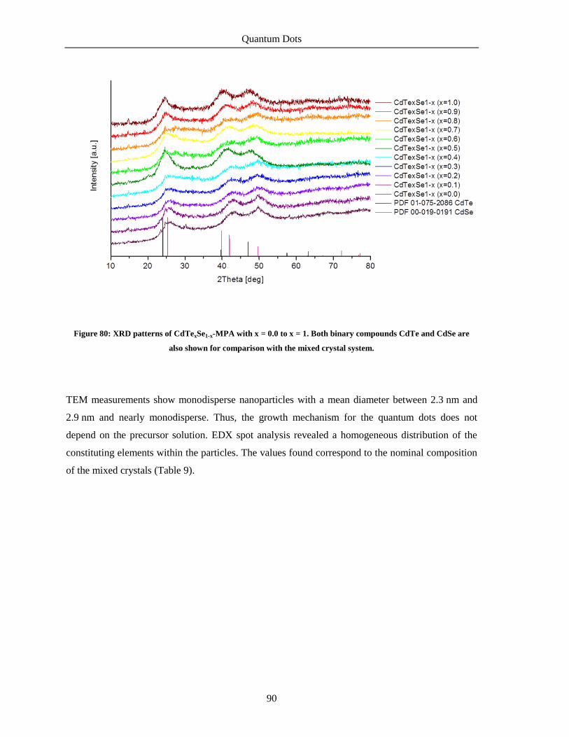
Quantum Dots
90
Figure 80: XRD patterns of CdTexSe1-x-MPA with x = 0.0 to x = 1. Both binary compounds CdTe and CdSe are
also shown for comparison with the mixed crystal system.
TEM measurements show monodisperse nanoparticles with a mean diameter between 2.3 nm and
2.9 nm and nearly monodisperse. Thus, the growth mechanism for the quantum dots does not
depend on the precursor solution. EDX spot analysis revealed a homogeneous distribution of the
constituting elements within the particles. The values found correspond to the nominal composition
of the mixed crystals (Table 9).
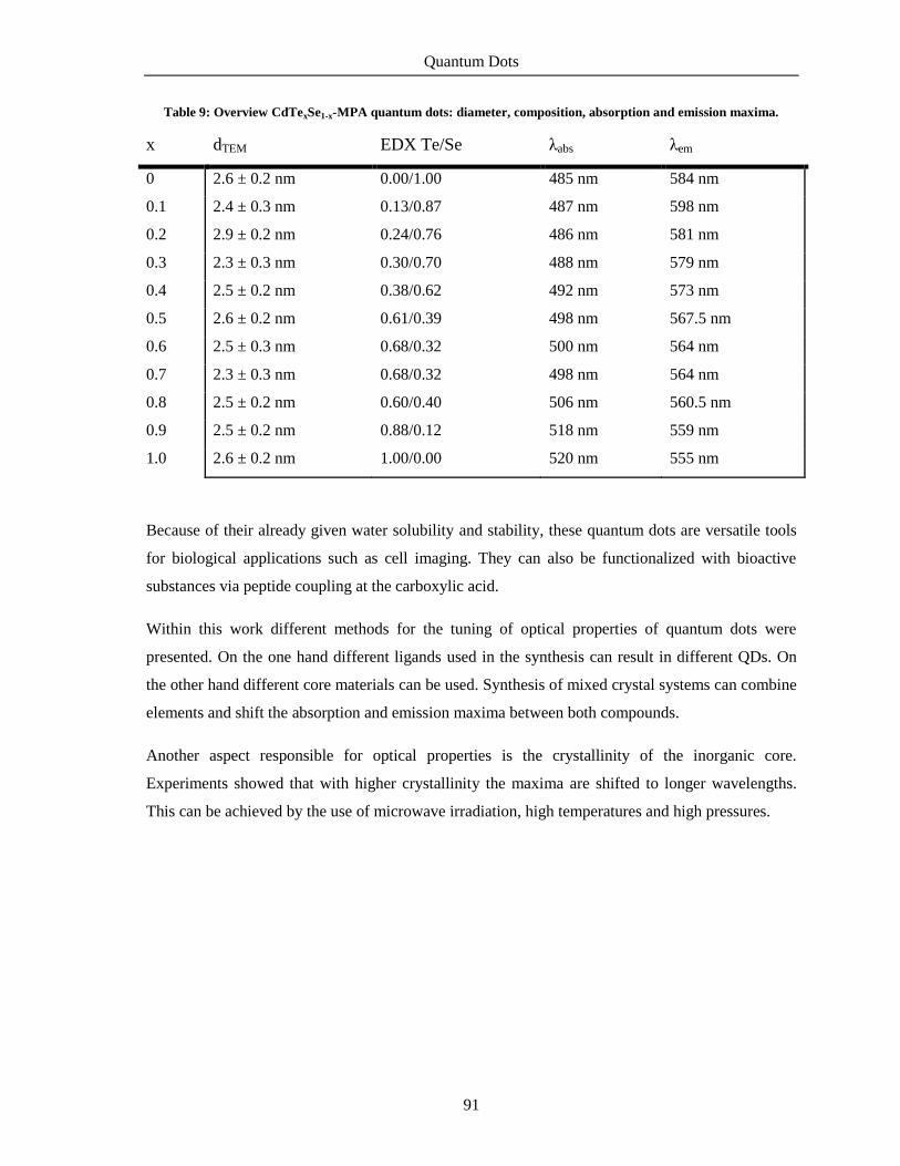
Quantum Dots
91
Table 9: Overview CdTexSe1-x-MPA quantum dots: diameter, composition, absorption and emission maxima.
x dTEM EDX Te/Se λabs λem
0 2.6 ± 0.2 nm 0.00/1.00 485 nm 584 nm
0.1 2.4 ± 0.3 nm 0.13/0.87 487 nm 598 nm
0.2 2.9 ± 0.2 nm 0.24/0.76 486 nm 581 nm
0.3 2.3 ± 0.3 nm 0.30/0.70 488 nm 579 nm
0.4 2.5 ± 0.2 nm 0.38/0.62 492 nm 573 nm
0.5 2.6 ± 0.2 nm 0.61/0.39 498 nm 567.5 nm
0.6 2.5 ± 0.3 nm 0.68/0.32 500 nm 564 nm
0.7 2.3 ± 0.3 nm 0.68/0.32 498 nm 564 nm
0.8 2.5 ± 0.2 nm 0.60/0.40 506 nm 560.5 nm
0.9 2.5 ± 0.2 nm 0.88/0.12 518 nm 559 nm
1.0 2.6 ± 0.2 nm 1.00/0.00 520 nm 555 nm
Because of their already given water solubility and stability, these quantum dots are versatile tools
for biological applications such as cell imaging. They can also be functionalized with bioactive
substances via peptide coupling at the carboxylic acid.
Within this work different methods for the tuning of optical properties of quantum dots were
presented. On the one hand different ligands used in the synthesis can result in different QDs. On
the other hand different core materials can be used. Synthesis of mixed crystal systems can combine
elements and shift the absorption and emission maxima between both compounds.
Another aspect responsible for optical properties is the crystallinity of the inorganic core.
Experiments showed that with higher crystallinity the maxima are shifted to longer wavelengths.
This can be achieved by the use of microwave irradiation, high temperatures and high pressures.

Quantum Dots
92

Summary/Zusammenfassung
93
6. Summary/Zusammenfassung
6.1 Summary
In conclusion this work presents a successful application of newly developed conjugates of
bioactive molecule and nanoparticle. A remarkable enhancement of receptor activation was
achieved by multivalent presentation of active moieties supported on gold nanoparticles.
For this purpose diverse gold nanoparticles were synthesized and functionalized with biogenic
amines. Different synthetic approaches were used to obtain gold nanoparticles between 4 nm and 25
nm. Small gold nanoparticles of 4 nm, 6 nm and 7 nm were synthesized in organic solution; larger
gold nanoparticles of 14 nm and 25 nm were synthesized by citrate reduction of the precursor salt in
aqueous solution. All types were rather monodisperse in size. Next they were equipped with
mercaptocarboxylic acids of different chain lengths via ligand exchange reaction making all gold
nanoparticles water soluble. This kind of ligand provides an additional group for functionalization
at the ligand periphery. Only with 11-mercaptoundecanoic acid (MUDA) was the resulting colloidal
solution stable and could be used for further modifications. Molecules possessing an amino group
can be coupled onto the gold nanoparticles via EDC/NHS (1-ethyl-3-(3-
dimethymaminopropyl)carbodiimide/N-hydroxysuccinimide) mediated peptide coupling reaction.
Four different amines were immobilized on gold nanoparticles of 14 nm diameter: histamine (Au-
MUDA-HA), dopamine (Au-MUDA-DA), serotonin (Au-MUDA-5HT) and carbachol (Au-
MUDA-CB).
Histamine functionalized 14 nm gold nanoparticles were applied as new agonists in ligand-receptor
interaction (Figure 81). As a functional read-out system, chloride secretion resulting from
stimulation of neuronal and epithelial histamine H1 and H2 receptors was measured in Ussing
chamber experiments. Compared to native histamine the multivalent presentation of histamine
resulted in a potentiation of activation ability. Extremely low concentrations were necessary to
induce a chloride secretion across rat colonic epithelium. For comparison the monovalent analogue
AcS-MUDA-HA and gold nanoparticles without an active moiety Au-MUDOLS were also
synthesized and tested. The measured effects could be attributed to Au-MUDA-HA as both
references showed no activity at low concentrations. Only Au-MUDOLS harmed the tissue
irreversibly. The observed effects could be explained by a high local density of histaminic moieties

Summary/Zusammenfassung
94
in close proximity. Tests with 7 nm and 25 nm gold nanoparticles did not show consistent results in
the experimental setup and no statement of size dependence can be made.
Figure 81: Histamine functionalized gold nanoparticles and native histamine.
Dopamine functionalized gold nanoparticles Au-MUDA-DA were not able to induce a chloride
secretion at the administered concentrations. The colloidal solution changed color indicating an
alteration. However multivalent presentation of serotonin on the gold nanoparticles Au-MUDA-
5HT was effective. Chloride secretion could be measured in Ussing chamber experiments. Again
very low concentrations were needed for activation of receptors. The stable acetylcholine derivative
carbachol immobilized at the gold nanoparticles Au-MUDA-CB also showed excellent results in
Ussing chamber experiments. Au-MUDA-CB evoked a response at picomolar concentrations. Gold
nanoparticles with only free carboxylate moieties Au-MUDA were not inert in the experimental
setup and they were able to activate receptors unspecifically. Again all enhanced interactions can be
referred to the multivalent supporting of the active moieties.
In a further part of this work quantum dots (QDs) for cellular imaging were synthesized. CdTe and
CdSe based nanoparticles emit in the visible spectrum of light when their core size is smaller than
the Exciton bohr radius. QDs were synthesized directly in aqueous solution using different
mercaptocarboxylic acids as capping ligands. The optical properties were determined. For
biological application the QDs were covered with a ZnS shell. The presence of the additional shell
has only slight influence on the optical properties of the QDs. Both passivated and unpassivated
nanoparticles were applied for cell imaging. Carboxylic acid terminated QDs showed a better cell

Summary/Zusammenfassung
95
uptake than the sulfonated analogue, passivated QDs showed a faster cell uptake. CdTe/ZnS-TGA
(thioglycolic acid) showed the best results in the cell imaging study.
Additionally the ternary system consisting of both parts of CdTe and CdSe was synthesized and
characterized. Optical properties of the mixed crystal system shift linearly between both binary
compounds. The size of the QDs did not differ significantly. This synthetic method can be used for
fine tuning of emitting-properties without changing the particle size, which is necessary in some
biological applications.
6.2 Zusammenfassung
In dieser Arbeit wurde eine erfolgreiche Anwendung von neu entwickelten Konjugaten aus
bioaktivem Molekül und Nanopartikel vorgestellt. Es konnte eine erhebliche Verbesserung in der
Rezeptoraktivierung durch multivalente Präsentation von aktiven Substanzen auf Goldnanopartikeln
erzielt werden.
Hierzu wurden diverse Goldnanopartikel synthetisiert und funktionalisiert. Verschiedene
synthetische Ansätze wurden ausgewählt, um Goldnanopartikel zwischen 4 nm und 25 nm zu
erhalten. Kleine Goldnanopartikel mit 4 nm, 6 nm und 7 nm Größe wurden in organischer Lösung
hergestellt, größere Goldnanopartikel mit 14 nm und 25 nm Größe wurden mittels Citrat-Reduktion
eines Goldsalzes synthetisiert. Alle dargestellten Partikel waren relativ monodispers in ihrer Form.
Anschließend wurden Mercaptocarbonsäuren verschiedener Kettenlängen mittels
Ligandenaustauschreaktion auf die Partikel gebracht, wodurch auch die Partikel aus der organischen
Synthese wasserlöslich wurden. Diese Art Liganden beinhaltet eine zusätzliche funktionelle Gruppe
zur Funktionalisierung an der Ligandenperipherie. Nur mit 11-Mercaptoundecansäure (MUDA)
konnten stabile kolloidale Lösungen erhalten werden, die für weitere Modifikationen genutzt
werden können. Moleküle mit einer Aminogruppe können über EDC/NHS (1-Ethyl-3-(3-
dimethymaminopropyl)carbodiimide/N-Hydroxysuccinimide) vermittelte Peptidkupplung auf den
Goldnanopartikeln angebracht werden. Vier verschiedene Amine wurden auf Goldnanopartikel mit
14 nm Durchmesser immobilisiert: Histamin (Au-MUDA-HA), Dopamin (Au-MUDA-DA),
Serotonin (Au-MUDA-5HT) und Carbachol (Au-MUDA-CB).
Histamin-funktionalisierte Goldnanopartikel mit 14 nm Größe wurden als neue Agonisten in der
Ligand-Rezeptor-Wechselwirkung (Abbildung 81) angewendet. Gemessen wurde die Chlorid-
Sekretion durch die Stimulierung von neuronalen und epithelialen Histamin H1 und H2 Rezeptoren

Summary/Zusammenfassung
96
in Ussing-Kammer-Experimenten gemessen. Im Vergleich zu nativem Histamin führte die
multivalente Präsentation von Histamin zu einer Potenzierung der Aktivierungsfähigkeit. Es waren
extrem niedrigen Konzentrationen notwendig, um eine Chlorid-Sekretion am Ratten Kolonepithel
zu induzieren. Zum Vergleich wurden auch das monovalente Analogon AcS-MUDA-HA und
Goldnanopartikel ohne eine aktive Einheit Au-MUDOLS synthetisiert und getestet. Die
gemessenen Effekte konnten so Au-MUDA-HA zugeschrieben werden, da beide Referenzen keine
Aktivität bei geringen Konzentrationen zeigten. Nur Au-MUDOLS zerstörte das Gewebe
irreversibel. Die beobachteten Effekte können durch eine hohe lokale Dichte von Histamin-
Einheiten in unmittelbarer Nähe erklärt werden. Die ebenfalls getesteten Goldnanopartikel mit 7 nm
und 25 nm Größe zeigten keine konsistenten Ergebnisse und es kann keine Aussage über die
Größenabhängigkeit gemacht werden.
Dopamin-funktionalisierte Goldnanopartikel Au-MUDA-DA waren nicht in der Lage, ein Chlorid-
Sekretion bei den verabreichten Konzentrationen zu induzieren. Die Farbänderung der kolloidalen
Lösung deutet darauf hin, dass eine Veränderung der Struktur stattgefunden hat. Allerdings zeigte
die multivalente Präsentation von Serotonin auf den Goldnanopartikeln Au-MUDA-5HT eine
Wirkung. Eine Chlorid-Sekretion konnte in den Ussing-Kammer-Experimenten gemessen werden.
Auch hier wurden sehr geringe Konzentrationen für die Aktivierung der Rezeptoren benötigt. Das
stabile Acetylcholin-Derivat Carbachol immobilisiert auf Goldnanopartikeln Au-MUDA-CB zeigte
auch hervorragende Ergebnisse in den Ussing-Kammer-Experimenten. Au-MUDA-CB zeigte eine
Antwort im pikomolaren Bereich auf. Goldnanopartikel mit nur freien Carboxylat-Einheiten Au-
MUDA waren nicht inert; sie waren in der Lage, Rezeptoren unspezifisch zu aktivieren. Alle
gemessenen Effekte können der multivalenten Präsentation der aktiven Einheiten zugeschrieben
werden.
Im anderen Teil dieser Arbeit wurden Quantenpunkte (QDs) für zelluläre Bildgebung synthetisiert.
CdTe- und CdSe- basierte Nanopartikel emittieren im sichtbaren Bereich des Lichtes, wenn ihre
Kerngröße kleiner als der Exciton-Bohr-Radius ist. QDs wurden direkt in wässriger Lösung unter
Verwendung verschiedener Mercaptocarbonsäuren als Capping-Liganden synthetisiert. Die
optischen Eigenschaften wurden bestimmt. Für die biologischen Anwendungen wurden die QDs mit
einer ZnS-Schale ummantelt. Das Vorhandensein der zusätzlichen Hülle hat nur geringen Einfluss
auf die optischen Eigenschaften der Quantenpunkte. Beide passivierten und nicht passivierten
Nanopartikel wurden zur zellulären Bildgebung angewendet. Carbonsäuren terminierte QDs zeigten
eine bessere Zellaufnahme als die sulfonierten Analogen, passivierte QDs zeigten eine schnellere
Zellaufnahme. CdTe/ZnS-TGA (Thioglycolsäure) wiesen die besten Ergebnisse in der Studie auf.

Summary/Zusammenfassung
97
Zusätzlich wurde das ternäre System, bestehend aus CdTexSe1-x, synthetisiert und charakterisiert.
Die optischen Eigenschaften des Mischkristallsystems liegen zwischen den beiden binären
Verbindungen und korrelieren linear mit der Zusammensatzung. In der Größe der QDs lag keine
signifikante Abweichung. Diese Synthesemethode kann für die Feinabstimmung der Emissions-
Eigenschaften genutzt werden, ohne dass die Partikelgröße dabei verändert wird, was in einigen
biologischen Anwendungen notwendig ist.

Summary/Zusammenfassung
98

Outlook
99
7. Outlook
After the successful immobilization of the four neurotransmitters and the ability of these conjugates
to activate receptors, further experiments will be done to explain these multivalent interactions as
there is little research in this field of interest. Histamine functionalized gold nanoparticles with a
diameter of 14 nm were proved to act as their monovalent analogues and were excellent activators
for receptor functionality. Another attempt can be made on the synthesis of dopamine
functionalized gold nanoparticles. These conjugates have versatile applications. Because of the
basic pH value, the catechol moiety oxidized in situ, reaction conditions and synthetic strategy must
be optimized. The multivalent presentation of carbachol on 14 nm gold nanoparticles also showed
encouraging first results. The effect of those conjugates on the cholinergic systems should be
studied in depth.
Investigations ought to be done on the reactivity of receptor blockers immobilized on gold
nanoparticles. It can be presumed that the activation concentration of multivalently presented
blockers is also lower than that of the molecular agents. The use of receptor blockers is far more
widespread and applied in therapy. Atropine is an interesting representative of receptor blockers. It
addresses muscarinic receptors and is an anticholinergic blocking the effect of acetylcholine in the
body. Immobilization cannot be accomplished as before, the molecule first has to be modified and
provided with a linker anchoring at the gold surface.
Moreover the size dependence of the effects should be investigated in detail. A variety of
nanoparticles with different core sizes but the same ligands at the surface can be compared in their
activating ability. The core materials can also be exchanged for cheaper materials or materials with
additional properties and used for biological tests. Iron oxide based nanoparticles can also be
synthesized relatively easily in different core sizes from affordable precursors and their
biocompatibility is also very high. Furthermore they inherit the property of superparamagnetism. A
direct synthesis in aqueous solution yielding monodisperse nanoparticles could be developed.
Another closer look ought to be taken at the molecular interactions at the interface between
nanoparticle and receptor. The multivalent interactions contributing to the huge potency of the
conjugates can be determined in detail helping to understand the mechanisms. Sophisticated cell
analysis can be used to gain insight in subcellular structures and reactions. This might help to
develop new nanomaterials for therapy. For this purpose the nanoparticles can be equipped with a

Outlook
100
fluorescent dye. With the help of fluorescence microscopy the location of the nanoparticles in the
tissue can be visualized. It should also be examined whether the gold nanoparticles are able to
overcome barriers like cell walls. The surface can be covered with ligands favoring a cross over
membranes. If this is successful it might be possible to set a controlled transport across the barrier.
Here again a size dependency should be investigated. Functionalized quantum dots can also be
applied for cell imaging to give information about the remains of the nanoparticulate materials.
Besides all these applications also the toxicity of the nanoparticles should be kept in mind. Size and
ligand dependent cytotoxicity can often be observed for nanoparticles in biological systems.
Especially the gold core and the time in the body after the reaction should be investigated in detail.
Biocompatibility of the nanoparticles is crucial for further applications.
Multivalent presentation of active substances proved to enhance the interactions and gives
promising outlook. Systematic investigations and the synthesis of novel conjugates will open the
door to new developments in nanomedicine and nanomaterial based applications.

Experimental Section
101
8. Experimental Section
8.1 Chemicals, Solvents and Consumables
All chemicals were purchased from Acros Organics, Alfa Aesar, Sigma Aldrich, Merck,
Novabiochem or Fluka at the highest purity grade available, solvents were purchased anhydrous and
with 99.9% purity unless otherwise noted. All solvents were distilled prior to use. All reactions
under inert conditions were carried out in argon atmosphere (99.99%, Messer Griesheim)
employing oven- and flame-dried glassware.
Dry DMF was prepared as followed: DMF was refluxed under argon with sodium and
benzophenone for several hours until the solution turned blue. Anhydrous DMF was stored over
molecular sieve 4 Å under argon.
For the synthesis of nanoparticles ultrapure water from Millipore was used.
For dialysis of the particle solution a membrane of regenerated cellulose with different pore size by
Roth was used. The membrane was treated according to the producer which means soaking for
30 min in distilled water followed by washing. All dialyses were prepared at room temperature in
the given solvent for a given time.
Flash column chromatography and filtration was performed using Merck silica gel 60 Å (0.040 –
0.063 mm mesh size). Analytical thin-layer chromatography (TLC) was performed using pre-coated
polyester sheets Polygram® SIL G/UV254 Machery-Nagel, 0.2 mm silica gel with fluorescent
indicator.
8.2 Analytics
NMR Analysis:
1H and
13C NMR spectra were recorded on Bruker BioSpin Avance III AV600 (600 MHz), AV400
(400 MHz) or AV200 (200 MHz) spectrometers using TMS as an internal standard with chemical
shifts given in ppm relative to TMS (δ = 0.00 ppm) or the respective solvent peaks. 1H NMR data

Experimental Section
102
are reported as follows: chemical shifts (multiplicity [ppm], coupling constants [Hz], integration,
classification). Multiplicity is recorded as s = singlet, br s = broadened singlet, d = doublet, t =
triplet, q quartet, m = multiplet. For 13
C NMR, chemical shifts and structural assignments are
reported where applicable.
IR:
The IR measurements were performer on a Bruker Alpha IR spectrometer utilizing KBr cells
(d = 0.5 mm).
UV/Vis:
The UV/Vis spectra were recorded with an Agilent 8453 spectrophotometer (Agilent Technologies
Inc., Santa Clara, CA, USA).
CHN-Analysis:
Elemental analyses were obtained with a Carlo Erba 1106 (balance: Mettler Toledo UMX-2)
analyzer.
ESI:
Mass spectra were recorded on a Finnigan LCQDuo spectrometer using methanol solutions of the
respective compounds.
TEM:
The characterization of the particle cores was accomplished by a transmission electron microscope
CM30/STEM by Philps with a LaB6 cathode, 300 kV equipped with a GATAN digital camera.
10 µL of the colloidal solution was placed on a carbon-coated copper grid, followed by natural
evaporation of the solvent at room temperature. For determination of the size and standard deviation
100 particles were measured.
DLS and ζ-potential:
The hydrodynamic radius in solution was determined by a STABISIZER PMX-200 from Particle
Metrix GmbH via dynamic light scattering setting set zero 20 s, 3 x 180 s per run, 3 runs. Zeta-
potentials were measured on a NanoZS by Malvern.

Experimental Section
103
XRD:
The crystal structure of the nanoparticles was verified by powder X-ray diffraction in reflection
mode on a PANalytical X’Pert PRO instrument with Cu Kα radiation (λ = 1.54 Å) at an operating
voltage of 40 kV and a current of 40 mA (divergence aperture 2°).
Crystallite size was calculated with Scherrer equation (d = crystallite size, k = shape factor 0.9, λ =
irradiation wavelength, β1/2 = reflection width at half height, θ = diffraction angle).
𝑑 = 𝑘 ∙ 𝜆
𝛽1/2 ∙ cos 𝜃
TGA:
Thermogravimetric measurements were done with a Netzsch STA409PC thermoscale with an
attached quadrupole mass spectrometer QMG421 by Balzers, ionization energy 70 eV.

Experimental Section
104
8.3 Synthesis of the Nanoparticles
Citrate-coordinated gold nanoparticles (Au-Citrate Ø 14 nm)
A solution of 50 mg (0.127 mmol) HAuCl4∙3 H2O in 195 mL of
Millipore water was heated to reflux for 20 min. Under vigorous
stirring a solution of 224 mg (0.52 mmol) of sodium citrate in 5 mL of Millipore water was added
quickly. The reaction mixture was held at 80 °C for 2 h. Then the solution was cooled to 0 °C in an
ice bath and filtered (0.2 µm pore size). A clear red solution with a particle concentration of 2.7 nM
was obtained and stored at 4 °C.
1H NMR (400 MHz, D2O): δ/ppm = 2.61 (bs, CH2); IR (KBr disc): ν/cm
-1 = 3426.8 (νO-H), 1598.2
(νC=O), 1397.6, 1258.4, 618.0; TEM: d = 14.0 ± 0.9 nm; UV/Vis: λmax = 522 nm; DLS:
dhydr = 17 ± 3 nm.
Citrate-coordinated gold nanoparticles (Au-Citrate Ø 25 nm)
A solution of 2.5 mg (0.018 mmol) HAuCl4·3 H2O in 72 mL of
Millipore water was heated to reflux for 10 min. Under vigorous
stirring a solution of 9 mg (0.03 mmol) of sodium citrate in 0.9 mL of Millipore water was added
quickly. The reaction mixture was held at 80 °C for 30 min. The solution turned first blue then
purple. Subsequently the solution was cooled to 0 °C in an ice bath and filtered (0.2 µm pore size).
A clear red solution with a particle concentration of 0.5 nM was obtained and stored at 4 °C.
1H NMR (400 MHz, D2O): δ/ppm = 3.12 (bs, OH), 2.75 (d, J = 15.4 Hz, 2 H, CH2), 2.64 (d, J =
15.4 Hz, 2 H, CH2); IR (KBr disc): ν/cm-1
= 3426.8 (νO-H), 1598.2 (νC=O), 1397.6, 1258.4, 618.0;
TEM: d = 25.3 ± 2.4 nm; UV/Vis: λmax = 529 nm; DLS: dhydr = 26 ± 10 nm.
DT-coordinated gold nanoparticles (Au-DT Ø 4 nm)
62 mg AuPPh3Cl (0.125 mmol) were mixed with 0.06 mL 1-dodecanethiol
(0.25 mmol, 2 equiv.) in 10 mL chloroform to form a clear solution. An amount of 108 mg
(1.25 mmol, 10 equiv.) of tert-butylamin-borane complex was added and the solution turned dark
red. The mixture was heated with stirring at 65 °C for 5 h and then cooled down to room

Experimental Section
105
temperature. Then 40 mL of ethanol were added and the precipitate separated by centrifugation. The
black powder was washed with ethanol three times, dried under vacuum. The gold nanoparticles
were resolved in 10 mL of chloroform to give a dark red solution with a particle concentration of
6.3 µM.
1H NMR (400 MHz, CDCl3): δ/ppm = 1.59-1.52 (m, 6 H, CH2), 1.43-1.39 (m, 2 H, CH2), 1.30-1.22
(m, 10 H, CH2), 1.13-1.09 (m, 4 H, CH2), 0.88 (t, J = 6.9 Hz, 3 H, CH3); IR (KBr disc): ν/cm-1
=
2921.8 (s, νC-H), 2850.9 (s, νC-H), 1488.0, 1385.6, 948.3; TEM: d = 4.0 ± 1.0 nm; UV/Vis:
λmax = 507 nm, DLS: dhydr = 5 ± 1 nm.
DT-coordinated gold nanoparticles (Au-DT Ø 7 nm)
247 mg AuPPh3Cl (0.5 mmol) were mixed with 0.25 mL 1-dodecanethiol
(1 mmol, 2 equiv.) in 40 mL toluene to form a clear solution. An amount of 435 mg (5 mmol,
10 equiv.) of tert-butylamin-borane complex was added and the solution turned dark red. The
mixture was heated with stirring at 55 °C for one hour and then cooled down to room temperature.
Then 40 mL of ethanol were added and the precipitate separated by centrifugation. The black
powder was washed with ethanol three times, dried under vacuum. The gold nanoparticles were
resolved in 10 mL of chloroform to give a dark red solution with a particle concentration of 7.5 µM.
The use of benzene instead of toluene results in gold nanoparticles with a mean diameter of 6 nm.
1H NMR (400 MHz, CDCl3): δ/ppm = 1.60-1.49 (m, 10 H, CH2), 1.33-1.19 (m, 12 H, CH2), 0.88 (t,
J = 7.2 Hz, 3 H, CH3); IR (KBr disc): ν/cm-1
= 2917.1 (s, νC-H), 2844.2 (s, νC-H), 1454.8, 1432.8,
716.1, 687.7, 517.7; TEM: d = 7.1 ± 0.8 nm; UV/Vis: λmax = 518 nm; DLS: dhydr = 8 ± 1 nm.
DMAP-coordinated gold nanoparticles (Au-DMAP Ø 6 nm)
An aqueous solution of HAuCl4 (24 mg in 2 mL) was added to a solution of
tetraoctylammonium bromide (TOAB) in toluene (153 mg in 5 mL) and the mixture was stirred
until all the tetrachloroaurate had transferred into the organic phase, to give a deep orange solution.
A fresh solution of NaBH4 (27 mg in 1.5 mL) was added slowly whilst stirring. The toluene phase
immediately turned to ruby red. After stirring for 18 hours, the toluene phase containing TOAB-
stabilized gold NPs was washed with 0.1 M H2SO4, 0.1 M NaOH and H2O (each three times).
Subsequently, an aqueous solution of DMAP (300 mg in 24 mL) was added to the TOAB-stabilized
gold NP solution. Phase transfer of the solution took place simultaneously and a ruby red aqueous
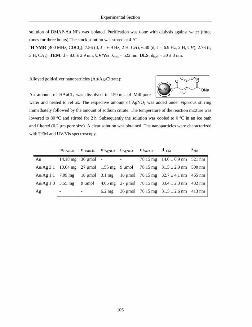
Experimental Section
106
solution of DMAP-Au NPs was isolated. Purification was done with dialysis against water (three
times for three hours).The stock solution was stored at 4 °C.
1H NMR (400 MHz, CDCl3): 7.86 (d, J = 6.9 Hz, 2 H, CH), 6.40 (d, J = 6.9 Hz, 2 H, CH), 2.76 (s,
3 H, CH3); TEM: d = 8.6 ± 2.9 nm; UV/Vis: λmax = 522 nm; DLS: dhydr = 30 ± 3 nm.
Alloyed gold/silver nanoparticles (Au/Ag-Citrate):
An amount of HAuCl4 was dissolved in 150 mL of Millipore
water and heated to reflux. The respective amount of AgNO3 was added under vigorous stirring
immediately followed by the amount of sodium citrate. The temperature of the reaction mixture was
lowered to 80 °C and stirred for 2 h. Subsequently the solution was cooled to 0 °C in an ice bath
and filtered (0.2 µm pore size). A clear solution was obtained. The nanoparticles were characterized
with TEM and UV/Vis spectroscopy.
mHAuCl4 nHAuCl4 mAgNO3 nAgNO3 mNa3Cit dTEM λabs
Au 14.18 mg 36 µmol - - 78.15 mg 14.0 ± 0.9 nm 521 nm
Au/Ag 3:1 10.64 mg 27 µmol 1.55 mg 9 µmol 78.15 mg 31.5 ± 2.9 nm 500 nm
Au/Ag 1:1 7.09 mg 18 µmol 3.1 mg 18 µmol 78.15 mg 32.7 ± 4.1 nm 465 nm
Au/Ag 1:3 3.55 mg 9 µmol 4.65 mg 27 µmol 78.15 mg 33.4 ± 2.3 nm 432 nm
Ag - - 6.2 mg 36 µmol 78.15 mg 31.5 ± 2.6 nm 413 nm

Experimental Section
107
8.4 Ligand Exchange Reactions
General procedure for ligand exchange reaction in water:
I Two-phase system: The ligand (6∙105 equiv.) was dissolved in 5 mL of water and under vigorous
stirring 1 mL of organic solvent including the nanoparticles was added. The reaction mixture was
stirred for 16 h at room temperature whilst a phase transfer could be observed. The organic phase
was removed and the aqueous phase was dialysed against water. The purified colloidal solution was
stored at 4 °C.
II One-phase system: 10 mL of the nanoparticle solution were stirred at room temperature and 6∙105
equivalents of the ligand were added. NaOH was added until a basic solution was obtained. After
16 h the solution was purified via dialysis against water and stored at 4 °C.
Ligand exchange at Au-Citrate 14 nm with different ligands:
According to the general procedure II ligand exchange reactions at Au-
Citrate nanoparticles were performed with 11-mercaptoundecanoic acid (MUDA),
6-mercaptohexanoic acid (MHA), 3-mercaptopropionic acid (MPA), lipoic acid (LA). The resulting
nanoparticles were characterized via UV/Vis.
Ligand amountligand λabs
Au-MPA MPA 9 µL xx
Au-MHA MHA 15 mg xx
Au-MUDA MUDA 22 mg 524 nm
Au-LA LA 20 mg 531 nm
Ligand exchange with MUDA at Au-DT and Au-Citrate:
According to the general procedure I 1 mL of Au-DT (4 nm or 7 nm)
were added to a solution of 44 mg MUDA (0.2 mmol) and 20 mg NaOH (0.5 mmol, pH>8) in 9 mL
water. Dialysis against water three times with 300 mL (MWCO 3500) was performed.

Experimental Section
108
According to general procedure II 10 mL 44 mg MUDA (0.2 mmol) and 20 mg NaOH (0.5 mmol,
pH>8) were added to a solution of Au-Citrate (14 nm or 25 nm). Dialysis against water three times
with 300 mL, MWCO 3500) was performed.
The nanoparticles were characterized with TEM, DLS and UV/Vis. Au-MUDA 14 nm was
synthesized for the publication and characterized additionally with 1H NMR and IR.
dTEM dDLS λabs cNP
4 nm 3.8 ± 0.7 nm 5 ± 1 nm 507 nm 1.26 µM
7 nm 7.5 ± 0.4 nm 8 ± 2 nm 524 nm 1.5 µM
14 nm 14.5 ± 1.2 nm 24 ± 7 nm 527 nm 1 nM
25 nm 25.9 ± 2.3 nm 32 ± 8 nm 532 nm 0.5 nM
Ligand exchange with MUDA at alloyed Au/Ag nanoparticles:
According to general procedure II 10 mL 44 mg MUDA (0.2 mmol) and
20 mg NaOH (0.5 mmol, pH>8) were added to citrate-stabilized gold, silver and the alloyed
nanoparticles. Dialysis against water three times with 300 mL (MWCO 3500) was performed.
The nanoparticles were characterized with UV/Vis.
Particle λabs
Au Au-MUDA 524 nm
Au/Ag 3:1 Au0.75Ag0.25-MUDA 499 nm
Au/Ag 1:1 Au0.5Ag0.5-MUDA 473 nm
Au/Ag 1:3 Au0.25Ag0.75-MUDA 444 nm
0:1 Ag-MUDA 424 nm
Ligand exchange with MUDOLS at Au-DT and Au-Citrate:
The ligand MUDOLS was synthesized separately.
According to the general procedure I 1 mL of Au-DT (7 nm) were added to a solution of 60 mg
MUDOLS (0.2 mmol) and 20 mg NaOH (0.5 mmol, pH>8) in 9 mL water. Dialysis against water
three times with 300 mL (MWCO 3500) was performed.
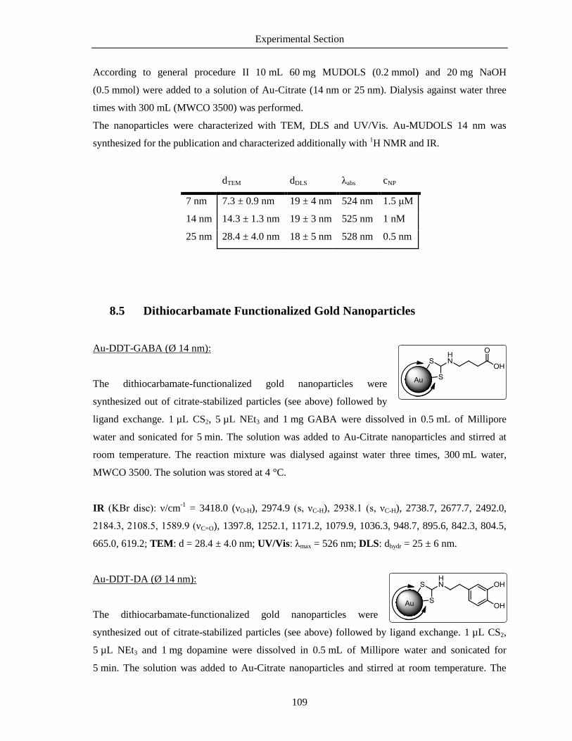
Experimental Section
109
According to general procedure II 10 mL 60 mg MUDOLS (0.2 mmol) and 20 mg NaOH
(0.5 mmol) were added to a solution of Au-Citrate (14 nm or 25 nm). Dialysis against water three
times with 300 mL (MWCO 3500) was performed.
The nanoparticles were characterized with TEM, DLS and UV/Vis. Au-MUDOLS 14 nm was
synthesized for the publication and characterized additionally with 1H NMR and IR.
dTEM dDLS λabs cNP
7 nm 7.3 ± 0.9 nm 19 ± 4 nm 524 nm 1.5 μM
14 nm 14.3 ± 1.3 nm 19 ± 3 nm 525 nm 1 nM
25 nm 28.4 ± 4.0 nm 18 ± 5 nm 528 nm 0.5 nm
8.5 Dithiocarbamate Functionalized Gold Nanoparticles
Au-DDT-GABA (Ø 14 nm):
The dithiocarbamate-functionalized gold nanoparticles were
synthesized out of citrate-stabilized particles (see above) followed by
ligand exchange. 1 µL CS2, 5 µL NEt3 and 1 mg GABA were dissolved in 0.5 mL of Millipore
water and sonicated for 5 min. The solution was added to Au-Citrate nanoparticles and stirred at
room temperature. The reaction mixture was dialysed against water three times, 300 mL water,
MWCO 3500. The solution was stored at 4 °C.
IR (KBr disc): ν/cm-1
= 3418.0 (νO-H), 2974.9 (s, νC-H), 2938.1 (s, νC-H), 2738.7, 2677.7, 2492.0,
2184.3, 2108.5, 1589.9 (νC=O), 1397.8, 1252.1, 1171.2, 1079.9, 1036.3, 948.7, 895.6, 842.3, 804.5,
665.0, 619.2; TEM: d = 28.4 ± 4.0 nm; UV/Vis: λmax = 526 nm; DLS: dhydr = 25 ± 6 nm.
Au-DDT-DA (Ø 14 nm):
The dithiocarbamate-functionalized gold nanoparticles were
synthesized out of citrate-stabilized particles (see above) followed by ligand exchange. 1 µL CS2,
5 µL NEt3 and 1 mg dopamine were dissolved in 0.5 mL of Millipore water and sonicated for
5 min. The solution was added to Au-Citrate nanoparticles and stirred at room temperature. The

Experimental Section
110
reaction mixture was dialysed against water: three times, 300 mL water, MWCO 3500. The solution
was stored at 4 °C.
IR (KBr disc): ν/ cm-1
= 3431.7 (νO-H), 2922.3 (s, νC-H), 2851.1 (s, νC-H), 1635.2 (νC=O), 1488.0,
1403.4, 1045.2, 948.5, 771.7, 463.8; TEM: d = 25.9 ± 1.1 nm; UV/Vis: λmax = 543 nm; DLS:
dhydr = 35 ± 7 nm.
8.6 Positively Charged Gold Nanoparticles
Au-CA-NAc (Ø 14 nm)
According to the general procedure II 12 mg NAc-CA∙HCl (0.1 mmol)
were added to a solution of Au-Citrate (cNP = 1 nM) and stirred for 20 hours. The reaction mixture
was dialysed against water: three times, 300 mL water, MWCO 3500. The solution was stored at
4 °C. A clear violet solution with a particle concentration of 1 nM was obtained and stored at 4 °C.
1H NMR (400 MHz, D2O): δ/ppm = 3.09 (s, 3H, CH3), 2.58 (d, J = 15.6 Hz, 2 H, CH2), 2.44 (d, J =
15.6 Hz, 2 H, CH2); IR (KBr disc): ν/cm-1
= 3442.7 (νO-H), 2919.8 (s, νC-H), 2580.3, 2359.3, 2336.4,
1636.7 (νC=O), 1384.3; TEM: d = 14.1 ± 3.6 nm, UV/Vis: λmax = 525 nm, DLS: dhydr = 14 ± 4 nm.
Cysteamine-coordinated gold nanoparticles (Au-CA Ø 30 nm)
Briefly, 16.4 mg of cysteamine hydrochloride (0.2 mmol) were dissolved in
1 mL water. 400 µL (85 µmol) were added quickly to a solution of 22 mg HAuCl4∙3 H2O (65 µmol)
in 20 mL water. The mixture was stirred for 20 min at room temperature in the dark. Then, 10 µL of
freshly prepared NaBH4 solution (3.8 mg in 10 mL, 0.1 µmol) was added quickly under vigorous
stirring. After 30 min the mixture turned red and was stirred for another 16 h. Due to instability and
incompatibility with the dialysis material, the particles were subsequently functionalized.
The nanoparticles were characterized with TEM, UV/Vis and DLS.
TEM: d = 30.5 ± 5.3 nm; UV/Vis: λmax = 525 nm; DLS: dhydr = 60 ± 31 nm.

Experimental Section
111
8.7 Mixed Ligand Shell
Au-MUDA/MUDOLS (Ø 14 nm)
Stock solutions of MUDA and MUDOLS in water with a
concentration of 10 mM were prepared. Stoichiometric amounts of
MUDA and MUDOLS were added to a solution of Au-Citrate under vigorous stirring. The reaction
mixtures were purified via dialysis against water: 3 x 3 h with 300 ml, MWCO 3.5. Clear violet
solutions were obtained and stored at 4 °C. 1: Au-MUDA, 2: Au-MUDA/MUDOLS 2:1, 3: Au-
MUDA/MUDOLS 1:1, 4: Au-MUDA/MUDOLS 1:2, 5: Au-MUDOLS. The nanoparticles were
characterized with UV/Vis, DLS and ζ-potential.
Shell λabs dhydr ζ potential Conductivity
Au-Citrate - 521 nm 17 ± 3 nm -28 ± 6 mV 1.11 µS/cm
1 Au-MUDA 1:0 526 nm 24 ± 7 nm -57 ± 17 mV 8.91 µS/cm
2 Au-MUDA/OLS 2:1 526 nm 34 ± 4 nm -58 ± 23 mV 0.67 µS/cm
3 Au-MUDA/OLS 1:1 529 nm 38 ± 4 nm -63 ± 12 mV 0.58 µS/cm
4 Au-MUDA/OLS 1:2 526 nm 32 ± 4 nm -68 ± 12 mV 0.55 µS/cm
5 Au-MUDOLS 0:1 525 nm 19 ± 3 nm -70 ± 13 mV 0.23 µS/cm

Experimental Section
112
8.8 Functionalization of the Particles Shell
General procedure for the EDC/NHS mediated coupling in water:
10 mL of the nanoparticle solution were transferred in a glass vessel washed with aqua regia and
distilled water. 6∙105 equivalents of the respecting amine, EDC and NHS were dissolved in 0.5 mL
of water with 12∙105 equiv. of NEt3 and added to the nanoparticle solution. The reaction mixture
was stirred overnight and subsequently dialysed against water (3 x 3 h with 300 mL, MWCO 3500).
Au-MUDA-DA
According to the general procedure, dopamine was
attached at Au-MUDA nanoparticles 14 nm. A clear red
solution was obtained at stored at 4 °C.
7 nm:
TEM: d = 7.8 ± 1.2 nm; UV/Vis: λmax = 526 nm; DLS: dhydr = 9 ± 2 nm.
14 nm:
1H NMR (400 MHz, D2O): δ/ppm = 6.80 (d, J = 7.8 Hz, 1 H, CH), 6.74 (s, 1 H, CH), 6.54 (d, J =
7.9 Hz, 1 H, CH), 5.84 (s, 1 H, NH), 3.46 (q, 2 H, CH2), 2,85 ( t, 2 H, CH2), 2.67 (t, 2 H, CH2), 2.32
(s, 3 H, CH3), 2.14 (t, 2 H, CH2), 1.64-1.49 (m, 4 H, CH2), 1.39-1.17 (m, 14 H, CH2); IR (KBr
disc): ν/cm-1
= 3431.1, 3018.7 (νO-H), 2919.7 (s, νC-H), 2849.9 (s, νC-H), 1704.4 (νC=O, amide), 1656.4,
1488.1, 1404.3, 1384.9, 1290.3, 1231.9, 1081.1, 1003.7, 956.4, 948.6, 815.7, 720.4, 662.9; TEM:
d = 14.7 ± 1.2 nm; UV/Vis: λmax = 525 nm; DLS: dhydr = 26 ± 6 nm.
Au-MUDA-HA
According to the general procedure, histamine was attached at
Au-MUDA nanoparticles with three different sizes. A clear red
solution was obtained at stored at 4 °C.
7 nm:
1H NMR (400 MHz, D2O): δ/ppm = 7.62 (s, 1 H, CH), 7.30 (s, 1 H, CH), 6.90 (m, 1 H, NH), 3.15
(m, 2 H, CH2), 2.99 (m, 2 H, CH2), 2.84 (m, 2 H, CH2), 2.79-2.70 (m, 6 H, CH2), 2.09-2.03 (m, 1 H,

Experimental Section
113
CH2), 1.85-1.74 (m, 2 H, CH2), 1.66-1.58 (m, 1 H, CH2), 1.52-1.37(m, 1 H, CH2), 1.33-1.11 (m,
10 H, CH2); TEM: d = 7.3 ± 0.9 nm; UV/Vis: λmax = 524 nm; DLS: dhydr = 11 ± 2 nm.
14 nm:
1H NMR (400 MHz, D2O): δ/ppm = 7.55 (s, 1 H, CH), 6.81 (s, 1 H, CH), 2.73-2.86 (m, 2 H, CH2
(HA)), 2.57-2.71 (m, 3 H, CH2 (HA) + NH)), 2.41-2.53 (m, 2 H), 2.21-2.34 (m, 4 H), 2.10-2.21 (m,
3 H), 2.03 (t, J = 7.7 Hz, 1 H), 1.52-1.68 (m, 1 H), 1.36-1.48 (m, 1 H), 1.18 (bs, 8 H, CH2); IR (KBr
disc): ν/cm-1
= 3421.1 (νO-H), 3018.8, 2921.4 (s, νC-H), 2850.0 (s, νC-H), 1641.1 (νC=O, amide),
1596.3 (νC-NH), 1488.2, 1404.2, 957.1, 948.6; TEM: d = 14.3 ± 0.7 nm; UV/Vis: λmax = 528 nm;
DLS: dhydr = 19 ± 3 nm
25 nm:
1H NMR (400 MHz, D2O): δ/ppm = 7.55 (s, 1 H, CH), 6.81 (s, 1 H, CH), 2.86-2.72 (m, 2 H, CH2),
2.70-2.57 (m, 2 H, CH2), 2.50-2.43 (m, 2 H, CH2), 2.33-2.22 (m, 4 H, CH2), 2.20-2.11 (m, 3 H,
CH2), 2.10-2.00 (m, 1 H, CH2), 1.66-1.50 (m, 1 H, CH2), 1.46-1.36 (m, 1 H, CH2), 1.31-1.12 (m,
8 H, CH2);TEM: d = 25.7 ± 1.2 nm; UV/Vis: λmax = 534 nm; DLS: dhydr = 32 ± 8 nm
Au-MUDA-5HT
According to the general procedure, serotonin (5-
hydroxytryptamine) was attached at Au-MUDA
nanoparticles 14 nm. The reaction mixture was stirred under argon atmosphere. The solution was
dialysed with degassed water under argon atmosphere and stored under argon atmosphere. A clear
red solution was obtained at stored at 4 °C.
1H NMR (400 MHz, D2O): δ/ppm = 7.40-7.30 (m, 1 H, CH), 7.25-7.16 (m, 1 H, CH), 7.08-7.00 (m,
1 H, CH), 6.68-6.75 (m, 1 H, CH), 3.28-3.21 (m, 2 H, CH2), 2.99-2.92 (m, 2 H, CH2), 2.15-2.08 (m,
2 H, CH2), 1.88-1.80 (m, 2 H, CH2), 1.55-1.42 (m, 4 H, CH2), 1.36-1.31 (m, 1 H, CH2), 1.29-1.22
(m, 2 H, CH2); IR (KBr disc): ν/cm-1
= 3439.5 (νO-H), 2921.3 (s, νC-H), 2848.0 (s, νC-H), 1629.3 (νC=O,
amide), 1488.1, 1384.4, 948.3, 617.1; TEM: d = 14.1 ± 0.9 nm; UV/Vis: λmax = 528 nm; DLS:
dhydr = 16 ± 4 nm.

Experimental Section
114
Au-MUDA-CB
According to the general procedure, carbachol was
attached at Au-MUDA nanoparticles 14 nm. The solution
was dialysed three times for 2 hours each. The solution was stored under argon atmosphere in an
Eppendorf cup.
1H NMR (400 MHz, D2O): δ/ppm = 4.47-4.41 (m, 2 H, CH2), 3.66-3.60 (m, 2 H, CH2), 3.14 (s,
3 H, CH3), 3.09-3.00 (m, 4 H, CH2), 1.88-1.78 (m, 2 H, CH2), 1.40-1.16 (m, 12 H, CH2), 1.03-0.98
(m, 2 H, CH2); IR (KBr disc): ν/cm-1
= 3426.7 (νO-H), 2921.0 (s, νC-H), 2848.8 (s, νC-H), 1640.8 (νC=O,
amide), 1576.0, 1487.8, 1465.0, 1408.3, 1342.9, 1238.0, 1099.6, 948.7, 721.5, 663.4; TEM:
d = 14.5 ± 0.7 nm; UV/Vis: λmax = 532 nm; DLS: dhydr = 17 ± 6 nm.
Au-CA-His:
According to the general procedure, L-histamine was
attached at Au-CA nanoparticles 30 nm. A clear red
solution was obtained at stored at 4 °C.
1H NMR (400 MHz, D2O): δ/ppm = 7.73 (s, 1 H, CH), 7.64 (s, 1 H, CH), 7.39 (s, 1 H, CH), 3.11-
3.08 (m, 2 H, CH2), 2.80-2.75 (m, 1 H, CH2), 1.80-1.77 (m, 4 H, CH2); TEM: d = 30.7 ± 7.2 nm;
UV/Vis: λmax =527 nm; DLS: dhydr = 45 ± 9 nm.

Experimental Section
115
8.9 Synthesis of the Ligands
11-(Acetylthio)undecanoic acid (AcS-MUDA):
11-(Acetylthio)undecanoic acid was synthesized as reported in the literature
[163]: 500 mg 11-bromoundecanoic acid (1.88 mmol) were dissolved in 10 mL dry DMF.
Separately, 322 mg KSAc (2.82 mmol, 1.5 equiv.) were dissolved in 5 mL dry DMF and added
dropwise to the solution. After stirring for 1.5 h 50 mL of water was added and a white solid
appeared. The precipitate was filtered off, washed three times with water and dried in vacuum.
413.2 mg of a colorless solid (1.6 mmol, 84%) could be obtained.
1H NMR (400 MHz, CDCl3): δ/ppm = 2.78 (t, J = 8 Hz, 2 H, CH2), 2.28 (m, 5 H, CH2 + CH3),
1.61-1.52 (m, 2 H, CH2), 2.52-2.26 (m, 2 H, CH2), 1.32-1.16 (m, 12 H, CH2); 13
C-NMR (100 MHz,
CDCl3): δ/ppm = 196.3 (Cq, C=O, acetyl), 179.5 (Cq, C=O), 33.9, 31.0 (CH3), 29.5, 29.4, 29.3, 29.2,
29.2, 29.1, 29.0, 28.8, 24.7; IR (KBr disc): ν/cm-1
= 2920.4 (s, νC-H), 2849.9 (s, νC-H), 1697.5 (νC=O,
amide), 1432.7, 1408.9, 1290.6, 1138.8, 950.4, 926.1, 631.5; ESI: [M+Na]+ = 283.1335 (calcd:
[M+Na]+ = 283.1338); CHN: found C 59.84%, H 9.22% (calcd C 59.96%, H 9.29%)
2,5-Dioxopyrrolidin-1-yl-11-(acetylthio)undecanoate (AcS-MUDA-NHS):
195 mg NHS (1.7 mmol, 1.1 equiv.) and 700 mg AcS-MUDA
(1.55 mmol) were dissolved in DCM under stirring at room
temperature. 350 mg DCC (1.7 mmol, 1.1 equiv.) were separately dissolved in 5 mL DCM and
added slowly. After 30 min a white precipitate appeared. The reaction mixture was stirred
overnight. The solvent was removed under reduced pressure. 506 mg (1.42 mmol, 92%) of a
colorless powder were obtained.
1H NMR (400 MHz, CDCl3): δ/ppm = 2.81-2.77 (m, 6 H, CH2), 2.53 (t, J = 7.3 Hz, 2 H, CH2), 2.25
(s, 3 H, CH3), 1.71-1.63 (m, 2 H, CH2), 1.52-1.47 (m, 2 H, CH2), 1.38-1.17 (m, 12 H, CH2);
13C NMR (100 MHz, CDCl3): δ/ppm = 196.1 (Cq, C=O, acetyl), 169.3 (2 x Cq, C=O), 168.7 (Cq,
C=O), 30.9 (CH3), 30.7, 29.5, 29.3, 29.2, 29.2, 29.1, 29.0, 28.8, 28.7, 25.6 (2 x CH2), 24.6; IR (KBr
disc): ν/cm-1
= 2917.6 (s, νC-H), 2848.4 (s, νC-H), 1817.2, 1788.4 (νC=O), 1736.4 (νC=O), 1682.1 (νC=O,
amide), 1541.1, 1465.0, 1425.9, 1413.9, 1366.9, 1208.1, 1129.6, 1076.0, 639.1; ESI: [M+Na]+ =
380.1502 (calcd: [M+Na]+ = 380.1600); CHN: found C 56.22%, H 7.71%, N 4.82% (calcd C
57.12%, H 7.61%, N 3.92%)

Experimental Section
116
S-(11-((2-(1H-imidazol-4-yl)ethyl)amino)-11-oxoundecyl) ethanethioate (AcS-MUDA-HA):
97 mg AcS-MUDA-NHS (0.27 mmol) were dissolved in DMF
to form a clear solution. 34 mg HA∙2 HCl (0.30 mmol,
1.1 equiv.) and a few drops NEt3 were added and the solution was stirred overnight. Subsequently
50 mL water were added and a white solid appeared. The precipitate was filtered off and washed
three times with water. The crude product was dissolved in CHCl3 and filtered again. After
evaporation 70 mg (0.19 mmol, 70%) of a colorless powder was obtained.
1H NMR (400 MHz, CDCl3): δ/ppm = 7.50 (s, 1 H, CHar), 6.75 (s, 1 H, CHar), 6.33 (br s, 1 H, NH),
3.52-3.42 (m, 2 H, CH2 (HA)), 2.79-2.69 (m, 2 H, CH2 (HA)), 2.53-2.39 (m, 2 H, CH2), 2.56 (s,
3 H, CH3), 2.10 (t, J = 7.2 Hz, 2 H, CH2), 1.59-1.43 (m, 4 H, CH2), 1.26-1.14 (m, 12 H, CH2);
13C NMR (100 MHz, CDCl3): δ/ppm = 196.4 (Cq, C=O, acetyl), 173.7 (Cq, C=O, amide), 134.8
(CH, Car), 127.6 (Cq, Car), 118.6 (CH, Car), 39.2 (CH2 (HA)), 36.9 (CH2 (HA)), 30.7 (CH3), 29.5,
29.3, 29.3, 29.2, 29.1, 28.7, 28.4, 26.9, 25.7; IR (KBr disc): ν/cm-1
= 2920.7 (νC-H), 2850.6 (νC-H),
1691.9 (νC=O), 1638.9 (νC=O), 1575.2 (νCN-H), 1466.1, 1452.7, 1255.8, 1090.7, 623.3; ESI: [M+H]+ =
354.2221 (calcd: [M+H]+ = 354.2210); CHN: found C 59.72%, H 8.67%, N 11.37% (calcd C
61.15%, H 8.84%, N 11.89%)
S-(11-((2-(5-hydroxy-1H-indol-3-yl)ethyl)amino)-11-
oxoundecyl) ethanethioate (AcS-MUDA-5HT):
97 mg AcS-MUDA-NHS (0.27 mmol) were dissolved in
DMF to form a clear solution. 64 mg 5HT∙ HCl (0.30 mmol, 1.1 equiv.) and a few drops NEt3 were
added and the solution was stirred overnight. Subsequently 50 mL water were added and a white
solid appeared. The precipitate was filtered off and washed three times with water. The crude
product was dissolved in CHCl3 and filtered again. After evaporation 33 mg (0.08 mmol, 29%) of a
colorless powder were obtained.
1H NMR (400 MHz, CDCl3): δ/ppm = 8.02 (s, 1 H, NH), 7.19-7.12 (m, 1 H, CH), 6.99-6.88
(m,2 H, CH), 6.78-6.72 (m, 1 H, CH), 6.27 (s, 1 H, NH), 5.59 (bs, 1 H, OH), 3.54-3.46 (m, 2 H,
CH2), 2.85-2.75 (m, 4 H, CH2), 2.25 (s, 3 H, CH3), 2.05 (t, J = 7.5 Hz, 2 H, CH2), 1.93-1.82 (m, 4 H,
CH2), 1.23-1.15 (m, 12 H, CH2); 13
C NMR (100 MHz, CDCl3): δ/ppm = 195.5, 172.4, 155.9, 149.1,
130.4, 127.0, 121.9, 111.1, 110.8, 102.1, 48.0, 38.6, 35.9, 32.9, 29.9, 29.6, 28.5, 28.3, 28.2, 28.1,
28.0, 24.6, 23.9.

Experimental Section
117
S-(11-((3,4-dihydroxyphenethyl)amino)-11-oxoundecyl) ethanethioate (AcS-MUDA-DA):
97 mg AcS-MUDA-NHS (0.27 mmol) were dissolved in
4 mL DMF to form a clear solution. 57 mg DA∙HCl
(0.30 mmol, 1.1 equiv.) in 1 mL DMF were added and the solution was stirred overnight.
Subsequently 50 mL water were added and a white solid appeared. The precipitate was filtered off
and washed three times with water. The crude product was dissolved in CHCl3 and filtered again.
After evaporation 54 mg (0.14 mmol, 52%) of a colorless powder were obtained.
1H NMR (400 MHz, CDCl3): δ/ppm = 6.80 (d, J = 7.80 Hz, 1 H, CH), 6.74 (s, 1 H, CH), 6.54 (d, J
= 7.92, 1 H, CH), 5.84 (s, 1 H, NH), 3.46 (q, J = 6.28 Hz, 2 H, CH2), 2.85 (t, J = 7.39 Hz, 2 H,
CH2), 2.67 (t, J = 7.03 Hz, 2 H, CH2), 2.32 (s, 3 H, CH3), 2.14 (t, J = 8.10 Hz, 2 H, CH2), 1.64-1.49
(m, 4 H, CH2), 1.39-1.17 (m, 4 H, CH2); 13
C NMR (100 MHz, CDCl3): δ/ppm = 196.7, 174.5,
144.5, 143.2, 130.5, 120.4, 115.4, 115.1, 40.9, 36.8, 34.9, 30.7, 29.5, 29.3, 29.3, 29.2, 29.1, 29.1,
29.0, 28.7, 28.3; IR (KBr disc): ν/cm-1
= 2919.7 (νC-H), 2849.9 (νC-H), 1704.4 (νC=O), 1656.4 (νC=O,
amide), 1488.1, 1404.3, 1384.9, 1231.9, 956.4, 948.6; 662.9; ESI: [M+H]+ = 418.27 (calcd:
[M+Na]+ = 418.20); CHN: found C 63.22%, H 8.49%, N 3.66% (calcd: C 63.76%, H 8.41%, N
3.54%)
Sodium 11-mercaptoundecyl sulfate (MUDOLS):
In a flame-dried vessel under argon atmosphere, 353 mg of 11-
mercaptoundecanol (1.72 mmol) were dissolved in 3 ml dry DMF. Separately, 395 mg of SO3∙DMF
complex (2.50 mmol, 1.5 equiv.) were dissolved in 1 mL dry DMF and added to the solution. After
stirring for 1 h at room temperature the solvent was removed under reduced pressure (OPV) and the
residue was dissolved in 4 mL of 1 M NaOH. A white precipitate appeared. 15 mL of ethanol were
added and washed three times. The product was dried in vacuum. 280 mg of a colorless solid
(0.91 mmol, 53%) could be obtained, the dimer was formed.
1H NMR (200 MHz, DMSO-d6): δ/ppm = 3.68 (t, J = 6.6 Hz, 2 H, CH2), 3.35 (t, J = 6.3 Hz, 2 H,
CH2), 2.27-2.61 (m, 4 H, CH2), 2.56-2.49 (m, 2 H, CH2), 1.67-1.52 (m, 4 H, CH2), 1.36-1.24 (m,
10 H, CH2); 13
C NMR (200 MHz, DMSO-d6): δ/ppm = 65.6, 60.7, 32.5, 29.1, 29.0, 28.9, 28.8, 28.6,
28.5, 27.7, 25.5; IR (KBr disc): ν/cm-1
= 2918.7 (s, νC-H),, 2849.9 (s, νC-H), 1614.8, 1470.0, 1261.7
& 1205.1 & 1128.1 (s, νR-OSO2-OR), 1068.8, 966.4, 831.8, 718.9, 628.7, 586.3; ESI: [M+Na]+ =
633.16 (calcd: [M+Na]+ = 633.16); CHN: C11H23ClNa2O4S2 found C 35.68%, H 6.01% (calcd: C
36.21%, H 6.35%).

Experimental Section
118
8.10 Synthesis of Quantum Dots
Mercaptocarboxylic acid functionalized cadmium telluride
nanoparticles (CdTe-ligand)
55 mg of Te powder (0.43 mmol, 2 equiv.) and 42 mg NaBH4
(1.11 mmol, 5.3 equiv.) were dissolved in 5 mL of degassed water under argon atmosphere and
heated to 70 °C for 1.5 h. In parallel 56 mg Cd(OAc)2∙2H2O (0.21 mmol) and 4.8 equiv. of the
respective ligand were dissolved in 12 mL of degassed water, NaOH was added until the solution
cleared up. Both precursor solutions were combined and heated up to 100 °C for 4 h and then
quenched with an ice bath. The solution was dialysed against water three times (MWCO 3500) and
stored at 4 °C. The particles were characterized with TEM, DLS, UV/Vis and fluorescence
spectroscopy.
Ligand Amount dTEM dDLS λabs λem
CdTe-TGA TGA 33 µL 4.1 ± 0.3 nm 8 ± 1 nm 540 nm 594 nm
CdTe-MPA MPA 42 µL 4.3 ± 0.4 nm 7 ± 1 nm 498 nm 574 nm
CdTe-MPSA MPSA 180 mg 4.9± 0.4 nm 5 ± 1 nm 478 nm 544 nm
Mercaptocarboxylic acid functionalized cadmium selenide
nanoparticles (CdSe-ligand)
34 mg of Se powder (0.43 mmol, 2 equiv.) and 42 mg NaBH4
(1.11 mmol, 5.3 equiv.) were dissolved in 5 mL of degassed water under argon atmosphere at rt for
1.5 h. In parallel 56 mg Cd(OAc)2∙2 H2O (0.21 mmol) and 4.8 equiv. of the respective ligand were
dissolved in 12 mL of degassed water, NaOH was added until the solution cleared up. Both
precursor solutions were combined and heated up to 100 °C. The reaction was quenched with an ice
bath after 4 hours. The solution was dialysed against water three times (MWCO 3500) and stored at
4 °C. The particles were characterized with TEM, DLS, UV/Vis and fluorescence spectroscopy.

Experimental Section
119
Ligand Amount dTEM dDLS λabs λem
CdSe-TGA TGA 33 µL 3.5 ± 0.4 nm 7 ± 2 nm 540 nm 670 nm (br)
CdSe-MPA MPA 42 µL 3.9 ± 0.2 nm 5 ± 1 nm 473 nm 543 nm
Mercaptocarboxylic acid functionalized cadmium sulfide nanoparticles
(CdS-ligand)
34 mg of Na2S (0.4 mmol, 2 equiv.) were dissolved in 10 mL of
degassed water under argon atmosphere. In parallel 54 mg Cd(OAc)2∙2H2O (0.2 mmol) and 42 µL
of MPA (0.48 mmol, 2.4 equiv.) were dissolved in 20 mL of degassed water, NaOH was added until
the solution cleared up. The sulfide precursor solution was added and the reaction mixture was
heated up to 100 °C. The reaction was quenched with an ice bath after 4 h. The particle solution was
dialysed against water three times (MWCO 3500) and stored at 4 °C. The particles were
characterized with TEM and DLS measurements, UV/Vis and fluorescence spectroscopy.
Ligand Amount dTEM dDLS λabs λem
CdS-MPA MPA 42 µL 3.7 ± 0.5nm 5 ± 1 nm 430 nm 616 nm
Passivation with ZnS of mercaptocarboxylic acid functionalized
quantum dots (CdTe/ZnS-ligand)
To a purified quantum dot solution in water 17 mg ZnCl2
(0.12 mmol), 9 mg Na2S (0.12 mmol) and 0.48 mmol ligand were added and stirred under argon for
3 hours. The pH was adjusted to ≥8 with NaOH. The reaction mixture was heated to 60 °C for 3 h.
The particle solution was purified with dialysis against water (MWCO 3500) and stored at 4 °C.
The particles were characterized with TEM and DLS measurements, UV/Vis and fluorescence
spectroscopy and XRD analysis
Ligand Amount dTEM dDLS λabs λem
CdTe/ZnS-TGA TGA 33 µL 4.3 ± 0.2 nm 11 ± 2 nm 542 nm 590 nm
CdTe/ZnS-MPA MPA 42 µL 4.5 ± 0.3 nm 6 ± 1 nm 500 nm 574 nm

Experimental Section
120
Synthesis of CdTexSe1-x quantum dots
CdTexSe1-x quantum dots were synthesized using standard Schlenk
technique. The precursor solutions of NaHTe and NaHSe were freshly prepared For NaHTe 52 mg
Te powder (0.4 mmol) and 37 mg NaBH4 (1 mmol, 2.5 equiv.) were dissolved in 10 mL of
degassed water and under argon atmosphere. The reaction mixture was heated to 75 °C for 1 h and
then cooled to room temperature giving a red solution. For NaHSe 34 mg Se powder (0.4 mmol)
and 37 mg NaBH4 (1 mmol, 2.5 equiv.) were dissolved in 10 mL of degassed water and under argon
atmosphere, cooled in an ice bath for 3 h and then heated up to room temperature giving a colorless
solution. In the meantime a third flask was loaded with 54 mg Cd(OAc)2∙2H2O (0.2 mmol) , 42 µL
MPA (0.48 mmol) and 20 mL of water. NaOH was added until the solution cleared up. The mixture
was heated up to 100 °C. Then both precursor solutions were added simultaneously to the Cd2+
precursor solution in different molar rations (x = 0.1 to 0.9) and stirred for 4 h. The reaction mixture
was quenched with an ice bath and the particle solution was dialyses against water (three times,
MWCO 3500). The particles were characterized with TEM and DLS, UV/Vis and fluorescence
spectroscopy, XRD analysis and EDX spot analysis.
x dTEM dDLS EDX Te/Se λabs λem
CdSe-MPA 0 2.6 ± 0.2 nm 5 ± 1 nm 0.00/1.00 485 nm 584 nm
CdTe0.1Se0.9-MPA 0.1 2.4 ± 0.3 nm 6 ± 2 nm 0.13/0.87 487 nm 598 nm
CdTe0.2Se0.8-MPA 0.2 2.9 ± 0.2 nm 8 ± 2 nm 0.24/0.76 486 nm 581 nm
CdTe0.3Se0.7-MPA 0.3 2.3 ± 0.3 nm 7 ± 2 nm 0.30/0.70 488 n 579 nm
CdTe0.4Se0.6-MPA 0.4 2.5 ± 0.2 nm 7 ± 2 nm 0.38/0.62 492 nm 573 nm
CdTe0.5Se0.5-MPA 0.5 2.6 ± 0.2 nm 6 ± 2 nm 0.61/0.39 498 nm 567.5 nm
CdTe0.6Se0.4-MPA 0.6 2.5 ± 0.3 nm 6 ± 1 nm 0.68/0.32 500 nm 564 nm
CdTe0.7Se0.3-MPA 0.7 2.3 ± 0.3 nm 5 ± 2 nm 0.68/0.32 498 nm 564 nm
CdTe0.8Se0.2-MPA 0.8 2.5 ± 0.2 nm 6 ± 2 nm 0.60/0.40 506 nm 560.5 nm
CdTe0.9Se0.1-MPA 0.9 2.5 ± 0.2 nm 8 ± 2 nm 0.88/0.12 518 nm 559 nm
CdTe-MPA 1.0 2.6 ± 0.2 nm 7 ± 1 nm 1.00/0.00 520 nm 555 nm

Experimental Section
121
8.11 Biological Functionality
Animals
Female and male Wistar rats with a body mass of 200 - 250 g were used. The animals were bred
and housed at the institute for Veterinary Physiology and Biochemistry of the Justus-Liebig-
University Giessen at an ambient temperature of 22.5 °C and air humidity of 50 - 55% on a 12 h :
12 h light-dark cycle with free access to water and food until the time of the experiment.
Experiments were approved by the named animal welfare officer of the Justus-Liebig-University
(administrative number 487_M) and performed according to the German and European animal
welfare law.
Solutions
The standard solution for the Ussing chamber experiments was a buffer solution containing: NaCl
107 mmol l−1
, KCl 4.5 mmol l−1
, NaHCO3 25 mmol l−1
, Na2HPO4 1.8 mmol l−1
, NaH2PO4 0.2 mmol
l−1
, CaCl2 1.25 mmol l−1
, MgSO4 1 mmol l−1
and glucose 12 mmol l−1
. The solution was gassed with
carbogen (5% CO2 in 95% O2, v/v); pH was 7.4. For the Cl−-free buffer, NaCl and KCl were
substituted by Na gluconate and K gluconate (KGluc), respectively. The Ca2+
concentration in the
Cl−-free buffer was increased to 5.75 mmol l
−1 to compensate for the Ca
2+-buffering properties of
gluconate.
Tissue preparation
Animals were killed by stunning followed by exsanguination. The serosa and tunica muscularis
were stripped away by hand to obtain a mucosa-submucosa preparation of the distal colon. Briefly,
the colon was placed on a small plastic rod with a diameter of 5 mm. A circular incision was made
near the anal end with a blunt scalpel, and the serosa together with the tunica muscularis was gently
removed in a proximal direction. Two segments of the distal colon of each rat were prepared.
Short-circuit current measurements
The mucosa-submucosa preparation was fixed in a modified Ussing chamber bathed with a volume
of 3.5 mL on each side of the mucosa. The tissue was incubated at 37 °C and short-circuited by a
computer-controlled voltage-clamp device (Ingenieur Büro für Mess- und Datentechnik Mussler,
Aachen, Germany) with correction for solution resistance. Tissue conductance (Gt) was measured
every minute by the voltage deviation induced by a current pulse (±50 μA, duration 200 ms) under
open-circuit conditions. Short-circuit current (Isc) was continuously recorded on a chart-recorder. Isc

Experimental Section
122
is expressed as μEq h−1
cm−2
,e. g. the flux of a monovalent ion per time and area, with
1 μEq h−1
cm−2
= 26.9 μA cm−2
. At the end of each experiment, tissue viability was tested by
administration of the cholinergic agonist, carbachol (5 × 10⁻⁵
mol l−1
at the serosal side) and/or the
administration of forskolin (5 × 10⁻⁶
mol l−1
at the mucosal and the serosal side), an activator of
adenylate cyclase(s), which induce a strong Ca2+
-respective cAMP-dependent Cl− secretion.
Drugs
Au-MUDOLS, Au-MUDA-HA, carbachol, cimetidine, histamine dihydrochloride, and mepyramine
were dissolved in aqueous stock solutions. Forskolin (Calbiochem, Bad Soden, Germany) was
dissolved in ethanol (final maximal concentration 0.25%, v/v). Tetrodotoxin was dissolved in
2 × 10⁻²
mol l−1
citrate buffer. If not indicated otherwise, drugs were from Sigma (Taufkirchen,
Germany).
Statistics
Results are given as means ± SEM, with the number (n) of investigated tissues. When means of
several groups had to be compared, an analysis of variance was performed followed by post hoc
Tukey's-test. For the comparison of two groups, a Mann-Whitney U-test was applied. P < 0.05 was
considered to be statistically significant.

References
123
9. References
1 Ostwald, W. (1914) Die Welt der vernachlässigten Dimensionen. Eine Einführung in die
moderne Kolloidchemie mit besonderer Berücksichtigung ihrer Anwendungen, Steinkopff
Verlag, Dresden.
2 Special Issue: Nanotechnology & Nanomaterials, Nanotoxicology & Nanomedicine (2014).
Angew. Chem. Int. Ed., 53 (46), 12265–12642.
3 Goesmann, H. and Feldmann, C. (2010) Nanoparticulate Functional Materials. Angew. Chem.
Int. Ed., 49 (8), 1362–1395.
4 Faraday, M. (1857) Experimental Relations of Gold (and other Metals) to Light. Phil. Trans. R.
Soc. Lond., 147, 145–181.
5 Kunckels, J. (1676) Nützliche Observationes oder Anmerkungen von Auro und Argento
Potabili, Schutzens, Hamburg.
6 Graham, T. (1861) Liquid Diffusion Applied to Analysis. Phil. Trans. R. Soc., 151, 183–190.
7 GDCh (ed) (2014) Nano. Ein Magazin der Gesellschaft Deutscher Chemiker, Spektrum der
Wissenschaft Verlagsgesellschaft mbH, Heidelberg.
8 Savage, G. (1975) Glass and Glassware, Octopus Books, London.
9 British Museum The Lycurgus Cup. http://blog.britishmuseum.org/2014/04/30/the-lycurgus-
cup-transformation-in-glass/; 02.02.2016.
10 Dörfler, H.-D. (2002) Grenzflächen und kolloid-disperse Systeme, Springer Verlag.
11 LaMer, V. and Dinegar, R.H. (1950) Theory, Production and Mechanism of Formation of
Monodisperse Hydrosols. J. Am. Chem. Soc., 72 (11), 4847–4854.
12 Chang, J. and Waclawik, E.R. (2014) Colloidal semiconductor nanocrystals: Controlled
synthesis and surface chemistry in organic media. RSC Adv., 4 (45), 23505–23527.
13 Kamat, P.V. (2002) Photophysical, Photochemical and Photocatalytic Aspects of Metal
Nanoparticles. J. Phys. Chem. B, 106 (32), 7729–7744.
14 Daniel, M.-C. and Astruc, D. (2004) Gold Nanoparticles: Assembly, Supramolecular
Chemistry, Quantum-Size-Related Properties, and Applications toward Biology, Catalysis, and
Nanotechnology. Chem. Rev., 104 (1), 293–346.
15 Schimpf, S., Lucas, M., Mohr, C., Rodemerck, U., Brückner, A., Radnik, J., Hofmeister, H.,
Claus, P. (2002) Supported gold nanoparticles: in-depth catalyst characterization and
application in hydrogenation and oxidation reactions. Catal. Today, 72 (1-2), 63–78.

References
124
16 Turkevich, J., Stevenson, P.C., Hillier, J. (1951) A study of the nucleation and growth
processes in the synthesis of colloidal gold. Discuss. Faraday Soc., 11, 55–75.
17 Frens, G. (1973) Controlled Nucleation for the Regulation of the Particle Size in Monodisperse
Gold Suspensions. Nature Phys. Sci., 241 (105), 20–22.
18 Frens, G. (1972) Particle size and sol stability in metal colloids. Kolloid-Z. u. Z. Polymere
(Kolloid-Zeitschrift und Zeitschrift für Polymere), 250 (7), 736–741.
19 Brust, M., Walker, M., Bethell, D., Schiffrin, D.J., Whyman, R. (1994) Synthesis of Thiol-
derivatised Gold Nanoparticles in a Two-phase Liquid-Liquid System Synthesis of Thiol-
derivatised Gold Nanoparticles in a Two-phase Liquid-Liquid System. J. Chem. Soc., Chem.
Commun. (7), 801–802.
20 Brust, M., Fink, D., Bethell, D., Schiffrin, D.J., Kiely, C. (1995) Synthesis and Reactions of
Functionalized Gold Nanoparticles. J. Am. Chem. Soc., Chem. Commun. (16), 1655–1656.
21 Kolb, U., Quaiser, S.A., Winter, M., Reetz, M.T. (1996) Investigation of Tetraalkylammonium
Bromide Stabilized Palladium/Platinum Bimetallic Clusters Using Extended X-ray Absorption
Fine Structure Spectroscopy. Chem. Mater., 8 (8), 1889–1894.
22 Perez, H., Pradeau, J.-P., Albouy, P.-A., Perez-Omil, J. (1999) Synthesis and Characterization
of Functionalized Platinum Nanoparticles. Chem. Mater., 11 (12), 3460–3463.
23 Subramanian, V., Wolf, E., Kamat, P.V. (2001) Semiconductor - Metal Composite
Nanostructures. To What Extent Do Metal Nanoparticles Improve the Photocatalytic Activity
of TiO2 Films? J. Phys. Chem. B, 105 (46), 11439–11446.
24 Gittins, D.I. and Caruso, F. (2001) Spontaneous Phase Transfer of Nanoparticulate Metals from
Organic to Aqueous Media. Angew. Chem. Int. Ed., 40 (16), 3001–3004.
25 Rucareanu, S., Gandubert, V.J., Lennox, R.B. (2006) 4-(N,N-Dimethylamino)pyridine-
Protected Au Nanoparticles: Versatile Precursors for Water- and Organic-Soluble Gold
Nanoparticles. Chem. Mater., 18 (19), 4674–4680.
26 Zheng, N., Fan, J., Stucky, G.D. (2006) One-Step One-Phase Synthesis of Monodisperse
Noble-Metallic Nanoparticles and Their Colloidal Crystals. J. Am. Chem. Soc., 128 (20), 6550–
6551.
27 Schmid, G., Pfeil, R., Boese, R., Bandermann, F., Meyer, S., Calis, G.H.M., van der Velden,
Jan W. A. (1981) Au55[P(C6H5)3]12CI6 - ein Goldcluster ungewöhnlicher Größe. Chem. Ber.,
114 (11), 3634–3642.
28 Nikoobakht, B. and El-Sayed, M.A. (2003) Preparation and Growth Mechanism of Gold
Nanorods (NRs) Using Seed-Mediated Growth Method. Chem. Mater., 15 (10), 1957–1962.

References
125
29 Schwartzberg, A.M., Olson, T.Y., Talley, C.E., Zhang, J.Z. (2006) Synthesis, characterization,
and tunable optical properties of hollow gold nanospheres. J. Phys. Chem. B, 110 (40), 19935–
19944.
30 Alkilany, A.M., Abulateefeh, S.R., Mills, K.K., Yaseen, A.I.B., Hamaly, M.A., Alkhatib, H.S.,
Aiedeh, K.M., Stone, J.W. (2014) Colloidal stability of citrate and mercaptoacetic acid capped
gold nanoparticles upon lyophilization: effect of capping ligand attachment and type of
cryoprotectants. Langmuir, 30 (46), 13799–13808.
31 Giersig, M. and Mulvaney, P. (1993) Preparation of ordered colloid monolayers by
electrophoretic deposition. Langmuir, 9 (12), 3408–3413.
32 Njoki, P., Lim, I.-I., Mott, D., Park, H.-Y., Khan, B., Mishra, S., Sujakumar, R., Luo, J., Zhong,
C.-J. (2007) Size Correlation of Optical and Spectroscopic Properties for Gold Nanoparticles.
J. Phys. Chem. C, 111 (40), 14664–14669.
33 Kelly, K.L., Coronado, E., Zhao, L.L., Schatz, G.C. (2003) The Optical Properties of Metal
Nanoparticles: The Influence of Size, Shape, and Dielectric Environment. J. Phys. Chem. B,
107 (3), 668–677.
34 Duonghong, D., Ramsden, J., Grätzel, M. (1982) Dynamics of Interfacial Electron-Transfer
Processes in Colloidal Semiconductor Systems. J. Am. Chem. Soc., 104 (11), 2977–2985.
35 Rossetti, R. and Brus, L. (1982) Electron-hole recombination emission as a probe of surface
chemistry in aqueous cadmium sulfide colloids. J. Phys. Chem., 86 (23), 4470–4472.
36 Henglein, A. (1982) Photochemistry of Colloidal Cadmium Sulfide 2. Effects of Adsorbed
Methyl Viologen and of Colloidal Platinum. J. Phys. Chem., 86 (23), 2291–2293.
37 Brus, L.E. (1983) A simple model for the ionization potential, electron affinity, and aqueous
redox potentials of small semiconductor crystallites. J. Chem. Phys., 79 (11), 5566.
38 Alivisatos, A.P. (1996) Semiconductor Clusters, Nanocrystals, and Quantum Dots. Science,
271 (5251), 933–937.
39 Murray, C.B., Norris, D.J., Bawendi, M.G. (1993) Synthesis and Characterization of Nearly
Monodisperse CdE (E = S, Se, Te) Semiconductor Nanocrystallites. J. Am. Chem. Soc., 115
(19), 8706–8715.
40 Liu, W., Howarth, M., Greytak, A.B., Zheng, Y., Nocera, D.G., Ting, A.Y., Bawendi, M.G.
(2008) Compact biocompatible quantum dots functionalized for cellular imaging. J. Am. Chem.
Soc., 130 (4), 1274–1284.
41 Biju, V., Itoh, T., Anas, A., Sujith, A., Ishikawa, M. (2008) Semiconductor quantum dots and
metal nanoparticles: syntheses, optical properties, and biological applications. Anal. Bioanal.
Chem., 391 (7), 2469–2495.

References
126
42 Zhang, H., Zhou, Z., Yang, B., Gao, M. (2003) The Influence of Carboxyl Groups on the
Photoluminescence of Mercaptocarboxylic Acid-Stabilized CdTe Nanoparticles. J. Phys.
Chem. B, 107 (1), 8–13.
43 Zhang, Y.-h., Zhang, H.-s., Ma, M., Guo, X.-f., Wang, H. (2009) The influence of ligands on
the preparation and optical properties of water-soluble CdTe quantum dots. Appl. Surf. Sci.,
255 (9), 4747–4753.
44 Hannachi, L. and Bouarissa, N. (2008) Electronic structure and optical properties of CdSexTe1-x
mixed crystals. Superlattices Microstruct., 44 (6), 794–801.
45 Gaponik, N., Talapin, D.V., Rogach, A.L., Hoppe, K., Shevchenko, E.V., Kornowski, A.,
Eychmüller, A., Weller, H. (2002) Thiol-Capping of CdTe Nanocrystals: An Alternative to
Organometallic Synthetic Routes. J. Phys. Chem. B, 106 (29), 7177–7185.
46 Rogach, A.L., Katsikas, A., Kornowski, A., Su, D., Eychmüller, A., Weller, H. (1996)
Synthesis and Characterization of Thiol-Stabilized CdTe Nanocrystals. Ber. Bunsenges. Phys.
Chem., 100 (11), 1772–1778.
47 Dong, C. and Ren, J. (2012) Water-soluble mercaptoundecanoic acid (MUA)-coated CdTe
quantum dots: one-step microwave synthesis, characterization and cancer cell imaging.
Luminescence, 27 (3), 199–203.
48 He, Y., Sai, L.-M., Lu, H.-T., Hu, M., Lai, W.-Y., Fan, Q.-L., Wang, L.-H., Huang, W. (2007)
Microwave-Assisted Synthesis of Water-Dispersed CdTe Nanocrystals with High Luminescent
Efficiency and Narrow Size Distribution. Chem. Mater., 19 (3), 359–365.
49 Zhang, H., Wang, L., Xiong, H., Hu, L., Yang, B., Li, W. (2003) Hydrothermal Synthesis for
High-Quality CdTe Nanocrystals. Adv. Mater., 15 (20), 1712–1715.
50 Wang, C.L., Zhang, H., Zhang, J.H., Li, M.J., Sun, H.Z., Yang, B. (2007) Application of
Ultrasonic Irradiation in Aqueous Synthesis of Highly Fluorescent CdTe/CdS Core-Shell
Nanocrystals. J. Phys. Chem. C, 111 (6), 2465–2469.
51 Wang, Q., Xu, Y., Zhao, X., Chang, Y., Liu, Y., Jiang, L., Sharma, J., Seo, D.-K., Yan, H.
(2007) A facile one-step in situ functionalization of quantum dots with preserved
photoluminescence for bioconjugation. J. Am. Chem. Soc., 129 (20), 6380–6381.
52 Jeong, S., Achermann, M., Nanda, J., Ivanov, S., Klimov, V.I., Hollingsworth, J.A. (2005)
Effect of the thiol-thiolate equilibrium on the photophysical properties of aqueous CdSe/ZnS
nanocrystal quantum dots. J. Am. Chem. Soc., 127 (29), 10126–10127.
53 Bailey, R.E. and Nie, S. (2003) Alloyed semiconductor quantum dots: tuning the optical
properties without changing the particle size. J. Am. Chem. Soc., 125 (23), 7100–7106.

References
127
54 Jiang, W., Singhal, A., Zheng, J., Wang, C., Chan, W.C.W. (2006) Optimizing the Synthesis of
Red- to Near-IR-Emitting CdS-Capped CdTexSe1-x Alloyed Quantum Dots for Biomedical
Imaging. Chem. Mater., 18 (20), 4845–4854.
55 Piven, N., Susha, A.S., Döblinger, M., Rogach, A.L. (2008) Aqueous Synthesis of Alloyed
CdSexTe1-x Nanocrystals. J. Phys. Chem. C, 112 (39), 15253–15259.
56 Yang, F., Xu, Z., Wang, J., Zan, F., Dong, C., Ren, J. (2013) Microwave-assisted aqueous
synthesis of new quaternary-alloyed CdSeTeS quantum dots; and their bioapplications in
targeted imaging of cancer cells. Luminescence, 28 (3), 392–400.
57 Medinitz, I., Uyeda, T., Goldman, E., Mattoussi, H. (2005) Quantum dot bioconjugates for
imaging, labelling and sensing. Nature Mater., 4 (6), 435–446.
58 Kairdolf, B.A., Smith, A.M., Stokes, T.H., Wang, M.D., Young, A.N., Nie, S. (2013)
Semiconductor quantum dots for bioimaging and biodiagnostic applications. Annu. Rev. Anal.
Chem., 6 (1), 143–162.
59 Wang, C., Gao, X., Su, X. (2010) In vitro and in vivo imaging with quantum dots. Anal.
Bioanal. Chem., 397 (4), 1397–1415.
60 Bruchez, M., Moronne, M., Gin, P., Weiss, S., Alivisatos, A.P. (1998) Semiconductor
Nanocrystals as Fluorecent Biological Labels. Science, 281 (5385), 2013–2016.
61 He, Y., Lu, H.-T., Sai, L.-M., Su, Y.-Y., Hu, M., Fan, C.-H., Huang, W., Wang, L.-H. (2008)
Microwave Synthesis of Water-Dispersed CdTe/CdS/ZnS Core-Shell-Shell Quantum Dots with
Excellent Photostability and Biocompatibility. Adv. Mater., 20 (18), 3416–3421.
62 Hu, H.-Y., Dou, X.-R., Jiang, Z.-L., Tang, J.-H., Xie, L., Xie, H.-P. (2012) Cytotoxicity and
cellular imaging of quantum dots protected by polyelectrolyte. J. Pharm. Anal., 2 (4), 293–297.
63 Hoshino, A., Fujioka, K., Oku, T., Suga, M., Sasaki, Y.F., Ohta, T., Yasuhara, M., Suzuki, K.,
Yamamoto, K. (2004) Physicochemical Properties and Cellular Toxicity of Nanocrystal
Quantum Dots Depend on Their Surface Modification. Nano Lett., 4 (11), 2163–2169.
64 Porta, F., Speranza, G., Krpetić, Ž., Dal Santo, V., Francescato, P., Scarì, G. (2007) Gold
nanoparticles capped by peptides. Mater. Sci. Eng., 140 (3), 187–194.
65 Mirkin, C.A., Letsinger, R.L., Mucic, R.C., Storhoff, J.J. (1996) A DNA-based method for
rationally assembling nanoparticles into macroscopic materials. Nature, 382 (6592), 607–609.
66 Storhoff, J.J., Elghanian, R., Mucic, R.C., Mirkin, C.A., Letsinger, R.L. (1998) One-Pot
Colorimetric Differentiation of Polynucleotides with Single Base Imperfections Using Gold
Nanoparticle Probes. J. Am. Chem. Soc., 120 (9), 1959–1964.
67 An, P., Zuo, F., Wu, Y.P., Zhang, J.H., Zheng, Z.H., Ding, X.B., Peng, Y.X. (2012) Fast
synthesis of dopamine-coated Fe3O4 nanoparticles through ligand-exchange method. Chinese
Chemical Letters, 23 (9), 1099–1102.

References
128
68 Bain, C.D., Troughton, E.B., Tao, Y.-T., Evall, J., Whitesides, G.M., Nuzzo, R.G. (1989)
Formation of Monolayer Films by the Spontaneous Assembly of Organic Thiols from Solution
onto Gold. J. Am. Chem. Soc., 111 (1), 321–335.
69 Love, J.C., Estroff, L.A., Kriebel, J.K., Nuzzo, R.G., Whitesides, G.M. (2005) Self-assembled
monolayers of thiolates on metals as a form of nanotechnology. Chemical reviews, 105 (4),
1103–1169.
70 Lin, S.-Y., Tsai, Y.-T., Chen, C.-C., Lin, C.-M., Chen, C.-h. (2004) Two-Step
Functionalization of Neutral and Positively Charged Thiols onto Citrate-Stabilized Au
Nanoparticles. J. Phys. Chem. B, 108 (7), 2134–2139.
71 Kassam, A., Bremner, G., Clark, B., Ulibarri, G., Lennox, R.B. (2006) Place exchange
reactions of alkyl thiols on gold nanoparticles. J. Am. Chem. Soc., 128 (11), 3476–3477.
72 Henglein, A. and Giersig, M. (1999) Formation of Colloidal Silver Nanoparticles: Capping
Action of Citrate. J. Phys. Chem. B, 103 (44), 9533–9539.
73 Schmid, G. and Corain, B. (2003) Nanoparticulated Gold: Syntheses, Structures, Electronics,
and Reactivities. Eur. J. Inorg. Chem., 2003 (17), 3081–3098.
74 Weisbecker, C.S., Merritt, M.V., Whitesides, G.M. (1996) Molecular Self-Assembly of
Aliphatic Thiols on Gold Colloids. Langmuir, 12 (16), 3763–3772.
75 Bain, C.D. and Whitesides, G.M. (1989) Modeling Organic Surfaces with Self-Assembled
Monolayers. Adv. Mater., 4 (28), 506–512.
76 Yu, C.-J. and Tseng, W.-L. (2008) Colorimetric Detection of Mercury(II) in a High-Salinity
Solution Using Gold Nanoparticles Capped with 3-Mercaptopropionate Acid and Adenosine
Monophosphate. Langmuir, 24 (21), 12717–12722.
77 Bellino, M.G., Calvo, E.J., Gordillo, G. (2004) Adsorption kinetics of charged thiols on gold
nanoparticles. Phys. Chem. Chem. Phys., 6 (2), 424.
78 Porter, M.D., Bright, T.B., Allara, D.L., Chidsey, C.E.D. (1987) Spontaneously organized
molecular assemblies. 4. Structural characterization of n-alkyl thiol monolayers on gold by
optical ellipsometry, infrared spectroscopy, and electrochemistry. J. Am. Chem. Soc., 109 (12),
3559–3568.
79 Amoli, B.M., Gumfekar, S., Hu, A., Zhou, Y.N., Zhao, B. (2012) Thiocarboxylate
functionalization of silver nanoparticles: Effect of chain length on the electrical conductivity of
nanoparticles and their polymer composites. J. Mater. Chem., 22 (37), 20048.
80 Wu, L., Wang, Z., Shen, B. (2013) Large-scale gold nanoparticle superlattice and its SERS
properties for the quantitative detection of toxic carbaryl. Nanoscale, 5 (12), 5274–5278.

References
129
81 Niidome, T., Nakashima, K., Takahashi, H., Niidome, Y. (2004) Preparation of primary amine-
modified gold nanoparticles and their transfection ability into cultivated cells. Chem. Commun.
(17), 1978–1979.
82 Hostetler, M.J., Templeton, A.C., Murray, R.W. (1999) Dynamics of Place-Exchange
Reactions on Monolayer-Protected Gold Cluster Molecules. Langmuir, 15 (11), 3782–3789.
83 Montalti, M., Prodi, L., Zaccheroni, N., Baxter, R., Teobaldi, G., Zerbetto, F. (2003) Kinetics
of Place-Exchange Reactions of Thiols on Gold Nanoparticles. Langmuir, 19 (12), 5172–5174.
84 Templeton, A.C., Hostetler, M.J., Warmoth, E.K., Chen, S., Hartshorn, C.M., Krishnamurthy,
V.M., Forbes, M.D.E., Murray, R.W. (1998) Gateway Reactions to Diverse, Polyfunctional
Monolayer-Protected Gold Clusters. J. Am. Chem. Soc., 120 (19), 4845–4849.
85 Templeton, A.C., Wuelfing, W.P., Murray, R.W. (2000) Monolayer-Protected Cluster
Molecules. Acc. Chem. Res., 33 (1), 27–36.
86 Oh, E., Susumu, K., Blanco-Canosa, J.B., Medintz, I.L., Dawson, P.E., Mattoussi, H. (2010)
Preparation of stable maleimide-functionalized au nanoparticles and their use in counting
surface ligands. Small, 6 (12), 1273–1278.
87 Gibson, J.D., Khanal, B.P., Zubarev, E.R. (2007) Paclitaxel-functionalized gold nanoparticles.
J. Am. Chem. Soc., 129 (37), 11653–11661.
88 Templeton, A.C., Hostetler, M.J., Kraft, C.T., Murray, R.W. (1998) Reactivity of Monolayer-
Protected Gold Cluster Molecules: Steric effects. J. Am. Chem. Soc., 120 (8), 1906–1911.
89 Thode, C.J. and Williams, M.E. (2008) Kinetics of 1,3-dipolar cycloaddition on the surfaces of
Au nanoparticles. J. Colloid Interface Sci., 320 (1), 346–352.
90 Lee, S.H., Bae, K.H., Kim, S.H., Lee, K.R., Park, T.G. (2008) Amine-functionalized gold
nanoparticles as non-cytotoxic and efficient intracellular siRNA delivery carriers. Int. J.
Pharm., 364 (1), 94–101.
91 Zhu, M., Schneider, M., Papastavrou, G., Akari, S., Möhwald, H. (2001) Controlling the
Adsorption of Single Poly(styrenesulfonate) Sodium on NH3+-Modified Gold Surfaces on a
Molecular Scale. Langmuir, 17 (21), 6471–6476.
92 Hostetler, M.J., Wingate, J.E., Zhong Chuan-Jian, Harris, J.E., Vachet, R.W., Clark, M.R.,
Londono, D., Green, S.J., Stokes, R.W., Wignall, G.D., Glish, G.L., Porter, M.D., Evans, N.D.,
Murray, R.W. (1998) Alkanethiolate Gold Cluster Molecules with Core Diameters from 1.5 to
5.2 nm: Core and Monolayer Properties as a Function of Core Size. Langmuir, 14 (1), 14–30.
93 Dhawan, A., Sharma, V., Parmar, D. (2009) Nanomaterials: A challenge for toxicologists.
Nanotoxicology, 3 (1), 1–9.
94 Sperling, R.A., Rivera Gil, P., Zhang, F., Zanella, M., Parak, W.J. (2008) Biological
applications of gold nanoparticles. Chem. Soc. Rev., 37 (9), 1896–1908.

References
130
95 Mout, R., Moyano, D.F., Rana, S., Rotello, V.M. (2012) Surface functionalization of
nanoparticles for nanomedicine. Chem. Soc. Rev., 41 (7), 2539–2544.
96 Elsabahy, M. and Wooley, K.L. (2012) Design of polymeric nanoparticles for biomedical
delivery applications. Chem. Soc. Rev., 41 (7), 2545–2561.
97 Dreaden, E.C., Alkilany, A.M., Huang, X., Murphy, C.J., El-Sayed, M.A. (2012) The golden
age: gold nanoparticles for biomedicine. Chem. Soc. Rev., 41 (7), 2740–2779.
98 Khandare, J., Calderón, M., Dagia, N.M., Haag, R. (2012) Multifunctional dendritic polymers
in nanomedicine: opportunities and challenges. Chem. Soc. Rev., 41 (7), 2824–2848.
99 Barenholz, Y. (2012) Doxil®-the first FDA-approved nano-drug: lessons learned. J. Controlled
Release, 160 (2), 117–134.
100 Shi, J., Xiao, Z., Kamaly, N., Farokhzad, O.C. (2011) Self-assembled targeted nanoparticles:
evolution of technologies and bench to bedside translation. Acc. Chem. Res., 44 (10), 1123–
1134.
101 Huang, J.G., Leshuk, T., Gu, F.X. (2011) Emerging nanomaterials for targeting subcellular
organelles. Nano Today, 6 (5), 478–492.
102 Dobrovolskaia, M.A. and McNeil, S.E. (2007) Immunological properties of engineered
nanomaterials. Nature Nanotechnology, 2 (8), 469–478.
103 Yameen, B., Choi, W.I., Vilos, C., Swami, A., Shi, J., Farokhzad, O.C. (2014) Insight into
nanoparticle cellular uptake and intracellular targeting. J. Controlled Release, 190, 485–499.
104 Derfus, A.M., Chan, W.C.W., Bhatia, S.N. (2004) Probing the Cytotoxicity of Semiconductor
Quantum Dots. Nano Lett., 4 (1), 11–18.
105 Mammen, M., Choi, S.-K., Whitesides, G.M. (1998) Polyvalente Wechselwirkungen in
biologischen Systemen: Auswirkungen auf das Design und die Verwendung multivalenter
Liganden und Inhibitoren. Angew. Chem., 110 (20), 2908–2953.
106 Gestwicki, J.E., Cairo, C.W., Strong, L.E., Oetjen, K.A., Kiessling, L.L. (2002) Influencing
Receptor−Ligand Binding Mechanisms with Multivalent Ligand Architecture. J. Am. Chem.
Soc., 124 (50), 14922–14933.
107 Cairo, C.W., Gestwicki, J.E., Kanai, M., Kiessling, L.L. (2002) Control of Multivalent
Interactions by Binding Epitope Density. J. Am. Chem. Soc., 124 (8), 1615–1619.
108 Kiessling, L.L., Gestwicki, J.E., Strong, L.E. (2006) Synthetic multivalent ligands as probes of
signal transduction. Angew. Chem. Int. Ed., 45 (15), 2348–2368.
109 Kiessling, L.L., Gestwicki, J.E., Strong, L.E. (2000) Synthetic multivalent ligands in the
exploration of cell-surface interactions. Curr. Opin. Chem. Biol., 7 (4), 696–703.
110 Dernedde, J., Enders, S., Reissig, H.-U., Roskamp, M., Schlecht, S., Yekta, S. (2009) Inhibition
of selectin binding by colloidal gold with functionalized shells. Chem. Commun. (8), 932–934.

References
131
111 Saada, M.-C., Montero, J.-L., Vullo, D., Scozzafava, A., Winum, J.-Y., Supuran, C.T. (2011)
Carbonic Anhydrase Activators: Gold Nanoparticles Coated with Derivatized Histamine,
Histidine, and Carnosine Show Enhanced Activatory Effects on Several Mammalian Isoforms.
J. Med. Chem., 54 (5), 1170–1177.
112 Panigrahi, S., Basu, S., Praharaj, S., Pande, S., Jana, S., Pal, A., Ghosh, S., Pal, T. (2007)
Synthesis and Size-Selective Catalysis by Supported Gold Nanoparticles: Study on
Heterogeneous and Homogeneous Catalytic Process. J. Phys. Chem. C, 111 (12), 4596–4605.
113 Sivaraman, S.K., Kumar, S., Santhanam, V. (2011) Monodisperse sub-10 nm gold
nanoparticles by reversing the order of addition in Turkevich method - the role of chloroauric
acid. J. Colloid Interface Sci., 361 (2), 543–547.
114 Biver, T., Corti, A., Eltugral, N., Lorenzini, E., Masini, M., Paolicchi, A., Pucci, A., Ruggeri,
G., Secco, F., Venturini, M. (2012) Analysis of 4-dimethylaminopyridine (DMAP)-gold
nanoparticles behaviour in solution and of their interaction with calf thymus DNA and living
cells. J. Nanopart. Res., 14 (2), 681–693.
115 Link, S., Wang, Z.L., El-Sayed, M.A. (1999) Alloy Formation of Gold−Silver Nanoparticles
and the Dependence of the Plasmon Absorption on Their Composition. J. Phys. Chem. B, 103
(18), 3529–3533.
116 Wilcoxon, J. (2009) Optical Absorption Properties of Dispersed Gold and Silver Alloy
Nanoparticles. J. Phys. Chem. B, 113 (9), 2647–2656.
117 Sun, L., Luan, W., Shan, Y.J. (2012) A composition and size controllable approach for Au-Ag
alloy nanoparticles. Nanoscale Res. Lett., 7 (1), 225.
118 Briñas, R.P., Maetani, M., Barchi, J.J. (2013) A survey of place-exchange reaction for the
preparation of water-soluble gold nanoparticles. J. Colloid. Interf. Sci., 392, 415–421.
119 Eustis, S. and El-Sayed, M.A. (2006) Why gold nanoparticles are more precious than pretty
gold: noble metal surface plasmon resonance and its enhancement of the radiative and
nonradiative properties of nanocrystals of different shapes. Chem. Soc. Rev., 35 (3), 209–217.
120 Nuzzo, R.G. and Allara, D.L. (1983) Adsorption of bifunctional organic disulfides on gold
surfaces. J. Am. Chem. Soc., 105 (13), 4481–4483.
121 Zhao, Y., Pérez-Segarra, W., Shi, Q., Wei, A. (2005) Dithiocarbamate assembly on gold. J.
Am. Chem. Soc., 127 (20), 7328–7329.
122 Arndt, T., Schupp, H., Schrepp, W. (1989) Self-Assembled And Langmuir-Blodgett Films Of
Thiocarbaminates: A Comparative Study. Thin Solid Films, 178 (1-2), 319–326.
123 Sharma, J., Chhabra, R., Yan, H., Liu, Y. (2008) A facile in situ generation of dithiocarbamate
ligands for stable gold nanoparticle-oligonucleotide conjugates. Chem. Commun. (18), 2140–
2142.

References
132
124 Kailasa, S.K. and Wu, H.-F. (2012) One-pot synthesis of dopamine dithiocarbamate
functionalized gold nanoparticles for quantitative analysis of small molecules and
phosphopeptides in SALDI- and MALDI-MS. Analyst, 137 (7), 1629–1638.
125 Mehta, V.N., Kumar, M.A., Kailasa, S.K. (2013) Colorimetric Detection of Copper in Water
Samples Using Dopamine Dithiocarbamate-Functionalized Au Nanoparticles. Ind. Eng. Chem.
Res., 52 (12), 4414–4420.
126 Banerjee, S., Kar, S., Perez, J.M., Santra, S. (2009) Quantum Dot-Based OFF/ON Probe for
Detection of Glutathione. J. Phys. Chem. C, 113 (22), 9659–9663.
127 Takeuchi, H., Omogo, B., Heyes, C.D. (2013) Are Bidentate Ligands Really Better than
Monodentate Ligands For Nanoparticles? Nano Lett., 13 (10), 4746–4752.
128 Zhang, M., Liu, Y.-Q., Ye, B.-C. (2011) Colorimetric assay for sulfate using positively-charged
gold nanoparticles and its application for real-time monitoring of redox process. Analyst, 136
(21), 4558–4562.
129 Jv, Y., Li, B., Cao, R. (2010) Positively-charged gold nanoparticles as peroxidase mimic and
their application in hydrogen peroxide and glucose detection. Chem. Commun., 46 (42), 8017–
8019.
130 Portilla, L. and Halik, M. (2014) Smoothly tunable surface properties of aluminum oxide core-
shell nanoparticles by a mixed-ligand approach. ACS Appl. Mater. Interfaces, 6 (8), 5977–
5982.
131 Thermo Scientific NHS/Sulfo-NHS, 1–4.
132 Staros, J.V., Wright, R.W., Swingle, D.M. (1986) Enhancement by N-
Hydroxysulfosuccinimide of Water-Soluble Carbodiimide-Mediated Coupling Reactions. Anal.
Bioanal., 156 (1), 220–222.
133 Roskamp, M., Enders, S., Pfrengle, F., Yekta, S., Dekaris, V., Dernedde, J., Reissig, H.-U.,
Schlecht, S. (2011) Multivalent interaction and selectivities in selectin binding of
functionalized gold colloids decorated with carbohydrate mimetics. Org. Biomol. Chem., 9
(21), 7448.
134 Seiler, N., Demisch, L., Schneider, H. (1971) Biochemie und Funktion von biogenen Aminen
im Zentralnervensystem. Angew. Chem., 83, 53–88.
135 Dreyer, D.R., Miller, D.J., Freeman, B.D., Paul, D.R., Bielawski, C.W. (2012) Elucidating the
Structure of Poly(dopamine). Langmuir, 28 (15), 6428–6435.
136 Lee, H., Dellatore, S.M., Miller, W.M., Messersmith, P.B. (2007) Mussel-Inspired Surface
Chemistry for Multifunctional Coatings. Science, 318 (5849), 426–430.

References
133
137 Bu, Y. and Lee, S. (2012) Influence of Dopamine Concentration and Surface Coverage of Au
Shell on the Optical Properties of Au, Ag, and Ag core Au shell Nanoparticles. ACS Appl.
Mater. Interfaces, 4 (8), 3923–3931.
138 Lu, C.-C., Zhang, M., Li, A.-J., He, X.-W., Yin, X.-B. (2011) 3,4-Dihydroxy-L-phenylalanine
for Preparation of Gold Nanoparticles and as Electron Transfer Promoter in H2O2 Biosensor.
Electroanalysis, 23 (10), 2421–2428.
139 Xu, C., Xu, K., Gu, H., Zheng, R., Liu, H., Zhang, X., Guo, Z., Xu, B. (2004) Dopamine as A
Robust Anchor to Immobilize Functional Molecules on the Iron Oxide Shell of Magnetic
Nanoparticles. J. Am. Chem. Soc., 126 (32), 9938–9939.
140 Yuen, A.K.L., Hutton, G.A., Masters, A.F., Maschmeyer, T. (2012) The interplay of catechol
ligands with nanoparticulate iron oxides. Dalton Trans., 41 (9), 2545–2559.
141 Shultz, M.D., Reveles, J.U., Khanna, S.N., Carpenter, E.E. (2007) Reactive Nature of
Dopamine as a Surface Functionalization Agent in Iron Oxide Nanoparticles. J. Am. Chem.
Soc., 129 (9), 2482–2487.
142 Montet, X., Funovics, M., Montet-Abou, K., Weissleder, R., Josephson, L. (2006) Multivalent
Effects of RGD Peptides Obtained by Nanoparticle Display. J. Med. Chem., 49 (20), 6087–
6093.
143 Nichols, D.E. and Nichols, C.D. (2008) Serotonin receptors. Chem. Rev., 108 (5), 1614–1641.
144 Cappelli, A., Manini, M., Paolino, M., Gallelli, A., Anzini, M., Mennuni, L., Del Cadia, M.,
Rienzo, F. de, Menziani, M.C., Vomero, S. (2011) Bivalent Ligands for the Serotonin 5-HT3
Receptor. ACS Med. Chem. Lett., 2 (8), 571–576.
145 Paolino, M., Mennuni, L., Giuliani, G., Anzini, M., Lanza, M., Caselli, G., Galimberti, C.,
Menziani, M.C., Donati, A., Cappelli, A. (2014) Dendrimeric tetravalent ligands for the
serotonin-gated ion channel. Chem. Commun., 50 (62), 8582–8585.
146 Park, K. (2013) Facing the Truth about Nanotechnology in Drug Delivery. ACS Nano, 7 (9),
7442–7447.
147 Shenhar, R. and Rotello, V.M. (2003) Nanoparticles: Scaffolds and Building Blocks. Acc.
Chem. Res., 36 (7), 549–561.
148 Fasting, C., Schalley, C.A., Weber, M., Seitz, O., Hecht, S., Koksch, B., Dernedde, J., Graf, C.,
Knapp, E.-W., Haag, R. (2012) Multivalency as a Chemical Organization and Action Principle.
Angew. Chem. Int. Ed., 51 (42), 10472–10498.
149 Greger, R. (2000) Role of CFTR in the Colon. Annu. Rev. Physiol., 62 (1), 467–491.
150 Böhme, M., Diener, M., Rummel, W. (1991) Calcium- and cyclic-AMP-mediated secretory
responses in isolated colonic crypts. Pflügers Arch. Eur. J. Phy., 419 (2), 144–151.

References
134
151 Bell, A., Althaus, M., Diener, M. (2015) Communication between mast cells and rat
submucosal neurons. Pflug. Arch. Eur. J. Phy., 467 (8), 1809–1823.
152 Schultheiss, G., Hennig, B., Schunack, W., Prinz, G., Diener, M. (2006) Histamine-induced ion
secretion across rat distal colon: involvement of histamine H1 and H2 receptors. Eur. J.
Pharmacol., 546 (1-3), 161–170.
153 Wang, Y.Z. and Cooke, H.J. (1990) H2 receptors mediate cyclical chloride secretion in guinea
pig distal colon. Am. J. Physiol., 258 (6), G887–G893.
154 Strabel, D. and Diener, M. (1995) Evidence against direct activation of chloride secretion by
carbachol in the rat distal colon. Eur. J. Pharmacol., 274 (1-3), 181–191.
155 Bridges, R.J., Rummel, W., Simon B. (1983) Forskolin induced chloride secretion across the
isolated mucosa of rat colon descendens. Naunyn-Schmiedeberg's Arch. Pharmacol., 323 (4),
355–360.
156 Hug, F., Diener, M., Scharrer, E. (1996) Modulation by fish oil diet of eicosanoid-induced
anion secretion in the rat distal colon. Z. Ernahrungswiss., 35 (4), 323–331.
157 Garrison, J.C. and Rall, T.W. (1990) Autacoids; drug therapy of inflammation., in Goodmann
and Gilman's The Pharmacological Basis of Therapeutics (eds A.G. Gilman, T.W. Rall, A.S.
Nies, P. Taylor), Pergamon Press, New York, 8th edition, pp. 574–599.
158 Brunton, L.L. (1990) Agents for control of gastric acidity and treatment of peptid ulcers., in
Goodmann and Gilman's The Pharmacological Basis of Therapeutics (eds A.G. Gilman, T.W.
Rall, A.S. Nies, P. Taylor), Pergamon Press, New York, 8th edition, pp. 897–913.
159 Panula, P., Chazot, P.L., Cowart, M., Gutzmer, R., Leurs, R., Liu, W.L.S., Stark, H.,
Thurmond, R.L., Haas, H.L. (2015) International Union of Basic and Clinical Pharmacology.
XCVIII. Histamine Receptors. Pharmacol. Rev., 67 (3), 601–655.
160 Kenyon, J. and Gobbins, W.R. (1977) Effects of Low-Chloride Solutions On Action Potentials
of Sheep Cardiac Purkinje Fibers. J. Gen. Physiol., 70 (5), 635–660.
161 Tavernaro, I.K. (2014) Multivalente Präsentation potenzieller Inhibitoren der Selektin-Ligand-
Wechselwirkungen durch biokompatible Nanopartikel. Justus-Liebig-Universität Gießen.
Dissertation.
162 Scherrer, P. (1918) Bestimmung der Größe und der inneren Struktur von Kolloidteilchen
mittels Röntgenstrahlen. Gött. Nachr., 2, 98–100.
163 Tahir, M.N., Théato, P., Müller, Werner E G, Schröder, H.C., Janshoff, A., Zhang, J., Huth, J.,
Tremel, W. (2004) Monitoring the formation of biosilica catalysed by histidine-tagged
silicatein. Chem. Commun. (24), 2848–2849.

Appendix
135
10. Appendix
10.1 Abbreviations and Symbols
5HT Serotonin, 5-hydroxytryptamine
CA Cysteamine
CB Carbachol
d Diameter; doublet (NMR)
DA Dopamine
DLS Dynamic light scattering
DTC Dithiocarbamate
e. g. Exempli gratia for example
EDC 1-Ethyl-3-(3-dimethylamino-
propyl)carbodiimide
EDX Energy dispersive X-ray
spectroscopy
equiv. Equivalent
FDA Food and drug administration
GABA γ-Aminobutanoic acid
HA Histamine
IC50 Half maximal inhibitory
concentration
IR Infra-red
Isc Short circuit current
LA Lipoic acid, 6,8-dithioocatnoic
acid
L-Dopa Levodopa, L-3,4-dihydroxy -
phenylalanine
LED Light emitting diode
m Meter; multiplett (NMR)
MHA 6-Mercapto-1-hexanoic acid
min Minute
MPA 3-Mercapto-1-propanoic acid
MPSA 3-Mercapto-1-propanesulfonic
acid soldium salt
MR Magnetic resonance
MS Mass spectrometry
MUDA 11-Mercapto-1-undecanoic
acid
MUDOL 11-Mercapto-1-undecanol
MUDOLS 11-Mercaptoundecyl sulfate
sodium salt
MWCO Molecular weight cut off
NAc-CA N-Acetylcysteamine
NEt3 Triethylamine
NHS N-Hydroxysuccinimide
nm Nanometer, 10-9
m
NMR Nuclear magnetic resonance
NP Nanoparticle
PEG Poly(ethylene glycol)
o/n Over night
q Quartet (NMR)
QY Quantum yield
QD Quantum dot
RT Room temperature
s Singlet
SAM Self-assembled monolayer
SPION Superparamagnetic iron oxide
nanoparticle
t Triplet
TEM Transmission electron
microscopy

Appendix
136
TGA 2-Mercaptoacetic acid,
thioglycolic acid;
thermogravimetric analysis
TMAH Tetramethylammonium
hydroxide
TMS Trimethylsilane
TOAB Tetraoctylammonium bromide
TOP Trioctylphosphine
TOPO Trioctylphosphine oxide
UV/Vis Ultraviolett/visible
XRD X-Ray diffraction
ζ
potential
Zeta-potential
λ Wavelength [nm]
ν Vibration [cm-1
]
10.2 Calculation of core diameters
The concentration of the particle solution was determined under the assumption that a fully
conversion of used salt amounts took place. The average radius of the NP is taken from TEM
analysis counting 100 particles.
An example is given for Au nanoparticles:
Volume per particle 𝑉 =4
3· 𝜋 · 𝑟3
Mass per particle 𝑚 = 𝜌 · 𝑉 𝜌(𝐴𝑢) = 19.32 g/cm3
Mol per particle 𝑛Au pro NP = 𝑚 · 𝑀 𝑀 (𝐴𝑢) = 196.97 g/mol
Number NP 𝑁NP = 𝑛Au ges
𝑛Au per NP
Mol NP 𝑀NP =𝑁NP
𝑁𝐴 𝑁𝐴 = 6.022 × 10²³ mol−1
Concentration NP 𝑐(𝑁𝑃) = 𝑀NP
𝑉

Appendix
137
10.3 List of Figures
Figure 1: Length scale for classification of nanomaterials. Adapted from [3] .................................... 1
Figure 2: The Lycurgus cup contains nanoparticles of gold and silver (British Museum). [9] .......... 3
Figure 3: Gold as bulk material and dispersed in water as colloids. ................................................... 4
Figure 4: Top-down and bottom-up approach for the synthesis of nanoparticles. .............................. 5
Figure 5: LaMer diagram to describe nucleation and nucleus growth. Adapted from [12] ................ 6
Figure 6: Growth and stabilization of nanoparticles. Adapted from [3] ............................................. 7
Figure 7: UV/Vis spectra (normalized) for Au nanoparticles with different core sizes in aqueous
solution. Adapted from [32] ...................................................................................................... 10
Figure 8: Plasmon resonance: scheme for oscillation of the electron cloud at a spherical
nanoparticle. Adapted from [33] ............................................................................................... 10
Figure 9: Density of states in a semiconductor crystal in dependence of the dimension. Adapted
from [38] ................................................................................................................................... 11
Figure 10: Size-dependent photoluminescence (a+b) and absorption and emission spectra (c) of
CdSe. Adapted from [41] .......................................................................................................... 12
Figure 11: Quantum dot core materials and their emission wavelengths. Adapted from [57] .......... 14
Figure 12: Confocal fluorescence microscopy images of cells incubated with quantum dots. Two
different kinds of modified CdTe coloring nuclei (red) and cytoplasm (green). Adapted from
[59] ............................................................................................................................................ 14
Figure 13: Photostability of a traditional dye compared to quantum dots. Adapted from [61] ........ 15
Figure 14: General scheme for the functionalization of nanoparticles. ............................................ 16
Figure 15: Mercaptocarboxylic acids for ligand exchange reactions. ............................................... 17
Figure 16: Different reaction types for covalent attachment of bioactive molecules........................ 19
Figure 17: Different mechanisms of binding with monovalent and multivalent ligands. ................. 25
Figure 18: TEM images of colloidal gold nanoparticles with different sizes synthesized via the
Stucky approach (A, B), the Brust-Schiffrin method (C) and the Turkevich method (D, E). .... 29
Figure 19: Reaction scheme for the synthesis of citrate-stabilized gold nanoparticles. .................... 30
Figure 20: Reaction scheme for the synthesis of dodecanethiol-stabilized gold nanoparticles in
organic solution. ........................................................................................................................ 31
Figure 21: Reaction scheme for the biphasic synthesis of DMAP-stabilized gold nanoparticles ..... 32
Figure 22: UV/Vis absorption spectra of all gold nanoparticles synthesized. For bigger
nanoparticles the maximum of the plasmon resonance shifts to longer wavelengths (inset). ... 33

Appendix
138
Figure 23: Reaction scheme for the synthesis of alloyed nanoparticles with the citrate reduction
route. .......................................................................................................................................... 34
Figure 24: UV/Vis absorption spectra of Au/Ag alloy nanoparticles. The photograph shows the
colloidal solutions. The maxima of the plasmon resonance shift linearly between both Au and
Ag nanoparticles. The spectra are normalized for a better comparison between the different
alloys. ........................................................................................................................................ 35
Figure 25: Reaction scheme for the ligand exchange at citrate-stabilized gold nanoparticles with
mercaptocarboxylic acids. ......................................................................................................... 36
Figure 26: UV/Vis absorption spectra of gold nanoparticles stabilized different ligands compared to
Au-Citrate are shown. For short chained ligands MHA and LA no stable solution could be
obtained. .................................................................................................................................... 37
Figure 27: UV/Vis absorption spectra of gold nanoparticles with different sizes after the ligand
exchange reaction with MUDA. All colloidal solutions are stable. .......................................... 38
Figure 28: UV/Vis absorption spectra of Au/Ag alloy nanoparticles after the ligand exchange with
MUDA. All colloidal solutions are stable. The spectra are normalized for better comparison
between the different alloys. ..................................................................................................... 38
Figure 29: UV/Vis absorption spectra of gold nanoparticles after the ligand exchange with MUDA,
MUDOL and MUDOLS. No stable solution of alcohol-terminated nanoparticles could be
obtained. .................................................................................................................................... 39
Figure 30: UV/Vis absorption spectra of gold nanoparticles with different core sizes after the ligand
exchange with MUDOLS. All colloidal solutions are stable. ................................................... 40
Figure 31: Reaction scheme of the formation of DTC-anchors followed by the ligand exchange at
citrate-stabilized gold nanoparticles. ......................................................................................... 41
Figure 32: UV/Vis absorption spectra of gold nanoparticles after the exchange with DTC-ligands. 41
Figure 33: Reaction scheme for the ligand exchange with NAc-CA at Au-Citrate followed by
deprotection of the amino moiety. ............................................................................................. 42
Figure 34: Reaction scheme for the synthesis of gold nanoparticles with free amino moieties. ....... 43
Figure 35: Variation of the reaction temperature for the synthesis of amino-stabilized gold
nanoparticles. ............................................................................................................................. 44
Figure 36: Variation of the molar ratios for the synthesis of amino-stabilized gold nanoparticles. .. 44
Figure 37: Surface structure of gold nanoparticles with two different ligand containing either
carboxylic acid (MUDA) or sulfated alcohol (MUDOLS) termination. ................................... 45
Figure 38: UV/Vis absorption spectra of gold nanoparticles with a mixed ligand shell of both
MUDA and MUDOLS are shown. All colloidal solutions are stable. ...................................... 46

Appendix
139
Figure 39: Reaction pathway of the formation of the stable amide bond between carboxylate and
amine mediated by EDC and NHS. A: unstable reactive o-acylisourea ester, B: semi-stable
amine-reactive NHS ester, C: stable amide bond. ..................................................................... 48
Figure 40: Formation of Au-MUDA-NHS active ester in solution. The product is not stable and
hydrolyzed within hours. ........................................................................................................... 48
Figure 41: Coupling of amines to MUDA-functionalized gold nanoparticles by a NHS/EDC
mediated reaction. The resulting nanoparticles are stable in aqueous solution. ........................ 49
Figure 42: Bioactive amines histamine, dopamine, serotonin (5-hydroxytryptamine) and carbachol.
................................................................................................................................................... 49
Figure 43: UV/Vis spectra of gold nanoparticles (14 nm) functionalized with histamine (HA),
dopamine (DA), serotonin (5HT) and carbachol (CB). Au-MUDA is plotted for comparison
with the precursor solution. ....................................................................................................... 50
Figure 44: Histamine functionalized gold nanoparticles with three different core sizes: reaction
scheme and TEM images. A dTEM = 7.3 ± 0.9 nm, dhydr = 11 ± 2 nm; B dTEM = 14.7 ± 1.2 nm,
dhydr = 26 ± 6 nm; C dTEM = 25.7 ± 1.2 nm, dhydr = 32 ± 8 nm. .................................................. 51
Figure 45: Dopamine functionalized gold nanoparticles with two different core sizes: reaction
scheme and TEM images. A dTEM = 7.8 ± 1.2 nm, dhydr = 9 ± 2 nm; B dTEM = 15 ± 1 nm, dhydr =
26 ± 6 nm. ................................................................................................................................. 52
Figure 46: Mechanism of the formation of polydopamine under aerobic conditions. [135] ............ 53
Figure 47: Serotonin functionalized gold nanoparticles: reaction scheme and TEM image. Diameter
dTEM = 14.1 ± 0.9 nm, dhydr = 16 ± 4 nm. ................................................................................... 54
Figure 48: Carbachol functionalized gold nanoparticles: reaction scheme and TEM image. Diameter
dTEM = 14.5 ± 0.7 nm, dhydr = 17 ± 6 nm. ................................................................................... 55
Figure 49: L-Dopa functionalized gold nanoparticles: reaction scheme and TEM image. Diameter
dTEM = 30.7 ± 7.2nm, dhydr = 45 ± 9 nm. .................................................................................... 56
Figure 50: Au-MUDA-HA 7 nm at the serosal side induced a time-dependent increase in the Isc,
forskolin was added for the control of proper biological functionality; each peak is an addition
of gold nanoparticle samples. Five different samples were tested. ........................................... 57
Figure 51: Au-MUDA at the serosal side induced a time-dependent increase in Isc, forskolin was
added for the control of proper biological functionality; each peak is an addition of gold
nanoparticles. Four different samples were tested. ................................................................... 58
Figure 52: Au-MUDA-DA 7 nm (left) and 14 nm (right) at the serosal side induced a time
dependent increase in Isc, forskolin was added for the control of biological functionality. ...... 59

Appendix
140
Figure 53: Au-MUDA-5HT at the serosal side induced a time-dependent increase in Isc, forskolin
was added for the control of proper biological functionality; each peak is an addition of gold
nanoparticles. Four different samples were tested; n = 7. ......................................................... 59
Figure 54: Au-MUDA CB induced an increase in the Isc, native carbachol induced an increase in the
Isc at different concentrations; n = 6. ......................................................................................... 60
Figure 55: Potency of Au-MUDA-CB versus native carbachol; an equivalent potency between Au–
MUDA–CB and carbachol was obtained at 1 pM Au-MUDA-CA and 500nM CB. ................ 61
Figure 56: Synthetic procedure for gold nanoparticles with a mean diameter of 14 nm (NP 1)
followed by ligand exchange with 11-mercaptoundecanoic acid (NP 2) and functionalization
with histamine (NP 3, Au–MUDA–HA). Double arrows at the Au core represent the ligand. 65
Figure 57: IR spectra of (a) Au–Citrate NP 1, (b) Au–MUDA NP 2 and (c) Au–MUDA–HA NP 3;
vibration for the amide bond is found at 1641 cm−1
. ................................................................. 66
Figure 58: (A) UV/Vis spectra of the stepwise functionalization of Au–Citrate NP 1, Au–MUDA
NP 2 and Au–MUDA–HA NP 3 nanoparticles. (B) TEM images and DLS measurements with
values for the distribution of the hydrodynamic diameters of (i) Au–Citrate NP 1 d = 14.0 ± 0.9
nm, dhydr = 16.7 ± 3.0 nm, (ii) Au–MUDA NP 2 d = 14.5 ± 1.2 nm, dhydr = 24.1 ± 6.7 nm and
(iii) Au–MUDA–HA NP 3 d = 14.3 ± 0.7 nm, dhydr = 19.3 ± 2.9 nm; d = diameter. ................ 67
Figure 59: (A) Synthetic procedure for the preparation of Au–MUDOLS NP 4, (B) UV/Vis spectra
of Au–Citrate NP 1 and Au–MUDOLS NP 4, (C) TEM images and DLS measurements with
values for the distribution of the hydrodynamic diameters of (i) Au–Citrate NP 1 d = 14.0 ± 0.9
nm, dhydr = 16.7 ± 3.0 nm, (ii) Au–MUDOLS NP 4 d = 14.3 ± 1.3 nm, dhydr = 18.6 ± 2.5 nm; d
= diameter. ................................................................................................................................. 68
Figure 60: Synthesis of the monovalent analogue AcS–MUDA–HA (3) starting from 11-
bromoundecanoic acid 1. Reaction conditions: (a) KSOAc, DMF, 2 h, rt, (b) NHS, DCM, 18 h,
rt, (c) EDC, HA·2HCl, NEt3, DCM, 6 h, rt. .............................................................................. 68
Figure 61: Au–MUDA–HA (10−11
mol l−1
at the serosal side, arrow) induced an increase in Isc (A),
which was almost abolished in the absence of chloride (0 Cl−; black bar, (B). At the end of the
experiments, the secretagogues carbachol (5 × 10⁻⁵
mol l−1
at the serosal side, white bars) and
forskolin (forsk; 5 × 10⁻⁶
mol l−1
at the mucosal and the serosal side, grey bars, (A & B),
known to induce chloride secretion, were administered. Line interruptions are caused by
omission of time intervals in order to synchronize the tracings of individual records to the
administration of drugs. Values are means (symbols) ± SEM (parallel continuous lines), n = 6–
8. ................................................................................................................................................ 69
Figure 62: Au–MUDA–HA (10−11
, 5 × 10⁻¹¹
, 10−10
mol l−1
, administered cumulatively at the serosal
side, arrows) induced an increase in Isc. (B) AcS–MUDA–HA (10−7, 5
× 10−7
, 10−6
mol l−1
, at

Appendix
141
the serosal side, arrows) was ineffective. (C) Au–MUDOLS (10−12
, 5 × 10⁻¹²
, 10−11
mol l−1
, at
the serosal side, arrows) failed to change the Isc. All particles except Au–MUDOLS preserved
the secretory function of the tissues as they did not impair the effect of the secretagogue
forskolin (forsk; 5 × 10⁻⁶
mol l−1
at the mucosal and the serosal side, grey bars). Values are
means (symbols) ± SEM (parallel continuous lines), n = 4–8. ................................................. 71
Figure 63: (A) Histamine induced a concentration-dependent increase in Isc. (B) shows the potency
of Au–MUDA–HA versus histamine; an equivalent potency between Au–MUDA–HA and
histamine was obtained with 10−11
mol l−1
and 10−5
mol l−1
, respectively. Values are given as
increase in Isc above baseline in short-circuit current (ΔIsc) just before administration of the
corresponding drug and are means ± SEM, n = 6–8. ................................................................ 72
Figure 64: Mercaptocarboxylic acids used for the synthesis of quantum dots. ................................ 79
Figure 65: Reaction scheme for the synthesis of CdTe quantum dots in aqueous solution. ............. 79
Figure 66: Absorption and emission spectra of CdTe quantum dots. ............................................... 80
Figure 67: Reaction scheme for the synthesis of CdSe quantum dots in aqueous solution. ............. 80
Figure 68: Absorption and emission spectra of CdSe quantum dots. ............................................... 81
Figure 69: Reaction scheme for the synthesis of CdS quantum dots in aqueous solution. ............... 81
Figure 70: Absorption and emission spectra of CdS quantum dots. ................................................. 82
Figure 71: Reaction scheme for the passivation of CdTe quantum dots with a ZnS shell. ............... 82
Figure 72: Summary of absorption and emission spectra of CdTe and CdSe quantum dots with
different ligands. ....................................................................................................................... 83
Figure 73: Summary of absorption and emission spectra of passivated CdTe quantum dots and their
precursors. ................................................................................................................................. 84
Figure 74: Overview of CdTe quantum dots used for biological applications. ................................ 85
Figure 75: Crypts incubated with the QDs for 1 h in a Tyrode buffer solution; fluorescence intensity
before and afterwards was compared. X-axis exposure time, Y-axis in arbitrary units. ........... 86
Figure 76: Crypts incubated with the QDs for 1 h in a Tyrode buffer solution; fluorescence intensity
before and afterwards was compared. X-axis exposure time, Y-axis in arbitrary units. ........... 87
Figure 77: Crypts incubated with the QDs for 1 h in a buffer solution (tyrode). .............................. 87
Figure 78: Synthetic procedure for the mixed crystal system CdTexSe1-x. ....................................... 88
Figure 79: Emission (λex = 350 nm) and absorption maxima of CdTexSe1-x-MPA with x = 0.0 to x =
1.0 Inset: Photography of CdTexSe1-x-MPA with x = 0.1 to x = 1 under the UV lamp (λex =
366 nm). .................................................................................................................................... 89
Figure 80: XRD patterns of CdTexSe1-x-MPA with x = 0.0 to x = 1. Both binary compounds CdTe
and CdSe are also shown for comparison with the mixed crystal system. ................................ 90
Figure 81: Histamine functionalized gold nanoparticles and native histamine. ................................ 94

Appendix
142
10.4 List of Tables
Table 1: Summary of the nanoparticle characterization methods. .................................................... 20
Table 2: Reaction conditions for the synthesis of Au-Citrate nanoparticles in aqueous solution.
[112] .......................................................................................................................................... 30
Table 3: Three different gold nanoparticles synthesized through reduction of HAuCl4 with different
amounts of Na3Citrate. TEM and DLS values are given for the determination of the core size.
Scale bar is 20 nm. .................................................................................................................... 31
Table 4: Different gold nanoparticles were synthesized in different organic solvents. TEM and DLS
values are given for the determination of the core size. ............................................................ 32
Table 5: Reaction conditions for the deprotection of the amino moiety at gold nanoparticles. ........ 43
Table 6: Zeta-Potential measurements of gold nanoparticles with different mixed ligand shells. .... 47
Table 7: Effects of additive concentrations of Au–MUDA–HA 14 nm on Isc and H1 and H2
receptors. ................................................................................................................................... 70
Table 8: Quantum dots stabilized with different ligands and the analogues passivated with a ZnS
shell. .......................................................................................................................................... 85
Table 9: Overview CdTexSe1-x-MPA quantum dots: diameter, composition, absorption and emission
maxima. ..................................................................................................................................... 91

Appendix
143
11. Publications and Presentations
Publications:
Friederike Gasiorek, Ervice Pouokam, Martin Diener, Sabine Schlecht, Mathias S.
Wickleder: Effects of Multivalent Histamine Supported on Gold Nanoparticles: Activation
of Histamine Receptors by Derivatized Histamine at Subnanomolar Concentrations Org.
Biomol. Chem. 2015, 13, 9984-9992.
Oral Presentations:
“Dopamine-functionalized Nanoparticles – Synthesis, Characterization and Application”
Nanodays Köln, September 5-7, 2013
“Multivalent Presentation of Bioactive Amines on Nanoparticles “
Nanodays Aachen, September 4-6, 2014
“Synthesis, Characterization and Application of Gold Nanoparticles with Multivalently
Presented Histamine”
17. JCF-Frühjahrssymposium, March 25-28, 2015
“Quantum Dots for Cellular Imaging”
GGL 8th Annual Conference Gießen, September 30 / October 1, 2015
Poster Presentations:
“Synthesis of Water Soluble Gold Nanoparticles for Biomedical Applications”
Wissenschaftsforum Darmstadt, September 1-4, 2013; GGL 6th Annual Conference Gießen,
September 11-12, 2013
“Multivalent Presentation of Bioactive Ligands on Functionalized Noble Metal
Nanoparticles”
INASCON Middelfahrt, Denmark, September 11-14, 2014
“Synthesis, Characterization and Application of Gold Nanoparticles with Multivalently
Presented Histamine”
GGL 7th Annual Conference Gießen, September 17-18 2014; 17. JCF-Frühjahrssymposium,
March 25-28, 2015
“Multivalent Presentation of Histamine on Bioactive Nanoparticles”
Multivalenzsymposium Berlin, October 23-24, 2014
“Structural and Optical Properties of CdTexSe1-x Nanoparticles”
Materialforschungstag Marburg, July 9, 2015
“Synthesis and Characterization of Water-Dispersed CdX (X = Se, Te) Quantum Dots for
Cellular Imaging”
Wissenschaftsforum Dresden, August 30-September 3, 2015

Appendix
144
12. Lebenslauf
Der Lebenslauf wurde aus der elektronischen Version der Arbeit entfernt.

Acknowledgement
145
13. Acknowledgement
Beim Verfassen dieser Arbeit bin ich von vielen Seiten fachlich und auch menschlich
unterstützt worden. An dieser Stelle sei den Personen herzlich gedankt.
Im Einzelnen möchte ich mich ausdrücklich bei Prof. Dr. Siegfried Schindler bedanken, der die
Betreuung dieser Arbeit übernommen hat und mir stets Vertrauen und Freiheit beim Bearbeiten des
Themas entgegen gebracht hat.
Ebenfalls bedanken möchte ich mich bei Prof. Dr. Mathias S. Wickleder, der mich in seine
Arbeitsgruppe aufgenommen hat und den Nanopartikeln eine Zukunft gibt.
Sabine Schlecht danke ich für die Vergabe des interessanten Themas.
Meinen Kooperationspartnern Prof. Dr. Martin Diener und Dr. Ervice Pouokam möchte ich für
die Durchführung der Experimente in der Veterinärphysiologie und die tolle Zusammenarbeit über
die letzten drei Jahre danken.
Bedanken möchte ich mich auch bei Anne S. Schulze für die Anfertigung der TEM-Bilder und
bei Oliver Falkenbach für die XRD-Messungen.
Dr. Christian Logemann, Felix Machka und Ken James danke ich für die Korrekturen, Anne S.
Schulze und Jolanthe Lintl danke ich für sorgfältiges und aufmerksames Lesen der Arbeit.
Für die stets gute Zusammenarbeit und zahlreichen anregenden Diskussionen danke ich den
Mitgliedern der Arbeitsgruppen Schindler und Wickleder.
Ich danke auch allen Mitarbeitern des Fachbereiches, die diese Arbeit mit Messungen,
Ratschlägen und Diskussionen unterstützt haben.
Bei Michael Linden möchte ich mich für seine große Hilfsbereitschaft, nicht nur während
der Promotion, bedanken, auf welche ich mich immer verlassen konnte.
Nicht zuletzt geht ein großes Dankeschön an meine Familie für die bedingungslose
Unterstützung während der gesamten Studienzeit. Felix Machka danke ich für den stetigen
Zuspruch und die viele Geduld in den letzten Jahren.

Acknowledgement
146

Erklärung
147
14. Erklärung
Versicherung nach § 17 der Promotionsordnung
Ich erkläre: Ich habe die vorgelegte Dissertation selbstständig und ohne unerlaubte
fremde Hilfe und nur mit den Hilfen angefertigt, die ich in der Dissertation
angegeben habe. Alle Textstellen, die wörtlich oder sinngemäß aus veröffentlichten
Schriften entnommen sind, und alle Angaben, die auf mündlichen Auskünften
beruhen, sind als solche kenntlich gemacht. Ich stimme einer evtl. Überprüfung
meiner Dissertation durch eine Antiplagiat Software zu. Bei den von mir
durchgeführten und in der Dissertation erwähnten Untersuchungen habe ich die
Grundsätze guter wissenschaftlicher Praxis, wie sie in der „Satzung der Justus-
Liebig-Universität Gießen zur Sicherung guter wissenschaftlicher Praxis“
niedergelegt sind, eingehalten.
Ort, Datum Unterschrift