synthesis and neurotoxicity profile of 2,4,5
TRANSCRIPT
HAL Id: hal-02384825https://hal.archives-ouvertes.fr/hal-02384825
Submitted on 5 Feb 2021
HAL is a multi-disciplinary open accessarchive for the deposit and dissemination of sci-entific research documents, whether they are pub-lished or not. The documents may come fromteaching and research institutions in France orabroad, or from public or private research centers.
L’archive ouverte pluridisciplinaire HAL, estdestinée au dépôt et à la diffusion de documentsscientifiques de niveau recherche, publiés ou non,émanant des établissements d’enseignement et derecherche français ou étrangers, des laboratoirespublics ou privés.
Synthesis and Neurotoxicity Profile of2,4,5-Trihydroxymethamphetamine and Its 6-( N
-Acetylcystein- S -yl) ConjugateAnne Neudörffer, Melanie Mueller, Claire-Marie Martinez, Annis Mechan,
Una Mccann, George Ricaurte, Martine Largeron
To cite this version:Anne Neudörffer, Melanie Mueller, Claire-Marie Martinez, Annis Mechan, Una Mccann, et al.. Syn-thesis and Neurotoxicity Profile of 2,4,5-Trihydroxymethamphetamine and Its 6-( N -Acetylcystein- S-yl) Conjugate. Chemical Research in Toxicology, American Chemical Society, 2011, 24 (6), pp.968-978. �10.1021/tx2001459�. �hal-02384825�
Synthesis and Neurotoxicity Profile of 2,4,5-Trihydroxymethamphetamine and its 6-(N-Acetylcystein-S-yl)Conjugate
Anne Neudörffer†, Melanie Mueller‡, Claire-Marie Martinez†, Annis Mechan‡, Una McCann§,George A. Ricaurte*,‡, and Martine Largeron*,†
† UMR 8638 CNRS - Université Paris Descartes, Synthèse et Structure de Molécules d’IntérêtPharmacologique, Faculté des Sciences Pharmaceutiques et Biologiques, 4 Avenue del’Observatoire, 75270 Paris cedex 06, France‡ Department of Neurology, Johns Hopkins University School of Medicine, 5501 Hopkins BayviewCircle, Baltimore, Maryland 21224§ Department of Psychiatry and Behavioral Sciences, Johns Hopkins University School ofMedicine, 5501 Hopkins Bayview Circle, Baltimore, Maryland 21224
AbstractThe purpose of the present study was to determine if trihydroxymethamphetamine (THMA), ametabolite of methylenedioxymethamphetamine (MDMA, “ecstasy”) or its thioether conjugate, 6-(N-acetylcystein-S-yl)-2,4,5-trihydroxymethamphetamine (6-NAC-THMA), plays a role in thelasting effects of MDMA on brain serotonin (5-HT) neurons. To this end, novel high-yieldsyntheses of THMA and 6-NAC-THMA were developed. Lasting effects of both compounds onbrain serotonin (5-HT) neuronal markers were then examined. A single intraventricular injectionof THMA produced a significant lasting depletion of regional rat brain 5-HT and 5-hydroxyindoleacetic acid (5-HIAA), consistent with previous reports that THMA harbors 5-HTneurotoxic potential. The lasting effect of THMA on brain 5-HT markers was blocked by the 5-HTuptake inhibitor fluoxetine, indicating persistent effects of THMA on 5-HT markers, like those ofMDMA, are dependent on intact 5-HT transporter function. Efforts to identify THMA in thebrains of animals treated with a high, neurotoxic dose (80 mg/kg) of MDMA were unsuccessful.Inability to identify THMA in brains of these animals was not related to the unstable nature of theTHMA molecule, because exogenous THMA administered intracerebroventricularly could bereadily detected in the rat brain for several hours. The thioether conjugate of THMA, 6-NAC-THMA, led to no detectable lasting alterations of cortical 5-HT or 5-HIAA levels, indicating thatit lacks significant 5-HT neurotoxic activity. The present results cast doubt on the role of eitherTHMA or 6-NAC-THMA in the lasting serotonergic effects of MDMA. The possibility remainsthat different conjugated forms of THMA, or oxidized cyclic forms (e.g. the indole of THMA)play a role in MDMA-induced 5-HT neurotoxicity in vivo.
*CORRESPONDING AUTHOR: Tel:(+) 33 1 53 73 96 46; Fax: (+) 33 1 44 07 35 88, [email protected];[email protected] Information Available. 1H and 13C NMR spectra of THMA and its synthetic intermediates, together with 6-(N-acetylcystein-S-yl)-THMA; analytical HPLC chromatograms of THMA and of 6-(N-acetylcystein-S-yl)-THMA. This information isavailable free of charge via the Web at http://pubs.acs.org.
NIH Public AccessAuthor ManuscriptChem Res Toxicol. Author manuscript; available in PMC 2012 June 20.
Published in final edited form as:Chem Res Toxicol. 2011 June 20; 24(6): 968–978. doi:10.1021/tx2001459.
NIH
-PA Author Manuscript
NIH
-PA Author Manuscript
NIH
-PA Author Manuscript
Introduction3,4-Methylenedioxymethamphetamine (MDMA, “ecstasy”) is a drug of abuse that produceslasting decreases in various pre-synaptic serotonergic neuronal markers including 5-HT, itsmajor metabolite, 5-hydroxyindoleacetic acid (5-HIAA), its biosynthetic enzyme,tryptophan hydroxylase (TPH) and its membrane re-uptake site, the 5-HT transporter(SERT) (1,2). These decrements in 5-HT axonal markers are accompanied by anatomicalsigns of 5-HT axonal damage (3,4) and a lasting decrease in anterograde [3H]prolinetransport from the dorsal raphe nucleus to the forebrain (5). Collectively, these lastingchemical and anatomical alterations after MDMA exposure have been widely interpreted asbeing indicative of a toxic effect of MDMA on brain 5-HT axon terminals (1, 2, 6). Suchneurotoxic effects of MDMA have been documented in a variety of species, including non-human primates. In most species (including primates), MDMA selectively damages brain 5-HT neurons (1, 2, 7). For uncertain reasons, in mice, MDMA selectively damages braindopamine (DA) neurons (8). Despite knowledge of MDMA’s neurotoxic properties for morethan two decades (9), the mechanisms by which MDMA damages brain axons and axonterminals are not known (2, 10).
Research demonstrating that central administration of MDMA fails to produce 5-HTneurotoxicity (11–14) has led to the suspicion that systemic metabolism of peripherallyadministered MDMA is required for the expression of neurotoxicity. This suspicion hasgenerally been taken to suggest that a toxic metabolite is responsible for the lasting effectsof MDMA on brain 5-HT neurons. In this regard, thioether conjugates of MDMAmetabolites have drawn particular attention (15–18).
Of various potentially toxic metabolites of MDMA (Scheme 1), recent evidence indicatesthat several metabolites do not appear to be directly involved. These include 3,4-dihydroxymethamphetamine (HHMA) (14, 19, 20) and 4-hydroxy-3-methoxymethamphetamine (HMMA) (20), both of which are phase I and phase IImetabolites generated through O-demethylenation, and 3,4-dihydroxyamphetamine (HHA)(21), which is produced via N-demethylation followed by O-demethylenation. In particular,exposure to these metabolites does not result in lasting 5-HT deficits. More recently, thethioether conjugate of HHMA, 5-(N-acetylcystein-S-yl)-HHMA (5-NAC-HHMA) has beenimplicated in MDMA neurotoxicity (18) but efforts to replicate these findings have beenunsuccessful (20).
One metabolite of MDMA that has not yet been excluded as the mediator of MDMAneurotoxicity is 2,4,5-trihydroxymethamphetamine (THMA) (23, 24). This metabolite,arising from the ring hydroxylation biotransformation step (Scheme 1), is a particularlyintriguing candidate because it has structural similarity to the well known DA neurotoxin, 6-hydroxydopamine (6-OHDA), has been reported to be formed endogenously in the liver(22), and has already been shown to produce lasting 5-HT deficits when administeredcentrally (23, 24). Of note, however, THMA has never been detected in the brains ofanimals treated with MDMA.
Although extensive work has been done with thioether conjugates of HHMA (18, 20, 25–28), there are no published reports on thioether conjugates of THMA. Notably, thioetherconjugates of 6-OHDA have been detected in brains of rats and mice given 6-OHDAcentrally (29), suggesting that similar processes may occur with THMA. To our knowledge,synthesis of the thioether conjugate of THMA has yet to be reported.
The goal of the present investigation was to accomplish high-yield syntheses of THMA 4and its thioether conjugate, 6-(N-acetylcystein-S-yl)-2,4,5-trihydroxymethamphetamine (6-NAC-THMA) 5 (Scheme 1), and to determine if either of these MDMA metabolites plays a
Neudörffer et al. Page 2
Chem Res Toxicol. Author manuscript; available in PMC 2012 June 20.
NIH
-PA Author Manuscript
NIH
-PA Author Manuscript
NIH
-PA Author Manuscript
role in MDMA neurotoxicity. In particular, after confirming earlier findings that centrallyadministered THMA produces lasting effects on brain 5-HT neurons (23, 24), we sought to:1) determine if the lasting effect of THMA on 5-HT can be blocked with a 5-HT transporterblocker, fluoxetine, which is known to protect against MDMA neurotoxicity (30); 2) employnewly developed liquid chromatographic-mass spectrometric (LC-MS) methods to ascertainif THMA can be detected in the brains of rats treated with peripherally administeredneurotoxic dose of MDMA; 3) test if the inherent instability of the THMA molecule impactsits detectability in brain tissue; and 4) determine if the thioether conjugate of THMA, 6-NAC-THMA 5, produces lasting effects on brain 5-HT neuronal markers. The presentresults cast doubt on the role of THMA and its thioether conjugate, 6-NAC-THMA, inMDMA neurotoxicity.
Experimental ProceduresChemical
All reagents and solvents (HPLC grade) were commercial products of the highest availablepurity and were used as supplied.
Analytical thin-layer chromatography was carried out on silica gel Macherey-NagelPolygram SIL G/UV 254 (0.25 mm). Column chromatography was performed on Macherey-Nagel Si 60 M silica gel (40–63 μm). Melting points were measured on a Köfler apparatus.
HPLC was carried out using a Waters system consisting of a 600E multisolvent deliverysystem, a Rheodyne-type loop injector, and a 2487 dual-channels UV–visible detector set at254 and 278 nm. A mixture of two solvents A and B constituted the mobile phase. Solvent Awas prepared by adding 1‰ concentrated trifluoroacetic acid (TFA) to deionized water.Solvent B was prepared by adding 0.5‰ TFA to a 1:1 (v/v) mixture of methanol anddeionized water. Semipreparative reversed-phase HPLC was performed using a 250 × 20mm, 5 μm Kromasil C18 column and a 2 mL loop injector, whereas for the analyticalreversed-phase HPLC, a 250 × 4.6 mm, 5 μm Kromasil C18 column, together with a 50 μLloop injector, were used.
1H NMR and 13C NMR spectra were performed on a Brüker AC-300 spectrometer operatingat 300 and 75 MHz, respectively. Chemical shifts are expressed as δ units (part per million)downfield from TMS (tetramethylsilane). The measurements were carried out using thestandard pulse sequences. The carbon type (methyl, methylene, methine, or quaternary) wasdetermined by DEPT experiments. 1H and 13C NMR spectra of all compounds are includedin the supporting information as a proof of their identity. High-resolution mass spectra(HRMS) were recorded on a LTQ-Orbitrap spectrometer operating in positive ion mode.UV/vis spectra were recorded on a VARIAN Cary 100 spectrophotometer.
(±)-2,4,5-trihydroxymethamphetamine hydrobromide (THMA, HBr) was synthesized in foursteps (Scheme 2) from commercially available 2,4,5-trimethoxybenzaldehyde andnitroethane, through a procedure close to those previously reported for the synthesis of(±)-3,4-dihydroxymethamphetamine hydrobromide (HHMA, HBr) with some modifications(31– 34).
2,4,5-Trimethoxy-β-methyl-β-nitrostyrene (1)A solution of 2,4,5-trimethoxybenzaldehyde (2.94 g, 15.0 mmol) and of ammonium acetate(1.18 g, 15.3 mmol) in 60 mL of nitroethane was heated to reflux for 45 min. Afterevaporation under reduced pressure, H2O (50 mL) was added to the resulting orange oil, andthe reaction mixture was extracted with ethyl acetate (2 × 100 mL). The organic phase wasdried over MgSO4 and filtered off. Evaporation of the solvent under reduced pressure
Neudörffer et al. Page 3
Chem Res Toxicol. Author manuscript; available in PMC 2012 June 20.
NIH
-PA Author Manuscript
NIH
-PA Author Manuscript
NIH
-PA Author Manuscript
afforded 1 in 99.5% (3.77 g, 14.90 mmol) as an orange solid. mp 94–96 °C; (literature (34)mp 93–95 °C) Spectroscopic data were identical to those previously reported (34).
2,4,5-trimethoxyphenylacetone (2)Compound 1 (1.52 g, 6.0 mmol) was dissolved at 50 °C in 4.8 mL of toluene. Then, 2.0 g ofFe (36.0 mmol, 6 equiv) (electrolytic powder), 81.1 mg of ferric chloride (0.3 mmol, 0.05equiv), 19.2 mL of water and 2.1 mL of 35% aqueous solution of HCl (2 equiv) weresuccessively added and the resulting mixture was heated to reflux for 6 h. After filtrationthrough Celite, the residue was washed with ethyl acetate (120 mL). The resulting organicphase was washed with 50 mL of water, dried over MgSO4, filtered off and the solvent wasevaporated. After column chromatography (toluene/acetone 92.5/7.5), the crude pale yellowsolid was recrystallized from diethyl ether affording compound 2 (1.08 g, 4.8 mmol) in80.5% yield. mp 55–57 °C; 1H NMR (CDCl3) δ 2.15 (s, 3H, CH3), 3.63 (s, 2H, CH2), 3.81(s, 3H, OCH3), 3.84 (s, 3H, OCH3), 3.90 (s, 3H, OCH3), 6.55 (s, 1H, H3), 6.68 (s, 1H,H6). 13C NMR (CDCl3) δ 29.1, 44.8, 56.0, 56.1, 56.5, 97.4, 114.6, 114.8, 142.8, 148.8,151.5, 207.3. HRMS (ESI) m/z calcd for [M + Na]+ 247.0946; found, 247.0942.
2,4,5-trimethoxymethamphetamine (3)To a stirred solution of compound 2 (1.0 g, 4.5 mmol) in dry dichloromethane (10 mL)cooled at −5 °C, was added a 2 M solution of methylamine in THF (10.1 mL, 20.25 mmol,4.5 equiv). After the reaction mixture was stirred at room temperature for 1h, 1.15 mL ofglacial acetic acid (4.5 equiv) was added, followed by sodium tri-acetoxyborohydride(Na(AcO)3BH) (1.43 g, 6.75 mmol, 1.5 equiv) in small portions. Then, the reaction mixturewas stirred at room temperature for 3 h, and quenched with 2.5 M sodium hydroxideaqueous solution (5 mL). The aqueous layer was extracted with diethyl ether (3 × 100 mL)and the combined ether extracts were washed with 2.5 M sodium hydroxide aqueoussolution (5 mL) dried over anhydrous MgS04, and evaporated to dryness to give compound3 as a white solid (1.07 g, 4.5 mmol) in quantitative yield. mp 74–76 °C; 1H NMR (CDCl3)δ 1.01 (d, J = 6.0 Hz, 3H), 1.55 (broad s, 1H), 2.36 (s, 3H), 2.52 (m, J = 13.0 Hz, 1H), 2.64(m, J = 13.0 Hz, 1H), 2.73 (m, 1H), 3.76 (s, 3H), 3.80 (s, 3H), 3.85 (s, 3H), 6.49 (s, 1H),6.66 (s, 1H). 13C NMR (CDCl3) δ 19.8, 34.0, 37.5, 55.3, 56.1, 56.3, 56.6, 97.7, 115.1, 119.4,142.7, 147.9, 151.9. HRMS (ESI) m/z calcd for [M + H]+ 240.1600; found, 240.1594.
(±)-2,4,5-Trihydroxymethamphetamine hydrobromide (THMA, HBr) (4)THMA was previously described as the hydrochloric salt (24). A 48% aqueous solution ofHBr (5.2 mL) (46 mmol) was added to compound 3 (954 mg, 4.0 mmol). The resultingsolution was heated to reflux for 2.0 h, under inert atmosphere. After evaporation underreduced pressure, the brown solid was recrystallized successively in dichloromethane-MeOH 90:10 v/v, and chloroform-isopropanol 85:15 v/v mixture, affording THMA, HBr 4as a white solid (845 mg, 3.04 mmol) in 76% yield. The degree of purity for compound 4(99%) was determined by analytical HPLC (eluent, solvent A/solvent B 93/7; flow rate, 0.6mL min−1). mp 172–174 °C; UV absorption (0.2 M aqueous HCl) λmax = 292 nm, ε = 4300mol−1 L cm−1. 1H NMR (D2O) δ 1.14 (d, J = 6,5 Hz, 3H), 2.57 (s, 3H), 2.69 (m, 2H), 3.37(m, 1H), 6.38 (s, 1H), 6.60 (s, 1H). 13C NMR (D2O) δ 14.8, 30.1, 32.9, 55.8, 104.2, 113.9,118.9, 137.1, 143.9, 147.7. Note, when the 1H NMR spectra of THMA was performed inDMSO D6, three additional signals were recorded at 8.09 (1H, OH, D2O exchanged), 8.38(2H, OH, D2O exchanged) and 8.74 (2H, MeNH2
+, D2O exchanged) that confirmed thepresence of the three hydroxyl groups at C2, C4 and C5, and that of the protonatedsecondary amino group on the side chain. As earlier reported, HRMS did not allow to obtainthe exact mass of THMA but that of 5,6-dihydroxyindole species. Only, derivatization withmethyl chloroformate provided a means of converting the highly reactive THMA to a stablederivative then avoiding the cyclization into indole (22). HRMS (ESI) m/z calcd for [M +
Neudörffer et al. Page 4
Chem Res Toxicol. Author manuscript; available in PMC 2012 June 20.
NIH
-PA Author Manuscript
NIH
-PA Author Manuscript
NIH
-PA Author Manuscript
Na]+ 200.06822; found, 200.06823 (5,6-dihydroxyindole species). However, it was possibleto detect THMA by LC-MS (see Figure 5 below).
ElectrochemistryControlled-potential electrolyses were carried out in a cylindrical three-electrode dividedcell (9 cm diameter), using a Voltalab 32 electrochemical analyser (Radiometer,Copenhagen). In the main compartment, a cylindrical platinum grid (area = 60 cm2) servedas the anode (working electrode). A platinum sheet was placed in the concentric cathodiccompartment (counter-electrode), which was separated from the main compartment with aglass frit. The reference electrode was an Ag/AgCl electrode, to which all potentials quotedare referred.
6-(N-acetylcystein-S-yl)-2,4,5-trihydroxymethamphetamine 5. Procedure AA solution of THMA, HBr (69.5 mg, 0.25 mmol), in 0.2 M HCl (250 mL), was oxidizedunder nitrogen at 10°C at a platinum grid whose potential was fixed at + 1.0 V versus Ag/AgCl. After the consumption of 2 electrons per molecule, 2 equiv of N-acetylcysteine (83mg, 0.50 mmol) were added to the pale orange solution, which slowly turned yellow. After50 min, the reaction mixture was frozen at −80 °C and then freeze-dried. The residue wassubdivided into fractions of about 25 mg. Each fraction was dissolved in 2 mL of water andthen purified by semipreparative reversed-phase HPLC, using a mixture of solvent A/solventB 78/22 as the eluent (flow rate: 4.5 mL min−1, detection: 292 and 264 nm). Fractionscontaining 6-(N-acetylcystein-S-yl)-2,4,5-trihydroxy-methamphetamine 5 were collected,immediately frozen at −80 °C, and then freeze-dried. Compound 5 (diastereomeric mixture)was isolated as a pale pink solid (64 mg, 0.18 mmol) in 71% yield. Its degree of purity(96.5%) was determined by analytical HPLC (eluent, solvent A/B 90/10; flow rate, 0.65 mLmin−1). mp > 110 °C (dec.); UV absorption (0.2 M aqueous HCl) λmax = 253 (ε = 2420mol−1 L cm−1) and 307 nm (ε = 3118 mol−1 L cm−1). 1H NMR (D2O) δ 1.30 (d, J = 6.5 Hz,3H), 1.76 (s, 3H), 2.56 (s, 3H), 3.03 (m, 3H), 3.27 (m, 1H), 3.29 (m, 1H), 4.23 (m, 1H), 6.38(s, 1H), 6.44 (s, 1H). 13C (CDCl3) Diastereoisomer A: δ 14.8, 21.4, 30.2, 31.1, 34.5, 53.2,56.4, 105.2, 117.6, 120.3, 139.9, 143.9, 147.9, 173.8; Diastereoisomer B: δ 14.8, 21.4, 30.2,31.2, 34.5, 53.4, 56.4, 105.2, 117.7, 120.4, 139.9, 143.9, 147.9, 173.8. Note, when the 1HNMR spectra of THMA was performed in DMSO D6, four additional signals were recordedat 8.26 (1H, NHCOMe, D2O exchanged), 8.38 (2H, OH, D2O exchanged), 9.04 (1H, OH,D2O exchanged) and 9.42 (1H, CO2H, D2O exchanged) that confirmed the presence of thethree hydroxyl groups at C2, C4 and C5, that of the acetylated amino group and that of thecarboxylic acid function. As mentioned in the case of THMA, HRMS did not allow to obtainthe exact mass of 6-NAC-THMA but that of its corresponding 5,6-dihydroxyindole species.HRMS (ESI) m/z calcd for [M + Na]+ 361.08282; found, 361.08286.
For biological evaluation, a second purification was performed by semipreparative reversed-phase HPLC, using a mixture of solvent A/solvent B 92/8 as the eluent (flow rate: 10.5 mLmin−1). Then, compound 5 could be isolated with 99.5% purity grade (see the supportinginformation).
Procedure BA solution of THMA, HBr 4 (69.5 mg, 0.25 mmol) in 10−2 M sodium phosphate buffer (250mL, pH 7.4) containing 5 equiv of N-acetylcysteine (208 mg, 1.25 mmol) was oxidizedunder nitrogen at 10 °C at a platinum grid whose potential was fixed at + 0.4 V versus Ag/AgCl. After the consumption of 1.75 electron per molecule, the resulting red solution wasacidified to pH 2.0 with 0.2 M HCl. The resulting solution was frozen at −80 °C and thenfreeze-dried. Semipreparative reversed-phase HPLC was performed using the followingmobile phase gradient (flow rate: 5.0 mL min−1): 0–10 min, mixture of solvent A/solvent B
Neudörffer et al. Page 5
Chem Res Toxicol. Author manuscript; available in PMC 2012 June 20.
NIH
-PA Author Manuscript
NIH
-PA Author Manuscript
NIH
-PA Author Manuscript
80/20; 10–70 min, mixture of solvent A/solvent B from 80/20 to 100% solvent B. Fractionscontaining 6-(N-acetylcystein-S-yl)-2,4,5-trihydroxymethamphetamine 5 were collected,immediately frozen at −80 °C, and then freeze-dried. Compound 5 was isolated in 27% yield(24.0 mg, 0.07 mmol). 21% of the starting THMA (10.5 mg, 0.05 mmol) were alsorecovered because the electrolysis was stopped before the end of the oxidation (after theconsumption of 1.75 electron per molecule) since, in pH 7.4 buffer, compound 5 underwentoxidative cyclization into an indole species which rapidly polymerized as do other indoles ofthis nature (29).
Enzymatic oxidationMushroom tyrosinase (12500 units) was added to a solution of THMA, HBr 4 (69.5 mg,0.25 mmol) containing 5 equiv of N-acetylcysteine (208 mg, 1.25 mmol) in 10−2 M sodiumphosphate buffer (250 mL, pH 7.4) at 37°C. The solution became immediately red. After 10min of reaction at 37°C, the solution was acidified to pH 2.0 with concentrated HCl, frozenat −80 °C and then freeze-dried. Semipreparative reversed-phase HPLC was performedusing the following mobile phase gradient (flow rate: 5.0 mL min−1): 0–10 min, mixture ofsolvent A/solvent B 80/20; 10–80 min, mixture of solvent A/solvent B from 80/20 to 100%solvent B. Fractions containing 6-(N-acetylcystein-S-yl)-2,4,5-trihydroxymethamphetamine5 were collected, immediately frozen at −80 °C, and then freeze-dried. Compound 5 wasisolated in 21.5% yield (19.2 mg, 0.05 mmol). Note 14.5% of THMA (7.5 mg, 0.04 mmol)were also recovered.
Biological StudiesMale albino Sprague-Dawley rats weighing approximately 300 grams were used. Animalswere housed in a colony room maintained at 22 ± 2 °C with free access to food and water.Lighting in the room was automatically regulated on a 12:12 h light: dark cycle. Animalswere single housed throughout. The facilities used are accredited by the AmericanAssociation for the Assessment and Accreditation of Laboratory Animal Care. Allexperimental procedures were approved by the Institutional Animal Care and UseCommittee at the Johns Hopkins University School of Medicine and were in accordancewith the National Institutes of Health Guide for the Care and Use of Laboratory AnimalsHousing.
To evaluate of the lasting effects of THMA and 6-NAC-THMA on brain 5-HT axonalmarkers, five experiments were performed. The purpose and design of each of theseexperiments were as follows:
Experiment 1 assessed regional brain 5-HT and 5-HIAA levels one week afteradministration of newly synthesized THMA. THMA, as the hydrobromide salt, wasadministered into the right lateral ventricle of rats at two different doses (250 and 1000nmol). Doses refer to the base form. These doses were selected on the basis of previousstudies (23, 24). Rats received either 10 μL of artificial cerebrospinal fluid (aCSF) (controlgroup, N = 5), 250 nmol of THMA (N = 6), or 1000 nmol of THMA (N = 7).
Experiment 2 determined if the lasting effect of THMA on central 5-HT neuronal markerscould be blocked with a 5-HT transporter blocker, fluoxetine. There were four treatmentgroups (n=5–7 per group): 1) SAL/aCSF; 2) Fluox/aCSF; 3) SAL/THMA, 4) Fluox/THMA.Rats received saline or fluoxetine (10 mg/kg, i.p.) 65 and 5 min prior to injection of THMA(150 μg) into the lateral ventricle. THMA was dissolved in aCSF immediately prior tointraventricular injection. One week after treatment, rats were sacrificed for determination ofbrain 5-HT and 5-HIAA levels.
Neudörffer et al. Page 6
Chem Res Toxicol. Author manuscript; available in PMC 2012 June 20.
NIH
-PA Author Manuscript
NIH
-PA Author Manuscript
NIH
-PA Author Manuscript
Experiment 3 tested for the presence of THMA in the brain of animals treated with a highneurotoxic dose of MDMA (80 mg/kg; s.c.). Six rats were treated with MDMA: 3 wereexamined 1.5 h after MDMA administration; the other 3 were examined 3 hours afterMDMA administration. Cortical and hippocampal tissue from these animals was analyzedfor THMA, MDMA and various MDMA metabolites, as below. These times for analysiswere selected on the basis of prior pharmacokinetic studies of MDMA in the rat indicatingthat these times correspond to peak concentrations of MDMA [i.e. times when maximumconcentrations of precursor would be available for potential conversion into THMA (20)].Also, after intracerebroventricular administration, the highest levels of THMA were evident30 to 120 minutes later (see Figure 6).
Experiment 4 determined if THMA, an inherently unstable molecule, could be reliablymeasured in brain tissue after central doses that produce a lasting effect on brain 5-HT and5-HIAA, namely 250 and 1000 nmol (see Figure 2). THMA (250 or 1000 nmol) was infusedinto the right lateral ventricle of rats (N = 12 for each dose). Animals were then divided intosubgroups (N = 3 at each time point) and sacrificed at various time points (0.5, 1, 2, and 4 h)after treatment. Regional brain levels of THMA were measured, as below.
Experiment 5 assessed regional brain 5-HT and 5-HIAA levels two weeks afteradministration of 6-NAC-THMA, the thioether conjugate of THMA. 6-NAC-THMA wasadministered directly into the right frontal cortex. Rats received either 1 μL of aCSF (controlgroup, N = 6) or 42 nmol of 6-NAC-THMA (N = 8), or 52 nmol of 5,7-dihydroxytryptamine(5,7-DHT) (positive control group, N = 6, single dose). Animals treated with aCSF and 6-NAC-THMA received four consecutive doses, with each dose administered 12 h apart. Theneurotoxin 5,7-DHT was administered in a single dose. Dose and frequency chosen are thesame that were used in previous studies to test for the neurotoxic potential of variousthioether conjugates derived from HHMA and/or HHA (15, 16, 18, 20).
Intracerebroventricular administrationAnimals were anesthetized with a sodium pentobarbital (60 mg/kg, i.p.) and the head wasshaved and placed into a stereotaxic apparatus. Using a surgical scalpel, a mid-saggitalincision was made to expose the skull. A small burr hole was made with a hand drill: (−)0.92 mm from bregma, (−) 1.4 mm lateral to the midline. A 10 μL Hamilton 7000 seriesglass syringe (Hamilton Co., Reno, NV) containing the various injection solutions wasinserted (−) 3.5 mm dorsoventrally (35). Artificial CSF serving as a vehicle control wasprepared as described previously by Miller et al. (15). Ten microliter of the drug solutionwas injected manually into the right ventricle at a rate of 10 μL over 2 min. After theinjection was completed, the needle was left in the ventricle for an additional 2 min.
Intracortical administrationAnimals were anesthetized with xylazine (25 mg/kg, i.p.) and ketamine (35 mg/kg i.p.).Guide cannulae (20 gauge; Plastic One, Roanoke, VA) were surgically implanted into theright frontal cortex [anteroanterior, 3.0 mm; mediolateral, 2.0 mm; dorsoventral, 2.0 mm(35)]. Cannulae were fixed to the skull with dental acrylic (Ortho-Jet, Lang Dental,Wheeling, IL) and two stainless steel screws. Dummy cannulae were placed in the guidecannulae, and animals were individually housed and allowed a 7-day recovery period. Thedummy cannulae were replaced with internal cannulae (24 gauge; Plastic One) connected toPE 20 tubing that in turn was connected to a 1 μL Hamilton 7000 series glass syringe(Hamilton Co., Reno, NV) containing the various injection solutions. Artificial CSF servedas a vehicle control and was prepared as described previously by Miller et al. (20). Onemicroliter of the drug solution was injected manually into the frontal cortex at a rate of 0.2μL over 5 min for a total of four consecutive doses, with each dose administered 12 h apart.
Neudörffer et al. Page 7
Chem Res Toxicol. Author manuscript; available in PMC 2012 June 20.
NIH
-PA Author Manuscript
NIH
-PA Author Manuscript
NIH
-PA Author Manuscript
After the injection was completed, the internal cannulae were left in the cortex for anadditional 2 min. Animals were awake but gently restrained during the injections. Afterinjection, the dummy cannulae were replaced.
Brain DissectionRats were killed by decapitation and the striatum, hippocampus and cerebral cortex weredissected free as previously described (36). Immediately after dissection, tissue parts werewrapped in aluminum foil and stored in liquid nitrogen until assay.
Regional brain 5-HT and 5-HIAA determinationTissue samples were analyzed for their content of 5-HT and 5-HIAA 1 or 2 weeks after drugtreatment by means of high performance liquid chromatography coupled withelectrochemical detection (HPLC-EC) as previously described (7).
Determination of brain THMA and MDMA metabolite concentrationsFor determination of brain concentrations of THMA, samples were prepared and analyzedusing LC-MS methods. In particular, aliquots of rat cortex (approximately 100 mg) andhippocampus (approximately 50 mg) were weighed and for each microgram of tissue, 10 μLof internal standard solution (pholedrine) was added. After homogenization in 0.01 M HCl(preserved with 6 % of each 250 mmol SMBS and 250 mmol EDTA) with a Polytronhomogenization unit (model PT 10–35, 15 s, setting 6; Kinematica Inc., Bohemia, NY), thesamples were centrifuged (16,000g for 10 min), and the supernatant was transferred toautosampler vials. Aliquots (5μL) were injected into the LC-MS system. All samples wereanalyzed using an Agilent Technologies (AT, Waldbronn, Germany) AT Series 1100 LC/MSD, VL version, using electrospray ionization (ESI) in positive ionization mode, includingan AT 1100 Series HPLC system which consisted of a degasser, a quaternary pump, acolumn thermostat, and an autosampler. Isocratic elution was performed on a Zorbax 300-SCX column (Narrow-Bore 2.1 × 150 mm, 5 μm) and a Zorbax SCX guard column (4.6 ×12.5 mm, 5 μm). The mobile phase consisted of 5 mM aqueous ammonium formate adjustedto pH 3 with formic acid (eluent A) and acetonitrile (eluent B). Samples were analyzed inpositive (selected ion monitoring) SIM mode with the following ions: m/z 198 (target ion),167 for THMA, m/z 196 (t), 165 for the cyclic indoline form of THMA, and m/z 166 (t), 135for pholedrine (IS). Fragmentor voltage, 100 V. The linear range was 5 to 100 μg/g forTHMA as well as for the indoline species. The lowest point of the calibration curve wasdefined as the limits of quantitation of the method (5μg/g). For determination of MDMA andits metabolites HHMA, HMMA, and MDA, samples were prepared and analyzed usingpreviously described LC-MS methods (37).
StatisticsThe significance of differences between means was determined using one-way analysis ofvariance (ANOVA) followed by Tukey’s multiple comparison test. Statistical analyses wereperformed using Prism, Version 3.02 (GraphPad Software, Inc. San Diego, CA, USA).Differences were considered significant if p < 0.05.
Results and DiscussionSynthesis of THMA
THMA was synthesized in a straightforward manner (Scheme 2), starting fromcommercially available 2,4,5-trimethoxybenzaldehyde. Briefly, the previously reportedKnoevenagel condensation beetween 2,4,5-trimethoxybenzaldehyde and nitroethane (34)quantitatively led to β-methyl-β-nitrostyrene 1, which was converted, after reduction of the
Neudörffer et al. Page 8
Chem Res Toxicol. Author manuscript; available in PMC 2012 June 20.
NIH
-PA Author Manuscript
NIH
-PA Author Manuscript
NIH
-PA Author Manuscript

nitro group and hydrolysis of the imine function, into the corresponding 2,4,5-trimethoxyphenylacetone 2 in 80.5% yield. Then, acid-catalyzed reductive amination bysodium triacetoxyborohydride (38) quantitatively yielded 2,4,5-trimethoxymethamphetamine 3. Finally, complete demethylation of 3 using hydrobromicacid heated at reflux, afforded THMA 4 in 76% yield. Interestingly, this four step reactionsequence produced THMA in a markedly improved overall yield (60%) when comparedwith that of the previously described procedure, starting from commercially available 3,4-(methylenedioxy)phenol, for which the overall yield did not exceed 5% after seven steps(24). Although it was previously reported that THMA could be synthesized from 2,4,5-trimethoxybenzaldehyde by modification of the procedure for 4-hydroxy-3-methoxymethamphetamine, no details of the experiments were provided (22). So, theexperiments and the spectroscopic characterizations are thoroughly detailed in this work.
Electrosynthesis of 6-NAC-THMA 5As previously described in the case of 6-hydroxydopamine (39), 2,4,5-trihydroxymethamphetamine was poorly stable in potassium phosphate buffer (pH 7.4) andrapidly air oxidized to a putative p-quinone species. At pH 7.4, the ionization of the phenolgroup at the 4 position [pKa = 4–5, as reported for 6-OHDA (39)] facilitated thedeprotonation of the secondary amine function at the origin of the intramolecularcyclization, leading to an indoline intermediate. This was converted into a redox active 5,6-dihydroxyindole species which, after subsequent oxidation reaction, afforded insolublemelanin-like polymers (Scheme 3, path a). To overcome this problem, we decided to adaptour electrochemical procedure, previously described for the preparation of thioetherconjugates of HHMA (40, 41), to the synthesis of 6-NAC-THMA 5. So, the anodicoxidation of THMA 4 was conducted under acidic conditions (Scheme 3, path b), in theabsence of N-acetylcysteine (NAC) because 6-NAC-THMA 5 was also electroactive at theapplied potential, giving no desired additional oxidation products.
Controlled potential electrolysis was used as a preparative method for the isolation of 6-NAC-THMA 5. When the controlled potential of the platinum anode was fixed at + 1.0 Vversus Ag/AgCl, which is at a potential for which THMA could be oxidized to the p-quinone species, a coulometric value of 2.0 ± 0.1 was found for the number of electronsinvolved in the oxidation of one molecule of THMA into the transient p-quinone species.The latter was rather stable in aqueous 0.2 M HCl as shown by monitoring the UVabsorption spectrum in the course of the electrolysis (Figure 1). After the application of thepotential, a decrease in the UV absorption band shown by THMA at 292 nm (ε = 4300mol−1 L cm−1) was observed, while two new bands at 264 and 389 nm developed. Spectralchanges showed three isosbestic points at 230, 283 and 309 nm, indicating that a simpleequilibrium between two species was shifted (Figure 1, spectra a-e). Subsequent addition of2 equiv of N-acetylcysteine resulted in the slow discoloration of the pale orange solution dueto the formation of the catechol-thioether conjugate 5, which was identified by the change inthe UV absorption spectrum (Figure 1, spectra e-i), showing new absorption maxima at 261and 298 nm. Treatment of the electrolysis solution afforded, after semipreparative reversed-phase HPLC (See Experimental Section), 6-NAC-THMA 5 in 71% yield.
In a second experiment aimed at comparing the results of anodic oxidation with that ofenzymatic procedure reported below, the electrolysis was performed in potassium phosphatebuffer (pH 7.4), in the presence of NAC, in other words, under the experimental conditionsrequired for enzymatic oxidation. After the application of the potential, the electrolysissolution rapidly became red, in agreement with the formation of quinonoid species. This wassubstantiated by monitoring the UV absorption spectrum in the course of the electrolysis. Adecrease in the UV absorption band at 292 nm was recorded while new bands at 278 and489 nm developed. Contrary to what has been observed under acidic conditions, spectral
Neudörffer et al. Page 9
Chem Res Toxicol. Author manuscript; available in PMC 2012 June 20.
NIH
-PA Author Manuscript
NIH
-PA Author Manuscript
NIH
-PA Author Manuscript
changes did not show isosbestic points. Consequently, to limit the decomposition of theelectrogenerated catechol-thioether conjugate 5 which was electroactive at the appliedpotential, the electrolysis was stopped after the consumption of 1.75 electron per molecule.Treatment of the electrolysis solution afforded, after semipreparative reversed-phase HPLC,6-NAC-THMA 5 in 27% yield.
Enzymatic procedure for the synthesis of 6-NAC-THMA 5To ensure that 6-NAC-THMA 5 could be formed enzymatically, THMA was oxidized withmushroom tyrosinase in the presence of 5 equiv of NAC (See Experimental Section), amethod previously reported for the synthesis of thioether conjugates of HHMA (18, 25, 42).Because of the rapid degradation of the solution at 37°C, the reaction was stopped after only10 min by acidification of the reaction mixture to pH 2.0 with concentrated HCl. Aftersemipreparative reversed-phase HPLC, 6-NAC-THMA 5 was isolated in 21% yield.Although 6-NAC-THMA 5 can be prepared enzymatically, this procedure was not adaptedfor routine synthesis because it was too expensive and not suitable for yielding substantialamounts of thioether conjugates. In this respect, the electrosynthesis performed under acidicconditions proved to be particularly attractive for routine synthesis, leading to 6-NAC-THMA 5 in high yield (71%).
Biological studies of THMA 4THMA 4 administered into the right lateral ventricle of rats produced a significant lastingdepletion of 5-HT and 5-HIAA in the cortex and hippocampus (Figure 2). 5-HT and 5-HIAA levels were also reduced in the striatum but the reductions did not achieve statisticalsignificance. In contrast, significant effects on DA were evident in the striatum (Figure 3).Taken together, these results confirm previous reports that THMA has the potential toproduce lasting effects on brain 5-HT and DA neurons (23, 24).
The lasting effect of THMA on brain 5-HT neurons could be blocked with the 5-HT uptakeinhibitor fluoxetine (Figure 4). This observation indicates that the lasting effect of THMAon brain 5-HT neurons, like that of MDMA, is dependent on intact 5-HT transporterfunction (30, 43). Further, it suggests that THMA could play a role in MDMA’s long-termeffects on brain 5-HT neurons.
If THMA is, in fact, the metabolite that mediates MDMA neurotoxicity, its presence in brainwould be anticipated. We therefore sought to identify THMA in the brain of MDMA-treatedanimals. While other O-demethylenated MDMA metabolites of MDMA (HHMA andHMMA) could be readily and reliably detected in the brain of rats treated with a highneurotoxic dose of MDMA (80 mg/kg), THMA could not be detected in either the cortex orhippocampus, at either 1.5 or 3 hours after MDMA administration (Figure 5).
Recognizing that our inability to detect THMA in the brain of MDMA-treated rats might bedue to the inherent instability of the THMA molecule, we next carried out studies todetermine the stability of THMA in vivo. In particular, we administered THMA to ratsintracerebroventricularly, then attempted to measure THMA in various rat brain regions atseveral times after THMA administration. Figure 6 shows time-concentration profiles ofTHMA in the cortex, hippocampus and striatum of rats treated identically as those whoselasting 5-HT and 5-HIAA depletions are shown in Figure 2. THMA levels peakedapproximately 0.5 –1 h after THMA administration and could be measured up to 4 hours.These results demonstrate that it is feasible to detect and reliably measure THMA (and itsindoline formed via cyclization – not shown) in rat brain tissue for several hours after itsintracerebroventricular administration. Thus, it appears that our inability to identify THMAin the rat brain after peripheral MDMA administration is not related to instability of the
Neudörffer et al. Page 10
Chem Res Toxicol. Author manuscript; available in PMC 2012 June 20.
NIH
-PA Author Manuscript
NIH
-PA Author Manuscript
NIH
-PA Author Manuscript
THMA molecule. The absence of detectable levels of THMA after MDMA administrationcasts doubt on the view that THMA, at least in its free (unconjugated) form, mediatesMDMA’s lasting effects on brain 5-HT neurons. At this time, it cannot be stated withabsolute certainty that other conjugated forms of THMA, such a sulfate or glucuronicconjugates, are not present in brains of MDMA-treated rats because the present THMAanalyses were done without performing conjugate cleavage (because the cleavage procedureresults in THMA degradation and disappearance).
Notably, inspection of Figures 2 and 6 shows that there was no correlation between THMAconcentrations and lasting 5-HT deficits in various rat brain regions. That is, higherconcentrations of THMA were not associated with greater 5-HT deficits. This may be yetanother indication that THMA does not mediate the long-term effects of MDMA on brain 5-HT neurons because, in general, there is an excellent correlation between brain MDMAconcentrations and lasting 5-HT deficits (20). In considering the lack of correlation betweenTHMA concentrations and regional brain 5-HT deficits, it is also important to take intoaccount that 5-HT terminals in different brain regions may differ in their susceptibility tovarious neurotoxicants, including THMA. Further, it is possible that 5-HT terminals in thestriatum are less affected because some THMA in this brain region may be sequesteredwithin DA terminals, effectively reducing the amount of THMA to which 5-HT terminalsare exposed.
Biological studies of 6-NAC-THMA 5Given reports implicating thioether conjugates of O-demethylenated metabolites of MDMAas mediators of MDMA neurotoxicity (10, 15–18, 27, 28, but see 20), we next assessed theneurotoxic potential of 6-NAC-THMA 5, a thioether conjugate of THMA. These studiesinvolved direct injection of 6-NAC-THMA 5 into cortical tissue, using the known 5-HTneurotoxin, 5,7-DHT, as a positive control. As expected, 5,7-DHT produced lastingdepletions of both 5-HT and 5-HIAA in brain (Figure 7). In contrast, 6-NAC-THMA 5 waswithout long-term effects on brain 5-HT neuronal markers. These results suggest that 6-NAC-THMA 5 lacks significant 5-HT neurotoxic potential, and do not support the view thatthis particular thioether conjugate of THMA is responsible for mediating MDMA-induced 5-HT neurotoxicity.
ConclusionsTHMA produces lasting effects on brain 5-HT neurons. The lasting effect of THMA onbrain 5-HT neurons, like that of MDMA, is dependent upon intact 5-HT transporterfunction. In contrast to other O-demethylenated MDMA metabolites (HHMA, HMMA),THMA was not detected in brain tissue of rats treated peripherally with a high neurotoxicdose of MDMA. Inability to detect THMA in the brain after peripheral MDMAadministration was not due to instability of the THMA molecule, because exogenous THMAadministered centrally could be readily detected in rat brain for several hours afterintracerebroventricular administration. The thioether conjugate of THMA, 6-NAC-THMA,lacks 5-HT neurotoxic potential, as evidenced by the fact that it fails to produce lastingeffects on brain 5-HT axonal markers. Taken together, these observations suggest thatneither THMA 4 nor 6-NAC-THMA 5 is directly responsible for MDMA neurotoxicity butleave open the possibility that forms of THMA different than those measured here (e.g.,different THMA conjugates or oxidized cyclic forms) could be involved. Additionalresearch is required to test these possibilities.
Supplementary MaterialRefer to Web version on PubMed Central for supplementary material.
Neudörffer et al. Page 11
Chem Res Toxicol. Author manuscript; available in PMC 2012 June 20.
NIH
-PA Author Manuscript
NIH
-PA Author Manuscript
NIH
-PA Author Manuscript
AcknowledgmentsFunding Sources
C.M.M. is a PhD student funded by the French MENRT. This Research was supported by the joint grants of the« Mission Interministérielle de Lutte contre la Drogue et la Toxicomanie » (MILDT) and the « Institut National dela Santé et de la Recherche Médicale » (INSERM). (Appel à projets commun 2007 MILDT-INSERM « Recherchesur les drogues et la toxicomanie ») (to M.L.) as well as by NIH grants DA 05707 and DA 01796401 (to G.A.R.).
References1. Green AR, Mechan AO, Elliott JM, O’Shea E, Colado MI. The pharmacology and clinical
pharmacology of 3,4-methylenedioxymethamphetamine (MDMA, “ecstasy”). Pharmacol Rev.2003; 55:463–508. [PubMed: 12869661]
2. Sakar S, Schmued L. Neurotoxicity of ecstasy (MDMA): an overview. Curr Pharm Biotechnol.2010; 11:460–469. [PubMed: 20420572]
3. Molliver ME, Berger UV, Mamounas LA, Molliver DC, O’Hearn E, Wilson MA. Neurotoxicity ofMDMA and related compounds: anatomic studies. Ann N Y Acad Sci. 1990; 600:649–661.[PubMed: 1979216]
4. Commins DL, Vosmer G, Virus RM, Woolverton WL, Schuster CR, Seiden LS. Biochemical andhistological evidence that methylenedioxymethylamphetamine (MDMA) is toxic to neurons in therat brain. J Pharmacol Exp Ther. 1987; 241:338–345. [PubMed: 2883295]
5. Callahan BT, Cord BJ, Ricaurte GA. Long-term impairment of anterograde axonal transport alongfiber projections originating in the rostral raphe nuclei after treatment with fenfluramine ormethylenedioxymethamphetamine. Synapse. 2001; 40:113–121. [PubMed: 11252022]
6. Yamamoto BK, Moszczynska A, Gudelsky GA. Amphetamine toxicities: classical and emergingmechanisms. Ann N Y Acad Sci. 2010; 1187:101–121. [PubMed: 20201848]
7. Mechan A, Yuan J, Hatzidimitriou G, Irvine RJ, McCann UD, Ricaurte GA. Pharmacokineticprofile of single and repeated oral doses of MDMA in squirrel monkeys: relationship to lastingeffects on brain serotonin neurons. Neuropsychopharmacology. 2006; 31:339–350. [PubMed:15999148]
8. Easton N, Marsden CA. Ecstasy: are animal data consistent between species and can they translateto humans? J Psychopharmacol. 2006; 20:194–210. [PubMed: 16510478]
9. Schmidt CJ, Wu L, Lovenberg W. Methylenedioxymethamphetamine: a potentially neurotoxicamphetamine analogue. Eur J Pharmacol. 1986; 124:175–178. [PubMed: 2424776]
10. Capela JP, Carmo H, Remiao F, Lourdes Bastos M, Meisel A, Carvalho F. Molecular and cellularmechanisms of ecstasy-induced neurotoxicity: an overview. Mol Neurobiol. 2009; 39:210–271.[PubMed: 19373443]
11. Schmidt CJ, Taylor VL. Direct central effects of acute methylenedioxy-methamphetamine onserotonergic neurons. Eur J Pharmacol. 1988; 156:121–131. [PubMed: 2463176]
12. Paris JM, Cunningham KA. Lack of serotonin neurotoxicity after intraraphe microinjection of(±)-3,4-methylenedioxymethamphetamine (MDMA). Brain Res Bull. 1992; 28:115–119.[PubMed: 1347247]
13. Esteban B, O’Shea E, Camarero J, Sanchez V, Green AR, Colado MI. 3,4-Methylenedioxymethamphetamine induces monoamine release, but not toxicity, whenadministered centrally at a concentration occurring following a peripherally injected neurotoxicdose. Psychopharmacology (Berl). 2001; 154:251–260. [PubMed: 11351932]
14. Escobedo I, O’Shea E, Orio L, Sanchez V, Segura M, de la Torre R, Farre M, Green AR, ColadoMI. A comparative study on the acute and long-term effects of MDMA and 3,4-dihydroxymethamphetamine (HHMA) on brain monoamine levels after i. p or striataladministration in mice. Br J Pharmacol. 2005; 144:231–241. [PubMed: 15665862]
15. Miller RT, Lau SS, Monks TJ. 2,5-Bis-(glutathion-S-yl)-alpha-methyldopamine, a putativemetabolite of (±)-3,4-methylenedioxyamphetamine, decreases brain serotonin concentrations. EurJ Pharmacol. 1997; 323:173–180. [PubMed: 9128836]
Neudörffer et al. Page 12
Chem Res Toxicol. Author manuscript; available in PMC 2012 June 20.
NIH
-PA Author Manuscript
NIH
-PA Author Manuscript
NIH
-PA Author Manuscript
16. Bai F, Lau SS, Monks TJ. Glutathione and N-acetylcysteine conjugates of α-methyldopamineproduce serotonergic neurotoxicity: possible role in methylenedioxyamphetamine-mediatedneurotoxicity. Chem Res Toxicol. 1999; 12:1150–1157. [PubMed: 10604863]
17. Monks TJ, Jones DC, Bai F, Lau SS. The role of metabolism in 3,4-(+)-methylenedioxyamphetamine and 3,4-(+)-methylenedioxymethamphetamine (ecstasy) toxicity.Ther Drug Monit. 2004; 26:132–136. [PubMed: 15228153]
18. Jones DC, Duvauchelle C, Ikegami A, Olsen CM, Lau SS, de la Torre R, Monks TJ. Serotonergicneurotoxic metabolites of ecstasy identified in rat brain. J Pharmacol Exp Ther. 2005; 313:422–431. [PubMed: 15634943]
19. Steele TD, Brewster WK, Johnson MP, Nichols DE, Yim GK. Assessment of the role of alpha-methylepinine in the neurotoxicity of MDMA. Pharmacol Biochem Behav. 1991; 38:345–351.[PubMed: 1676172]
20. Mueller M, Yuan J, Felim A, Neudörffer A, Peters FT, Maurer HH, McCann UD, Largeron M,Ricaurte GA. Further studies on the role of metabolites in (±)-3,4-methylenedioxymethamphetamine-induced serotonergic neurotoxicity. Drug Metab Dispos. 2009;37:2079–2086. [PubMed: 19628751]
21. McCann UD, Ricaurte GA. Major metabolites of (±)-3,4-methylenedioxyamphetamine (MDA) donot mediate its toxic effects on brain serotonin neurons. Brain Res. 1991; 545:279–282. [PubMed:1860050]
22. Lim HK, Foltz RL. Ion trap tandem mass spectrometric evidence for the metabolism of 3,4-(methylenedioxy)methamphetamine to the potent neurotoxins 2,4,5-trihydroxymethamphetamineand 2,4,5-trihydroxyamphetamine. Chem Res Toxicol. 1991; 4:626–632. [PubMed: 1687259]
23. Zhao Z, Castagnoli N, Ricaurte GA, Steele T, Martello M. Synthesis and neurotoxicologicalevaluation of putative metabolites of the serotonergic neurotoxin 2-(methylamino)-1-[3,4-(methylenedioxy)phenyl]propane[(Methylenedioxy)methamphetamine]. Chem Res Toxicol. 1992;5:89–94. [PubMed: 1349835]
24. Johnson M, Elayan I, Hanson GR, Foltz RL, Gibb JW, Lim HK. Effects of 3,4-dihydroxymethamphetamine and 2,4,5-trihydroxymethamphetamine, two metabolites of 3,4-methylenedioxymethamphetamine, on central serotonergic and dopaminergic systems. JPharmacol Exp Ther. 1992; 261:447453.3.
25. Erives GV, Lau SS, Monks TJ. Accumulation of neurotoxic thioether metabolites of 3,4-(±)-methylenedioxymethamphetamine in rat brain. J Pharmacol Exp Ther. 2008; 324:284–291.[PubMed: 17906065]
26. Pizarro N, de la Torre R, Joglar J, Okumura N, Perfetti X, Lau SS, Monks TJ. Serotonergicneurotoxic thioether metabolites of 3,4-methylenedioxymethamphetamine (MDMA, « ecstasy »):synthesis, isolation and characterization of diastereoisomers. Chem Res Toxicol. 2008; 21:2272–2279. [PubMed: 19548351]
27. Perfetti X, O’Mathuna B, Pizarro N, Cuyas E, Khymenets O, Almeida B, Pellegrini M, Pichini S,Lau SS, Monks TJ, Farré M, Pascual A, Joglar J, de La Torre R. Neurotoxic thioether adducts ofMDMA identified in human urine after ecstasy ingestion. Drug Metab Dispos. 2009; 37:1448–1455. [PubMed: 19349378]
28. Capela JP, Macedo C, Branco PS, Ferreira LM, Lobo AM, Fernandes E, Remiao F, Bastos ML,Dirnagl U, Meisel A, Carvalho F. Neurotoxicity mechanisms of thioether ecstasy metabolites.Neuroscience. 2007; 146:1743–1757. [PubMed: 17467183]
29. Liang Y-O, Plotsky PM, Adams RN. Isolation and identification of an in vivo reaction product of6-hydroxydopamine. J Med Chem. 1977; 20:581–583. [PubMed: 850243]
30. Schmidt CJ. Neurotoxicity of the psychedelic amphetamine, methylenedioxymethamphetamine. JPharmacol Exp Ther. 1987; 240:1–7. [PubMed: 2433425]
31. Borgman RJ, Baylor MR, McPhillips JJ, Stitzel RE. α-Methyldopamine derivatives. Synthesis andpharmacology. J Med Chem. 1974; 17:427–430. [PubMed: 4830540]
32. Morgan PH, Beckett AH. Synthesis of some N-oxygenated products of 3,4-dimethoxyamphetamine and its N-alkyl derivatives. Tetrahedron. 1975; 31:2595–2601.
33. Cannon JG, Perez Z, Long JP, Rusterholtz DB, Flynn JR, Costall B, Fortune DH, Naylor RJ. N-alkyl derivatives of (±)-α-methyldopamine. J Med Chem. 1979; 22:901–907. [PubMed: 573798]
Neudörffer et al. Page 13
Chem Res Toxicol. Author manuscript; available in PMC 2012 June 20.
NIH
-PA Author Manuscript
NIH
-PA Author Manuscript
NIH
-PA Author Manuscript
34. Milhazes N, Cunha-Oliveira T, Martins P, Garrido J, Oliveira C, Rego AC, Borges F. Synthesisand cytotoxicity profile of 3,4-methylenedioxymethamphetamine (“ecstasy”) and its metaboliteson undifferentiated PC12 cells: a putative structure-toxicity relation ship. Chem Res Toxicol.2006; 19:1294–1304. [PubMed: 17040098]
35. Paxinos, G.; Watson, C. The Brain in Stereotaxic Coordinates. Academic Press, Inc; New York:1986.
36. Heffner TG, Hartman JA, Seiden LS. A rapid method for the regional dissection of the rat brain.Pharmacol Biochem Behav. 1980; 13:453–456. [PubMed: 7422701]
37. Mueller M, Peters FT, Ricaurte GA, Maurer HH. Liquid chromatographic-electrospray ionizationmass spectrometric assay for simultaneous determination of 3,4-methylenedioxymethamphetamineand its metabolites 3,4-methylenedioxyamphetamine, 3,4-dihydroxymethamphetamine, and 4-hydroxy-3-methoxymethamphetamine in rat brain. J Chromatogr B Analyt Technol Biomed LifeSci. 2008; 874:1199–124.
38. Jozwiak K, Yiu-Ho Woo A, Tanger MJ, Toll L, Jimenez L, Kozocas JA, Plazinska A, Xiao RP,Wainer IW. Comparative molecular field analysis of fenoterol derivatives: a platform towardshighly selective and effective β2-adrenergic receptor agonists. Bioorg Med Chem. 2010; 18:728–736. [PubMed: 20036561]
39. Blank CL, Kissinger PT, Adams RN. 5,6-dihydroxyindole formation from oxidized 6-hydroxydopamine. Eur J Pharmacol. 1972; 19:391–394. [PubMed: 4640864]
40. Felim A, Urios A, Neudörffer A, Herrera G, Blanco M, Largeron M. Bacterial plate assays andelectrochemical methods: an efficient tandem for evaluating the ability of catechol-thioethermetabolites of MDMA (“ecstasy”) to induce toxic effects through redox-cycling. Chem ResToxicol. 2007; 20:685–693. [PubMed: 17355154]
41. Felim A, Neudörffer A, Monnet FP, Largeron M. Environmentally friendy expeditious one-potelectrochemical synthesis of bis-catechol-thioether metabolites of ecstasy: in vitro neurotoxiceffects in the rat hippocampus. Int J Electrochem Sci. 2008; 3:266–281.
42. Macedo C, Branco PS, Ferreira LM, Lobo AM, Capela JP, Fernandes E, de Lourdes Bastos M,Carvalho F. Synthesis and cyclic voltammetry studies of 3,4-methylenedioxymethamphetamine(MDMA) human metabolites. J Health Sci. 2007; 53:31–42.
43. Sanchez V, Camarero J, Esteban B, Peter MJ, Green AR, Colado MI. The mechanisms involved inthe long-lasting neuroprotective effect of fluoxetine against MDMA (“ecstasy”)-induceddegeneration of 5-HT nerve endings in rat brain. Br J Pharmacol. 2001; 134:46–57. [PubMed:11522596]
Neudörffer et al. Page 14
Chem Res Toxicol. Author manuscript; available in PMC 2012 June 20.
NIH
-PA Author Manuscript
NIH
-PA Author Manuscript
NIH
-PA Author Manuscript
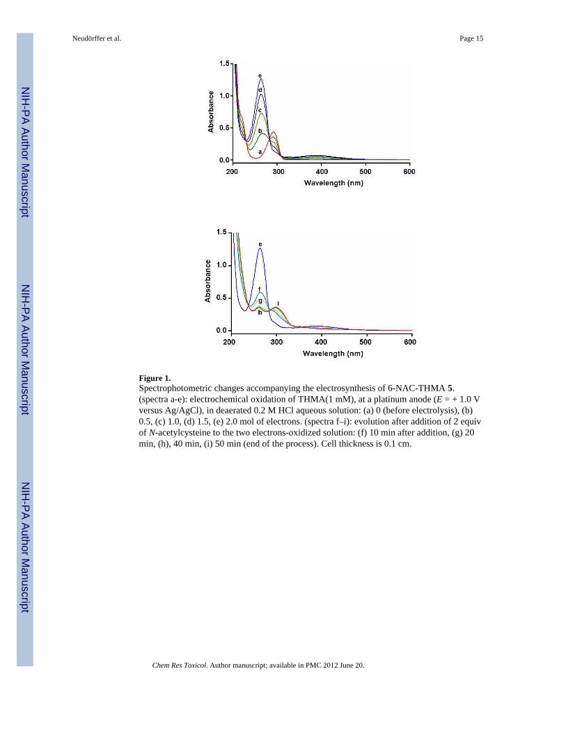
Figure 1.Spectrophotometric changes accompanying the electrosynthesis of 6-NAC-THMA 5.(spectra a-e): electrochemical oxidation of THMA(1 mM), at a platinum anode (E = + 1.0 Vversus Ag/AgCl), in deaerated 0.2 M HCl aqueous solution: (a) 0 (before electrolysis), (b)0.5, (c) 1.0, (d) 1.5, (e) 2.0 mol of electrons. (spectra f–i): evolution after addition of 2 equivof N-acetylcysteine to the two electrons-oxidized solution: (f) 10 min after addition, (g) 20min, (h), 40 min, (i) 50 min (end of the process). Cell thickness is 0.1 cm.
Neudörffer et al. Page 15
Chem Res Toxicol. Author manuscript; available in PMC 2012 June 20.
NIH
-PA Author Manuscript
NIH
-PA Author Manuscript
NIH
-PA Author Manuscript
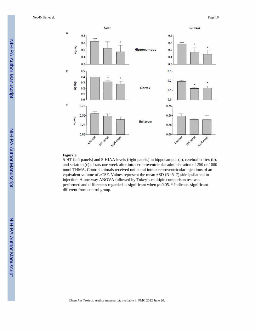
Figure 2.5-HT (left panels) and 5-HIAA levels (right panels) in hippocampus (a), cerebral cortex (b),and striatum (c) of rats one week after intracerebroventricular administration of 250 or 1000nmol THMA. Control animals received unilateral intracerebroventricular injections of anequivalent volume of aCSF. Values represent the mean ±SD (N=5–7) side ipsilateral toinjection. A one-way ANOVA followed by Tukey’s multiple comparison test wasperformed and differences regarded as significant when p<0.05. * Indicates significantdifferent from control group.
Neudörffer et al. Page 16
Chem Res Toxicol. Author manuscript; available in PMC 2012 June 20.
NIH
-PA Author Manuscript
NIH
-PA Author Manuscript
NIH
-PA Author Manuscript
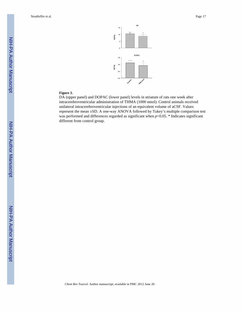
Figure 3.DA (upper panel) and DOPAC (lower panel) levels in striatum of rats one week afterintracerebroventricular administration of THMA (1000 nmol). Control animals receivedunilateral intracerebroventricular injections of an equivalent volume of aCSF. Valuesrepresent the mean ±SD. A one-way ANOVA followed by Tukey’s multiple comparison testwas performed and differences regarded as significant when p<0.05. * Indicates significantdifferent from control group.
Neudörffer et al. Page 17
Chem Res Toxicol. Author manuscript; available in PMC 2012 June 20.
NIH
-PA Author Manuscript
NIH
-PA Author Manuscript
NIH
-PA Author Manuscript
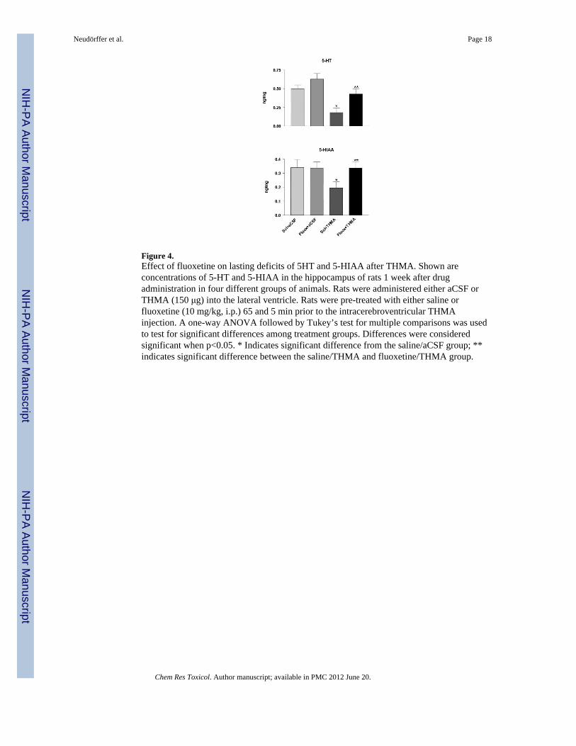
Figure 4.Effect of fluoxetine on lasting deficits of 5HT and 5-HIAA after THMA. Shown areconcentrations of 5-HT and 5-HIAA in the hippocampus of rats 1 week after drugadministration in four different groups of animals. Rats were administered either aCSF orTHMA (150 μg) into the lateral ventricle. Rats were pre-treated with either saline orfluoxetine (10 mg/kg, i.p.) 65 and 5 min prior to the intracerebroventricular THMAinjection. A one-way ANOVA followed by Tukey’s test for multiple comparisons was usedto test for significant differences among treatment groups. Differences were consideredsignificant when p<0.05. * Indicates significant difference from the saline/aCSF group; **indicates significant difference between the saline/THMA and fluoxetine/THMA group.
Neudörffer et al. Page 18
Chem Res Toxicol. Author manuscript; available in PMC 2012 June 20.
NIH
-PA Author Manuscript
NIH
-PA Author Manuscript
NIH
-PA Author Manuscript
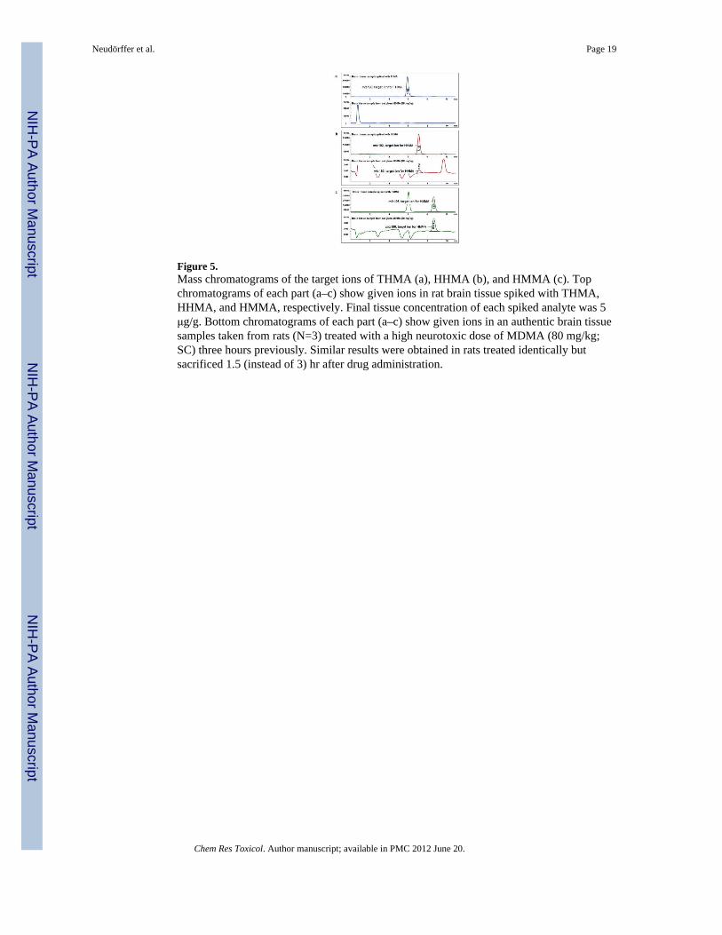
Figure 5.Mass chromatograms of the target ions of THMA (a), HHMA (b), and HMMA (c). Topchromatograms of each part (a–c) show given ions in rat brain tissue spiked with THMA,HHMA, and HMMA, respectively. Final tissue concentration of each spiked analyte was 5μg/g. Bottom chromatograms of each part (a–c) show given ions in an authentic brain tissuesamples taken from rats (N=3) treated with a high neurotoxic dose of MDMA (80 mg/kg;SC) three hours previously. Similar results were obtained in rats treated identically butsacrificed 1.5 (instead of 3) hr after drug administration.
Neudörffer et al. Page 19
Chem Res Toxicol. Author manuscript; available in PMC 2012 June 20.
NIH
-PA Author Manuscript
NIH
-PA Author Manuscript
NIH
-PA Author Manuscript
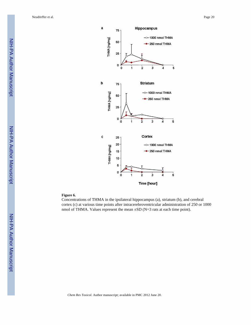
Figure 6.Concentrations of THMA in the ipsilateral hippocampus (a), striatum (b), and cerebralcortex (c) at various time points after intracerebroventricular administration of 250 or 1000nmol of THMA. Values represent the mean ±SD (N=3 rats at each time point).
Neudörffer et al. Page 20
Chem Res Toxicol. Author manuscript; available in PMC 2012 June 20.
NIH
-PA Author Manuscript
NIH
-PA Author Manuscript
NIH
-PA Author Manuscript
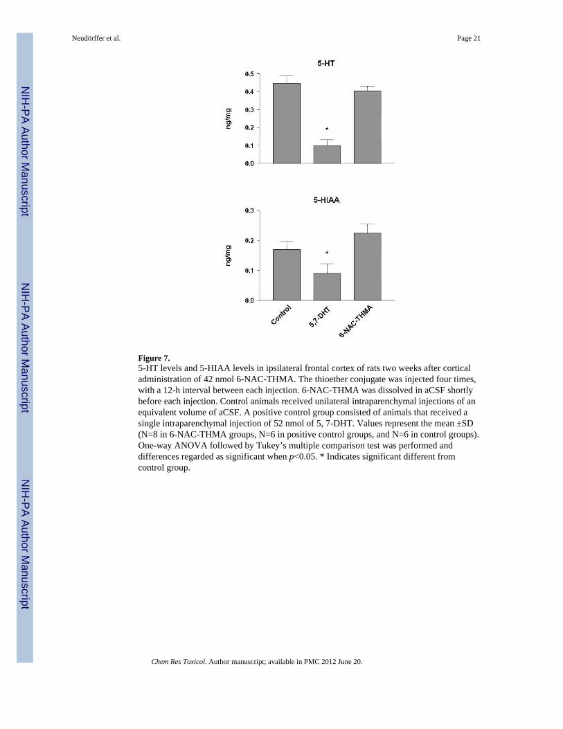
Figure 7.5-HT levels and 5-HIAA levels in ipsilateral frontal cortex of rats two weeks after corticaladministration of 42 nmol 6-NAC-THMA. The thioether conjugate was injected four times,with a 12-h interval between each injection. 6-NAC-THMA was dissolved in aCSF shortlybefore each injection. Control animals received unilateral intraparenchymal injections of anequivalent volume of aCSF. A positive control group consisted of animals that received asingle intraparenchymal injection of 52 nmol of 5, 7-DHT. Values represent the mean ±SD(N=8 in 6-NAC-THMA groups, N=6 in positive control groups, and N=6 in control groups).One-way ANOVA followed by Tukey’s multiple comparison test was performed anddifferences regarded as significant when p<0.05. * Indicates significant different fromcontrol group.
Neudörffer et al. Page 21
Chem Res Toxicol. Author manuscript; available in PMC 2012 June 20.
NIH
-PA Author Manuscript
NIH
-PA Author Manuscript
NIH
-PA Author Manuscript
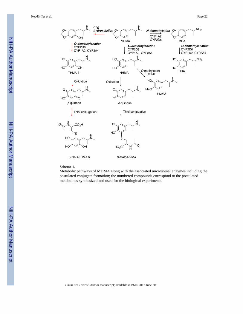
Scheme 1.Metabolic pathways of MDMA along with the associated microsomal enzymes including thepostulated conjugate formation; the numbered compounds correspond to the postulatedmetabolites synthesized and used for the biological experiments.
Neudörffer et al. Page 22
Chem Res Toxicol. Author manuscript; available in PMC 2012 June 20.
NIH
-PA Author Manuscript
NIH
-PA Author Manuscript
NIH
-PA Author Manuscript
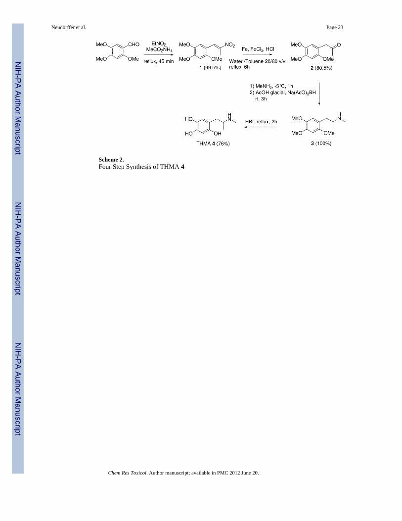
Scheme 2.Four Step Synthesis of THMA 4
Neudörffer et al. Page 23
Chem Res Toxicol. Author manuscript; available in PMC 2012 June 20.
NIH
-PA Author Manuscript
NIH
-PA Author Manuscript
NIH
-PA Author Manuscript
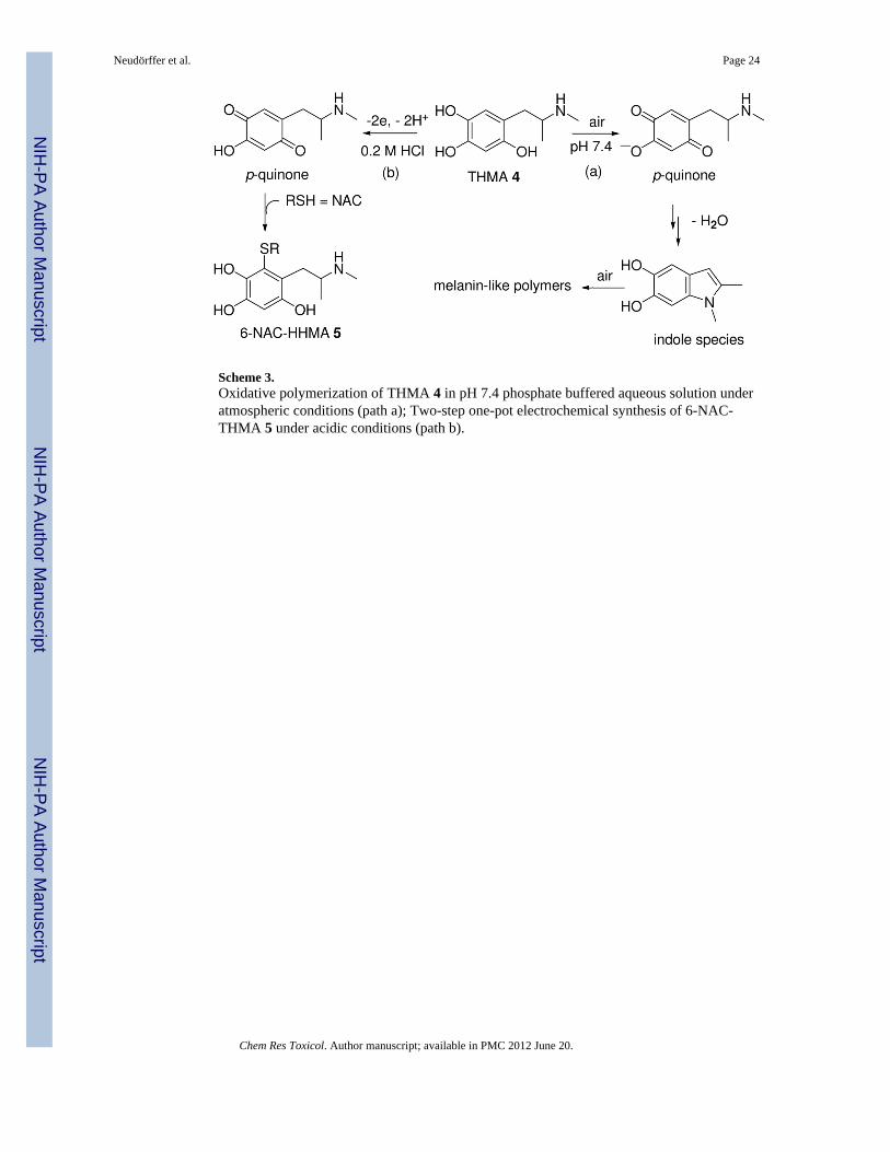
Scheme 3.Oxidative polymerization of THMA 4 in pH 7.4 phosphate buffered aqueous solution underatmospheric conditions (path a); Two-step one-pot electrochemical synthesis of 6-NAC-THMA 5 under acidic conditions (path b).
Neudörffer et al. Page 24
Chem Res Toxicol. Author manuscript; available in PMC 2012 June 20.
NIH
-PA Author Manuscript
NIH
-PA Author Manuscript
NIH
-PA Author Manuscript