synthesis and nmr analysis in solution of oligo(3-hydroxyalkanoic acid) derivatives with the side...
TRANSCRIPT

Synthesis and NMR Analysis in Solution of Oligo(3-hydroxyalkanoic acid)Derivatives with the Side Chains of Alanine, Valine, and Leucine(�-Depsides): Coming Full Circle from PHB to �-Peptides to PHB
Matthias Albert1) and Dieter Seebach*
Laboratorium f¸r Organische Chemie, ETH-Hˆnggerberg, CH-8093 Z¸rich
and
Elke Duchardt1)2) and Harald Schwalbe*2)
Department of Chemistry, Francis Bitter Magnet Laboratory, Massaschusetts Institute of Technology,170 Albany Street, Cambrige, MA 02139, USA
Oligomers of 3-hydroxyalkanoic acids that contain two, three, and six residues with and withoutO-terminal (tBu)Ph2Si and C-terminal PhCH2 protection have been synthesized in such a way that the sidechains on the oligoester backbone were those of the proteinogenic amino acids Ala (Me), Val (CHMe2), andLeu (CH2CHMe2). The enantiomerically pure 3-hydroxyalkanoates were obtained by Noyori hydrogenation ofthe corresponding 3-oxo-alkanoates with [Ru((R)-binap)Cl2](binap� 2,2�bis(diphenylphosphanyl)-1,1�-bi-naphthalene)/H2 (Scheme 1), and the coupling was achieved under the conditions (pyridine/(COCl)2, CH2Cl2,� 78�) previously employed for the synthesis of various oligo(3-hydroxybutanoic acids) (Schemes 2 and 3). TheCotton effects in the CD spectra of the new oligoesters provided no hints about chiral conformation (cf. a helix)in MeOH, MeCN, octan-1-ol, or CF3CH2OH solutions (Figs. 1 and 2). Detailed NMR investigations in CDCl3solution (Figs. 3 ± 6, and Tables 1 ± 5) of the hexa(3-hydroxyalkanoic acid) with the side chains of Val (HC), Ala(HB), Leu (HH), Val, Ala, Leu (from O- to C-terminus; 3) gave, on the NMR time-scale, no evidence for thepresence of any significant amount of a 21- or a 31-helical conformation, comparable to those identified instretched fibers of poly[(R)-3-hydroxybutanoic acid], or in lamellar crystallites and in single crystals of linearand cyclic oligo[(R)-3-hydroxybutanoic acids], or in the corresponding �-peptide(s) (the oligo(3-aminoalkanoicacid) analogs; 1 ± 3). Thus, the extremely high flexibility (averaged or −random-coil× conformation) of thepolyester chain (CO�O rotational barrier ca. 13 kcal/mol; no hydrogen bonding), as compared to polyamidechains (CO�NH barrier ca. 18 kcal/mol; hydrogen bonding) has been demonstrated once again. The possibleimportance of this structural flexibility, which goes along with amphiphilic properties, for the role of PHB inbiology, in evolution, and in prebiotic chemistry is discussed. Structural similarities of natural potassium-channeling proteins and complexes of oligo(3-hydroxybutanoates) with Na�, K�, or Ba2� are alluded to (Figs. 7±9).
1. Indroduction. ± The short-chain version of poly[(R)-3-hydroxybutanoic acid] (c-PHB), 1, has been detected in numerous biological systems, and we believe that it is fairto say that the biopolymer PHB is present in all living organisms3). The low-molecular-weight PHB has been shown to cause phospholipid membranes to become permeablefor cations, such as Na, K, Rb, Ca or Ba, under voltage-driven (patch-clampexperiments [5]) and under concentration-driven (artificial vesicles [6]) conditions.Furthermore, a Ca polyphosphate-PHB complex consisting of ca. 140 HB and 70phosphate units and containing ca. 35 Ca2� ions has been identified as a Ca-specific ion
��������� ����� ���� ± Vol. 85 (2002) 633
1) Part of the projected Ph. D. Theses of M. A. and E. D., ETH-Z¸rich and MIT, respectively.2) Present Address: Institut f¸r Organische Chemie, Johann Wolfgang Goethe-Universit‰t Frankfurt, Marie
Curie-Strasse 11, D-60439 Frankfurt.3) For review articles, see [1 ± 4].

channel, extractable from the inner cell wall of genetically competent E. coli [7] [8]. c-PHB and polyphosphate have recently been identified as part of the microbial KcsApotassium channel, in which an amino acid residue bears the polyester chain(complexing Ca ¥ PPi) [9].
In view of the biological importance of PHB, it is essential to learn as much aspossible about its structure. Two distinct folding patterns of the polyester chain havebeen characterized: a 21 helix in stretched fibers [10] and in lamellar crystallites[11] [12] of the polymer, and both 21 and 31 helices in crystals of cyclic oligomers(−oligolides×) of HB [13 ± 16] (Fig. 1,a, and b). In contrast, all attempts to find apreferred conformation (secondary structure) of the polyester chain in solution havefailed so far; among the methods used were CD [17] [18] and NMR [17]4) spectroscopy,as well as FRET5) measurements [21]. While there are indications for the presence ofsecondary structures in the ensemble of molecules on the very short timescale of UV/VIS spectroscopy [17] [18] [21], no preferred conformation could be detected on theslow NMR timescale [17]. On the other hand, inspection of the 31 helix (Fig. 1,b)indicated that replacement of the chain-bound O-atoms by NH groups would lead toNH ¥¥¥O�CH-bonding, and thus to stabilization of the helix. This was, indeed, the case(Fig. 1,c), and has led to our entry into the field of �-peptides [22], systematicinvestigations of which showed that the 31 or 314 helix observed in solutions of the �-hexapeptide H-(�-HVal-�-HAla-�-HLeu)2-OH (2) may be due to preferred backboneconformations and not only due to multiple H-bonding [22]. Thus, we thought itworthwhile to have a look at a �-hexadepside, the O-analog 3 of the �-hexapeptide6),
��������� ����� ���� ± Vol. 85 (2002)634
For the description of the NMR spectra with full assignement of all residues in the chain, we needed to haveabbreviations to designate the six residues of 3 unambiguously; we chose 1- and 4-HC, 2- and 5-HB, and 3- and6-HH (hydroxy-caproic, -butyric and -heptanoic acid).
4) 13C-NMR Experiments with solid-phase copolymers of poly(�-hydroxybutyrate-co-�-hydroxyvalerate)(P(3HB-3HV)) [19] or P(3HB-3HV) in chloroform solution [20] have also been reported.
5) FRET�Fluorescence Resonance Energy Transfer.6) A �-depsipeptide consisting of six �-amino acids and a central �-hydroxybutanoic acid (HB) residue has
been previously described [23]. Also, chimeric, MHC-binding oligomers, which, contain �- and �-amino, aswell as �-hydroxy acids, have been synthesized [24]. Oligomers of �-hydroxy acids with proteinaceous sidechains have not previously been reported; they are �-depsides, homologs of �-depsides, which have beenprepared from lactic acid (the Ala analog), mandelic acid (the phenylglycine analog), and 2-hydroxy-3-methylbutanoic acid (the Val analog); no �-depsidic structure has been published; for CD spectra, see [25].

with different and larger side chains, which hopefully better stabilize certain backboneconformations7) and allow easier NMR assignments of the sequential residues due toincreased resolution as compared to HB oligomers with identical Me groups8).
2. Preparation of the Hexakis(3-hydroxy-alkanoic acid) 3. ± Linear and cyclicoligomers of (R)-3-hydroxyvalerate (HV) and (R)-3-hydroxybutyrate (HB) have beenpreviously synthesized by Seebach and co-workers [12 ± 16] [28]. While the enantio-merically pure building blocks required for these oligomers were obtained byhydrolysis of the biopolymer PHB/PHV [29], �-hydroxy acid derivatives withsubstituents other than Me or Et in the �-position had to be prepared byenantioselective synthesis. The methodology (Scheme 1) chosen for that purpose, i.e.,
��������� ����� ���� ± Vol. 85 (2002) 635
Fig. 1. Models of the different helical conformations of PHB and �3-oligopeptides. a) The 21 helix, b) the 31 helixof a PHB chain, c) the 31 or 314 helix of a �3-peptide.
7) For the preferred confomations of various types of esters, see [26].8) Oligomer 3 is an oligo(3-hydroxyalkanoic acid) (OHB) with different side chains in specific positions. This
kind of OHB has not been previously described, while there is a host of literature on poly(3-hydroxyalkanoic acids) (PHA) with uniform or irregularly placed side chains [27].
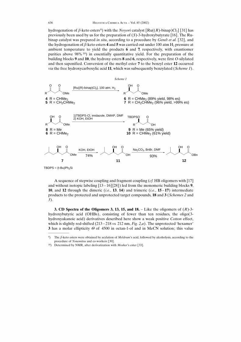
hydrogenation of �-keto esters9) with the Noyori catalyst [Ru((R)-binap)Cl2] [31] haspreviously been used by us for the preparation of (S)-3-hydroxybutyrate [16]. The Ru-binap catalyst was prepared in situ, according to a procedure by Gene√t et al. [32], andthe hydrogenation of �-keto esters 4 and 5was carried out under 100 atmH2 pressure atambient temperature to yield the products 6 and 7, respectively, with enantiomerpurities above 98%10) in essentially quantitative yield. For the preparation of thebuilding blocks 9 and 10, the hydroxy esters 8 and 6, respectively, were first O-silylatedand then saponified. Conversion of the methyl ester 7 to the benzyl ester 12 occurredvia the free hydroxycarboxylic acid 11, which was subsequently benzylated (Scheme 1).
A sequence of stepwise coupling and fragment coupling (cf.HB oligomers with [17]and without isotopic labeling [13 ± 16] [28]) led from the monomeric building blocks 9,10, and 12 through the dimeric (i.e., 13, 14) and trimeric (i.e., 15 ± 17) intermediateproducts to the protected and unprotected target compounds, 18 and 3 (Schemes 2 and3).
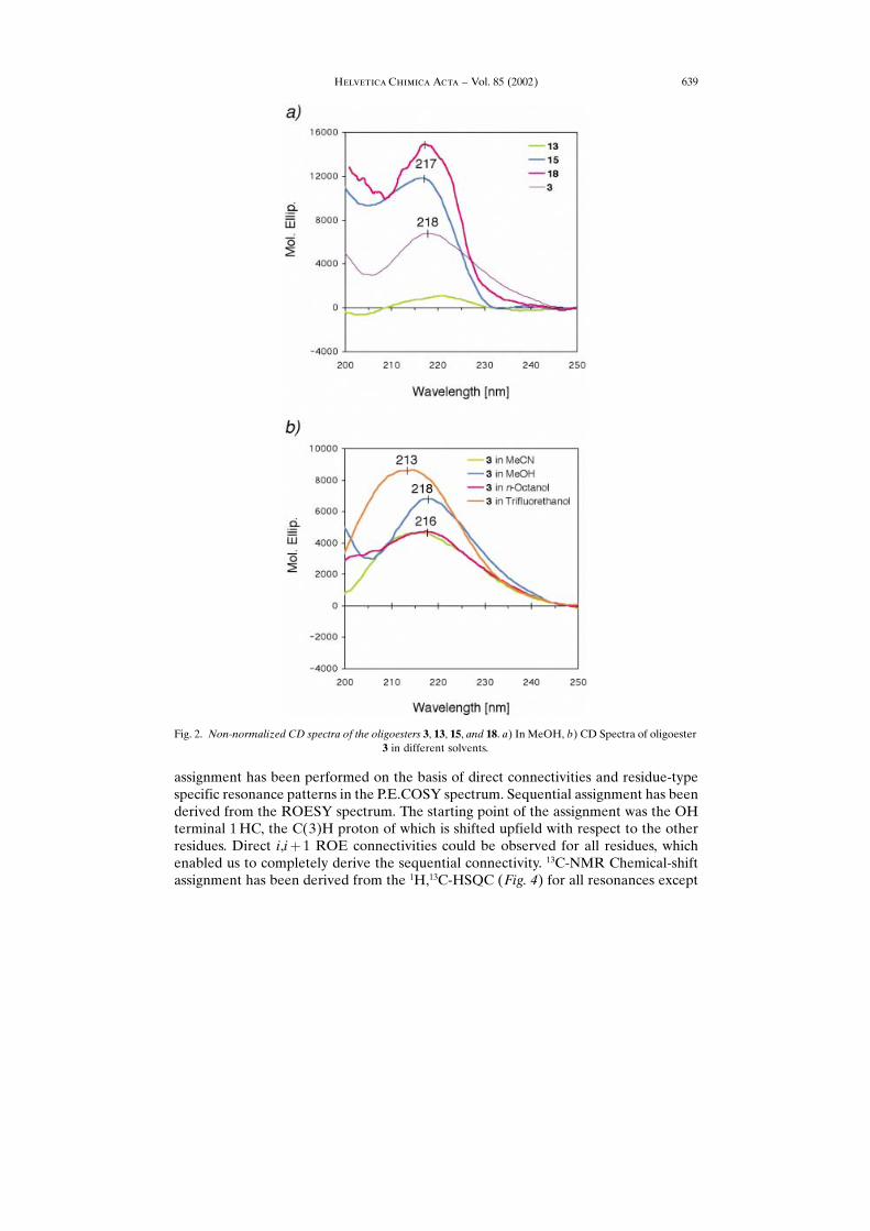
3. CD Spectra of the Oligomers 3, 13, 15, and 18. ± Like the oligomers of (R)-3-hydroxybutyric acid (OHBs), consisting of fewer than ten residues, the oligo(3-hydroxyakanoic acid) derivatives described here show a weak positive Cotton effect,which is slightly red-shifted (213 ± 218 vs. 212 nm, Fig. 2,a). The unprotected −hexamer×3 has a molar ellipticity � of 4500 in octan-1-ol and in MeCN solution; this value
Scheme 1
R
OH
OMe
O
R
TBDPSO
OH
O
OH
OMe
O OH
OH
O OH
OBn
O
R OMe
OO
R
OH
OMe
O
KOH, EtOH Na2CO3, BnBr, DMF
8 R = Me6 R = CHMe2
9 R = Me (65% yield)10 R = CHMe2 (61% yield)
1)TBDPS-Cl, imidazole, DMAP, DMF2) KOH, EtOH
7 11 1274% 93%
4 R = CHMe25 R = CH2CHMe2
6 R = CHMe2 (89% yield, 98% es)7 R = CH2CHMe2 (96% yield, >99% es)
[Ru((R)-binap)Cl2], 100 atm. H2
TBDPS = (t-Bu)Ph2Si
��������� ����� ���� ± Vol. 85 (2002)636
9) The �-keto esters were obtained by acylation ofMeldrum×s acid, followed by alcoholysis, according to theprocedure of Yonemitsu and co-workers [30].
10) Determined by NMR, after derivatization with Mosher×s ester [33].

��������� ����� ���� ± Vol. 85 (2002) 637
Scheme 2
HO O
O
OBn
O
TBDPSO O
O
O
O
OBn
O
TBDPSO O
O
OBn
O
TBDPSO OH
O
HO OBn
O
TBDPSO OH
O
1) 10, CH2Cl2, (COCl)2
2) CH2Cl2, 14, 0o, then pyridine
HF · pyridineCH2Cl2, 0o
9 12
13
14 10
15
77%
92%
1) 9, CH2Cl2, (COCl)2
2) CH2Cl2, 12, 0o, then pyridine
77%

increases as the polarity of the solvent increases: �� 7000 and 8500 in MeOH andCF3CH2OH, respectively (Fig. 2,b).
The higher intensities of the Cotton effects from the fully or partially protectedderivatives (Fig. 2,a) is due to the presence of Ph groups in the O- and C-terminalprotection ((t-Bu)Ph2Si, CH2Ph). The CD spectrum of the �-peptide analog 2 of theoligoester 3 gives rise to a much stronger Cotton effect (�� 40000 at 216 nm in MeOH[23 ± 25] [34]). Thus, the CD spectra provide no indication for the presence of a helicalsecondary structure in the solution of the oligo(hydroxy acid) 3.
4. NMR Investigation of the Hexakis(hydroxy acid) 3. ± Spectral Assignment. The1H-NMR chemical shifts have been assigned from a P.E.COSY spectrum (Fig. 3)together with a ROESY spectrum (data not shown), annotations are given in Fig. 3(top: annotated units 1 to 3; bottom: annotated units 4 to 6). Intra-residual resonance
��������� ����� ���� ± Vol. 85 (2002)638
Scheme 3
TBDPSO O
O
O
O O
HO O
O
O
O O
TBDPSO O
O
O
O O
OBnH
O O
O
O
O O
OH
OH OBn
1) 16, CH2Cl2, (COCl)2
2) CH2Cl2, 17, -78o, then pyridine
HF.pyridineCH2Cl2, 0o
H2,Pd-CMeOH, 0o
15
16 (yield 83%) 17 (yield 87%)
218 (yield 65%)
1) HF.pyridine, CH2Cl2, 0o
2) H2,Pd-C, MeOH, 0o
23 (yield 94%)
assignment has been performed on the basis of direct connectivities and residue-typespecific resonance patterns in the P.E.COSY spectrum. Sequential assignment has beenderived from the ROESY spectrum. The starting point of the assignment was the OHterminal 1HC, the C(3)H proton of which is shifted upfield with respect to the otherresidues. Direct i,i� 1 ROE connectivities could be observed for all residues, whichenabled us to completely derive the sequential connectivity. 13C-NMR Chemical-shiftassignment has been derived from the 1H,13C-HSQC (Fig. 4) for all resonances except
��������� ����� ���� ± Vol. 85 (2002) 639
Fig. 2. Non-normalized CD spectra of the oligoesters 3, 13, 15, and 18. a) In MeOH, b) CD Spectra of oligoester3 in different solvents.

��������� ����� ���� ± Vol. 85 (2002)640
Fig. 3. P.E.COSY Spectrum of 3, assignment of the first three (top) and the last three units (bottom). The HHC(4)H2 protons have not been stereospecifically assigned and are, therefore, denoted according to their relative
chemical shift (Up�upfield, Down�downfield).

the C�O moieties, which have been assigned from an 1H,13C-HMBC spectrum. Thechemical-shift assignments are listed in Table 1. The resonances of all repetition unitsare resolved except for 3HH and 6HH, and 2HB and 5HB, which overlap partiallywith one another. In addition, the 3HH unit shows interresidual signal overlap with2HB, which renders the analysis of this residue more difficult.Conformational Analysis: Assignment of the Diastereotopic C(2)H2 Protons. To
stereospecifically assign the diastereotopic C(2)H2 protons, vicinal coupling constantshave been measured and interpreted for the hexamer 3. In addition, H,H-distanceinformation has been derived from integration of intra-residual ROE (rotating-frameOverhauser enhancement) cross-peaks. 3J(C(2)H2,C(3)H) coupling constants extract-ed from the P.E.COSY spectrum (Fig. 3) and qualitative 3J(C(2)H2,C(4)) couplingconstants derived from the 1H,13C-HMBC spectrum (Fig. 5) are given in Table 2
together with the ratios RUp�DownROE �ROE�C�2�HDown�C�i�H�ROE�C�2�HUp�C�i�H� for i� 3,4 of the intra-
residual C(2)H2,C(3)H and the C(2)H2,C(4)HROEs. Here, the annotationsDown andUp signify the relative chemical shift of the two C(2)H2 protons. The three possiblerotamers (ap, (�)-sc, and (�)-sc) are shown in Fig. 6, together with the characteristicrelative sizes of 3J coupling constants and ROE intensities.
��������� ����� ���� ± Vol. 85 (2002) 641
Fig. 4. a) Natural-abundance 1H,13C-HSQC spectrum of 3. The grey-shaded region has been magnified in b.Asterisks indicate impurities c) C(3)H/(C1) region of a natural-abundance 1H,13C-HMBC spectrum. C(1)
assignments are given.
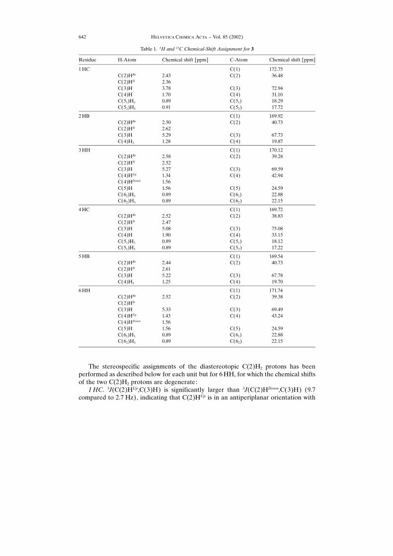
The stereospecific assignments of the diastereotopic C(2)H2 protons has beenperformed as described below for each unit but for 6HH, for which the chemical shiftsof the two C(2)H2 protons are degenerate:1HC. 3J(C(2)HUp,C(3)H) is significantly larger than 3J(C(2)HDown,C(3)H) (9.7
compared to 2.7 Hz), indicating that C(2)HUp is in an antiperiplanar orientation with
��������� ����� ���� ± Vol. 85 (2002)642
Table 1. 1H and 13C Chemical-Shift Assignment for 3
Residue H-Atom Chemical shift [ppm] C-Atom Chemical shift [ppm]
1HC C(1) 172.75C(2)HRe 2.43 C(2) 36.48C(2)HSi 2.36C(3)H 3.78 C(3) 72.94C(4)H 1.70 C(4) 31.10C(51)H3 0.89 C(51) 18.29C(52)H3 0.91 C(52) 17.72
2HB C(1) 169.92C(2)HRe 2.50 C(2) 40.73C(2)HSi 2.62C(3)H 5.29 C(3) 67.73C(4)H3 1.28 C(4) 19.87
3HH C(1) 170.12C(2)HRe 2.58 C(2) 39.28C(2)HSi 2.52C(3)H 5.27 C(3) 69.59C(4)HUp 1.34 C(4) 42.94C(4)HDown 1.56C(5)H 1.56 C(5) 24.59C(61)H3 0.89 C(61) 22.88C(62)H3 0.89 C(62) 22.15
4HC C(1) 169.72C(2)HRe 2.52 C(2) 38.83C(2)HSi 2.47C(3)H 5.08 C(3) 75.08C(4)H 1.90 C(4) 33.15C(51)H3 0.89 C(51) 18.12C(51)H3 0.89 C(52) 17.22
5HB C(1) 169.54C(2)HRe 2.44 C(2) 40.73C(2)HSi 2.61C(3)H 5.22 C(3) 67.78C(4)H3 1.25 C(4) 19.70
6HH C(1) 171.74C(2)HRe 2.52 C(2) 39.38C(2)HSi
C(3)H 5.33 C(3) 69.49C(4)HUp 1.43 C(4) 43.24C(4)HDown 1.56C(5)H 1.56 C(5) 24.59C(61)H3 0.89 C(61) 22.88C(62)H3 0.89 C(62) 22.15

��������� ����� ���� ± Vol. 85 (2002) 643
Fig. 5. Natural-abundance 1H,13C-HMBC spectrum of 3. C(2)H2,C(4) Cross-peaks are indicated.
Table 2. 3J(C(2)H2,C(3)H) Coupling Constants Extracted from the P.E.COSY Spectrum of 3, Relative 3J(C(2)H2,CB)Coupling Constants from the HMBC Spectrum. � : Larger value, � : smaller value, n.d.: not determined.
RUp�DownROE �ROE�C�2�HDown� C�i�H�ROE�C�2�HUp� C�i�H� for i� 3 and i� 4 are given.
Residue Atomidentity
Stereo-specificassign-ment
3J(C(4),C(2)H2)[Hz]
3J(C(4),C(2)H2)[Hz]
ROE�C�2�HDown� C�4�H�ROE�C�2�HUp� C�4�H�
ROE�C�2�HDown� C�3�H�ROE�C�2�HUp� C�3�H�
1HC C(2)HDown C(2)HRe 2.7 � 0.97 1.89C(2)HUp C(2)HSi 9.7 �
2HB C(2)HDown C(2)HSi 7.6 � 0.81 0.28C(2)HUp C(2)HRe 4.6 �
3HH C(2)HDown C(2)HRe 5.3 � n.d. 0.46C(2)HUp C(2)HSi � 5.3 �
4HC C(2)HDown C(2)HRe 5.9 � n.d. n.d.C(2)HUp C(2)HSi 4.9 �
5HB C(2)HDown C(2)HSi 7.4 � 0.74 0.71C(2)HUp C(2)HRe 4.9 �
6HH C(2)HDown C(2)HSi n.d. n.d. n.d. n.d.C(2)HUp C(2)HRe n.d. n.d.
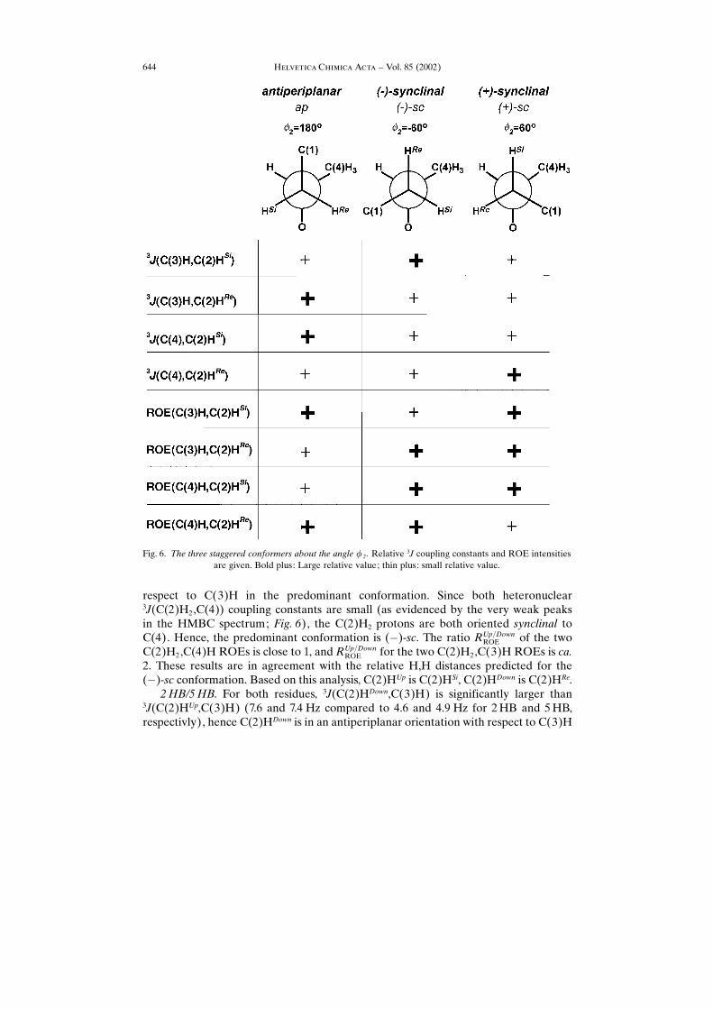
respect to C(3)H in the predominant conformation. Since both heteronuclear3J(C(2)H2,C(4)) coupling constants are small (as evidenced by the very weak peaksin the HMBC spectrum; Fig. 6), the C(2)H2 protons are both oriented synclinal toC(4). Hence, the predominant conformation is (�)-sc. The ratio RUp�DownROE of the twoC(2)H2,C(4)H ROEs is close to 1, and RUp�DownROE for the two C(2)H2,C(3)H ROEs is ca.2. These results are in agreement with the relative H,H distances predicted for the(�)-sc conformation. Based on this analysis, C(2)HUp is C(2)HSi, C(2)HDown is C(2)HRe.2HB/5HB. For both residues, 3J(C(2)HDown,C(3)H) is significantly larger than
3J(C(2)HUp,C(3)H) (7.6 and 7.4 Hz compared to 4.6 and 4.9 Hz for 2HB and 5HB,respectivly), hence C(2)HDown is in an antiperiplanar orientation with respect to C(3)H
��������� ����� ���� ± Vol. 85 (2002)644
Fig. 6. The three staggered conformers about the angle � 2 . Relative 3J coupling constants and ROE intensitiesare given. Bold plus: Large relative value; thin plus: small relative value.

in the predominant conformation for both units. Both 3J(C(2)H2,C(4)) couplingconstants are small. This indicates a predominant (�)-sc-conformation with a reverseddiastereotopic assignment of the C(2)H2 protons as compared to 1HC: C(2)HUp isC(2)HRe, C(2)HDown is C(2)HSi.
The observed differences in RUp�DownROE for the two HB units, however, are not inagreement with only a pure (�)-sc-conformation. This observation has not been furtheranalyzed due to the insensitivity of ROEs to conformational averaging.3HH/4HC. The two 3J(C(2)H2,C(3)H) coupling constants are almost equal for
4HC (5.9 and 4.9 Hz), indicating that both C(2)H2 protons are in a synclinalorientation with respect to C(3)H. The overlap of the 3HHC(2)HUp,C(3)H cross-peakmakes a quantification of the 3J(C(3)H,C(2)HUp) coupling constant impossible.Qualitative analysis, however, shows that 3J(C(2)HUp,C(3)H) is smaller than3J(C(2)HDown,C(3)H) in this unit. The 3HH 3J(C(3)H,C(2)H2) coupling constantsare of similar size as the coupling constants found for 4HC. Both 3J(C(2)H2,C(4))coupling constants are small for both units. The C(2)H2 proton with the slightly larger3J(C(3)H,C(2)H2) has also the larger 3J(C(4),C(2)H2) coupling constant, which showsthat the predominant conformation is (�)-sc, and ap is second most populated in bothunits. Therefore, C(2)HDown is C(2)HRe and C(2)HUp is C(2)HSi.NMR Analysis: Backbone Angle �2. The populations of the three conformations
about the angle � 2 based on the stereospecific assignment, as well as a detaileddescription of the coupling-constant analysis are given in Table 3. 3J(C(2)H2,C(3)H)Coupling constants and the conformation about the angle � 2 are averaged for all unitscompared to 3J(C(2)H2,C(3)H) coupling constants derived from the generalizedKarplus equation [35] (see caption of Table 3).
For 3HH, 3J(C(3)H,C(2)HRe) is slightly smaller than 3J(C(3)H,C(2)HRe) for allother units. Although exact populations could not be determined for this unit, the (�)-sc conformation can be assumed to be predominant similar to 4HC, which has acomparably small 3J(C(3)H,C(2)HRe). For 6HH, the conformation about � 2 could notbe determined due to the degenerate chemical shift of the two C(2)H2 protons.
Table 3. 3J(C(2)H2,C(3)H) Coupling Constants and Resulting Populations of the Three Staggered Conforma-tions about the Angle �2 . The populations P have been calculated using the Pachler analysis equations:3J(C(2)HRe,C(3)H)�P(�)-scJ(�)-sc�PapJap�P(�)-scJ(�)-sc,3J(C(2)HSi,C(3)H)�PapJ(�)-sc�P(�)-scJap�P(�)-scJ(�)-sc and1�Pap�P(�)-sc�P(�)-sc
. Jap, J(�)-sc and J(�)-sc can be obtained from the generalized Karplus equation [35]:3J(H,H)� 13.7 cos2�� 0.73 cos���i�ci[0.56� 2.47 cos2(zi�� 16.9 ��ci � )]. In this equation, � is the dihedralangle between the two protons, zi is the orientation factor of substituent i, �ci is the electronegativity differencebetween substituent i and the respective proton. Not only the direct substituents ia are considered but also theatoms bound to the i,a substituents (i,b). Thus, �ci is determined according to �ci��ci,a� 0.14 S �ci,b (O: �ci�1.3, C: �ci� 0.4, H: �ci� 0.0). The 3J(C(2)H2,C(3)H) coupling constant are 11.63 for ap, 3.55 for (�)-sc, and
1.97 Hz for (�)-sc.
Residue Pap [%] P(�)-sc [%] P(�)-sc [%]
1HC 4.5 77.0 18.52HB 23.7 54.8 21.53HH ± ± ±4HC 33.6 23.3 43.15HB 27.0 52.9 20.06HH ± ± ±
��������� ����� ���� ± Vol. 85 (2002) 645

However, since the two C(2)H2 protons resonate at the same chemical shift, it can beassumed that the 6HH unit is completely unstructured.
In both helical secondary structures proposed, the conformation about � 2 is close to(�)-sc (�62.4� for a 21 and � 52.1� for a 31 helix). In principle, either one of the twohelices could, therefore, occupy a fraction of the conformational ensemble of thehexameric PHB analogue 3 of up to the product of the (�)-sc population of eachinvolved residue. Excluding the totally averaged carboxy terminal 6HH unit, themaximum possible helix content of the remaining five units of 3 is 7.5%. The HB unitsare similar to the repetition units in PHB. NMR Studies on linear 20mer PHBmolecules [17] show that the conformational equilibrium about the angle � 2 (54.6%(�)-sc, 35.4% ap, and 10% (�)-sc) differs from the one in the HB units (54.8%(52.9%)(�)-sc, 23.7%(27%) ap, 21.5%(20.0%) (�)-sc for 2HB (5HB)) in 3. While thepopulations of the predominant (�)-sc conformation are similar, the ap conformation issignificantly more populated than the (�)-sc conformation in the 20mer PHB ascompared to the hexamer 3, where the populations of ap and (�)-sc are almost equal.In principle, the more averaged conformation of the two HB residues may be due to thesmaller size of the hexamer 3 in comparison to the 20mer PHB molecule, which makesit more flexible. However, studies on 3-HB dimers and trimers [36] indicate that the3J(C(3)H,C(2)H2) coupling constants in this smaller compounds are comparable tothose in the 20mer PHB. The conformational average of 4HC and the two HH unitsdiffers from that in the 20mer PHB. This shows that incorporation of the bulkier sidechains changes the conformation of the PHB backbone. Furthermore, the conforma-tion of the two HB units (2HB and 5HB) in 3 is influenced by the presence of themodified units.
The similarity in coupling constants of the two HB units, conformational average ofwhich about � 2 differs significantly both from 4HC and the two HH units, however,suggests that the units adopt a conformation primarily governed by their specific sidechains.Conformational Analysis: ROE Data. The overall conformation of the hexamer 3
has been analyzed by ROE distance information. All interresidual ROESY cross-peaksare given in Table 4, together with the relative peak intensities.
The i,i� 2 and i,i� 3 cross-peaks can be indicative for the 21 or the 31 helix,respectively. The shortest interresidual distance (Table 5) for a 31 helix is the i,i� 3C(2)HSi,C(3)H distance of 3.2 ä; for a 21 helix, it is the i,i� 2 C(2)HRe,C(4)H3 with3.2 ä. Both distances are at least 8 ä in the respective other helix.
A total of three i,i� 2 cross-peaks and one i,i� 3 cross-peak have been observed.Two of the three i,i� 2 cross-peaks involve C(2)H2 protons. One is between1HC,C(2)HSi and 3HH,C(4)H2. The corresponding C(4)H3,C(2)HRe cross-peak,which would be indicative for a 21 helix (Table 5), was not observed.
The other i,i� 2 cross-peak has been observed between 3HH,C(6)H3 and5HB,C(2)HSi. Although no information on the distance between these protons isavailable, it can be assumed that they are far apart in either of the helix conformations,where the bulky side chains are pointing away from the helix center. The i,i� 3 cross-peak is not indicative for the presence of a 31 helix. Since none of the cross-peaks thatare indicative for either of the proposed helices could be found in the ROESYspectrum, whereas other nonindicative ROEs are observed, none of the helices can be
��������� ����� ���� ± Vol. 85 (2002)646

assumed to be predominantly populated. This is in agreement with the analysis of theconformation about the angle � 2 (see above).
5. Discussion and Conclusions. ± The conclusion from the NMR measurement of 3is clear-cut: in the hexameric PHB analog 3, all repetition units undergo conforma-tional averaging, albeit to a different extent. The predominant conformation about theangle � 2 for the units 1HC, 2HB, and 5HB is (�)-sc. This is in agreement with bothproposed secondary structures. However, the units 3HH and 4HC are predominantlyin the (�)-sc conformation about � 2 . In addition, ROE data show that neither of the twohelices is present to a detectable extent on the timescale of NMR spectroscopy. Theconformational averaging of the carboxy-terminal 6HH unit is more pronounced thanthat of all the other units. In the 1HC unit, the (�)-sc conformation is more populatedthan in the other units. This can be due to the (transient) formation of an intra-residual
��������� ����� ���� ± Vol. 85 (2002) 647
Table 4. Inter-residual ROEs Extracted from a ROESY Spectrum of 3. H,H Distances have been taken frommodel structures of the two proposed PHB helices. H,H Distances involving C(5)- and C(6)-side-chain protonscould not be determined (n.d.). An estimate of the peak intensity is given. For the degenerate 6HH C(2)H2
protons, the distance to C(2)HRe as well as C(2)HSi is given.
Residue
1
Atomidentity1
Residue
2
Atomidentity2
Residuedistance
Atom distance [ä] ROE Intensity
31 21
1HC C(3)H 2HB C(4)H3 i,i� 1 6.2 5.3 weakC(2)HRe 2HB C(4)H3 i,i� 1 4.9 5.4 weakC(2)HRe 2HB C(3)H i,i� 1 4.7 4.1 weakC(2)HSi 2HB C(4)H3 i,i� 1 5.1 5.3 strongC(4)H 3HH C(4)HDown i,i� 2 � 8 6.0 overlappingC(2)HSi 3HH C(4)HDown i,i� 2 � 8 4.8 strongC(4)H 4HC C(4)H i,i� 3 6.4 � 8.0 weakC(2)HSi 5HB C(4)H3 i,i� 4 7.0 � 8.0 weak
2HB C(2)HSi 3HH H(6)H3 i,i� 1 n.d. n.d strongC(2)HSi 3HH C(4)HDown i,i� 1 5.1 5.3 weak
3HH C(3)H 4HC C(3)H i,i� 1 4.9 4.5 strongC(5)H 5HB C(2)HSi i,i� 2 n.d. n.d overlapping
4HC C(3)H 5HB C(4)H3 i,i� 1 6.2 5.3 weakC(3)H 5HB C(3)H i,i� 1 4.9 4.5 weak
5HB C(3)H 6HH C(4)HDown i,i� 1 6.2 5.3 weakC(3)H 6HH C(3)H i,i� 1 4.9 4.5 overlappingC(3)H 6HH C(6)H3 i,i� 1 n.d. n.d. strongC(3)H 6HH C(2)H i,i� 1 6.5/6.9 4.4/5.6 weakC(4)H3 6HH C(3)H i,i� 1 6.2 6.5 weak
Table 5. Characteristic Inter-Residual Distances in the Two Proposed Helices
Atom identity Atom identity Residue distance Atom distance [ä]1 2 31 21
C(4)H C(2)HRe i,i� 2 � 8.0 3.2C(4)H C(2)HSi i,i� 2 7.0 4.8C(3)H C(2)HRe i,i� 3 4.6 � 8.0C(3)H C(2)HSi i,i� 3 3.2 � 8.0
H-bond between the free O-terminal OH group and the adjacent ester carbonyl O-atom, as reported earlier [32]. The conformation about the angle � 2 of all the units of 3deviates from that found in linear PHB derivatives [17], indicating that the side-chainmodifications have indeed altered the backbone conformation, but our going full circlefrom PHB to �-peptides to PHB has not led to the discovery of a predominantsecondary structure! In a series of four papers, including the present one, we have nowestablished the extremely high conformational flexibility of the polyester backbone inPHB [17] [21] [37]. A preferred conformation of oligo(3-hydroxybutyrates) inhomogeneous solution could be detected neither by state-of-the-art NMR spectroscopywith isotopically [17] or structurally (the present paper) labeled, nor by FRETmeasurements [21] with fluorescence-labeled OHBs, nor by GROMOS96 molecular-dynamics simulations of the hexamers 1 (n� 1) and 311). At the risk of writing down atruism: there is a tremendous difference between the worlds of polyesters, like PHB 1without, and polyamides, like �-peptides (cf. 2) with, H-bonding! While the latter formwell-defined, predictable, structure-dependent helices, sheets, or turns, the former haverandom or averaged structures under the same conditions, as studied by the samemethods. The structural flexibility of PHB ± along with its hydrolytic instability ± maybe the reason why it has not shown up in crystal structures of proteins, although it hasbeen identified as an appendage on proteins [9]12). The structural flexibility13) of PHBgoes with its amphiphilic properties: it can behave like a polar or like a nonpolarcompound, it can be hydrophilic and hydrophobic: it may be called a −chemicalchameleon×! Each C�O group of the polymer is a dipole (and thus a H-bond acceptorand a ligand for metal ions [38]). The higher polymers are soluble neither in polar nornonpolar solvents, and show a peculiar solubility behavior by dissolving well only inchlorinated solvents [1] [39] ± the solution can function as ion-transporting bulkmembrane [40]. On the other hand, OHBs can be incorporated in phospholipidbilayers ± functioning as ion carriers or ionophores [5 ± 7]. In blood serum, PHB ismostly associated with albumin [1] [41], the abundant transport protein for lipophiliccompounds ± but also the carrier of 40% of the serum Ca2� content. PHB mixes withLiClO4, to form a solid, conductive electrolyte composition14), and it can be solubilizedin THF by the addition of LiCl [11] [12] [43] [44]. Beautiful demonstrations of the−mixed× nature of PHB were obtained, years ago, from crystal structures of the cyclictriesters (triolides �[O�CHR�CH2�CO]3�, R�Me, Et), and of inclusion com-pounds, and their Na�, K�, and Ba2� complexes (Fig. 7).
Returning to the structure of the PHB chain, we realize that the two helices (Figs. 1and 8) have totally different features and patterns: the 31 helix has a resulting dipolemoment with an O-terminal positive and a C-terminal negative poles, with a lipophilicsurface covered by Me groups, and without the capability of complexing ions (Fig. 8,c).The 21 helix has no resulting dipole moment, both, the C�O and the C�Me bonds
��������� ����� ���� ± Vol. 85 (2002)648
11) See preceding report in this issue [37].12) Reusch ; see the discussion with references in [1]. Poly[(R)-3-hydroxybutyryl]-conjugated proteins; cf.
farnesyl, palmitoyl, phosphoryl, sulfatyl, glycosyl, hypusinyl derivatives of proteins (Fig. 15 in [1]).13) Adjectives like evasive, elusive, volatile, or fickle come to mind!14) Poly(ethylene oxide) dissolves Li salts to form crystalline electrolytes with discrete composition. The
recently published structure of these crystalline complexes [42] strongly resemble to the channel-likearrangement of triolide complexes with Na�, K� , and Ba 2� (Fig. 7).

point in approximately perpendicular directions with respect to the helix axis,rendering the helix surface amphiphilic and providing for ideal complexation of ionsin between parallel or anti-parallel strands of helices (Fig. 8,a). The flexibility of thebackbone would thus allow PHB chains to enter and be incorporated in bilayers as the31 helical conformer, and switch to the 21 helical form under the influence of metal ions(cf. the proposed structures of channels, pores, and PHB ¥Ca ¥ polyphospate complexes[1] [6] [7], and see Figs. 7 and 8,b).
��������� ����� ���� ± Vol. 85 (2002) 649
Fig. 7. Crystal structures containing cyclic hydroxybutyrate (HB) and hydroxyvalerate (HV) trimers (triolides):models for polar and nonpolar ion channels. Grey: C and C,C bonds; red: O; blue: N; yellow: S; violet: Na� ;white:H. a) Antiparallel stacks of HB triolide molecules (the arrows indicate the directions of the C�O dipolemoments in each molecule (2.60 D [45]) and stack [14]. b) View along a channel filled with Et2O molecules inthe crystal of HB/HV trimers (−mixolides×); the stacks are arranged as in a, but, due to the presence of both Meand Et side chains, the packing of the stacks is not perfect, so that Et2O molecules fill the gaps [15]. c) and d)Two views along a channel filled with Na� ¥ 2H2O; the channel walls are formed by HV triolides, which turn alltheir C�OO-atoms inside to provide coordination sites for Na� and H-bond acceptor atoms for H2O, the −glue×between the ions and the wall; the SCN� counter-ions stick in the wall; the composition of the crystal is HV-
triolide ¥NaSCN ¥ 2 H2O ¥ 0.5 MeCN [15].

��������� ����� ���� ± Vol. 85 (2002)650
Fig. 8. Structures and properties of the amphiphilic 21 and lipophilic 31 helices of poly[(R)-3-hydroxybutyrate](PHB). Grey: C and C,C bonds; red: O; violet: Na�. a) Side view and projection along the axis of the left-handed 21 helix of a PHB chain [13] [15]; the helix surface is amphiphilic, covered by C�O and MeC groups,which point in perpendicular directions; there is no resulting dipole moment. b) Two antiparallel 21-helical PHBcolumns complexing a Na� ion; cf. the proposed PHB ion-channel structures [5 ± 7] [12] [15] [46] and theproteinaceous K� channel in Fig. 9. c) Side view and projection along the right-handed 31 helix of a PHB chain;the helix has a large resulting dipole moment (see arrow; cf. polarization of phospholipid bilayers in cell walls),
its surface is lipophilic, covered with Me groups [13] [15].
There is a striking resemblance between the geometries of a 21-helical PHB ioncomplex, as depicted in Fig. 8,b, and the so-called selectivity filter of the highlypreserved part of natural-protein potassium channels consisting of an array of glycine,valine, threonine, and tyrosine residues, in which four subunits provide amide C�OO-atoms in a proper arrangement for complexation and passage of the partiallydesolvated K ions [47] (Fig. 9)15). Recent synthetic model chemistry of the potassiumchannel in combination with NMR spectroscopy, indeed, suggest cooperative formingand breaking of four H-bonds in the channel to accomodate ion passage through thefilter [49]16).
As Reusch [4] [52] and Seebach et al. [1] [2] [51] have speculated before, PHB mightbe a primordial or ancient biopolymer, which might have taken the role of an ion-channel material in anRNAworldwithout proteins, when both genetic information andenzymatic activity were provided by RNA. Phospholipids and PHB may have beencongeners in this RNA world, both being formed by coupling of acetoacetic acid (3-oxobutanoic acid) units in a reducing atmosphere, PHB by simple reduction andpolymerizing, and esterification, and the lipids by two reduction steps and C,C-bondformations17) (see, e.g., the hypothetical proposals for the origin of life as forwarded byW‰chtersh‰user [54]). Structural similarities between PHB and protein channels mayoriginate from the evolution of the latter after the design of the former.
We thank C. Griesinger for helpful discussions. We would like to express our gratitude to Zeneca BioProducts, Billingham, GB, for supplying us with PHB and Novartis Pharma AG, Basel, for continuing financialsupport (D. S., A. M.). This work was supported by the MIT, the Karl-Winnacker Foundation, the Alfred P.Sloan Foundation, the NIH (NCRR Programme), and the Fonds der Chemischen Industrie (H. S. and E. D.).
Experimental Part
1. General. The solvents used were either puriss., p. a. quality or distilled over appropriate drying agents.FC: Fluka silica gel 60 (40 ± 63 �m) at ca. 0.3 bar. M.p.: B¸chi 510 apparatus; not corrected. Optical rotation:Perkin-Elmer 241 polarimeter (10 cm, 1 ml cell) at r.t. CD Spectra: Jasco J-710, recording from 200 to 250 nm at20� ; 1-mm rectangular cell; average of five scans, corrected for the baseline; molar ellipticity in deg ¥ cm2 ¥ dmol�1
(� in nm); smoothing by Jasco software. IR Spectra: Perkin-Elmer 782 spectrophotometer. NMR: Allmeasurements for the structual investigations have been performed on Bruker DRX-600 spectrometers in the
��������� ����� ���� ± Vol. 85 (2002) 651
15) There are additional, intriguing connections between PHB and the microbial KcsA potassium channel,which consist of four protein subunits [44] [47]: the crystal structure, from which the presentations in Fig. 9were prepared, was obtained with a protein from which 25% of the amino acid residues of the naturalprotein had been removed. According to investigations by Reusch, there is an amino acid in the removedpart of the protein that bears a PHB ester chain complexing polyphosphate [4] [9]. Point mutation in theremoved part leads to a protein that lacks PHB, oligomerizes poorly to the tetrameric quarternarystructure, and leads to a channel that does not gate like the natural system. The role of the PHB appendagein the potassium-channeling mechanism of the membrane-bound protein is unknown at this stage. Wethank R. N. Reusch for communicating unpublished results to D. Seebach.
16) Interestingly, the Schultz group has just published the preparation of two (depsipeptide) KcsAmutants withan (S)-2-hydroxy-3-(4-hydroxyphenyl)propionate unit (the O analog of Tyr) in position 145 or an (S)-2-hydroxy-3-phenylpropionate unit (the O analog of Phe) in position 147 of the selectivity filter [50].
17) The microbial enzyme by which the high-molecular-weight storage PHB is assembled fromMe�CH(OH)�CH2�CO�S-Coenzyme A is well-known [3], and the mechanism has been studied ingreat detail [53]. The Claisen condensation of acetic acid to 3-oxobutanoic acid is common to thebiosynthetic pathways leading to PHB [3] and to fatty acids (see textbooks of biochemistry andmonographs on enzymatic reaction mechanisms).

Francis Bitter Magnet Laboratory and the Department of Chemistry at MIT (NIH grant 1S10RR13886-01) at20�. The spectra have been recorded on a 10-mg sample in 500 �l CHCl3; Bruker AMX-500 (1H: 500 MHz, 13C:125 MHz), AMX-400 (1H: 400 MHz, 13C: 100 MHz); chemical shifts � in ppm downfield from internal Me4Si(�0 ppm); J values in Hz. MS: Finnigan MAT TSQ-7000 (ESI); in m/z (% of basis peak). Elemental analyseswere performed by the Microanalytical Laboratory of the Laboratorium f¸r Organische Chemie, ETH-Z¸rich.
2. Preparation of the Acid Chloride. General Procedure 1 (GP 1). Similarly to the procedure in [28], thecarboxylic acid was dissolved in CH2Cl2, and oxalyl chloride (1.5 equiv.), and one drop DMF were added. Themixture was stirred at r.t., until the gas evolution ceased (2 ± 8 h). The solvent was removed in vacuo, and theoily residue was dried under high vacuum.
3. Coupling of the Acid Chloride with the Corresponding Alcohol. General Procedure 2 (GP 2). Similarly tothe procedure in [28], the well-dried acid chloride was dissolved under Ar in CH2Cl2, and cooled to � 78�. Afterthe addition of a soln. of the appropriate alcohol (1.0 equiv.) in CH2Cl2, a soln. of pyridine (1.0 equiv.) in CH2Cl2was slowly injected. The mixture was allowed to warm to r.t. within 12 h and stirred for another 10 h. Subsequentdilution with Et2O was followed by thorough washing with � HCl (2�), sat. NaHCO3 and sat. NaCl solns. Theorg. phase was dried (MgSO4) and concentrated in vacuo.
��������� ����� ���� ± Vol. 85 (2002)652
Fig. 9. Sections from the crystal structure of the Strepto-myces lividans potassium channel (KcsA) consisting offour protein molecules [47] (see also Footnote15)). Colorcoding as in Figs. 7 and 8, except for violet, which is K.The channel part shown is called selectivity filter andconsists of four identical peptide sequences of theprotein subunits (C4 symmetry) pointing the C�Ogroups in such a way that a wall of amide carbonyl O-atoms results, providing complexation for (partiallydesolvated) K� ions to travers. Compare with the Na�
channel in Fig. 7,c and d, and with the complex shown inFig. 8,b! For recent review articles on ion channel
structures, see [48].

4. Removal of the Benzyl Ester Protecting Group. General Procedure 3 (GP 3). The benzyl-ester-protectedoligoester was dissolved in MeOH, and a catalytic amount of 10% Pd/C was added. The apparatus wasevacuated and flushed three times with H2. After the mixture was stirred under H2 (balloon) for 18 h,subsequent filtration through Celite and concentration in vacuo yielded the crude carboxylic acid.
5. (t-Bu)Ph2SiO Deprotection. General Procedure 4 (GP 4). The appropriate (tert-butyl)diphenylsilyl etherwas dissolved in CH2Cl2 in a polyether flask and cooled to 0�. A soln. of 70% HF ¥ pyridine was added, and thebiphasic system was vigorously stirred for 20 min. The emulsion was poured into H2O, Et2O was added, and theorg. phase was separated. The org. phase was washed subsequently with H2O (2�), sat. NaHCO3 (3�) and sat.NaCl. solns., dried (MgSO4), and evaporated under reduced pressure.
6. Preparation of the [Ru((R)-binap)Cl2] Catalyst. General Procedure 5 (GP 5). Similarly to the procedurein [32], [Ru(cod)-(2-methylallyl)2] complex and binap (�2,2�-bis(diphenylphosphanyl)-1,1�-binaphthalene)were dissolved in degassed acetone, and a soln. of HCl in MeOH was slowly added. The resulting orange soln.was stirred for 1 h, and the solvent was removed in vacuo to yield the dihalogeno complex, which was useddirectly without further purification.
Methyl 4-Methyl-3-oxopentanoate (4). According to the procedure in [30], a soln. of Meldrum×s acid(50.0 g, 0.35 mol) and pyridine (55.0 ml, 53.9 g, 0.68 mol, 1.9 equiv.) in CH2Cl2 (250 ml) was cooled to 0�.Isobutanoyl chloride (36.7 ml, 37.3 g, 0.35 mol) was added slowly. The orange soln. was stirred for 1 h at 0� andthen at r.t. for 1 h. The mixture was subsequently washed with 1� HCl (3�) and sat. NaCl solns. The org. phasewas dried (MgSO4), and the solvent was evaporated under reduced pressure. The red oily crude product wasrefluxed 4 h in MeOH (100 ml). After removal of the solvent in vacuo, the residue was distilled (63�, 13 Torr)over a 50-cm Vigreux column. Subsequent FC (SiO2; Pentane/ Et2O 5 :1) yielded 4 (17.0 g, 0.12 mol, 29%).Colorless oil. 1H-NMR (200 MHz, CDCl3): keto tautomer: 1.12 (d, J� 6.9, Me2CH); 2.71 (sept., J� 6.9,Me2CH); 3.50 (s, CH2); 3.72 (s, MeO); enol tautomer: 1.12 (d, J� 7.1,Me2CH); 2.40 (sept., J� 7.1, Me2CH); 3.72(s, MeO); 4.98 (d, J� 0.8, CH�COH).
Methyl 5-Methyl-3-oxohexanoate (5). A soln. of Meldrum×s acid (30.0 g, 0.21 mol) and pyridine (33.0 ml,32.3 g, 0.41 mol, 1.9 equiv.) in CH2Cl2 (150 ml) was cooled to 0�. Isopentanoyl chloride (25.3 g, 25.6 ml,0.21 mol) was added slowly. The orange soln. was stirred for 1 h at 0� then at r.t. for 1 h. The mixture wassubsequently washed with 1� HCl (3�) and sat. NaCl solns. The org. phase was dried (MgSO4), and the solventwas evaporated under reduced pressure. The red oily crude product was refluxed 4 h in MeOH (300 ml). Afterremoval of the solvent in vacuo, the residue was distilled (14 Torr, 80�) over a 50-cm Vigreux column.Subsequent FC (SiO2; Pentan/ Et2O 5 :1) yielded 5 (12.7 g, 0.08 mol, 39%). Colorless oil. 1H-NMR (200 MHz,CDCl3): 0.91 (d, J� 6.6,Me2CH); 2.02 ± 2.20 (m, Me2CH); 2.39 (d, 7.06, Me2CHCH2); 3.40 (s, COCH2CO); 3.71(s, MeO).
Methyl (S)-4-Methyl-3-hydroxypentanoate (6). The catalyst was prepared in situ according to GP 5 with[Ru-(cod)(2-methylallyl)2] complex (10.1 mg, 32 mmol), (R)-binap (23.5 mg, 38 mmol, 1.2 equiv.), and 0.24�methanolic HCl (0.29 ml, 69 mmol, 2.2 equiv.). A degassed soln. of 4 (5.00 g, 34.7 mmol) in MeOH (5 ml) wasadded to the dried catalyst. This orange suspension was immediately placed in an autoclave, which was purgedwith H2 (3�) and pressurized under 100 atm. After stirring at r.t. for 72 h, the solvent was removed in vacuo, andthe residue was distilled under reduced pressure (13 Torr, 78�) to yield 6 (4.50 g, 30.3 mmol, 89%). Colorless oil.[�]r�t�D ��43.9 (c� 1.00, CHCl3). 1H-NMR (200 MHz, CDCl3): 0.92 (d, J� 5.8, Me�C(4)); 0.95 (d, J� 5.8,Me�C(4)); 1.71 (oct., J� 5.8, H�C(4)); 2.40 (dd, J� 16.2, 9.1, 1 H�C(2)); 2.52 (dd, J� 16.2, 3.7, 1 H�C(2));2.78 (s, OH); 3.71 (s, MeO); 3.78 (ddd, J� 3.7, 5.8, 9.1, H�C(3)).
Methyl (R)-5-Methyl-3-hydroxyhexanoate (7). The catalyst was prepared in situ according to GP 5 with[Ru-(cod)(2-methylallyl)2] complex (9.2 mg, 28 mmol), (R)-binap (21.5 mg, 34 mmol, 1.2 equiv.) and 0.24�methanolic HCl (0.26 ml, 64 mmol, 2.2 equiv.). A degassed soln. of 5 (5.00 g, 31.6 mmol) in MeOH (5 ml) wasadded to the dried catalyst. This orange suspension was immediately placed in an autoclave, which was purgedwith H2 (3�) and pressurized under 100 atm. After stirring at r.t. for 72 h, the solvent was removed in vacuo, andthe residue was distilled under reduced pressure (12 Torr, 83�) to yield 7 (4.85 g, 30.3 mmol, 96%). Colorless oil.[�]r�t�D ��15.8 (c� 1.00, CHCl3). 1H-NMR (400 MHz, CDCl3): 0.91 (d, J� 6.3,Me2CH); 1.18 (ddd, J� 13.9, 8.6,4.6, 1 H�C(4)); 1.48 (ddd, J� 13.9, 9.0, 5.6, 1 H�C(4)); 1.70 ± 1.88 (m, H�C(5)); 2.39 (dd, J� 16.5, 8.2,1 H�C(2)); 2.49 (dd, J� 16.5, 3.4, 1 H�C(2)); 2.67 (s, OH); 3.7 (s, MeO); 4.04 ± 4.12 (m, H�C(3)).
(R)-3-[(tert-Butyl)diphenylsilyloxy]butyric Acid (9). According to the procedure in [28], a soln. of methyl3-hydroxybutyrate 8 (10.0 g, 84.6 mmol) and 1H-imidazole (7.48 g, 109 mmol, 1.3 equiv.) in DMF (250 ml) wascooled to 0�. At this temp., TBDPS-Cl (23.2 g, 84.1 mmol; containing 5 mol-% silanol) was added and themixture was warmed to r.t. within 2 h. After 2 h, 4-(dimethylamino)pyridine (DMAP; 10.3 g, 84.6 mmol) wasadded, and the solution was stirred further for 18 h. The solvent was removed under high vacuum at 40�, and the
��������� ����� ���� ± Vol. 85 (2002) 653

residue diluted with Et2O (200 ml). The org. phase was washed subsequently with 1� HCl, sat. NaHCO3, andsat. NaCl solns., dried (MgSO4), and evaporated in vacuo. The resulting crude product was dissolved in a cooled(ice bath) soln. of KOH (7.12 g, 127 mmol, 1.5 equiv.) in EtOH (500 ml), and the resulting soln. was allowed towarm to r.t. within 12 h. The solvent was evaporated, and H2O was added. The H2O sol. was extracted with Et2O(2�), acidified at pH 1 with conc. HCl, and subsequently extracted with CH2Cl2 (3�). The CH2Cl2 phase wasdried and evaporated. Recrystallization (hexane, 3� ) yielded 9 (18.8 g, 55 mmol, 65%). White solid. M.p. 71 ±72�. [�]r�t�D ��5.94 (c� 1.00, CHCl3). 1H-NMR (300 MHz, CDCl3): 1.07 (s, t-Bu); 1.15 (d, J� 6.2, Me); 2.51 (d,J� 5.68, H�C(2)); 4.21 ± 4.31 (m, H�C(3)); 7.35 ± 7.48 (m, 4 arom. H); 7.66 ± 7.70 (m, 11 arom. H).
(S)-3-[(tert-Butyl)diphenylsilyloxy]-4-methylpentanoic Acid (10): A soln. of 6 (4.00 g, 27.4 mmol) and 1H-imidazole (2.41 g, 35.6 mmol, 1.3 equiv.) in DMF (250 ml) was cooled to 0� and treated with TBDPS-Cl (7.55 g,27.4 mmol; containing 5 mol-% silanol). The mixture was stirred at 0� for 2 h and then warmed to r.t. within 2 h.After 2 h, DMAP (3.34 g, 27.4 mmol) was added, and the mixture was stirred further for 18 h. The solvent wasremoved under high vacuum at 40�, and the residue was diluted with Et2O (200 ml). The org. phase wassubsequently washed with 1� HCl, sat. NaHCO3, and sat. NaCl solns., dried (MgSO4), and evaporated underreduced pressure. The resulting crude product was dissolved in a cooled (ice bath) soln. of KOH (2.31 g,41.1 mmol, 1.5 equiv.) in EtOH (150 ml), and the resulting soln. was allowed to warm at r.t. within 12 h. Thesolvent was evaporated, and H2O was added. The aq. solution was extracted with Et2O (2�), acidified to pH 1with conc. HCl, and subsequently extracted with CH2Cl2 (3�). The CH2Cl2 phases were dried and evaporated.Recrystallization (hexane; 3� ) yielded 10 (1.71 g, 16.7 mmol, 61%). White solid. M.p. 104 ± 105�. [�]r�t�D ��2.0(c� 1.00, CHCl3). IR (CHCl3): 3072m, 2961s, 2858m, 2700m, 2623m, 1955w, 1894w, 1822w, 1709s, 1465m, 1427m,1299m, 1206m, 1110s, 1077s, 1010m, 945m. 1H-NMR (400 MHz, CDCl3): 0.77 (d, J� 6.8, 1 Me�C(4)); 0.89 (d,J� 6.8, 1 H�C(4)); 1.04 (s, t-Bu); 1.73 (dsept., J� 6.9, 3.5, H�C(4)); 2.37 (dd, J� 15.4, 5.6, 1 H�C(2)); 2.49 (dd,J� 15.4, 6.9, 1 H�C(2)); 4.03 (ddd, J� 6.9, 5.6, 3.5, H�C(3)); 7.30 ± 7.45 (m, 4 arom. H); 7.62 ± 7.70 (m, 11 arom.H); 10.65 (br. s, COOH). 13C-NMR (100 MHz, CDCl3): 17.3; 17.4; 19.4; 27.0; 33.3; 38.1; 74.4; 127.4; 127.5; 129.6;129.7; 133.7; 133.9; 135.9; 136.0; 177.7. MS: 313.1 (48, [M� (t-Bu)]�); 295.1 (2), 271.3 (12), 241.1 (2), 235.1 (10),199.1 (100), 193.1 (5), 181.1 (5), 157.1 (2), 139.0 (18). Anal. calc. for C22H30O3Si (370.56): C 71.31, H 8.16; foundC 71.13, H 8.03.
(R)-3-Hydroxy-5-methylhexanoic Acid (11). The methyl ester 7 (4.00 g, 25 mmol) was dissolved in a cooled(ice bath) soln. of KOH (2.0 g, 38 mmol, 1.5 equiv.) in EtOH (100 ml). The resulting soln. was allowed to warmto r.t. within 12 h, and stirred further for 12 h at the same temp. The solvent was evaporated, and H2O (50 ml)was added. The aq. soln. was then extracted with Et2O, acidified with conc. HCl to pH 1, and extracted withCH2Cl2 (3�). The CH2Cl2 phase was dried (MgSO4) and evaporated. Recrystallization (hexane; 2� ) yielded11 (2.69 g, 18.4 mmol, 74%). Colorless needles. M.p. 64�. [�]r�t�D � 14.2 (c� 1.00, CHCl3) [55]: [�]r�t�D (ent-11)��14.2, (c� 1.00, CHCl3). 1H-NMR (300 MHz, CDCl3): 0.94 (d, J� 6.5, Me2CH); 1.18 (ddd, J� 13.7, 8.7, 4.7,1 H�C(4)); 1.48 (ddd, J� 13.7, 8.7, 5.6, 1 H�C(4)); 1.73 ± 1.87 (m, H�C(5)); 2.44 (dd, J� 16.6, 8.7, H�C(2));2.53 (dd, J� 16.6, 3.4, H�C(2)); 4.08 ± 4.17 (m, H�C(3)). EI-MS: 145.1 (�1, [M�H]�), 129.0 (2), 113.0 (3),89.0 (100), 71.0 (81). Anal. calc. for C7H14O3 (146.18): C 57.51, H 9.65; found C 57.55, H 9.73.
Benzyl (R)-3-Hydroxy-5-methylhexanoate (12). Compound 11 (5.00 g, 34 mmol) was dissolved in asuspension of NaHCO3 (10.9 g, 102 mmol, 3.0 equiv.) in DMF (100 ml). BnBr (6.4 g, 4.5 ml, 38 mmol, 1.1 equiv.)was added, and the suspension was stirred for 24 h at r.t. After the addition of H2O (500 ml), the soln. wasextracted with Et2O (4�). The org. phase was washed with sat. NaCl soln. and evaporated under reducedpressure. FC (pentane/Et2O 5 :1) yielded 12 (7.51 g, 31.8 mmol, 93%). Colorless oil. [�]r�t�D ��15.8 (c� 1.00,CHCl3). IR (CHCl3): 3585w (br.), 3007m, 2959m, 2871w, 1721s, 1497w, 1455w, 1405w, 1385m, 1317m, 1270m,1170s, 1072w, 1041w, 970w. 1H-NMR (400 MHz, CDCl3): 0.91 (d, J� 6.7, Me2CH); 1.18 (ddd, J� 13.8, 8.6, 4.4,1 H�C(4)); 1.48 (ddd, J� 13.8, 8.9, 5.5, 1 H�C(4)); 1.76 ± 1.84 (m, H�C(5)); 2.44 (dd, J� 16.5, 8.8, 1 H�C(2));2.53 (dd, J� 16.5, 3.3, 1 H�C(2)); 2.87 (d, J� 4.0, OH); 4.07 ± 4.14 (m, H�C(3)); 5.15 (s, PhCH2); 7.29 ± 7.44 (m,5 arom. H). 13C-NMR (75 MHz, CDCl3): 22.0; 23.3; 24.4; 41.8; 45.6; 66.1; 66.5; 128.3; 128.4; 128.6; 135.6; 172.8.EI-MS: 236.1 (4, M�), 190.1 (4), 179.1 (5), 150.0 (�1), 127.1 (2), 107.0 (44), 91.0 (100).
Benzyl (3R)-3-({[(3�R)-3�-(tert-Butyl)diphenylsilyloxy]butanoyl}oxy)-5-methylhexanoate (13). Prepara-tion of the acid chloride according to GP 1 with 9 (5.01 g, 14.6 mmol) and oxalyl chloride (1.88 ml, 2.78 g,21.9 mmol, 1.5 equiv.) in CH2Cl2 (50 ml). The acid chloride was subsequently coupled, according to GP 2, at �78� with 12 (3.44 g, 14.6 mmol) in CH2Cl2 (50 ml), in the presence of pyridine (1.73 g, 1.76 ml, 1.5 equiv.). FC(pentane/Et2O 7 :1) yielded 13 (6.3 g, 11.3 mmol, 77%). Colorless oil. [�]r�t�D ��7.9 (c� 1.00 , CHCl3). IR(CHCl3): 3007w, 2964m, 2930m, 2858m, 1964w, 1897w, 1820w, 1732s, 1471w, 1427m, 1381m, 1304m, 1172m, 1111s,997m. 1H-NMR (400 MHz, CDCl3): 0.84 (d, J� 6.5, Me�C(5)); 0.86 (d, J� 6.4, Me�C(5)); 1.03 (s, t-Bu); 1.11(d, J� 6.1, 3 H�C(4�)); 1.25 ± 1.32 (m, H�C(5)); 1.48 ± 1.59 (m, 2 H�C(4)); 2.34 (dd, J� 14.7, 7.4, 1 H, CH2);
��������� ����� ���� ± Vol. 85 (2002)654
2.48 (dd, J� 14.7, 5.3, 1 H, CH2); 2.51 (dd, J� 15.3, 6.0, 1 H, CH2); 2.58 (dd, J� 15.3, 6.7, 1 H, CH2); 4.20 ± 4.27(m, H�C(3�)); 5.04 (d, J� 12.3, 1 H, PhCH2); 5.08 (d, J� 12.3, 1 H, PhCH2); 5.21 ± 5.27 (m, H�C(3)); 7.27 ± 7.43(m, 11 arom. H); 7.65 ± 7.68 (m, 4 arom. H). 13C-NMR (100 MHz, CDCl3): 19.2; 22.0; 22.9; 23.3; 24.5; 26.9; 39.6;43.0; 44.5; 66.4; 68.9; 127.5; 127.6; 128.3; 128.4; 128.5; 129.6; 129.7; 134.0; 134.3; 135.7; 135.8; 135.9; 170.1; 170.5.EI-MS: 559.3 (4, [M�H]�), 503.2 (100, [M�C4H9]�); 483.3 (20, [M�C6H5]�), 375.1 (8), 285.1 (70), 265.1(44), 239.1 (10), 219.2 (19), 207.1 (24), 199.1 (47), 197.1 (21), 139.0 (25), 137.0 (22), 135.0 (45). Anal. calc. forC34H44O5Si (560.81): C 72.82, H 7.91; found C 73.00, H 8.16.
Benzyl (3R)-3-{[(3�R)-3�-Hydroxybutanoyl]oxy}-5-methylhexanoate (14). According to GP 4, 13 (1.95 g,3.47 mmol) was treated with 70% HF ¥ pyridine (1.44 ml, 50 mmol, 13 equiv.) in CH2Cl2 (25 ml) for 20 min. FC(pentane/ Et2O 1 :1) yielded 14 (1.06 g, 3.28 mmol, 95%). Colorless oil. [�]r�t�D ��11.3 (c� 1.00 , CHCl3). IR(CHCl3): 3540w, 3007w, 2961m, 2871w, 1731s, 1455w, 1390w, 1312w, 1266m, 1172m, 1123w, 1053w, 975w.1H-NMR (400 MHz, CDCl3): 0.90 (d, J� 6.5, Me�C(5)); 0.91 (d, J� 6.4, Me�C(5)); 1.20 (d, J� 6.3,3 H�C(4�)); 1.31 ± 1.40 (m, H�C(5)); 1.52 ± 1.67 (m, 2 H�C(4)); 2.32 (dd, J� 15.9, 8.8, 1 H, CH2); 2.41 (dd, J�15.9, 3.3, 1 H, CH2); 2.59 (dd, J� 15.4, 5.8, 1 H, CH2); 2.63 (dd, J� 15.4, 6.8, 1 H, CH2); 3.06 (d, J� 3.6, OH);4.11 ± 4.19 (m, H�C(3�)); 5.10 (d, J� 12.3, 1 H, PhCH2); 5.12 (d, J� 12.3, 1 H, PhCH2); 5.33 ± 5.40 (m,H�C(3)); 7.37 ± 7.41 (m, 5 arom. H). 13C-NMR (100 MHz, CDCl3): 22.1; 22.5; 22.9; 24.6; 39.7; 43.1; 64.4; 66.7;69.4; 128.4; 128.5; 128.6; 135.6; 170.4; 172.3. FAB�-MS: 323.2 (100, [M�H]�), 219.2 (12), 195.1 (6). Anal. calc.for C18H26O5 (322.40): C 67.06, H 8.13; found C 66.93, H 7.93.
Benzyl (3R)-3-{[(3�R)-3�-({(3��S)-3��-[(tert-Butyl)diphenylsilyloxy]-4��-methylpentanoyl}oxy)butanoyl]-oxy}-5-methylhexanoate (15): Preparation of the acid chloride according to GP 1 with 10 (644 mg, 1.74 mmol)and oxalyl chloride (0.22 ml, 331 mg, 2.61 mmol, 1.5 equiv.) in CH2Cl2 (50 ml). The acid chloride wassubsequently coupled according to GP 2 at � 78� with 14 (556 mg, 1.74 mmol) in CH2Cl2 (50 ml), in thepresence of pyridine (206 mg, 223 ml, 2.64 mmol, 1.5 equiv.). FC (pentane/Et2O 5 :1) yielded 15 (1.05 g,1.55 mmol, 89%). Colorless oil. [�]r�t�D ��4.25 (c� 1.00, CHCl3). IR (CHCl3): 3015w, 2961m, 2861w, 1734s,1470w, 1427w, 1388w, 1282m, 1172m, 1111m, 1059m. 1H-NMR (500 MHz, CDCl3): 0.78 (d, J� 6.9, Me�C(4��));0.86 (d, J� 6.5, Me�C(5)); 0.87 (d, J� 6.9, Me�C(4��)); 0.88 (d, J� 6.5, Me�C(5)); 1.04 (s, t-Bu); 1.11 (d, J�6.2, 3 H�C(4�)); 1.29 ± 1.35 (m, H�C(5)); 1.52 ± 1.60 (m, 2 H�C(4)); 1.69 (dsept., J� 6.9, 3.3, H�C(4��)); 2.29(dd, J� 15.3, 7.0, 1 H�C(2�)); 2.29 (d, J� 6.4, 2 H�C(2��)); 2.46 (dd, J� 15.3, 6.2, 1 H�C(2�)); 2.53 (dd, J� 15.4,5.7, 1 H�C(2)); 2.59 (dd, J� 15.4, 7.1, 1 H�C(2)); 4.08 (dt, J� 6.4, 3.3, H�C(3��)); 5.04 (dsept., 7.0, 6.2,H�C(3�)); 5.08 (d, J� 12.2, 1 H, PhCH2); 5.11 (d, J� 12.2, 1 H, PhCH2); 5.27 ± 5.32 (m, H�C(3)); 7.34 ± 7.43 (m,11 arom. H); 7.62 ± 7.70 (m, 4 arom. H). 13C-NMR (125 MHz, CDCl3): 17.0; 17.8; 19.4; 19.5; 22.0; 23.0; 24.6; 27.0;33.2; 39.1; 39.6; 40.7; 43.0; 66.5; 67.2; 69.3; 74.4; 127.4; 127.5; 128.3; 128.5; 128.6; 129.5; 129.6; 133.9; 134.4; 135.7;135.9; 136.0; 169.4; 170.0; 170.7. FAB�-MS: 697.5 (2, [M�Na]�), 673.5 (4, [M�H]�), 617.4 (100, [M�C4H9]�),597.5 (26), 313.2 (42), 311.2 (17), 309.2 (11), 293.2 (15), 225.1 (16), 199.1 (54), 197.2 (28), 183.1 (14), 139.0 (11),137.1 (15), 135.1 (33). Anal. calc. for C40H54O7Si (674.95): C 71.18, H 8.06; found: C 71.29, H 8.21.
(3R)-3-{[(3�R)-3�-({(3��S)-3��-[(tert-Butyl)diphenylsilyloxy]-4��-methylpentanoyl}oxy)butanoyl]oxy}-5-methylhexanoic Acid (16): Treatment of a soln. of 15 (480 mg, 0.71 mmol) in MeOH (10 ml) with Pd/C (100 mg)for 28 h according to GP 3 yielded 16 (350 mg, 0.59 mmol, 83%). Pure colorless viscous oil. [�]r�t�D ��0.3 (c�1.00, CHCl3). IR (CHCl3): 2961m, 2861w, 1732s, 1600w, 1471w, 1427w, 1387w, 1301w, 1262w, 1177m, 1111m,1059m. 1H-NMR (500 MHz, CDCl3): 0.77 (d, J� 6.9, CHCH3CH3); 0.88 (d, J� 6.9, CHCH3CH3); 0.89 (d, J�6.2, 3 H, Me2CH); 0.90 (d, J� 7.3, 3 H, Me2CH); 1.04 (s, t-Bu); 1.13 (d, J� 6.3, Me�C(3�)); 1.33 ± 1.38 (m,H�C(5)); 1.55 ± 1.60 (m, 2 H�C(4)); 1.70 (dsept., J� 6.9, 3.3, H�C(4��)); 2.34 ± 2.58 (m, 3 CH2); 4.06 ± 4.09 (m,H�C(3��)); 5.03 ± 5.10 (m, H�C(3�)); 5.28 (br. m, H�C(3)); 7.34 ± 7.44 (m, 6 arom. H); 7.63 ± 7.80 (m, 4 arom.H). 13C-NMR (125 MHz, CDCl3): 17.1; 17.7; 19.4; 19.5; 22.0; 23.0; 24.6; 27.1; 33.2; 38.9; 39.6; 40.9; 43.1; 67.4;69.4; 74.5; 127.4; 127.5; 129.5; 129.6; 133.9; 134.3; 135.9; 136.0; 169.7; 170.9; 175.0. FAB�-MS: 1025.2 (15, [M�Na�H]�), 1003.2 (3, [M�H]�), 945.1 (100), 925.2 (36), 419.0 (26), 352.9 (26), 329.0 (23), 312.9 (37), 311.0(53), 308.9 (18), 225.0 (39), 219.0 (24), 215.0 (19), 201.0 (25), 197.0 (42), 183.0 (27). Anal. calc. for C57H82O13Si(1003.35): C 68.23, H 8.24; found: C 68.40, H 8.04.
Benzyl (3R)-3-[((3�R)-3�-{[(3��S)-3��-Hydroxy-4��-methylpentanoyl]oxy}butanoyl)oxy]-5-methylhexanoate(17). According to GP 4, 15 (1.10 g, 1.62 mmol) was treated with 70% HF ¥ pyridine (0.62 ml, 21.1 mmol,13 equiv.) in CH2Cl2 (15 ml) for 20 min. FC (pentane/ Et2O 3 :1) yielded 17 (612 mg, 1.40 mmol, 87%).Colorless oil. [�]r�t�D ��10.1 (c� 1.00, CHCl3). IR (CHCl3): 3538w, 3025w, 2962m, 2871w, 1733s, 1497w, 1467w,1384m, 1307m, 1266m, 1175m, 1139m. 1H-NMR (500 MHz, CDCl3): 0.89 (d, J� 6.5, Me�C(5)); 0.90 (d, J� 6.0,Me�C(5)); 0.92 (d, J� 6.7, Me�C(4��)); 0.94 (d, J� 6.8, Me�C(4��)); 1.28 (d, J� 6.3, 3 H�C(4�)); 1.31 ± 1.41 (m,H�C(5)); 1.52 ± 1.60 (m, 2 H�C(4)); 1.70 (dsept., J� 5.7, 6.8, H�C(4��)); 2.36 (dd, J� 15.9, 9.7, H�C(2��)); 2.43(dd, J� 15.5, 5.4, 1 H�C(2�)); 2.44 (dd, J� 15.9, 2.7, H�C(2��)); 2.55 (dd, J� 15.5, 7.7, 1 H�C(2�)); 2.58 (dd, J�
��������� ����� ���� ± Vol. 85 (2002) 655
15.4, 5.5, 1 H�C(2)); 2.62 (dd, J� 15.3, 7.2, 1 H�C(2)); 2.95 (d, J� 3.9, OH); 3.76 (dddd, J� 9.7, 5.7, 3.9, 2.7,H�C(3��)); 5.10 (d, J� 12.2, 1 H, PhCH2); 5.13 (d, J� 12.2, 1 H, PhCH2); 5.27 (ddq, J� 7.7, 6.3, 5.4, H�C(3�));5.30 ± 5.35 (m, H�C(3)); 7.30 ± 7.38 (m, 5 arom. H). 13C-NMR (125 MHz, CDCl3): 17.7; 18.3; 19.8; 22.1; 22.9;24.6; 33.2; 38.8; 39.6; 40.8; 43.1; 66.6; 67.6; 69.5; 72.7; 128.3; 128.5; 128.6; 135.7; 169.6; 170.1; 172.7. FAB�-MS:458.9 (8, [M�Na]�), 437.0 (100, [M�H]�), 329.0 (8), 323.0 (41), 305.0 (5), 233.0 (4), 219.0 (24), 215.0 (9),201.0 (19), 128.9 (11), 114.9 (23). Anal. calc. for C34H44O5Si (436.55): C 66.03, H 8.31; found: C 65.82, H 8.19.
Benzyl (3R)-3-{[(3�R)-3�-({(3��S)-3��-[((3R���)-3���-{[(3����R)-3����-({(3�����S)-3�����-[(tert-butyl)diphenylsilyl-oxy]-4�����-methylpentanoyl}oxy)butanoyl]oxy}-5���-methylhexanoyl)oxy]-4��-methylpentanoyl}oxy)butanoyl]-oxy}-5-methylhexanoate (18). Preparation of the acid chloride according to GP 1 with 16 (475 mg, 0.81 mmol)and oxalyl chloride (0.10 ml, 154 mg, 1.21 mmol, 1.5 equiv.) in CH2Cl2 (30 ml). The acid chloride wassubsequently coupled according to GP 2 at � 78� with 17 (354 mg, 0.81 mmol) in CH2Cl2 (50 ml), in thepresence of pyridine (96 mg, 98 ml, 1.21 mmol, 1.5 equiv.). FC (pentane/Et2O 3 :1) yielded 18 (520 mg,0.51 mmol, 64%). Colorless oil. [�]r�t�D ��4.5 (c � 1.00, CHCl3). IR (CHCl3): 3037w, 2961m, 2871w, 1736s,1600w, 1467w, 1427w, 1384w, 1307w, 1172m, 1104m, 1060w. 1H-NMR (500 MHz, CDCl3): 0.78 (d, J � 6.9, 3 H,Me2CH); 0.87 ± 0.90 (m, 21 H, Me2CH); 1.04 (s, t-Bu); 1.14 (d, J� 6.3, MeCH); 1.23 (d, J� 6.3, MeCH); 1.30 ±1.38 (m, Me2CH); 1.52 ± 1.60 (m, 2 CHCH2CH); 1.69 (dsept., J� 6.9, 3.3, Me2CHCH); 1.85 ± 1.91 (m, Me2CH);2.34 ± 2.64 (m, 6 CH2); 4.07 (dt, J� 3.3, 6.4, H�C(3�����)); 5.03 ± 5.08 (m, CH2CH); 5.09 (d, J� 12.2, 1 H, PhCH2);5.12 (d, J� 12.2,1 H, PhCH2); 5.08 ± 5.14 (m, CH2CH); 5.16 ± 5.23 (m, CH2CH); 5.23 ± 5.28 (m, CH2CHCH2);5.23 ± 5.28 (m, CH2CHCH2); 7.30 ± 7.47 (m, 11 arom. H); 7.63 ± 7.70 (m, 4 arom. H). 13C-NMR (125 MHz,CDCl3): 17.0; 17.8; 19.4; 19.5; 22.0; 23.0; 24.6; 27.0; 33.2; 39.1; 39.6; 40.7; 43.0; 66.5; 67.2; 69.3; 74.4; 127.4; 127.5;128.3; 128.5; 128.6; 129.5; 129.6; 133.9; 134.4; 135.7; 135.9; 136.0; 169.4; 170.0; 170.7. FAB�-MS: 1025.2 (15,[M�Na]�), 1003.2 (3, M�), 945.1 (100), 925.2 (36), 419.0 (26), 352.9 (26), 329.0 (23), 312.9 (37), 311.0 (53),308.9 (18), 225.0 (39), 219.0 (24), 215.0 (19), 202.0 (25), 197.0 (42), 183.0 (27). Anal. calc. for C57H82O13Si(1003.36): C 68.23, H 8.24; found: C 68.40, H 8.04.
Benzyl (3R)-3-{[(3�R)-3�-{[(3��S)-3��-{[(3R���)-3���-{[(3����R)-3����-{[(3�����S)-3�����-Hydroxy-4�����-methylpenta-noyl]oxy}butanoyl]oxy}-5���-methylhexanoyl]oxy}-4��-methylpentanoyl]oxy}butanoyl]oxy}-5-methylhexanoate(19). According to GP 4, 18 (400 mg, 0.398 mmol) was treated with 70% HF ¥ pyridine (0.13 ml, 5.17 mmol,13 equiv.) in CH2Cl2 (15 ml) for 20 min. FC (pentane/ Et2O 2 :1) yielded 19 (289 mg, 378 mmol, 95%). Colorlessoil. [�]r�t�D ��3.0 (c� 1.00, CHCl3). IR (CHCl3): 3538w, 3025w, 2962m, 2871w, 1736s, 1600w, 1467w, 1384w,1307m, 1285m, 1261m, 1176m, 1139m, 1056m, 1003w. 1H-NMR (500 MHz, CDCl3): 0.87 ± 0.95 (m, 4Me2CH);1.24 (d, J� 6.3,MeCH); 1.31 (d, J� 6.3,MeCH); 1.33 ± 1.44 (m, 2Me2CH); 1.53 ± 1.61 (m, 2 CHCH2CH); 1.66 ±1.75 (m, Me2CHCH); 1.86 ± 1.92 (m, Me2CH); 2.36 (dd, J� 15.9, 9.7, 1 H, CH2); 2.44 (dd, J� 15.9, 2.7, 1 H, CH2);2.39 ± 2.65 (m, 5 CH2); 3.00 (d, J� 3.9, OH); 3.74 ± 3.78 (m, H�C(3�����)); 5.09 (d, J� 12.2, 1 H, PhCH2); 5.12 (d,J� 12.2, 1 H, PhCH2); 5.12 ± 5.14 (m, MeCHCH2); 5.17 ± 5.23 (m, MeCHCH2); 5.27 ± 5.34 (m, H�C(3���),H�C(3��), H�C(3�)); 7.31 ± 7.47 (m, 5 arom. H). 13C-NMR (125 MHz, CDCl3): 17.5; 17.7; 18.1; 18.4; 19.6; 19.9;22.0; 22.1; 23.0; 24.5; 24.6; 31.1; 33.2; 36.5; 38.9; 39.5; 39.6; 40.7; 40.8; 43.0; 43.1; 66.5; 67.6; 67.7; 69.4; 69.5; 72.7;74.7; 128.3; 128.5; 128.6; 135.7; 169.4; 169.5; 169.5; 168.7; 170.0; 172.7. FAB�-MS: 788.0 (20, [M�Na]�), 766.0(100, M�), 652.1 (4), 547.8 (3), 419.6 (6), 329.4 (4), 225.3 (3), 219.3 (5), 215.3 (3), 201.3 (7), 183.3 (4). Anal.calc. for C41H64O13 (764.95): C 64.38, H 8.43; found: C 64.40, H 8.28.
(3R)-3-{[(3�R)-3�-{[(3��S)-3��-{[(3R���)-3���-{[(3����R)-3����-{[(3�����S)-3�����-Hydroxy-4�����-methylpentanoyl]oxy}-butanoyl]oxy}-5���-methylhexanoyl]oxy}-4��-methylpentanoyl]oxy}butanoyl]oxy}-5-methylhexanoic Acid (3).Treatment of a soln. of 19 (50 mg, 66 mmol) in MeOH (4 ml) with Pd/C (12 mg) for 28 h according to GP 3yielded 3 (45 mg, 64 mmol, 97%). Colorless viscous oil. [�]r�t�D ��9.64 (c� 0.98, CHCl3). IR (CHCl3): 3692w,3517w, 3025w, 2963m, 2871w, 1737s, 1605w, 1467w, 1387w, 1307m, 1261m, 1101m, 1057w, 1004w. 1H-NMR(500 MHz, CDCl3): 0.90 ± 0.95 (m, 4Me2CH); 1.27 (d, J� 6.3, MeCH); 1.31 (d, J� 6.3, MeCH); 1.34 ± 1.44 (m,2 Me2CH); 1.55 ± 1.63 (m, 2 CHCH2CH); 1.67 ± 1.75 (m, Me2CHCH); 1.88 ± 1.95 (m, Me2CH); 2.35 ± 2.66 (m,6 CH2); 3.78 ± 3.82 (m, H�C(3�����)); 4.60 (br. s, COOH, OH); 5.09 ± 5.13 (m, CH); 5.21 ± 5.27 (m, CH); 5.28 ±5.36 (m, 3 CH). 13C-NMR (100 MHz, CDCl3): 17.3; 17.7; 18.1; 18.3; 19.7; 19.9; 22.1; 22.2; 22.9; 22.9; 24.6; 24.7;31.2; 3.2; 36.6; 38.8; 39.5; 39.7; 40.8; 43.0; 43.3; 67.7; 67.8; 69.6; 69.7; 72.9; 75.1; 169.6; 169.8; 169.9; 170.0; 172.8;173.2. FAB�-MS: 1416.6 (1), 719.4 (24), 697.4 (100, [M�Na]�); 675.4 (4, [M�H]�), 653.3 (2), 565.3 (2), 479.3(2), 351.3 (4), 329.3 (4), 215.2 (4), 201.2 (4). Anal. calc. for C34H58O13 (674.82): C 60.52, H 8.66; found: C 60.41,H 8.79.
��������� ����� ���� ± Vol. 85 (2002)656
REFERENCES
[1] D. Seebach, M. G. Fritz, Biol. Macromolecules 1999, 25, 217.[2] H.-M. M¸ller, D. Seebach, Angew. Chem. 1993, 105, 483; Angew. Chem. Int. Ed. 1993, 32, 477.[3] K. Sudesh, H. Abe, Y. Doi, Prog. Polym. Sci. 2000, 25, 1503; R. H. Marchessault, Trends Polym. Sci. 1996, 4,
163; A. Steinb¸chel, in −Biomaterials×, Ed. D. Byrom, Stockton Press, New York, 1991, p. 123 ± 213; H. G.Schlegel, −Allgemeine Mikrobiologie×, 7th edn. Thieme Verlag, Stuttgart, 1992.
[4] R. N. Reusch, Biochemistry (Moscow) 2000, 65, 280.[5] D. Seebach, A. Brunner, H. M. B¸rger, R. N. Reusch, L. L. Bramble, Helv. Chim. Acta 1996, 79, 507.[6] M. G. Fritz, P. Walde, D. Seebach, Macromolecules 1999, 32, 574.[7] S. Das, U. D. Lengweiler, D. Seebach, R. N. Reusch, Proc. Natl. Acad. Sci. U.S.A. 1997, 94, 9075.[8] R. N. Reusch, Biochemistry 2001, 40, 2075.[9] R. N. Reusch,Biochemistry 1999, 38, 15666, and personnal communications by R. N. Reusch to D. Seebach.
[10] M. Yokouchi, Y. Chatani, H. Tadokoro, K. Teranishi, H. Tani, Polymer 1973, 14, 267; R. J. Pazur, S.Raymond, P. J. Hocking, R. H. Marchessault, Polymer 1998, 39, 3065; R. J. Pazur, S. Raymond, P. J.Hocking, R. H. Marchessault, Macromolecules 1998, 31, 6585.
[11] D. Seebach, A. K. Beck, U. Br‰ndli, D. M¸ller, M. Przybylski, K. Schneider, Chimia 1990, 44, 112.[12] D. Seebach, H. M. Burger, H.-M. M¸ller, U. D. Lengweiler, A. K. Beck, K. E. Sykes, P. H. Barker, P. J.
Barham, Helv. Chim. Acta 1994, 77, 1099.[13] D. Seebach, U. Br‰ndli, P. Schnurrenberger, M. Przybylski, Helv. Chim. Acta 1988, 71, 155; D. Seebach, U.
Br‰ndli, H.-M. M¸ller, M. Dobler, M. Egli, M. Przybylski, K. Schneider, Helv. Chim. Acta 1989, 72, 1704;H.-M. M¸ller, M. Dobler, P. Zbinden, D. Seebach, Chimia 1991, 45, 376; D. Seebach, T. Hoffmann, F. N. M.K¸hnle, J. N. Kinkel, M. Schulte,Helv. Chim. Acta 1995, 78, 1525; A. Brunner, F. M. N. K¸hnle, D. Seebach,Helv. Chim. Acta 1996, 79, 319; D. A. Plattner, Dissertation ETH-Z¸rich, No. 10283, 1993; F. M. N. K¸hnle,Dissertation ETH-Z¸rich, No. 11782, 1996.
[14] D. A. Plattner, A. Brunner, M. Dobler, H.-M. M¸ller, W. Petter, P. Zbinden, D. Seebach,Helv. Chim. Acta1993, 76, 2004.
[15] D. Seebach, T. Hoffmann, F. N. M. K¸hnle, U. D. Lengweiler, Helv. Chim. Acta 1994, 77, 2007.[16] B. M. Bachmann, D. Seebach, Helv. Chim. Acta 1998, 81, 2430.[17] P. Waser, M. Rueping, D. Seebach, E. Duchardt, H. Schwalbe, Helv. Chim. Acta 2001, 84, 1821.[18] R. H. Marchessault, K. Okamura, C. J. Su, Macromolecules 1970, 3, 735.[19] N. Kamiya, M. Sakurai, Y. Inoue, R. Chu√ jo√ , Y. Doi,Macromolecules 1991, 21, 2178; D. L. VanderHart, W. J.
Orts, R. H. Marchessault, Macromolecules 1995, 28, 6394.[20] P. Dais, M. E. Nedea, R. H. Marchessault, Polymer 1992, 33, 4288.[21] M. Rueping, A. Dietrich, V. Buschmann, M. G. Fritz, M. Sauer, D. Seebach,Macromolecules 2001, 34, 7042.[22] D. Seebach, J. L. Matthews,Chem. Commun. 1997, 2015; D. Seebach, S. Abele, K. Gademann, G. Guichard,
T. Hintermann, B. Jaun, J. L. Matthews, J. V. Schreiber, L. Oberer, U. Hommel, H. Widmer, Helv. Chim.Acta 1998, 81, 932; K. Gademann, B. Jaun, D. Seebach, R. Perozzo, L. Scapozza, G. Folkers, Helv. Chim.Acta 1999, 82, 1; D. Seebach, J. V. Schreiber, S. Abele, X. Daura, W. F. van Gunsteren, Helv. Chim. Acta2000, 83, 34.
[23] D. Seebach, P. E. Ciceri, M. Overhand, B. Jaun, D. Rigo, L. Oberer, U. Hommel, R. Amstutz, H. Widmer,Helv. Chim. Acta 1996, 79, 2043.
[24] D. Seebach, S. Poenaru, G. Folkers, D. RognanHelv. Chim. Acta 1998, 81, 1181; S. Poenaru, J. R. Lamas, G.Folkers, J. A. Lo¬pez de Castro, D. Seebach, D. Rognan, J. Med. Chem. 1999, 42, 22318.
[25] O. Kuisle, E. Quinoa, R. Riguera, Tetrahedron Lett. 1999, 40, 1203; O. Kuisle, E. Quinoa, R. Riguera J. Org.Chem. 1999, 64, 8063.
[26] W. B. Schweizer, J. D. Dunitz, Helv. Chim. Acta 1982, 65, 1547.[27] H. Brandl, R. A. Gross, R. W. Lenz, R. C. Fuller,Appl. Environ. Microbiol. 1988, 54, 1977; R. G. Lageveen,
G. W. Huisman, H. Preusting, P. Katelaar, G. Eggink, B. Witholt, Appl. Environ. Microbiol. 1988, 54, 2924;K. Fritsche, R. W. Lenz, R. C. Fuller, Int. J. Biol. Macromol. 1990, 12, 92; R. W. Lenz, B.-W. Kim, H. W.Ulmer, K. Fritsche, in −Novel Biodegradable Microbial Polymers×, Ed. E. A. Dawes, Kluwer, Dordrecht,1990, p. 23 ± 35.
[28] U. D. Lengweiler, M. G. Fritz, D. Seebach, Helv. Chim. Acta 1996, 79, 670.[29] D. Seebach, M. Z¸ger,Helv. Chim. Acta 1982, 65, 495; D. Seebach, M. F. Z¸ger, Tetrahedron Lett. 1984, 25,
2747; D. Seebach, A. K. Beck, R. Breitschuh, K. Job, Org. Synth. , Coll. Vol. IX, 483.
��������� ����� ���� ± Vol. 85 (2002) 657
[30] Y. Oikawa, K. Sugano, O. Yonemitsu, J. Org. Chem. 1978, 43, 2087; Y. Oikawa, T. Yoshioka, K. Sugano, O.Yonemitsu, Org. Synth. , Coll. Vol. VI 1988, 359.
[31] M. Kitamura, T. Ohkuma, S. Inoue, N. Sayo, H. Kumbayashi, S. Akutagawa, T. Ohta, H. Takaya, R. Noyori,J. Am. Chem. Soc. 1988, 110, 629.
[32] J. P. Gene√t, C. Pinel, V. Ratovelomanana-Vidal, S. Mallart, X. Pfister, M. C. Cano De Andrade, J. A.Laffitte, Tetrahedron: Asymmetry 1994, 5, 665; J. P. Gene√t, Acros Organics Acta 1995, 4.
[33] J. D. Dale, H. S. Mosher, J. Am. Chem. Soc. 1973, 95, 512.[34] D. Seebach, M. Overhand, F. N. M. K¸hnle, B. Martinoni, L. Oberer, U. Hommel, H. Widmer,Helv. Chim.
Acta 1996, 79, 913.[35] M. Karplus, J. Chem. Phys. 1959, 30, 11; J. M. van de Ven, C. W. Hilbers, Eur. J. Biochem. 1988, 178, 1.[36] J. Li, J. Uzawa, Y. Doi, Bull. Chem. Soc. Jpn. 1998, 71, 1683.[37] P. J. Gee, F. A. Hamprecht, L. D. Schuler, W. F. van Gunsteren, E. Duchardt, H. A. Schwalbe, M. Albert, D.
Seebach Helv. Chim. Acta 2002, 85, 618.[38] D. Seebach, H. M. B¸rger, D. A. Plattner, R. Nesper, T. F‰ssler, Helv. Chim. Acta 1993, 76, 2581.[39] M. Terada, R. H. Marchessault, in −International Symposium on Biological Polyhydroxyalkanoates 1998×,
Ed. Y. Doi, RIKEN (Japan), p. 75.[40] H. M. B¸rger, D. Seebach, Helv. Chim. Acta 1993, 76, 2570.[41] T. Peters Jr., −All about Albumin: Biochemistry, Genetics, and Medical Application×, Academic Press, San
Diego, 1996.[42] Z. Gadjourova, Y. G. Andrev, D. P. Tunstall, P. G. Bruce, Nature (London) 2001, 412, 520.[43] D. Seebach, A. K. Beck, A. Studer, in −Modern Synthetic Methods 1995×, Vol. 7, Eds. B. Ernst, C. Leumann,
Verlag Helvetica Chimica Acta, Basel/VCH Weinheim, 1995, pp. 1 ± 178.[44] R. N. Reusch, W. H. Reusch, U.S. Patent, 5,266,422, November 30, 1993 (Chem. Abstr. 1994, 120, 111726g).[45] N. L. Allinger, Adv. in Phys. Org. Chem. 1976, 13, 1.[46] R. N. Reusch, H. L. Sadoff, Proc. Natl. Acad. Sci. U.S.A. 1988, 85, 4176.[47] D. A. Doyle, J. M. Cabral, R. A. Pfuetzner, A. Kuo, J. M. Gulbis, S. L. Cohen, B. T. Chait, R. MacKinnon,
Science (Washington) 1998, 280, 69.[48] G. Yellen,Curr. Opin. Neurobiol. 1999, 9, 267; L. Heginbotham,Nature (London) 1999, 6, 811; P. C. Biggin,
T. Roosild, S. Choe, Curr. Opin. Struct. Biol. 2000, 10, 456; D. C. Rees, G. Chang, R. H. Spencer, J. Biol.Chem. 2000, 275, 713.
[49] J. Mareque-Rivas, H. Schwalbe, S. J. Lippard, Proc. Natl. Acad. Sci. U.S.A. 2001, in press.[50] T. Lu, A. Y. Ting, J. Mainland, L. Y. Jan, P. G. Schultz, J. Yang, Nature Neuroscience 2001, 4, 239.[51] D. Seebach, A. Brunner, B. M. Bachmann, T. Hoffmann, F. N. M. K¸hnle, U. D. Lengweiler,Ernst Schering
Research Fundation, 1995, No. 28.[52] R. N. Reusch, A. G. Gruhn, in −International Symposium on Bacterial Polyhydroxyalkanoates Davos 1996×,
Ottawa, NRC Research Press, 1997, p. 10.[53] Y. Jia, T. J. Kappock, T. Frick, A. J. Sinskey, J. Stubbe, Biochemistry 2000, 39, 3927; Y. Jia, W. Yuan, J.
Wodzinska, C. Park, A. J. Sinskey, J. Stubbe,Biochemistry 2001, 40, 1011; U. M¸h, A. J. Sinskey, D. P. Kirby,W. S. Lane, J. Stubbe, Biochemistry 1999, 38, 826; J. Wodzinska, K. D. Snell, A. Rhomberg, A. J. Sinskey, K.Biemann, J. Stubbe, J. Am. Chem. Soc. 1996, 118, 6319.
[54] G. W‰chtersh‰user, Prog. Biophys. Mol. Biol. 1992, 58, 85; G. W‰chtersh‰user, in −Biochemical Principlesand Mechanisms of Biosynthesis and Biodegradation of Polymers×, Ed. A. Steinb¸chel, Wiley-VCH,Weinheim, 1999, p. 1 ± 8.
[55] K. Oertle, H. Beyeler, R. O. Duthaler, W. Lottenbach, M. Riediker, E. Steiner,Helv. Chim. Acta 1990, 73,353.
Received September 7, 2001
��������� ����� ���� ± Vol. 85 (2002)658