synthesis, characterisation, and...
TRANSCRIPT

SYNTHESIS, CHARACTERISATION, AND
CRYSTALLOGRAPHIC STUDIES OF SULFATHIAZOLE
SALTS AND RELATED SPECIES
SITI AINA MARDIA AKHMAD AZNAN
FACULTY OF SCIENCE
UNIVERSITY OF MALAYA
KUALA LUMPUR
2018
SYNTHESIS, CHARACTERISATION, AND
CRYSTALLOGRAPHIC STUDIES OF
SULFATHIAZOLE SALTS AND RELATED SPECIES
SITI AINA MARDIA AKHMAD AZNAN
DISSERTATION SUBMITTED IN FULFILMENT OF
THE REQUIREMENTS FOR THE DEGREE OF MASTER
OF SCIENCE
DEPARTMENT OF CHEMISTRY
FACULTY OF SCIENCE
UNIVERSITY OF MALAYA
KUALA LUMPUR
2018

ii
UNIVERSITY OF MALAYA
ORIGINAL LITERARY WORK DECLARATION
Name of Candidate: Siti Aina Mardia Binti Akhmad Aznan
Registration/Matric No: SGR120117
Name of Degree: Master of Science (Research and Dissertation)
Title of Project Paper/Research Report/Dissertation/Thesis (“this Work”):
Synthesis, Characterisation, and Crystallographic Studies of Sulfathiazole Salts and
Related Species.
Field of Study:
I do solemnly and sincerely declare that:
(1) I am the sole author/writer of this Work;
(2) This Work is original;
(3) Any use of any work in which copyright exists was done by way of fair
dealing and for permitted purposes and any excerpt or extract from, or
reference to or reproduction of any copyright work has been disclosed
expressly and sufficiently and the title of the Work and its authorship have
been acknowledged in this Work;
(4) I do not have any actual knowledge nor do I ought reasonably to know that
the making of this work constitutes an infringement of any copyright work;
(5) I hereby assign all and every rights in the copyright to this Work to the
University of Malaya (“UM”), who henceforth shall be owner of the
copyright in this Work and that any reproduction or use in any form or by any
means whatsoever is prohibited without the written consent of UM having
been first had and obtained;
(6) I am fully aware that if in the course of making this Work I have infringed
any copyright whether intentionally or otherwise, I may be subject to legal
action or any other action as may be determined by UM.
Candidate’s Signature Date:
Subscribed and solemnly declared before,
Witness’s Signature Date:
Name:
Designation:

iii
SYNTHESIS, CHARACTERISATION, AND CRYSTALLOGRAPHIC
STUDIES OF SULFATHIAZOLE SALTS AND RELATED SPECIES
ABSTRACT
Co-crystallisation of equimolar quantities of sulfathiazole (STL) with each of 1,4-
diazabicyclo[2.2.2]octane (DABCO) and piperazine (PIP) resulted in facile formation of
salts [DABCOH][STL_H] (1) and [PIPH][STL_H] (2), respectively. Crystallographic
studies show the formation of aniline-N–H...O(sulfonyl) hydrogen bonds between
anions to form supramolecular undulating and zigzag layers, respectively, with the
cations being connected to these by charge-assisted N–H...N(thioazole) interactions.
The salts formations were confirmed by 1H NMR, IR, Raman spectroscopies, CHN
elemental analysis, Single Crystal X-ray Diffraction (SCXRD), Powder X-ray
Diffraction (PXRD) and Differential Scanning Calorimetry (DSC) as well as melting
point. Solid state grinding competition experiments were monitored by PXRD. In a
sequence of experiments where STL was co-ground with a molar equivalent of PIP and
n equivalents of DABCO (with n increasing from 0.1 to 1.0 in 0.1 increments),
formation of salt 1 was observed. In related experiments where salt 2 was ground with
an equimolar amount of DABCO, substitution of PIPH+ by DABCOH
+ was evident,
i.e., postsynthetic metathesis had occurred to about 70% for dry grinding. Quantitative
yields were obtained in the case of liquid-assisted grinding (LAG) with a few drops of
ethanol after 1.4 equivalents of DABCO were added. These observations are primarily
correlated with differences in aniline-N–H...O(sulfonyl) hydrogen bonding that sustain
the layers.
Keywords: Crystallographic study, sulfathiazole salt and postsynthetic metathesis.

iv
SINTESIS, PENCIRIAN, DAN KAJIAN KRISTALOGRAFI KE ATAS GARAM
SULFATIAZOLA DAN SPESIES YANG BERKAITAN
ABSTRAK
Penghabluran bersama antara sulfatiozola (STL) dan 1,4-diazabisiklo[2.2.2]oktana
(DABCO) serta piperazina (PIP) dalam kuantiti mol yang sama menghasilkan garam
[DABCOH][STL_H] (1) dan [PIPH][STL_H] (2). Kajian pembelauan sinar-X
menunjukkan pembentukan ikatan hidrogen antara anilina-N–H...O(sulfonil) dengan
anion menghasilkan „supramolekul‟ yang beralun serta jalinan kation melalui interaksi
antara N–H...N(tiazola) membentuk lapisan zig-zag. Analisis 1H NMR, IR, Raman
spektroskopi, Analisis unsur CHN, Pembelauan Hablur Tunggal Sinar-X (SCXRD),
Pembelauan Serbuk Sinar-X (PXRD), Pengimbasan Pembezaan Kalorimeter (DSC)
serta takat lebur mengesahkan pembentukan garam 1 dan 2. Experimen persaingan bagi
kisaran keadaan pepejal dipantau menggunakan PXRD. Eksperimen yang seterusnya,
STL telah dikisar dengan kemolaran yang sama dengan PIP dan n mol DABCO (n
meningkat dari 0.1 ke 1.0 mol dengan kadar peningkatan 0.1). Pembentukan garam 1
telah dikenalpasti. Dalam kajian selanjutnya, garam 2 telah dikisar dengan kemolaran
yang sama dengan DABCO, penukar gantian PIPH+
oleh DABCOH+
telah diperolehi,
i.e., metatesis selepas sintetik telah berlaku sebanyak 70% bagi kisaran kering. Hasil
kuantitatif telah diperolehi dengan kisaran bantuan cecair dengan beberapa titis etanol
selepas 1.4 kesetaraan DABCO ditambah. Pemerhatian ini dikaitkan dengan perbezaan
ikatan hidrogen anilina-N–H...O(sulfonil) yang menyokong lapisan.
Kata kunci: Kajian kristalografi, garam sulfatiazola dan metatesis selepas sintetik.

v
ACKNOWLEDGEMENTS
I would like to express my greatest appreciation towards my supervisors, Prof. Dr.
Edward Richard Tom Tiekink and Prof. Dr Zanariah Abdullah for their inspiring
suggestions, valuable guidance and advice during the course of my research work.
Then, I would like to thank staff members of the Department of Chemistry, Faculty of
Science for their kind assistance in various ways in facilitating my research.
Not to forget, my sincere gratitude to all my colleagues who helped me in one way or
another and deepest appreciation to them especially the members of my group for being
such wonderful, understanding and supportive friends.
I would like to thank the University of Malaya for the research grants of UMRG
RG125/10AFR, High Impact Research MoE Grants UM.C/625/1/HIR/MoE/SC/03 and
UM.C/625/1/HIR/MoE/SC/12 from the Ministry of Higher Education, Malaysia for
financial assistance throughout the entire course.
Last but not least, I would like to dedicate this work to my beloved family especially my
parents and my husband who have given me an enormous amount of support and
encouragement throughout my whole education and for their continuous belief in me.

vi
TABLE OF CONTENTS
PAGE
Abstract iii
Abstrak iv
Acknowledgements v
Table of Contents vi
List of Figures ix
List of Tables xi
List of Symbols and Abbreviations xii
List of Appendices xiii
CHAPTER 1 INTRODUCTION 1
1.1 Crystal Engineering 1
1.2 Intermolecular Interactions 1
1.3 Mechanochemistry 2
1.4 Co-crystal in Pharmaceutical Industry 3
1.5 Why Sulfathiazole? 4
1.6 X-ray Crystallography 6
1.6.1 Single Crystal X-ray Diffraction 6
1.6.2 Powder X-ray Diffraction 6
1.6.3 Diffraction Patterns 7
1.7 Crystal 7
1.8 Objectives of Studies 8

vii
CHAPTER 2 EXPERIMENTAL 9
2.1 Chemicals 9
2.2 Spectroscopic Analyses 9
2.2.1 1H NMR Spectroscopic analysis 9
2.2.2 Infrared Spectroscopy 9
2.2.3 Raman Spectroscopic Analysis 9
2.2.4 Melting Point Determination 10
2.2.5 CHN Elemental Analysis 10
2.2.6 DSC Analysis 10
2.3 X-ray Crystallography 10
2.4 PXRD Analysis 10
2.5 Synthesis of Salts 11
2.5.1 Preparation of Salts 11
2.6 Preparation for Metathesis Experiments 12
2.7 X-ray Cystallography Studies 13
CHAPTER 3 RESULTS AND DISCUSSION 14
3.1 Synthesis and Characterisation 14
3.2 Infrared and Raman Spectrometry 16
3.3 Single Crystal X-ray Crystallography 19
3.4 Powder X-ray Diffraction 26
3.5 Differential Scanning Calorimetry 28
3.6 Postsynthetic Metathesis Monitored by PXRD 30

viii
CHAPTER 4 CONCLUSION 38
REFERENCES 39
LIST OF PUBLICATIONS 44
LIST OF PRESENTATIONS 45
APPENDIX A: 1H NMR AND IR

ix
LIST OF FIGURES
PAGE
Figure 1.1 Representation of drug-drug co-crystal and
combination drug
3
Figure 1.2 Chemical structures of DABCOH+, PIPH
+ and
STL_H-
4
Figure 2.1 Preparation of Salt 1 11
Figure 2.2 Preparation of Salt 2 12
Figure 3.1 Raman spectra for conformers and Salt 1 16
Figure 3.2 Raman spectra for conformers and Salt 2 17
Figure 3.3 IR spectra of Salt 1 18
Figure 3.4 IR spectra of Salt 2 18
Figure 3.5 Molecular structures and crystallographic numbering
for the 4-aminophenylsulfonyl(1,3-thiazol-
2yl)azanide anion and 1-azonia-4-
azabicyclo(2.2.2)octane cation in the structure of
Salt 1
19
Figure 3.6 Molecular structures and crystallographic numbering
for the 4-aminophenylsulfonyl(1,3-thiazol-
2yl)azanide anion and piperazinium cation in the
structure of Salt 2
19
Figure 3.7 View of the crystal packing in Salt 1 23
Figure 3.8 View of the crystal packing in Salt 2 25
Figure 3.9 PXRD trace of Salt 1 27
Figure 3.10 PXRD trace of Salt 2 27
Figure 3.11 DSC trace of Salt 1 28
Figure 3.12 DSC trace of Salt 2 28
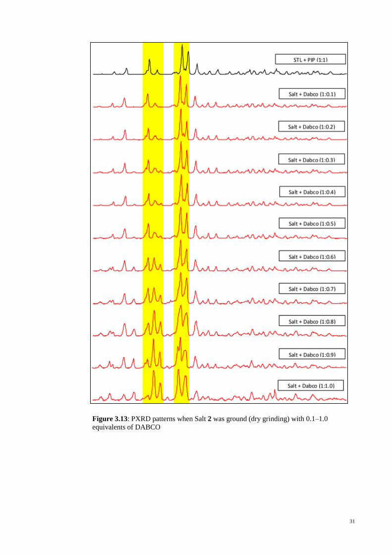
Figure 3.13 PXRD patterns when Salt 2 was ground (dry
grinding) with 0.1–1.0 equivalents of DABCO
31
Figure 3.14 PXRD patterns when Salt 2 underwent LAG
(ethanol) with 1.1–1.5 equivalents of DABCO, in
separate experiments
33
x
Figure 3.15 PXRD patterns when Salt 2 was ground (dry
grinding) with 1.0, 1.5 and 2.5 equivalents of
DABCO
34
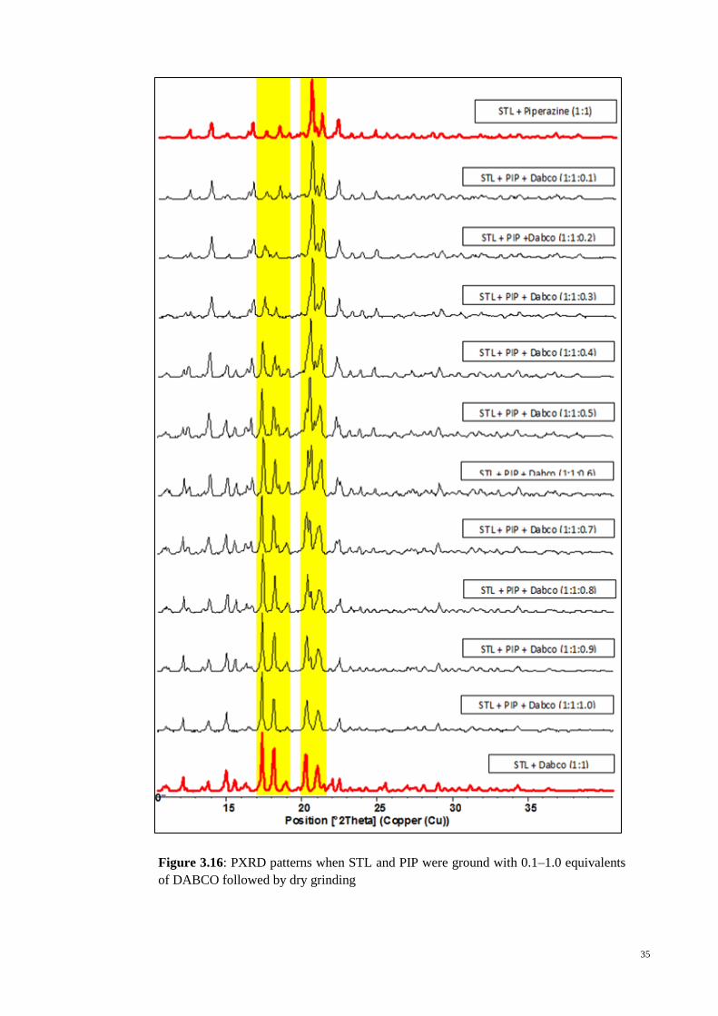
Figure 3.16 PXRD patterns when STL and PIP were ground with
0.1–1.0 equivalents of DABCO followed by dry
grinding
35
Figure 3.17 Representative of PXRD profiles 36

xi
LIST OF TABLES
PAGE
Table 1.1 Strength scale of different intermolecular interactions and
hydrogen bonds
2
Table 3.1 1H NMR data sulfathiazole (STL) and 4-
aminophenylsulfonyl(1,3-thiazol-2-yl)azanide anion
(STL_H-), anion in Salts 1 and Salt 2
14
Table 3.2 1H NMR data of DABCO, PIP, Salt 1, Salt 2 and STL,
DABCO and PIP
15
Table 3.3 IR (cm-1
) absorption for sulfathiazole (STL) and the 4-
aminophenylsulfonyl(1,3-thiazol-2-yl)azanide, [STL_H-],
anions in Salt 1, 2 and sulfathiazole polymorph form III
17
Table 3.4 Crystal data and refinement details for Salt 1 and salt 2 20
Table 3.5 Selected bond distances (Å) and and angles (°) for
[DABCOH][STL_H] (1), [PIPH][STL_H] (2)
21
Table 3.6 Summary of intermolecular interactions (A–H…B; Å, º)
operating in the crystal structures of Salt 1
22
Table 3.7 Summary of intermolecular interactions (A–H…B; Å, º)
operating in the crystal structures of Salt 2
24
Table 3.8 Percentage of Salt 1 formed from addition of DABCO into
Salt 2
32
Table 3.9 Percentage of Salt 1 formed from LAG (using ethanol) 33
Table 3.10 Percentage of salt 1 formed when STL and PIP were
ground with 0.1–1.0 equivalents of DABCO followed by
dry grinding.
36

xii
LIST OF SYMBOLS AND ABBREVIATIONS
APIs : Active Pharmaceutical Ingredients
oC : Degree Celsius
CDCl3 : Deuterated Chloroform
CHN Elemental Analysis : Carbon, Hydrogen and Nitrogen Elemental Analysis
δ : Chemical Shift
DMSO-d6 : Deuterated Dimethylsulfoxide
DABCO : 1-Azonia-4-azabicyclo(2.2.2)octane
FT-IR : Fourier Transform Infrared
J : Coupling Constant
kcal/mol : Kilocalorie per mole
LAG : Liquid Assisted Grinding
M : Molar
m : Multiplet
mL : Mililitre
mmol : Milimoles
NMR : Nuclear Magnetic Resonance
PIP : Piperazine
s : Singlet
Salt 1 : [DABCOH][STL_H] Salt
Salt 2 : [PIPH][STL_H] Salt
STL : Sulfathiazole
t : Triplet
v : Stretching Vibration

xiii
LIST OF APPENDICES
Appendix A: 1H NMR and IR Spectra
1
CHAPTER 1: INTRODUCTION
1.1 Crystal Engineering
Organic chemistry has long since evolved from its traditional synthetic chemistry to
the now contemporary research in crystal engineering, mostly driven by its relevance in
pharmaceutical industries, optical materials and materials science. The most precise
definition of crystal engineering was suggested by Desiraju, “…the understanding of
intermolecular interactions in the content of crystal packing and in the utilization of
such understanding in the design of new solids with desired physical and chemical
properties.” Desiraju (1989).
While both traditional synthetic chemistry and crystal engineering resemble each
other in its analysis and synthesis components, the latter predicts and designs new
functionalised solids with more reason and imagination by taking preformed molecules
with the intention to produce the desired product in a predetermined approach.
1.2 Intermolecular Interactions
The foundation of a crystal structure formed by various intermolecular interactions,
that are responsible for crystal assembly, relates to the study of supramolecular
chemistry. Hydrogen bonding plays an important role in crystal architecture due to its
structural robustness. The very strong negatively-charged hydrogen bond was proven as
one of the most superior forces with energy of 15-40 kcal in comparison to other
intermolecular interactions as shown in Table 1.1.

2
Table 1.1: Strength scale of different intermolecular interactions and hydrogen
bonds (Nangia, 2010)
Interaction type Energy (kcal mol-1
) Examples
Charge-assisted hydrogen bonds 15-40 O-H∙∙∙O-, F-H∙∙∙F
-
Coordinative bonds 20-45 M-N, M-O
Strong hydrogen bonds 5-15 O-H∙∙∙O, N-H∙∙∙O
Weak hydrogen bonds 1-4 C-H∙∙∙O, O-H∙∙∙
van der Waals interactions 0.5-2 CH3∙∙∙CH3,CH3∙∙∙Ph
Heteroatom interactions 1-2 N∙∙∙Cl, I∙∙∙I, Br∙∙∙Br
-stacking 2-10 Ph∙∙∙Ph, nucleobases
Adapted from (Nangia, 2010)
A molecule‟s structure in its crystalline form can be altered by introducing a new
substance which may generate more stable crystal packing. This “supramolecular
synthesis” approach may be achieved with several attempts. However, it is noted that
mechanical grinding has been the most popular and efficient method thus far (Trask et
al., 2004).
1.3 Mechanochemistry
The grinding of a solid is the form of mechanochemistry that has been used for many
years. Chemical reaction transformation is achieved by mechanical forces such as
grinding, milling processes and sonication. These techniques can be solvent free and
less energy consuming than standard solution reactions. It is a sustainable alternative to
conventional solution based and solvothermal chemical processes which impose
wastage of solvents in both lab and industry. Hence, mechanochemistry is known as a
clean and green technology for future practice.

3
In this study, co-crystals were produced by solid state grinding. Solvent drop
grinding was used as an alternative method to improve the conversion and enhance the
kinetics of the desired co-crystal formation. Co-crystals can be defined as quoted from
(Aakeröy & Salmon, 2005),
“Only compounds constructed from discrete neutral molecular species can be
considered as co-crystal. Consequently, all solids containing ions including complex
transition metal ions are excluded. Only co-crystals made from reactants that are solids
at ambient conditions were included (Boese et al., 2003). A co-crystal is a structurally
homogeneous crystalline material that contains two or more neutral building blocks that
are present in definite stoichiometric amounts. Based on these definitions, we are
essentially left with two families of compounds: binary donor-acceptor complexes and
hydrogen bonded co-crystals.”
1.4 Co-crystal in Pharmaceutical Industry
In the field of pharmaceutical development, a pure drug crystal property can be
enhanced by modifying a pharmaceutical co-crystal that contains a single active
pharmaceutical ingredient (API) and a relevant co-former combination.
Figure 1.1: Representation of drug-drug co-crystal and combination drug.
(Sekhon, 2012)

4
1.5 Why Sulfathiazole?
In the realm of the organic solid state, the study of postsynthetic metathesis was done
by Caira, Nassimbeni and Wildervank, (Caira et al., 1995). In the study, the sulfa drug
sulfadimidine–2-hydroxybenzoic acid was formed from co-crystallisation of
sulfadimidine and 2-hydroxybenzoic acid. The sample later on, was ground with a
stoichiometric amount of 2-aminobenzoic acid producing a new 1:1 co-crystal,
sulfadimidine–2-aminobenzoic acid (Caira et al., 1995).
Figure 1.2: Chemical structures of DABCOH+, PIPH
+ and STL_H
-
a. 1-azonia-4-azabicyclo(2.2.2)octane cation (DABCOH+)
b. piperazinium cation (PIPH+)
c. 4-aminophenylsulfonyl(1,3-thiazol-2-yl)azanide anion (STL_H-)
Due to its well-known anti-microbial activity and its five solvent free polymorphs,
sulfa drug sulfathiazole, Figure 1.2(c) has captured the attention of most crystal
engineers. (Drebushchak et al., 2008; Gelbrich et al., 2008; Grove & Keenan, 1941;
Parmar et al., 2007). Sulfathiazole has also attracted the attention of crystal engineers to
further a deeper understanding of characterising, controlling and imitate the formation
of this sulfa drug (Hu et al., 2013; Kelleher et al., 2006; Lawrence et al., 2010; McArdle

5
et al., 2010; Munroe et al., 2014; Munroe et al., 2012; Munroe et al., 2011; Sovago, et
al., 2014). The effectiveness of integrating solvent in the crystal structures of
sulfathiazole has been proved by the discovery of more than one hundred solvates of
sulfathiazole.
Sulfathiazole along with other sulfa-drugs are well known to form co-crystals (Caira,
2007). In the early days, sulfathiazole was featured prominently as pharmaceutically
inspired co-crystals (Stahly, 2009). Sulfathiazole was co-crystallised with proflavin with
equimolar proportions 1:1 and it was found in Flavazole® (Mcintosh et al., 1945).
However, the wide usage of antibiotics and the growing of microbial resistance, the
once highly successful sulfa drugs are now becoming ineffective (Wright et al., 2014).
In the area of developing “new forms of old drugs” to improve effectiveness, driven by
crystal engineering strategies (Almarsson & Zaworotko, 2004; Schultheiss & Newman,
2009; Shan & Zaworotko, 2008), this subject of study attracts the attention of the
medicinal chemistry (Elder et al., 2013; Friščić & Jones, 2010; Kawakami, 2012; Stahl
& Wermuth, 2002; Tilborg et al., 2014) and crystal engineering communities (Aakeröy
et al., 2014; Chierotti et al., 2013; Goud et al., 2014; Maddileti et al., 2014; Moradiya et
al., 2013). The experiment of 1:1 salt formation between sulfathiazole and each of 1,4-
diazabicyclo[2.2.2]octane (DABCO) and piperazine, which will form salt 1 and salt 2
respectively, will be reported in this document. Other interesting topic which will be
documented is the conversion of salt 2 to salt 1 by grinding salt 2 with DABCO
showing that postsynthetic metathesis can also be applicable in the solid state synthesis
of organic salts via mechanochemistry. Although the challenge of producing salt 1 and 2
is quite significant, but yet it will be proved producible once the results are reported in
this document.

6
1.6 X-ray Crystallography
1.6.1 Single Crystal X-ray Diffraction
Single X-ray diffraction is a non-destructive technique which can reveal the
information of the arrangement and architecture of atoms or ions within the crystals.
After the discovery of X-rays in the early 1890‟s, its relevance in the field of chemistry
was noted when it was found that X-ray could be diffracted by crystals. A three-
dimensional architecture of atoms of a crystal was first achieved via X-ray diffraction in
1913 by William Lawrence Bragg. He found X-ray diffraction of sodium chloride
crystal “each sodium is surrounded by six equidistant chlorines and each chlorine by six
equidistant sodiums”. From his findings he infers that sodium chloride crystal consists
of sodium ions, chloride ions and no discrete non-charge atoms of sodium and chloride
were found (Glusker & Trueblood, 2010).
Subsequently, Katherine Lonsdale was able to demonstrate that the benzene ring is a
flat hexagon in which all carbon- carbon double bonds are equal in length and not a ring
structure with alternating single and double bonds (Glusker & Trueblood, 2010).
The main purpose of performing crystal structure analysis by using X-ray or neutron
diffraction is to obtain detail information about the positions of individual atoms at the
atomic level in a 3-dimensional picture. The detailed information includes interatomic
distances, bond angles, planarity of a particular group of atoms, the angle between
planes, conformation at detail around bonds, information about molecular packing,
molecular motion in the crystal and molecular charge distribution.
1.6.2 Powder X-ray Diffraction
“Finger print identification” of numerous solid materials e.g. asbestos, quartz etc. are
always correlated with their powder diffraction patterns. In powder or polycrystalline
diffraction, it is important to have a sample with a smooth plane. The sample has to be

7
ground down to fine particles of about 0.002 mm to 0.005 mm cross section. The ideal
sample is homogeneous and the crystallites are randomly orientated and to have a
smooth flat surface, the sample is pressed into a sample holder. The solid fine
particles are randomly distributed exposing all possible h, k, l planes. Only crystallites
having reflecting planes (h, k, l) parallel to the specimen‟s surface will contribute to the
reflected intensities.
1.6.3 Diffraction Patterns
Goniometer configuration is determined by a diffraction pattern that consist of a plot
of reflected intensities versus the detector angle 2-theta (theta). Values of the 2-theta for
each peak can be calculated based on the wavelength of anode material used. Thus, it is
important to minimise the peak position to the interplanar spacing d which is correlated
to the h,k,l planes that generate the reflection event. The value of d-spacing will be
determined by the metrics of the unit cell. According to Bragg‟s law, a simple relation
for scattering angles can be calculated using 2dhkl sin θ = nλ. Hence, the dimension of
the unit cell can be resolved when the intensity (area under the peak) and the indices h,
k, l are known.
1.7 Crystal
“A crystal is defined as a solid that contains a very high degree of long-range three-
dimensional internal order of the component atoms, molecules or ions. Many studies
were conducted since early times, mostly with regards to the external features of
crystals” (Glusker & Trueblood, 2010).

8
However, it was Max von Laue who noticed that periodic internal organization of
crystals were able to diffract electromagnetic radiation of specific wavelength and he
was able to reason the distances between atoms or ions in details. A crystal‟s ability to
diffract was demonstrated in an experiment which also revealed that X-rays have wave-
like properties (Glusker & Trueblood, 2010).
Even though it‟s external appearance is of flat faces and straight edges seems like the
most obvious property of a crystal, this is not necessary or sufficient to define a crystal,
rather it is internal order and it‟s regular internal repetition quality. This was first
suggested by Johanes Kepler and some of the earliest pictures of crystals viewed under
microscope were published by Robert Hooke (Glusker & Trueblood, 2010).
Crystallisation can be achieved through many methods, most often from solution.
The steps to obtain crystals include saturation of solution, supersaturation. The most
crucial event is nucleation, where solute molecules meet in solution and form small
aggregates. More molecules are then laid out on the nucleus surface and eventually a
crystal forms. All crystals are built up of periodic three-dimensional translational
repetition of some basic structural pattern. This basic component is called “unit cell”.
1.8 Objectives of Studies
The objectives of this research were:
To synthesise and to study the correlations between molecular structure, crystal
packing and physical properties of the selected salts derived from Sulfathiazole.
To study if crystal packing efficiency is valid to be used as a benchmark for
stability of a compound.
To prove that LAG (Liquid Assisted Grinding) can be used to improve the
conversion of one compound (salt) into another.
9
CHAPTER 2: EXPERIMENTAL
2.1 Chemicals
Piperazine and sulfathiazole, 1,4-diazabicyclo[2.2.2]octane were purchased as
analytical grade from Merck and were used throughout the entire experiment . All
solvents and reagents were commercially available and used as obtained without further
purification unless otherwise stated.
2.2 Spectroscopic Analyses
2.2.1 1H NMR Spectroscopic Analysis
For 1H spectroscopic analysis, machines that were used include a Varian Inova
TM
500 NMR spectrometer performing at 500 MHz and JEOL, ECA 400 MHz NMR
spectrometer performing at 400 MHz. All of the NMR samples were prepared by
adding 20–30 mg of the respective sample to 1 mL of DMSO-d6. The NMR spectras
were recorded in DMSO-d6 at 25 C, using the DMSO residual proton at δ 2.49 as the
internal standard. Chemical shifts are reported in ppm on the δ scale and coupling
constants are given in Hz.
2.2.2 Infrared Spectroscopy
IR spectra in the range 4000 - 450 cm-1
were obtained by the Attenuated Total
Reflectance (ATR) technique on a Perkin Elmer RX1 FTIR spectrophotometer.
2.2.3 Raman Spectroscopic Analysis
Bruker Vector22 spectrophotometer in the range 4000 - 400 cm-1
was used to
measure raman scattering. Measurements were made at room temperature under
ambient conditions using a microscope objective to focus the incident laser light upon
individual grains. Excitation at 632.8 nm or 514.5 nm was used and no resonance
effects were observed. For easier comparison amongst spectra, the data presented have

10
been normalised and the background was subtracted by L. E. McNeil from Department
of Physics and Astronomy, University of North Carolina.
2.2.4 Melting Point Determination
Krüss KSP1N melting point apparatus was used to determine the melting point using
glass capillaries and were uncorrected.
2.2.5 CHN Elemental Analysis
The carbon, hydrogen and nitrogen elemental compositions of samples from this
work were obtained from a Perkin-Elmer PE 2400 CHN Elemental Analyser.
2.2.6 DSC Analysis
In the range between 30–400 °C at the rate of 10 °C/min, differential scanning
calorimetric data were recorded with a Perkin Elmer DSC 6 using a Tzero aluminium
pan.
2.3 X-ray Crystallography
Rigaku AFC12/SATURN724 diffractometer fitted with MoKα radiation (λ =
0.71073 Å) so that max was 27.5° was used to measure the intensity data for colourless
crystals of salt 1 and yellow salt 2. The measurement was done at 98 K and data
processing was performed with CrystalClear (Rigaku/MSC inc., 2004), while the
absorption correction applied to the data of salt 2 was with ABSCOR (Higashi, 1995).
2.4 PXRD Analysis
PANalytical Empyrean XRD system with Cu-Κα1 radiation (λ= 1.54056 Å) in the 2θ
range 5 - 90o with a slit size = 0.4785° was used to record powder X-ray diffraction.
Data were recorded with a comparison between experimental and calculated (from
CIF's) PXRD patterns. The software used for powder X-ray diffraction was
X'PertHighScore Plus (Almelo, 2009).
11
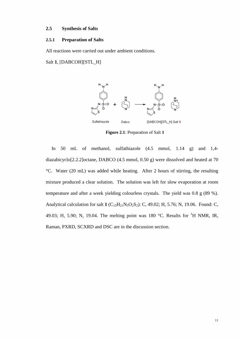
2.5 Synthesis of Salts
2.5.1 Preparation of Salts
All reactions were carried out under ambient conditions.
Salt 1, [DABCOH][STL_H]
Figure 2.1: Preparation of Salt 1
In 50 mL of methanol, sulfathiazole (4.5 mmol, 1.14 g) and 1,4-
diazabicyclo[2.2.2]octane, DABCO (4.5 mmol, 0.50 g) were dissolved and heated at 70
°C. Water (20 mL) was added while heating. After 2 hours of stirring, the resulting
mixture produced a clear solution. The solution was left for slow evaporation at room
temperature and after a week yielding colourless crystals. The yield was 0.8 g (89 %).
Analytical calculation for salt 1 (C15H21N5O2S2): C, 49.02; H, 5.76; N, 19.06. Found: C,
49.03; H, 5.90; N, 19.04. The melting point was 180 °C. Results for 1H NMR, IR,
Raman, PXRD, SCXRD and DSC are in the discussion section.

12
Salt 2, [PIPH][STL_H]
Figure 2.2: Preparation of Salt 2
In 50 mL of acetone, sulfathiazole (13.9 mmol, 3.55 g) and piperazine (13.9 mmol,
1.20 g) were dissolved and heated with stirring at 70 °C for 2 hours. After a week of
slow evaporation at room temperature, 1.85 g (87 %) of yellow crystals formed.
Analytical calculation for salt 2 (C13H19N5O2S2): C, 45.73; H, 5.61; N, 20.51. Found: C,
45.44; H, 5.44; N, 20.03%. The melting point of the crystals was ranging from 176–178
°C. Results for 1H NMR, IR, Raman, PXRD, SCXRD and DSC are in the discussion
section.
2.6 Preparation for Metathesis Experiments
DABCO of (0.1 molar equivalents, 0.004 g) was ground with crystals of salt 2 (0.122
g) using a mortar and pestle. Additional experiments of dry grinding were conducted in
0.1 molar equivalent increments to a maximum of 1.5. The dry grinding procedure was
carried out for at least 5 min followed by measurement with PXRD. LAG experiments
were also conducted with 1.1–1.5 molar equivalents of DABCO addition by using
ethanol as the solvent. For competition experiments, STL (1.95 mmol, 0.50 g) and PIP
(1.95 mmol, 0.169 g) were ground together for 5 min with addition of 0.1 molar
equivalent (0.022 g) of DABCO using a mortar and pestle. The rest of the experiments
were carried out similarly with 0.1 molar equivalent increments.

13
2.7 X-ray Crystallography Studies
The structures were solved by direct methods (Sheldrick, 2008), and full-matrix least
squares refinement was performed on F2 with anisotropic displacement parameters for
all non-hydrogen atoms (Sheldrick, 2008). The O- and N-bound hydrogen atoms were
located from difference maps and were refined with O–H = 0.84±0.01 Å and N–H =
0.88-0.92±0.01 Å; the C-bound hydrogen atoms were included in the refinement in their
idealized positions. A weighting scheme of the form w = 1/[2(Fo
2) + (aP)
2 + bP]
where P = (Fo2 + 2Fc
2)/3 was introduced in each case. The absolute structures of each
of crystal salt 1 and 2 were determined on the basis of difference in Friedel pairs
included in their respective data sets as confirmed by the values of the Flack parameters
(Flack, 1983), i.e., -0.02(7) and 0.06(5) for salt 1 and 2, respectively. The programs
WinGX (Farrugia, 2012), PLATON (Spek, 2003), ORTEP-3 for Windows (Farrugia,
2012), and DIAMOND were also used in the study.

14
CHAPTER 3: RESULTS AND DISCUSSION
3.1 Synthesis and Characterisation
Co-crystalisation experiments between sulfathiazole (STL) and each of 1,4-
diazabicyclo[2.2.2.]octane (DABCO) and piperazine (PIP), formed crystal salts 1 and
2, respectively, as shown in Figures 2.1 and 2.2.
The 1H NMR spectra conducted in DMSO-d6 solution for 1:1 ratios of the respective
pairs of co-formers clearly indicated salt formation as shown in Table 3.1. The absence
of the thiazole-H resonance observed in STL, indicated that proton transfer from
sulfathiazole had occurred. Upfield shifts were noted for all remaining protons within
thiazole ring. Full structural characterisation of salt was achieved through X-ray
crystallography.
Table 3.1: 1H NMR data sulfathiazole (STL) and 4-aminophenylsulfonyl-
(1,3-thiazol-2-yl)azanide anion (STL_H−), anions in Salts 1 and 2
H STL (ppm) Salt 1 (ppm) Salt 2 (ppm)
thiazole-H 12.37 - -
aniline-H 5.82 5.67 5.53
1 7.14 7.08 6.98
2 6.70 6.61 6.49
3 7.39 7.40 7.41
4 6.52 6.51 6.48

15
Given the solid-state observations and the facile formation of salts in solution, it was
thought worthwhile to perform competition experiments in DMSO-d6 solution
monitored by 1H NMR, see Table 3.1 for data. The methylene resonance for pure
DABCO occurred as a sharp singlet at 2.69 ppm and upon proton transfer from STL
in a solution containing a 1:1 stoichiometric mixture this resonance shifted downfield to
2.71 ppm. The comparable values for PIP/PIPH+ were 2.48 and 2.87 ppm,
respectively. In a separate NMR experiment, from Table 3.2, DMSO-d6 solution
containing a 1:1:1 molar ratio of STL, DABCO and PIP featured two sharp resonances
at 2.85 and 2.66 ppm with an integration ratio of 2 to 3, indicating preferential
protonation of PIP over DABCO. This observation is in accord with expectation in that
PIP is more basic than DABCO as seen for example in the calculated (“Advanced
Chemistry Development (ACD/Laboratories),” 2014) pKa values of 9.55±0.10 and
8.19±0.10, respectively, an observation correlated with steric pressures in DABCO and
tertiary amines in general. This suggests that the substitution of PIPH+ in salt 2 by
DABCOH+ leading to salt 1 is due to solid-state considerations.
Table 3.2: 1H NMR data of DABCO, PIP, Salt 1, Salt 2 and STL, DABCO and PIP
Solution containing Assignment
DABCO 2.69 methylene-H in DABCO
PIP 2.48 methylene-H in PIP
1 2.71 methylene-H in DABCOH+
2 2.87 methylene-H in PIPH+
STL, DABCO and PIP 2.85 methylene-H in PIPH+; integration = 8.35 = 8
2.66 methylene-H in DABCO; integration = 11.72 = 12

16
3.2 Infrared and Raman Spectroscopy
Another analysis that has proven useful in distinguishing between co-crystal/salt
formation is Raman spectroscopy (Brittain, 2009; Roy et al., 2013). The most
impressive finding from the Raman measurements in the present study was that the
spectra recorded from the crystals grown from a solution containing the two constituents
and from the crystals prepared by grinding the constituent powders together are virtually
identical, apart from some minor variations in relative intensity which may be due to
variations in the orientation of the individual crystallites from which the measurements
were made. These spectra differed significantly from those of the precursor molecules
as shown in Figures 3.1 and 3.2. Peaks observed at 513 and 1269 cm-1
in the spectrum
of the crystals produced by grinding but did not appear in the spectrum of the solution-
grown crystals. This agreement between the Raman-active vibrational modes of the two
types of crystals clearly shows that the local bonding in the two species is essentially the
same, i.e. that the two methods of fabrication produce the same crystal structure.
Figure 3.1: Raman spectra for conformers and Salt 1

17
Figure 3.2: Raman spectra for conformers and Salt 2
From the data tabulated in Table 3.3, it is evident that sulfathiazole exhibits IR bands
consistent with those of form III (Hu et al., 2010), confirming the conclusions of the
PXRD study.
Table 3.3: IR (cm-1
) absorption for sulfathiazole (STL) and the 4-aminophenylsulfonyl-
(1,3-thiazol-2-yl)azanide, [STL_H-], anions in Salt 1, 2 and sulfathiazole polymorph form III
Mode STL
(cm-1
)
Salt 1 (cm-1
) Salt 2 (cm-1
) Sulfathiazole
polymorph
form III (cm-1
)
-SO2N–H 3274 – –
-NH2 (sym) 3317 3352 (+35) 3345 (+28) 3280
-NH2 (asym) 3350 3412 (+62) 3449 (+99) 3320
-SO2- (sym) 1131 1121 (-10) 1117 (-14) 1133
-SO2- (asym) 1322 1320 (-2) 1311 (-11) 1323
C–N 1267 1234 (-33) 1241 (-26) 1530
C–N(thiazole) 1572 1655 (+83) 1635 (+63) 1530
C–S(thiazole) 924 948 (+24) 940 (+16) 1072 a The values in parentheses are the corresponding values, i.e., the frequency difference between the same mode of anionic-STL
and STL in cm-1, i.e., = ([STL_H]) - (STL).

18
The most obvious difference in the IR spectra relates to the absence of (N–H) at
3274 cm-1 in the salts indicating proton transfer had occurred (Figures 3.3 and 3.4).
It is the most explicit difference in the IR spectra that relates to this matter. As for
ν(C=N) and ν(C–Nthiazole), systematic shifts to higher and lower frequency were
observed respectively, together with reduced and increased bond orders in the salts
according to the corresponding ν(C=N) and ν(C–Nthiazole) as shown in Table 3.3. At the
same time small shifts were noted for sym, symm(SO2), significant shifts are evident for
bands due to amino group.
Figure 3.3: IR spectra of Salt 1
Figure 3.4: IR spectra of Salt 2

19
3.3 Single Crystal X-ray Crystallography
The molecular structures of the asymmetric unit of salt 1 contains a 4-
aminophenylsulfonyl(1,3-thiazol-2-yl)azanide anion (STL_H-) and a 1-azonia-4-
azabicyclo(2.2.2)octane (DABCOH+) cation (1) or a piperazinium (PIPH
+) cation (2) as
shown in Figures 3.5 and 3.6.
Figure 3.5: Molecular structures and crystallographic numbering for the
4-aminophenylsulfonyl(1,3-thiazol-2-yl)azanide anion and
1-azonia-4- azabicyclo(2.2.2)octane cation in the structure of Salt 1
Figure 3.6: Molecular structures and crystallographic numbering for the
4-aminophenylsulfonyl(1,3-thiazol-2-yl)azanide anion and piperazinium cation in
the structure of Salt 2
Details of cell data, X-ray data collection, and structure refinement are given in Table
3.4.
20
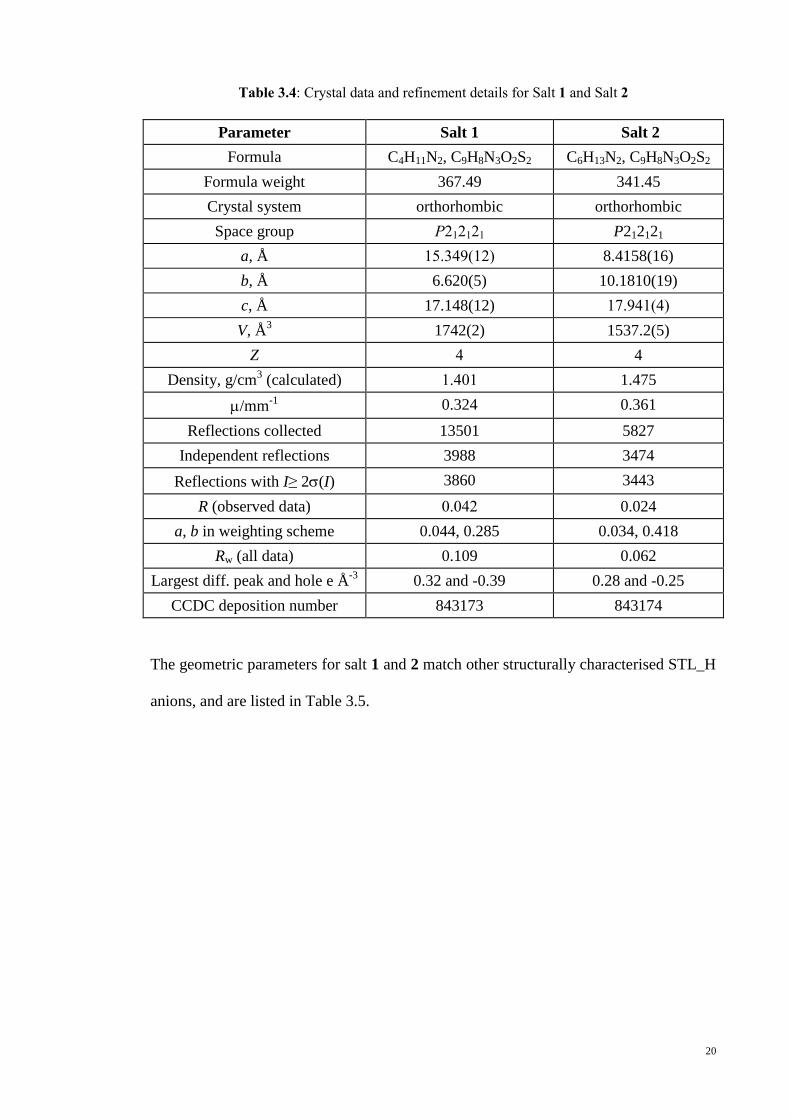
Table 3.4: Crystal data and refinement details for Salt 1 and Salt 2
Parameter Salt 1 Salt 2
Formula C4H11N2, C9H8N3O2S2 C6H13N2, C9H8N3O2S2
Formula weight 367.49 341.45
Crystal system orthorhombic orthorhombic
Space group P212121 P212121
a, Å 15.349(12) 8.4158(16)
b, Å 6.620(5) 10.1810(19)
c, Å 17.148(12) 17.941(4)
V, Å3 1742(2) 1537.2(5)
Z 4 4
Density, g/cm3 (calculated) 1.401 1.475
/mm-1
0.324 0.361
Reflections collected 13501 5827
Independent reflections 3988 3474
Reflections with I≥ 2(I) 3860 3443
R (observed data) 0.042 0.024
a, b in weighting scheme 0.044, 0.285 0.034, 0.418
Rw (all data) 0.109 0.062
Largest diff. peak and hole e Å-3
0.32 and -0.39 0.28 and -0.25
CCDC deposition number 843173 843174
The geometric parameters for salt 1 and 2 match other structurally characterised STL_H
anions, and are listed in Table 3.5.

21
Table 3.5: Selected bond distances (Å) and and angles (°) for [DABCOH][STL_H] (1),
[PIPH][STL_H] (2)
Compound Salt 1
n = 0
Salt 2
n = 0
Sn2-Nn2 1.586(2) 1.5839(13)
Sn2-On1 1.4744(18) 1.4549(11)
Sn2-On2 1.4608(18) 1.4583(11)
Cn1-Sn1 1.781(2) 1.7644(14)
Cn3-Sn1 1.736(3) 1.7291(15)
Cn1-Nn1 1.323(3) 1.3276(18)
Cn2-Nn1 1.388(3) 1.3848(18)
Cn1-Nn2 1.360(3) 1.3558(18)
Cn7-Nn3 1.375(3) 1.370(2)
Cn2-Cn3 1.361(4) 1.351(2)
S...O 3.060(3) 3.0295(12)
N...O - -
Cn1-Sn1-Cn3 90.11(12) 89.82(7)
On1-Sn2-On2 114.92(10) 115.42(7)
On1-Sn2-Nn2 113.31(11) 113.36(7)
On1-Sn2-Cn4 105.64(10) 106.08(7)
On2-Sn2-Nn2 106.37(11) 106.03(7)
On2-Sn2-Cn4 107.92(11) 106.76(7)
Nn2-Sn2-Cn4 108.43(10) 108.90(7)
Cn1-Nn1-Cn2 112.1(2) 111.48(12)
Cn1-Nn2-Sn2 119.89(16) 120.06(10)
Sn2-Nn2-Cn1-Sn1 -7.1(3) 1.23(18)
Cn1-Nn2-Sn2-Cn4 -65.6(2) -71.25(13)
Dihedral angle between thiazole
and aniline rings
89.67(11) 84.83(7)

22
Due to its notable characteristic of conformational flexibility (Parkin et al., 2008)
one of the crystallographically characterised sulfonamide STL_H anions, relative
orientation of the sulfoxide and thiazole residues enables the formation of an
intramolecular S←O interaction (Nakanishi et al., 2007), as shown in Figure 2.1. From
Table 3.5 the result of these conformational observations is that each STL_H anion
adopts an approximate U-shape (the dihedral angles between the five- and six-
membered rings are 89.67(11)° and 84.83(7)°), respectively which proves important in
determining the crystal packing patterns.
Geometric data denoting the hydrogen bonding and other intermolecular
interactions operating in the crystal structures of salt 1 and 2 are collected in Table 3.6
and Table 3.7.
Table 3.6: Summary of intermolecular interactions (A–H…B; Å, º) operating in the crystal
structures of Salt 1
A H B H…B A…B A-
H…B
Symmetry
operation
N3 H1n O1 2.09(2) 2.945(3) 171(3) ½+x, ½-y, 1-z
N3 H2n O2 2.138(19) 2.975(3) 158(3) ½+x, -½-y, 1-z
N4 H3n N1 1.81(2) 2.733(4) 176(3) x, y, z
C8 H8 O2 2.52 3.461(4) 172 ½+x, ½-y, 1-z
C3 H3 Cg(C4-C9) 2.86 3.611(4) 137 1-x, ½+y, ½-z
C10 H10a Cg(S1,N1,C1-C3) 2.89 3.773(4) 149 x, -1+y, z

23
Figure 3.7: View of the crystal packing in Salt 1
(a) Undulating supramolecular layer comprising anions consolidated by aniline-N–
H...O(sulfonyl) hydrogen bonding, shown as orange dashed lines, connected to the cations
via ammonium-N–H...N(thiazole) hydrogen bonding (blue dashed lines).
(b) Projection down the b-axis of the unit cell contents highlighting the interdigitation of layers.
For the crystal structure of salt 1, the aggregation of anions into undulating
layers in the ab-plane, via aniline-N–H...O(sulfonyl) hydrogen bonds, with the thiazole
slits protruding almost in a perpendicular appearance to either side, Figure 3.7.
Affiliated with the anionic layers by charge-assisted N–H...N (thiazole) hydrogen bonds
are the DABCOH+ cations, Figure 3.7(a). Interdigitate neutral layers were formed along
the c-axis and it is shown in Figure 3.7(b). Additional stabilisation to this arrangement is
supported by phenyl-C8–H...O2 interactions. However, neither the azanide-N2 nor

24
amine-N5 atoms form significant intermolecular interactions. The primary interactions
between layers are of the type thiazole- and ammonium-C–H... as indicated in Table
3.6.
Table 3.7: Summary of intermolecular interactions (A–H…B; Å, º) operating in the crystal
structures of Salt 2
A H B H…B A…B A-H…B Symmetry
operation
N3 H1n O2 2.359(13) 3.226(2) 177.4(16) -x, -½+y, 1½-z
N3 H2n O2 2.290(15) 3.1436(19) 165.4(14) -1+x, y, z
N4 H3n N1 1.817(14) 2.7373(18) 170.3(14) -½+x, ½-y, 2-z
N4 H4n N5 1.966(15) 2.8639(19) 163.3(15) -½+x, ½-y, 2-z
N5 H5n Cg(S1,N1,C1-C3) 2.503(16) 3.3559(16) 155.5(14) ½+x, ½-y, 2-z
C11 H11b Cg(C4-C9) 2.50 3.3391(17) 143 ½+x, ½-y, 2-z

25
Figure 3.8: View of the crystal packing in Salt 2
(a) Zigzag supramolecular layer comprising anions consolidated by aniline-N–H...O(sulfonyl)
hydrogen bonding, shown as orange dashed lines, connected to the cations via ammonium-
N–H...N(thiazole) hydrogen bonding (blue dashed lines) which are obscured in this view.
(b) Projection down the a-axis of the unit cell contents highlighting the interdigitation of the
layers and with connections between cations mediated by ammonium-N–H...N(amine)
hydrogen bonding (blue dashed lines).
(c) Supramolecular chain comprising cations connected by ammonium-N–H...N(amine)
hydrogen bonding and linked to the anion layers by N–H...(thiazole) interactions, shown
as purple dashed lines.
26
The supramolecular aggregation in salt 2 on the other hand, closely resembles
the crystal structure of salt 1, ie. via aniline-N–H...O(sulfonyl) hydrogen bonds, the
anions aggregate into zig-zag layers. In this case, it involves only one of the sulfonyl-O2
atoms which is bifurcated; the sulfonyl-O1 atom forms a close intramolecular C–H...O
contact. The cations are associated with the layers as in salt 1, Figure 3.8(a). The layers
inter-digitate along the c-axis and associate via ammonium-N–H...N(amine) hydrogen
bonds, Figure 3.8(b); additional stabilisation to the arrangement from ammonium-C–
H... interactions serve to connect the layers. The residual acidic hydrogen, i.e. residing
on the amine-N5, forms a relatively rare N–H... interaction (Coupar et al., 1996; Knop
et al., 1994), as illustrated in Figure 3.8(c).
In terms of hydrogen bonding, a common feature of the crystal structures is the
formation of charge assisted cation-N–H...N(thiazole) hydrogen bonds. Both aniline
hydrogen atoms participate in aniline-N–H...O(sulfonyl) hydrogen bonding with two
different sulfonyl-O atoms in the case of salt 1 but only one sulfonyl-O atom in the
structure of salt 2. An additional conventional hydrogen bond occurs in salt 2, namely
charge assisted N–H...N hydrogen bonds between cations.
3.4 Powder X-ray Diffraction
Powder X-ray diffraction experiments were carried out on STL. It was identified by
PXRD as form III (Hu et al., 2010) and co-ground with each of DABCO and PIP in
order to determine whether the facile salt formation observed in solution can be
translated into the solid-state. As can be seen from Figure 3.9 and 3.10, salt 1 and salt 2
were prepared quantitatively in bulk form by dry grinding and were crystallographically
characterised.

27
Figure 3.9: PXRD trace of Salt 1
Figure 3.10: PXRD trace of Salt 2
a. Green trace: Pure sulfathiazole.
b. Black trace: DABCO
c. Red trace: PXRD trace of salt after dry grinding.
d. Blue trace: Calculated PXRD based on the single crystal structure.
From Figure 3.9, it can be concluded that co-crystal salt 1 was formed based on
identical trace between the red trace which is PXRD trace salt after dry grinding and the
blue trace which was calculated PXRD based on single crystal structure.
28
On the other hand, Figure 3.10 shows that co-crystal salt 2 was formed from the
comparison trace between the red trace (PXRD trace salt after dry grinding) and blue
trace (calculated PXRD based on single crystal structure).
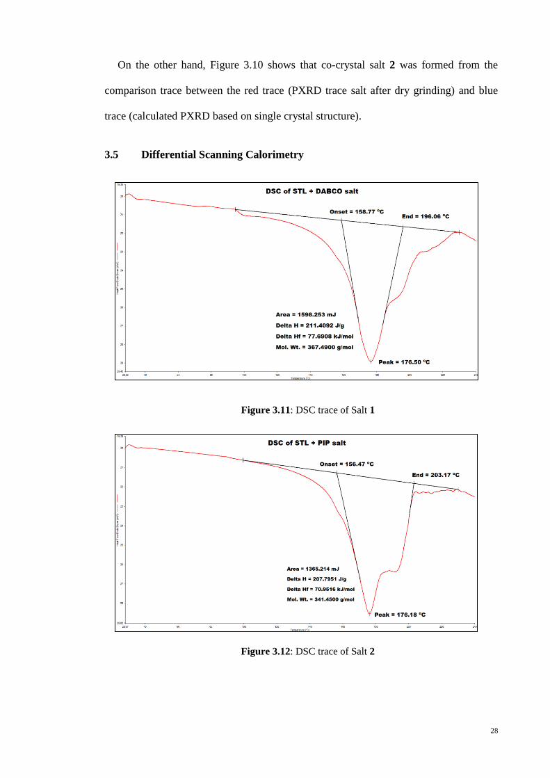
3.5 Differential Scanning Calorimetry
Figure 3.11: DSC trace of Salt 1
Figure 3.12: DSC trace of Salt 2

29
The DSC of salt 1 and 2 were similar, whereby each of them exhibits a significant
endothermic peak around 176 °C corresponding to melting as recorded in Figure 3.11
and Figure 3.12. In the case of salt 1, the endothermic peak was at 176.5 °C with onset
and end temperatures of 158.7 and 196.1 °C, respectively, and with Hmelting = 77.7
kJ/mol. For salt 2, the endothermic step was at 176.2 °C (onset-end 156.5-203.2 °C)
with Hmelting = 71.0 kJ/mol. The progress of the solid state reactions are nicely
correlated with the DSC results which showed that Hmelting was about 10% higher for
salt 1 over salt 2. However, a consideration of the calculated densities, i.e. 1.401 g/cm3
for salt 1 and 1.475 g/cm3 for salt 2 suggests that salt 2 might be the more compact
structure, although an elongation of the c-axis in salt 2 compare to salt 1, the axis along
which layers stack, has already been noted. The reduced proportion of hydrogen bonds
in salt 2 probably contributes to the lower Hmelting where the bond can be easily break
when expose to heat.

30
3.6 Postsynthetic Metathesis Monitored by PXRD
This topic was inspired by the literature precedent (Caira et al., 1995). When
crystals of salt 1 were ground with various quantities of PIP, no significant changes
were evident in the measured PXRD patterns. Piperazine is hygroscopic and can be
transformed rapidly into a liquid/amorphous state under ambient conditions. This could
be a factor as has been noted by Braga, Grepioni and Lampronti. (Braga et al., 2011) in
their supramolecular metathesis experiments with isomeric tartaric acids and pyrazine
which was noted to readily sublime. Apparently, when DABCO was ground with
crystals of salt 2, the PXRD indicated that partial supramolecular metathesis had
occurred as the peculiar PXRD pattern of crystal salt 1 appeared. Hence, a series of dry
grinding experiments were conducted in order to observe quantitatively where crystals
of salt 2 were ground with 0.1 stoichiometric increments of DABCO. These
experiments were perfectly reproducible and the same results were obtained when salt 2
was synthesised in powder form by grinding rather than as powdered single crystals.
Allied PXRD experiments were conducted where STL was co-ground with one
molar equivalent of PIP and n equivalents of DABCO, with n varying in increments of
0.1 up to 1. Even when n was as low as 0.3 for DABCO, 26% of the sample comprised
of salt 1 and this trend was maintained throughout the experiment, a clear preference for
the formation of salt 1 over salt 2 was demonstrated in Figure 3.16.
31
Figure 3.13: PXRD patterns when Salt 2 was ground (dry grinding) with 0.1–1.0
equivalents of DABCO

32
Table 3.8: Percentage of Salt 1 formed from addition of DABCO into Salt 2
Mole equivalent of
DABCO added to Salt 2
Percentage of Salt 1 formed
0.1 1.7
0.2 15.6
0.3 15.6
04 16.3
0.5 15.0
0.6 37.4
0.7 44.7
0.8 53.1
0.9 60.0
1.0 70.2
As shown in Figure 3.13 and Table 3.8, after 0.3 or more equivalents of DABCO
were added, Rietveld refinement (Almelo, 2009) indicates that the conversion increased
to a maximum of 70% after one molar equivalent was added.

33
Figure 3.14: PXRD patterns when Salt 2 underwent LAG (ethanol) with 1.1–1.5 equivalents
of DABCO
Table 3.9: Percentage of Salt 1 formed from LAG (using ethanol)
Mole equivalent of DABCO added to Salt
2
Percentage of Salt 1 formed
1.1 95.0
1.2 96.2
1.3 97.4
1.4 99.5
1.5 99.5
As shown in Figure 3.14 and Table 3.9, after addition of 1.4 equivalents of DABCO
to salt 2 with a few drops of ethanol (LAG), quantitative conversion to salt 1 can be
seen clearly with percentage of conversion of 99.5%.

34
Figure 3.15: PXRD patterns when Salt 2 was ground (dry grinding) with 1.0, 1.5 and 2.5
equivalents of DABCO
From Table 3.8, at 1.0 molar equivalent of DABCO addition, the conversion to salt 1
was 70.2%, while at 2.0 molar equivalents, conversion to salt 1 was 93.0%. From figure
3.15, at 2.5 molar equivalents, the percentage of conversion rose to 97.6% with peaks
assignable to unreacted DABCO, marked with asterisks were clearly evident in Figure
3.15.
35
Figure 3.16: PXRD patterns when STL and PIP were ground with 0.1–1.0 equivalents
of DABCO followed by dry grinding

36
Table 3.10: Percentage of Salt 1 formed when STL and PIP were ground with 0.1–1.0
equivalents of DABCO followed by dry grinding
Mole equivalent of DABCO added to
1:1 STL and PIP
Percentage of salt 1 formed
0.1 4.2
0.2 15.7
0.3 25.9
0.4 38.6
0.5 47.4
0.6 57.6
0.7 68.3
0.8 76.1
0.9 83.1
1.0 98.8
Figure 3.17: Representative of PXRD profiles
a. Ground crystals of salt 2
b. Ground crystals of salt 2 with 0.5 molar equivalents of DABCO
c. Ground crystals of salt 2 with 1.0 molar equivalents of DABCO
d. Ground crystals of salt 2 with 1.5 molar equivalents of DABCO after LAG with
ethanol
e. Ground crystals of salt 1

37
Figure 3.17 shows representative PXRD profiles (relative intensity versus 2-Theta),
traces shown in (b) and (c) show partial supramolecular metathesis up to a maximum of
70% in (c), and quantitative substitution in (d) yielding salt 1, with the aid of LAG
(ethanol). From Table 3.10, percentage of salt 1 formed when STL and PIP were ground
with 0.1–1.0 equivalents of DABCO followed by dry grinding shows the preferential of
formation of salt 1 over salt 2.
A comparison of the hydrogen bonding in each structure provides a clue as to why
the transformation of salt 2 to salt 1 occurs. With more acidic hydrogen atoms in salt 2
and therefore more conventional hydrogen bonds, two structural consequences occur.
Crucially in salt 2, with hydrogen bonds occurring between cations, absent in salt 1, the
layers are squeezed into a zigzag topology as opposed to the undulating layers in salt 1.
This projects the sulfanyl-O1 atoms out of plane so these atoms do not form hydrogen
bonds. The sulfanyl-O1 atoms are therefore accessible for interaction with incoming
DABCO upon grinding experiments with salt 2. The driving force for the exchange is
related to the observation that the aniline-N–H…O(sulfanyl) hydrogen bonds involving
the bifurcated O2 in salt 2 are systematically longer and weaker than the analogous
hydrogen bonds in salt 1, where each sulfanyl-O atom forms a strong aniline-N–
H…O(sulfanyl) hydrogen bond. These structural differences along with the propensity
of PIP to sublime combine to give a plausible explanation for the progress of the post
synthetic metathetical reaction.

38
CHAPTER 4: CONCLUSION
Facile salt formation indicated in solution for STL and each of DABCO and PIP is
vindicated by solution and dry grinding experiments. Even though the layered crystal
structures of salt 1 and salt 2 are similar, salt 1 exhibits more efficient crystal packing,
corroborated by packing efficiency calculations and DSC. This observation allows solid
state postsynthetic metathesis where DABCO can displace PIP in salt 2 to form salt 1.
Such metathetical reactions are rare for organic compounds (Braga et al., 2011; Caira et
al., 1995), and the present study demonstrates that these can be conducted for salts as
well as for species comprising neutral components. Further investigations into this
phenomenon are underway.
Through the study of crystal engineering, the efficacy of this organic compound
which has five solvent-free polymorphs and has proven to be capable of forming co-
crystals with other sulfa drugs can be in enhanced. Formerly, sulfathiazole (STL) was
widely used as a common oral and topical antimicrobial until it became ineffective due
to growing of microbial resistance as a result of the use of antibiotics. However,
sulfathiazole can be combined with other active pharmaceutical ingredients to form a
new co-crystal salt with potential chemotherapeutic benefits.
39
REFERENCES
Aakeröy, C. B., Forbes, S., & Desper, J. (2014). Altering physical properties of
pharmaceutical co-crystals in a systematic manner. CrystEngComm, 16(26), 5870–
5877.
Aakeröy, C. B., & Salmon, D. J. (2005). Building co-crystals with molecular sense and
supramolecular sensibility. CrystEngComm, 7(72), 439–448.
Advanced Chemistry Development (ACD/Laboratories). (2014). [computer software].
ACD/Laboratories: Toronto, Ontario, Canada.
Ali, H. R. H., Edwards, H. G. M., & Scowen, I. J. (2009). Insight into thermally induced
solid‐state polymorphic transformation of sulfathiazole using simultaneous in situ
Raman spectroscopy and differential scanning calorimetry. Journal of Raman
Spectroscopy, 40(8), 887–892.
Almarsson, O., & Zaworotko, M. J. (2004). Crystal engineering of the composition of
pharmaceutical phases. Do pharmaceutical co-crystals represent a new path to
improved medicines? Chemical Communications, (17), 1889–1896.
Almelo, B. V. (2009). X‟Pert HighScore Plus. PANalytical. [computer software]. The
Netherlands.
Basics of X-Ray Diffraction: Diffraction spectra (1999). Retrieved from
http://www.geo.umass.edu/courses/geo311/xrdbasics.pdf.
Boese, R., Kirchner, M. T., Billups, W. E., & Norman, L. R. (2003). Cocrystallization
with acetylene: molecular complexes with acetone and dimethyl sulfoxide.
Angewandte Chemie International Edition, 42(17), 1961–1963.
Braga, D., Grepioni, F., & Lampronti, G. I. (2011). Supramolecular metathesis: co-
former exchange in co-crystals of pyrazine with (R, R)-,(S, S)-,(R, S)-and (S, S/R,
R)-tartaric acid. CrystEngComm, 13(9), 3122–3124.
Brittain, H. G. (2009). Vibrational Spectroscopic Studies of Cocrystals and Salts. 2. The
Benzylamine− Benzoic Acid System. Crystal Growth & Design, 9(8), 3497–3503.
Caira, M. R. (2007). Sulfa drugs as model cocrystal formers. Molecular Pharmaceutics,
4(3), 310–6.
Caira, M. R., Nassimbeni, L. R., & Wildervanck, A. F. (1995). Selective formation of
hydrogen bonded cocrystals between a sulfonamide and aromatic carboxylic acids
in the solid state. Journal of the Chemical Society, Perkin Transactions 2, (12),
2213.
Chierotti, M. R., Gaglioti, K., Gobetto, R., Braga, D., Grepioni, F., & Maini, L. (2013).
From molecular crystals to salt co-crystals of barbituric acid via the carbonate ion
and an improvement of the solid state properties. CrystEngComm, 15(37), 7598–
7605.

40
Coupar, P. I., Ferguson, G., & Glidewell, C. (1996). Piperazine–4, 4‟-Sulfonyldiphenol
(1/2): a Self-Assembled Channel Structure. Acta Crystallographica Section C:
Crystal Structure Communications, 52(12), 3052–3055.
Desiraju, G. R. (1989). Crystal Engineering: The Design of Organic Solids (1st ed.).
Amsterdam: Elsevier Science.
Drebushchak, T. N., Boldyreva, E. V, & Mikhailenko, M. A. (2008). Crystal structures
of sulfathiazole polymorphs in the temperature range 100–295 K: A comparative
analysis. Journal of Structural Chemistry, 49(1), 84–94.
Elder, D. P., Holm, R., & de Diego, H. L. (2013). Use of pharmaceutical salts and
cocrystals to address the issue of poor solubility. International Journal of
Pharmaceutics, 453(1), 88–100.
Farrugia, L. J. (2012). WinGX and ORTEP for Windows: an update. Journal of Applied
Crystallography, 45(4), 849–854.
Flack, H. D. (1983). On Enantiomorph-Polarity Estimation. Acta Crystallographica
Section A: Foundations of Crystallography, 39(6), 876–881.
Friščić, T., & Jones, W. (2010). Benefits of cocrystallisation in pharmaceutical
materials science: an update. Journal of Pharmacy and Pharmacology, 62(11),
1547–1559.
Gelbrich, T., Hughes, D. S., Hursthouse, M. B., & Threlfall, T. L. (2008). Packing
similarity in polymorphs of sulfathiazole. CrystEngComm, 10(10), 1328.
Glusker, J. P., & Trueblood, K. N. (2010). Crystal Structure Analysis: A Primer (IUCr
Texts on Crystallography). Oxford University Press, USA.
Goud, N. R., Khan, R. A., & Nangia, A. (2014). Modulating the solubility of
sulfacetamide by means of cocrystals. CrystEngComm, 16(26), 5859–5869.
Grove, D. C., & Keenan, G. L. (1941). The Dimorphism of Sulfathiazole. Journal of the
American Chemical Society, 63(1), 97–99.
Higashi, T. (1995). ABSCOR [computer software]. Rigaku Corporation, Tokyo, Japan.
Hu, Y., Erxleben, A., Hodnett, B. K., Li, B., McArdle, P., Rasmuson, Å. C., & Ryder,
A. G. (2013). Solid-State Transformations of Sulfathiazole Polymorphs: The
Effects of Milling and Humidity. Crystal Growth & Design, 13(8), 3404–3413.
Hu, Y., Erxleben, A., Ryder, A. G., & McArdle, P. (2010). Quantitative analysis of
sulfathiazole polymorphs in ternary mixtures by attenuated total reflectance
infrared, near-infrared and Raman spectroscopy. Journal of Pharmaceutical and
Biomedical Analysis, 53(3), 412–420.
Hu, Y., Gniado, K., Erxleben, A., & McArdle, P. (2014). Mechanochemical reaction of
sulfathiazole with carboxylic acids: formation of a cocrystal, a salt, and
coamorphous solids. Crystal Growth & Design, 14(2), 803–813.

41
Kawakami, K. (2012). Modification of physicochemical characteristics of active
pharmaceutical ingredients and application of supersaturatable dosage forms for
improving bioavailability of poorly absorbed drugs. Advanced Drug Delivery
Reviews, 64(6), 480–495.
Kelleher, J. M., Lawrence, S. E., & Moynihan, H. A. (2006). Effect of the steric demand
and hydrogen bonding capability of additives on the crystal polymorphism of
sulfathiazole. CrystEngComm, 8(4), 327.
Knop, O., Cameron, T. S., Bakshi, P. K., Linden, A., & Roe, S. P. (1994). Crystal
chemistry of tetraradial species. Part 5. Interaction between cation lone pairs and
phenyl groups in tetraphenylborates: crystal structures of Me3S+, Et3S+, Me3SO+,
Ph2I+, and 1-azoniapropellane tetraphenylborates. Canadian Journal of Chemistry,
72(8), 1870–1881.
Lawrence, S. E., McAuliffe, M. T., & Moynihan, H. A. (2010). Mimics of a R22(8)
Hydrogen-Bond Dimer Motif: Synthesis and Influence on the Crystallisation of
Sulfathiazole and Sulfapyridine. European Journal of Organic Chemistry, 2010(6),
1134–1141.
Lee, I. S., Lee, A. Y., & Myerson, A. S. (2008). Concomitant polymorphism in confined
environment. Pharmaceutical Research, 25(4), 960–968.
Maddileti, D., Swapna, B., & Nangia, A. (2014). High solubility crystalline
pharmaceutical Forms of blonanserin. Crystal Growth & Design, 14(5), 2557–
2570.
McArdle, P., Hu, Y., Lyons, A., & Dark, R. (2010). Predicting and understanding
crystal morphology: the morphology of benzoic acid and the polymorphs of
sulfathiazole. CrystEngComm, 12(10), 3119.
Mcintosh, J., Robinson, R. H. M., & Selbie, F. R. (1945). Acridine-Sulphonamide
Compounds as Wound Antiseptics Clinical Trials of Flavazole. The Lancet,
246(6361), 97–99.
Moradiya, H., Islam, M. T., Woollam, G. R., Slipper, I. J., Halsey, S., Snowden, M. J.,
& Douroumis, D. (2013). Continuous Cocrystallization for Dissolution Rate
Optimization of a Poorly Water-Soluble Drug. Crystal Growth & Design, 14(1),
189–198.
Munroe, Á., Croker, D. M., Rasmuson, Å. C., & Hodnett, B. K. (2014). Solution-
Mediated Polymorphic Transformation of FV Sulphathiazole. Crystal Growth &
Design, 14(7), 3466–3471.
Munroe, A., Croker, D., Rasmuson, Å. C., & Hodnett, B. K. (2011). Analysis of FII
crystals of sulfathiazole: epitaxial growth of FII on FIV. CrystEngComm, 13(3),
831–834.
Munroe, Á., Rasmuson, Å. C., Hodnett, B. K., & Croker, D. M. (2012). Relative
Stabilities of the Five Polymorphs of Sulfathiazole. Crystal Growth & Design,
12(6), 2825–2835.

42
Nakanishi, W., Nakamoto, T., Hayashi, S., Sasamori, T., & Tokitoh, N. (2007). Atoms-
in-molecules analysis of extended hypervalent five-center, six-electron (5c-6e)
C2Z2O interactions at the 1,8,9-positions of anthraquinone and 9-
methoxyanthracene systems. Chemistry - A European Journal, 13(1), 255–268.
Nangia, A. (2010). Supramolecular chemistry and crystal engineering. Journal of
Chemical Sciences, 122(3), 295–310.
Parkin, A., Collins, A., Gilmore, C. J., & Wilson, C. C. (2008). Using small molecule
crystal structure data to obtain information about sulfonamide conformation. Acta
Crystallographica Section B: Structural Science, 64(1), 66–71.
Parmar, M. M., Khan, O., Seton, L., & Ford, J. L. (2007). Polymorph Selection with
Morphology Control Using Solvents. Crystal Growth & Design, 7(9), 1635–1642.
Rigaku/MSC inc. (2004). CrystalClear User Manual. Rigaku Corporation, Tokyo,
Japan.
Roy, S., Chamberlin, B., & Matzger, A. J. (2013). Polymorph Discrimination Using
Low Wavenumber Raman Spectroscopy. Organic Process Research &
Development, 17(7), 976–980.
Schultheiss, N., & Newman, A. (2009). Pharmaceutical Cocrystals and Their
Physicochemical Properties. Crystal Growth & Design, 9(6), 2950–2967.
Sekhon, B. S. (2012). Drug-drug co-crystals. DARU Journal of Pharmaceutical
Sciences, 20(1), 45.
Shan, N., & Zaworotko, M. J. (2008). The role of cocrystals in pharmaceutical science.
Drug Discovery Today, 13(9–10), 440–6.
Sheldrick, G. M. (2008). A short history of SHELX. Acta Crystallographica Section A,
64(1), 112–122.
Sovago, I., Gutmann, M. J., Hill, J. G., Senn, H. M., Thomas, L. H., Wilson, C. C., &
Farrugia, L. J. (2014). Experimental Electron Density and Neutron Diffraction
Studies on the Polymorphs of Sulfathiazole. Crystal Growth & Design, 14(3),
1227–1239.
Spek, A. L. (2003). Single-crystal structure validation with the program PLATON.
Journal of Applied Crystallography, 36(1), 7–13.
Stahl, P. H., & Wermuth, C. G. (2002). Handbook of pharmaceutical salts: properties,
selection, and use (Vol. 2). Weinheim, Germany: Wiley-Vch.
Stahly, G. P. (2009). A Survey of Cocrystals Reported Prior to 2000. Crystal Growth &
Design, 9(10), 4212–4229.
Tilborg, A., Norberg, B., & Wouters, J. (2014). Pharmaceutical salts and cocrystals
involving amino acids: a brief structural overview of the state-of-art. European
Journal of Medicinal Chemistry, 74, 411–426.

43
Trask, A. V, Motherwell, W. D. S., & Jones, W. (2004). Solvent-drop grinding: green
polymorph control of cocrystallisation. Chemical Communications, 0, 890–891.
Wright, P. M., Seiple, I. B., & Myers, A. G. (2014). The evolving role of chemical
synthesis in antibacterial drug discovery. Angewandte Chemie (International Ed. in
English), 53(34), 8840–69.
44
LIST OF PUBLICATIONS
Aznan, A. M. A., Abdullah, Z., Lee, V. S., & Tiekink, E. R. T. (2014). Crystal structure
of a new monoclinic polymorph of N-(4-methylphenyl)-3- nitropyridin-2-amine.
Acta Crystallographica Section E: Structure Reports Online, 70(8), 58–61.
Aznan, A. M. A., Abdullah, Z., Khoo, C. H., Chen, B. J., See, T. H., Sim, J. H., …
Tiekink, E. R. T. (2015). Three ammonium salts of sulfathiazole: Crystallography
and anti-microbial assay. Zeitschrift Fur Kristallographie - Crystalline Materials,
230(6), 385–396.
Aznan, A. M. A., Safwan, A. P., Abdullah, Z., Kaulgud, T., Arman, H. D., … Tiekink,
E. R. T. (2014). Postsynthetic Metathesis in an All Organic Two-Dimensional
Array Mediated by Hydrogen Bonding. Crystal Growth & Design, 14, 5794–5800.

45
LIST OF PRESENTATIONS
Dalton 2014 Conference, “Supramolecular Methathesis: cation exchange in salts
derived from the sulfa-drug, Sulfathiazole”, Poster presentation (International), 15-17
April 2014, University of Warwick, Coventry, England.
5 th
UM-NUS-CU Trilateral Mini Symposium and Scientific Meeting 2014,
“Supramolecular Methathesis: cation exchange in salts derived from the sulfa-drug,
Sulfathiazole”, Poster presentation (International), 11-12 February 2014, University of
Malaya, Kuala Lumpur, Malaysia.
University of Malaya Chemical Crystallography Symposium 2014, “Supramolecular
Methathesis: cation exchange in salts derived from the sulfa-drug, Sulfathiazole”, Poster
and oral presentation (National), 28 May 2014, University of Malaya, Kuala Lumpur,
Malaysia.
University of Malaya Pharmaceutical Co-Crystal Symposium 2014, “Supramolecular
Methathesis: cation exchange in salts derived from the sulfa-drug, Sulfathiazole”, Poster
and oral presentation (National), 19 July 2014, University of Malaya, Kuala Lumpur,
Malaysia.

APPENDIX
APPENDIX A: 1H NMR and IR

1H
NM
R S
pec
tru
m (
DM
SO
-d6
, 400 M
Hz)
of
Su
lfa
thia
zole

1H
NM
R S
pec
tru
m (
DM
SO
-d6
, 400 M
Hz)
of
DA
BC
O

1H
NM
R S
pec
tru
m (
DM
SO
-d6
, 400 M
Hz)
of
Pip
erazi
ne

1H
NM
R S
pec
tru
m (
DM
SO
-d6
, 400 M
Hz)
of
ST
L, D
AB
CO
& P
IP

1H
NM
R S
pec
tru
m (
DM
SO
-d6
, 400 M
Hz)
of
Salt
1, [D
AB
CO
H][
ST
L_H
]
1H
NM
R S
pec
tru
m (
DM
SO
-d6
, 400 M
Hz)
of
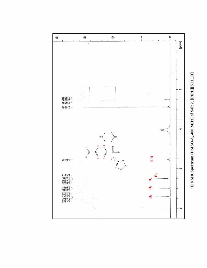
Salt
2, [P
IPH
][S
TL
_H
]

IR S
pec
tru
m o
f S
alt
1,
[DA
BC
OH
][S
TL
_H
]