synthesis of glyconanomaterials: a review
TRANSCRIPT
* Corresponding author E-mail: [email protected]
© 2016 NSP
Natural Sciences Publishing Cor.
Int. J. Nano. Chem. 2 No. 3, 53-74 (2016) 53
International Journal of Nanomaterials and Chemistry
http://dx.doi.org/10.18576/ijnc/020301
Synthesis of Glyconanomaterials: A Review
Verinder Kaur and Ritu Mahajan*
Department of Applied Sciences, Chandigarh University, National Highway 95, Chandigarh-Ludhiana Highway Sahibzada Ajit Singh
Nagar, Punjab 140413, India.
Received: 14 May 2016, Revised: 16 Jul.2016, Accepted: 30 Jul. 2016.
Published online: 1 Sep. 2016.
Abstract: Glyconanomaterials broadly defined as carbohydrate presenting structures below 100nm in size, exhibit
remarkable chemical and physical properties with high potential for modern biomedical applications. The results
demonstrate that this approach to carbohydrate presentation at nonmaterial surfaces leads to efficient and selective binding
to cognate proteins, enabling new applications in carbohydrate-lectin recognition, profiting, biosensing, screening, cell
imaging and bacteria detection.
Keywords: Glyconanomaterials, Carbohydrates, Imaging, Therapy, biomolecules.
1 Introduction
Carbohydrates are the most abundant biomolecules in
nature and essential elements in a wide range of processes
in living systems. [1-5] Besides their uses as structural
materials and energy sources, they are to large extent
mediating recognition events through their interactions with
proteins and other biological entities.[6-8] Complex
carbohydrate structures are thus involved in, for example,
cell communication and trafficking, tumor genesis and
progression, immune responses, fertilization, apoptosis, and
infection.[9-11] The field has recently experienced a
dramatic upsurge, much on account of the very strong
developments in carbohydrate synthesis, glycan analysis
methods, and nanotechnology[12-15]
Nanomaterials as scaffolds for carbohydrate ligand display
have recently emerged, and glyconanomaterials have thus
been synthesized, demonstrating great potential in
biomedical imaging, diagnostics, and therapeutics.[16-18]
Compared with molecular scaffolds, nanomaterials as
ligand carriers offer a number of attractive features.
Nanomaterials, being small in size, have high specific
surface areas and can therefore accommodate high density
ligands promoting multivalent interactions with their
binding partners.[19] The ligand density can be modulated
by the size and shape of the nanomaterial, and multiple
epitopes of the same ligand can be exposed and presented
in a three-dimensional format. Nanomaterials possess
unique optical, electronic, magnetic, and mechanical
properties as well as chemical reactivities [20-21] These
properties, together with their nanosized dimensions, allow
for their incorporation into cells for in vitro and in vivo
imaging, drug-delivery, and targeting tumor cells. This
opens up a wide range of possibilities, the potential of
which is just emerging. [22]
Nanomaterials constitute a class of structures that have
unique physiochemical properties and are excellent
scaffolds for presenting carbohydrates, important
biomolecules that mediate a wide variety of important
biological events.[23-25] The fabrication of carbohydrate-
presenting nanomaterials, glyconanomaterials, is of high
interest and utility, combining the features of nanoscale
objects with biomolecular recognition.[26] The structures
can also produce strong multivalent effects, where the
nanomaterial scaffold greatly enhances the relatively weak
affinities of single carbohydrate ligands to the
corresponding receptors, and effectively amplifies the
carbohydrate-mediated interactions. Glyconanomaterials
are thus an appealing platform for biosensing
applications.[27-30] In this review, we discuss the
chemistry for conjugation of carbohydrates to
nanomaterials, summarize strategies, the limitations and
future perspectives of these emerging glyconanomaterials
sensing systems are furthermore discussed.
Nanoparticles are the subject of numerous papers and
reports and are full of promises for electronic, optical,
magnetic and biomedical applications. [31-3] Although
metallic nanoparticles have been functionalized with
peptides, proteins and DNA during the last 20 years,
carbohydrates has not been used with this purpose until
2001. Since the first synthesis of gold nanoparticles
functionalized with carbohydrates (glyconanoparticles) was

54 V. Kaur, R. Mahajan: Synthesis of Glyconanomaterials: A Review…
© 2016 NSP
Natural Sciences Publishing Cor.
reported, the number of published articles has considerably
increased. [33] This review article is the progress in the
development of nanoparticles functionalized with
biological relevant oligosaccharides.
Nanomaterials have unique optical, electronic, or magnetic
properties, thus explaining their potential applications in
complex biosystems when coupled with biomolecules, such
as DNA, peptides, proteins, or carbohydrates. [34] With a
large surface-to-volume ratio and homogeneity in aqueous
solutions, various biomolecule conjugated nanomaterials
are exploited for elucidating biological interactions. During
the past decade, biomolecule-conjugated nanoparticles
(NPs) have been prepared and used in diagnostics, creative
therapeutics, biomolecular interactions, and in vivo cell
imaging.[35] For example, Mirkin et al. developed an
ultrasensitive bio-barcode detection method based on
oligonucleotide-conjugated gold NP (AuNP) for
biomarkers in small amounts in complex biofluids. [36]
(Fig.1)
Figure 1. Bio-barcode detection method based on
oligonucleotide-conjugated gold NP (AuNP)
Weissleder et al. fabricated antibody-conjugated iron oxide
NPs and used them to enhance T2 signals in magnetic
resonance imaging. [37] Since they have unique magnetic
properties, diverse functionalized magnetic nanoparticles
(MNPs) have been designed and prepared to purify target
proteins from crude cell lysate by simple magnetic
separation. Recently, the authors combined antibody-
conjugated MNP with matrix-assisted laser
desorption/ionization–time of flight (MALDI–TOF) mass
spectrometry (MS) as a rapid and cost-effective detection
method for diagnosing disease markers in human sera. [38]
Biomolecule-modified quantum dots (QDs) have also been
demonstrated as having promising applications in in
vivo imaging, including cell trafficking and targeting.
Besides metallic NPs, carbon nanotubes (CNTs) have also
been demonstrated to be powerful carriers and to be useful
in biological systems because of their high surface
utilization efficiency and good size uniformity. [39-40].
2 Preparation of Glyconanomaterials
A critical step in the preparation of glyconanomaterials is
the surface coupling chemistry for attaching carbohydrates
to the nanomaterial. Nanomaterials come in different forms,
sizes, and shapes. Conjugation chemistry should therefore
be designed by taking into consideration the chemical
nature of the nanomaterial to afford efficient ligand
coupling and to provide optimal ligand presentation. [41-
42]
2.1 Conjugation of Carbohydrates to
Nanomaterials
Two general strategies for nanomaterial functionalization
can be discerned, based on either noncovalent or covalent
protocols. Both approaches are associated with advantages
and drawbacks, although covalent protocols are generally
preferred due to the considerably higher stabilities of the
constructs. [43]
2.2 Noncovalent Attachment
A variety of glyconanomaterials based on physisorption of
carbohydrate ligands to the material surface has been
reported.[44-45] The attachment relies on noncovalent
interactions, including, for example, hydrogen bonding,
Coulombic interactions, and hydrophobic effects. A method
for producing metallic glyconanoparticles through
electrostatic adsorption was reported by Yang and co-
workers, in which metal/chitosan nanocomposites were
prepared on a range of different metals including Au, Ag,
Pt, and Pd. [46] The nanoparticles were synthesized by
reducing metal salts in the presence of chitosan, resulting in
simultaneous ligand adsorption. Rosenzweig et al.
synthesized dextran-coated quantum dots (QDs) where
negatively charged carboxymethyldextran was adsorbed
onto QDs by mixing with positively charged polylysine via
electrostatic interactions.[47] Khiar et al. functionalized
carbon nanotubes (CNTs) with pyrene-modified
neoglycolipids.[48] Carbohydrate-conjugated, self-
assembled CNT bundles could be exfoliated, yielding
individual functionalized nanotubes. As noticed from these
examples, a notable advantage of the physisorption strategy
is that the reaction conditions are relatively mild, and
minimal chemical derivatization is required for the
nanomaterial substrates and the carbohydrate ligands.
Nevertheless, the physical adsorption is relatively random
and disordered compared to covalent linkages. In addition,
the association is not sufficiently strong, which may lead to
potential bond breakage during interactions, as well as
increased nonspecific or unexpected interactions with the
target molecules. This can significantly affect the
specificity and sensitivity in applications such as biological
sensing and recognition. However, as demonstrated in the
mentioned examples, oligomer/polymer-based ligands can
to some extent circumvent the stability problems.[49-50]

Int. J. Nano. Chem. 2, No. 3, 53-74 (2016) / http://www.naturalspublishing.com/Journals.asp 55
© 2016 NSP
Natural Sciences Publishing Cor.
3 Covalent Attachment
The most commonly used method for conjugating
carbohydrate structures to nanomaterials is based on
covalent attachment. Among the various nanomaterials,
gold nanoparticles (Au NPs) are the most extensively used
scaffold materials especially in fundamental studies due to
their ease of preparation, exceptional stability, and high
reproducibility.[51] Au NPs of different sizes, shapes, and
controlled dispersity can now be synthesized using simple
solution-based methods. The well-established thiol– and
disulfide–Au chemistry, first applied to nanoparticles using
a two-phase system by Brust et al., allows the preparation
of Au NPs with well-defined surfaces. [52] These surface
ligands serve as a protective layer to provide high stability
for the nanomaterials in media ranging from organic
solvents to biological milieus. The chemistry has been
widely adopted to prepare Au NPs modified with various
functional groups, and biological molecules including
DNA, proteins, peptides, and carbohydrates have all been
successfully introduced into the system. Penadés and co-
workers reported the first synthesis of carbohydrate-
functionalized Au NPs. [53] (Fig.2)
Figure 2. First synthesis of carbohydrate-functionalized Au
NPs.
The trisaccharide determinant of the Lewisx (Lex) antigen
was derivatized with an alkylthiol, and Lex-coated Au NPs
were prepared by reducing HAuCl4 with NaBH4 in
presence of the thiol-derivatized Lex. Based on this
strategy, Au NPs functionalized with monosaccharide’s
(glucose), disaccharides (maltose), and tetrasaccharides
(Ley) were prepared and applied to the studies of various
biological interactions.[54] Later, several other research
groups utilized a similar strategy to produce Au and Ag
glyconanoparticles using thiolated carbohydrates.[55-57]
Furthermore, thiolated carbohydrate derivatives have been
adopted in the preparation of glyco-quantum dots (GQDs).
Additional coupling methods based on the reaction of
complementary functional groups have also been developed
to facilitate the conjugation of carbohydrates other than the
thiolated derivatives. Examples include coupling N-
hydroxysuccinimide (NHS)-functionalized dextran to
amine-functionalized Ag NPs and amine-derivatized
carbohydrates to aldehyde-functionalized Au NPs.
Current methods for the preparation of carbohydrate-
conjugated nanomaterials generally require the use of
derivatized carbohydrates, amenable to coupling to the
chosen nanomaterial surface. Un-derivatized carbohydrate
structures present a considerable challenge. A few reported
examples apply to flat substrates in microarray
construction. One approach used hydrazide-modified gold
films, where the hydrazide reacted with the terminal
aldehyde group of the carbohydrates.[58] A similar
approach employed amine-functionalized surfaces and the
coupling of the carbohydrates took place by reductive
amination. In both cases, reducing carbohydrates are
necessary and, for monosaccharide’s, the coupled products
often became acyclic and lost their binding affinities. [59-
60]
A simple method for attaching un-derivatized
carbohydrates to gold and iron oxide nanoparticles is based
on the well-established procedure for the covalent
attachment of molecules and materials to solid substrates
using functionalized perfluorophenylazides (PFPAs).The
azide moiety on PFPAs can be activated by UV light,
converting into the highly active nitrene that undergoes
insertion reaction into diverse CH bonds and addition
reaction to C=C bonds. Polymers carbon nanotubes,
graphene and small organic molecules have been
successfully immobilized onto PFPA-modified flat
substrates and nanoparticles, providing highly robust and
stable linkages. [61-62] Carbohydrates are another category
of substances that are well-suited for this photo initiated
immobilization chemistry. Carbohydrates have a number of
CH bonds that can be used for the insertion reaction with
PFPA, while leaving OH groups intact for the binding
interactions with lectins. [63] More importantly, the
coupling chemistry does not require chemical derivatization
of the carbohydrates. This is especially attractive for higher
carbohydrate structures, the synthesis of which are often
complex and time-consuming due to the stereochemistry
control and multiple protection/deprotection steps involved
in the site-specific glycosylation and derivatization
reactions.[64] The photocoupling reaction is also facile and
efficient, taking place in a few minutes at room temperature
in the ambient environment. Photochemical methods for
carbohydrate attachment have been explored and reported
in the literature.[65-66] Sprenger and co-workers employed
carbenes to attach glycans and glycoconjugates in the
fabrication of microarrays.[67] (Fig.3)

56 V. Kaur, R. Mahajan: Synthesis of Glyconanomaterials: A Review…
© 2016 NSP
Natural Sciences Publishing Cor.
Figure 3. Carbenes to attach glycans and glycoconjugates
in the fabrication of microarrays
The photoactive aryltrifluoromethyldiazirine was
conjugated to dextran and was then applied to glass slides
to form a photoactive coating. Activation by UV light
produced highly reactive carbenes, which attached glycans
via insertion reactions. In the method of Wang et al., the
photoactive species was a phthalimide chromophore that
induces H abstraction and subsequent recombination
reaction with neighboring molecules.[68] This
photochemistry was adopted to covalently attach
unmodified mono-, oligo-, and polysaccharides on glass
slides. PFPA as the photocoupling agent was used by
Addadi and co-workers in the preparation of hyaluronan-
coated polystyrene beads.[69] PFPA was first coupled onto
amino-capped polystyrene beads, and hyaluronan was
subsequently immobilized by UV irradiation.(Fig.4)
Figure 4. PFPA was first coupled onto amino-capped
polystyrene beads, and hyaluronan was subsequently
immobilized by UV irradiation.
PFPAs can be employed to conjugate monosaccharides and
oligosaccharides to nanomaterials. Compared with
polysaccharides, mono- and oligo-saccharides are smaller
carbohydrate structures and more challenging for this
coupling chemistry. In principle, only one covalent bond is
needed to attach the entire molecule to the surface. The
probability of bond formation increases with the number of
CH bonds, or the size of the carbohydrate structure. Indeed,
our results showed that the coupling yield increased from
57% for d-mannose, to 74% and 81% for 2-O-α-d-
mannopyranosyl-d-mannopyranose (Man2) and 3, 6-di-O-
(α-d-mannopyranosyl)-d-mannopyranose (Man3),
respectively.
The presence of PFPA on the NPs was confirmed by NMR,
FTIR, and X-ray photoelectron spectroscopy (XPS). UV–
vis spectroscopy and transmission electron microscopy
(TEM) images of PFPA/Au NPs showed excellent
dispersibility and stability of these nanoparticles in organic
solvents. To couple carbohydrates to the NPs, a solution of
PFPA NPs mixed with the carbohydrate ligand was
irradiated with 280-nm UV light for 5 min (Fig. 1) to yield
nanoparticles that were well-dispersed and readily soluble
in water. (Fig.5)
Figure5. Synthesis of Au NPs functionalized with PFPA-
thiol and subsequent photoinitiated coupling of
carbohydrates.
Carbohydrates constitute the most abundant organic matter
in nature, serving as structural components and energy
sources, and mediating a wide range of cellular activities.
The emergence of nanomaterials with distinct optical,
magnetic, and electronic properties has witnessed a rapid
adoption of these materials for biomedical research and
applications.[70-72] Nanomaterials of various shapes and
sizes having large specific areas can be used as multivalent
scaffolds to present carbohydrate ligands. (Fig.6) The
resulting glyconanomaterials effectively amplify the
glycan-mediated interactions, making it possible to use
these materials for sensing, imaging, diagnosis and therapy.

Int. J. Nano. Chem. 2, No. 3, 53-74 (2016) / http://www.naturalspublishing.com/Journals.asp 57
© 2016 NSP
Natural Sciences Publishing Cor.
Figure6.Various types of glyconanomaterials have been de
veloped and used in imaging, diagnosis, and therapeutics.
4 Preparation of Glyconanomaterials: Carbon
nanomaterials
Carbon-based nanomaterials have a long history, from the
oldest nanomaterial of amorphous carbon, to the newly
discovered fullerenes, carbon nanotubes (CNTs) and
graphene. [73-75] These materials continue to break
records of material and physical properties, and hold
promise to impact a wide range of fields, including
electronics, sensing, imaging and therapeutics. However,
several disadvantages limit their biological applications,
such as poor water solubility, lack of reactive functionality,
and potentially high cytotoxicity. An effective way to
overcome these limitations is to introduce an organic
coating on the carbon materials. Carbohydrates are in these
sense suitable candidates that not only increase the
biocompatibility and solubility, but also introduce
molecular recognition features to the carbon materials,
which can impact cellular interactions and uptake of these
entities.
One approach to carbohydrate conjugation is through non-
covalent interactions between carbon materials and
modified carbohydrates. In this case, carbohydrates are
chemically derivatized with non polar moieties such as
lipids polyaromatic hydrocarbons, or porphyrins which are
capable of interacting with the hydrophobic carbon
materials. The resulting carbon materials are not chemically
functionalized, and their properties are thus preserved.
Bertozzi and co-workers coated single-wall CNTs
(SWCNTs) with poly (methyl vinyl ketone) having a
18 lipid tail that could self-assemble on the SWCNTs
through hydrophobic effects. [76] (Fig.7).
The polymer was functionalized with α-D-N-
acetylgalactosamine (α-GalNAc) as pendant groups. The
resulting mucin mimic-modified SWCNTs resisted non-
specific protein adsorption, and could also recognize
the Helix pomatia agglutinin (HPA) and α-GalNAc-binding
lectin.
Surface functionalization of nanomaterials is an area of
current investigation that supports the development of new
biomaterials for applications in biology and medicine. [77-
78] The synthesis, characterization, and antibacterial
properties of the first examples of antibiotic-labeled
graphitic carbon nanofibers (GCNFs) covalently
functionalized with amino glycoside and quinolone
antibiotics were described. Ruthenium tetroxide oxidation
of herringbone GCNFs gave higher amounts of surface
carboxyl groups than previous methods. These carboxyl
groups served as sites of attachment for antibiotics by acyl
substitution. Bioassay of this novel, functionalized GCNFs
using serial dilution and optical density methods
demonstrated that antibiotic-labeled GCNFs possess
significant antibacterial activity against Pseudomonas
aeruginosa.
Figure 7. single-wall CNTs (SWCNTs) coated with poly
(methyl vinyl ketone) having a 18 lipid tail that could self-
assemble on the SWCNTs through hydrophobic effects
In another study, the group functionalized monosaccharides
with pyrene, which were subsequently adsorbed on
SWCNTs. The modified SWCNTs were used to promote
cell adhesion and study dynamic cellular activities. The
same concept was used in a work by Lin et al. where
pristine graphene was functionalized with pyrene-modified
maltose.[79] Upon adsorption; graphene quenched the
pyrene fluorescence, which was then recovered by addition
of the lectin Concanavalin A (Con A). This displacement-
type assay provided a means for lectin sensing, where the
detection limit was estimated to be 0.8 nM in the case of
Con A.
Covalent modification requires a chemical reaction between
the carbon nanomaterial and the carbohydrate. [80] In this
case, either the carbon nanomaterial or the carbohydrate, or
both, need to be chemically functionalized. Carbon
nanomaterials are relatively inert chemically, and therefore,
methods for the chemical functionalization of carbon
materials often involve the use of reactive intermediates
such as azomethineylides, radicals, carbenes, and nitrenes.
Among the carbon materials, fullerenes, especially
buckminsterfullerene C60, have the richest and the most
58 V. Kaur, R. Mahajan: Synthesis of Glyconanomaterials: A Review…
© 2016 NSP
Natural Sciences Publishing Cor.
established functionalization chemistry. Well-defined
fullerene derivatives can be synthesized, and the number of
functional groups can be precisely controlled. [81] For CNTs
and graphene, the most common way to achieve covalent
functionalization is to use the oxidized forms. Oxidation
generates oxygen-containing functional groups, such as
epoxy and carboxylic acid moieties, which can then be used
to react with, e.g., amine-functionalized carbohydrates. In
the case of graphene, the vast majority of the literature uses
the oxidized form, which can be prepared inexpensively
from graphite to produce single-layer graphene oxide in
large quantities. Single-layer pristine graphene, on the other
hand, is still difficult to obtain, especially on a larger scale.
Aryl diazonium salts are among the most used reagents to
functionalize pristine CNTs and graphene. The reaction has
been suggested to proceed via aryl radicals that are
generated by electron transfer from the CNTs or graphene
to the aryl diazonium ions after elimination of N2.[82] This
chemistry was for example used by Torres et al., who
prepared SWCNTs and graphene coated with α-D-
mannosyl (Man) dendrons following a sequential
functionalization approach.[83] A diazonium salt, prepared
from 4-[(trimethylsilyl)ethynyl)]aniline with isoamylnitrite,
was activated under microwave irradiation and reacted with
SWCNTs and graphene. The carbohydrates were
subsequently conjugated to the material via a copper-
catalyzed alkyne-azide cycloaddition (CuAAC) reaction
using azide-functionalized-α-D-mannosyl dendrons.
Another useful functionalization method relies on
azomethineylides, which undergo 1,3-dipolarcycloaddition
with CNTs to form pyrrolidine derivatives.[84] This method
can also be applied to indirect carbohydrate
functionalization as exemplified in a study by Hong et al.[85-
86] The azomethineylides were in this case generated from
α-amino acids and an aldehyde, and carboxy-functionalized
D-N-acetyl glucosamine(GlcNAc) structures were then
conjugated to the resulting pyrrolidinyl CNTs .Nitrenes
constitute another widely used reactive intermediate to
introduce functional groups directly on pristine carbon
nanomaterials. Generated by thermal or light activation,
these reactive intermediates are perceived to undergo
cheletropic cycloaddition reactions with alkenes to form
aziridines Azide-functionalized carbohydrates was used to
functionalize CNTs in refluxing chlorobenzene to render
CNTs water soluble. This chemistry is very efficient, and
perfluorophenylnitrene formation was reported to
functionalize different pristine carbon nanomaterials.
Unlike the singlet phenyl nitrene that primarily ring
expands to form the dehydroazepine, the singlet
perfluorophenylnitrene can undergo efficient C=C addition
reactions due to its longer lifetime and higher activation
energy barrier for the ring expansion reaction. Therefore,
reaction with perfluorophenylazide (PFPA) resulted in
covalent functionalization of fullerenes, CNTs and
graphene. The properties of the materials can be tailored by
the PFPA functionality. For instance, pristine graphene can
be made soluble in water or common organic solvents, and
CNTs can be derivatized with polymer brushes through the
conjugation of an atom-transfer radical-polymerization
(ATRP) initiator to PFPA.[87-88] Using PFPA-NHS to
functionalize CNTs or graphene, the material can be further
conjugated with amine-functionalized carbohydrates. The
resulting materials selectively recognized carbohydrate-
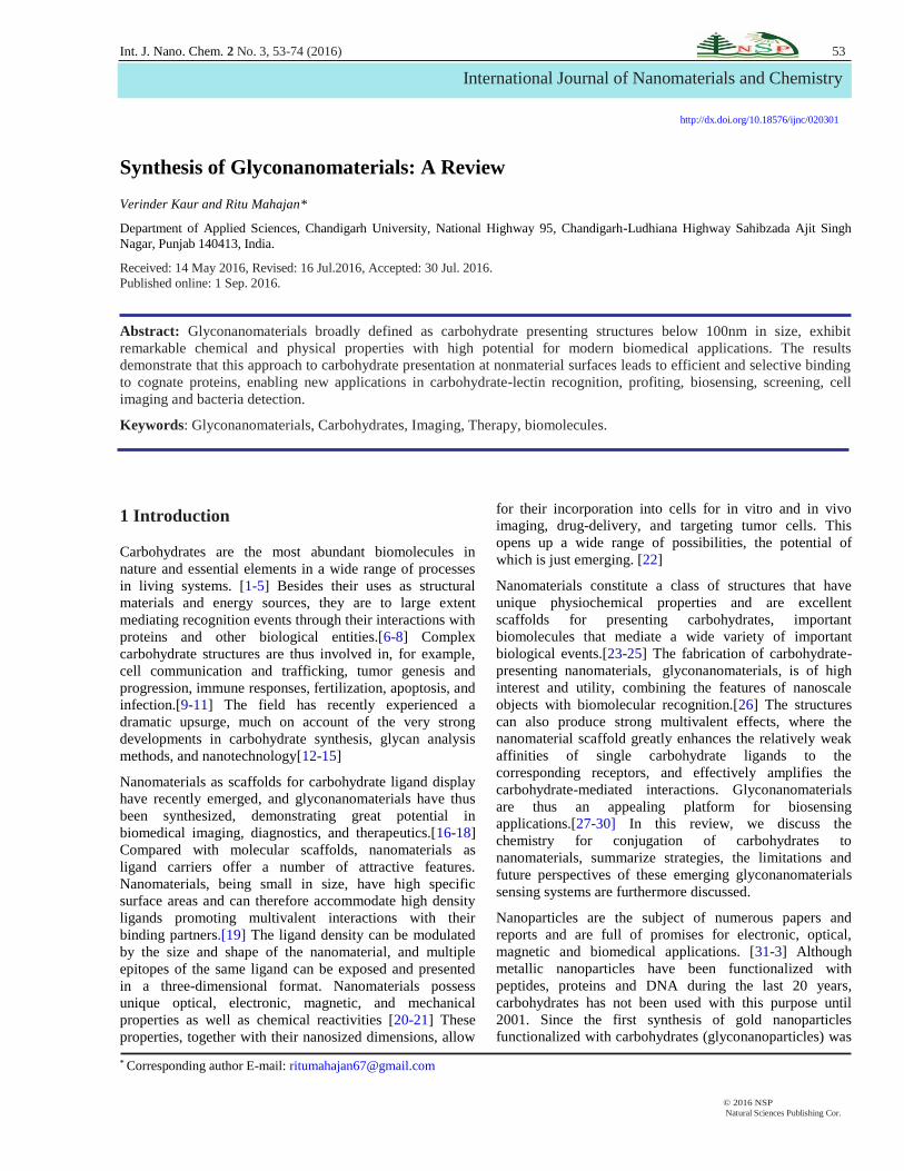
binding lectins.(Fig. 8) Methods for direct functionalization
of pristine CNTs or graphene using (a), aryl diazonium salt,
(b) azomethineylide,(c) alkyl azide, and (d) PFPA.
Figure 8.Carbohydrate conjugation through microwave-
assisted functionalization of single-walled carbon
nanotubes using perfluorophenyl azides
To take full advantage of the remarkable applications of
carbon nanotubes in different fields, there is a need to
develop effective methods to improve their water
dispersion and biocompatibility while maintaining their
physical properties. In this sense, current approaches suffer
from serious drawbacks such as loss of electronic structure
together with low surface coverage in the case of covalent
functionalizations, or instability of the dynamic hybrids
obtained by non-covalent functionalizations. Thus, the
molecular basis of an original strategy was examined that
combines the advantages of both functionalizations without
their main drawbacks. [89-90] The hierarchical self-
assembly of diacetylenic-based neoglycolipids into highly
organized and compacted rings around the nanotubes,
followed by photo polymerization leads to the formation of
nanotubes covered with glyconano rings with a shish
kebab-type topology exposing the carbohydrate ligands to
the water phase in a multivalent fashion. The glyco
nanotubes obtained are fully functional, and able to
establish specific interactions with their cognate receptors.
[91-92] In fact, by taking advantage of this selective binding,
an easy method to sense lectins as a working model of toxin
detection was developed based on a simple analysis of
TEM images. (Fig.9). Remarkably, different experimental
settings to assess cell membrane integrity, cell growth
kinetics and cell cycle demonstrated the cellular
biocompatibility of the sugar-coated carbon nanotubes
compared to pristine single-walled carbon nanotubes.

Int. J. Nano. Chem. 2, No. 3, 53-74 (2016) / http://www.naturalspublishing.com/Journals.asp 59
© 2016 NSP
Natural Sciences Publishing Cor.
Figure 9. Self-assembly of diacetylenic-based
neoglycolipids into highly organized and compacted rings
around the nanotubes, followed by photo polymerization
leads to the formation of nanotubes covered with glyconano
rings.
A new approach was developed for the non-covalent
functionalization of multiwalled carbon nanotubes
(MWNTs), which allows a presentation of carbohydrate on
their surface by hydrophobic interactions.[93-95] The
approach is based on the self-assembly of a sugar-based
amphiphile on MWNTs in alcohol/water mixtures, which
has been investigated by means of ultraviolet (UV), Raman
spectra, Fourier transform infrared spectroscopy (FTIR), X-
ray photoelectron spectroscopy (XPS), X-ray diffraction
(XRD), and high-resolution transmission electron
microscopy (HRTEM). It was demonstrated that alcohols
not only can promote the self-assembly of the amphiphile
on MWNTs but also can regulate the amount and
conformation of the assembled amphiphile. The adsorption
of bovine serum albumin (BSA) onto functionalized
MWNTs was studied and characterized with circular
dichroism (CD) spectra. The results showed that the
conformation of adsorbed BSA has been well preserved. It
has been demonstrated that, the functionalized MWNTs
with disaccharide groups on their exterior surface have a
good dispersibility in water and are biocompatible, may
have a potential application of molecular recognition.
The utilization of a nanomaterial wrapped in biologically
relevant molecules to study and solve biomedical problems
is a new and stimulating field of research.[96-97] One of
the most salient features of using nanomaterials, such as
nanoparticles, nanorods, nanowires and carbon nanotubes
in biology is their ability to carry multiple copies of a single
drug or various active principles with different, ideally
synergistic, modes of action.[98] Consequently, those
diseases or biological processes whose biological targets
require a multivalent display of the active epitope, are
expected to benefit from the application of a nanometric
platform. Illustrative examples of such events are those
mediated by carbohydrates, which include cell adhesion,
inflammation, tumor cell metastasis, and pathogen
infections. It has been shown that the weak interaction
between an individual ligand and the corresponding
specific lectin is compensated by the multivalent display of
carbohydrates through the so called cluster effect.[99] On
the other hand, single-walled carbon nanotubes (SWCNTs)
as interesting 1D nanomaterials, are actually being actively
investigated as vehicles for the in vivo smart delivery of
biologically relevant cargoes including drugs, proteins, and
nucleic acids, as nanometric sensors and for cancer
treatment. However, concerns about their potential toxicity
have reduced much of the original enthusiasm about their
promising clinical applications. Nevertheless, recent
investigations, including a pilot study, on the in vivo
behavior of SWCNTs, have concluded that conveniently
functionalized water soluble SWCNTs are completely
cleared from the body via the biliary and renal pathway,
and are non toxic.[100]
While covalent, and non-covalent, approaches have been
followed for the dispersion of SWCNTs in aqueous media,
the latter one is highly desirable as it conserves the
nanotubes structure, while the former one has been shown
to disrupt their p-network, leading to possible losses in their
mechanical, electrical, and bio- sensing properties.(Fig.10).
Figure 10. Dispersion of SWCNTs in aqueous media
In this review, we discuss the utilization of carbon
nanotubes as molecular platforms for a multivalent
presentation of biologically relevant saccharide
epitopes.[101-102] The strategy is based on the utilization
of neutral pyrene functionalized neoglycolipids that interact
with a CNT’s surface giving rise to a nanometric material
with a multivalent display of carbohydrates, much like the
glycocalyx on the cell surface (Fig.11). Between the
pyrene tail and the glycoligand, the designed amphiphilic
compound I exhibits an advantageous variable spacer
derived from tetraethylene glycol for the fine-tuning of the
hydrophilic-hydrophobic balance of the pyrene-
polyethlyene glycol-sugar (Py-PEG-Sugar) I.
A selection of glycosylated polyacrylate nanoparticles has
been reported by radical-initiated emulsion polymerization
in aqueous media. Using ethyl acrylate as a co-monomer,
carbohydrate acrylates were incorporated into the poly

60 V. Kaur, R. Mahajan: Synthesis of Glyconanomaterials: A Review…
© 2016 NSP
Natural Sciences Publishing Cor.
(ethyl acrylate) framework to give stable emulsions of
glyconanoparticles with an average particle size of around
40 nm.
Figure 11. Specificity acquired by the aggregates toward
specific receptors.(A). Schematic representation of:
selective recognition of CNT- Py-PEG- Lac-5 (I), inhibition
of lectin binding by adding monovalent lactose (II),
absence of selective interaction of PNA-FITC with CNT-
Py-PEG-Man-6 displaying an a-mannose epitope on their
surface (III). (B) Fluorescence spectra of I: PNA-FITC with
MWCNTs-Py-PEG-Lac-5, I-a: PNA-FITC with SWCNTs-
Py-PEG-Lac-5, II: MWCNTs-Py-PEG-Lac-5 with PNA-
FITC, previously incubated with lactose, III, PNA-FITC
with MWCNTs-Py-PEG-Man-6. [PNA-FITC] ¼ 0.82 mM.
Using this technique a variety of glyconanoparticles were
prepared from 3-O-acryloyl-1,2:5,6-di-O-isopropylidene-a-
D-glucofuranose, 1-O-acryloyl-2,3:5,6-di-O-
isopropylidene-a-D-mannofuranose, 6-O-acryloyl-1,2:3,4-
di-O-isopropylidene-a-D-galactopyranose, 2-N-acryloyl-
1,3,4,6-tetra-O-acetyl-b-D-glucosamine, 5-O-acryloyl-2,3-
isopropylidene-1-methoxy-b-D-ribofuranose and 4-N-
acetyl-50-O-acryloyl-20,30-O-isopropylidene cytidine.
Scanning electron microscopy, dynamic light scattering and
proton NMR analysis of the emulsions indicated essentially
100% incorporation of the carbohydrate acrylate monomer
into the polymer with the exception of O-benzyl- and O-
benzoyl-protected carbohydrate acrylates, which gave
incomplete incorporation.[103-104] Formation of larger
glyconanoparticles of 80 nm with (unprotected) 3-O-
acryloyl-D-glucose and 5-O-acryloyl-1-methoxy-b-D-
ribofuranose revealed the influence of free hydroxyl groups
in the monomer on the particle size during polymerization,
a feature which is also apparently dependent on the amount
of carbohydrate in the matrix. This methodology allows for
a new, simple route to the synthesis of polymeric
glyconanoparticles with potential applications in targeted
drug delivery and materials development.
5 Metal Nanoparticles
Metal nanoparticles, for example Au, Ag, and Cu
nanoparticles, show distinct optical properties that are
different from bulk materials due to the quantum
confinement effect resulting from the reduction of the
particle size.[105-107] The collective oscillation of
electrons in the metal nanoparticles, generated by light
illumination, is highly sensitive to the dielectric
environment close to the nanoparticle surface. The so-
called localized surface plasmon resonance (LSPR),
provides a powerful means to monitor the molecular events
occurring at the particle surface . Many studies have taken
advantage of the change in LSPR, often resulting in a color
change visible by naked eyes, as means to study
carbohydrate-mediated interactions or as a detection
mechanism for sensing carbohydrates. Gold
glyconanoparticles as elements of the nano world belong to
a group of particles with diameters not exceeding 100
nm.[108-109]. This size scale makes them conformable to
common biomolecules. A gold glyconanoparticles consists
of three different parts: the gold core, the linkers, and
saccharide ligands. The glycocalyx-like surface of these
particles mimics the presentation of carbohydrate epitopes
of cell surface glycoconjugates. As a consequence, gold
glyconanoparticles provide inimitable tools for probing and
manipulating the mechanisms of biological processes based
on carbohydrate interactions. Each component of the gold
glyconanoparticles has a profound effect on the
nanoparticles properties. Carbohydrate-conjugated Au
nanoparticles (AuNPs) were employed to differentiate
plant-legume lectins. Various AuNPs were treated lectins,
and changes in LSPR were subjected to linear discriminant
analysis to successfully differentiate all lectins.( Fig. 12).

Int. J. Nano. Chem. 2, No. 3, 53-74 (2016) / http://www.naturalspublishing.com/Journals.asp 61
© 2016 NSP
Natural Sciences Publishing Cor.
Figure 12.Assembling different antennas of the gp120 high
mannose-type glycans on gold nanoparticles provides
superior binding to the anti-HIV antibody 2G12 than the
individual antennas.
Other studies in chemical and bio-sensing used Au and Ag
nanoparticles in surface-enhanced Raman spectroscopy
(SERS).[110-112] For example, lactose-functionalized Ag
nanoparticles were used by Graham et al. to probe the
interaction with Con A, where the SERS intensity could be
enhanced for lectin detection at pico molar level .[113]
Among the metal nanoparticles, AuNPs are most widely
used due to their relative inert nature and ease of
preparation in comparison to other metal nanoparticles. In
most cases, the carbohydrates were derivatized with a thiol
or a disulfide structure, and the carbohydrate conjugation
was accomplished by either a one-pot protocol or a two-
step process. Early examples by Penadés and coworkers
demonstrated the one-pot synthetic method, where
disulfide-derivatized oligosaccharides were dissolved in
methanol and then mixed with an aqueous tetrachloroauric
acid solution.[114] The carbohydrate-conjugated AuNPs
were subsequently obtained following addition of NaBH4
under vigorous stirring. Similarly, Iyer and coworkers
thiolated the trisaccharide portion of globotriaosylceramide
Gb3, which was then directly added into HAuCl4 solution
followed by NaBH4 reduction. The trisaccharide-
conjugated AuNPs showed selective inhibition towards
Shiga toxins 1 and 2. The two-step process involves the
synthesis of AuNPs followed by the addition of thiolated
carbohydrates. A ligand exchange reaction occurs in the
second step, where the ligand such as citric acid on the as-
prepared AuNPs is replaced by the thiolated carbohydrate
as a result of the higher bond strength between thiols and
Au than carboxylic acid. For example, thiolated lactose and
glucose derivatives prepared by Russell et al. were bound to
citrate-coated AuNPs by ligand replacement. Apart from
these examples, a large number of other studies have
involved the synthesis of Au glyconanoparticles following
either of these two methods.[115-117] Post-modification of
nanomaterials with carbohydrate structures is another
method to conjugate carbohydrates on metal nanoparticles.
In this case, a functional group is introduced on the metal
nanoparticle surfaces, and carbohydrates, either derivatized
or underivatized, are then conjugated to the nanoparticles
through a coupling reaction. This method was, for example,
used by Kataoka et al. who synthesized AuNPs with an
acetal-terminated PEG-SH ligand. The acetal was then
converted to aldehyde, which in turn was used to attach p-
amino phenyl-lactose and p-amino phenyl-α–D-mannose by
reductive amination. In a two-step process, AuNPs were
first treated with a thiol- or disulfide-functionalized PFPA.
Light activation of the PFPA then resulted in the covalent
conjugation of carbohydrates to the AuNPs. In this case, the
carbohydrates were used in their native form and no
chemical derivatization was needed. (Fig.13). Using this
methodology, various carbohydrates including
monosaccharide’s, oligosaccharides and polysaccharides
were conjugated, as well as reducing- or non-reducing
carbohydrates to AuNPs without affecting their binding
affinities.
Figure 13.Synthesis and characterization of glucose-
functional glycopolymers and gold nanoparticles: study of
their potential interactions with ovine red blood cell
A method to measure the binding affinity of glyco
nanoparticle (GNP)-protein interactions was reported which
was based on a fluorescent competition binding assay,
which yielded the apparent dissociation constant (Kd) of
GNPs with the interacting protein. Au nanoparticles
conjugated with underivatized mono-, oligo-, poly-
saccharides were synthesized using photocoupling
chemistry.[118-119] The affinities of these GNPs with
lectins were measured and were several orders of
magnitude higher than the corresponding free ligands with
lectins. The effect of ligand display on the binding affinity
of GNPs was furthermore studied where GNPs of varying
linker type, spacer length, ligand density, and nanoparticle
size were prepared and Kd values determined. The long
spacer linker containing hydrocarbon and ethylene oxide
units gave the highest binding affinity as well as assay
sensitivity. The binding affinity increased with ligand
density in general, showing a drastic increase in affinity at
low ligand density. In addition, the affinity enhancement
was more pronounced on smaller NPs than the larger ones.
These results not only demonstrate that the binding affinity
of GNPs is highly influenced by how the ligands are
presented on the nanoparticles, but also pave the way for
tailor-made glyconanomaterials with tunable affinity by
way of ligand display.

62 V. Kaur, R. Mahajan: Synthesis of Glyconanomaterials: A Review…
© 2016 NSP
Natural Sciences Publishing Cor.
The ability to produce monomolecular coatings with well-
defined structural and functional properties is of key
importance in biosensing, drug delivery, and many recently
developed applications of nano-technology.[120-122]
Organic chemistry has proven to be a powerful tool to
achieve this in many research areas. Thus, three oligo
(lactosides) were glycosylated in a (1 → 3) manner, and
which are further functionalized with amide-linked short
alkanethiol spacers. The oligosaccharides (di-, tetra-, and
hexasaccharide) originate from the inexpensive and readily
available lactose disaccharide. These thiolated derivatives
were immobilized onto gold surfaces, and the thus formed
self-assembled monolayer’s (SAMs) on planar gold were
characterized by wettability, ellipsometry and infrared
reflection–absorption spectroscopy. Further, the ability of
these SAMs to stabilize gold nanoparticles in saline
solutions was also demonstrated, indicating that the
oligosaccharides may be used as stabilizing agents in gold
nanoparticle-based assays.(Fig. 14).
Figure 14. Synthesis of PFPA-Au NPs and subsequent
coupling of α-1,4-mannobiose.
A strategy based on the utilization of neutral pyrene
functionalized neoglycolipids I that interact with a CNT’s
surface giving rise to biocompatible nanomaterials which
are able to engage specific ligand-lectin interactions similar
to glycoconjugates on the cell membrane is reported.[123]
The utilization of a nanomaterial wrapped in biologically
relevant molecules to study and solve biomedical problems
is a new and stimulating field of research. [124]One of the
most salient features of using nanomaterials, such as
nanoparticles, nanorods, nanowires and carbon nanotubes
in biology is their ability to carry multiple copies of a single
drug or various active principles with different, ideally
synergistic, modes of action. Consequently, those diseases
or biological processes whose biological targets require a
multivalent display of the active epitope are expected to
benefit from the application of a nanometric platform.
Illustrative examples of such events are those mediated by
carbohydrates, which include cell adhesion, inflammation,
tumor cell metastasis, and pathogen infections.[125] It has
been shown that the weak interaction between an individual
ligand and the corresponding specific lectin is compensated
by the multivalent display of carbohydrates through the so
called cluster effect.[126] On the other hand, single-walled
carbon nanotubes (SWCNTs) as interesting 1D
nanomaterials, are actually being actively investigated as
vehicles for the in vivo smart delivery of biologically
relevant cargoes including drugs, proteins, and nucleic
acids, as nanometric sensors, and for cancer treatment.
However, concerns about their potential toxicity have
reduced much of the original enthusiasm about their
promising clinical applications. Nevertheless, recent
investigations, including a pilot study, on the in vivo
behavior of SWCNTs, have concluded that conveniently
functionalized water soluble SWCNTs are completely
cleared from the body via the biliary and renal pathway,
and are non toxic. While covalent, and non-covalent,
approaches have been followed for the dispersion of
SWCNTs in aqueous media, the latter one is highly
desirable as it conserves the nanotubes structure, while the
former one has been shown to disrupt their p-network,
leading to possible losses in their mechanical, electrical,
and biosensing properties.[127]
The ability to produce monomolecular coatings with well-
defined structural and functional properties is of key
importance in biosensing, drug delivery, and many recently
developed applications of nano-technology.[128] Organic
chemistry has proven to be a powerful tool to achieve this
in many research areas. The synthesis of three oligo
(lactosides) glycosylated in a (1 → 3) manner is reported
and which are further functionalized with amide-linked
short alkanethiol spacers. [129]The oligosaccharides (di-,
tetra-, and hexasaccharide) originate from the inexpensive
and readily available lactose disaccharide. These thiolated
derivatives were immobilized onto gold surfaces, and the
thus formed self-assembled monolayer’s (SAMs) on planar
gold were characterized by wettability, ellipsometry and
infrared reflection–absorption spectroscopy. Further, the
ability of these SAMs to stabilize gold nanoparticles in
saline solutions was also demonstrated, indicating that the
oligosaccharides may be used as stabilizing agents in gold
nanoparticle-based assays. [130]
QDs such as CdSe, CdTe, CdS and ZnS are crystalline
semiconductor nanoparticles display unique electronic
properties resulting from the size-dependent quantum
confinement. Broad absorption, tunable and narrow
emission made them promising nanomaterials for imaging
and sensing. When functionalized with carbohydrates, the
water solubility and biocompatibility of the modified QDs
is enhanced, in addition to exerting molecular recognition
abilities. Similar to metal nanoparticles, carbohydrate
conjugation to QDs can be accomplished by one-pot
synthesis, ligand exchange reaction, or post-modification.
Examples of the one-pot synthesis involved mixing
disulfide-functionalized carbohydrate structures with
Cd(NO3)2.4H2O at pH10, followed by the drop wise
addition of Na2S. As QDs were formed via the combination

Int. J. Nano. Chem. 2, No. 3, 53-74 (2016) / http://www.naturalspublishing.com/Journals.asp 63
© 2016 NSP
Natural Sciences Publishing Cor.
of Cd2+ andS2- in basic solution, the carbohydrate disulfide
ligands were directly attached to the QDs forming
stabilizing layers .In an example of the ligand exchange
protocol, Surolia and coworkers synthesized carbohydrate-
conjugated CdSe-ZnS core-shell QDs by treating as-
prepared QDs with thiol-functionalized lactose, melibiose
and maltotriose.[131] Seeberger and coworkers further
developed a controllable glyco-QDs synthesis method by
using a continuous-flow microreactor.Cd and Se precursors
were injected into a heating chamber followed by coating
with Zn and S precursors in a second chamber.[132] The
size and fluorescence emission of the QDs could be
controlled by the precursor concentrations, temperature and
flow rate. Carbohydrate conjugation was accomplished in
the last step in a ligand exchange chamber using thiol-
functionalized carbohydrate ligands such as α-D-
mannosides or β-D-galactosides. In the post-modification
method, a functional group is introduced to QDs, which is
then coupled to carbohydrates. For example, Wang and
coworkers modified CdS QDs with carboxy-terminated
alkylthiol, which were then used to conjugate amine-
derivatized carbohydrates using standard coupling reagents
for amidation.[133] Other coupling methods such as thiol-ene
and reductive amination have also been demonstrated to
introduce carbohydrates onto QDs. For
instance,Seeberger’s group synthesized a series of
carbohydrate-capped CdSe/ZnS core-shell QDs using the
thiol-ene reaction .[134] To introduce double bonds on the
nanoparticles, the QDs were first functionalized through
ligand exchange with an amino-terminated PEG2000-
linked dihydrolipoic acid structure, followed by
conjugation with amaleimide moiety. Thiol-functionalized
α-D-mannosides-D-galactosides and β-D-galactosamine
derivatives were subsequently conjugated to the QDs
through a thiol-ene reaction. In another example, Jana et al.
synthesized glyco-QDs by reductive amination. The amine-
functionalized QDs were prepared by encapsulating
hydrophobic QDs into a polymer prepared by reverse
micelle polymerization of acrylates containing N-(3-
aminopropyl)methacrylamide. Carbohydrate
immobilization was subsequently achieved by adding
maltose, lactose and dextran to the amino-functionalized
QDs in the presence of Na(CN)BH3 at pH 9 in borate
buffer followed by dialysis.
6 MNPs
MNPs constitute an important class of nanomaterials
suitable for biomedical imaging such as magnetic
resonance imaging (MRI), and therapeutics such as
hyperthermia treatment. The most frequently used MNPs in
these applications are iron oxide nanoparticles including
magnetite (Fe3O4) and maghemite (α-Fe2O3).[128] Iron
oxide nanoparticles can be readily prepared using simple
protocols to give particles in the size range of 5-20 nm.
These nanoparticles can be readily dispersed in aqueous
solutions to form homogeneous and stable suspensions.
Iron oxide nanoparticles have excellent biocompatibility
and are highly desirable for in vivo studies. In fact,
Feraheme, a product based on carbohydrate-coated
magnetite nanoparticles, have already been in clinical use
for the treatment of iron deficiency anaemia.[135]
To prepare carbohydrate-conjugated MNPs, similar
strategies to those of QDs can be adopted.For example,
Horák et al. reported a one-pot protocol where D-mannose
was directly added to a reaction mixture with FeCl3and
FeCl2in the presence of NH4OH.[130] In this case, D-
mannose was thought to act as a metal-coordinating ligand
that bound to the nanoparticles by chelation to
Fe(II)/Fe(III). In this context, carboxylic acid- and
phosphate-functionalized carbohydrates are also effective
in binding to iron oxide nanoparticles. For example,
lactobionic acid, D-gluconic acid, Ficolland carboxy-
terminated glycolipids were used by Kekkonen et al. and
Baccile et al. to stabilize MNPs.[136-137] These MNPs
showed increasing stability with increasing carbohydrate
ligand size. In the case of ligand exchange reactions,
phosphate-functionalized, per acetylated mannose,
rhamnose and ribose derivatives were used by Lartigue et
al. to replace the original ligands on their on oxide
nanoparticles such as oleic acid/oleylamine.[138] Removal of
the acetyl protection groups resulted in significant increase
in water solubility of the resulting nanoparticles .Other
routes include initial nanoparticle functionalization to
introduce a functional group, followed by carbohydrate
conjugation. For example, Huang and coworkers prepared
magnetite nanoparticles coated with sialic acid (Sia) using
amide coupling.[139] The magnetite nanoparticles were
synthesized by NH4OH-induced co-precipitation of
FeCl3 and FeSO4 in the presence of dextran as a coating
ligand. Amino groups were subsequently introduced by
treating the dextran layer with epichlorohydrin/NaOH
followed by ammonia. The partially protected sialic acid
derivative having a carboxylic acid end group was then
conjugated to amine-MNPs by amide coupling, and the
final glyco-MNPs were obtained after removing the
protecting groups in aqueous NaOH. Other examples, from
our group, involved the preparation of PFPA-functionalized
iron oxide nanoparticles by treating the particles with
PFPA-phosphate. Carbohydrate conjugation was then
achieved photochemically by irradiating the dispersion of
iron oxide nanoparticles and carbohydrate followed by
dialysis.
7 SNPs
SNPs are widely used in biochemistry due to their
outstanding biocompatibility, water dispersability, stability
and functionality . Among different SNPs formats,
mesoporous SNPs have over the last 20 years been
developed to possess unique and advantageous properties
such as tunable particle size, pore size and shape. These
properties have enabled their use as drug delivery systems
in for example anti-cancer therapy. Mesoporous SNPs were
in this case loaded with multiple drugs and functionalized

64 V. Kaur, R. Mahajan: Synthesis of Glyconanomaterials: A Review…
© 2016 NSP
Natural Sciences Publishing Cor.
with active targeting ligands, including carbohydrates.[140]
The resulting SNPs were able to selectively target tumor
cells, including multi-drug resistance (MDR) cells, leading
to cancer cell death without damaging normal tissue (Table
1).
Table 1: Properties of typical nanomaterials and their
biomedical applications.
Nanomaterials Intrinsic properties
Biomedical applications
Category Examples
Metallic Au, Ag SPR Biosensing, drug
delivery, bioimagin
Semiconduct CdS, Fluorescence, Immunoassays,
Or CdSe Luminescence bioimaging, biosensing
Magnetic Fe3O4 Magnetism MRI, drug delivery
Carbon- CNTs, Electronic and mechanical Drug and gene delivery,
Based Fullerene properties, conductivity therapy, biosensing
SNPs surfaces can be efficiently functionalized with
carbohydrates by post-modification methods, generally
involving initial functionalization of the SNPs, and
subsequent conjugation of derivatized or underivatized
carbohydrates.[136] Several different conjugation
chemistries have here been used, including CuAAC, amide
coupling, nucleophilic substitution, and photocoupling. For
example, Basuet al. synthesized azide-functionalized SNPs
using ((azidomethyl) phenylethyl)-trimethoxysilane, and
conjugated alkyne-functionalized carbohydrate derivatives
in the presence of CuSO4/sodium ascorbate or
CuI/diisopropylethylamine, while heating to 70oC in a
microwave reactor.[141-143] Liu et. al. utilized amide
coupling to conjugate galactose (Gal) derivatives onto
SNPs.[144] The SNPs surface wasfirstfunctionalizedwithN-
(β-ethylenamine)-γ-propylaminetriethyloxylsilane and
lactobionic acid was then coupled to the amino-
functionalized surface in the presence of amide coupling
reagents. Gary-Bobo et al. prepared mannose-
functionalized mesoporous SNPs by coupling aminopropyl-
functionalized SNPs to squarate ester-derivatized α-
mannose.[145] For the photo initiated carbohydrate
conjugation to SNPs, a method developed was the
nanoparticles were first functionalized with PFPA-silane,
after which the carbohydrates were conjugated by
irradiation in the presence of carbohydrates.[146] Using this
methodology, carbohydrate-functionalized SNPs were
prepared from different mono- and oligosaccharides.
Over the past decade, diagnostics and therapeutics have
changed gradually towards the use of more specific and
targeted approaches.[146-147] The most profound impact
has been in the nanotechnology sectors, where an explosion
in directing biomolecules to specific biomarkers has
illustrated great potentials not only in detection but also in
targeted therapy. Increased knowledge of the diseases at the
molecular level catalyzed a shift towards identifying new
biological indicators. In particular, carbohydrate-mediated
molecular recognitions using nano-vehicles are likely to
increasingly affect medicine opening a new area of
biomedical applications. This article provides an overview
of the recent progress made in recruiting the “sugar code”
functionalized on various nano-platforms to decipher
cellular information for both in vitro and in vivo
applications.[148-149] Today’s glyco-technologies are
enabling better detection with great therapeutic
potentials.(Table 1). Tomorrow they are likely to bring a
full understanding of the “cell-glyconanomaterial bio-
conversation” where major biomedical problems will be
overcome translating insights from the “glyco-nanoworld”
into clinical practice.[150-152]
Creative Glyconanoparticles provides a simple and
environmental friendly method to prepare
glyconanoparticles (GNPs). Nanoparticles functionalized
with glycans can be applied as powerful solid-phase
chemical tools for the study of protein–carbohydrate
interactions using nanoscale properties for detection of
binding events. This method allows the easy synthesis of
stable glyconanoparticles with reduced dispersion and
controlled size.[149-150] Creative Glyconanoparticles has
been reported that boost up the study and applications of
carbohydrates in glycobiology, biomedicine and material
science. (Fig.15)
Figure 15.Applications of Glyconanoparticle
8 Characterization of Glyconanomaterials
Glyconanomaterials are synthesized under various
conditions using specific chemistry and reagents. These
materials must therefore be carefully evaluated to fully
characterize the structure, composition, density of surface
ligands, and biological activities in order to obtain proper
correlation with their performances.[151-153] Conventional
chemical analytic techniques that are insensitive to flat

Int. J. Nano. Chem. 2, No. 3, 53-74 (2016) / http://www.naturalspublishing.com/Journals.asp 65
© 2016 NSP
Natural Sciences Publishing Cor.
substrates can be readily adopted to the significantly
increased specific surface areas of nanomaterials for
nanomaterial characterization.[154-155] In the paper by
Brust et al. on the preparation of thiol-capped Au NPs, the
products were characterized by FTIR showing the presence
of alkanethiol and TEM revealing the size and shape of the
nanoparticles.[156] With the rapid development of
advanced analytical tools, especially sensitive surface
characterization techniques, nanomaterials can now be
analyzed more accurately, providing in-depth
understanding of the chemical and physical properties of
glyconanomaterials. NMR, FTIR, and surface-enhanced
Raman spectroscopy (SERS) offer detailed structural
analysis of nanomaterials and surface ligands. Thermo
gravimetric analysis (TGA) yields the amount of organic
components on the nanomaterials, from which the ligand
densities can be derived.[157] Elemental analysis and XPS
provide information on the elemental composition and
chemical state of the bulk nanomaterials and the surface
ligands. A combination of microscopy techniques, scanning
probe techniques (STM, AFM), TEM, and small-angle X-
ray scattering (SAXS) reveals the physical characteristics
of size, shape, and assembly behavior of the
nanomaterials.[158-160] Caution should be used when
analyzing the results as the experimental conditions applied
to each technique (vacuum, ambient, solution) can
significantly impact the outcome. Microscopic techniques
can also be used to directly visualize the interactions of
glyconanomaterials with their binding partners. When d-
mannose-functionalized iron oxide nanoparticles were
treated with Escherichia coli strain ORN178, the
nanoparticles selectively bound to the FimH lectin on the
bacteria, which was clearly shown by TEM. The surfaces
can be further characterized by taking advantage of the
unique properties offered by the nanomaterials.[161-162]
Classic examples are metal nanoparticles, which exhibit
plasmon resonance that is highly sensitive to the surface
constituents and can be conveniently monitored
colorimetrically, as the molecular recognition event occurs
at, or close to, the surface of the nanoparticles.[163-165]
Carbohydrate–lectin interactions of free ligands in solution
have been studied by many biochemical and biophysical
methods including NMR spectroscopy, surface plasmon
resonance (SPR), X-ray crystallography, isothermal
titration calorimetry (ITC), and fluorescence
spectroscopy.[166-168] Quantitative analysis of
glyconanomaterials is investigated to a lesser extent and
only a few protocols were reported to determine the binding
affinity of glyconanoparticles.[169-170] Lin and coworkers
used SPR to analyze the multivalent interactions between
mannose-, glucose-, or galactose-encapsulated gold
nanoparticles with Con A. A competition binding study was
carried out where equilibria were established between
mannopyranoside attached on the SPR sensor, Con A, and
varied concentrations of mannose-encapsulated Au NPs.[171]
The dissociation constant Kd of mannose/Au NPs with Con
A was determined to be 2.3 nM, representing a binding
affinity over 5 orders of magnitude higher than that of the
free d-mannopyranoside with Con A in solution (Kd = 470
µm measured by ITC. In the system developed by Wu and
co-workers, magnetite/gold core/shell nanoparticles coated
with proteins were allowed to interact with carbohydrate
ligands on a glycan array. A magnetic field was applied to
amplify the protein–carbohydrate interactions and the
signals were visualized and quantified using a silver
enhancement reagent. Apparent Kd values of 66 nm, 61 nm,
and 57 nm were determined for Man, Man4, and Man9
ligands with Con A, respectively. [172]
Binding Affinity of Glyconanomaterials Biomedical
imaging, therapeutics, medical diagnosis, and drug delivery
are among the many areas glyconanomaterials have the
potential to impact. The interaction of glyconanomaterials
with biological receptors and targets is a critical process
involved in these applications and the binding affinity is
thus an important parameter for evaluating the performance
of glyconanomaterials.[173-175] (Fig.16).
Figure 16: Multivalent glycocyclopeptides: toward nano-
sized glycostructures
Fluorescence-based competition assay was developed to
determine the binding affinity of glyconanoparticles with
lectins.[176-177] In the assay, a fixed concentration of a
free ligand (for example, d-mannose) and varying amounts
of ligands bound to Au NPs were incubated with
fluorescein isothiocyanate (FITC)-labeled Con A. The
solution was then centrifuged and the fluorescence intensity
of the supernatant was measured. Two equilibria co-exist in
the system: FITC-Con A with free d-mannose and FITC-
Con A with d-mannose bound on nanoparticles (Fig. 17).
Since very low concentrations of Con A and free d-
mannose were used, it was assumed that no agglomeration
occurred. Both interactions are reversible, and steady
equilibria are reached rapidly.
In order to calculate the binding affinity constant, the
concentration of the carbohydrate ligand on Au NPs must
be determined.[178-180] The colorimetric assay of
anthrone-sulfuric acid was adopted to measure the ligand
density on the nanoparticles. A calibration curve was first
established using the corresponding free carbohydrate, and
the amount of surface-bound ligand on the Au NPs was
subsequently determined. The fluorescence intensity

66 V. Kaur, R. Mahajan: Synthesis of Glyconanomaterials: A Review…
© 2016 NSP
Natural Sciences Publishing Cor.
measured from the competition studies was plotted against
the concentration of d-mannose on the Au NPs (Fig. 4).The
result was a typical concentration-response curve for
ligand–receptor binding, validating the assumptions made
for the system. The concentration of ligands displaying
50% of specific binding (IC50) value was subsequently
derived and the apparent dissociation constant (Kd2)
calculated using the Cheng–Prusoff equation (Eq.1).
Kd2=IC50/1+[M]/Kd1
Equation 1: where [M] is the concentration of free ligand,
i.e., d-mannose, Kd1 is dissociation constant of free ligand
to Con A, and Kd2 is the apparent dissociation constant of
surface bound d-mannose to Con A.
Figure 17: Equilibria involved in the competition binding
assay. b) Concentration dependent fluorescence intensity
curve (right). [Man] is the concentration of d-mannose on
NPs determined using the anthrone/H2SO4 colorimetry
assay.
9 Ligand Presentation and Binding Affinity
Nanomaterials, being three-dimensional in shape and small
in size, are capable of hosting ligands in higher densities in
comparison to their flat counterparts due to greatly
increased specific surface areas.[181-182] This has
significant implication for glyconanomaterials where the
substrate configuration could dictate the ligand presentation
and cooperativity and thus impact the interactions of the
glyconanomaterials with their binding partners. The result
is markedly enhanced affinities of these glyconanomaterials
with the relevant biological targets. Data showed that the
apparent Kd of d-mannose tethered on Au NPs with Con A
can be as low as 0.43 nm (Table 2), representing a binding
affinity of over six orders of magnitude higher than for
free d-mannopyranoside with Con A. These results
demonstrate that nanoparticles are excellent scaffolds for
amplifying the weak affinities of carbohydrate ligands with
lectins. Similar observations were reported by Sun and co-
workers where single-walled carbon nanotubes
functionalized with mannose and galactose selectively
bound anthrax spores, inducing aggregation of the spores in
the presence of Ca2+.[183] In contrast, carbohydrate-
conjugated polystyrene beads did not exhibit the observed
affinity towards anthrax spores. This was attributed to the
ability of nanotubes to promote multivalent interactions of
the carbohydrate ligands with receptors on the spores.[184]
Table 2: Binding affinity of D-mannose on Au NPs with
Con A. Au NPs were functionalized with PFPA–thiol
before d-mannose was coupled.
PFPA-thiol Spacer Kd [nM]
1. CH2CH2 19± 2.2
2. CH2(CH2)4CH2 15± 2.0
3. CH2(CH2)9CH2 5.3± 0.72
4. CH2(CH2)9CH2(OCH2CH2)4 0.43± 0.044
Unlike the free ligand that has the translational and
rotational freedom in solution, the surface-bound ligand is
no longer an unrestricted entity. Each ligand becomes a
member of the nanomaterial carrier and can act
cooperatively when interacting with their binding
partners.[185-186] The efficiency of the ligand association
with the binding site, i.e., the binding affinity, is sensitive
to a number of factors: how the ligand is attached, i.e., the
coupling chemistry, the type and length of the spacer
connecting the ligand and the nanomaterial, the
flexibility/rigidity of the spacer, the density of ligands, and
the distance between them.
10 Ligand Density
Ligand density is another important parameter affecting the
binding of surface-tethered carbohydrates.[187-188] A few
studies report the impact of ligand density on the binding
affinity of carbohydrates immobilized on a flat surface, but
the topic has not been extensively investigated for
glyconanomaterials. Wong and co-workers used a
fluorescence assay to analyze mannose–Con A interaction
on glycan microarrays. Kd decreased from 214 nM to 76.8
nM when the mannose printing concentration increased
from 0.6 µm to 80 µm.[189] However, Kd increased to 80.4
nm when the mannose printing concentration increased to
100 µm. Corn and co-workers employed the technique of
SPR imaging to study carbohydrate–protein interactions
using carbohydrate microarrays.[190] The binding affinity,
measured by the adsorption coefficient (KADS), increased
slightly from 5.0 × 10⁶ M−1 to 5.6 × 10⁶ M−1 when the
surface mannose concentration increased from 10% to 50%,
but remained unchanged up to 100% of the surface Using
the photocoupling chemistry, glyconanoparticles were
synthesized and the relationship between ligand density and
the binding affinity of the resulting glyconanoparticles was
studied.[191-192] The ligand density was controlled by adding
varying amounts of 1-hexanethiol to PFPA–thiol 3, and
treating Au NPs with the mixed thiols before d-mannose
was coupled. The ligand density was measured by the
anthrone-sulfuric acid assay, and the apparent Kd values
were determined by the fluorescence competition assay.

Int. J. Nano. Chem. 2, No. 3, 53-74 (2016) / http://www.naturalspublishing.com/Journals.asp 67
© 2016 NSP
Natural Sciences Publishing Cor.
Results show that the binding affinity increased with the
ligand density (Table 2). Interestingly, the binding affinity
of the glyconanoparticles with only 2.8% of surface
coverage was over 3,800 times higher than that for free d-
mannopyranoside with Con A in solution. The binding
affinity in123±16creased 5.7 times from 123 nm to 21.4
nm when the ligand density increased only 2.6 times from
2.0 to 5.3 nmol mg−1 NPs. In both cases, multivalency
could well be in play, demonstrating the enormous power
of the ligand cooperativity.(Table 3).
Table 3: Binding affinity versus ligand density. The Au
NPs were functionalized with mixed thiols of 1-hexanethiol
and PFPA–thiol 3.
% of PFPA-
Thiol 3
D-Mannose
density
[nmol mg-1 Au
NPs]
Surface
Coverage [%] Kd [nM]
10% 2.0±0.54 2.8 123±16
30% 5.3±0.83 7.4 21.4±7.3
50% 11.2±2.32 16 16.3±5.4
70% 24.3±2.78 34 14.4±2.4
90%% 35.2±1.29 49 12.7±4.0
95% 42.6±3.81 59 9.4±2.3
98% 50.3±4.17 70 6.7±1.4
100% 57.4±3.21 80 5.3±1.6
mannose density.
11 Conclusions
Combining nanotechnology with glycobiology has
triggered an exponential growth of research activities for
the design of novel functional bionanomaterials
(glyconanotechnology). More specifically, recent advances
in the tailored and versatile synthesis of glycosylated
nanoparticles (glyconanoparticles), considered as synthetic
mimetics of natural glycoconjugates, have paved the way
towards diverse biomedical applications. The accessibility
of a wide variety of these structured nanosystems, in terms
of shape, size, and organization around stable nanoparticles,
has readily contributed to their development and
application in nanomedicine. In this context, glycosylated
gold nanoparticles, glycosylated quantum dots, fullerenes,
single-wall nanotubes, and self-assembled
glyconanoparticles using amphiphilic glycopolymers or
glycodendrimers have received considerable attention for
their application in powerful imaging, therapeutic, and
biodiagnostic devices.
Together with sensitive detection devices, inhibitors of
bacterial adhesion to host tissue, and cancer vaccines in
therapeutic systems (including photo sensitizers for
photodynamic therapies), these novel bionanomaterials are
finding widespread relevance.
Acknowledgement
The Authors are thankful for all the guidance and support to
Chandigarh University, National Highway 95, Chandigarh-
Ludhiana Highway, SahibzadaAjit Singh Nagar, Punjab
140413, India.
List of Abbreviations
DNA: Deoxyribo Nucleic Acid
NPs: nanoparticles
AuNP: Gold : nanoparticle
MNPs: magnetic nanoparticles
MALDI–TOF: matrix assisted laser desorption
ionization-time of flight
MS: mass spectrometry
QDs: quantum dots
CNTs: carbon nanotubes
HAuCl4: Chloroauric acid
NaBH4 : sodium borohydride
GQDs: glyco-quantum dots
NHS: N-hydroxysuccinimide
UV: Ultraviolet
PFPA: perfluorophenyl azide
SWCNTs: single-wall carbon nanotubes
α-GalNAc: α-D-N-acetylgalactosamine
HPA: Helix pomatia agglutinin
GCNFs: graphitic carbon nanofibers
Con A: Concanavalin A
C60: buckminsterfullerene
GlcNAc: D-N-acetylglucosamine
ATRP: atom-transfer radical-polymerization
PFPA-NHS: perfluorophenyl azide- N-
Hydroxysuccinimide
MWNTs: multiwalled carbon nanotubes
XRD: X-ray diffraction
HRTEM: high-resolution transmission electron
microscopy
BSA: bovine serum albumin
CD: circular dichroism
Py-PEG: Pyrene-polyethylene glycol
PNA-FITC: Fluorescein isothiocyanate- Peptide
nucleic acid
LSPR: Localized surface plasmon resonance
spectroscopy
SERS: surface-enhanced Raman spectroscopy
Gb3: globotriaosylceramide
GNP: : glyco nanoparticle
Kd: dissociation constant
SAMs: self-assembled monolayer’s
p-network: Polymer network
MRI: magnetic resonance imaging
Sia: sialic acid
SNP: Single-nucleotide Polymorphism.
CuAAC : Copper-Catalyzed Azide-Alkyne
Cycloaddition
MDR: multi-drug resistance
NMR: Nuclear magnetic resonance
FTIR: Fourier transform Infrared spectroscopy
TGA: Thermo gravimetric analysis
SAXS: small-angle X-ray scattering

68 V. Kaur, R. Mahajan: Synthesis of Glyconanomaterials: A Review…
© 2016 NSP
Natural Sciences Publishing Cor.
STM: Scanning tunneling spectroscopy
AFM: AFM
TEM: Transmission electron microscopy
SPR: surface plasmon resonance
ITC: isothermal titration calorimetry
FITC: fluorescein isothiocyanate
KADS : adsorption coefficient
References
[1] Berg, J. M.; Tymoczko, J. L.; Strye, L. Biochemistry. Fifth
Ed. New York, 2002.
[2] Cummings, R. D.; Smith, D. F.The selectin family of
carbohydrate-binding proteins: structure and importance of
carbohydrate ligands for cell adhesion, Bioassays. 1992,
14(12), 849-56.
[3] Mahajan, R.; Khare, N. K.; Khare, A. Synthesis of
Neoglycoproteins as Artificial Antigens, J. Carb. Chem.
1994, 13(1), 63.
[4] Nangia, M, P.; Conklin, J.; Hogan, V.; Raz, A.
Carbohydrate-binding proteins in cancer, and their ligands
as therapeutic agents, Trends in Molecular Medicine, 2002,
8(4), 187-192.
[5] Richards, M.R.; Lowary, T.L. Chemistry and Biology of
Galactofuranose-Containing Polysaccharides.
Chembiochem. 2009, 10, 1920–1938.
[6] Liu, F.T.; Rabinovich, G.A. The Evolution of the Immune
System: Conservation and Diversification. Nat. Rev.
Cancer. 2005, 5, 29. Edited by Davide Malagoli.
[9] Rubinstein, N.; Ilarregui, J.M.; Toscano, M.;
Rabinovich, G. A.; The role of galectins in the initiation,
amplification and resolution of the inflammatory response.
Tissue Antigens, 2004, 64, 1–12.
[10] Dube, D. H.; Bertozzi, C. R. Glycans in cancer and
inflammation-potential for therapeutics and diagnostics.
Nature Reviews Drug Discovery, 2005, 4(6), 477-88.
[11] Fuster, M.M.; Esko, J.D. Structural Characterization of
Protein Glycosylation Utilizing Fragmentation. Nat. Rev.
Cancer. 2005, 5, 526.
[12] Mahajan, R.; Khare, N. K.; Khare, A. Standardized
Intermediates: Synthesis of Model Bacteria O-Antigens. A
regio-/stereoselective synthesis of 1, 2- Trans disaccharides
as allyl glycosides suitable for chain extension. Ind. J.
Chem, 1997, 36(B), 745.
[13] Ohtsubo, K.; Marth, J.D. Glycosylation in cellular
mechanisms of health and disease. Cell. 2006, 126(5), 855-
67.
[14] Wang, X.; Matei, E.; Deng, L.; Koharudin, L.;
Gronenborn, A. M.; Ramström, O.; Yan, M. Sensing lectin-
glycan interactions using lectin super-microarrays and
glycans labeled with dye-doped silica nanoparticles,
Biosens. Bioelectron. 2013, 47, 258–264.
[15] Koch, C.C. Nanostructured Materials: Synthesis, Properties,
and Applications of Oxide Nanomaterials. Processing
properties, and potential applications. 2002, Norwich, New
York: Noyes Publications.
[16] Gonsalves, K.; Halberstadt, C.; Laurencin, C.T.; Nair, L.;
Biomedical Nanostructures.2007, Hoboken, New Jersey:
John Wiley & Sons, Inc.
[17] Rosi, N.L.; Mirkin, C.A.; Nanostructures in
Biodiagnostics. Chem Rev. 2005, 105, 1547–1562.
[18] Allen, T.M. PEGylated Protein Drugs: Basic Science and
Clinical Applications
[19] Nature Rev Drug Discov. 2002, 2, 750.
[20] Zeng, S.; Baillargeat, D.; Ho, H. P.; Yong, K.T.
Nanomaterials Enhanced Surface Plasmon Resonance For
Biological And Chemical Sensing Applications. Chemical
Society Reviews, 2014, 43 (10), 3426–3452.
[21] Seo, J.H.; Adachi, K.; Lee, B.K.; Kang, D.G.; Kim, Y.K.;
Kim, K.R.; Lee, H.Y.; Kawai, T.; Cha, H.J.; Bioconjugate
Chem., 2007, 18(6), 2197-201.
[22] Lee, R. J.; Low, P. S. Delivery of liposomes into cultured
KB cells via folate receptor-mediated endocytosis. J. Biol.
Chem., 1994, 269(5), 3198-204.
[23] Ruoslahti, E.; Bhatia, S. N.; Sailor, M. J. Targeting of drugs
and nanoparticles to tumors. J. Cell Biol., 2010,188(6), 759-
68.
[24] Green, M.R.; Manikhas, G.M.; Orlov, S.; Afanasyev, B.; Ma
khson, A.M.; Bhar, P.; Hawkins, M. J. A novel Cremophor-
free, albumin-bound particle form of paclitaxel for the
treatment of advanced non-small-cell lung cancer. Ann.
Oncol., 2006, 17(8), 1263-8.
[25] Fuente, J. M.; Penadés, S. Glyconanoparticles: Types,
synthesis and applications in glycoscience, biomedicine and
material science. General Subjects, 2006, 1760(4), 636–651.
[26] Takuya, K.; Shingo, Y.; Martina, O.; Thomas, R. Synthesis
And Bio-Applications Of Carbohydrate. Gold
Nanoconjugates With Nanoparticle And Nanolayer Forms,
Materials Science And Engineering, 2011, 31(6), 1221-
1229.
[27] Kerativitayanan, P.; Carrow, J.K.; Gaharwar, A. K.
Nanomaterials for Engineering Stem Cell Responses, Adv
Health Mater. 2015, 4(11), 1600–27.
[28] Adak, A. K, Li, B. Y.; Lin, C. C. Advances In
Multifunctional Glycosylated Nanomaterials: Preparation
And Applications In Glycoscience, Carbohydrate Research,
2015, 405, 2-12.
[29] Lepenies, B.; Yin, J.; Seeberger, P. H. Applications of
Synthetic Carbohydrates to Chemical Biology, Current
Opinion in Chemical Biology, 2010, 14(3), 404-11.
[30] Juan, G.; Isabel, G.; Nuria, G.; Daniel, P.; Penades. S.
Specific Labelling of Cell Populations in Blood With
Targeted Immuno-Fluorescent/Magnetic
Glyconanoparticles, Biomaterials, 2011, 32(36), 9818-9825.
[31] Jun, F.; Hideaki, N.; Hiroshi, M.; The Epr Effect: Unique
Features Of Tumor Blood Vessels For Drug Delivery,
Factors Involved and Limitations and Augmentation of the

Int. J. Nano. Chem. 2, No. 3, 53-74 (2016) / http://www.naturalspublishing.com/Journals.asp 69
© 2016 NSP
Natural Sciences Publishing Cor.
Effect, Advanced Drug Delivery , Reviews, 2011,
63(3), 136-151.
[32] Yen-Jun C.; Xichun Z.; Zhengwei P.; Craig,T. A
Convenient Method For Synthesis of Glyconanoparticles for
Colorimetric Measuring Carbohydrate? Protein Interactions,
Biochemical and Biophysical Research
Communications, 2009, 389(1), 22-27.
[33] Kyung, R.; L.; Jong, M.; P.; Han, N.; C.; Won, Y. L. Gold
Glyconanoparticle-Based Colorimetric Bioassay for the
Determination Of Glucose In Human Serum, Microchemical
Journal, 2013, 106, 154-159.
[34] Raemdonck, K.; Martens, T. F.; Braeckmans, K.;
Demeester, J.; De Smedt, S. C. Polysaccharide-Based
Nucleic Acid Nanoformulations, Advanced Drug Delivery
Reviews, 2013, 65(9), 1123-1147.
[35] Srivastava, S. S.; Mahajan, R.; Khare, N. K.; Khare A
Stereo Selective Synthesis of Oligomers as Allyl Glycosides
related to Antigenic Determinants of Bacterial- O-
lipopolysaccharides. Carbohydrate Letters, 1999, 3(4), 247.
[36] Liu, L. H.; Yan, M. Perfluorophenyl Azides: New
Applications in Surface Functionalization and Nanomaterial
Synthesis, Acc. Chem. Res., 2010, 43 (11), 1434–1443.
[37] Mahajan, R.; Khare, N. K. Biocatalysts for Oligosaccharide
synthesis. Trends in Carbohydrate Research, 2010, 2(2), 26-
53.
[38] Mahajan, R.; Sharma, K. Strategic Management aspects of
Indian Pharmaceutical Industry” Asian Journal of
Management Research, 2011, 2(1), 622-636
[39] Mirkin, C, A.; Letsinger, R. L.; Mucic, R. C.; Storhoff, J. J.;
A DNA-Based Method For Rationally Assembling
Nanoparticles Into Macroscopic Materials, Nature, 1996,
382(6592), 607-9.
[40] Sun, E. Y.; Weissleder, R.; Josephson, L. Continuous
Analyte Sensing With Magnetic Nanoswitches. Small, 2006,
2(10), 1144-1147.
[41] Nanjing, H.; Kitjanit, Neranon.; Olof, R.; Mingdi, Y.;
Glyconanomaterials For Biosensing Applications,
Biosensors And Bioelectronics, 2016, 76, 113-130.
[42] Tao , W.A.; Wollscheid, B.; O'Brien, R.; Eng, J. K.; Li, X.
J.; Bodenmiller, B.; Watts, J. D.; Hood, L.; Aebersold R.
Quantitative phosphoproteome analysis using a dendrimer
conjugation chemistry and tandem mass spectrometry. Nat.
Methods. 2005, 2(8), 591-8.
[43] Lam C. W., James J.T., McCluskey R., Arepalli S.; Hunter
R.L. A Review of carbon nanotubes toxicity and assessment
of potential occupational and environmental health risks.
Crit Rev. Toxicol. 2006, 36(3), 189–217.
[44] Chan, W. C.; Maxwell, D. J.; Gao, X.; Bailey, R. E.; Han,
M.; Nie, S. Luminescent quantum dots for multiplexed
biological detection and imaging. Curr Opin
Biotechnol. 2002, 13(1), 40-6.
[45] Jayaraman, N. Multivalent ligand presentation as a central
concept to study intricate carbohydrate–protein interactions.
Chem. Soc. Rev., 2009, 38, 3463-3483
[46] Drechsler, U.; Erdogan, B.; Rotello, V. M. Nanoparticles:
Scaffolds for Molecular Recognition Chem. Eur. J., 2004,
10(22), 5570- 5579.
[47] Frey, N. A; Peng, S.; Cheng, K; Sun, S.
Magnetic Nanoparticles: Synthesis, Functionalization, And
Applications In Bioimaging And Magnetic Energy Storage.
Chem Soc Rev. 2009, 38(9), 2532-42.
[48] Li, H. Y.; Yue. B. Y.; Yu, J. G.; Wu, X. W.; Zhou,W. X.;
Zhou, N.; Teng, J.; Zhong, M.;Diiodocarbene Modified
Graphene: Preparation, Characterization and Its Application
as a Novel Adsorbent for Aqueous Removal of Pb(II) ,
Nanosci. Nanotechnol. Lett. 2016, 8, 387-392.
[49] Xuesong Li, X.; Jinho, W. C.; Kim., A. S.;, Dongxing, J.
N.; Yang, R.; Piner, A.; Velamakanni, Jung, I.; Tutuc E.;
Banerjee, S.N.; Colombo .L.; Ruoff, R. S. Large-Area
Synthesis of High-Quality and Uniform Graphene Films on
Copper Foils, Science, 2009, 324(5932),1312-1314.
[50] Chen, Y.; Ji, T.;Rosenzweig , Z. Synthesis of
Glyconanospheres Containing Luminescent CdSe−ZnS
Quantum Dots, Nano Letters, 2003, 3 (5), 581–584.
[51] Khiar,N. ; Leal,M.P.; Baati, R. C.; Ruhlmann, C.;
Mioskowski, P.; Schultz, I. ;Fernández. Tailoring Carbon
Nanotube Surfaces with Glyconanorings: New
Bionanomaterials With Specific Lectin Affinity, Chem.
Commun. 2009, 4121–4123.
[52] Plachá, Dand.; Rummeli, M. H. Nanoparticles for
Nanocomposites and Their Characterization, J. Nanosci.
Nanotechnol. 2016, 16, 7781-7782.
[53] Dutko, O.;, Plachá, D.; Mikeska, M.; Simha, G.; Wróbel,
M. W.; Bachmatiuk, A.; Rümmeli, Mark H. Comparison of
Selected Oxidative Methods for Carbon Nanotubes:
Structure and Functionalization Study. J. Nanosci.
Nanotechnol. 2016, 16, 7822-7825
[54] Tourinho, P.S.; Van Gestel, C.A.M.;Lofts ,S.; Svendsen
,C.;Soares, A.M.V.M.; Loureiro, S.; Metal-Based
Nanoparticles In Soil: Fate, Behavior, And Effects On Soil
Invertebrates, Environ Toxicol Chem. 2012, 31(8):1679-92.
[55] Brust, M.; Fink, J.; Bethell, D.; Schiffrin, D. J.; Kiely, C.
Synthesis and Reactions of Functionalized Gold
Nanoparticles. J Chem Soc, Chem Commun. 1995, 16,
1655–1656.
[56] Penades,S.; Fuhrhop, J. H. Host-Guest Chemistry: Mimetic
Approaches to Study Carbohydrate Recognition. 2002,
Volume 218.
[57] Sears, P.; Wong, C. H. Toward automated synthesis of
oligosaccharides and glycoproteins. Science. 2001, 23,
291(5512), 2344-50.
[58] Soledad Penades,S.; Fuhrhop, J. H. Host-Guest Chemistry:
Mimetic Approaches to Study Carbohydrate Recognition.
2002, Volume 218.
[59] Mozafari, M.R..; Johnson, C..; Hatziantoniou, S..;
Demetzos, C.; Nanoliposomes and their applications in food
nanotechnology. J. Liposome Res. 2008; 18(4), 309–327.
[60] Tourinho, P.S.; Van Gestel,C.A.M.; Lofts, S.; Svendsen, C.;
Soares, A.M.V.M..; Loureiro, S. Metal-based nanoparticles

70 V. Kaur, R. Mahajan: Synthesis of Glyconanomaterials: A Review…
© 2016 NSP
Natural Sciences Publishing Cor.
in soil: fate, behavior, and effects on soil invertebrates.
Toxicol. Chem. 2012; 31(8), 1679–1692.
[61] Reddy, V. R. Gold Nanoparticles: Synthesis and
Applications. Nanoscale Multifunctional Materials: Science
& Applications, 2006, 1791.
[62] Fox, A.; Stewart, G. C.; Waller, L.N.; Fox, K.F.; Harley,
W.M.; Price, R.L.; Carbohydrates and glycoproteins of
Bacillus anthracis and related bacilli: targets for
biodetection, J. Microbiol. Methods, 2003, 54, 143–152.
[63] Fadel,, T. R.; Steenblock, E. R.; Stern, E. Li. N.; Wang,
X.; Haller, G. L.; Pfefferle, L. D.; Fahmy , T. M.; Enhanced
cellular activation with single walled carbon nanotube
bundles presenting antibody stimuli. Nano Lett. 2008, 8(7),
2070-76.
[64] Khosravi, D. K.; Mozafari, R. Nanoliposome Potentials in
Nanotherapy: A Concise Overview. Int. J. Nanosci.
Nanotechnol. 2010, 6, 3–13.
[65] ZhangY.; So, M. K.; Rao, J. Protease-Modulated Cellular
Uptake of Quantum Dots. Nano Lett. Nano Lett. 2006 ,6(9),
1988–1992.
[66] Guzman, J., and Gates, B. C. Catalysis by supported gold:
correlation between catalytic activity for CO oxidation and
oxidation states of gold. J. Am. Chem. Soc.2004, 126(9),
2672-3.
[67] Ya-Chun, Y.; Wei-Lung, T.; 1, 4-Benzenediboronic-Acid-
Induced Aggregation Of Gold Nanoparticles: Application
To Hydrogen Peroxide Detection And Biotin–Avidin-
Mediated Immunoassay With Naked-Eye Detection,
Analytical Chemistry, 2016, 88 (10), 5355-5362.
[68] Isabel, G.; Ana, S.; Malou, H.; Lacey, M.G.; Penadés,
S.; Luis , M. Liz-M.; Glycans As Biofunctional Ligands For
Gold Nanorods: Stability And Targeting In Protein-Rich
Media, Journal Of The American Chemical Society, 2015,
137 (10), 3686-3692.
[69] Stine, K. J. Carbohydrate Nanotechnology, Carbohydr. Res.
2007, 342. 448– 459. John Wiley & Sons.
[70] Sprenger, N.; Duncan, P.I. Sialic Acid Utilization. Advances
In Nutrition, 2012, 3, 392s–397s.
[71] Wang, X.; Ramström , O.; Yan , M.; Glyconanomaterials:
Synthesis, Characterization, And Ligand Presentation, Adv
Mater. , 2010,22(17),1946–1953.
[72] Dvir, G.; Ben, L.; Maria, P.; Viviana, F.; Dan,
O.; Steve, W.; Addadi, L., Structural Basis for the Brilliant
Colors of the Sapphirinid Copepods. J. Am. Chem.
Soc., 2015, 137 (26), 8408–8411.
[73] Thygesen, M.B.; Sauer, J.;Jensen, K.J. Chemoselective
Capture of Glycans for Analysis on Gold Nanoparticles:
Carbohydrate Oxime Tautomers Provide Functional
Recognition by Proteins. Chem.Eur. J. 2009, 15(7), 1649–
1660.
[74] Otsuka, H.; Akiyama, Y.; Nagasaki, Y.; Kataoka,
K.Quantitative and Reversible Lectin-Induced Association
of Gold Nanoparticles Modified with α-Lactosyl-ω-
mercapto-poly(ethylene glycol). J. Am. Chem. Soc., 2001,
123(34), 8226–8230.
[75] Laurent, S.; Forge, D.; Port, M.; Roch, A.; Robic, C.; Elst,
L.; Muller, R.; Magnetic Iron Oxide Nanoparticles:
Synthesis, Stabilization, Vectorization, Physicochemical
Characterizations, And Biological Applications. Chem
Rev., 2008, 108(6), 2064–2110.
[76] Gupta, A.K.; Gupta, M. Synthesis and Surface Engineering
of Iron Oxide Nanoparticles for Biomedical
Applications. Biomaterials. 2005, 26(18), 3995–4021.
[77] Olsen, L. R.; Dessen, A.; Gupta, D.; Sabesan,
S.; Sacchettini, J.C.; Brewer, C.F. X-ray crystallographic
studies of unique cross-linked lattices between four isomeric
biantennary oligosaccharides and soybean agglutinin.
Biochemistry. 1997, 36(49), 15073-80.
[78] Sears, P.; Wong,C.H. Toward automated synthesis of
oligosaccharides and glycoproteins.Science, 2001,
291(5512), 2344-50.
[79] Bertozzi, C. R.; Kiessling, L.L. Chemical glycobiology.
Science. 2001, 23, 291(5512), 2357-64.
[80] Crocker, P.RPaulson, J. C.; Varki A. Siglecs and their roles
in the immune system. Nat Rev. Immunol. 2007, 7(4), 255-
66.
[81] Sharon, N. Carbohydrates as future anti-adhesion drugs for
infectious diseases.
Biochimica et Biophysica Acta (BBA) - General Subjects,
2006, 1760(4), 527-537.
[82] Y. Lin,Y.; Zhang,L.; Yao,W.; Qian,H.; Ding,D.; Wu,
I.;Jiang, X. ACS Appl. Mater. Interfaces, 2011, 3(4), 995–
1002.
[83] Wang, H. F.; Gu, L. R.; Lin, Y.; Lu, F.S.; Meziani, M. J.;
Luo, P.G.J.; Wang, W.; Cao, L, Sun Yp. J. Am. Chem.
Soc.2006, 128(41), 13364-5.
[84] Kroto, H.W.; Heath, J. R.; Obrien, S. C.; Curl, R. F.;
Smalley, R. E. C60: Buckminsterfullerene. Nature, 1985,
318 (6042), 162–163.
[85] Chua , C.K., Pumera, M. Covalent chemistry on grapheme,
Chem. Soc. Rev., 2013,42, 3222-3233.
[86] Ragoussi M. E.; Casado, S.; Viana R. R.; Torres.T.
Selective carbohydrate–lectin interactions in covalent
graphene- and SWCNT-based molecular recognition
systems. Chem. Sci. 2013, 4(10), 4035.
[87] Karousis, N.; Tagmatarchis, N.; Tasis, D. Current Progress
on the Chemical Modification of Carbon
Nanotubes. Chemical Reviews, 2010, 110(9): 5366–5397.
[88] Huang, D.; Liao, F.; Molesa, S.; Redinger, D.; Subramanian,
V. Plastic-Compatible Low Resistance Printable Gold
Nanoparticle Conductors for Flexible Electronics. Journal of
the Electrochemical Society. 2003, 150(7), G412-G417.
[89] Zhang, H.; Huang, X.; Li, L.; Zhang, G.; Hussain, I.; Li, Z.;
Tan, B. Photoreductive Synthesis of Water-Soluble
Fluorescent Metal Nanoclusters. Chem. Commun., 2012, 48,
567–569.
[90] Ragoussi, M. E.; Casado, S.; Viana, R. R.; Torres, T.
Selective carbohydrate–lectin interactions in covalent

Int. J. Nano. Chem. 2, No. 3, 53-74 (2016) / http://www.naturalspublishing.com/Journals.asp 71
© 2016 NSP
Natural Sciences Publishing Cor.
graphene- and SWCNT-based molecular recognition
systems, Chemical Science, 2015, 4(10), 4035 -42.
[91] Ejaz ,M.; Yamamoto ,S.; Ohno, K.; Tsujii ,Y.; Fukuda, T.
Controlled Graft Polymerization of Methyl Methacrylate on
Silicon Substrate by the Combined Use of the
Langmuir−Blodgett and Atom Transfer Radical
Polymerization Techniques. Macromolecules, 1998, 31 (17),
5934–5936.
[92] Ohno, K.; Koh, K.-m.; Tsujii, Y.; Fukuda, T. Synthesis of
Gold Nanoparticles Coated with Well-Defined, High-
Density Polymer Brushes by Surface-Initiated Living
Radical Polymerization. Macromolecules 2002, 35(24),
8989-8993.
[93] P. M. Ajayan, Yakobson, B. I.; R. E. Smalley, R. E.
Fullerene Nanotubes: C1, 000,000 and Beyond, American
Scientist. 1997, 84(4), 324-27.
[94] Xu,L.; Qi,X.; Li,X.; Yu Bai,Y.; Liu H, Recent advances
in applications of nanomaterials for sample preparation.
Talanta. 2016, 146, 714-26.
[95] Božič,M.; Vivod,V.; Vogrinčič,R.; Ban,I.; Jakša,G.;
Hribernik,S.; Darinka Fakin,D.; Kokol, V. Enhanced
catalytic activity of the surface modified TiO2-MWCNT
nanocomposites under visible light. J Colloid Interface Sci.
2016, 465, 93-105.
[96] Mhlanga, S.D.; Mondal, K.C.; Carter, R.; Witcomb , M.J.;
Coville,. Nj.; The Effect Of Catalyst Preparation Method On
The Catalytic Synthesis Of Multiwalled Carbon Nanotubes
Using Fe-Co/Caco3 Catalysts. S Afr J Chem., 2009,62, 67-
76.
[97] Gupta, D.; Bhattacharyya, L.; Fant, J.; Macaluso, F.;
Sabesan, S.; Brewer ,C.F. Observation of Unique Cross-
Linked Lattices Between Multiantennary Carbohydrates and
Soybean Lectin - Presence Of Pseudo-2-Fold Axes Of
Symmetry In Complex Type
Carbohydrates. Biochemistry, 1994, 33, 7495–7504.
[98] Chien, Y.; Jan, M.; Adak, A.; Tzeng, H.; Lin, Y.; Chen, Y.;
Wang, K.; Chen, C.; Lin ,C.Chem Bio Chem ., 2008, 9(7),
1100–1109.
[99] Derk , J.; Klein , E.; Benjamin, G.; Lia, A.Temperature-
Sensitive Micrometer-Thick Layers Of Hyaluronan Grafted
On Microspheres, J. Am. Chem. Soc., 2006, 128 (4), 1119-
1124.
[100] Mathias, B.; Some Recent Advances In Nanostructure
Preparation From Gold And Silver Particles: A Short
Topical Review, Colloids Surf. A, 2002, 202, 175–186.
[101] Huang, X.; Li, B.; Li, L.; Zhang, H.; Majeed, I.;
Hussain, I.; Tan, B.; Facile Preparation Of Highly Blue
Fluorescent Metal Nanoclusters In Organic Media. J. Phy.
Chem. C, 2012, 116(1), 448–455.
[102] De, J K. P.; Geus, J.W.; Carbon Nanofibres: Catalytic
Synthesis and Applications. Catal Rev. 2000, 42(4), 481-
510.
[103] Monthioux, M.; Kuznetsov, V.L.; Who Should Be
Given The Credit For The Discovery Of Carbon Nanotubes?
Carbon, 2006, 44(9), 1621-1623.
[104] Kumar, V.; Thakur, M. K. Chemical Functionalization
of Carbon Nanomaterials: Chemistry and Applications.
Carbon, 2006, 44, 1621-1623.
[105] Bertozzi, C.Rl.; Kiessling,L.; Chemical Glycobiology,
Science, 2001, 291(5512), 2357–2364.
[106] Murray, C. B.; Kaganm, C.R.; Bawendi, M,G.;
Synthesis And Characterisation Of Monodisperse
Nanocrystals And Close-Packed Nanocrystal Assemblies.
Annu Rev Mater Sci., 2000, 30, 545-610.
[107] Paull, R.; Wolfe, J.; Hebert, P.; Sinkula, M.; Investing
In Nanotechnology. Nature Biotechnology, 2003, 21, 1134-
1147.
[108] Haddon, R. C.; Laura, P.; Zanello, B.Z.; Hui, H; Bone
Cell Proliferation On Carbon Nanotubes, Nano Letters ,
2006, 6 (3): 562–67.
[109] Karousis, N.; Tagmatarchis, N.; Tasis, D.; Current
Progress on the Chemical Modification of Carbon
Nanotubes. Chemical Reviews, 2010, 110(9), 5366–5397.
[110] Kroto, H.W.; Heath, J. R.; Obrien, S. C.; Curl, R. F.;
Smalley, R. E.; C60: Buckminsterfullerene, Nature, 1985 ,
318 (6042), 162–163.
[111] Chun, K.C.; Martin, P.; Covalent Chemistry
on Grapheme, Chem. Soc. Rev., 2013, 42, 3222-3233.
[112] Jaehyeung, P.; Mingdi, Y.; Covalent Functionalization
Of Graphene With Reactive Intermediates, Acc. Chem.
Res., 2013, 46 (1),181–189.
[113] Usrey, Ml; Lippmann, Es; Strano, M.S.;Evidence For A
Two-Step Mechanism In Electronically Selective Single-
Walled Carbon Nanotube Reactions, Journal Of The
American Chemical Society, 2005, 127 (46), 16129–35.
[114] Chen, Z; Thiel, W; Hirsch, A.; Reactivity Of The
Convex And Concave Surfaces Of Single-Walled Carbon
Nanotubes (Swcnts) Towards Addition Reactions:
Dependence On The Carbon-Atom
Pyramidalization.". Chemphyschem : A European Journal
Of Chemical Physics And Physical Chemistry, 2003, 4 (1):
93–7.
[115] Tsuge, O.; Kanemasa , S.; Recent Advances In
Azomethine Ylide Chemistry, Advances In Heterocyclic
Chemistry, 1989, 45: 231–349.
[116] Maggini, M.; .Scorrano , G .; Prato, M.; Addition Of
Azomethine Ylides To C60: Synthesis, Characterization,
And Functionalization Of Fullerene Pyrrolidines". J. Am.
Chem. Soc., 1993, 115 (21): 9798–9799.
[117] Jm, D. L. F.; Penades S. Glyconanoparticles: Types,
Synthesis And Applications In Glycoscience, Biomedicine
And Material Science. Biochimica et Biophysica Acta-
General Subjects. 2006, 1760, 636–651.
[118] Yakobson, B. I.; Smalley, R. E. Fullerene Nanotubes:
C1,000,000 and Beyond, American Scientist. 1997, 84(4),
324-27.
[119] Mojca, B.; Vera, V.; Robert, V.; Irena, B.; Gregor,
J.; Silvo, H.; Darinka, F.;Vanja, K. Enhanced Catalytic
Activity Of The Surface Modified Tio2-Mwcnt
72 V. Kaur, R. Mahajan: Synthesis of Glyconanomaterials: A Review…
© 2016 NSP
Natural Sciences Publishing Cor.
Nanocomposites Under Visible Light, J Colloid Interface
Sci., 2016, 465, 93-105.
[120] Sudibya, H. G.; Ma, J.; Dong, X.; Ng, S.; Li, L. J.; Liu,
X., W.; Chen, P.; Interfacing Glycosylated Carbon-
Nanotube-Network Devices With Living Cells To Detect
Dynamic Secretion Of Biomolecules, Angew. Chem. Int.
Ed., 2009, 48, 2723–2726.
[121] Tasis, D.; Tagmatarchis, N.; Bianco, A.; Prato, M.
Chemistry of Carbon Nanotubes. Chem. Rev. 2006,
106(3), 1105–1136.
[122] Britz, D. A.; Khlobystov, A. N. Noncovalent
Interactions of Molecules with Single Walled Carbon
Nanotubes. Chem. Soc. Rev. 2006, 35, 637–659.
[123] Palazzi X.; Kergozien-Framery S. Carcinogenicity
evaluation for the application of carbon nanotubes as
biomaterials in rasH2 mice, Exp. Toxicol. Pathol. 2009, 61,
433-41.
[124] Madani S. Y.; Naderi N.; Dissanayake O.; Tan A.;
Seifalian A. M. A new era of cancer treatment: carbon
nanotubes as drug delivery tools. Int. J. Nanomed. 2011, 6,
2963- 2979.
[125] Buford M. C.; Hamilton R. F. Jr.; Holian A. A
comparison of dispersing media for various engineered
carbon nanoparticles. Part. Fibre Toxicol. 2007, 4, 6.
[126] X. Chen, U. C. Tam, J. L. Czlapinski, G. S. Lee, D.
Rabuka, A. Zettl.;Bertozzi, C. R. Interfacing Carbon
Nanotubes with Living Cells. J. Am. Chem. Soc., 2006,
128(19), 6292-93.
[127] Pantarotto,D.; Briand,J. ; Prato , M.; Bianco,A.,
Translocation of bioactive peptides across cell membranes
by carbon nanotubes. Chem. Commun., 2004,7(1), 16-7.
[128] Taab, H. A.; Riegler, N.; Diederich, F.; Krieger,
C.; Schweitzer, D.; [3.3]- And [4.4](2,7)Pyrenophanes As
Excimer Models: Synthesis, Molecular Structure, And
Spectroscopic Properties. Chem. Ber., 1984, 117, 246–259.
[129] Karousis, N.; Sato, Y.; Suenaga, Y.; Tagmatarchis,
N. Direct Evidence For Covalent Functionalization Of
Carbon Nanohorns By High-Resolution Electron
Microscopy Imaging Of C60conjugated Onto Their
Skeleton. Carbon , 2012, 50, 3909–3914.
[130] Jian, G.; Xin-Shan, Y.; Protecting Groups In
Carbohydrate Chemistry: Influence On Stereoselectivity Of
Glycosylations, Molecules, 2010, 15(10), 7235-7265.
[131] Crich, D.; Subramanian, V.; Hutton, T.K. Β, Selective
Glucosylation In The Absence Of Neighboring Group
Participation: Influence Of The 3,4-O-Bisacetal Protecting
System. Tetrahedron 2007, 26, 5042-5049.
[132] Beyene,H.T. ; Chakravadhanula,V.S.K.; Hanisch,C.;
Elbahri,M.; Strunskus,T. ; Zaporojtchenko,V.; . Kienle, L.;
Faupel ,F. Preparation And Plasmonic Properties Of
Polymer-Based Composites Containing Ag–Au Alloy
Nanoparticles Produced By Vapor Phase Co-Deposition,
Journal Of Mater Sci,2010, 45(1), 5865–5871.
[133] Ghorbani,H.R.; Safekordi,A.; Attar, H.; Rezayat ,
S.M.;Biological And Non-Biological Methods For Silver
Nanoparticles Synthesis, Journal Of Chem. Biochem. Eng.
Q., 2011, 25(3), 317–326.
[134] Babu, P.; Surolia, A.; Gupta, S.; Biphenyl ethers
conjugated CdSe/ZnS core/Shell quantum dots and
interpretation of the mechanism of amyloid fibril disruption,
Biomaterials, 2010, 31(26), 6809-6822.
[135] Disney, M. D.; Zheng, J.; Swager, T. M.; Seeberger, P.
H. Detection of bacteria with carbohydrate-functionalized
fluorescent polymers. J Am Chem Soc. 2004, 126(41),
13343–13346.
[136] Wang,X.; Ramström, O.; Yan, M.;Quantitative
Analysis Of Multivalent Ligand Presentation On Gold
Glyconanoparticles And The Impact On Lectin Binding,
Anal Chem. 2010 , 82(21), 10.
[137] Matthew D. Disney, M.D.; Juan Zheng, J.;Timothy M.
Swager, T. M.;Peter H. Seeberger P. H. Detection of
Bacteria with Carbohydrate-Functionalized Fluorescent
Polymers. J. Am. Chem. Soc., 2004, 126 (41), 13343–
13346.
[138] Hirai,M.; Kumar , A. Optical Properties And
Microstructure Of Silver-Copper Nanoparticles Synthesized
By Pulsed Laser Deposition, Journal Of Electronic
Materials, 2007,36(12),1574–1578.
[139] Kekkonen, V.; Lafreniere, N.; Ebara, M. S. A.; Sawa
, Y. ; Narain , R . Synthesis and characterization of
biocompatible magnetic glyconanoparticles. J Magn Magn
Mater. 2009, 321, 1393–1396.
[140] Baccile, N.; Noiville , R.; Stievano, L.; Van Bogaert,
I. Sophorolipids-functionalized iron oxide nanoparticles.
Phys Chem Chem Phys, 2013, 15:1606–1620.
[141] Lartigue, L.; Innocenti , C.; Kalaivani , T.; Awwad,
A.; Sanchez Duque M. D. M.; Guari, Y.; Larionova, J.;
Guérin, C.; Montero, J.L.G.; Barragan, M. V. al. Water-
dispersible sugar-coated iron oxide nanoparticles. An
evaluation of their Relaxometric and magnetic hyperthermia
properties. J Am Chem Soc. 2011, 133(27), 10459–10472.
[142] Huang, C.; Bonduelle, J.; Thévenot, S.;
Lecommandoux , A. 2012, 134 (2), 747–750.
[143] Isaac, K.; Wang, X.; Varughese, B.; Isaacs, L.; Ehrman,
S. H.; English, D. S. Magnetic Iron Oxide Nanoparticles For
Biorecognition: Evaluation Of Surface Coverage And
Activity. J. Phys. Chem. B 2006, 110 (4), 1553–1558.
[144] Verma, K.; Faria, J. P.; Antia, H. M.; Basu, S.;
Mazumdar, A.; Monteiro, M. J. P. F. G.; Appourchaux, T;
Chaplin, W J.; García, R. A.; Metcalfe, T. S.Asteroseismic
Estimate of Helium Abundance of a Solar Analog Binary
System,The Astrophysical Journal,2013, 770, 138.
[145] Verma, K.; Antia, H. M.; Basu, S.; Mazumdar, A. A
Theoretical Study of Acoustic Glitches in Low-mass Main-
sequence Stars, The Astrophysical Journal, 2014, 794, 115.
[146] Hekker, S.; Elsworth, Y.; Basu, S.; Mazumdar, A.;
Aguirre, V.S.; Chaplin, W.J. Tests of the asymptotic large
frequency separation of acoustic oscillations in solar-type
and red-giant stars, Monthly Notices of the Royal
Astronomical Society, 434(2), 1668-73.

Int. J. Nano. Chem. 2, No. 3, 53-74 (2016) / http://www.naturalspublishing.com/Journals.asp 73
© 2016 NSP
Natural Sciences Publishing Cor.
[147] Liu, L. H.; Yan, M. Simple method for the covalent
immobilization of graphene. Nano Lett. 2009, 9(9), 3375–
3378.
[148] Brevet, D.; Gary-Bobo, M.; Raehm, L.; Richeter, S.;
Hocine, O.; Amro, K.; Loock, B.; Couleaud, P.; Frochot,
C.; Morère, A. Mannose-Targeted Mesoporous Silica
Nanoparticles For Photodynamic Therapy. Chem
Commun. 2009, 28(12), 1475–1477.
[149] Gupta, A. K.; Gupta, M. Synthesis and Surface
Engineering of Iron Oxide Nanoparticles for Biomedical
Applications. Biomaterials, 2005, 26(18), 3995- 4021.
[150] Samal,A. K.; Polavarapu,L.; Rodal-Cedeira, S. Liz-
Marzán, L. M. ; Pérez-Juste,J.; Pastoriza-Santos, I. Size
Tunable Au@Ag Core-Shell Nanoparticles: Synthesis And
Surface-Enhanced Raman Scattering Properties, Langmuir,
2013, 29(48), 15076–15082.
[151] Sonay,A. Y.; Caglayan, A. B.; Çulha,M. Synthesis Of
Peptide Mediated Au Core–Ag Shell Nanoparticles As
Surface-Enhanced Raman Scattering Labels, Plasmonics,
2012, 7(1), 77–86.
[152] Gangliang,H. E. A. Recent progress on the applications
of multifunctional glyconanoparticles. Curr Pharm Des,
2013, 22(46). ISSN (Print): 1381-6128.
[153] Zhou, X.; O'shea, S.;, Li, S.;. Amplified
Microgravimetric Gene Sensor Using Au Nanoparticle
Modified Oligonucleotides. Chem.Comm. 2000,11, 953–
954.
[154] Reynolds, A.; Haines, A.; Russell, D. Gold
Glyconanoparticles For Mimics And Measurement Of Metal
Ion-Mediated Carbohydrate-Carbohydrate
Interactions. Langmuir. 2006, 22(3), 1156–1163.
[155] Timmy, F.; Thomas, E.; Daniel, A.; Bo, L.;
Konradsson,P. Synthesis Of Oligo(Lactose)-Based Thiols
And Their Self-Assembly Onto Gold Surfaces.Colloids And
Surfaces B, Biointerfaces. 2012, 135(1):153–65.
[156] Yagüe,C., Moros, M., Grazú, V., Arruebo, M.,
Santamaría, J. Synthesis And Stealthing Study Of Bare And
Pegylated Silica Micro- And Nanoparticles As Potential
Drug-Delivery Vectors., Chemical Engineering Journal,
2008, 137(1), 45-53.
[157] Peter M.; Binudal N. S.; Soumya S.; Nair S.V.; Furuike
T.; Tamura H.; Kumar R. J , Nanocomposite Scaffolds Of
Bioactive Glass Ceramic Nanoparticles Disseminated
Chitosan Matrix For Tissue Engineering Applications,
Carbohydrate Polymers, 2010, 79, 284-289.
[158] Li, J.; Chem, Y.;Yin, Y.; Yao, F.; Yao, K. Modulation
Of Nano-Hydroxyapatite Size Via Formation On Chitosan–
Gelatin Network Film In Situ. Biomaterials, 2007, 28(5),
781-790.
[159] Brust, M.; Walker, M.; Bethell, D.; Schiffrin, D. J.
Whyman, R. J. Chem. Soc, Chem. Commun. 1994, 801-802.
[160] Assali, M.; Leal,M.P.; Fernández,I.; Baati,
R.; Mioskowski , C.; Khiar, N.;Non-Covalent
Functionalization Of Carbon Nanotubes With Glycolipids:
Glyconanomaterials With Specific Lectin-Affinity, ,
2009,5, 948-950.
[161] Ronkainen, N. J.; Okon, S. L. Nanomaterial – Based
Eectrochemical Immunosensor for Clinically Significant
Biomarkers. Materials. 2014, 7(6), 4669-4709.
[162] Grau, V.; Moros, M.; Espinl, C. S. Nanocarries as
Nanomedicines: Design Concepts and Recent Advances.
Frontiers of Nanoscience, 2012, 4, 337-440.
[163] Improved Non-Covalent Biofunctionalization Of Multi-
Walled Carbon Nanotubes Using Carbohydrate Amphiphiles
With A Butterfly-Like Polyaromatic Tail, Nano Research,
3(11):764-778.
[164] Capek ,I.Dispersions, Novel Nanomaterial Sensors And
Nanoconjugates Based On Carbon Nanotubes. Advances In
Colloid And Interface Science, 2009, 150(2), 63–89.
[165] Hassan, M.; Azzazy, M. M.; Mansour, S.
C.Nanodiagnostics: A New Frontier for Clinical Laboratory
Medicine. Clinical Chemistry. 2006, 52(7), 1238-1246.
[166] T.; Stefan C. T. Svensson, Konradsson, P. Synthesis Of
Tri-, Penta-, And Heptasaccharides, Functionalized With
Orthogonally N-Protected Amino Residues At The
Reducing And Non-Reducing Ends, Tetrahedron . 2012,
68(33), 6712–6720.
[167] Chao, W.; Yuan, -L. Z.; Wen , B. Y.; Xiao,P. H.;
Baker, D. C .;Synthesis Of A Mannose-Capped
Disaccharide With A Thiol Terminus, Arkivoc, 2009, 14,
171-180.
[168] Ramírez, H. Y.; Lin , C. H.;Chao, C. C.; Hsu, Y. Y.;
Huang, W. T.; Chen, S. Tseng, Y. T. H. C.;Chang, W. H.;
Lin, S. D.; Cheng, S. J.; Optical Fine Structures Of Highly
Quantized Ingaas/Gaas Self-Assembled Quantum Dots.
Phys. Rev. B, 2010, 81 (3), 245324.
[169] Rangnathan, R.; drian, G.; Trinchi , C. T. I.; Vasudevan,
D. Core Shell Quantum Dots: Properties and Applications.
Journal of Alloy and Compounds. 2015, 636, 395-404.
[170] Disney , M.; Zheng , J.; Swager, T.; Seeberger, P.
Detection Of Bacteria With Carbohydrate-Functionalized
Fluorescent Polymers. J Am Chem Soc. 2004, 126(41),
13343–13346.
[171] Marradi, M.; Gianvincenzo, D. P.; Enríquez, N.P.;
Martínez, Á. Om.; Chiodo , F. Y.; Angulo, J. A.; Penadés,
S. Gold Nanoparticles Coated With Oligomannosides Of
Hiv-1 Glycoprotein Gp120 Mimic The Carbohydrate
Epitope Of Antibody 2g12. J Mol Biol. 2011, 410(5), 798–
810.
[172] Baccile , N.; Noiville, R.; Stievano, L.; Bogaert, V.
I.Sophorolipids-Functionalized Iron Oxide Nanoparticles,
Phys Chem Chem Phys.,2013, 15, 1606–1620.
[173] Olsen, L.R.; Dessen, A.; Gupta, D.; Sabesan, S.;
Sacchettini, J. C.; Brewer, C. F. X-ray crystallographic
studies of unique cross-linked lattices between four isomeric
biantennary oligosaccharides and soybean
agglutinin.Biochemistry. 1997, 36(49), 15073-80.
[174] Lartigue, L.; Innocenti , C.; Kalaivani , T.; Awwad, A.;
Sanchez,D. M.; Guari, Y.; Larionova, J.; Guérin. C.;
Montero. Jlg.; Barragan, M. V.; Water-Dispersible Sugar-
Coated Iron Oxide Nanoparticles. An Evaluation Of Their
Relaxometric And Magnetic Hyperthermia Properties. J Am
Chem Soc. 2011, 133(27), 10459–10472.

74 V. Kaur, R. Mahajan: Synthesis of Glyconanomaterials: A Review…
© 2016 NSP
Natural Sciences Publishing Cor.
[175] Huang,J.; Bonduelle,C.; Thévenot, J.; Lecommandoux,
S.;Heise,A. Biologically Active Polymersomes from
Amphiphilic Glycopeptides. J. Am. Chem. Soc.,
2012, 134(1), 119–122.
[176] Banerjee, S.; Wong ,S. S. Synthesis And
Characterization Of Carbon Nanotube-Nanocrystal
Heterostructures. Nano Lett., 2002, 2(3), 195–200.
[177] Chen, Y.Vedala, H.; Kotchey, G.; Audfray, A.; Cecioni
,S.; Imberty ,A.; Vidal ,S.; Star, A.; Electronic Detection Of
Lectins Using Carbohydrate-Functionalized Nanostructures:
Graphene Versus Carbon Nanotubes.ACS Nano.,2011, 6(1),
760–770.
[178] Kuzmany, H.; Kukovecz , A.; Simon, F.; Holzweber,
M.; Kramberger, C.; Pichler, T.; Functionalization Of
Carbon Nanotubes. Synth Met. 2004, 141,113–122.
[179] Georgakilas, V.; Otyepka, M.; Bourlinos, A. B.;
Chandra , V.; Kim, N. D.; Kemp, K. C.; Hobza, P.; Zboril,
R.; Kim, K. S.; Functionalization Of Graphene: Covalent
And Non-Covalent Approaches, Derivatives And
Applications.Chem Rev. 2012,112(11), 6156–6214.
[180] Liu, L. H.; Yan, M.; Simple Method for the Covalent
Immobilization of Graphene. Nano Lett. 2009, 9(9), 3375–
3378.
[181] Perro, A.; Meunier, F.; Schmitt, V.; Ravaine,
S.;Production Of Large Quantities Of "Janus. Nanoparticles
Using Wax-In-Water Emulsions". Colloids and Surfaces A:
Physicochemical and Engineering Aspects, 2009, 332 (1),
57–62.
[182] Sunasee, R.; Narain, R.; Glycopolymers And
Glyconanoparticles In Biomolecular Recognition Processes
And Vaccine Development. Macromol Biosci. 2013, 13(1),
9–27.
[183] Ahmed, M.; Wattanaarsakit, P.; Narain, R. Recent
Advances In The Preparation Of Glycopolymer
Bioconjugates. Eur Polym J. 2013, 49(10), 3010–3033.
[184] Kheireddine, E. L. B. Xuefei , H. Glyco-
Nanomaterials: Translating Insights From The “Sugar-Code
to Biomedical Applications. Current Medicinal Chemistry,
2011, 18, 2060-2078.
[185] Sun, M.; Zhang, H. Y.; Liu , B. W.; Liu , Y.
Construction of a supramolecular polymer by bridged
bis(permethyl-β-cyclodextrins with porphyrins and its
highly efficient magnetic resonance imaging.
Macromolecules. 2013, 46(11), 4268–4275.
[186] Kheireddine El-Boubbou, ,Xuefei Huan, Current
Medicinal Chemist Glyco-Nanomaterials: Translating
Insights From The “Sugar-Code” To Curr Med Chem.
2011, 18(14), 2060-78.
[187] Kalevi , K.; Paola, E.; Kim, B.; Ernest, K.P,
Nanoparticles In Cancer. Current Radiopharmaceuticals,
2008, 1(1), 30-36.
[188] Katz , E.; Willner, I. Integrated Nanoparticle-
Biomolecule Hybrid Systems: Synthesis, Properties, And
Applications, Angew Chem Int Ed., 2004, 43, 6042-6108.
[189] Akira I, M. S.; Hiroyuki, H.; Takeshi, K.; Medical
Application of Functionalized Magnetic Nanoparticles.
Journal of Bioscience and Bioengineering. 2005, 100(1), 1-
11.
[190] Kulkarni, A.; Fuller, C.; Korman, H.; Weiss, A.;, Iyer,
S. S.; Glycan Encapsulated Gold Nanoparticles Selectively
Inhibit Shiga Toxins 1 And 2. Bioconjugate Chem. 2010,
21(8), 1486–1493.
[191] Disney, M. D.; Zheng, J.; Swager, T. M.; Seeberger,
P. H. Detection Of Bacteria With Carbohydrate-
Functionalized Fluorescent Polymers. J Am Chem
Soc. 2004, 126(41), 13343–13346.
[192] S. K.; Chi-Huey Wong,C. H. Quantitative Analysis of
Carbohydrate−Protein Interactions Using Glycan
Microarrays: Determination of Surface and Solution
Dissociation Constants. J Am Chem Soc. 129(36), 11177-
84
[193] Pfaff , A.; Schallon, A.; Ruhland, T. M.; Majewski, A.
P.; Schmalz, H.; Freitag, R.; Müller, A. H. E. Magnetic
And Fluorescent Glycopolymer Hybrid Nanoparticles For
Intranuclear Optical Imaging. Biomacromolecules.2011,
12(10), 3805–3811.
[194] Van Kasteren , S. I.; Campbell, S. J.; Serres, S.;
Anthony, D. C.; Sibson, N. R.; Davis, B. G.;
Glyconanoparticles Allow Pre-Symptomatic In Vivo
Imaging Of Brain Disease. Proc Natl Acad Sci (USA). 2009,
106(1), 18–23.
[195] Balaji, S.; Xinfeng, S.;; Frank, W. X.; Hongbing, L.;
Vincent, C.;Wilson, L. J.; Mikos, A. G.; Jansen, J. A. In
Vivo Biocompatibility of Ultra-Short Single-Walled Carbon
Nanotube/Biodegradable Polymer Nanocomposites for Bone
Tissue Engineering. Bone, 2008, 43 (2), 362–370.