synthesis of locust bean gum new derivatives and … · synthesis of locust bean gum new...
TRANSCRIPT

FAC
ULD
AD
E DE FA
RM
ÁC
IA
Luis Braz . Synthesis of Locust Bean G
um new
derivatives and their application in nanoparticulate drug delivery system
s
Synthesis of Locust Bean Gum
new derivatives and their
application in nanoparticulate drug delivery systems
Luis Braz
Synthesis of Locust Bean Gum new derivatives and their application in nanoparticulate drug delivery systems
Luis Braz
D 2016
D.FFU
P 2016
PHD IN PHARMACEUTICAL SCIENCES
PHARMACEUTICAL TECHNOLOGY SPECIALTY

i
This page was intentionally left in blank

ii
iii
Luis Manuel Lima Verde de Braz
Synthesis of Locust Bean Gum new derivatives and their application in nanoparticulate drug
delivery systems
Thesis submitted in fulfilment of the requirements to obtain the PhD degree in Pharmaceutical Sciences,
Pharmaceutical Technology Speciality, Faculty of Pharmacy of University of Porto
Work developed under supervision of Prof. Dr. Bruno Sarmento and co-supervision of Prof. Dr. Ana M Rosa da Costa and Prof. Dr. Domingos de Carvalho Ferreira
May, 2016
iv
The full reproduction of this thesis is allowed for research purposes only, through a written declaration of the person concerned, to which he commits to. Luis Manuel Lima Verde de Braz
v
“Don’t count the days, make the days count.”
Muhammad Ali
To Guida,
Diogo and Inês

vi
This page was intentionally left in blank

vii
ACKNOWLEDGEMENTS
I would like to express my deep gratitude to all the people and institutions that received,
helped and supported me during this work. Thus, I would like to acknowledge:
My supervisor Professor Bruno Sarmento and my co-supervisors Professor Domingos
Ferreira and Professor Ana Costa for all the support during the development of this work
and thesis.
Professor Bruno Sarmento, my supervisor, from Instituto de Engenharia Biomédica
(INEB) and Instituto de Investigação e Inovação em Saúde (I3S) da Universidade do
Porto, Porto, Portugal, and from Instituto de Investigação e Formação Avançada em
Ciências e Tecnologias da Saúde (IINFACTS) do Instituto Superior de Ciências de Saúde
do Norte (ISCS-N) da Cooperativa de Ensino Superior Politécnico e Universitário
(CESPU), Gandra Portugal, for accepting me as a PhD student, for the support and
scientific guidance during the development of this work and the writing of the thesis. I also
acknowledge his patience and, most of all, friendship.
Professor Domingos Ferreira, my co-supervisor, from Laboratório de Tecnologia
Farmacêutica da Faculdade de Farmácia, Universidade do Porto (FFUP), Porto, Portugal,
for the support, friendship, understanding and availability provided to make possible the
development of this work.
Professor Ana Costa, my co-supervisor, from Centro de Investigação em Química do
Algarve (CIQA), Universidade do Algarve, Faro, Portugal and from Faculdade de Ciências
e Tecnologia, Universidade do Algarve (FCT-UAlg), Faro, Portugal, for accepting the
challenge of being my PhD co-supervisor, for all the support and scientific guidance
during the development of this work and the writing of the thesis. Also for her patience, for
being very present during the bench work, especially during the challenging chemical
modifications performed in locust bean gum. Thank you also for sharing with me the most
difficult and most exultant moments during the performance of this work, and for her
dedicated friendship.
Industrial Farense for kindly providing locust bean gum.

viii
Professor Ana Grenha from Centro de Investigação em Biomedicina (CBMR) da
Universidade do Algarve, Faro, Portugal and from Centro de Ciências do Mar (CCMAR)
da Universidade do Algarve, Faro, Portugal and from FCT-UAlg, for all the support given
during the development of this work, especially for lending a space in her laboratory and
allow me to use her material and reagents.
All the colleagues at CBMR, particularly Susana Rodrigues for all the friendship and
support during the development of this work. Thank you for feeding the cells and seeding
the plates when I was unable to be there.
Professor João Lourenço from CIQA, FCT-UAlg and Centro de Química Estrutural (CQE),
Instituto Superior Técnico, Universidade de Lisboa, Lisboa, Portugal for assistance in the
X-ray diffraction analysis.
Professor Marta Covo from Research Unit on Applied Molecular Biosciences (UCIBIO) at
Rede de Química e Tecnologia (REQUIMTE), Departmento de Química, and
CENIMAT/I3N, Departmento de Ciência de Materiais, Faculdade de Ciências e
Tecnologia, Universidade Nova de Lisboa, Caparica, Portugal, for assistance in the NMR
analysis.
Professor Carlos Gamazo from Departamento de Microbiología y Parasitología da
Universidad de Navarra (UNAV), Pamplona, Spain, for receiving me in his group at UNAV
during my short stay, and for all the support in the in vivo studies performed there. Thank
you for the short moment during all this time when I really felt like a PhD student.
Ana Camacho (now Professor Ana Camacho, congratulations!) from Departamento de
Microbiología y Parasitología da Universidad de Navarra (UNAV), Pamplona, Spain, for
assistance during the in vivo experiments performed in UNAV.
Marinella from CBMR for assistance in the day 0 blood sample collection in the in vivo
assays performed in UAlg.
Patrícia Madureira from CBMR for assistance in the SDS-PAGE and immunoblotting
analyses performed in UAlg.
All my colleagues at Escola Superior de Saúde da Universidade do Algarve (ESSUAlg) for
their support and friendship.
ix
My family, especially my wife, Guida, and my kids, Diogo and Inês, the reason for all this.
Thank you for all the support in the good and bad moments. Diogo, sorry for having
missed your 3rd birthday, but I’m glad I didn’t miss your first official goal (against Benfica,
just for the records!).
This work was supported by National Portuguese funding through FCT – Fundação para a
Ciência e a Tecnologia, projects PTDC/SAU-FCF/100291/2008, PEst-
OE/EQB/LA0023/2013, PEst-OE/QUI/UI4023/2011 and UID/Multi/04378/2013. PROTEC
grant from Direção Geral do Ensino Superior (SFRH/PROTEC/67422/2010), and mobility
grant from ShareBiotech are also acknowledged.

x
This page was intentionally left in blank

xi
ABSTRACT
Polymeric nanoparticles have been demonstrating to be very promising in oral delivery of
biopharmaceuticals, including for vaccination purposes. In this respect, they should focus
on optimizing antigen association efficiency, provide stability, tailor the release and elicit
high levels of long-lasting antibody and cellular immune responses. Nanoparticles may
benefit oral immunization due to the predominant uptake of particulates by Peyer patches.
The M cells have been pointed as the primary targets to consider for nanoparticles. After
nanoparticle uptake, subsequent internalization by professional antigen presentation cells
is expected to occur, mediating the following immune response. Additionally, nanoparticle
matrix materials might further help on the potentiation of an immune response, and the
use of mucoadhesive polymers, the surface chemistry and/or surface ligand conjugation
play an important role. Locust bean gum (LBG) may contribute in a strong manner for the
improvement of nanoparticle abilities regarding an application in oral immunization, as the
chemical composition of this polysaccharide includes mannose residues that may provide
a preferential targeting of M cells and/or dendritic cells.
The development of LBG-based nanoparticles for an application in oral immunization was,
thus, proposed in this thesis. Nanoparticle production occurred by mild polyelectrolyte
complexation, requiring the chemical modification of LBG. Three LBG derivatives were
synthesized, namely a positively charged ammonium derivative (LBGA) and negatively
charged sulfate (LBGS) and carboxylate (LBGC) derivatives. Glycidyltrimethylammonium
chloride was the alkylating agent allowing to obtain LBGA, a N,N-dimethylformamide
sulfur trioxide (SO3DMF) complex was the sulfating agent in the synthesis of LBGS, and
2,2,6,6-tetramethylpiperidine-1-oxyl the oxidizing agent used to produce LBGC. The
derivatives were characterized by Fourier transform infrared spectroscopy, elemental
analysis, nuclear magnetic resonance spectroscopy, gel permeation chromatography and
x-ray diffraction. Since a pharmaceutical application was aimed, a toxicological analysis of
the derivatives was required. The assessment of the metabolic activity of intestinal Caco-2
cells following exposure (3 h or 24 h) to LBG and derivatives was performed by the MTT
test, demonstrating the general safety of LBG derivatives at concentrations up to 1.0
mg/mL, with the exception of LBGA. Similar observations resulted from a complementary
cytotoxicity assessment evaluating cell membrane integrity (LDH release assay).
Several nanoparticle formulations were produced using LBGA or chitosan (either in the
free amine, CS, or in the hydrochloride salt form, CSup) as positively charged polymers,
and LBGC or LBGS as negatively charged counterparts. The nanoparticle formulations
were obtained with production yields up to 58%, while sizes varied between 180 and 830

xii
nm and zeta potential between -28 mV and +48 mV, depending on the qualitative and
quantitative composition. Morphological characterization performed on chosen
formulations (LBGA/LBGS and CSup/LBGS) by transmission electronic microscopy
suggested that nanoparticles presented a solid and compact structure with spherical-like
shape. CSup/LBGS nanoparticles, which were later selected for the subsequent stage of
antigen association, demonstrated to be stable in suspension for at least 3 months when
stored at 4 ºC. LBGA/LBGS and CSup/LBGS nanoparticle formulations induced high cell
viability in Caco-2 cells after 3 h and 24 h of exposure, when tested at concentrations up
to 1.0 mg/mL (MTT assay), which was a remarkable event particularly considering the
observation of some toxicity of the bare LBGA derivative. The LDH release assay
evidenced some cytotoxicity of the CSup/LBGS formulation (24 h; 1.0 mg/mL), not shown
by the MTT assay.
Two model antigens (a particulate cellular extract of Salmonella Enteritidis HE, and a
soluble antigen - ovalbumin, OVA) were associated to CSup/LBGS nanoparticles with
efficiency around 30%. The process was verified to not induce any deleterious effect on
antigen structural integrity, while the antigenicity was retained. Nanoparticles exhibited
adequate physicochemical properties for an application in oral immunization (size of 180 –
200 nm; positive zeta potential of 10 – 13 mV) and demonstrated to restrain the release of
the antigens. Regarding the latter, a very limited release of HE in both simulated gastric
and intestinal fluids was observed, while OVA released a maximum of 40% in the former
medium. In vivo studies encompassed the administration of either HE-loaded or OVA-
loaded nanoparticles to BALB/c mice. During five (HE) or six (OVA) weeks after oral and
subcutaneous immunization, the systemic (IgG1 and IgG2a) and mucosal (IgA)
immunological responses were evaluated. The adjuvant effect of the CSup/LBGS
nanoparticles in obtaining an immunological response after oral immunization was
demonstrated, although this was only provided when the soluble antigen OVA was used.
On the contrary, an absence of effect was observed when the particulate antigen HE was
tested. Nanoparticles were further found to elicit a balanced Th1/Th2 immune response,
which is a relevant observation regarding an effective immunological protection.
Overall, LBGS was the synthesized derivative showing better ability for complexation with
chitosan regarding the objective of producing nanoparticles with adequate properties for
oral immunization. Additionally, a preliminary indication on the potential of the system for
oral immunization is provided, although this is dependent on the antigen type.
Keywords: Locust bean gum, oral immunization, ovalbumin, polymeric nanoparticles,
Salmonella Enteritidis antigenic complex
xiii
RESUMO
As nanopartículas poliméricas têm demonstrado grande potencial na administração oral
de biofármacos, incluindo em vacinação. Neste âmbito devem focar a otimização da
eficiência de encapsulação, proporcionar estabilidade, modular a libertação e induzir
níveis elevados e duradouros de resposta imunológica humoral e celular. As
nanopartículas podem beneficiar esta abordagem devido à captura predominante de
material particulado pelas placas de Peyer. As células M têm sido apontadas como
principais alvos a considerar e, após internalização, é expectável a captura subsequente
por células apresentadoras de antigénios profissionais, mediando a resposta imune que
se segue. Adicionalmente, a matriz das nanopartículas pode potenciar a resposta imune
e a utilização de polímeros mucoadesivos, com a sua química de superfície
eventualmente aliada à conjugação superficial de ligandos, têm um papel importante. A
goma de alfarroba (LBG) pode contribuir fortemente para melhorar a aplicação das
nanopartículas em imunização oral, porque a sua composição química inclui resíduos de
manose que podem proporcionar uma vetorização para as células M e/ou dendríticas.
O desenvolvimento de nanopartículas de LBG para imunização oral é assim proposta
nesta tese. A produção das nanopartículas ocorreu por complexação polieletrolítica,
requerendo a modificação química da LBG. Três derivados foram sintetizados, um
derivado aminado carregado positivamente (LBGA) e os derivados sulfatado (LBGS) e
carboxilado (LBGC), com carga negativa. O cloreto de glicidiltrimetilamónio foi o agente
alquilante para obtenção da LBGA, o complexo de trióxido de enxofre e N,N-
dimetilformamida (SO3DMF), o agente sulfatante na síntese da LBGS, e a 2,2,6,6-
tetrametilpiperidina-1-oxil foi o agente oxidante na produção da LBGC. Os derivados
foram caraterizados por espectroscopia de infravermelho de transformada de Fourier,
ressonância magnética nuclear, análise elementar, cromatografia de permeação de gel e
difração de raios-X. A intenção de uma aplicação farmacêutica implicou a análise
toxicológica dos derivados. A avaliação da atividade metabólica de células Caco-2 após
exposição (3 h ou 24 h) à LBG ou aos derivados sintetizados foi realizada por MTT, que
mostrou que, com exceção da LBGA, os materiais induziram viabilidades acima dos 70%
quando testados em concentrações até 1 mg/mL. Um ensaio complementar que avalia a
integridade da membrana celular (libertação de LDH) conferiu resultados semelhantes.
Foram produzidas várias formulações de nanopartículas que utilizaram LBGA ou
quitosano como polímero carregado positivamente e LBGC ou LBGS como polímero
negativo. As nanopartículas foram obtidas com rendimento de produção até 58%,
enquanto os tamanhos variaram entre 180 e 830 nm e o potencial zeta entre -28 mV e

xiv
+48 mV, dependendo da composição qualitativa e quantitativa. A caraterização
morfológica realizada em algumas formulações (LBGA/LBGS e CSup/LBGS) por
microscopia eletrónica de transmissão sugere que as nanopartículas apresentam uma
estrutura sólida e compacta e forma aproximadamente esférica. As nanopartículas de
CSup/LBGS, posteriormente selecionadas para a associação de antigénio, demonstraram
manter a estabilidade físico-química por pelo menos 3 meses quando armazenadas a 4
ºC. As nanopartículas de LBGA/LBGS e CSup/LBGS revelaram ausência de
citotoxicidade em células Caco-2 após 3 h e 24 h de exposição, quando em
concentrações até 1.0 mg/mL (ensaio MTT), uma observação relevante considerando a
forte citotoxicidade do derivado LBGA. O ensaio de libertação de LDH revelou maior
citotoxicidade da formulação CSup/LBGS (24 h; 1.0 mg/mL), não observada no ensaio
MTT.
Dois antigénios modelo (um extrato celular particulado de Salmonella Enteritidis – HE, e
um antigénio solúvel – ovalbumina, OVA) foram associados às nanopartículas
CSup/LBGS com eficácia aproximada de 30%. Um estudo de estabilidade revelou
ausência de efeito negativo do processo de associação sobre a integridade estrutural do
antigénio, mantendo a sua antigenicidade. As nanopartículas exibiram propriedades
físico-químicas adequadas para uma aplicação em imunização oral (tamanho de 180 –
200 nm; potencial zeta positivo de 10 – 13 mV) e demonstraram retardar a libertação dos
antigénios. Neste sentido, observou-se uma libertação muito limitada de HE em meios
gástrico e intestinal simulados, enquanto a OVA libertou no máximo 40% no primeiro
meio. Ensaios in vivo incluíram a administração de nanopartículas contendo HE ou OVA
a ratinhos BALB/c. Durante cinco/seis semanas após imunização oral e subcutânea, a
resposta imunológica sistémica (IgG1 e IgG2a) e mucosa (IgA) foi avaliada. O efeito
adjuvante das nanopartículas de CSup/LBGS na resposta imunológica após imunização
oral foi demonstrado, apesar de se ter verificado apenas para o antigénio solúvel OVA.
Pelo contrário, verificou-se uma ausência de efeito quando se testou HE, um antigénio
particulado. As nanopartículas proporcionaram ainda um equilíbrio na resposta Th1/Th2,
o que é relevante para uma proteção imunológica eficiente.
De forma geral, o LBGS foi o derivado sintetizado que evidenciou maior capacidade para
complexação com quitosano com vista à produção de nanopartículas com propriedades
adequadas para imunização oral. Além disso, há uma indicação preliminar do potencial
do sistema para imunização oral, apesar de este depender do tipo de antigénio.
Palavras-chave: complexo antigénico de Salmonella Enteritidis, goma de alfarroba,
imunização oral, nanopartículas poliméricas, ovalbumina

xv
LIST OF PUBLICATIONS AND COMMUNICATIONS
Publications
Braz, L., Dionísio, M., Grenha, A., 2011. Chitosan-based nanocarriers: effective
vehicles for mucosal protein delivery. In: S.P. Davis (Ed.), Chitosan: manufacturing,
properties and usage. Nova Science Publishers, New York. p. 365-412
Braz, L., Rodrigues, S., Fonte, P., Grenha, A., Sarmento, B., 2011. Mechanisms of
chemical and enzymatic chitosan biodegradability and its application on drug delivery.
In: G. Felton (Ed.), Biodegradable Polymers: Processing, Degradation and
Applications. Nova Science Publishers, New York. p. 325-364
Braz, L., Grenha, A., Corvo, M., Lourenço, J.P., Ferreira, D., Sarmento, B., Rosa da
Costa, A.M. Synthesis and characterization of Locust Bean Gum derivatives and their
application in the production of nanoparticles, submitted for publication to Carbohydrate
Polymers
Braz, L., Camacho, A., Grenha, A., Ferreira, D., Rosa da Costa, A.M., Gamazo, C.,
Sarmento, B., Chitosan/Sulfated Locust Bean Gum nanoparticles: In vitro and in vivo
evaluation towards an application in oral immunization, submitted for publication to
International Journal of Pharmaceutics
Oral communications
Braz, L.; Grenha, A.; Sarmento, B.; Rosa da Costa, A. Novel Locust Bean Gum
nanoparticles for protein delivery, XVIII International Conference on Bioencapsulation,
Oporto – Portugal, September 2010
Braz, L., Grenha, A., Ferreira, D., Rosa da Costa, A.M., Sarmento, B. Locust Bean
Gum based nanoparticulate system for oral antigen delivery. 9th Central European
Symposium on Pharmaceutical Technology, Dubrovnik – Croatia, September 2012
Poster presentations
Braz, L.; Grenha, A.; Sarmento, B.; Rosa da Costa, A. Synthesis of Locust Bean Gum
New Derivatives for Drug Delivery Applications. 70th FIP World Congress of
Pharmacy/Pharmaceutical Sciences, Lisbon – Portugal, August 2010
Braz, L., Grenha, A., Ferreira, D., Rosa da Costa, A.M., Sarmento, B. Modification of a
galactomannan-based polysaccharide and application in drug delivery. I Simpósio
Nacional de Nanociência e Nanotecnologia Biomédica, Lisbon – Portugal, May 2011

xvi
Braz, L., Grenha, A., Ferreira, D., Rosa da Costa, A.M., Sarmento, B. Cytotoxicity
evaluation of Locust Bean Gum derivatives and their application in drug delivery. 3rd
PharmSciFair, Prague – Czech Republic, June 2011
Silva, A., Grenha, A., Rosa da Costa, A.M., Braz, L. Análise multifactorial dos
parâmetros que influenciam o rendimento de purificação da goma de alfarroba. IV
Congresso Ibero-Americano de Ciências Farmacêuticas, Lisbon – Portugal, June 2011
Braz, L., Grenha, A., Ferreira, D., Rosa da Costa, A.M., Sarmento, B. Locust Bean
Gum derivatives for nanometric drug delivery. 19th Portuguese-Spanish Conference on
Controlled Drug Delivery, Oporto – Portugal, October 2011
Braz, L., Grenha, A., Ferreira, D., Rosa da Costa, A.M., Sarmento, B. Locust Bean
Gum based nanoparticles for oral antigen delivery. 2nd International Conference on
Pharmaceutics and Novel Drug Delivery Systems, San Francisco - EUA, February
2012
Braz, L., Camacho, A., Grenha, A., Ferreira, D., Rosa da Costa, A.M., Sarmento, B.,
Gamazo, C. Salmonella Enteritidis oral immunization mediated by chitosan-sulphated
locust bean gum nanoparticles. 11th International Conference of the European Chitin
Society, Oporto - Portugal, May 2013
Braz, L., Grenha, A., Ferreira, D., Rosa da Costa, A.M., Sarmento, B. Ovalbumin-
loaded sulphated locust bean gum nanoparticles for oral immunization. 3rd Conference
on Innovation in Drug Delivery: Advances in Local Drug Delivery, Pisa - Italy,
September 2013
Braz, L., Grenha, A., Ferreira, D., Rosa da Costa, A.M., Sarmento, B. Locust Bean
Gum-based nanoparticles as antigens carriers. Nano2013.pt - II Simpósio Nacional de
Nanociência e Nanotecnologia Biomédica, Lisbon – Portugal, October 2013
Braz, L.; Grenha, A.; Ferreira, D.; Rosa da Costa, A.M.; Sarmento, B., Immunization
study of OVA encapsulated in locust bean gum based nanoparticles, 9th World Meeting
on Pharmaceutics, Biopharmaceutics and Pharmaceutical Technology, Lisbon -
Portugal, April 2014

xvii
TABLE OF CONTENTS
ACKNOWLEDGEMENTS ............................................................................................................. vii
ABSTRACT .................................................................................................................................. xi
RESUMO .................................................................................................................................. xiii
LIST OF PUBLICATIONS AND COMMUNICATIONS ...................................................................... xv
TABLE OF CONTENTS ............................................................................................................... xvii
LIST OF FIGURES ...................................................................................................................... xxi
LIST OF TABLES ...................................................................................................................... xxiv
ABBREVIATIONS ..................................................................................................................... xxv
CHAPTER 1 .................................................................................................................................. 1
1. General introduction .......................................................................................................... 3
1.1. Nanoparticles as carriers in drug delivery ................................................................... 3
1.1.1. Methods for nanoparticle production ....................................................................................... 5
1.1.2. Materials for nanoparticle production ....................................................................................... 8
1.1.2.1. Locust bean gum ............................................................................................................ 11
1.1.2.1. Chitosan .......................................................................................................................... 13
1.2. Nanoparticle application in oral immunization ......................................................... 16
1.2.1. General concepts in immunization .......................................................................................... 16
1.2.2. Oral immunization ................................................................................................................... 18
1.2.3. The role of nanoparticles: Locust bean gum as potential adjuvant ......................................... 23
CHAPTER 2 ................................................................................................................................ 25
2. Motivations and Objectives .............................................................................................. 27
CHAPTER 3 ................................................................................................................................ 29
3. Synthesis and characterization of Locust Bean Gum derivatives and their application in the
production of nanoparticles ...................................................................................................... 31
3.1. Introduction ............................................................................................................. 31
3.2. Materials and methods ............................................................................................ 33
3.2.1. Materials .................................................................................................................................. 33
3.2.2. Cell line ..................................................................................................................................... 33

xviii
3.2.3. Synthesis of Locust Bean Gum derivatives .............................................................................. 34
3.2.3.1. Purification of Locust Bean Gum .................................................................................... 34
3.2.3.2. Sulfation of Locust Bean Gum ........................................................................................ 34
3.2.3.3. Carboxylation of Locust Bean Gum ................................................................................ 35
3.2.3.4. Quaternary ammonium salt of Locust Bean Gum .......................................................... 36
3.2.4. Chemical characterization of Locust Bean Gum derivatives .................................................... 36
3.2.4.1. Fourier transform infrared (FTIR) spectroscopy ............................................................. 36
3.2.4.2. Elemental analysis .......................................................................................................... 36
3.2.4.3. Nuclear magnetic resonance (NMR) spectroscopy ........................................................ 37
3.2.4.4. GPC/SEC3 analysis ........................................................................................................... 37
3.2.4.5. X‐ray diffraction (XRD) .................................................................................................... 37
3.2.5. Production of Locust Bean Gum‐based nanoparticles ............................................................. 37
3.2.5.1. CS/LBGS and CS/LBGC nanoparticles ............................................................................. 38
3.2.5.2. LBGA/LBGS nanoparticles ............................................................................................... 38
3.2.6. Characterization of Locust Bean Gum‐based nanoparticles .................................................... 39
3.2.6.1. Size, polydispersion index and ζ potential ...................................................................... 39
3.2.6.2. Production yield ............................................................................................................. 39
3.2.6.3. Morphological analysis ................................................................................................... 39
3.2.7. Safety evaluation ..................................................................................................................... 40
3.2.8. Statistical analyses ................................................................................................................... 41
3.3. Results and discussion ............................................................................................. 42
3.3.1. Synthesis and chemical characterization of Locust Bean Gum derivatives ............................. 42
3.3.2. Characterization of nanoparticles ............................................................................................ 52
3.3.2.1. CS/LBGS and CS/LBGC nanoparticles ............................................................................. 53
3.3.2.2. LBGA/LBGS nanoparticles ............................................................................................... 58
3.3.3. Safety evaluation ..................................................................................................................... 60
3.4. Conclusions ............................................................................................................. 69
CHAPTER 4 ............................................................................................................................... 71
4. Chitosan/Sulfated Locust Bean Gum nanoparticles: in vitro and in vivo evaluation towards
an application in oral immunization ......................................................................................... 73
4.1. Introduction ............................................................................................................ 73
4.2. Materials and methods ............................................................................................ 74
4.2.1. Materials .................................................................................................................................. 74
4.2.2. Cell line..................................................................................................................................... 75
4.2.3. Production of Locust Bean Gum‐based nanoparticles ............................................................. 75

xix
4.2.3.1. CSup/LBGS nanoparticles ............................................................................................... 76
4.2.3.2. Association of a bacterial antigenic complex to CSup/LBGS nanoparticles ................... 76
4.2.3.3. Association of OVA to CSup/LBGS nanoparticles ........................................................... 77
4.2.4. Characterization of CSup/LBGS nanoparticles ......................................................................... 77
4.2.4.1. Size, zeta potential and polidispersion index ................................................................. 77
4.2.4.2. Production yield ............................................................................................................. 77
4.2.4.3. Association efficiency ..................................................................................................... 78
4.2.4.4. Morphological analysis ................................................................................................... 78
4.2.5. In vitro evaluation of CSup/LBGS nanoparticles ...................................................................... 79
4.2.5.1. Evaluation of the structural integrity and antigenicity of the loaded antigens .............. 79
4.2.5.1.1. HE‐loaded CSup/LBGS nanoparticles......................................................................... 79
4.2.5.1.2. OVA‐loaded CSup/LBGS nanoparticles ...................................................................... 79
4.2.5.2. Stability evaluation on storage ....................................................................................... 80
4.2.5.3. In vitro release in SGF and SIF ........................................................................................ 80
4.2.5.4. Safety evaluation of unloaded nanoparticles ................................................................. 81
4.2.6. In vivo evaluation of the immune response in BALB/c mice .................................................... 82
4.2.6.1. HE‐loaded CSup/LBGS nanoparticles .............................................................................. 82
4.2.6.2. OVA‐loaded CSup/LBGS nanoparticles ........................................................................... 83
4.2.7. Statistical analyses ................................................................................................................... 84
4.3. Results and discussion .............................................................................................. 84
4.3.1. Characterization of unloaded CSup/LBGS nanoparticles ......................................................... 84
4.3.2. Characterization of loaded nanoparticles ................................................................................ 89
4.3.2.1. HE‐loaded CSup/LBGS .................................................................................................... 89
4.3.2.2. OVA‐loaded CSup/LBGS .................................................................................................. 93
4.3.3. In vitro evaluation of Locust Bean Gum‐based nanoparticles ................................................. 96
4.3.3.1. Evaluation of the structural integrity and antigenicity of the loaded antigens .............. 96
4.3.3.2. Stability evaluation on storage ....................................................................................... 98
4.3.3.3. In vitro release in SGF and SIF ........................................................................................ 99
4.3.3.4. Safety evaluation of unloaded nanoparticles ............................................................... 101
4.3.4. In vivo evaluation of the immune response in BALB/c mice .................................................. 106
4.3.4.1. HE‐loaded CSup/LBGS nanoparticles ............................................................................ 106
4.3.4.2. OVA‐loaded CSup/LBGS nanoparticles ......................................................................... 111
4.4. Conclusions ............................................................................................................ 116
CHAPTER 5 .............................................................................................................................. 119
5. General conclusions ........................................................................................................ 121
Future perspectives ................................................................................................................. 122
xx
References ............................................................................................................................. 125
xxi
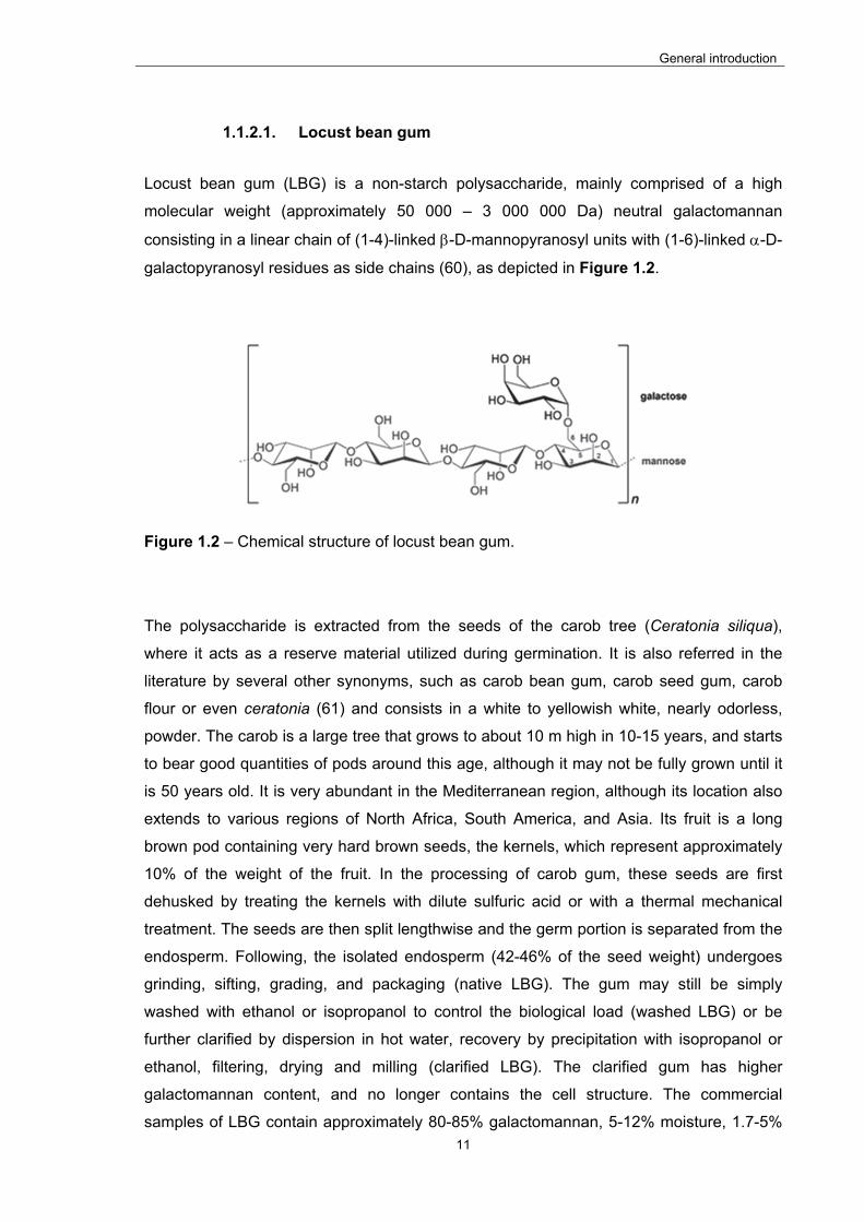
LIST OF FIGURES
Figure 1.1 – Schematic representation of the preparation of nanoparticles by the method
of polyelectrolyte complexation. .......................................................................................... 7
Figure 1.2 – Chemical structure of locust bean gum. ....................................................... 11
Figure 1.3 – Chemical structure of chitosan. .................................................................... 14
Figure 1.4 – Contribution of efficacy, safety, feasibility and cost to the development of
vaccines (118). .................................................................................................................. 16
Figure 3.1 – Scheme of the chemical modifications introduced in LBG. .......................... 44
Figure 3.2 – FTIR spectra of purified Locust Bean Gum (LBG) and its ammonium (LBGA),
carboxylate (LBGC) and sulfate (LBGS) derivatives. ........................................................ 45
Figure 3.3 – FTIR spectra of Locust Bean Gum sulfate derivatives (LBGS) obtained in
method 1 (M1) and method 2 (M2). B1, B2 and B3 refer to batch 1, 2 and 3, respectively.
.......................................................................................................................................... 47
Figure 3.4 – 1H-NMR of (a) LBG, (b) LBGA, (c) LBGS, and (d) LBGC; the big singlet
centered at 4.7 ppm is due to HOD (identified in grey). .................................................... 49
Figure 3.5 – 13C-NMR of LBGC. ....................................................................................... 50
Figure 3.6 – XRD patterns of (a) pristine locust bean gum (LBG), (b) sulfated LBG
(LBGS), (c) ammonium LBG (LBGA), and (d) carboxylated LBG (LBGC). ....................... 52
Figure 3.7 – Effect of charge ratio (-/+) on the zeta potential of CS/LBGS nanoparticles. 54
Figure 3.8 – Effect of charge ratio (-/+) on the zeta potential of CS/LBGC nanoparticles. 57
Figure 3.9 – TEM microphotograph of LBGA/LBGS = 1:2 (w/w) nanoparticles. .............. 60
Figure 3.10 - Caco-2 cell viability measured by the MTT assay after 3 h exposure to
increasing concentrations of bulk Locust Bean Gum, purified Locust Bean Gum (LBG) and
its ammonium (LBGA), carboxylate (LBGC) and sulfate (LBGS) derivatives. Data
represent mean ± SEM (n ≥ 3, six replicates per experiment at each concentration).
Dashed line indicates 70%. * P < 0.05 compared with DMEM. ........................................ 62
Figure 3.11 - Caco-2 cell viability measured by the MTT assay after 24 h exposure to
increasing concentrations of bulk Locust Bean Gum, purified Locust Bean Gum (LBG) and
its ammonium (LBGA), carboxylate (LBGC) and sulfate (LBGS) derivatives. Data
represent mean ± SEM (n ≥ 3, six replicates per experiment at each concentration).
Dashed line indicates 70%. * P < 0.05 compared with DMEM. ........................................ 62
Figure 3.12 – Caco-2 cell viability measured by the LDH release assay after 24 h
exposure to 1 mg/mL solutions of bulk Locust Bean Gum, purified Locust Bean Gum
(LBG) and its ammonium (LBGA), carboxylate (LBGC) and sulfate (LBGS) derivatives.
Data represent mean ± SEM (n ≥ 3, three replicates per experiment). * P < 0.05 compared
with DMEM. ....................................................................................................................... 65

xxii
Figure 3.13 – A549 cell viability measured by the MTT assay after 3 h and 24 h exposure
to increasing concentrations of sulfate locust bean gum (LBGS) derivative. Data represent
mean ± SEM (n ≥ 3, six replicates per experiment at each concentration). Dashed line
indicates 70%. * P < 0.05 compared with respective control (DMEM). ............................. 66
Figure 3.14 – Calu-3 cell viability measured by the MTT assay after 3 h and 24 h
exposure to increasing concentrations of sulfate locust bean gum (LBGS) derivative. Data
represent mean ± SEM (n ≥ 3, six replicates per experiment at each concentration).
Dashed line indicates 70%. * P < 0.05 compared with respective control (DMEM). ......... 66
Figure 3.15 – Caco-2 cell viability measured by the MTT assay after 3 h exposure to
increasing concentrations of ammonium Locust Bean Gum (LBGA) derivative, sulfate
Locust Bean Gum (LBGS) derivative and LBGA/LBGS nanoparticles (NP). Data represent
mean ± SEM (n ≥ 3, six replicates per experiment at each concentration). Dashed line
indicates 70%. * P < 0.05 compared with DMEM. ............................................................ 67
Figure 3.16 – Caco-2 cell viability measured by the MTT assay after 24 h exposure to
increasing concentrations of ammonium Locust Bean Gum (LBGA) derivative, sulfate
Locust Bean Gum (LBGS) derivative and LBGA/LBGS nanoparticles (NP). Data represent
mean ± SEM (n ≥ 3, six replicates per experiment at each concentration). Dashed line
indicates 70%. * P < 0.05 compared with DMEM. ............................................................ 68
Figure 4.1 – Effect of charge ratio (-/+) on the zeta potential of CSup/LBGS nanoparticles.
.......................................................................................................................................... 87
Figure 4.2 - TEM microphotograph of CSup/LBGS 1:2 (w/w) nanoparticles. ................... 88
Figure 4.3 – TEM microphotograph of HE-loaded CSup/LBGS (1:2, w/w; 8% HE)
nanoparticles. .................................................................................................................... 93
Figure 4.4 – Immunoblot (a) and SDS-PAGE (b) analyses of free HE (1) and HE released
from HE-loaded CSup/LBGS nanoparticles (2) (PS: standard proteins). ......................... 96
Figure 4.5 – SDS-PAGE (a) and immunoblot (b) analyses of free OVA (1) and OVA
released from OVA-loaded CSup/LBGS nanoparticles (2) (PS: standard proteins). ........ 97
Figure 4.6 – Size (square marks) and zeta potential (triangular marks) evolution as
function of time upon storage at 4 ºC of CSup/LBGS 1:2 (w/w) unloaded nanoparticles
(empty marks) and HE-loaded nanoparticles (8% w/w; filled marks); (mean ± SD, n ≥ 3).
.......................................................................................................................................... 99
Figure 4.7 – Antigen released overtime from CSup/LBGS nanoparticles in simulated
gastric fluid (SGF) and simulated intestinal fluid (SIF), at 37 ºC (mean ± SD; n ≥ 3). ..... 100
Figure 4.8 – Caco-2 cell viability measured by the MTT assay after 3 h exposure to
increasing concentrations of Chitosan (CSup), sulfate Locust Bean Gum (LBGS)
derivative and CSup/LBGS nanoparticles (NP). Data represent mean ± SEM (n ≥ 3, six
xxiii
replicates per experiment at each concentration). Dashed line indicates 70%. * P < 0.05
compared with DMEM. .................................................................................................... 103
Figure 4.9 – Caco-2 cell viability measured by the MTT assay after 24 h exposure to
increasing concentrations of Chitosan (CSup), sulfate Locust Bean Gum (LBGS)
derivative and CSup/LBGS nanoparticles (NP). Data represent mean ± SEM (n ≥ 3, six
replicates per experiment at each concentration). Dashed line indicates 70%. * P < 0.05
compared with DMEM. .................................................................................................... 104
Figure 4.10 – Caco-2 cell viability measured by the LDH release assay after 24 h
exposure to 1 mg/mL solutions of Chitosan (CSup), sulfate Locust Bean Gum (LBGS)
derivative and CSup/LBGS nanoparticles (NP). Data represent mean ± SEM (n ≥ 3, three
replicates per experiment). * P < 0.05 compared with DMEM. ....................................... 105
Figure 4.11 – Immunogenicity of HE after oral administration in mice. Serum A) IgG1 and
B) IgG2a systemic response, and C) IgA mucosal response after oral immunization of 5
female BALB/c mice with 200 µg of HE solution (HE), 200 µg of encapsulated HE (NP-
HE) and the corresponding mass of blank nanoparticles (NP). In the HE group the results
of one mouse were rejected due to the high initial absorbance (week 0) (mean ± SEM; n ≥
4). .................................................................................................................................... 108
Figure 4.12 – Immunogenicity of HE after S.C. administration in mice. Serum A) IgG1 and
B) IgG2a systemic response, and C) IgA mucosal response after S.C. immunization of 5
female BALB/c mice with 40 µg of HE solution (HE), 40 µg of encapsulated HE (NP-HE)
and the corresponding mass of blank nanoparticles (NP) (mean ± SEM; n = 5). ........... 110
Figure 4.13 – Immunogenicity of OVA after oral administration in mice. Serum anti-OVA
A) IgG1; B) IgG2a and C) faecal anti-OVA IgA response in BALB/c mice (n = 6) after oral
immunization with 100 µg of OVA solution (OVA) or 100 µg of encapsulated OVA (NP-
OVA). Antibody titers were determined in pooled serum samples at days 0, 7, 14, 21 and
28 post-administration. .................................................................................................... 113
Figure 4.14 – Immunogenicity of OVA after S.C. administration in mice. Serum anti-OVA
A) IgG1; B) IgG2a and C) faecal anti-OVA IgA response in BALB/c mice (n = 6) after S.C.
immunization with 20 µg of OVA solution (OVA) or 20 µg of encapsulated OVA (NP-OVA).
Antibody titers were determined in pooled faecal samples at days 0, 7, 14, 21 and 28
post-administration. ......................................................................................................... 115

xxiv
LIST OF TABLES
Table 1.1 - Advantages and limitations of nanoparticles for drug delivery applications. .... 5
Table 1.2 – Pros and cons of polysaccharides as nanoparticle matrix materials (53). ....... 9
Table 1.3 – Advantages and disadvantages of oral administration (141). ........................ 19
Table 1.4 – Toll-like receptors (TLRs) and their specific ligands (150)............................. 20
Table 1.5 – Some of the most frequent C-type lectins and their specific ligands (152, 153).
.......................................................................................................................................... 21
Table 3.1 – Elemental analysis data from the sulfate (LBGS), carboxylate (LBGC) and
ammonium (LBGA) derivatives of locust bean gum (LBG). .............................................. 46
Table 3.2 – GPC analysis of purified Locust Bean Gum (LBG), and its ammonium
(LBGA), carboxylate (LBGC) and sulfate (LBGS) derivatives. .......................................... 51
Table 3.3 - Physicochemical characteristics and production yield of CS/LBGS unloaded
nanoparticles (mean ± SD; n ≥ 3). Different letters represent significant differences in each
parameter (P < 0.05). ........................................................................................................ 53
Table 3.4 - Physicochemical characteristics and production yield of CS/LBGC unloaded
nanoparticles (mean ± SD; n ≥ 3). Different letters represent significant differences in each
parameter (P < 0.05). ........................................................................................................ 56
Table 3.5 - Physicochemical characteristics and production yield of LBGA/LBGS unloaded
nanoparticles (mean ± SD; n ≥ 3). Different letters represent significant differences in each
parameter (P < 0.05). ........................................................................................................ 58
Table 4.1 - Physicochemical characteristics and production yield of CSup/LBGS unloaded
nanoparticles (mean ± SD; n ≥ 3). Different letters represent significant differences in each
parameter (P < 0.05). ........................................................................................................ 85
Table 4.2 – Physicochemical characteristics, production yield and association efficiency of
HE antigens in CSup/LBGS nanoparticles (mean ± SD; n ≥ 3). Different letters represent
significant differences in each parameter, evaluated separately for each mass ratio (P <
0.05). ................................................................................................................................. 91
Table 4.3 – Physicochemical characteristics, production yield end association efficiency of
OVA in CSup/LBGS nanoparticles (mean ± SD; n ≥ 3). Different letters represent
significant differences in each parameter (P < 0.05). ........................................................ 95

xxv
ABBREVIATIONS
ABTS: 3-ethylbenzthiazoline-6-sulfonic acid
AE: Association efficiency
AIDS: Acquired immune deficiency syndrome
ANOVA: One-way analysis of variance
APCs: Antigen-presenting cells
AS03: Adjuvant system 03
AS04: Adjuvant system 04
AUC: Area under the curve
BCA: Bicinchoninic acid
BCG: Bacillus Calmette-Guérin
BSA: Bovine serum albumin
CD: Cluster of differentiation
CD4+: CD4 positive T cell
CD8+: CD8 positive T cell
CpG DNA: Cytosine – phosphate – guanine deoxyribonucleic acid
CS: Chitosan
CSup: Ultrapure chitosan
DCs: Dendritic cells
DC-SIGN: Dendritic cell-specific intercellular adhesion molecule-3-grabbing non-integrin
DMEM: Dulbecco’s modified Eagle’s medium
DMF: Dimethylformamide
DMSO: Dimethyl sulfoxide
DNA: Deoxyribonucleic acid

xxvi
DO: Degree of oxidation
DS: Degree of substitution
dsRNA: Double-stranded ribonucleic acid
EDTA: Ethylenediamine tetraacetic acid
ELISA: Enzyme-linked immunosorbent assay
FAE: Follicle associated epithelium
FBS: Fetal bovine serum
FDA: Food and drug administration
FTIR: Fourier transform infrared
GALT: Gut-associated lymphoid tissue
GlcNAc: N-acetylglucosamine
GPC/SEC3: Triple detection gel permeation chromatography
GRAS: Generally recognized as safe
GTMAC: N-glycidyl-N,N,N-trimethylammonium chloride
HE: Immunogenic subcellular extract obtained from whole Salmonella Enteritidis
HPLC: High performance liquid chromatography
ICAM-3: Intercellular adhesion molecule 3
IFN-γ: Interferon-gamma
IgA: Immunoglobulin A
IgG1: Immunoglobulin G1
IgG2a: Immunoglobulin G2a
IL-10: Interleukin 10
IL-17: Interleukin 17
IL-2: Interleukin 2

xxvii
IL-22: Interleukin 22
IL-4: Interleukin 4
IL-6: Interleukin 6
IPN: Interpenetrating polymer network
ISO: International Organization for Standardization
LBG: Locust bean gum
LBGA: Locust bean gum ammonium derivative
LBGC: Locust bean gum carboxylate derivative
LBGS: Locust bean gum sulfate derivative
LC: Loading capacity
LDH: Lactate dehydrogenase
LPS: Lipopolysaccharide
M cells: Microfold cells
M/G: Mannose/galactose ratio
ManNAc : N-acetylmannosamine
MBL: Mannose-binding lectin
MF59: Oil in water emulsion composed of squalene, polysorbate 80, sorbitan trioleate and
citrate buffer
MHC I: Major histocompatibility complex I
MHC II: Major histocompatibility complex II
Mn: Number average molecular weight
MPLA: Monophosphoryl lipid A
MTT: Thiazolyl blue tetrazolium bromide
Mw: Weight average molecular weight

xxviii
NMR: Nuclear magnetic resonance
NP: Nanoparticles
OD: Optical density
OVA: Ovalbumin
PAMP: Pathogen-associated molecular pattern
PBS: Phosphate buffered saline
PBS-T: Phosphate buffered saline-tween
PdI: Polydispersion index
PLGA: Poly(d,l-lactide-co-glycolide)
PPs: Peyer's patches
PRR: Pattern recognition receptors
PS: Proteins standard
PVA: Polyvinyl alcohol
PY: Production yield
Rg: Radius of gyration
SDS: Sodium dodecyl sulfate
SDS-PAGE: Sodium dodecyl sulfate - polyacrylamide gel electrophoresis
SGF: Simulated gastric fluid
SIF: Simulated intestinal fluid
SO3.DMF: N,N-Dimethylformamide sulfur trioxide
SP-A: Surfactant protein A
SP-D: Surfactant protein D
ssRNA: Single-stranded ribonucleic acid
TCRs: T cell receptors

xxix
TEM: Transmission electron microscopy
TEMPO: 2,2,6,6-tetramethylpiperidine-1-oxyl
TGF-β: Transforming growth factor β
Th: T helper
Th1: T-helper type 1
Th17: T-helper type 17
Th2: T-helper type 2
Th3: T-helper type 3
TLR: Toll-like receptor
XRD: X-ray diffraction

General introduction
1
CHAPTER 1
GENERAL INTRODUCTION
The information presented in this chapter was partially published in the following
publications:
Braz, L., Dionísio, M., Grenha, A., 2011. Chitosan-based nanocarriers: effective vehicles
for mucosal protein delivery. In: S.P. Davis (Ed.), Chitosan: manufacturing, properties and
usage. Nova Science Publishers, New York. p. 365-412.
Braz, L., Rodrigues, S., Fonte, P., Grenha, A., Sarmento, B., 2011. Mechanisms of
chemical and enzymatic chitosan biodegradability and its application on drug delivery. In:
G. Felton (Ed.), Biodegradable Polymers: Processing, Degradation and Applications.
Nova Science Publishers, New York. p. 325-364.

General introduction
2
This page was intentionally left in blank

General introduction
3
1. General introduction
1.1. Nanoparticles as carriers in drug delivery
The recent decades have brought to the market many new biomolecules that have been
identified as having therapeutic potential. This boom is directly related with advances in
the biotechnological industry, making available a very considerable number of molecules
that are protein-based. These molecules are usually called biopharmaceuticals, meaning
that they are biological in nature and manufactured using biotechnology (1). A
considerably wide variety of molecules is included in this group, from protein and
peptides, to antigens and nucleic acids. In many cases their promise is so high that they
are thought to occupy in the future an undisputed place alongside other established
therapies. Although therapeutically promising, biopharmaceuticals are very instable
compounds and their administration is extremely challenging, due to inherent
physicochemical and biopharmaceutical properties (2, 3). This is the main reason why
parenteral delivery frequently represents the unique administration possibility, as is often
verified for vaccines, for example. Nevertheless, the parenteral route involves an invasive
and painful administration, thus not being easily accepted by the patients and many times
leading to therapeutic incompliance (3, 4). A gap is, thus, clearly identified which needs to
be filled, compelling researchers to invest in this area in order to find adequate
alternatives that permit effective, safe, cheap and comfortable administration of
biopharmaceutical molecules through other routes. Comparing with the modality of
parenteral delivery, mucosal administration is advantageous as systemic pathway, mainly
because it is non-invasive, reducing patient discomfort, but also because it generally does
not require the involvement of skilled personnel for the administration, thus reducing the
costs of the process (3, 5). In this manner, the development of non-injectable delivery
systems for mucosal administration could enhance significantly patient’s compliance,
thereby leading to increased therapeutic benefits.
The therapeutic action of proteins and protein-based molecules is not only limited by the
potential degradation in biological environments, but also compromised by their low ability
to reach the therapeutic site of action (3, 6, 7). In fact, these limitations are related either
with the presence of a great number of functional groups susceptible of chemical
degradation, or with the high hydrophilic character of the proteins, which results in poor
permeability (8, 9). In addition, drug delivery via mucosal routes faces other major
restrictions, including specific mechanisms of defense, the possibility to induce immune
reactions at the delivery site and, generally, limitations in the surface area available for
General introduction
4
absorption (10). As such, a meaningful challenge for current pharmaceutical scientists has
been the need to develop suitable vehicles that permit delivering macromolecules through
alternative routes of administration. These drug delivery carriers should exhibit a sort of
characteristics, including capacity for high drug association and the ability to enhance
drug physicochemical stability, providing protection to encapsulated drugs from the
moment of carrier production until release. Furthermore, in many cases, the carriers are
expected to regulate the drug release profile, while allowing an intimate contact of
molecules with mucosal barriers, contributing for their epithelial permeation. In such a
task, there is a consensus in that the materials and methods used to prepare the referred
vehicles play relevant roles (11, 12).
Directing the research efforts towards the development of adequate vehicles for the
purpose of drug delivery through distinct routes of administration, resulted in the
appearance of several drug delivery systems like nanoparticles and microspheres.
Nanoparticulate-based technologies have reached a position of evidence and
nanoparticles are one of the most approached systems. The International Organization for
Standardization (ISO) defines nanoparticles as particles with at least one dimension less
than 100 nm (13). This definition is however not consensual in the field of drug delivery
and there are many authors considering that nanoparticles are carriers with dimensions
between 10 and 1000 nm (14-17). The interest in nanosystems (submicron sized
systems) relies on several differentiating features that include an increased surface-to-
volume ratio, in many cases displaying surface functionality, which offers high potential for
the association of biopharmaceuticals (15). Biological transport processes have been
reported to be affected by the physical attributes of nanocarriers, both anatomically and
down to the cellular ad subcellular levels (18). Actually, nanocarriers have been reported
to increase drug absorption by reducing the resistance of the epithelium to drug transport
in a localized area or by carrying the drug across the epithelium (19). In this regard,
transport has been described as more favorable for nanoparticles than for microparticles
(2, 20, 21). In addition, colloidal carriers are reported to have improved capacity to interact
with mucosal epithelial membranes (20), maximum interaction being reported to occur for
systems within 50 – 500 nm (8). Colloidal carriers have also shown several times the
ability to control the drug release profile of encapsulated molecules (22-24). Importantly,
specifically regarding the delivery of biopharmaceuticals, an improvement of molecule
stability, bioavailability, targeting, uptake and biological activity has been shown (24-26).
Table 1.1 summarizes the advantages and drawbacks of nanoparticles for drug delivery
applications.
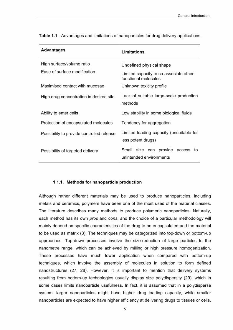
General introduction
5
Table 1.1 - Advantages and limitations of nanoparticles for drug delivery applications.
Advantages Limitations
High surface/volume ratio Undefined physical shape
Ease of surface modification Limited capacity to co-associate other functional molecules
Maximised contact with mucosae Unknown toxicity profile
High drug concentration in desired site Lack of suitable large-scale production
methods
Ability to enter cells Low stability in some biological fluids
Protection of encapsulated molecules Tendency for aggregation
Possibility to provide controlled release Limited loading capacity (unsuitable for
less potent drugs)
Possibility of targeted delivery Small size can provide access to
unintended environments
1.1.1. Methods for nanoparticle production
Although rather different materials may be used to produce nanoparticles, including
metals and ceramics, polymers have been one of the most used of the material classes.
The literature describes many methods to produce polymeric nanoparticles. Naturally,
each method has its own pros and cons, and the choice of a particular methodology will
mainly depend on specific characteristics of the drug to be encapsulated and the material
to be used as matrix (3). The techniques may be categorized into top-down or bottom-up
approaches. Top-down processes involve the size-reduction of large particles to the
nanometre range, which can be achieved by milling or high pressure homogenization.
These processes have much lower application when compared with bottom-up
techniques, which involve the assembly of molecules in solution to form defined
nanostructures (27, 28). However, it is important to mention that delivery systems
resulting from bottom-up technologies usually display size polydispersity (29), which in
some cases limits nanoparticle usefulness. In fact, it is assumed that in a polydisperse
system, larger nanoparticles might have higher drug loading capacity, while smaller
nanoparticles are expected to have higher efficiency at delivering drugs to tissues or cells.

General introduction
6
In a limit situation, this means that, even if the drug carrier has high association efficiency,
the efficacy of the delivery may be poor (30). Therefore, fabrication processes should be
rigorously optimized to provide a compromise between satisfactory association efficiency
and the most suitable size for the established objective. Interestingly, a recent
technological development related to a top-down process termed particle replication in
non-wetting templates, which is a modified soft lithography technique, has demonstrated
independent control over nanoparticle size, as well as other parameters that include
shape, modulus (stiffness) and surface chemistry (29, 31).
Bottom-up techniques might also be classified according to whether the formulation of
nanoparticles involves a polymerization reaction or is achieved directly from a preformed
polymer (32). Methods involving polymerization are divided in emulsion polymerization
and interfacial polymerization (32). When nanoparticles are prepared from preformed
polymers, which is the most common approach, the diversity of techniques increases,
involving methods based on emulsification, polymer desolvation, or intermolecular
electrostatic interactions (11, 32). Contrary to low molecular weight drugs,
biopharmaceuticals possess organized structures (proteins for example may have
secondary, tertiary, or even quaternary structure) with labile bonds and side chains of
chemically reactive groups. This specific structure defines the exact properties and
activities of the molecules and, therefore, it is crucial to ensure its preservation during the
association procedures, as its disruption or the modification of side chains can lead to loss
of activity of the molecule. This fragile nature requires that methods selected for
association do not damage the molecule structure, reduce their biological activity, or
render them immunogenic (3).
Methods based on the establishment of intermolecular electrostatic interactions are one of
the most reported and are applied when the matrix of nanoparticles is composed by at
least one polyelectrolyte, that is, a polymer that exhibits charged groups when in solution.
The principle of this methodology is the ability of polyelectrolytes to establish stable links
with oppositely charged groups (33). Two different methods are described based on
electrostatic interaction. Ionic gelation is the used denomination when the polyelectrolyte
is a polymer with gelling ability (such as chitosan or alginate, for instance) and its gelation
is induced by small anionic molecules, such as phosphate, citrate and sulfate groups. A
very typical example is that of chitosan nanoparticles prepared by interaction of chitosan
amino groups with the phosphate groups of tripolyphosphate (34). In turn, polyelectrolyte
complexation is the name of the technique when the groups mediating the interaction are
provided by two oppositely charged polyelectrolytes, instead of involving one small
molecule (35). The latter approach is often referred to as complex coacervation or

General introduction
7
interfacial coacervation (36, 37), and includes for example chitosan/alginate (38),
chitosan/carrageenan (39) or chitosan/dextran sulfate (40) nanoparticles. Figure 1.1
displays a schematic representation of the method of polyelectrolyte complexation to
produce nanoparticles. The assembly of nanoparticles is easily and immediately observed
upon pouring a solution of one polyelectrolyte over a solution of the oppositely charged
polyelectrolyte, under mild stirring.
The popularity of the method is mainly due to the fact that it usually involves a complete
hydrophilic environment and mild preparation conditions (41), avoiding the use of organic
solvents or high shear forces and making association of labile drugs, such as
biopharmaceuticals, an easier task (33, 42).
Figure 1.1 – Schematic representation of the preparation of nanoparticles by the method of polyelectrolyte complexation.
The literature indicates that, in order to obtain nanocarriers with pre-established
characteristics by this method, an optimization of the process should be performed

General introduction
8
focusing aspects such as the concentration of polyelectrolytes, the stirring conditions and
the conditions of centrifugation (43).
1.1.2. Materials for nanoparticle production
The selection of appropriate materials for drug delivery approaches should be driven by
several requirements, including biocompatibility, biodegradability, versatility and low
overall costs of production (11, 44). As mentioned above, polymeric nanoparticles are
one of the most representative of nanomedicines, as polymers are among the most
versatile building units, permitting an easy tailoring of their properties to meet specific
requirements (44). By definition, polymeric nanoparticles can be produced using either
synthetic or natural polymers. The former are reported frequently, but in many cases they
display unsatisfactory biocompatibility, which limits potential clinical applications (45). On
the contrary, natural polymers comply more easily with the requirements mentioned above
(46). Actually, it has been claimed that one of the ways of avoiding the potential toxicity of
nanocarriers, an issue believed to be one of the most relevant in preventing diverse
clinical applications so far, may be using natural materials (45). Moreover, these have
some remarkable merits over synthetic ones, namely improved capacity for cell adhesion
and mechanical properties similar to natural tissues (47).
The class of natural polymers is divided in proteins and polysaccharides. The latter have
found a wide range of applications, as there is an extensive variety of materials available
for exploration, comparing with proteins. Furthermore, many polysaccharides are obtained
from plants or marine organisms, therefore being less probable to induce adverse
immunological reactions, as compared with proteins (44). Polysaccharides are complex
carbohydrates, composed of monosaccharides joined together by glycosidic bonds (44,
48). The most common basic units composing these carbohydrate polymers include
several monosaccharides such as D-glucose, D-fructose, D-galactose, L-galactose, D-
mannose, L-arabinose and D-xylose. Some polysaccharides comprise in their structure
simple sugar acids (glucuronic, mannuronic and iduronic acids) and also monosaccharide
derivatives, like the amino sugars D-glucosamine and D-galactosamine, as well as their
derivatives N-acetylneuraminic acid and N-acetylmuramic acid (49). The presence of
several of these sugar units on the side chain of carbohydrate polymers makes them good
candidates for targeted delivery by carbohydrate recognizing receptors found on the
surface of several cells (50, 51).
General introduction
9
Polysaccharides might have algal, plant, microbial or animal origin, but they all share
general properties of natural polymers, including the propensity for biocompatibility, low
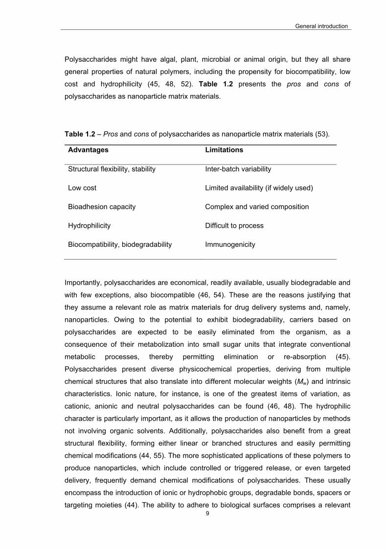
cost and hydrophilicity (45, 48, 52). Table 1.2 presents the pros and cons of
polysaccharides as nanoparticle matrix materials.
Table 1.2 – Pros and cons of polysaccharides as nanoparticle matrix materials (53).
Advantages Limitations
Structural flexibility, stability Inter-batch variability
Low cost Limited availability (if widely used)
Bioadhesion capacity Complex and varied composition
Hydrophilicity Difficult to process
Biocompatibility, biodegradability Immunogenicity
Importantly, polysaccharides are economical, readily available, usually biodegradable and
with few exceptions, also biocompatible (46, 54). These are the reasons justifying that
they assume a relevant role as matrix materials for drug delivery systems and, namely,
nanoparticles. Owing to the potential to exhibit biodegradability, carriers based on
polysaccharides are expected to be easily eliminated from the organism, as a
consequence of their metabolization into small sugar units that integrate conventional
metabolic processes, thereby permitting elimination or re-absorption (45).
Polysaccharides present diverse physicochemical properties, deriving from multiple
chemical structures that also translate into different molecular weights (Mw) and intrinsic
characteristics. Ionic nature, for instance, is one of the greatest items of variation, as
cationic, anionic and neutral polysaccharides can be found (46, 48). The hydrophilic
character is particularly important, as it allows the production of nanoparticles by methods
not involving organic solvents. Additionally, polysaccharides also benefit from a great
structural flexibility, forming either linear or branched structures and easily permitting
chemical modifications (44, 55). The more sophisticated applications of these polymers to
produce nanoparticles, which include controlled or triggered release, or even targeted
delivery, frequently demand chemical modifications of polysaccharides. These usually
encompass the introduction of ionic or hydrophobic groups, degradable bonds, spacers or
targeting moieties (44). The ability to adhere to biological surfaces comprises a relevant

General introduction
10
advantage in drug delivery applications (48), as these frequently imply an interaction with
cell surfaces. In this regard, the reactive functional groups of polysaccharides allow the
formation of non-covalent bonds with cell surfaces, affording enhanced residence time
and, consequently, increased drug absorption (45, 52). Notwithstanding the relevance of
the advantages mentioned above, some drawbacks should also be taken into account,
such as the possibility of generating immunogenic responses and the polymer variability
related to origin and supplier (47). Regarding the latter, plant-derived polysaccharides
pose potential challenges, as structural differences might occur according to the location
and plant collection season (46).
As understood from what is described above, although polysaccharides have been
traditionally included in formulations as inert materials, modern pharmaceutical design
involves these excipients in increasingly relevant roles. In this manner, their application
usually intends to endow the dosage forms with multi-functional abilities, such as
controlled release, stabilization, emulsification or bioadhesiveness, among others (46, 48).
Nevertheless, the set of polysaccharide properties confers a relevant versatility that adds
to a wide range of biological abilities, thus generally contributing to an increased
application in drug carrier production.
The application of polysaccharides in the production of nanoparticles by polyelectrolyte
complexation or ionic gelation is widely reported (34, 38-40, 56, 57). As these methods
require hydrophilic materials that exhibit opposite charges, in addition to an absence of
toxicity, chitosan is the only natural polycationic polysaccharide that satisfies these needs
(52). On the contrary, there are many negatively charged polysaccharides that can be
used for this end, including alginate, hyaluronic acid, dextran sulfate, chondroitin sulfate
and carrageenan, among many others. However, the interest on using other materials that
do not exhibit charge has also been demonstrated occasionally and, in this regard, the
synthesis of charged derivatives of these polysaccharides has been reported to serve the
strategy, as described for pullulan (58, 59).
Below, the general characteristics of the polysaccharides locust bean gum and chitosan,
which were used in this work to produce nanoparticles by polyelectrolyte complexation,
are detailed.
General introduction
11
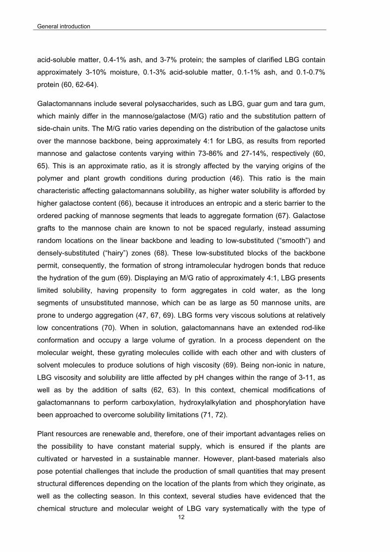
1.1.2.1. Locust bean gum
Locust bean gum (LBG) is a non-starch polysaccharide, mainly comprised of a high
molecular weight (approximately 50 000 – 3 000 000 Da) neutral galactomannan
consisting in a linear chain of (1-4)-linked -D-mannopyranosyl units with (1-6)-linked -D-
galactopyranosyl residues as side chains (60), as depicted in Figure 1.2.
Figure 1.2 – Chemical structure of locust bean gum.
The polysaccharide is extracted from the seeds of the carob tree (Ceratonia siliqua),
where it acts as a reserve material utilized during germination. It is also referred in the
literature by several other synonyms, such as carob bean gum, carob seed gum, carob
flour or even ceratonia (61) and consists in a white to yellowish white, nearly odorless,
powder. The carob is a large tree that grows to about 10 m high in 10-15 years, and starts
to bear good quantities of pods around this age, although it may not be fully grown until it
is 50 years old. It is very abundant in the Mediterranean region, although its location also
extends to various regions of North Africa, South America, and Asia. Its fruit is a long
brown pod containing very hard brown seeds, the kernels, which represent approximately
10% of the weight of the fruit. In the processing of carob gum, these seeds are first
dehusked by treating the kernels with dilute sulfuric acid or with a thermal mechanical
treatment. The seeds are then split lengthwise and the germ portion is separated from the
endosperm. Following, the isolated endosperm (42-46% of the seed weight) undergoes
grinding, sifting, grading, and packaging (native LBG). The gum may still be simply
washed with ethanol or isopropanol to control the biological load (washed LBG) or be
further clarified by dispersion in hot water, recovery by precipitation with isopropanol or
ethanol, filtering, drying and milling (clarified LBG). The clarified gum has higher
galactomannan content, and no longer contains the cell structure. The commercial
samples of LBG contain approximately 80-85% galactomannan, 5-12% moisture, 1.7-5%
General introduction
12
acid-soluble matter, 0.4-1% ash, and 3-7% protein; the samples of clarified LBG contain
approximately 3-10% moisture, 0.1-3% acid-soluble matter, 0.1-1% ash, and 0.1-0.7%
protein (60, 62-64).
Galactomannans include several polysaccharides, such as LBG, guar gum and tara gum,
which mainly differ in the mannose/galactose (M/G) ratio and the substitution pattern of
side-chain units. The M/G ratio varies depending on the distribution of the galactose units
over the mannose backbone, being approximately 4:1 for LBG, as results from reported
mannose and galactose contents varying within 73-86% and 27-14%, respectively (60,
65). This is an approximate ratio, as it is strongly affected by the varying origins of the
polymer and plant growth conditions during production (46). This ratio is the main
characteristic affecting galactomannans solubility, as higher water solubility is afforded by
higher galactose content (66), because it introduces an entropic and a steric barrier to the
ordered packing of mannose segments that leads to aggregate formation (67). Galactose
grafts to the mannose chain are known to not be spaced regularly, instead assuming
random locations on the linear backbone and leading to low-substituted (“smooth”) and
densely-substituted (“hairy”) zones (68). These low-substituted blocks of the backbone
permit, consequently, the formation of strong intramolecular hydrogen bonds that reduce
the hydration of the gum (69). Displaying an M/G ratio of approximately 4:1, LBG presents
limited solubility, having propensity to form aggregates in cold water, as the long
segments of unsubstituted mannose, which can be as large as 50 mannose units, are
prone to undergo aggregation (47, 67, 69). LBG forms very viscous solutions at relatively
low concentrations (70). When in solution, galactomannans have an extended rod-like
conformation and occupy a large volume of gyration. In a process dependent on the
molecular weight, these gyrating molecules collide with each other and with clusters of
solvent molecules to produce solutions of high viscosity (69). Being non-ionic in nature,
LBG viscosity and solubility are little affected by pH changes within the range of 3-11, as
well as by the addition of salts (62, 63). In this context, chemical modifications of
galactomannans to perform carboxylation, hydroxylalkylation and phosphorylation have
been approached to overcome solubility limitations (71, 72).
Plant resources are renewable and, therefore, one of their important advantages relies on
the possibility to have constant material supply, which is ensured if the plants are
cultivated or harvested in a sustainable manner. However, plant-based materials also
pose potential challenges that include the production of small quantities that may present
structural differences depending on the location of the plants from which they originate, as
well as the collecting season. In this context, several studies have evidenced that the
chemical structure and molecular weight of LBG vary systematically with the type of
General introduction
13
cultivar and growth conditions (46, 47). Furthermore, dehulling and milling processes, as
well as dissolution temperature, are reported to influence the chemical (mannose and
galactose content) and rheological properties of LBG (47, 68, 73, 74). This probably
justifies, at least in part, why LBG is considered polydisperse from a chemical point of
view, presenting three types of structural variation: 1) degree of galactose substitution, 2)
patterning of galactose side groups and 3) chain length; all directly related with
biosynthesis mechanisms (47). This is possibly one of the major limitations compromising
a more frequent application of LBG in the pharmaceutical field. LBG is Generally
Recognized as Safe (GRAS) by the Food and Drug Administration (FDA) and is approved
in most areas of the world (European Union, United States of America, Japan, Australia,
etc.), being used as thickening, gelling and stabilizing agent in both food and cosmetic
industries (75, 76). In food industry it is a food additive, coded as E-410 in the European
Union (77). Recently it has been indicated as a very useful excipient for pharmaceutical
applications mainly due to its ability as controlled release excipient in tablets. However,
other relevant properties and abilities have been demonstrated which contribute for its
increasing use. The synergistic interaction between xanthan gum and LBG leading to the
formation of thermally reversible gels is well-known. This effect has been ascribed to a
denaturant effect of LBG that disturbs the helix-coil equilibrium of xanthan, displacing it
from an ordered conformation to a more flexible one, facilitating the formation of
heterotopic junctions between both polymers (78). A mucoadhesive behaviour has been
indicated for this polysaccharide (79), although of lower potency comparing with others
like chitosan. Additionally, there are indications on LBG biodegradability (64, 75),
especially if administered orally, as enzymatic degradation mediated by β-mannanase (80,
81) or colonic bacteria (82) is expected to occur. Low toxicity and availability at low cost
(75, 76, 83) have also been referred, contributing for the increased interest on the
polymer.
1.1.2.1. Chitosan
Chitosan is a (1–4)-linked 2-amino-2-deoxy-β-D-glucan (Figure 1.3) that is obtained from
the deacetylation of chitin, the second most abundant natural polymer after cellulose (84).
The most usual source of chitin is the exoskeleton of crustaceans, but it exists in insects
and fungi. Despite the widespread occurrence, up to now the main commercial sources of
chitin have been crab and shrimp shells. Chitosan is the polymer considered to be
obtained when chitin is deacetylated to such a degree that it becomes completely soluble
in dilute aqueous acidic systems (85). The partial deacetylation is performed under
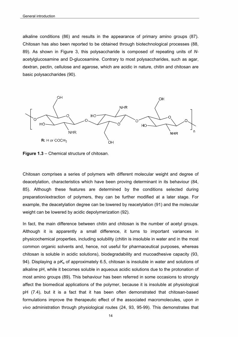
General introduction
14
alkaline conditions (86) and results in the appearance of primary amino groups (87).
Chitosan has also been reported to be obtained through biotechnological processes (88,
89). As shown in Figure 3, this polysaccharide is composed of repeating units of N-
acetylglucosamine and D-glucosamine. Contrary to most polysaccharides, such as agar,
dextran, pectin, cellulose and agarose, which are acidic in nature, chitin and chitosan are
basic polysaccharides (90).
Figure 1.3 – Chemical structure of chitosan.
Chitosan comprises a series of polymers with different molecular weight and degree of
deacetylation, characteristics which have been proving determinant in its behaviour (84,
85). Although these features are determined by the conditions selected during
preparation/extraction of polymers, they can be further modified at a later stage. For
example, the deacetylation degree can be lowered by reacetylation (91) and the molecular
weight can be lowered by acidic depolymerization (92).
In fact, the main difference between chitin and chitosan is the number of acetyl groups.
Although it is apparently a small difference, it turns to important variances in
physicochemical properties, including solubility (chitin is insoluble in water and in the most
common organic solvents and, hence, not useful for pharmaceutical purposes, whereas
chitosan is soluble in acidic solutions), biodegradability and mucoadhesive capacity (93,
94). Displaying a pKa of approximately 6.5, chitosan is insoluble in water and solutions of
alkaline pH, while it becomes soluble in aqueous acidic solutions due to the protonation of
most amino groups (89). This behaviour has been referred in some occasions to strongly
affect the biomedical applications of the polymer, because it is insoluble at physiological
pH (7.4), but it is a fact that it has been often demonstrated that chitosan-based
formulations improve the therapeutic effect of the associated macromolecules, upon in
vivo administration through physiological routes (24, 93, 95-99). This demonstrates that
General introduction
15
the presence of chitosan in the site of action as a dissolved form is not a critical issue
regarding the therapeutic action of the carried molecules, although it is certainly decisive
in the preparation of formulations. Directly regarding the influence of deacetylation degree
on solubility, highly deacetylated chitosans (85% or more) are readily soluble in solutions
with pH up to 6.5 (94).
Chitosan is included in marketed dietary supplements, as it attaches to fat and prevents
its absorption, inducing weight loss (100). It is well known as a non-toxic, biocompatible
and biodegradable polymer (101, 102), characteristics that render it very attractive for the
biomedical and pharmaceutical fields. Given the structural similarity to
glycosaminoglycans, it has been widely proposed for biomedical applications, including
wound healing and tissue engineering (89). Additionally, it has long been a reference in
the field of drug delivery, mainly owing to the reported mucoadhesive properties (103) and
the ability to enhance the permeability of drugs (23, 93, 95, 96, 98, 104-109). The
electrostatic interactions occurring between cationic amino groups of chitosan and the
negatively charged mucin have been reported as the main driving force for its strong
mucoadhesion (110). There is, thus, a direct relation between mucoadhesive capacity and
the deacetylation degree, because highly deacetylated chitosan has more positively
charged amino groups available to mediate the interaction with the negatively charged
mucus components, leading to increased mucoadhesion (107). This explains that most
scientific research reporting the pharmaceutical application of chitosan is performed with
highly deacetylated chitosan. The ability to improve the permeability of drug molecules is
described to be related with both transcellular and paracellular transport. While in
transcellular pathway the effect is mostly related with the mucoadhesive capacity, in the
case of the paracellular route it is attributed to a specific interaction with tight junctions,
leading to their transient opening (111). In the latter case, although it is suggested that the
temporary disruption of tight junctions is due to multiple mechanisms, it was demonstrated
that a specific type of transmembrane proteins (claudins) is possibly playing a major role
(112).
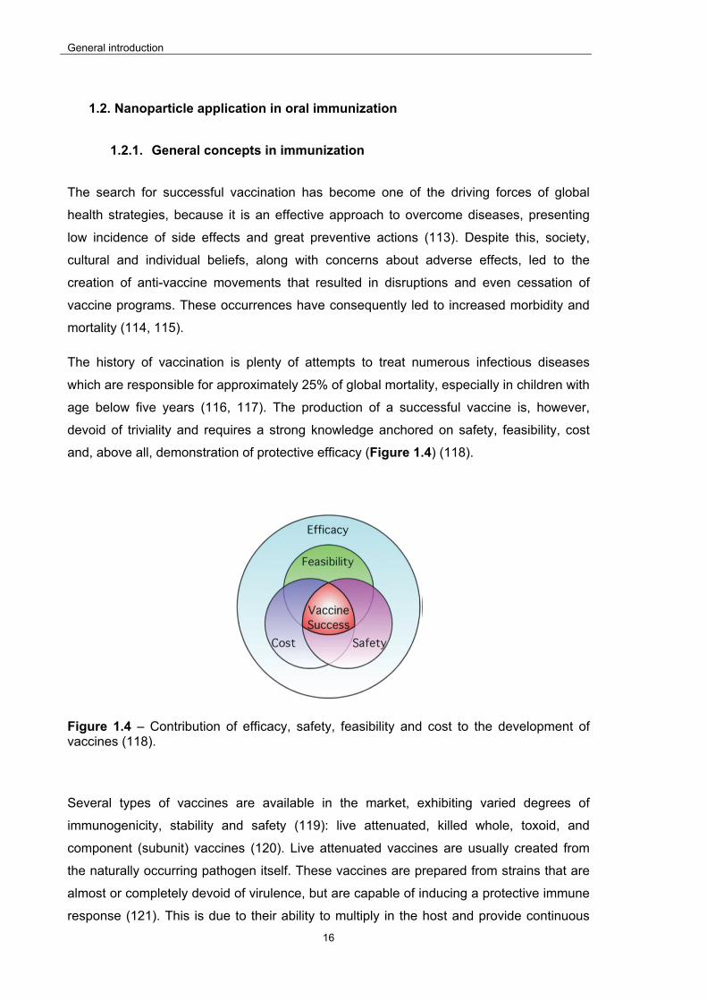
General introduction
16
1.2. Nanoparticle application in oral immunization
1.2.1. General concepts in immunization
The search for successful vaccination has become one of the driving forces of global
health strategies, because it is an effective approach to overcome diseases, presenting
low incidence of side effects and great preventive actions (113). Despite this, society,
cultural and individual beliefs, along with concerns about adverse effects, led to the
creation of anti-vaccine movements that resulted in disruptions and even cessation of
vaccine programs. These occurrences have consequently led to increased morbidity and
mortality (114, 115).
The history of vaccination is plenty of attempts to treat numerous infectious diseases
which are responsible for approximately 25% of global mortality, especially in children with
age below five years (116, 117). The production of a successful vaccine is, however,
devoid of triviality and requires a strong knowledge anchored on safety, feasibility, cost
and, above all, demonstration of protective efficacy (Figure 1.4) (118).
Figure 1.4 – Contribution of efficacy, safety, feasibility and cost to the development of vaccines (118).
Several types of vaccines are available in the market, exhibiting varied degrees of
immunogenicity, stability and safety (119): live attenuated, killed whole, toxoid, and
component (subunit) vaccines (120). Live attenuated vaccines are usually created from
the naturally occurring pathogen itself. These vaccines are prepared from strains that are
almost or completely devoid of virulence, but are capable of inducing a protective immune
response (121). This is due to their ability to multiply in the host and provide continuous

General introduction
17
antigenic stimulation over a period of time, thus often requiring just a single boost (122).
Nevertheless, the application of these vaccines comprises some limitations, which include
the possibility to cause severe infections in immunocompromised individuals (120), high
risk of reverting back to the virulent form (122), and the need for an effective cold chain for
their distribution. The latter is a clear limitation to their application in developing countries,
as these might lack the necessary infrastructure (123). Examples of live attenuated
vaccines are smallpox, polio (Sabin), measles, mumps, rubella, varicella and adenovirus
(119, 122). Inactivated (killed) vaccines (such as influenza, hepatitis A, polio (Salk)) (119,
122) contain killed or inactivated microorganisms. These are not able to cause infection,
but they are still able to stimulate a protective immune response, however usually
requiring multiple doses to maintain immunity (122, 124). Toxoid vaccines consist in
inactivated toxins but stimulate the production of neutralizing antibodies (tetanus,
diphtheria) (122, 125). Subunit vaccines use a defined portion of the pathogen, such as
proteins, peptides or nucleic acids (120, 122), and their interest mainly relies on the
provided increased safety, as these cannot revert to a virulent form, as well as on the
observed lack of contaminants remaining from the original pathogenic organism (122).
Despite of this, the application of these formulations results in poor immunogenic effect
when administered alone (120), demanding the use of an adjuvant in order to enhance the
immunological response (122, 126).
Adjuvants are a highly heterogeneous group of compounds which share the characteristic
of providing a modulation or/and enhancement of immune response (127). The
mechanism mediating the interaction of adjuvants with the immune system is highly
variable, as is the type of induced immunomodulation process, which consists, in short, in
Th1, Th2, Th3, Th17 or/and T regulatory mediated response. The Th1 subset is
characterized by the secretion of cytokines such as interleukin 2 (IL-2) and interferon-γ
(IFN-γ), to assist in cell-mediated immune response. The Th2 subset assists preferentially
in antibody immune responses after secreting cytokines like interleukin 4 (IL-4) (120). Th3
cells secrete interleukin 10 (IL-10) and/or transforming growth factor β (TGF-β), which are
immunosuppressive cytokines that inhibit the proliferation of and cytokine production by
effector T cells, including Th1 cells and Th2 cells (128). Th17 cells secrete interleukin 17
and 22 (IL-17 and IL-22, respectively) and play an important role in host defense against
bacterial and fungal infection, especially at mucosal surfaces (129). Several functions for
regulatory T cells include are suggested, which include prevention of autoimmune
diseases by maintaining self-tolerance; suppression of allergy, asthma and pathogen-
induced immunopathology; feto-maternal tolerance; and oral tolerance (130). Adjuvants
are described to act through different mechanisms, including the formation of a depot for

General introduction
18
the antigen, acting as an intermediate to target the antigen to immune cells, a mechanism
that is assumed to occur when antigen delivery is mediated by particulate delivery
systems, and even the stimulation of immune cells themselves (120, 131). Many
compounds and strategies are described as adjuvants (120), but only aluminum-based
mineral salt (alum), MF59 (an o/w emulsion composed of <250 nm droplets formed when
squalene (4.3% v/v), polysorbate 80 (0.5% v/v) and sorbitan trioleate (0.5% v/v) are
emulsified in citrate buffer), Adjuvant System 03 (AS03) (o/w emulsion + α tocopherol)
and Adjuvant System 04 (AS04) (composed of monophosphoryl lipid A (MPLA) and alum)
are approved for human usage (120, 122, 131, 132).
In addition to the urgent need to develop vaccines for emerging diseases, such as Zika or
Ebola, there are well-known infectious diseases like malaria, tuberculosis and acquired
immune deficiency syndrome (AIDS), that are still lacking an effective vaccine (133-135).
The Bacillus Calmette-Guérin (BCG) vaccine has been available for many years against
tuberculosis and the first vaccine against malaria was approved in 2015, but they are both
referred as having low-medium efficacy (134, 135). This evidences the need for innovative
strategies to the development of effective vaccines.
1.2.2. Oral immunization
The majority of vaccines available nowadays are administered through injection. The need
for this procedure increases general vaccination costs and requires trained personnel to
perform the administration, apart from being painful and uncomfortable, which often
results in diminished patient compliance. In addition, the administration itself presents
safety risks related to infection, needle reuse and disposal (119, 136), not to mention the
need for cold-chain distribution (123). Oral vaccination emerges, in this context, as a
strategy to overcome some of the mentioned drawbacks, benefiting from the general
advantages of oral administration, but also basing its success on specific immune features
of the intestine which revealed crucial for immunization. Table 1.3 presents the main
advantages and disadvantages of oral administration.
Contrary to parenteral immunization, which is mainly effective at producing systemic
immune responses, oral vaccines elicit both systemic and mucosal immunity, further
providing much higher patient’s compliance and being more easily distributed, which is
relevant for developing countries (119, 137, 138). Additionally, it is estimated that 90% of
all mammalian infections originate at mucosal surfaces and, thus, mucosal sites are the
primary access locations for most human pathogens (138). This reinforces the relevance
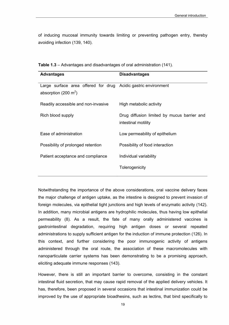
General introduction
19
of inducing mucosal immunity towards limiting or preventing pathogen entry, thereby
avoiding infection (139, 140).
Table 1.3 – Advantages and disadvantages of oral administration (141).
Advantages Disadvantages
Large surface area offered for drug
absorption (200 m2)
Acidic gastric environment
Readily accessible and non-invasive High metabolic activity
Rich blood supply Drug diffusion limited by mucus barrier and
intestinal motility
Ease of administration Low permeability of epithelium
Possibility of prolonged retention Possibility of food interaction
Patient acceptance and compliance Individual variability
Tolerogenicity
Notwithstanding the importance of the above considerations, oral vaccine delivery faces
the major challenge of antigen uptake, as the intestine is designed to prevent invasion of
foreign molecules, via epithelial tight junctions and high levels of enzymatic activity (142).
In addition, many microbial antigens are hydrophilic molecules, thus having low epithelial
permeability (8). As a result, the fate of many orally administered vaccines is
gastrointestinal degradation, requiring high antigen doses or several repeated
administrations to supply sufficient antigen for the induction of immune protection (126). In
this context, and further considering the poor immunogenic activity of antigens
administered through the oral route, the association of these macromolecules with
nanoparticulate carrier systems has been demonstrating to be a promising approach,
eliciting adequate immune responses (143).
However, there is still an important barrier to overcome, consisting in the constant
intestinal fluid secretion, that may cause rapid removal of the applied delivery vehicles. It
has, therefore, been proposed in several occasions that intestinal immunization could be
improved by the use of appropriate bioadhesins, such as lectins, that bind specifically to

General introduction
20
the mucosal surface and increase the carrier/antigen residence time (144). The decoration
with suitable ligands could, in addition, mediate specific recognition by receptors on
antigen presenting or sampling cells that direct the type and intensity of the subsequent
immune response (145).
The immune response takes place, for example, in the course of an infection. It starts with
an attraction of leukocytes to the infected region and interaction of the pathogen with the
immune cells driven by pathogen-associated molecular pattern (PAMP) molecules, which
have characteristic molecular patterns of infectious agents. These molecules are
recognized by innate immune receptors called pattern recognition receptors (PRR)
present on epithelial cells and antigen-presenting cells (APCs) and cause cell activation
and subsequent migration (146). Toll-like receptors (TLRs) and C-type lectin receptors are
the PRR families most involved in immune responses (147). While the former are
expressed either on the cell surface or inside the endosomes (148), the latter are
expressed on cell surface (149). Table 1.4 presents the different TLRs and their specific
ligands.
Table 1.4 – Toll-like receptors (TLRs) and their specific ligands (150).
TLRs Ligands
TLR-1/ TLR-2 Triacylated lipopeptides
TLR-2/ TLR-6 Diacylated lipopeptides
TLR-3 Viral dsRNA
TLR-4 Lipopolysaccharide
TLR-5 Flagellin
TLR-7 Viral ssRNA
TLR-8 Viral ssRNA
TLR-9 Bacterial or viral CpG DNA
TLR-11 Profilin
TLR-10, 12, 13 Unknown
CpG DNA: cytosine – phosphate – guanine deoxyribonucleic acid; dsRNA: double-stranded ribonucleic acid; ssRNA: single-stranded ribonucleic acid
C-type lectin receptors are a set of cell surface receptors that are specialized on the
recognition of carbohydrate molecules, including mannose, N-acetylglucosamine, L-
fucose, glucose and galactosamine, among others (151), as depicted in Table 1.5.
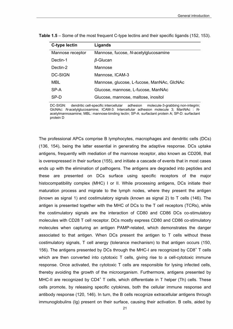
General introduction
21
Table 1.5 – Some of the most frequent C-type lectins and their specific ligands (152, 153).
C-type lectin Ligands
Mannose receptor Mannose, fucose, N-acetylglucosamine
Dectin-1 β-Glucan
Dectin-2 Mannose
DC-SIGN Mannose, ICAM-3
MBL Mannose, glucose, L-fucose, ManNAc, GlcNAc
SP-A Glucose, mannose, L-fucose, ManNAc
SP-D Glucose, mannose, maltose, inositol
DC-SIGN: dendritic cell-specific intercellular adhesion molecule-3-grabbing non-integrin; GlcNAc: N-acetylglucosamine; ICAM-3: Intercellular adhesion molecule 3; ManNAc : N-acetylmannosamine; MBL: mannose-binding lectin; SP-A: surfactant protein A; SP-D: surfactant protein D
The professional APCs comprise B lymphocytes, macrophages and dendritic cells (DCs)
(136, 154), being the latter essential in generating the adaptive response. DCs uptake
antigens, frequently with mediation of the mannose receptor, also known as CD206, that
is overexpressed in their surface (155), and initiate a cascade of events that in most cases
ends up with the elimination of pathogens. The antigens are degraded into peptides and
these are presented on DCs surface using specific receptors of the major
histocompatibility complex (MHC) I or II. While processing antigens, DCs initiate their
maturation process and migrate to the lymph nodes, where they present the antigen
(known as signal 1) and costimulatory signals (known as signal 2) to T cells (146). The
antigen is presented together with the MHC of DCs to the T cell receptors (TCRs), while
the costimulatory signals are the interaction of CD80 and CD86 DCs co-stimulatory
molecules with CD28 T cell receptor. DCs mostly express CD80 and CD86 co-stimulatory
molecules when capturing an antigen PAMP-related, which demonstrates the danger
associated to that antigen. When DCs present the antigen to T cells without these
costimulatory signals, T cell anergy (tolerance mechanism) to that antigen occurs (150,
156). The antigens presented by DCs through the MHC-I are recognized by CD8+ T cells
which are then converted into cytotoxic T cells, giving rise to a cell-cytotoxic immune
response. Once activated, the cytotoxic T cells are responsible for lysing infected cells,
thereby avoiding the growth of the microorganism. Furthermore, antigens presented by
MHC-II are recognized by CD4+ T cells, which differentiate in T helper (Th) cells. These
cells promote, by releasing specific cytokines, both the cellular immune response and
antibody response (120, 146). In turn, the B cells recognize extracellular antigens through
immunoglobulins (Ig) present on their surface, causing their activation. B cells, aided by
General introduction
22
Th cells, undergo extensive proliferation and generate both high-affinity memory B cells
and long-lived plasma cells (157), the latter being capable to secrete large amounts of
specific antibody (158). During this process, memory T and B cells are produced, which
can quickly proliferate and eliminate the infecting agent (146).
Immune responses occurring in the intestinal area are mediated by the so called gut-
associated lymphoid tissue (GALT). This comprises diffusely-scattered cells in the lamina
propria, intra-epithelial lymphocytes, isolated lymphoid follicles present throughout the
intestine and, most importantly, Peyer's patches (PPs) (141, 159). The latter are the
structures where immune cells of the intestinal mucosa are mainly located (154), being
privileged targets for mucosal vaccination due to their crucial role in intestinal mucosal
immunity (160). The immune cells from the PPs, such as the DCs, are separated from the
lumen by the Follicle Associated Epithelium (FAE) (159), which is composed of
enterocytes, goblet cells and microfold cells (M cells) (136, 154). Several works suggest
that PPs are the predominant site of uptake of nano- and microparticles, a process in
which the antigen-sampling M cells play an important role (136). The M cells are,
however, present in a small number in the intestinal tract, representing only 1 out of 10
million epithelial cells (approximately 5% in humans) (161). In parallel, they constitute 10–
30% of the epithelial cells of the FAE above the PPs (162). These cells are considered the
main entrance for pathogens invading the organism (163, 164) and are characterized by a
disorganized brush border and a reduced mucus layer at the apical side, because in FAE
there are less mucus secreting goblet cells than on the rest of the intestinal epithelium
(165, 166). Furthermore, M cells contain small cytoplasmatic vesicles, few lysosomes and
short microvilli (136, 159), and there is evidence that they can transport the antigen
without any degradation, even in the absence of a protective carrier (167). Importantly,
although the clear existence of a mannose receptor has not been described yet, M cells
have been reported to favorably recognize mannose units and mannosylated carriers
have been described to target M cells (80, 168, 169). The basolateral chamber of M cells
is deeply invaginated forming a pocket hosting APCs: lymphocytes, macrophages and
DCs (136, 154). These characteristics of M cells make them particularly suited for the task
of antigen uptake, because they favor antigen interactions with the apical membrane,
provide optimized antigen endocytosis, shorten and facilitate antigen access to the
basolateral compartment, and finally favor rapid and straightforward interactions between
immune cells and APCs that are present at the basal side (154). Therefore, M cells have
been signaled as potentials targets to take into account in the design of oral vaccination
strategies.

General introduction
23
1.2.3. The role of nanoparticles: Locust bean gum as potential adjuvant
Polymeric nanoparticles have been demonstrating to be very promising in oral delivery of
biopharmaceuticals, as many works report their effective role in the enhancement of oral
drug bioavailability by facilitating cell internalization (19, 170). Their reduced size provides
an intimate contact with epithelia and, in several occasions, they have shown the capacity
to carry the encapsulated molecules through the epithelium (8, 19).
With respect to oral vaccination, the design of suitable antigen delivery systems should
focus on optimizing antigen association efficiency, ensuring the maintenance of its stability
during association, tailoring release kinetics and eliciting high levels of long-lasting
antibody and cellular immune responses. Nanoparticles may provide extra benefits in oral
immunization strategies, because PPs have shown to be a predominant site for uptake of
particulates (136). Given their role in intestinal mediated immunization, M cells are the
primary targets to consider and it has been demonstrated that particle uptake depends on
various factors, such as particle size, surface charge, concentration, stability and ligand
conjugation (170). In this context, it is important to highlight that nanoparticles have been
showing to be better taken up by M cells as compared to microparticles (171-173). Once
M cell uptake is observed, subsequent uptake by professional APCs, such as
macrophages and DCs, is expected to occur, mediating the following immune response.
Importantly, a great advantage of formulating antigens in nanocarriers lies in the
exploitation of the intrinsic capacity of the referred phagocytic cells (macrophages, DCs,
etc.), located in the PPs, to internalize foreign particulate materials (174). Another
remarkable issue comes from studies on mucosal vaccination that have demonstrated
that particulate antigens are often more immunogenic than antigens in solution, under the
indication that particulate antigens are more likely to be trafficked across the mucosa and
taken up by APCs (175).
A careful selection of nanoparticle matrix materials might further help on the potentiation
of an immune response. In this context, mucoadhesive polymers may help on the
prolongation on the intestinal residence time (176), potentiating the uptake by M cells.
Importantly, the physicochemical characteristics of polymeric nanoparticles can be
modulated by tuning polymer properties and surface chemistry, while their specificity may
also be augmented by surface ligand conjugation (15). LBG, one of the polysaccharides
used in this work for nanoparticle production, may contribute in a strong manner for the
improvement of nanoparticle abilities regarding an application in oral immunization
mediated by nanoparticles. The greatest potentiality that is identified comes from the
proper chemical structure, which is composed of mannose and galactose residues. Given
General introduction
24
the favorable contact with mannose residues, mentioned above, LBG might act as vector
with preferential targeting ability towards these cells. Furthermore, it has mucoadhesive
characteristics (79), although these are not as significant as those described for chitosan,
which is the other polysaccharide used as matrix material in nanoparticle production.
Finally, as dendritic cells are described to express mannose receptors (155), LBG
nanoparticles are also expected to have a privileged contact with these cells, after being
sampled by M cells.

Motivations and objectives
25
CHAPTER 2
MOTIVATIONS AND OBJECTIVES
Motivations and objectives
26
This page was intentionally left in blank
Motivations and objectives
27
2. Motivations and Objectives
Successful vaccination approaches are required to face some of the most threatening
diseases worldwide. Additionally, as many of the most concerning situations occur in
developing countries, alternatives to the parenteral administration, that has costly
requirements (cold chain, sterility, skilled personnel, etc.), are demanded (119, 123, 136).
Oral immunization has thus been reaching a position of evidence in this regard, not only
because the oral route is the main port of entry of pathogens and permits easy
administration (138), but also due to specific structural abilities of the intestinal area
(Peyer’s patches, M cells, antigen presenting cells) regarding immunization approaches
(136, 154). Some of these structures are known to establish a preferential contact with
particulates (136). Therefore, the use of nanoparticles as antigen carriers has been
proposed as a valuable strategy to induce immunological responses. These nanoparticles
may be further functionalized or display in their structure moieties that provide an extra
improvement of the targeting ability. In this regard, the use of locust bean gum (LBG) as
matrix material may contribute for the improvement of nanoparticle abilities regarding oral
immunization due to the mannose content that mediates a privileged contact with M cells
and dendritic cells (80, 155, 169). Mucoadhesive materials may also be beneficial,
providing prolonged intestinal residence time (176), and thus, potentiating the uptake by M
cells. LBG has been reported to exhibit mucoadhesive characteristics (79), although other
polysaccharides, like chitosan, are known to display much stronger mucoadhesiveness
(103).
Taking into account the motivations referred above, this work is aimed at producing
nanoparticles based on LBG for an application in oral immunization. To demonstrate the
feasibility of the approach, Salmonella Enteritidis antigenic complex (HE) and ovalbumin
(OVA) were used as model antigens.
To accomplish the referred general objective, several partial objectives were considered,
which are disclosed below:
1) To synthesize charged derivatives of LBG (sulfated, carboxylated and ammonium) and
confirm the effective derivatization using adequate techniques;
2) To produce LBG-based nanoparticles by polyelectrolyte complexation and select a
formulation with suitable properties for antigen association and application as carrier in
oral immunization;
Motivations and objectives
28
3) To associate the model antigens HE and OVA and investigate their release profile in
media relevant for oral delivery applications;
4) To evaluate the cytotoxicity of the synthesized LBG derivatives and LBG-based
nanoparticles in human intestinal epithelial cells (Caco-2);
5) To evaluate in vivo the immunological response to a selected formulation of LBG-based
nanoparticles associating the model antigens individually.
In this manner, it is expected to develop a natural, biocompatible polymeric
nanoparticulate system which displays ability for the efficient association of antigens and
suitable physicochemical properties for an application in oral vaccination.

Locust bean gum derivatives and production of nanoparticles
29
CHAPTER 3
LOCUST BEAN GUM DERIVATIVES AND PRODUCTION OF NANOPARTICLES
The information presented in this chapter was submitted for publication in Carbohydrate
Polymers:
Braz, L., Grenha, A., Corvo, M., Lourenço, J.P., Ferreira, D., Sarmento, B., Rosa da
Costa, A.M. Synthesis and characterization of Locust Bean Gum derivatives and their
application in the production of nanoparticles, submitted for publication
Locust bean gum derivatives and production of nanoparticles
30
This page was intentionally left in blank

Locust bean gum derivatives and production of nanoparticles
31
3. Synthesis and characterization of Locust Bean Gum derivatives and their
application in the production of nanoparticles
3.1. Introduction
Biopolymers, among them polysaccharides, are an attractive class of polymers, as they
are derived from natural sources, normally easily available, relatively cheap, many times
biodegradable, and that can be modified by suitable chemical reagents. The most
common basic unit of polysaccharides is the monosaccharide D-glucose, although D-
fructose, D-galactose, L-galactose, D-mannose, L-arabinose, D-xylose, and the amino
sugars D-glucosamine and D-galactosamine, as well as simple sugar acids, like
glucuronic acid and iduronic acid, and sulfated monosaccharides are also frequently
present (62).
Locust bean gum (LBG), also known as carob bean gum, is obtained from the endosperm
of carob tree (Ceratonia siliqua) seeds, where it is a reserve material. The tree is very
abundant in the Mediterranean region, including the Algarve. The gum corresponds to a
galactomannan, and commercial samples of LBG contain approximately 80-85%
galactomannan, while the remaining content is ascribed to proteins and impurities. LBG is
reported as biocompatible, biosorbable, biodegradable, non-teratogenic and non-
mutagenic, and its degradation products are excreted readily. Classified by the FDA as
GRAS material, it is approved in most areas of the world for use in the food industry as
thickener, stabilizer, emulsifier, and gelling agent (E410), as well as in the pharmaceutical
industry as excipient in drug formulations, and in biomedical applications (60, 62-64). LBG
is mainly comprised of a high molecular weight (approximately 50 000 – 3 000 000 Da)
neutral galactomannan consisting in a linear chain of (1-4)-linked -D-mannopyranosyl
units with (1-6)-linked -D-galactopyranosyl residues as side chains. The mannose and
galactose contents have been reported to be 73-86% and 27-14%, respectively, which
corresponds to a mannose:galactose (M/G) ratio of approximately 4:1 (60). For that
reason, and in spite of the uneven distribution of galactose units along the mannose
backbone, leading to low-substituted (“smooth”) and densely-substituted (“hairy”) zones
(68), LBG is typically represented as shown in Figure 1.2 (chapter 1, general
introduction). Being non-ionic in nature, its viscosity and solubility are little affected by pH
changes within the range of 3-11, as well as by the addition of salts (62, 63).
Recently, there has been a growing interest in the chemical functionalization of
polysaccharides, particularly those non-animal derived, mainly by making use of the free
Locust bean gum derivatives and production of nanoparticles
32
hydroxyl groups distributed along their backbone, in order to create derivatives with
tailored properties for desired applications (48). Carboxymethylation of polysaccharides is
a well-known etherification process achieved by reaction with monochloroacetic acid, and
leading to products with a variety of promising properties, like increased hydrophilicity,
water solubility, and solution clarity. Carboxymethyl cellulose, guar gum, LBG, and
xanthan gum have been successfully used in the production of novel drug delivery
systems, like beads, microparticles, and nanoparticles (177). The sodium carboxymethyl
ether of LBG was combined with polyvinyl alcohol (PVA) for the production of
interpenetrating polymer network (IPN) hydrogel microspheres of buflomedil
hydrochloride, regarding a controlled drug delivery (178). The introduction of carboxyl
groups in a galactomannan isolated from the seeds of Leucaema leucocephala, with an
M/G ratio around 1.3, was successfully performed using 2,2,6,6-tetramethylpiperidine-1-
oxyl (TEMPO) as oxidizing agent. This reagent acts by oxidizing the free C-6 position of
the monosaccharide units, which, in this case, was demonstrated to be more effective on
the mannopyranose units than on the galactose ones. As expected, the modified polymer,
an anionic polyelectrolyte, showed greater water solubility than the original material. The
fact that in the course of this oxidation reaction aldehyde groups form as intermediate,
allowed the obtainment of an amphiphilic polymer by reductive amination with an amine
bearing a long alkyl chain (179). Cellulose (180) and cashew gum (181) were also
converted in the corresponding polyuronic acids by the above method. Sulfated LBG
derivatives with various degrees of substitution were obtained by the chlorosulfonic
acid/pyridine method, which presented significant chain stiffness, due to the electrostatic
effect (182). A mixed carboxymethyl sulfate derivative of LBG was prepared by carrying
out a sulfation reaction with SO3.DMF complex followed by carboxymethylation. Ionotropic
gelation of this LBG derivative induced by basic aluminum chloride in the presence the
potent analgesic tramadol hydrochloride, led to the formation of hydrogel beads
incorporating the drug. These hydrogels disintegrated very quickly in an acidic solution,
liberating almost all of their content in 15 min, thus constituting a promising system for
immediate dosage release formulations leading to instant analgesic action (183).
In this chapter, the chemical modification of LBG, aiming at the obtainment of charged
derivatives intended for the development of nanoparticulate carriers by polyelectrolyte
complexation, is described. Two anionic (sulfate and carboxylate) and one cationic
(trimethylammonium) derivatives were prepared. The former two were obtained by an
adaptation of a method applied in the sulfation of other polysaccharides, using an
SO3DMF complex as sulfating agent (184), and using the above described oxidizing
agent TEMPO (179), respectively. The latter was synthesized by reaction with the
Locust bean gum derivatives and production of nanoparticles
33
alkylating agent glycidyltrimethylammonium chloride (GTMAC), adapting a described
procedure (185).
3.2. Materials and methods
3.2.1. Materials
Locust bean gum (LBG) was a kind gift from Industrial Farense (Faro, Portugal). Chitosan
(CS, low molecular weight, deacetylation degree = 75 – 85%), glacial acetic acid,
chlorosulfuric acid (HClSO3), dimethylformamide (DMF), N-glycidyl-N,N,N-
trimethylammonium chloride (GTMAC), sodium hydroxide (NaOH), potassium hydroxide
(KOH), 2,2,6,6-tetramethypiperidine-1-oxyl (TEMPO), sodium bromide (NaBr), sodium
hypochlorite solution (NaClO), sodium borohydride (NaBH4), sodium nitrate (NaNO3),
sodium dihydrogen phosphate (NaH2PO4), sodium azide (NaN3), dialysis tubing (pore size
2000 Da), phosphotungstate dibasic hydrate, glycerol, phosphate buffered saline (PBS)
pH 7.4 tablets, Dulbecco’s modified Eagle’s medium (DMEM), penicillin/streptomycin
(10000 units/mL, 10000 µg/mL), non-essential amino acids, L-glutamine 200 mM, trypsin-
EDTA solution (2.5 g/L trypsin, 0.5 g/L EDTA), trypan blue solution (0.4%), thiazolyl blue
tetrazolium bromide (MTT), lactate dehydrogenase (LDH) kit, sodium dodecyl sulfate
(SDS), dimethyl sulfoxide (DMSO), hydrochloric acid (HCl 37%), sodium chloride (NaCl)
and potassium dihydrogen phosphate (KH2PO4) were purchased from Sigma-Aldrich
(Germany). Ethanol was supplied by VWR. Potassium bromide (KBr) was obtained from
Riedel-del-Haën (Germany). Fetal bovine serum (FBS) was obtained from Gibco (USA).
Ultrapure water (Mili-Q Plus, Milipore Iberica, Madrid, Spain) was used throughout. All
other chemicals were reagent grade.
3.2.2. Cell line
The Caco-2 cell line was obtained from the American Type Culture Collection (Rockville,
USA) and used between passages 77-93. Cell cultures were grown in 75 cm2 flasks in a
humidified 5% CO2/95% atmospheric air incubator at 37 ºC. Cell culture medium was
DMEM supplemented with 10% (v/v) FBS, 1% (v/v) L-glutamine solution, 1% (v/v) non-
essential amino acids solution and 1% (v/v) penicillin/streptomycin. Medium was changed
every 2-3 days and cells were subcultured weekly.

Locust bean gum derivatives and production of nanoparticles
34
3.2.3. Synthesis of Locust Bean Gum derivatives
3.2.3.1. Purification of Locust Bean Gum
The purification of LBG envisaged the removal of the protein content (3-7%) commonly
present in commercial samples (60). To do so, a standard procedure was followed (73,
78), in which LBG (5.0 g) was slowly dispersed in distilled water (1000 mL) previously
heated to 85 ºC, the dispersion being stirred for 1h. Then, the dispersion was cooled to
room temperature and, subsequently, centrifuged (22 000 x g, 20 ºC, 1 h). The
supernatant was collected and added to an equal volume of ethanol. The precipitate was
collected by vacuum filtration and added again to an equal volume of ethanol. After
subsequent collection by vacuum filtration, the precipitate was dried in a vacuum oven at
30 ºC during 72 h affording 3.9 g of white powder. The residue was grinded and stored
until further use.
3.2.3.2. Sulfation of Locust Bean Gum
Sulfation of LBG was performed by a method established for the sulfation of other
polysaccharides (184).
The sulfation agent, SO3.DMF, was prepared by slowly dropping 5 mL of HClSO3 into 25
mL of stirred DMF under cooling in an ice water bath, and continuing the stirring for 1.5 h.
The obtained solution was stored in the refrigerator until further use.
Method 1
Purified LBG (500 mg) was dispersed in DMF (35 mL) and stirred at 60 ºC for 30 min, in
order to provide the dispersion of LBG into the solvent. Then, the SO3.DMF complex was
added (9.3 mL) and the mixture reacted for 4 h under magnetic stirring. Subsequently, the
mixture was cooled down to room temperature in an ice bath, neutralized with 30% NaOH
solution until precipitation, and concentrated under reduced pressure at 60 ºC to
evaporate the solvent. The residue was dissolved in distilled water (30 mL) and dialyzed
against distilled water (5 L). The water was changed every 24 h and, after 3 days, the
solution was concentrated under reduced pressure at 40 ºC. Then, ethanol was added
into the concentrated solution, in order to precipitate the solute, and the dispersion was
concentrated under reduced pressure at 40 ºC. The previous step was repeated twice,

Locust bean gum derivatives and production of nanoparticles
35
and the last evaporation was performed until full evaporation of the solvent. The obtained
powder was dried in a vacuum oven at 40 ºC for 3 days, affording 407 mg of brownish
powder that was grinded and stored until further use.
Method 2
Purified LBG (500 mg) was slowly dispersed in distilled water (100 mL) previously heated
to 85 ºC, and the dispersion was stirred for 1 h. After that time, the dispersion was cooled
to room temperature and poured into an equal volume of ethanol. The precipitate was
collected, added to DMF (300 mL) and centrifuged (22 000 x g, 20 ºC, 20 min). The
precipitate was recovered and added to DMF (100 mL), resting overnight. This dispersion
was then filtered and the residue was again added to DMF (35 mL), this new mixture
being stirred at 60 ºC for 30 min, in order to provide the dispersion of LBG into the solvent.
Then, the SO3.DMF complex was added (9.3 mL) and the mixture reacted for 4 h under
magnetic stirring. Subsequently, the mixture was cooled down to room temperature in an
ice bath, neutralized with 30% NaOH solution until precipitation, and concentrated under
reduced pressure at 60 ºC to evaporate the solvent. The residue was dissolved in distilled
water (30 mL) and dialyzed against distilled water (5 L). The water was changed every 24
h and, after 3 days, the solution was concentrated under reduced pressure at 40 ºC.
Then, ethanol was added into the concentrated solution, in order to precipitate the solute,
and the dispersion was concentrated under reduced pressure at 40 ºC. The previous step
was repeated twice, and the last evaporation was performed until full evaporation of the
solvent. The obtained powder was dried in a vacuum oven at 40 ºC for 3 days, affording
363 mg of brownish powder that was grinded and stored until further use.
3.2.3.3. Carboxylation of Locust Bean Gum
The carboxylation of LBG was performed by oxidation with TEMPO (179). Purified LBG
(500 mg) was dissolved in 200 mL of distilled water under stirring at 80 ºC for 30 min.
After cooling down, the volume was adjusted to 200 mL and the solution was cooled in an
ice bath. Then, TEMPO (10 mg) and NaBr (50 mg) were added to the solution under
stirring. A 15% sodium hypochlorite solution (3.0 mL) with pH adjusted to 9.3 with 2 M HCl
solution was mixed with the polymer solution. The pH was maintained at 9.3 by addition of
a 0.05 M aqueous NaOH solution for 4 h. To stop the reaction, sodium borohydride (75
mg) was added and the solution was stirred for 45 min. Then the pH of the mixture was

Locust bean gum derivatives and production of nanoparticles
36
adjusted to 8 by addition of HCl before precipitation by 2 volumes of ethanol in presence
of NaCl (up to 10 g/L). The polymer was isolated by filtration under reduced pressure,
washed several times with ethanol, filtered and dried in a vacuum oven at 30 ºC during 3
days. A white powder (529 mg) was obtained, grinded and stored until further use.
3.2.3.4. Quaternary ammonium salt of Locust Bean Gum
The introduction of quaternary ammonium groups in LBG was achieved by alkylation with
glycidyltrimethylammonium chloride (GTMAC), as follows (185): an aqueous solution (10
mL) of KOH (0.550 g), was prepared in a round bottom flask, under stirring, at 60 ºC.
Then, purified LBG (506 mg) and 3.72 mL of GTMAC were added. After 5 h, an equal
amount of GTMAC was added to the mixture, which was allowed to react until the
completion of 24 h. It was then diluted with 20 mL of miliQ water, allowed to cool down to
room temperature, and neutralized with HCl (2M). The resulting solution was dialyzed for
3 days, the water being replaced every 24 h. Then, the LBGA solution was concentrated
under reduced pressure at 40 ºC and ethanol was added into the concentrated solution, in
order to precipitate the solute. The dispersion was concentrated under reduced pressure
at 40 ºC and ethanol was added again and evaporated under the same conditions until full
evaporation of the solvent. The obtained powder was dried in a vacuum oven at 40 ºC for
3 days, affording 423 mg of white powder that was grinded and stored until further use.
3.2.4. Chemical characterization of Locust Bean Gum derivatives
3.2.4.1. Fourier transform infrared (FTIR) spectroscopy
For recording FTIR spectra of purified LBG and their derivatives, samples were grounded
with KBr in a mortar and compressed into discs. For each spectrum, a 32-scan
interferogram was collected in transmittance mode with a 4 cm-1 resolution in the 4,000-
400 cm-1 region.
3.2.4.2. Elemental analysis
Elemental analysis data were obtained in a Thermo Finnigan, FLASH EA 1112 Series (C,
N, S) or in a Fisons Instruments, EA 1108 CHNS-O (O) elemental analyzer.
Locust bean gum derivatives and production of nanoparticles
37
3.2.4.3. Nuclear magnetic resonance (NMR) spectroscopy
All NMR spectra were acquired in a Bruker Avance III 400 spectrometer equipped with a
temperature control unit and a pulse gradient unit capable of producing magnetic field
pulsed gradients in the z-direction of 56.0 G/cm, operating at 400.15 MHz for hydrogen,
100.61 MHz for carbon, using a multinuclear reverse 5 mm probe (TXI). The samples
where dissolved in D2O. 1H NMR spectra were recorded with 8.22 KHz spectral window
digitized with 64 K points. The 13C spectrum was recorded between 0 and 238 ppm using
24,000 Hz spectral window digitized into 64 K points.
3.2.4.4. GPC/SEC3 analysis
Triple detection Gel Permeation Chromatography (GPC/SEC3) analysis was performed in
a modular system constituted by a degasser, HPLC pump (K-1001) and RI detector (K-
2300) from Knauer, and a viscometer and RALLS from Viscotek (Trisec Dual Detector
Model 270), using two PL aquagel-OH mixed 8 m, 300 x 7.5 mm columns.
For purified LBG, LBGC and LBGS the eluent was 0.2 M NaNO3, 0.01M NaH2PO4, 0.1%
w/v NaN3, pH=7, at 1mL/min; the samples were dissolved in the eluent at 1 mg/mL. For
LBGA the eluent was 0.5 M NaNO3, 0.01M KH2PO4, 0.1% w/v NaN3, pH=2, at the same
rate; the sample was dissolved at 1mg/mL in 10-2 M HCl.
3.2.4.5. X-ray diffraction (XRD)
Powder X‐ray diffractograms were recorded on a Panalytical X’Pert Pro diffractometer,
operating at 45 kV and 35 mA. The patterns of the pristine and modified samples were
recorded in the range 5‐45 degrees (2) with a step size of 0.0167 and a time per step of
2 000 seconds, using CuK radiation filtered by Ni and an X’Celerator detector. Prior to
the analysis, samples were reduced to a fine powder by grinding in a mortar.
3.2.5. Production of Locust Bean Gum-based nanoparticles
All nanoparticles were prepared by polyelectrolyte complexation method which consists in
the electrostatic interaction between the positive and negative charges of the different
polymers (35), as depicted in Figure 1.1 (chapter 1, general introduction).
Locust bean gum derivatives and production of nanoparticles
38
3.2.5.1. CS/LBGS and CS/LBGC nanoparticles
Five mass ratios of CS/LBGC (1:0.75; 1:1; 1:1.25; 1:1.5 and 1:2), and CS/LBGS (1:1;
1:1.25; 1:1.5; 1:2 and 1:2.5) were used to prepare the nanoparticles by polyelectrolyte
complexation. The stock solution of CS, dissolved in 1% (w/w) acetic acid, was prepared
to reach a final concentration of 1.0 mg/mL, while those of LBGC and LBGS, dissolved in
ultrapure water, had a final concentration of 2.0 mg/mL. The solutions were filtered with a
0.45 µm filter prior to use. The formulations were prepared by slowly adding 1.8 mL of
LBGC or LBGS to 1.0 mL of CS under gentle magnetic stirring at room temperature, as
shown in Figure 1.1 (chapter 1, general introduction). The concentration of CS was kept
constant at 1.0 mg/mL for the preparation of all formulations, while that of LBGC or LBGS
was modified to obtain the different ratios.
The suspensions of nanoparticles were mixed by magnetic stirring for 10 min and then
centrifuged in eppendorfs having a layer of 10 µL of glycerol, in order to facilitate the
following step of resuspension. The isolation of nanoparticles was performed by
centrifugation (Thermo Scientific-Heraeus Fresco 17, Germany) at 16 000 x g, for 30 min
at 15 ºC. After discarding the supernatants, the nanoparticles were resuspended with 200
µL of ultrapure water.
3.2.5.2. LBGA/LBGS nanoparticles
Three mass ratios of LBGA/LBGS were used to prepare the nanoparticles by
polyelectrolyte complexation, in particular 2/1, 1/1 and 1/2. The stock solution of LBGA,
dissolved in ultrapure water, was prepared to reach a final concentration of 0.5 mg/mL,
while that of LBGS, dissolved in ultrapure water, had a final concentration of 1.0 mg/mL.
The solutions were filtered with a 0.45 µm filter prior to use. The formulations 2/1, 1/1 and
1/2 were prepared by slowly adding 1.8 ml of LBGS to 1.0 ml of LBGA under gentle
magnetic stirring at room temperature, as shown in Figure 1.1 (chapter 1, general
introduction). The concentration of LBGA was kept constant at 0.5 mg/mL for the
preparation of all formulations, while that of LBGS was modified to obtain the different
ratios.
As described before, the suspensions of nanoparticles were mixed by magnetic stirring for
10 min and then centrifuged in eppendorfs with a layer of 10 µL of glycerol, in order to
facilitate the following step of resuspension. The isolation of nanoparticles was performed
by centrifugation (Thermo Scientific-Heraeus Fresco 17, Germany) at 16 000 x g, for 30
Locust bean gum derivatives and production of nanoparticles
39
min at 15 ºC. After discarding the supernatants, the nanoparticles were resuspended with
200 µL of ultrapure water.
3.2.6. Characterization of Locust Bean Gum-based nanoparticles
3.2.6.1. Size, polydispersion index and ζ potential
The size, ζ potential and polydispersion index (PdI) determination of the nanoparticles
were performed on freshly prepared samples. Size and PdI were measured by dynamic
light scattering and zeta potential was measured by laser Doppler anemometry, using a
Zetasizer Nano ZS (Malvern instruments, Malvern, UK). To prepare the samples, 20 µL of
each formulation were diluted in 1 mL of ultrapure water.
3.2.6.2. Production yield
For determination of nanoparticle production yield, the nanoparticles were prepared as
described in the previous sections, but without the use of the 10 µL of glycerol. After
discarding the supernatant of each formulation, the pellets were frozen and then dried on
a freeze-dryer (Alpha RVC, Christ, Germany). The yield of nanoparticle production (PY)
was calculated as follows:
PY = (Nanoparticle sediment weight/Total solids weight) x 100
where nanoparticle sediment weight is the weight after freeze-drying and total solids
weight is the total amount of solids added for nanoparticle formation.
3.2.6.3. Morphological analysis
The morphological examination of LBGA/LBGS nanoparticles was conducted by
transmission electron microscopy (TEM; JEM-1011, JEOL, Japan). The samples were
stained with 2% (w/v) phosphotungstic acid and placed on copper grids with carbon films
(Ted Pella, USA) for TEM observation.
Locust bean gum derivatives and production of nanoparticles
40
3.2.7. Safety evaluation
The in vitro cell viability and cytotoxicity of bulk LBG, purified LBG and the synthesized
derivatives was assessed in Caco-2 cells by the MTT assay and the LDH release assay,
respectively. LBGA/LBGS nanoparticles were evaluated using the MTT assay.
The cells were seeded at a density of 1×10⁴ cells/well in 96-well plates, in 100 µL of the
same medium used for culture in cell culture flasks, and were grown at 37 °C in a 5% CO2
atmosphere for 24 h before use.
The effect of polymeric solutions and nanoparticle suspensions was evaluated for 3 h and
24 h at three different concentrations (0.1, 0.5 and 1.0 mg/mL). A SDS solution (2%, w/v)
was used as a positive control of cell death, while cells incubated with DMEM served as
negative control. An additional control (DMEM+H2O) consisting in a mixture of DMEM and
H2O in the same ratio used for the samples was used, in order to evaluate the contribution
of polymers on cell viability. All formulations and controls were prepared as
solution/suspensions in pre-warmed cell culture medium without FBS immediately before
application to the cells.
To initiate the assay, culture medium of cells at 24 h in culture was replaced by 100 µL of
fresh medium without FBS containing the test samples or controls. A constant ratio (3:1)
between the culture medium and the solution/suspension of the materials was used. After
3 or 24 h of cell exposure, samples/controls were removed and 30 µL of the MTT solution
(0.5 mg/mL in PBS, pH 7.4) was added to each well. After 2 h, any generated formazan
crystals were solubilised with 50 µL of DMSO. Upon complete solubilisation of the
crystals, the absorbance of each well was measured by spectrophotometry (Infinite M200,
Tecan, Austria) at 540 nm and corrected for background absorbance using a wavelength
of 650 nm (186).
The relative cell viability (%) was calculated as follows:
Viability (%) = (A – S)/(CM – S)× 100
where A is the absorbance obtained for each of the concentrations of the test substance,
S is the absorbance obtained for the 2% SDS and CM is the absorbance obtained for
untreated cells (incubated with cell culture medium). The latter reading was assumed to
correspond to 100% cell viability. The assay was performed at least for three occasions
with six replicates at each concentration of test substance in each instance.
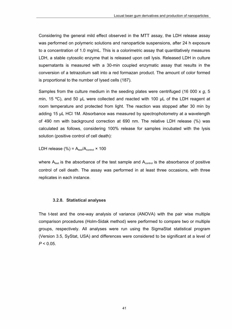
Locust bean gum derivatives and production of nanoparticles
41
Considering the general mild effect observed in the MTT assay, the LDH release assay
was performed on polymeric solutions and nanoparticle suspensions, after 24 h exposure
to a concentration of 1.0 mg/mL. This is a colorimetric assay that quantitatively measures
LDH, a stable cytosolic enzyme that is released upon cell lysis. Released LDH in culture
supernatants is measured with a 30-min coupled enzymatic assay that results in the
conversion of a tetrazolium salt into a red formazan product. The amount of color formed
is proportional to the number of lysed cells (187).
Samples from the culture medium in the seeding plates were centrifuged (16 000 x g, 5
min, 15 ºC), and 50 μL were collected and reacted with 100 μL of the LDH reagent at
room temperature and protected from light. The reaction was stopped after 30 min by
adding 15 μL HCl 1M. Absorbance was measured by spectrophotometry at a wavelength
of 490 nm with background correction at 690 nm. The relative LDH release (%) was
calculated as follows, considering 100% release for samples incubated with the lysis
solution (positive control of cell death):
LDH release (%) = Atest/Acontrol × 100
where Atest is the absorbance of the test sample and Acontrol is the absorbance of positive
control of cell death. The assay was performed in at least three occasions, with three
replicates in each instance.
3.2.8. Statistical analyses
The t-test and the one-way analysis of variance (ANOVA) with the pair wise multiple
comparison procedures (Holm-Sidak method) were performed to compare two or multiple
groups, respectively. All analyses were run using the SigmaStat statistical program
(Version 3.5, SyStat, USA) and differences were considered to be significant at a level of
P < 0.05.

Locust bean gum derivatives and production of nanoparticles
42
3.3. Results and discussion
Since commercial LBG contains about 3-7% protein in its constitution (60), it is essential
to go through a purification step prior to use, in order to avoid interference in the
modification reactions to be performed. Therefore, it was purified using a method based
on previously published protocols (73, 78), with a yield of approximately 78%. This purified
LBG was the material used for subsequent work, unless stated otherwise.
3.3.1. Synthesis and chemical characterization of Locust Bean Gum
derivatives
The syntheses of the three charged LBG derivatives were made by adaptation of
procedures described in the literature for the modification of other polysaccharides (179,
184, 185). To perform the sulfation reaction, SO3DMF was chosen as sulfating agent
(184), as it presents advantages over methods involving the manipulation of either
pyridine or sulfur trioxide (188-190). For the synthesis of the sulfate derivative, two
approaches were performed as described in the methodology. The difference mainly
resided in the processing of LBG prior to the addition of SO3DMF. For the introduction of
trimethylammonium groups in LBG, GTMAC was used as alkylating agent, which proved
to be efficient in the alkylation of other polysaccharides (58, 185, 191, 192). The reaction
mechanisms are as follows:
For the transformation of LBG into the corresponding polyuronic acid, TEMPO, a stable
nitroxyl radical, was chosen as oxidizing agent (179). This has proved to possess a high
CH O S O-
O
O
Me2N CH O Me2N
R O H
Me2N CH O+
R O SO3H
NaOH
R O SO3- Na+
HO S
O
O
Cl- HCl
R O H KOHR O- K+
O
N+(CH3)3Cl-N+(CH3)3Cl-RO
OHHCl

Locust bean gum derivatives and production of nanoparticles
43
efficiency in the conversion of high molecular weight polysaccharides. A highly selective
oxidation of C-6 primary hydroxyl to carboxylic groups can be achieved in an aqueous
solution of the polysaccharide at pH 9-11 with NaClO and catalytic amounts of TEMPO
and NaBr (179-181). The mechanism is as follows:
The methods described above are summarized in Figure 3.1.
OOH
HOO
OH
OOH
HOO
O
ONa
N+ O
N O
N OHNaBrO
NaBrNaClO
NaCl
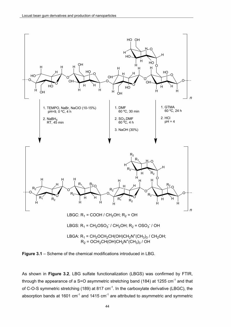
Locust bean gum derivatives and production of nanoparticles
44
Figure 3.1 – Scheme of the chemical modifications introduced in LBG.
As shown in Figure 3.2, LBG sulfate functionalization (LBGS) was confirmed by FTIR,
through the appearance of a S=O asymmetric stretching band (184) at 1255 cm-1 and that
of C-O-S symmetric stretching (189) at 817 cm-1. In the carboxylate derivative (LBGC), the
absorption bands at 1601 cm-1 and 1415 cm-1 are attributed to asymmetric and symmetric
LBGC: R1 = COOH / CH2OH; R2 = OH
LBGS: R1 = CH2OSO3- / CH2OH; R2 = OSO3
- / OH
LBGA: R1 = CH2OCH2CH(OH)CH2N+(CH3)3 / CH2OH; R2 = OCH2CH(OH)CH2N+(CH3)3 / OH
1. TEMPO, NaBr, NaClO (10-15%) pH=9, 0 ºC, 4 h
2. NaBH4 RT, 45 min
OO
HO
H
HOH
H
H
O
O
OH
H
HOH
H
H
O
O
H
OH
HH
H
OH
O
H
OH
HH
O
H
O
OH
HO
H
H
H
OH
HO
HO
HO
H
HO H
HO
HO
H HO
n
OO
R2
H
H
R1
H
H
O
O
R2
H
H
R1
H
H
O
O
H
R2
HH
H
R1O
H
R2
HH
O
H
O
OH
R2
R2
HR2
H
H
R1
R2
R2
HO
H
R2H
R2
H
n
1. DMF 60 ºC, 30 min 2. SO3.DMF 60 ºC, 4 h
3. NaOH (30%)
1. GTMA 60 ºC, 24 h 2. HCl pH = 4

Locust bean gum derivatives and production of nanoparticles
45
stretching vibration of –COO-, respectively (181). Since the quaternary ammonium groups
do not display characteristic IR absorption bands (193), evidence for formation of the
amino functionalized derivative (LBGA) comes from the broadening of the band at 1088
cm-1 (ether C-O symmetric stretching) and the new bands at 1479 and 914 cm-1 (C-H
scissoring in methyl groups of the ammonium and ether C-O asymmetric stretching,
respectively) (192).
Figure 3.2 – FTIR spectra of purified Locust Bean Gum (LBG) and its ammonium (LBGA), carboxylate (LBGC) and sulfate (LBGS) derivatives.

Locust bean gum derivatives and production of nanoparticles
46
In the elemental analysis, the weight percentages found for the analysed elements are
compiled in Table 3.1.
Table 3.1 – Elemental analysis data from the sulfate (LBGS), carboxylate (LBGC) and ammonium (LBGA) derivatives of locust bean gum (LBG).
Element
(%)
Polymer
LBGS
(M1)*
LBGS
(M2-B1)*
LBGS
(M2-B2)*
LBGS
(M2-B3)*
LBGC LBGA
N --- --- --- --- --- 3.84
C 25.55 35.06 23.94 28.42 37.39 43.39
S 7.77 3.50 9.78 7.41 --- ---
O --- --- --- --- 48.96 ---
*B1, B2 and B3 refer to LBGS derivatives from batches 1, 2 and 3, respectively; M1 and M2 refer to LBGS derivatives synthesized with methods 1 and 2, respectively
For LBGS, different degrees of substitution were obtained, even under the same reaction
conditions. For the sample of LBGS obtained by the first method (method 1), a C:S molar
ratio of 8.78 was obtained, which corresponds to a degree of substitution (DS) of 3.5.
Therefore, if the sulfate groups are assumed to be in the form of sodium salts, a molecular
formula between C30H47S3O34Na3 and C30H46S4O37Na4, to which corresponds a mean
molecular weight of 1166 g/mol, is derived. On the other hand, the samples of LBGS
obtained by the second method (method 2) presented a high variability on C:S molar ratio,
ranging from 26.76 in batch 1 to 6.55 in batch 2, and batch 3 presenting a value of 10.24.
These values correspond to values of DS of 1.22, 4.63, and 3, and to the mean molecular
weights of 932, 1282, and 1111 g/mol, respectively. As indicated in the methods and
stated above, the difference between the two methods only refers to a preliminary
treatment of LBG before the sulfation reaction. In the second method, a better dispersion
of LBG was promoted before the contact with the sulfating agent in an attempt to improve
the reaction. The need for this pre-treatment was motivated by the poor solubility of LBG
in DMF. Since in the sulfation reaction the polymer is used as a dispersion in the solvent,
it would be expected that a more effective dispersion would favour the reaction.
Notwithstanding and quite surprisingly, it was observed that, although the pre-treatment
afforded the highest value of DS (4.63), it also gave the lowest substitution (1.22), while in
Locust bean gum derivatives and production of nanoparticles
47
its absence an intermediate value of DS was obtained. This variation in DS translates, in
the FTIR spectra of the various samples, in different intensities of the band at 1255 cm-1
relative to other bands in the spectrum, with more substituted samples presenting a more
intense band (Figure 3.3).
Figure 3.3 – FTIR spectra of Locust Bean Gum sulfate derivatives (LBGS) obtained in method 1 (M1) and method 2 (M2). B1, B2 and B3 refer to batch 1, 2 and 3, respectively.
Assuming that a better dispersion of LBG leads to a higher reaction efficiency and affords
higher values of DS, it seems that the dispersibility of LBG in the reaction medium does
not directly correlate to the method used in its dispersion. One reason for the observed
variability in DS may be the fact that, contrary to what is observed in the reactions
described below (oxidation and alkylation), in which LBG progressively dissolves as the
reactions proceed, in this case a total solubilisation is never reached. This renders the
outcome of this reaction quite unpredictable and, therefore, this issue will have to be
tackled in future work. In fact, the reaction of LBG activated by pre-soaking in DMF and
dispersed in the same solvent, with solid SO3DMF complex, bellow 15 ºC, led to a DS of
approximately 4 (183). On the other hand, sulfation of LBG dispersed in formamide with

Locust bean gum derivatives and production of nanoparticles
48
SO3pyridine complex, under diverse conditions of reaction time, temperature, and amount
of sulfating agent, led to DS varying between approximately 2 and nearly 5 (182). Again
the soaking of LBG with the solvent prior to the reaction led to an intermediate DS relative
to the range obtained without any pre-treatment, although in the latter case a different
reagent and solvent were used. Nevertheless, only one batch per reaction conditions
seems to have been obtained in both these works, and therefore the state of dispersion of
LBG in each case may very well be the factor governing the substitution obtained, instead
of the parameters analysed. Moreover, in the latter work, no correlation or trend between
molecular weights of the obtained derivatives or depolymerisation of the parent
polysaccharide and degree of substitution is observed. On the contrary, a very erratic
dispersion of molecular weights with growing DS is obtained, pointing to a random
behaviour in this reaction. For LBGC, a C:O ratio of 1.02 was found, which corresponds to
a degree of oxidation (DO) of 4, meaning that all the free C-6 must have been oxidized.
Assuming all carboxylate groups to be in the sodium salt form, the molecular formula
would be C30H38O29Na4, and the molecular weight 955 g/mol. This value is not surprising,
in view of the effectiveness of the oxidizing system, although somewhat higher than DO
values observed for other galactomannans, which typically lay below 70% of the free units
(181). In LBGA, the C:N molar ratio was found to be 13.16, corresponding to a DS of 4.24.
If all the ammonium groups are in the form of chloride salt, this corresponds to a
molecular formula between C54H106O29N4Cl4 and C60H120O30N5Cl5, and the mean molecular
weight of 1454 g/mol. This corresponds to a full reaction of the free C-6 hydroxyl groups,
along with reaction on some secondary hydroxyls, in line with what was observed by us in
a similar modification performed in pullulan (58).
Figure 3.4 presents the 1H-NMR spectra of LBG and its three derivatives (LBGA, LBGS,
and LBGC). In the spectrum of LBG (Figure 3.4 a), there are three signals (5.21, 4.93,
and 3.74 ppm) resolved from the envelope of peaks between 3.8 and 4.4 ppm. The former
two are attributed to the anomeric protons H-1 of galactose and mannose units,
respectively, while the latter is due to the proton in the C-5 of mannose. In the spectrum of
LBGA (Figure 3.4 b) all the signals shifted upfield as a consequence of the alkylation of
hydroxyl groups, probably mostly the ones in C-6, as these are the most reactive. Due to
this shifting, the anomeric protons signals disappeared bellow the strong signal of HOD,
and the C-5 proton is probably now absorbing at 3.41. Nevertheless, this attribution may
not be correct, since the sample enormously swelled upon addition of D2O, reaching a
very dilute state and affording a spectrum with low signal intensities. A noteworthy aspect
of this spectrum is the intense singlet at 3.15 ppm, which indicates the presence of the
trimethylammonium methyl groups. The high intensity of this peak is consistent with the
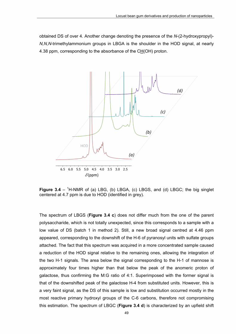
Locust bean gum derivatives and production of nanoparticles
49
obtained DS of over 4. Another change denoting the presence of the N-(2-hydroxypropyl)-
N,N,N-trimethylammonium groups in LBGA is the shoulder in the HOD signal, at nearly
4.38 ppm, corresponding to the absorbance of the CH(OH) proton.
Figure 3.4 – 1H-NMR of (a) LBG, (b) LBGA, (c) LBGS, and (d) LBGC; the big singlet centered at 4.7 ppm is due to HOD (identified in grey).
The spectrum of LBGS (Figure 3.4 c) does not differ much from the one of the parent
polysaccharide, which is not totally unexpected, since this corresponds to a sample with a
low value of DS (batch 1 in method 2). Still, a new broad signal centred at 4.46 ppm
appeared, corresponding to the downshift of the H-6 of pyranosyl units with sulfate groups
attached. The fact that this spectrum was acquired in a more concentrated sample caused
a reduction of the HOD signal relative to the remaining ones, allowing the integration of
the two H-1 signals. The area below the signal corresponding to the H-1 of mannose is
approximately four times higher than that below the peak of the anomeric proton of
galactose, thus confirming the M:G ratio of 4:1. Superimposed with the former signal is
that of the downshifted peak of the galactose H-4 from substituted units. However, this is
a very faint signal, as the DS of this sample is low and substitution occurred mostly in the
most reactive primary hydroxyl groups of the C-6 carbons, therefore not compromising
this estimation. The spectrum of LBGC (Figure 3.4 d) is characterized by an upfield shift

Locust bean gum derivatives and production of nanoparticles
50
of most of the signals and therefore not much information may be derived from it.
However, that is not the case of the 13C-NMR of this derivative (Figure 3.5), in which the
signals of the C-6 of galactose and unsubstituted mannose units, expected to appear at
61-62 ppm, are absent, in line with the full oxidation of these carbons estimated from the
elemental analysis data. Also a relatively broad signal appeared at nearly 176 ppm, due to
a poor resolution of the signals of the carboxylic acid carbons in galactose and mannose
units. Moreover, no signals attributable to aldehydic carbons (190-200 ppm) are observed
in the spectrum, thus confirming the complete oxidation to carboxylic acid.
Figure 3.5 – 13C-NMR of LBGC.
Table 3.2 presents the average molecular weights, polydispersity index (PdI), and radius
of gyration (Rg) of LBG and its derivatives. For the parent polysaccharide (LBG), these
are in general agreement with the literature (60, 74). Upon chemical modification, an
increase in both molecular weight and Rg was observed in LBGA, and a big decrease in
these parameters was patent in LBGC and LBGS. The increase identified in LBGA is
attributable to the presence of the introduced pendant chains, which lead to an increase in
the molar mass of the repeating unit and force the polymer, once in solution, and similarly
to what happens in the crystalline state (XRD results), to adopt a conformation that is
suitable to accommodate such bulky groups. The results observed in the LBGC and
LBGS derivatives suggest the occurrence of depolymerization during the chemical
modification, a common observation when the conditions of either the oxidation (181) or
the sulfation reaction (189) are applied. The latter was already stated in a similar
modification performed in pullulan (58). Moreover, no additional dehydration reactions,
with intra- and/or intermolecular crosslinking leading to a fraction of high molecular weight
chains, observed in sulfation reactions carried out at higher temperatures (188), occurred
in this case. The results obtained for LBGS were very similar among the two synthetic
methods (methods 1 and 2).
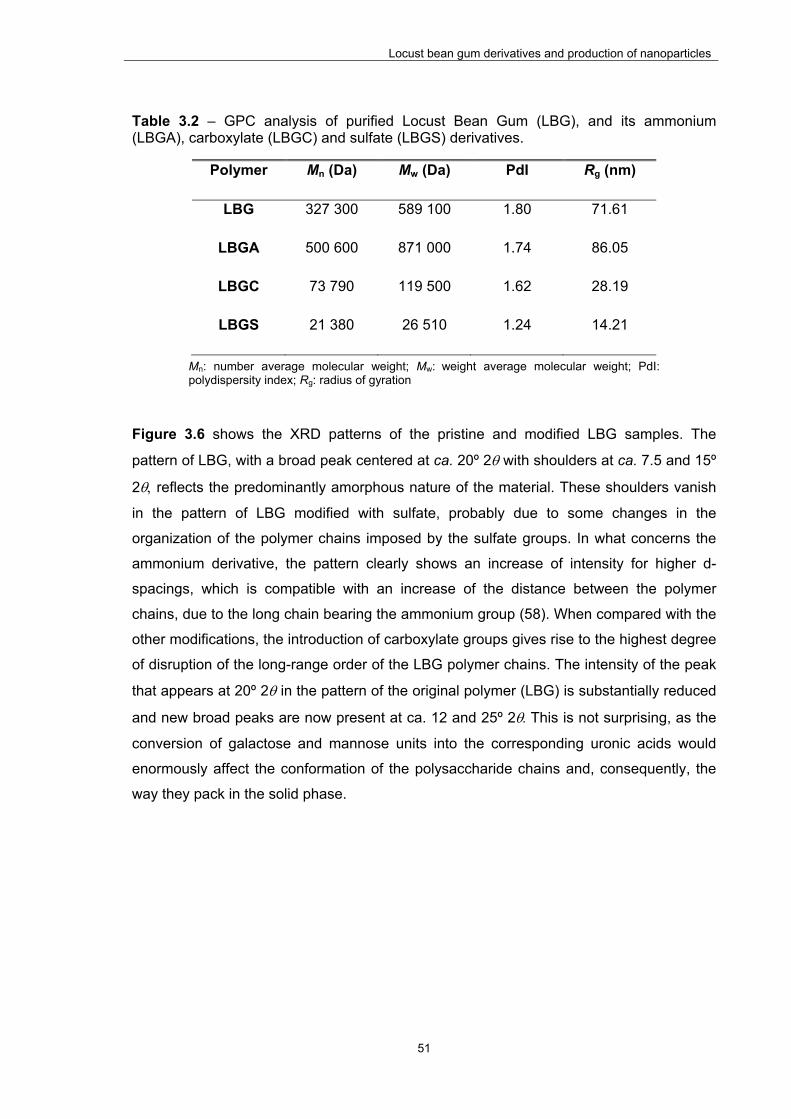
Locust bean gum derivatives and production of nanoparticles
51
Table 3.2 – GPC analysis of purified Locust Bean Gum (LBG), and its ammonium (LBGA), carboxylate (LBGC) and sulfate (LBGS) derivatives.
Polymer Mn (Da) Mw (Da) PdI Rg (nm)
LBG 327 300 589 100 1.80 71.61
LBGA 500 600 871 000 1.74 86.05
LBGC 73 790 119 500 1.62 28.19
LBGS 21 380 26 510 1.24 14.21
Mn: number average molecular weight; Mw: weight average molecular weight; PdI: polydispersity index; Rg: radius of gyration
Figure 3.6 shows the XRD patterns of the pristine and modified LBG samples. The
pattern of LBG, with a broad peak centered at ca. 20º 2 with shoulders at ca. 7.5 and 15º
2 reflects the predominantly amorphous nature of the material. These shoulders vanish
in the pattern of LBG modified with sulfate, probably due to some changes in the
organization of the polymer chains imposed by the sulfate groups. In what concerns the
ammonium derivative, the pattern clearly shows an increase of intensity for higher d-
spacings, which is compatible with an increase of the distance between the polymer
chains, due to the long chain bearing the ammonium group (58). When compared with the
other modifications, the introduction of carboxylate groups gives rise to the highest degree
of disruption of the long-range order of the LBG polymer chains. The intensity of the peak
that appears at 20º 2 in the pattern of the original polymer (LBG) is substantially reduced
and new broad peaks are now present at ca. 12 and 25º 2This is not surprising, as the
conversion of galactose and mannose units into the corresponding uronic acids would
enormously affect the conformation of the polysaccharide chains and, consequently, the
way they pack in the solid phase.

Locust bean gum derivatives and production of nanoparticles
52
Figure 3.6 – XRD patterns of (a) pristine locust bean gum (LBG), (b) sulfated LBG (LBGS), (c) ammonium LBG (LBGA), and (d) carboxylated LBG (LBGC).
3.3.2. Characterization of nanoparticles
The production of LBG derivatives described above endowed the polymer with charged
groups, enabling the preparation of nanoparticles by polyelectrolyte complexation. This is
a mild method occurring in hydrophilic medium, devoid of aggressive conditions such as
organic solvents or high shear forces, and involving electrostatic interactions between
oppositely charged polymers (108, 194). Three derivatives were synthesized which were
used in the production of different formulations of nanoparticles. The negatively charged
sulfate and carboxylate derivatives were complexed with chitosan to produce CS/LBGS
and CS/LBGC nanoparticles, respectively. In turn, the ammonium derivative was
complexed with the sulfate derivative in the innovative approach of producing LBG-only
nanoparticles (LBGA/LBGS). The results regarding the physicochemical characterization
of the referred nanoparticle formulations are displayed and discussed below.
5 15 25 35 452/degrees
a
b
cd

Locust bean gum derivatives and production of nanoparticles
53
3.3.2.1. CS/LBGS and CS/LBGC nanoparticles
The first approach towards the formulation of CS/LBGS and CS/LBGC nanoparticles
involved the production of carriers having higher or at least the same amount of LBG
derivative comparing to chitosan. In this regard, the starting mass ratios selected for the
production of the referred formulations of nanoparticles were 1:1, 1:1.5 and 1:2. In the
course of the experiments, the need to test other ratios was identified, not necessarily
being coincident for each formulation, thus justifying the slight differences observed
between the two formulations.
Table 3.3 displays the physicochemical characteristics of CS/LBGS nanoparticles. For the
production of these nanoparticles, LBGS corresponding to method 1 was used. With
CS/LBGS mass ratios varying between 1:1 and 1:2.5, and recalling that CS amount
remains constant in all formulations, it was verified that nanoparticle size generally
increased with increasing amounts of LBGS. The minimum size was 364 nm (CS/LBGS =
1:1, w/w) and the highest size was 589 nm (CS/LBGS = 1:2.5, w/w) (P < 0.05).
Table 3.3 - Physicochemical characteristics and production yield of CS/LBGS unloaded nanoparticles (mean ± SD; n ≥ 3). Different letters represent significant differences in each parameter (P < 0.05).
CS/LBGS
(w/w)
Size
(nm)
PdI Zeta potential
(mV)
Production
yield (%)
1:1 364.1 ± 30.0 a 0.34 ± 0.09 +45.6 ± 1.2 d 37.3 ± 5.6 h
1:1.25 403.7 ± 37.7 ab 0.40 ± 0.06 +40.0 ± 0.8 e 58.1 ± 2.7 i
1:1.5 pp* 1.0 ± 0.0 -5.9 ± 4.4 f n.d.
1:2 500.3 ± 59.6 bc 0.47 ± 0.08 -23.9 ± 2.7 g 56.6 ± 7.2 i
1:2.5 589.0 ± 69.5 c 0.54 ± 0.03 -28.5 ± 5.0 g n.d.
n.d.: not determined; pp: precipitate; *slight precipitation compromised the measurement of this parameter
The registered increase in size as higher amount of LBGS is included in the formulations
as compared with CS, might be explained by the increase of total mass of polymers that is
present. This effect was also reported in other works using the same nanoparticle
production method (39, 195). Precipitation was found to occur for an intermediate
formulation (CS/LBGS = 1:1.5, w/w), being coincident with a zeta potential close to zero (-
5.9 mV) that possibly is not sufficient to provide particle repulsion, thus leading to

Locust bean gum derivatives and production of nanoparticles
54
aggregation. A clear Tyndall effect was observed in all the other nanoparticle formulations.
The formulations 1:1 and 1:1.25 (w/w) exhibited a strong positive zeta potential of more
than 40 mV. The incorporation of a higher amount of LBGS, from formulation 1:1 to 1:1.25
(w/w) resulted in a corresponding decrease in the zeta potential from +46 mV to +40 mV
(P < 0.05). The formulations 1:2 and 1:2.5 (w/w) presented a complete shift in the zeta
potential as the nanoparticles became negatively charged with zeta potential reaching -29
mV. Again, the incorporation of a higher amount of LBGS led to a nominal decrease in the
zeta potential, although this is not statistically significant. This absolute shift of
nanoparticle charge occurring between the formulations 1:1.25 and 1:2 (w/w) reflects the
higher amount of LBGS that is present in the nanoparticles but also demonstrates that
both polymers have different charge density. Zeta potential results are perfectly in line
with the charge ratios that were calculated for each formulation of nanoparticles, as is
depicted in Figure 3.7. This figure shows the effect of charge ratios on the zeta potential
of CS/LBGS nanoparticles prepared with varying polymeric ratios.
Figure 3.7 – Effect of charge ratio (-/+) on the zeta potential of CS/LBGS nanoparticles.
For each polymer, by dividing the charge of the repeating unit by its molar mass, a charge
per mass ratio may be obtained. CS has a higher charge per mass ratio than LBGS (4.72
x 10-3 vs 3.00 x 10⁻³ charges/g, respectively), which justifies why formulations CS/LBGS =
1:1 and 1:1.25 (w/w) have a -/+ charge ratio below 1. The strong positive zeta potential (>
40 mV) of these nanoparticles is due to the predominance of positive charges. In turn, the
occurrence of precipitation in the formulation 1:1.5 (w/w) was coincident with a charge
‐40
‐30
‐20
‐10
0
10
20
30
40
50
60
0.0 0.5 1.0 1.5 2.0
Zeta potential (mV)
Charge ratio (‐/+)

Locust bean gum derivatives and production of nanoparticles
55
ratio around 1, justifying that the determined zeta potential was close to neutrality. In fact,
although a 1:1 -/+ charge stoichiometry might not imply the occurrence of complete
charge neutralization, due to different charge spacing in the intervenient species and to
steric limitations (195), one may assume a preferential interaction between the sulfate and
the ammonium groups, both weakly hydrated, instead of with the strongly hydrated
counterions (196). This leads to mainly an intrinsic charge matching in detriment of an
extrinsic charge compensation and, thus, to a small deviation from neutrality. Finally, the
continued addition of the negative polymer (formulations CS/LBGS = 1:2 and 1:2.5, w/w)
produced an excess of negative charges, resulting in -/+ charge ratio above 1 and,
consequently, negatively charged nanoparticles. A similar behavior concerning the charge
ratios leading to either the precipitation or the formation of nanoparticles, was previously
described (195).
The polydispersity index varied between 0.3 and 0.5, which is considered high. Regarding
the production yield, very reasonable values for this nanoparticle production methodology,
were obtained. A yield of 37% was registered for formulation 1:1 (w/w) which increased to
58% (P < 0.05) for formulation 1:1.25 (w/w). This is a result of the proper mechanism of
nanoparticle formation, based on the neutralization of chitosan amino groups by the
sulfate groups of LBGS. The incorporation of a higher amount of LBGS provides an
additional amount of sulfate groups that interacted with chitosan, thus forming a higher
amount of nanoparticles (106). However, this effect occurs up to a certain limit. As
observed, further increasing the amount of LBGS led to precipitation, certainly because of
the demonstrated neutralization of charges, as referred above. On keeping increasing
LBGS mass, nanoparticles are again formed (CS/LBGS 1:2 and 1:2.5, w/w), this time with
an opposite charge and a high yield (57% for formulation 1:2, w/w).
The results obtained for CS/LBGC nanoparticles were rather different comparing to those
described above regarding CS/LBGS formulations. In this case, as shown in Table 3.4,
the initially approached formulation of CS/LBGC 1:1 (w/w) resulted in a size of 479 nm,
which is more than 30% higher than the corresponding CS/LBGS formulation (P < 0.05).
The formulation 1:1.5 (w/w) already presented precipitation, similarly to 1:2 (w/w) and
therefore the intermediate formulation 1:1.25 (w/w) was produced.

Locust bean gum derivatives and production of nanoparticles
56
Table 3.4 - Physicochemical characteristics and production yield of CS/LBGC unloaded nanoparticles (mean ± SD; n ≥ 3). Different letters represent significant differences in each parameter (P < 0.05).
CS/LBGC
(w/w) Size (nm) PdI
Zeta potential
(mV)
Production
yield (%)
1:0.75 489.9 ± 63.6 a 0.45 ± 0.04 +45.5 ± 13.0 b 49.0 ± 5.0 d
1:1 479.1 ± 30.8 a 0.51 ± 0.07 +42.2 ± 7.4 b 54.3 ± 7.0 d
1:1.25 828.8 ± 299.8 a 0.64 ± 0.15 +28.8 ± 7.3 b n.d.
1:1.5 pp* 1.0 ± 0.0 -2.5 ± 8.3 c n.d.
1:2 pp* 1.0 ± 0.0 -15.2 ± 7.4 c n.d.
n.d.: not determined; pp: precipitate; *slight precipitation compromised the measurement of this parameter
The registered size revealed a strong increase to 829 nm, although this is not statistically
significant as is accompanied by an extremely high standard deviation, which indicates
reproducibility issues. This formulation also presented a high polydispersity index and thus
was not characterized for production yield. An attempt was also performed to produce
nanoparticles at a CS/LBGC ratio of 1:0.75 (w/w), but the characteristics were very
similar, under all aspects, to those of ratio 1:1 (w/w). The polydispersity index was around
0.5 – 0.6, which is even higher than those registered for CS/LBGS nanoparticles,
reinforcing the difficulty in producing suitable nanoparticles with the LBGC derivative. The
zeta potentials are highly positive (around +45 mV), which probably contributes to the
system stability. The determination of the charge ratios involved in each formulation of
nanoparticles is depicted in Figure 3.8.

Locust bean gum derivatives and production of nanoparticles
57
Figure 3.8 – Effect of charge ratio (-/+) on the zeta potential of CS/LBGC nanoparticles.
As observed, formulations 1:0.75 and 1:1 (w/w) have a -/+ charge ratio between 0.5 and
0.7 which does not translate into significant differences in the zeta potential. Nanoparticles
1:1.25 (w/w) displayed a -/+ charge ratio of 0.85 which induced a nominal decrease of the
zeta potential to +29 mV, although not to a statistically significant level. As observed
above for CS/LBGS nanoparticles, reaching a -/+ charge ratio around 1 (formulation 1:1.5,
w/w) resulted in precipitation. However, in this case the continued addition of the negative
polymer to formulate CS/LBGC = 1:2 (w/w) nanoparticles still resulted in precipitation,
despite the -/+ charge ratio of 1.4. It is important to highlight that, while the resulting zeta
potential for this formulation was of -15 mV, in the CS/LBGS corresponding formulation
was -24 mV, which possibly permitted enough repulsion to stabilize the formed
nanoparticles.
The determined production yields are satisfactory for this methodology, as referred above,
being around 50%. When comparing the zeta potentials of these nanoparticles with those
obtained for CS/LBGS nanoparticles (Table 3.3), a similar trend is observed. In this
regard, increasing the amount of LBGC present in the formulation reflects in a decrease of
the surface charge, owing to the higher amount of negative groups being incorporated.
Similarly to CS/LBGS nanoparticles, the formulation 1:1.5 is the one showing neutrality
(zeta potential of -2.5 mV) and the further incorporation of LBGC led to a decrease in the
surface charge. The precipitation verified for the latter was possibly due to the fact that the
existing surface charges were not sufficient to ensure particle repulsion. The resemblance
of the trend, particularly regarding the shift of the zeta potential (occurring for mass ratio of
‐20
‐10
0
10
20
30
40
50
60
70
0.0 0.2 0.4 0.6 0.8 1.0 1.2 1.4
Zeta potential (mV)
Charge ratio (‐/+)

Locust bean gum derivatives and production of nanoparticles
58
1:1.5), suggests the similarity of charge density in both derivatives. In fact, LBGS has a
charge per mass ratio of 3.00 x 10⁻³ charges/g, as stated before, and LBGC has 3.14 x 10-
3 charges/g.
3.3.2.2. LBGA/LBGS nanoparticles
One of the great novelties of producing LBG charged derivatives is the possibility of using
these to produce, for the first time, LBG-only nanoparticles. Given the difficulties in
producing nanoparticles with the LBGC derivative, as stated above, it was decided to
produce the LBG-only nanoparticles using just LBGS as negative counterpart. The
nanoparticles were produced by complexation of this derivative (method 2 – 50/50
mixtures of batches 2 and 3) with the ammonium derivative (LBGA) by the same
methodology reported in the other cases (polyelectrolyte complexation).
After observing the precipitation of the formulation LBGA/LBGS 1:1 (w/w), possibly
resulting from a (-/+) charge ratio of 1.09, formulations 1:2 (w/w) and 2:1 (w/w) were
approached, which results are depicted in Table 3.5.
Table 3.5 - Physicochemical characteristics and production yield of LBGA/LBGS unloaded nanoparticles (mean ± SD; n ≥ 3). Different letters represent significant differences in each parameter (P < 0.05).
LBGA/LBGS
(w/w)
Size (nm) PdI Zeta potential
(mV)
Production
yield (%)
1:2 206.6 ± 5.0 a 0.13 ± 0.03 -27.8 ± 1.4 c 30.0 ± 8.6 e
1:1 pp - - -
2:1 368.3 ± 19.3 b 0.38 ± 0.05 +48.1 ± 1.5 d 16.7 ± 3.8 f
pp: precipitate
The formulation containing the highest amount of LBGS registered a size of 207 nm and a
low polydispersity index of 0.13. Naturally, the zeta potential was negative (-28 mV),
reflecting the higher content of negatively charged derivative, which translated into a (-/+)
charge ratio of 2.17. As expected, the formulation having more LBGA exhibited a strongly
positive zeta potential (+48 mV; P < 0.05), as a result of the (-/+) charge ratio of 0.54.
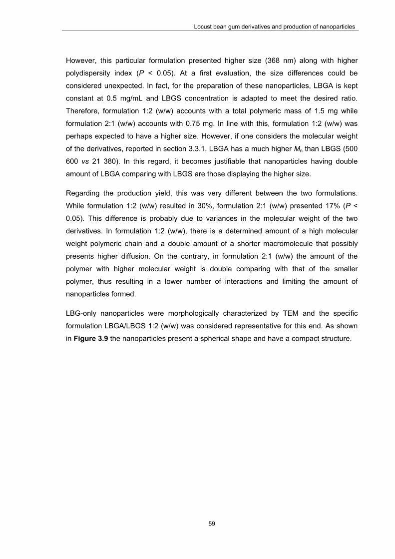
Locust bean gum derivatives and production of nanoparticles
59
However, this particular formulation presented higher size (368 nm) along with higher
polydispersity index (P < 0.05). At a first evaluation, the size differences could be
considered unexpected. In fact, for the preparation of these nanoparticles, LBGA is kept
constant at 0.5 mg/mL and LBGS concentration is adapted to meet the desired ratio.
Therefore, formulation 1:2 (w/w) accounts with a total polymeric mass of 1.5 mg while
formulation 2:1 (w/w) accounts with 0.75 mg. In line with this, formulation 1:2 (w/w) was
perhaps expected to have a higher size. However, if one considers the molecular weight
of the derivatives, reported in section 3.3.1, LBGA has a much higher Mn than LBGS (500
600 vs 21 380). In this regard, it becomes justifiable that nanoparticles having double
amount of LBGA comparing with LBGS are those displaying the higher size.
Regarding the production yield, this was very different between the two formulations.
While formulation 1:2 (w/w) resulted in 30%, formulation 2:1 (w/w) presented 17% (P <
0.05). This difference is probably due to variances in the molecular weight of the two
derivatives. In formulation 1:2 (w/w), there is a determined amount of a high molecular
weight polymeric chain and a double amount of a shorter macromolecule that possibly
presents higher diffusion. On the contrary, in formulation 2:1 (w/w) the amount of the
polymer with higher molecular weight is double comparing with that of the smaller
polymer, thus resulting in a lower number of interactions and limiting the amount of
nanoparticles formed.
LBG-only nanoparticles were morphologically characterized by TEM and the specific
formulation LBGA/LBGS 1:2 (w/w) was considered representative for this end. As shown
in Figure 3.9 the nanoparticles present a spherical shape and have a compact structure.

Locust bean gum derivatives and production of nanoparticles
60
Figure 3.9 – TEM microphotograph of LBGA/LBGS = 1:2 (w/w) nanoparticles.
3.3.3. Safety evaluation
Addressing the biocompatibility of materials to be used in drug delivery is a major issue in
formulation development (52, 197, 198). Additionally, current international guidelines
indicate the need to contextualize biocompatibility with a specific route of administration
and dose of the material (198). According to the guidelines issued by the International
Organization for Standardization (ISO), testing biocompatibility implies the performance of
a complete set of assays, addressing at first cellular morphology, membrane integrity and
metabolic efficiency, among other tests (197, 199-201). In this work we performed two of
the most used assays to test the toxicological effect of materials, which are the metabolic
assay MTT and the membrane integrity assay based on LDH release. The MTT assay
assesses cell metabolic efficiency, relying on the evaluation of enzymatic function. To do
so, after the exposure to the test materials, cells are incubated with yellow tetrazolium
(MTT) salts which are reduced to purple-blue formazan crystals by active mitochondrial
dehydrogenases (197). In this manner, a higher concentration of the formazan dye
corresponds to a higher amount of metabolically active cells, which is usually interpreted
as higher cell viability. The LDH release assay provides a mean to determine the amount
of LDH in cell culture medium upon exposure to potential toxicants. As LDH is a
cytoplasmic enzyme, its presence in the cell culture medium is an indicator of irreversible
cell death due to cell membrane damage (202, 203). The assay thus evaluates cell
membrane integrity and complements the results obtained by the MTT assay.

Locust bean gum derivatives and production of nanoparticles
61
Caco-2 cells are derived from a human colorectal carcinoma and are the most frequently
used as in vitro intestinal cell culture model. The epithelial cells in the intestinal region are
a heterogeneous population of cells that include enterocytes or absorptive cells, goblet
cells that secrete mucin, endocrine cells and M cells, among others. The most common
epithelial cells are the enterocytes that are responsible for the majority of the absorption of
both nutrients and drugs in the small intestine (204, 205). Despite their colonic origin,
Caco-2 cells undergo spontaneous differentiation in culture conditions to assume the
characteristics of small intestinal cells. These include morphological and functional
attributes, rendering the cell line a model of mature enterocytes (206). Owing to these
features, Caco-2 cells are the most reported in the studies of drug absorption and toxicity
(207, 208).
In this work, Caco-2 cells were used to evaluate the toxicological profile of LBG and the
synthesized derivatives. Figure 3.10 and Figure 3.11 represent the Caco-2 cell viability
obtained after exposure to the mentioned materials at different concentrations, for a
period of 3 h and 24 h. Cell viability values were calculated in relation to the 100% cell
viability considered for the incubation with DMEM (negative control of cell death). The
evaluation of LBG-based samples generally evidenced a mild effect on cell viability,
considered to be devoid of biological relevance. In fact, with the exception of LBGA, all the
other samples resulted in viabilities above 70% after 3 h or 24 h of exposure, when tested
at concentrations varying within 0.1 and 1.0 mg/mL. While at 3 h values remained above
88% in all conditions, the prolonged exposure until 24 h induced slight alterations.
However, these were in most cases devoid of physiological relevance and the only
remarkable effect resides in the decrease of the viability induced by the contact with
LBGC at the highest concentration tested (1.0 mg/mL) (P < 0.05) to a value around 70%.
Importantly, this is the value considered by ISO 10993-5 (201) as the level below which a
toxic effect is assumed to occur.
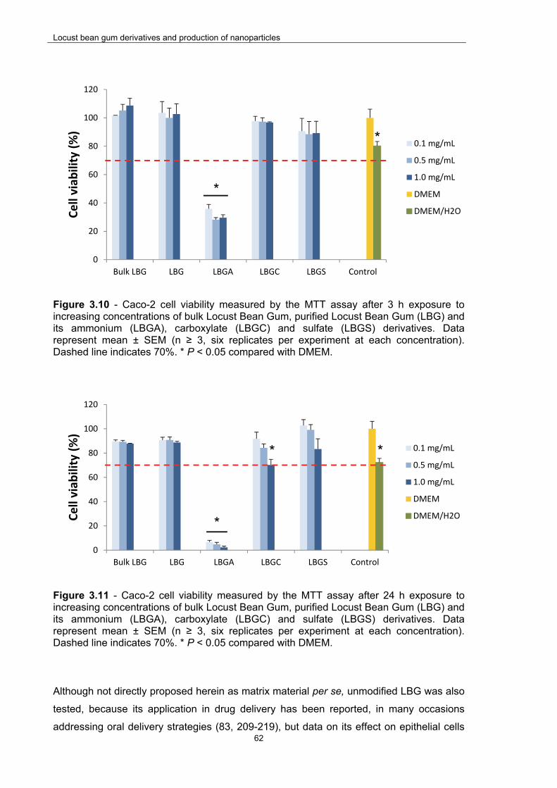
Locust bean gum derivatives and production of nanoparticles
62
Figure 3.10 - Caco-2 cell viability measured by the MTT assay after 3 h exposure to increasing concentrations of bulk Locust Bean Gum, purified Locust Bean Gum (LBG) and its ammonium (LBGA), carboxylate (LBGC) and sulfate (LBGS) derivatives. Data represent mean ± SEM (n ≥ 3, six replicates per experiment at each concentration). Dashed line indicates 70%. * P < 0.05 compared with DMEM.
Figure 3.11 - Caco-2 cell viability measured by the MTT assay after 24 h exposure to increasing concentrations of bulk Locust Bean Gum, purified Locust Bean Gum (LBG) and its ammonium (LBGA), carboxylate (LBGC) and sulfate (LBGS) derivatives. Data represent mean ± SEM (n ≥ 3, six replicates per experiment at each concentration). Dashed line indicates 70%. * P < 0.05 compared with DMEM.
Although not directly proposed herein as matrix material per se, unmodified LBG was also
tested, because its application in drug delivery has been reported, in many occasions
addressing oral delivery strategies (83, 209-219), but data on its effect on epithelial cells
0
20
40
60
80
100
120
Bulk LBG LBG LBGA LBGC LBGS Control
Cell viab
ility (%)
0.1 mg/mL
0.5 mg/mL
1.0 mg/mL
DMEM
DMEM/H2O
*
0
20
40
60
80
100
120
Bulk LBG LBG LBGA LBGC LBGS Control
Cell viab
ility (%)
0.1 mg/mL
0.5 mg/mL
1.0 mg/mL
DMEM
DMEM/H2O*
* *
*

Locust bean gum derivatives and production of nanoparticles
63
are not available on the literature. Moreover, a comparison between bulk LBG and LBG
was performed, revealing no significant differences, which indicates an absence of effect
of the purification process in the cytotoxic profile of the material. It is important to mention
that the results shown for LBGS sample correspond to the derivative obtained by the
second method of synthesis (method 2 – batch 1), which were similar to those registered
for the derivative obtained in the first method (method 1; data not shown).
As mentioned before, LBGA is the material that presents the most distinct behavior,
appearing as the exception to the mild effect observed for the tested materials. In fact, as
shown in Figure 3.10, a strong decrease of cell viability to approximately 30% is obtained
for any of the tested concentrations already upon a 3 h exposure. The effect is even more
drastic after 24 h, when a very low level of cell survival was registered (P < 0.05; Figure
3.11). Regarding concentration, there are no evidences of a statistically significant
concentration-dependent effect. The influence of surface charges on cytotoxicity remains
largely unresolved and sometimes the literature reports contradictory results. This is
possibly due to different characteristics of basic materials being used and also to
dissimilar assay conditions, which are frequently not described in sufficient detail.
Nevertheless, there are many indications suggesting that surface charge has a role on
cellular uptake (220, 221) and on the toxicological effect of substances. In this context,
positively charged materials have been frequently found to be more cytotoxic than neutral
or negatively charged counterparts, because positive charges provide a means for
stronger interaction with cell surfaces, in many cases associated with internalization of the
material (221-225). These statements are coincident with the results of our work, since the
neutral (bulk LBG and LBG) and negatively charged materials (LBGC and LBGS) are
devoid of a toxic effect. Another parameter that could be indicated as playing a significant
role on toxicity consists on the molecular weight of the polymers. In this regard, although it
could be suggested that smaller sizes have higher probability to be internalized by the
cells, the literature has been reporting no correlation (226). In this work, the molecular
weight of the polymers also seems to not be driving the cytotoxic behavior, as LBGS is the
smallest molecule and shows no toxic effect.
Comparing to LBGA, a very similar toxicological profile was observed for an ammonium
derivative of another polysaccharide, pullulan, which was synthesized using the same
methodology (58, 59). In that case, the assessment was performed in Calu-3 cells
(bronchial cell line) and cell viabilities around 50-60% were observed after 3 h, decreasing
to 40% at 24 h. Although a time-dependent effect is also clearly observed, the effect on
cell viability is not as strong as for LBGA. The first consideration to take into account is the
fact that the assessment was performed in different cell lines, which might translate into

Locust bean gum derivatives and production of nanoparticles
64
different sensitivity. Additionally, different charge density of the polymers might be
indicated as possible justification. In this regard, LBGA has a DS of 4.24, while the
corresponding pullulan derivative (ammonium pullulan) has a DS of 2 (58). A higher
number of positive charges results in stronger interactions and, thus, in lower cell viability.
Complementing this idea, a work reporting the cytotoxic effect of cationic pullulan
microparticles on human leukemic K562(S) cells, has established that toxicity increased
with the increase molar concentration of amino groups (227). In the work reporting the
cytotoxic evaluation of pullulan derivatives, a sulfate derivative of that polysaccharide was
also assessed. Similarly to what was observed for LBGS, the registered cell viability was
well above 80% (59).
Considering that polymer samples were solubilized in water and diluted with cell culture
medium prior to incubation with the cells, an additional control was performed consisting
in a mixture of DMEM and H2O in the same ratio used for the samples. This enables a real
evaluation of the contribution of the polymers on the final cell viability. As observed in
Figure 3.10 and Figure 3.11, the cell viability induced by this control varied between 72%
and 80%. Upon 3 h of contact there is a statistically significant difference between the
control (DMEM + H2O) and all samples but with LBGS (Figure 3.10). In fact, higher cell
viability is observed upon exposure to bulk LBG, LBG and LBGC, suggesting a positive
effect of the presence of the polymers. Interestingly, after 24 h exposure (Figure 3.11), a
shift is observed in the effect induced by LBGC and LBGS. In the former, the prolonged
contact with the cells at the two highest concentrations reverts the positive effect on cell
viability observed at 3 h. For LBGS, the results demonstrate that at the two lowest
concentrations, the more prolonged contact improves cell viability, which was not
registered at 3 h.
One of the most important information provided by the evaluation performed with the MTT
assay, is that only the more prolonged exposure to the highest concentration tested (1.0
mg/mL; 24 h) induced a relevant decrease of Caco-2 cell viability (exception for LBGA).
Therefore, it was deemed important to complement the results at these conditions by
means of the quantification of the amount of LDH released by Caco-2 cells. To perform
this assay, DMEM was used as negative control of LDH release and a lysis buffer was
used as positive control. Thus, the negative control (DMEM) corresponds to a normal cell
death, while the positive control (lysis buffer) represents 100% cell death.
Figure 3.12 shows the results of LDH release after 24 h exposure to the materials at the
concentration of 1.0 mg/mL. No statistically significant differences were observed between
the negative control (DMEM), bulk LBG, LBG, LBGC and LBGS, which means that these

Locust bean gum derivatives and production of nanoparticles
65
materials do not compromise Caco-2 cell membrane integrity, as no increased LDH
release was induced as compared with that observed upon incubation with cell culture
medium (DMEM). On the contrary, the contact with LBGA resulted in 90% LDH release,
which is considered comparable to that induced by the lysis buffer, thus indicating a high
cytotoxic effect that results in cell membrane disruption. The results obtained in this assay
reinforce those found in the MTT tests, confirming the high cytotoxicity of LBGA.
Figure 3.12 – Caco-2 cell viability measured by the LDH release assay after 24 h exposure to 1 mg/mL solutions of bulk Locust Bean Gum, purified Locust Bean Gum (LBG) and its ammonium (LBGA), carboxylate (LBGC) and sulfate (LBGS) derivatives. Data represent mean ± SEM (n ≥ 3, three replicates per experiment). * P < 0.05 compared with DMEM.
The overall results obtained with these complementary cytotoxicity assays indicate that,
with the exception of LBGA, LBG and negatively charged derivatives, present no
cytotoxicity towards this in vitro intestinal model. This was observed even for the highest
concentration tested (1.0 mg/mL) and for prolonged contact (24 h), suggesting their
relative safety for an application as matrix materials of oral drug delivery systems.
Complementarily, the effect on cell viability provided by LBGS (method 2 – batch 1) was
assessed in Calu-3 and A549 cells (respiratory epithelial cells) and the results, displayed
in Figure 3.13 and Figure 3.14, are in line with those observed for Caco-2 cells.
0
20
40
60
80
100
DMEM Bulk LBG LBG LBGA LBGS LBGC
LDH release (%)
*
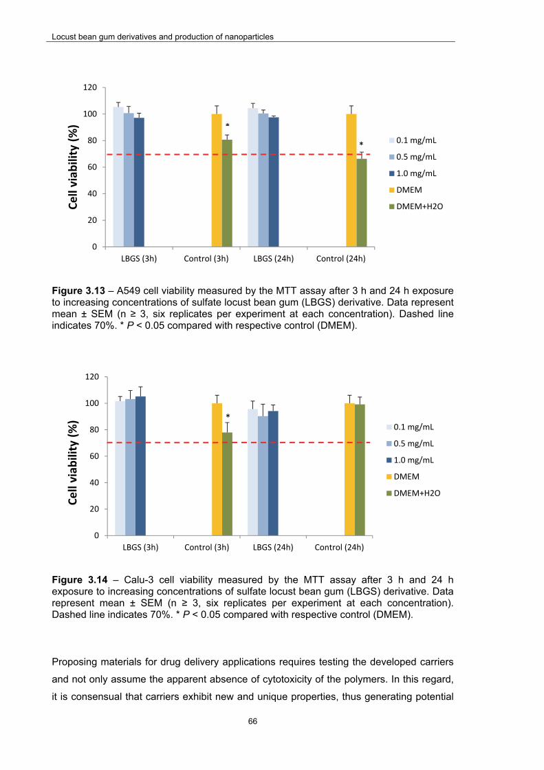
Locust bean gum derivatives and production of nanoparticles
66
Figure 3.13 – A549 cell viability measured by the MTT assay after 3 h and 24 h exposure to increasing concentrations of sulfate locust bean gum (LBGS) derivative. Data represent mean ± SEM (n ≥ 3, six replicates per experiment at each concentration). Dashed line indicates 70%. * P < 0.05 compared with respective control (DMEM).
Figure 3.14 – Calu-3 cell viability measured by the MTT assay after 3 h and 24 h exposure to increasing concentrations of sulfate locust bean gum (LBGS) derivative. Data represent mean ± SEM (n ≥ 3, six replicates per experiment at each concentration). Dashed line indicates 70%. * P < 0.05 compared with respective control (DMEM).
Proposing materials for drug delivery applications requires testing the developed carriers
and not only assume the apparent absence of cytotoxicity of the polymers. In this regard,
it is consensual that carriers exhibit new and unique properties, thus generating potential
0
20
40
60
80
100
120
LBGS (3h) Control (3h) LBGS (24h) Control (24h)
Cell viab
ility (%)
0.1 mg/mL
0.5 mg/mL
1.0 mg/mL
DMEM
DMEM+H2O
0
20
40
60
80
100
120
LBGS (3h) Control (3h) LBGS (24h) Control (24h)
Cell viab
ility (%)
0.1 mg/mL
0.5 mg/mL
1.0 mg/mL
DMEM
DMEM+H2O
*
*
*

Locust bean gum derivatives and production of nanoparticles
67
different risks as compared to the raw materials of the same chemistry (228), as observed
in other works (58, 59). In this regard, in addition to the evaluation of the polymer and the
synthesized derivatives, a preliminary evaluation of LBG-based nanoparticles was further
performed using the MTT assay. Although several formulations were proposed and
developed herein, that corresponding to LBG-only nanoparticles was selected for this step
due to the novelty of the polymer in nanoparticle production.
The viability of Caco-2 cells upon exposure to LBGA/LBGS nanoparticles is shown in
Figure 3.15 (3 h) and Figure 3.16 (24 h). The two developed formulations (LBGA/LBGS
2:1 and 1:2, w/w) were assessed. For formulation 2:1 (w/w) the comparison of results
obtained for each tested time revealed a statistically significant difference between
concentrations 0.1 and 1.0 mg/mL (P < 0.05). Formulation 1:2 (w/w) did not evidence
significant differences between all concentrations at the two tested times. A similar
observation was made after comparing the same concentrations for different times (3 h
and 24 h). The most remarkable result is that no significant effect on cell viability is
observed for both formulations at all concentrations, up to 24 h. Actually, the registered
viability was over 80% in all cases, which, as said before, is considered very acceptable
according to the ISO10993-5 (201).
Figure 3.15 – Caco-2 cell viability measured by the MTT assay after 3 h exposure to increasing concentrations of ammonium Locust Bean Gum (LBGA) derivative, sulfate Locust Bean Gum (LBGS) derivative and LBGA/LBGS nanoparticles (NP). Data represent mean ± SEM (n ≥ 3, six replicates per experiment at each concentration). Dashed line indicates 70%. * P < 0.05 compared with DMEM.
0
20
40
60
80
100
120
LBGA LBGS NP 2:1 NP 1:2 Control
Cell viab
ility (%)
0.1 mg/mL
0.5 mg/mL
1.0 mg/mL
DMEM
DMEM/H2O
*
* *
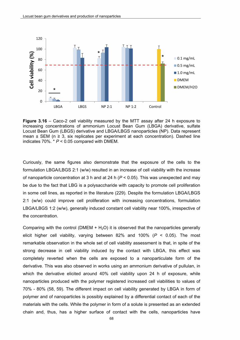
Locust bean gum derivatives and production of nanoparticles
68
Figure 3.16 – Caco-2 cell viability measured by the MTT assay after 24 h exposure to increasing concentrations of ammonium Locust Bean Gum (LBGA) derivative, sulfate Locust Bean Gum (LBGS) derivative and LBGA/LBGS nanoparticles (NP). Data represent mean ± SEM (n ≥ 3, six replicates per experiment at each concentration). Dashed line indicates 70%. * P < 0.05 compared with DMEM.
Curiously, the same figures also demonstrate that the exposure of the cells to the
formulation LBGA/LBGS 2:1 (w/w) resulted in an increase of cell viability with the increase
of nanoparticle concentration at 3 h and at 24 h (P < 0.05). This was unexpected and may
be due to the fact that LBG is a polysaccharide with capacity to promote cell proliferation
in some cell lines, as reported in the literature (229). Despite the formulation LBGA/LBGS
2:1 (w/w) could improve cell proliferation with increasing concentrations, formulation
LBGA/LBGS 1:2 (w/w), generally induced constant cell viability near 100%, irrespective of
the concentration.
Comparing with the control (DMEM + H2O) it is observed that the nanoparticles generally
elicit higher cell viability, varying between 82% and 100% (P < 0.05). The most
remarkable observation in the whole set of cell viability assessment is that, in spite of the
strong decrease in cell viability induced by the contact with LBGA, this effect was
completely reverted when the cells are exposed to a nanoparticulate form of the
derivative. This was also observed in works using an ammonium derivative of pullulan, in
which the derivative elicited around 40% cell viability upon 24 h of exposure, while
nanoparticles produced with the polymer registered increased cell viabilities to values of
70% - 80% (58, 59). The different impact on cell viability generated by LBGA in form of
polymer and of nanoparticles is possibly explained by a differential contact of each of the
materials with the cells. While the polymer in form of a solute is presented as an extended
chain and, thus, has a higher surface of contact with the cells, nanoparticles have
0
20
40
60
80
100
120
LBGA LBGS NP 2:1 NP 1:2 Control
Cell viab
ility (%)
0.1 mg/mL
0.5 mg/mL
1.0 mg/mL
DMEM
DMEM/H2O*
**

Locust bean gum derivatives and production of nanoparticles
69
comparatively a lower contact. Additionally, the number of positive charges available for
interaction with the negatively charged cells upon complexation with LBGS is significantly
decreased, thus decreasing the potential toxicity (226). This reinforces the need to
evaluate separately the carriers and the raw materials, as the former may exhibit different
properties, that may encompass different risks (228).
These preliminary results suggest an absence of overt toxicity of LBG-only nanoparticles,
thus potentiating possible applications, although it is recognized that further studies need
to be performed to reach a more accurate conclusion in this regard.
3.4. Conclusions
LBG demonstrated to be a good substrate for the production of charged derivatives,
permitting the synthesis of ammonium, sulfated and carboxylated LBG. Several
characterization techniques were used to confirm the presence of the new chemical
groups introduced in each new derivative.
Using a method of polyelectrolyte complexation, the produced derivatives were applied in
the preparation of different formulations of LBG-based nanoparticles, reported herein for
the first time. When the negatively charged derivatives (sulfated and carboxylated LBG)
were used, chitosan was the applied positively charged polyelectrolite. In turn, ammonium
LBG was complexed with sulfated LBG to obtain LBG-only nanoparticles. The
physicochemical characteristics of nanoparticles were highly dependent on their
composition and on the charge ratios applied in each complexation being performed.
Generally, the observed characteristics, with sizes around 200-400 nm in certain cases,
and tailorable zeta potential according to setup conditions, are suggested as adequate for
drug delivery applications.
A preliminary toxicological evaluation of LBG derivatives and the produced nanoparticles
was performed, assessing both the metabolic activity and the cell membrane integrity of
representative intestinal cells (Caco-2 cells) after an exposure of up to 24 h to
concentrations as higher as 1 mg/mL. Severe cytotoxicity was found for the ammonium
derivative of LBG, but this was clearly reverted after the assembly of nanoparticles, which
evidenced a very mild effect on Caco-2 cell viability. The results as a whole indicate the
possibility to use the synthesized LBG derivatives to produce nanoparticles for drug
delivery applications.

Locust bean gum nanoparticles: Application in oral immunization
70
This page was intentionally left in blank
Locust bean gum nanoparticles: Application in oral immunization
71
CHAPTER 4
LOCUST BEAN GUM NANOPARTICLES: APPLICATION IN ORAL IMMUNIZATION
The information presented in this chapter was submitted for publication in International
Journal of Pharmaceutics:
Braz, L., Camacho, A., Grenha, A., Ferreira, D., Rosa da Costa, A.M., Gamazo, C.,
Sarmento, B., Chitosan/Sulfated Locust Bean Gum nanoparticles: in vitro and in vivo
evaluation towards an application in oral immunization, submitted for publication
Locust bean gum nanoparticles: Application in oral immunization
72
This page was intentionally left in blank
Locust bean gum nanoparticles: Application in oral immunization
73
4. Chitosan/Sulfated Locust Bean Gum nanoparticles: in vitro and in vivo
evaluation towards an application in oral immunization
4.1. Introduction
The search for alternative vaccination approaches that may circumvent the limitations of
parenteral delivery is not new. Mucosal vaccination has thus been gaining popularity in
the recent decades and some mucosal vaccines are currently available in the market
(175, 230). Apart from leading to higher patient’s compliance, as the discomfort
associated with parenteral administration is avoided, mucosal vaccines permit an easier
chain of distribution, mainly because a cold-chain is not necessary, which is relevant for
developing countries (119, 137). The oral route is the one gathering higher interest
concerning this alternative vaccination concept, not only because of the general
advantages associated with oral administration, but also due to relevant features of the
intestine for immunization. In this regard, a special mention is due to the gut associated
mucosal tissue (GALT), which has sites where immune responses are initiated and
effector sites where adaptive immune responses are executed (175). The Peyer’s patches
existing in the GALT comprise the main location of immune cells associated to the
intestinal mucosa (154) and are separated from the intestinal lumen by the follicle
associated epithelium (FAE) (159). FAE is composed of enterocytes, goblet cells and
microfold cells (M cells) (136, 154), the latter being reported to have a thinner mucus
layer, and good ability for antigen uptake and transport to antigen presenting cells (APCs)
(231). Apart from these anatomical specificities, mucosal sites are considered locations of
primary access for most human pathogens (138), many accessing the organism through
the mouth. As mucosal immunization is expected to elicit both systemic and mucosal
immunity, the development of the latter at the intestinal level becomes a relevant tool
towards limiting or preventing pathogen entry, thus inhibiting the consequent infection
(139, 140).
Notwithstanding the evident ability of the intestinal area for antigen recognition, antigens
are biopharmaceuticals, thus being highly sensitive molecules that require specific
precautions regarding their formulation and delivery. Indeed, the protein-based structure
hinders the possibility of a direct oral administration, mainly because of the low gastric pH
and the high content of proteases (232, 233). Suitable particles are therefore demanded
for a successful oral immunization approach and nanoparticles have been indicated many
times as very useful in mediating this process. Apart from providing the associated
antigens with protection from the harsh gastrointestinal conditions referred above, their

Locust bean gum nanoparticles: Application in oral immunization
74
small size is expected to permit an intimate contact with the epithelial surface, where cells
with relevant roles in the generation of an immune response are located (234). In addition,
nanoparticles may also act as immunomodulator adjuvants, meaning that the particles
further mediate the development of the immune response. The particles may facilitate
both the antigen uptake and internalization by GALT and, also, the antigenic cross-
presentation by APCs (235). The use of The use of particle vehicles exhibiting targeting
moieties that have a favoured interaction with epithelial glycoconjugates that are
specifically activated by pathogens, such as the TLR family or the mannose receptor, has
been proposed as a strategy that mimics microbial behaviour in the development of
immune responses (235).
The uptake of particles is reported to primarily occur via the M cells (175), which have
been referred to provide a privileged contact with mannose residues (80, 169) and,
therefore, might be used as privileged target for mannose-containing particles. Locust
bean gum (LBG) is a polysaccharide of the class of galactomannans, thus having a
chemical structure composed of both galactose and mannose units (64), as depicted in
Figure 1.2, from chapter 1 (general introduction). Therefore, it potentially has the ability to
provide the said privileged contact between the particle and the M cells.
This paper proposes the design of nanoparticles based on LBG and chitosan, another
polysaccharide, to be used as antigen particles for oral immunization purposes. A
negatively charged derivative of LBG was produced (sulfated LBG) to enable the
production of nanoparticles by polyelectrolyte complexation. Two model antigens were
associated to the nanoparticles, an antigenic complex of Salmonella enterica serovar
Enteritidis and ovalbumin; and in vivo studies were performed.
4.2. Materials and methods
4.2.1. Materials
Chitosan (CSup) in the form of hydrochloride salt (Protasan® UP Cl 113, deacetylation
degree = 75% – 90%, molecular weight < 200 kDa), was purchased from Pronova
Biopolymer (Sandvika, Norway). Locust bean gum (LBG) was a kind gift from Industrial
Farense (Faro, Portugal). Immunogenic subcellular extract obtained from
whole Salmonella Enteritidis cells (HE) was kindly provided by Professor Carlos Gamazo
(University of Navarra, Spain). Ovalbumin (OVA), bovine serum albumin (BSA),
phosphotungstate dibasic hydrate, glycerol, phosphate buffered saline (PBS) pH 7.4

Locust bean gum nanoparticles: Application in oral immunization
75
tablets, Dulbecco’s modified Eagle’s medium (DMEM), penicillin/streptomycin (10000
units/mL, 10000 µg/mL), non-essential amino acids, L-glutamine 200 mM, trypsin-EDTA
solution (2.5 g/L trypsin, 0.5 g/L EDTA), trypan blue solution (0.4%), thiazolyl blue
tetrazolium bromide (MTT), lactate dehydrogenase (LDH) kit, sodium dodecyl sulfate
(SDS), Ponceau S red staining solution, protease inhibitor cocktail, dimethyl sulfoxide
(DMSO), HCl 37%, H2O2, 4-chloro-1-naphtol, NaCl, KH2PO4 and NaOH were purchased
from Sigma-Aldrich (Germany). XT sample buffer, Criterion XT bis-tris gel, XT MOPS
running buffer, Coomassie blue and Tris-glycine buffer were provided by Bio-Rad (USA)
and PBS-tween (PBS-T) and 3-ethylbenzthiazoline-6-sulfonic acid (ABTS) by VWR
(Portugal). Fetal bovine serum (FBS) was obtained from Gibco (USA), molecular mass
markers (Novex Sharp Pre-stained Protein Standard) from Invitrogen (Germany).
Peroxidase-conjugated goat anti-mouse IgG1, IgG2a and IgA antibodies were purchased
from Nordic Immunology (Netherlands) and skimmed milk from Continente (Portugal).
Ultrapure water (Mili-Q Plus, Milipore Iberica, Madrid, Spain) was used throughout. All
other chemicals were reagent grade.
4.2.2. Cell line
The Caco-2 cell line was obtained from the American Type Culture Collection (Rockville,
USA) and used between passages 77-93. Cell cultures were grown in 75 cm2 flasks in a
humidified 5% CO2/95% atmospheric air incubator at 37 ºC. Cell culture medium was
DMEM supplemented with 10% (v/v) FBS, 1% (v/v) L-glutamine solution, 1% (v/v) non-
essential amino acids solution and 1% (v/v) penicillin/streptomycin. Medium was changed
every 2-3 days and cells were subcultured weekly.
4.2.3. Production of Locust Bean Gum-based nanoparticles
All nanoparticles (NP) were prepared by polyelectrolyte complexation method which
consists in the electrostatic interaction between the positive and negative charges of the
different polymers (35).

Locust bean gum nanoparticles: Application in oral immunization
76
4.2.3.1. CSup/LBGS nanoparticles
Nine mass ratios of CSup/LBGS (1:1; 1:1.25; 1:1.5; 1:1.75; 1:2; 1:3; 1:3.25; 1:3.5 and 1:4)
were used to prepare the nanoparticles by polyelectrolyte complexation. The stock
solution of CSup, dissolved in ultrapure water, was prepared to reach a final concentration
of 1.0 mg/mL, while LBGS was dissolved in ultrapure water, at final concentration of 4.0
mg/mL. The solutions were filtered with a 0.45 µm filter prior to use. The formulations
were prepared by slowly adding 1.8 ml of LBGS to 1.0 ml of CSup under gentle magnetic
stirring at room temperature, as shown in Figure 1.1 (chapter 1, general introduction). The
concentration of CSup was kept constant at 1.0 mg/mL for the preparation of all
formulations, but that of LBGS was modified to obtain the different ratios.
After the addition of the LBGS solutions to the CSup solution was possible to see, almost
immediately, the Tyndall effect evidencing the formation of nanoparticles. The
suspensions of nanoparticles were mixed by magnetic stirring for 10 min and then
centrifuged in eppendorfs with a layer of 10 µL of glycerol, in order to facilitate the
following step of ressuspension. The isolation of nanoparticles was performed by
centrifugation (Thermo Scientific-Heraeus Fresco 17, Germany) at 16 000 x g, for 30 min
at 15 ºC. After discarding the supernatants, the nanoparticles were ressuspended with
200 µL of ultrapure water.
4.2.3.2. Association of a bacterial antigenic complex to CSup/LBGS
nanoparticles
An antigenic complex, HE, consisting of vesicles of outer membrane, was obtained from
Salmonella Enteritidis. Its association was performed to the CSup/LBGS 1:1.5 and 1:2
(w/w) nanoparticle formulations, which selection was mainly driven by the production
yield. The stock solution of the HE antigenic complex was prepared by dissolving it in
ultrapure water (0.4 mg/mL) using the ultra-sound bath during 15 min at room
temperature. The solution was filtered with a 0.22 µm low protein binding filter (Millex® -
GV, Millipore, Spain) prior to use. The NP-HE were prepared using the same methodology
used for the unloaded nanoparticles, but the concentration of LBGS solutions were
adjusted using different concentrations of HE solutions, in order to obtain a theoretical
content of 2%, 4% or 8% (w/w) of the total amount of polymers.

Locust bean gum nanoparticles: Application in oral immunization
77
4.2.3.3. Association of OVA to CSup/LBGS nanoparticles
The association of OVA was performed to the CSup/LBGS 1:2 (w/w) nanoparticle
formulation. The stock solution of OVA was prepared by dissolving it in ultrapure water
(0.3 mg/mL) under magnetic stirring during 15 min at room temperature. The solution was
filtered with a 0.22 µm low protein binding filter (Millex® - GV, Millipore, Spain) prior to use.
The NP-OVA was prepared using the same methodology used for the unloaded
nanoparticles, but the concentration of LBGS solution was adjusted using the stock OVA
solution, in order to obtain a theoretical content of 8% (w/w) of the total amount of
polymers.
4.2.4. Characterization of CSup/LBGS nanoparticles
4.2.4.1. Size, zeta potential and polidispersion index
The size, zeta potential and polidispersion index (PdI) determination of the nanoparticles
was performed on freshly prepared samples. Size and PdI were measured by dynamic
light scattering and zeta potential was measured by laser Doppler anemometry, using a
Zetasizer Nano ZS (Malvern instruments, UK). To prepare the samples, 20 µL of each
formulation were diluted in 1 mL of ultrapure water.
4.2.4.2. Production yield
For determination of nanoparticle production yield, the nanoparticles were prepared as
described in the previous sections but without the use of the 10 µL of glycerol. After
discarding the supernatant of each formulation, the pellets were frozen and then dried on
a freeze-dryer (Alpha RVC, Germany). The yield of nanoparticle production (PY) was
calculated as follows:
PY = (Nanoparticle sediment weight/Total solids weight) x 100
where nanoparticle sediment weight is the weight after freeze-drying and total solids
weight is the total amount of solids added for nanoparticle formation.

Locust bean gum nanoparticles: Application in oral immunization
78
4.2.4.3. Association efficiency
HE and OVA encapsulated in nanoparticles were quantified in each sample using the
Micro BCA Protein Assay Kit (Pierce, USA), which provides a colorimetric method
optimized to quantify reduced amounts of protein (0.5-20 μg/mL). A purple-coloured
water-soluble reaction product is formed by the chelation of two molecules of BCA with
one cuprous ion (Cu+1), which exhibits a strong absorbance at 562 nm that is linear with
increasing protein concentrations. High absorbance is therefore interpreted as high
protein concentration.
Different calibration curves were performed for each formulation (NP-HE or NP-OVA)
using ultrapure water as solvent. Supernatants obtained upon centrifugation of the
nanoparticle production media were incubated with the MicroBCA reagent (2 h, 37 ºC) in a
96-well plate. After that time, samples were analysed by spectrophotometry (Infinite M200
Tecan, Austria) at 562 nm. The supernatants of unloaded nanoparticles were used for
blank correction.
The protein association efficiency (AE) and loading capacity (LC) were calculated as
follows:
AE (%) = [(Total antigen amount – Free antigen amount)/Total antigen amount] x 100
LC (%) = [Total antigen amount – Free antigen amount)/Nanoparticle weight] x 100
4.2.4.4. Morphological analysis
The morphological examination of CSup/LBGS nanoparticles was conducted by
transmission electron microscopy (TEM; JEM-1011, JEOL, Japan). The samples were
stained with 2% (w/v) phosphotungstic acid and placed on copper grids with carbon films
(Ted Pella, USA) for TEM observation.

Locust bean gum nanoparticles: Application in oral immunization
79
4.2.5. In vitro evaluation of CSup/LBGS nanoparticles
4.2.5.1. Evaluation of the structural integrity and antigenicity of the
loaded antigens
4.2.5.1.1. HE-loaded CSup/LBGS nanoparticles
The integrity of HE antigens upon association was confirmed using SDS polyacrylamide
gel electrophoresis (SDS-PAGE) analysis. Pellets of fresh NP-HE and free HE were
dispersed in electrophoresis sample buffer (XT sample buffer) at a concentration of 1 mg
of HE/mL. The mixtures were left in ultrasound bath for 15 min and then heated at 100 ºC
for 10 min. After centrifugation (16 000 x g, 30 min, 15 ºC) the supernatants were
collected and heated at 100 ºC for 10 min. SDS-PAGE was performed with Criterion XT
bis-tris gel, run with XT MOPS running buffer at 200 mA for 1 h and finally stained with
Coomassie blue.
The antigenicity study was performed by immunoblotting using sera from a pool of mice
experimentally immunized with HE (40 µg, subcutaneously). After SDS-PAGE, the gel
was transferred to a nitrocellulose membrane (pore size of 0.45 mm; Whatman, UK) by
using a semidry electroblotter (Bio-Rad, USA) at 200 mA for 30 min, in a transfer buffer
(0.2 M glycine; 24 mM Tris; 20% methanol, pH 8.3). The blot was placed in blocking buffer
(3% skimmed milk PBS) overnight at 4 ºC. After washing with PBS-Tween (PBS-T) the
blot was incubated with serum diluted 1:100 in PBS-T with 1% (w/v) skimmed milk for 3 h.
After washing with PBS-T, the blot was incubated for 1 h at room temperature with the
peroxidase-conjugated goat anti-mouse IgG1 (Nordic Immunology) diluted 1:100 in PBS-T
with 1% (w/v) BSA. The blot was washed with PBS-T and developed by incubation in a
solution containing H2O2 and 4-chloro-1-naphtol for 3 min in the dark.
The apparent molecular masses of the proteins present in the antigenic extract were
determined by comparing their electrophoretic mobility with that of molecular mass
markers.
4.2.5.1.2. OVA-loaded CSup/LBGS nanoparticles
The integrity of OVA antigen upon association was confirmed by an SDS-PAGE analysis.
Pellets of fresh NP-OVA and OVA were dispersed in electrophoresis sample buffer (XT
sample buffer) at a concentration of 1 mg of OVA/mL. The mixtures were left in ultrasound

Locust bean gum nanoparticles: Application in oral immunization
80
bath for 15 min and then heated at 100 ºC for 10 min. After centrifugation (16 000 x g, 30
min, 15 ºC) the supernatants were collected and heated at 100 ºC for 10 min. SDS-PAGE
was performed in 10% polyacrylamide gel, run with tris-glycine buffer at 200 mA for 1 h
and transferred to a nitrocellulose membrane, as indicated below. Finally, the membrane
was stained with Ponceau S red staining solution and images were taken using the
ChemiDoc XRS+ imaging system (Bio-Rad, USA) after several washes with ultrapure
water.
The antigenicity study was performed by immunoblotting using sera from a pool of mice
experimentally immunized with OVA (20 µg, subcutaneously). After SDS-PAGE, the gel
was transferred to a nitrocellulose membrane (pore size of 0.45 mm; Whatman, UK) at
200 mA for 1 h, in a transfer buffer (0.2 M glycine; 24 mM Tris; 20% methanol, pH 8.3)
(wet transfer). The blot was placed in blocking buffer (5% skimmed milk PBS) during 3 h.
After washing with PBS-T, the blot was incubated with serum diluted 1:100 in PBS-T with
1% (w/v) skimmed milk for 2 h at room temperature and then overnight at 4 ºC. After
another washing step with PBS-T, the blot was incubated for 1 h at room temperature with
the peroxidase-conjugated goat anti-mouse IgG1 diluted 1:100 in PBS-T with 1% (w/v)
BSA. The blot was again washed with PBS-T and developed by incubation in a solution
containing H2O2 and 4-chloro-1-naphtol for 15 min in the dark.
The apparent molecular masses of the proteins present in the antigen were determined by
comparing their electrophoretic mobility with that of molecular mass markers.
4.2.5.2. Stability evaluation on storage
Aliquots of unloaded nanoparticle formulations (1:1; 1:1.5 and 1:2) and HE-loaded
formulations (1:1.5 and 1:2) were stored at 4 ºC. Nanoparticle size and zeta potential were
monitored as a function of time for 6 months, using the methodology described above (n ≥
3).
4.2.5.3. In vitro release in SGF and SIF
HE and OVA release profiles were determined in simulated gastric fluid (SGF) and
simulated intestinal fluid (SIF) (236). In these studies, 2.5 mg of NP-HE and 7.02 mg of
NP-OVA were incubated in SGF or SIF (37 ºC, 100 rpm), and at appropriate time intervals
samples were collected, centrifuged (16 000 x g, 10 min, 15 ºC) and the released HE or

Locust bean gum nanoparticles: Application in oral immunization
81
OVA determined by the microBCA (Pierce, USA) assay. Unloaded nanoparticles were
submitted to the same conditions and used as blank. All experiments were performed at
least in triplicate (n ≥ 3).
4.2.5.4. Safety evaluation of unloaded nanoparticles
The in vitro cell viability and cytotoxicity of CSup/LBGS nanoparticles, as well as that of
the raw materials involved in nanoparticle production, was assessed by the metabolic
assay MTT and the LDH release assay, respectively.
The cells were seeded at a density of 1×10⁴ cells/well in 96-well plates, in 100 µL of the
same medium used for culture in cell culture flasks, and were grown at 37 °C in a 5% CO2
atmosphere for 24 h before use. The effect on cell viability induced by three different
concentrations (0.1, 0.5 and 1.0 mg/mL) of unloaded nanoparticles, as well as that of raw
materials involved in nanoparticle production, was evaluated over 3 h and 24 h. A SDS
solution (2%, w/v) was used as a positive control of cell death, while cells incubated with
DMEM served as negative control. An additional control (DMEM+H2O) consisting in a
mixture of DMEM and H2O in the same ratio used for the samples was used, in order to
evaluate the contribution of materials on cell viability. All formulations and controls were
prepared as solution/suspensions in pre-warmed cell culture medium without FBS
immediately before application to the cells.
To initiate the assay, culture medium of cells at 24 h in culture was replaced by 100 µL of
fresh medium without FBS containing the test samples or controls. A constant ratio (3:1)
between the culture medium and the solution/suspension of the materials was used. After
3 or 24 h of cell exposure, samples/controls were removed and 30 µL of the MTT solution
(0.5 mg/mL in PBS, pH 7.4) was added to each well. After 2 h, any generated formazan
crystals were solubilised with 50 µL of DMSO. Upon complete solubilisation of the
crystals, the absorbance of each well was measured by spectrophotometry (Infinite M200,
Tecan, Austria) at 540 nm and corrected for background absorbance using a wavelength
of 650 nm (186).
The relative cell viability (%) was calculated as follows:
Viability (%) = (A – S)/(CM – S)× 100
where A is the absorbance obtained for each of the concentrations of the test substance,
S is the absorbance obtained for the 2% SDS and CM is the absorbance obtained for

Locust bean gum nanoparticles: Application in oral immunization
82
untreated cells (incubated with cell culture medium). The latter reading was assumed to
correspond to 100% cell viability. The assay was performed at least for three occasions
with six replicates at each concentration of test substance in each instance.
Considering the mild effect observed in the MTT assay, the LDH release assay was
performed on polymeric solutions and nanoparticle suspensions, after 24 h exposure to a
concentration of 1.0 mg/mL. This is a colorimetric assay that quantitatively measures
LDH, a stable cytosolic enzyme that is released upon cell lysis. Released LDH in culture
supernatants was measured with a 30-min coupled enzymatic assay that results in the
conversion of a tetrazolium salt into a red formazan product. The amount of color formed
is proportional to the number of lysed cells (187).
Samples from the culture medium in the seeding plates were centrifuged (16 000 x g, 5
min, 15 ºC), and 50 μL was collected and reacted with 100 μL of the LDH release reagent
at room temperature and protected from light. The reaction was stopped after 30 min by
adding 15 μL HCl 1N. Absorbance was measured by spectrophotometry at a wavelength
of 490 nm with background correction at 690 nm. The relative LDH release (%) was
calculated as follows, considering 100% release for samples incubated with the lysis
solution (positive control of cell death):
LDH release (%) = Atest/Acontrol × 100
where Atest is the absorbance of the test sample and Acontrol is the absorbance of positive
control of cell death. The assay was performed in at least three occasions, with three
replicates in each instance.
4.2.6. In vivo evaluation of the immune response in BALB/c mice
4.2.6.1. HE-loaded CSup/LBGS nanoparticles
The experiments were performed in compliance with the regulations of the responsible
committee of the University of Navarra (Pamplona, Spain) and in strict accordance with
good animal practice under the Declaration of Helsinki and the Directive 2010/63/EU. Six
groups of five female BALB/c mice (20 ± 1 g), 6 weeks old (Harlan Iterfauna Ibérica,
Spain), were starved 7 h before immunization and only allowed free access to water. The
groups were immunized orally with 200 µL of: 200 µg of HE in PBS, 200 µg of HE
encapsulated in NP (NP-HE in PBS), 2.33 mg of unloaded nanoparticles (NP in PBS); and
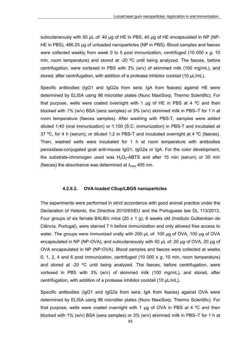
Locust bean gum nanoparticles: Application in oral immunization
83
subcutaneously with 50 µL of: 40 µg of HE in PBS, 40 µg of HE encapsulated in NP (NP-
HE in PBS), 466.25 µg of unloaded nanoparticles (NP in PBS). Blood samples and faeces
were collected weekly from week 0 to 5 post immunization, centrifuged (10 000 x g, 10
min, room temperature) and stored at -20 ºC until being analyzed. The faeces, before
centrifugation, were vortexed in PBS with 3% (w/v) of skimmed milk (100 mg/mL), and
stored, after centrifugation, with addition of a protease inhibitor cocktail (10 µL/mL).
Specific antibodies (IgG1 and IgG2a from sera; IgA from feaces) against HE were
determined by ELISA using 96 microtiter plates (Nunc MaxiSorp, Thermo Scientific). For
that purpose, wells were coated overnight with 1 µg of HE in PBS at 4 ºC and then
blocked with 1% (w/v) BSA (sera samples) or 3% (w/v) skimmed milk in PBS–T for 1 h at
room temperature (faeces samples). After washing with PBS-T, samples were added
diluted 1:40 (oral immunization) or 1:100 (S.C. immunization) in PBS-T and incubated at
37 ºC, for 4 h (serum); or diluted 1:2 in PBS-T and incubated overnight at 4 ºC (faeces).
Then, washed wells were incubated for 1 h at room temperature with antibodies
peroxidase-conjugated goat anti-mouse IgG1, IgG2a or IgA. For the color development,
the substrate-chromogen used was H2O2-ABTS and after 15 min (serum) or 30 min
(faeces) the absorbance was determined at λmax 405 nm.
4.2.6.2. OVA-loaded CSup/LBGS nanoparticles
The experiments were performed in strict accordance with good animal practice under the
Declaration of Helsinki, the Directive 2010/63/EU and the Portuguese law DL 113/2013.
Four groups of six female BALB/c mice (20 ± 1 g), 6 weeks old (Instituto Gulbenkian de
Ciência, Portugal), were starved 7 h before immunization and only allowed free access to
water. The groups were immunized orally with 200 µL of: 100 µg of OVA, 100 µg of OVA
encapsulated in NP (NP-OVA); and subcutaneously with 50 µL of: 20 µg of OVA, 20 µg of
OVA encapsulated in NP (NP-OVA). Blood samples and faeces were collected at weeks
0, 1, 2, 4 and 6 post immunization, centrifuged (10 000 x g, 10 min, room temperature)
and stored at -20 ºC until being analyzed. The faeces, before centrifugation, were
vortexed in PBS with 3% (w/v) of skimmed milk (100 mg/mL), and stored, after
centrifugation, with addition of a protease inhibitor cocktail (10 µL/mL).
Specific antibodies (IgG1 and IgG2a from sera; IgA from feaces) against OVA were
determined by ELISA using 96 microtiter plates (Nunc MaxiSorp, Thermo Scientific). For
that purpose, wells were coated overnight with 1 µg of OVA in PBS at 4 ºC and then
blocked with 1% (w/v) BSA (sera samples) or 3% (w/v) skimmed milk in PBS–T for 1 h at

Locust bean gum nanoparticles: Application in oral immunization
84
room temperature (faeces samples). After washing with PBS-T, a pool of samples were
added in twofold serial dilutions in PBS-T starting with 1:40, and incubated at 37 ºC, for 4
h (serum); or starting with 1:1 and incubated overnight at 4 ºC (faeces). Then, washed
wells were incubated for 1 h at room temperature with antibodies peroxidase-conjugated
goat anti-mouse IgG1, IgG2a or IgA. For the color development, the substrate-chromogen
used was H2O2-ABTS and after 15 min (serum) or 30 min (faeces) the absorbance was
determined at λmax 405 nm. The end titers were determined as the dilution of sample
giving the mean O.D. ≥ 0.2 the obtained from untreated mice sera.
4.2.7. Statistical analyses
The t-test and the one-way analysis of variance (ANOVA) with the pair wise multiple
comparison procedures (Holm-Sidak method) were performed to compare two or multiple
groups, respectively. All analyses were run using the SigmaStat statistical program
(Version 3.5, SyStat, USA) and differences were considered to be significant at a level of
P < 0.05.
4.3. Results and discussion
4.3.1. Characterization of unloaded CSup/LBGS nanoparticles
Several formulations of LBG-based nanoparticles were produced by a very mild
polyelectrolyte complexation, according to a procedure detailed in the experimental
section. Polyelectrolyte complexation is a process that involves electrostatic interaction
between oppositely charged groups. This procedure takes the advantage of occurring in a
hydrophilic environment with mild preparation conditions, avoiding the use of organic
solvents or high shear forces that might compromise the stability of encapsulated
materials (108, 194). LBG is a natural polymer with neutral charge, which hinders the
application of the mentioned methodology to directly obtain nanoparticles. In order to
overcome that relevant limitation, a sulfate derivative of LBG was produced, exhibiting a
negative charge. When CSup and LBGS solutions are mixed, an electrostatic interaction
is established between the negatively charged sulfate groups of LBGS and the positively
charged amino groups of CSup, leading to nanoparticle formation (Figure 1.1).
Considering the pH of the involved solutions (4.3 for CSup and 5.6 for LBGS), the
polymers display positive and negative charges, respectively. In the case of CSup, a

Locust bean gum nanoparticles: Application in oral immunization
85
degree of deacetylation of 86%, to which corresponds 0.86 positive charges per
monomer, and a Mw of 113 kDa, have been reported (237). The mean molar mass for the
monomer (198 g/mol) may be obtained by ponderation of the molar masses of the
acetylated and deacetylated units. In turn, for LBGS a C:S molar ratio of 26.76 was
determined, to which corresponds a DS of 1.22 and a mean molar mass of 932 g/mol for
the monomer. An Mw of 27 kDa was determined by GPC analysis.
As the aim of this work was to disclose the effect of LBGS in the production of
nanoparticles, the produced formulations accounted with a similar or higher amount of this
polymer comparing with CSup. Nine formulations of CSup/LBGS nanoparticles were
produced with polymeric mass ratios varying within 1:1 and 1:4. After the preparation
procedures, nanoparticles were characterized in terms of size, polydispersion index (PdI),
zeta potential and production yield. The detailed results are shown in Table 4.1.
Table 4.1 - Physicochemical characteristics and production yield of CSup/LBGS unloaded nanoparticles (mean ± SD; n ≥ 3). Different letters represent significant differences in each parameter (P < 0.05).
CSup/LBGS
(w/w) Size (nm) PdI
Zeta potential
(mV)
Production
yield (%)
1:1 174.8 ± 13.1 a 0.10 ± 0.005 +13.0 ± 2.7 b 9.1 ± 5.0 c
1:1.25 183.0 ± 4.7 a 0.10 ± 0.03 +14.5 ± 1.3 b n.d.
1:1.5 184.0 ± 4.1 a 0.09 ± 0.01 +13.0 ± 0.8 b 12.5 ± 3.9 c
1:1.75 182.5 ± 5.0 a 0.09 ± 0.01 +13.3 ± 1.0 b n.d.
1:2 183.0 ± 6.1 a 0.13 ± 0.02 +13.5 ± 0.6 b 30.2 ± 2.8 d
1:3 198.0 ± 26.0 a 0.12 ± 0.01 +13.0 ± 1.0 b n.d.
1:3.25 pp - - -
1:3.5 pp - - -
1:4 pp - - -
CSup: ultrapure chitosan; LBGS: sulfated locust bean gum; n.d.: not determined; PdI: polydispersity index; pp: precipitate

Locust bean gum nanoparticles: Application in oral immunization
86
Formulations with a higher amount of LBGS (ratios ≥ 1:3.25, w/w) resulted in precipitation.
Considering that a constant amount of CSup is used to produce all the formulations, the
observed precipitation is possibly due to the presence of an excess of anionic charges,
which neutralize CSup positive charges and, thus, reduce or eliminate electrostatic
repulsion, leading to precipitation. For all the other tested mass ratios, a clear Tyndall
effect was observed upon mixing the two polysaccharides, indicating the presence of
colloidal particles. Nanoparticles were thus successfully obtained for mass ratios varying
between 1:1 and 1:3. Surprisingly, the size of the particles did not present significant
variations among the tested ratios, being in all cases of approximately 180 nm. The ratio
1:3 resulted in nanoparticles with an average size of 198 nm but the standard deviation
increased 4-5 times (26 nm), which suggests the beginning of the destabilization of the
process of nanoparticle formation, which is reinforced by the precipitation occurred in the
following ratio (1:3.25, w/w). The absence of variations was not expected, as varying the
amount of one of the polymers, and therefore the amount of charges, should result in
different nanoparticle characteristics. The nanoparticles evidenced a very narrow PdI
(around 0.1) and a positive zeta potential around +13 mV. As observed for the size, it was
also unexpected that the zeta potential did not vary with the alteration of the mass ratios.
This effect is better analyzed considering the charge ratios involved in each formulation.
By dividing the charge of each repeating unit by its molar mass, a charge per mass ratio is
obtained for each polymer. In a 1/n formulation of CSup/LBGS, the -/+ charge ratio is
calculated by:
-/+ charge ratio = n . charge per mass (LBGS) / charge per mass (CSup)
Figure 4.1 demonstrates the influence of -/+ charge ratio on the zeta potential. As
observed, the charge ratio varies between 0.30 and 0.91 without significant effect on the
resulting zeta potential. This pattern is relatively similar to that observed in the
nanoparticles presented in the previous chapter, when charge ratios vary without reaching
a value around 1. In these CSup/LBGS nanoparticles, on reaching -/+ charge ratios of
0.98, 1.06 and 1.21 precipitation occurred. Although this behavior was unexpected, it has
been reported in other works regarding polysaccharide nanoparticles produced by the
same methodology (58, 59, 195, 238, 239).

Locust bean gum nanoparticles: Application in oral immunization
87
Figure 4.1 – Effect of charge ratio (-/+) on the zeta potential of CSup/LBGS nanoparticles.
It was previously demonstrated that the process leading to the assembly of polyelectrolyte
complexes has two major steps: the rapid formation of molecular or primary complex
particles, and a subsequent phase corresponding to the aggregation of primary particles
to secondary particles. While the primary particles are held together by long-range
electrostatic interactions, the formation of secondary particles involves short-range
dispersive interactions (240, 241). In this case, considering the differences between the
charge densities of the two polysaccharides (0.86 charges/monomer in CSup and 0.24
charges/monomer in LBGS), a very inefficient charge pairing should be expected.
Therefore, and also regarding the differences in the molecular weights of the
polysaccharides, in formulations 1:1 to 1:1.5, a reduced number of primary complexes
should form. The poor charge neutralization should result in enhanced electrostatic
repulsion between primary particles, leading to low dispersive attraction and smaller
particle sizes, as well as to low yields. By increasing the amount of LBGS, towards the 1:2
formulation, a larger number of primary particles form, which should tend to aggregate in
larger particles as the (-/+) charge ratio increases to 0.60. However, that was not the case,
with all formulations presenting almost invariant particle sizes, which should mean that
more particles formed; thus, the slight increase in yield observed in the latter formulation
(241). The fact that all formulations present similar surface potentials of ~13 mV, seems to
corroborate this hypothesis, as this potential should correspond to a repulsive electrostatic
force overcoming the dispersive interactions and therefore preventing the particles from
growing further. This behavior was observed in other works (238), normally associated to
the use of CSup. Therefore, another possible explanation may reside in conformational
0
5
10
15
20
25
0.0 0.2 0.4 0.6 0.8 1.0
Zeta potential (mV)
Charge ratio (‐/+)
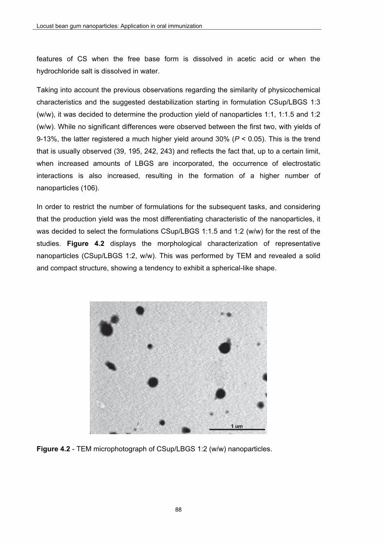
Locust bean gum nanoparticles: Application in oral immunization
88
features of CS when the free base form is dissolved in acetic acid or when the
hydrochloride salt is dissolved in water.
Taking into account the previous observations regarding the similarity of physicochemical
characteristics and the suggested destabilization starting in formulation CSup/LBGS 1:3
(w/w), it was decided to determine the production yield of nanoparticles 1:1, 1:1.5 and 1:2
(w/w). While no significant differences were observed between the first two, with yields of
9-13%, the latter registered a much higher yield around 30% (P < 0.05). This is the trend
that is usually observed (39, 195, 242, 243) and reflects the fact that, up to a certain limit,
when increased amounts of LBGS are incorporated, the occurrence of electrostatic
interactions is also increased, resulting in the formation of a higher number of
nanoparticles (106).
In order to restrict the number of formulations for the subsequent tasks, and considering
that the production yield was the most differentiating characteristic of the nanoparticles, it
was decided to select the formulations CSup/LBGS 1:1.5 and 1:2 (w/w) for the rest of the
studies. Figure 4.2 displays the morphological characterization of representative
nanoparticles (CSup/LBGS 1:2, w/w). This was performed by TEM and revealed a solid
and compact structure, showing a tendency to exhibit a spherical-like shape.
Figure 4.2 - TEM microphotograph of CSup/LBGS 1:2 (w/w) nanoparticles.

Locust bean gum nanoparticles: Application in oral immunization
89
4.3.2. Characterization of loaded nanoparticles
Bearing in mind the main objective of proposing CSup/LBGS nanoparticles for oral
vaccination, the first approach regarding the association of an antigen relied on using an
immunogenic subcellular extract obtained from whole Salmonella Enteritidis cells (HE)
(244, 245). After performing the in vivo studies, which results are reported in a subsequent
section (section 4.3.4), the need to test soluble antigens was identified, in order to permit
a more clear evaluation of the real adjuvant properties of the formulated nanoparticles.
Therefore, in a second stage of the work the protein ovalbumin (OVA) was associated as
model antigen. Additionally, as being one of the mostly used molecules for the antigen
effect, it enables further comparisons with the literature. As pertinent information, the HE
extracts are mainly composed of proteins (29%) and lipopolysaccharides (59%) (246). It is
well reported in the literature that lipopolysaccharide fractions have great ability to
generate immunological responses, since they are prototypical PAMP’s (247). Therefore,
taking into account that OVA is a protein, devoid of PAMP components, it is assumed as
less immunogenic than HE.
4.3.2.1. HE-loaded CSup/LBGS
As said above, the first demonstration of the usefulness of the developed nanoparticles to
act as adjuvants in a vaccination strategy relies on the determination of their ability to
associate antigens. In order to verify this, different amounts of the bacterial HE antigens
were tested and the resulting nanoparticles characterized. Departing from initial
concentrations of 2%, 4% and 8% (w/w) of the total amount of the polymers, an effective
and similar association was observed in all cases. In fact, the association efficiency varied
within 29-36% (Table 4.2), independently of both the initial concentration of antigen and
the formulation. These resulted in loading capacities up to 12%. Taking into account that
HE is negatively charged when in the LBGS-HE solution, and also considering the high
density of free amino groups present in the chitosan solution, it could be assumed that the
main factor affecting HE association to the nanoparticles was an electrostatic interaction.
This is in agreement with many other works reporting the association of protein-based
macromolecules to nanoparticles produced by polyelectrolyte complexation (40, 106, 248,
249). In turn, although it could be expected that HE and LBGS might compete in their
interaction with chitosan, the obtained results do not show an influence of LBGS content
on HE association. This may be due to the fact that LBGS could also interact with HE by
means of hydrophobic interactions, hydrogen bonding and other intermolecular forces
Locust bean gum nanoparticles: Application in oral immunization
90
(242). The absence of a concentration-dependent effect regarding HE was also
unexpected, although it has been observed in other works (39, 106).

91
Tab
le 4
.2 –
Phy
sico
chem
ical
cha
ract
eris
tics,
pro
duct
ion
yiel
d an
d as
soci
atio
n ef
ficie
ncy
of H
E a
ntig
ens
in C
Sup
/LB
GS
na
nopa
rtic
les
(mea
n ±
SD
; n
≥ 3)
. D
iffer
ent
lette
rs r
epre
sent
sig
nific
ant
diffe
renc
es in
eac
h pa
ram
eter
, ev
alua
ted
sepa
rate
ly
for
each
mas
s ra
tio (
P <
0.0
5).
CS
up
/LB
GS
(w/w
) H
E (
%)
Siz
e (n
m)
Pd
I
Zet
a
po
ten
tial
(mV
)
Pro
du
ctio
n
yiel
d (
%)
Ass
oci
atio
n
effi
cien
cy
(%)
Lo
adin
g
cap
acit
y (%
)
1:1.
5
0 18
4.0
± 4.
1a
0.09
± 0
.01
+13
.0 ±
0.8
e
12.5
± 3
.9i
- -
2 19
1.5
± 7.
8a
0.11
± 0
.02
+10
.0 ±
1.4
f 9.
6 ±
6.3
i 31
.2 ±
7.4
k 6.
4 ±
1.5
m
4 19
0.0
± 11
.3a
0.10
± 0
.01
+10
.0 ±
1.4
f 13
.1 ±
3.8
i 29
.0 ±
2.6
k 8.
5 ±
0.8
m
8 19
6.5
± 2.
1b
0.08
± 0
.01
+9.
5 ±
0.7 f
15.5
± 0
.0i
25.3
± 3
.2k
12.1
± 1
.5n
1:2
0 18
3.0
± 6.
1c
0.13
± 0
.02
+13
.5 ±
0.6
g
30.2
± 2
.8j
- -
2 19
2.0
± 9.
9c
0.11
± 0
.00
+10
.5 ±
0.7
h
28.5
± 5
.2j
35.5
± 9
.7l
2.4
± 0.
7o
4 19
3.0
± 11
.3c
0.13
± 0
.02
+11
.5 ±
0.7
h
29.6
± 3
.9j
34.8
± 3
.3l
4.5
± 0.
4p
8 20
2.5
± 10
.6d
0.13
± 0
.03
+12
.5 ±
0.7
g
30.1
± 3
.7j
31.9
± 4
.0l
7.8
± 1.
0q
CS
up: u
ltrap
ure
chito
san;
HE
: ant
igen
ic e
xtra
ct fr
om S
alm
on
ella
Ent
eriti
dis;
LB
GS
: sul
fate
d lo
cust
bea
n gu
m; P
dI: p
olyd
ispe
rsity
inde
x

Locust bean gum nanoparticles: Application in oral immunization
92
Table 4.2 further displays the physicochemical characteristics of HE-loaded CSup/LBGS
nanoparticles. The size of the particles was around 190-200 nm independently of the
specific formulation and the amount of HE associated. When comparing with the
corresponding unloaded nanoparticles, which had a size of 183-184 nm, no significant
differences were generally observed. An exception was only observed when 8% HE was
associated, for both formulations, with sizes reaching approximately 200 nm (P < 0.05),
but this observation is considered to be devoid of physiological relevance. The PdI of the
nanoparticles remained remarkably low after association of HE (around 0.1) and a very
slight decrease of 2-3 mV in zeta potential was generally observed (P < 0.05). Regarding
the latter, the only exception was for the formulation 1:2, again when 8% HE was
associated (zeta potential of +12.5 mV), in which no significant variation was observed
comparing with the equivalent unloaded nanoparticles. Regarding the yield of the process
of nanoparticle production, it was also observed an absence of effect upon association of
HE antigens, independently of the used concentration. Considering that size and zeta
potential values remained approximately similar after the association of the antigen, the
maintenance of the production yield is indicative of displacement of the polymers to permit
the incorporation of the active molecule.
Taking into account the properties exhibited by HE-loaded nanoparticles, it was decided to
select the formulation CSup/LBGS 1:2 with 8% of HE associated, to perform subsequent
studies. This selection was driven by the presence of a higher theoretical amount of LBG
and HE antigens. The morphological examination of this specific formulation was
performed by TEM (Figure 4.3) and revealed the maintenance of the solid and compact
structure of the unloaded nanoparticles, also showing a tendency to exhibit a spherical-
like shape.

Locust bean gum nanoparticles: Application in oral immunization
93
Figure 4.3 – TEM microphotograph of HE-loaded CSup/LBGS (1:2, w/w; 8% HE) nanoparticles.
As shown in Table 4.2, the developed HE-loaded CSup/LBGS nanoparticles evidence
adequate physicochemical properties for the objective of oral delivery aimed at mucosal
vaccination, with a size around 200 nm and a positive zeta potential. In fact, the size is
small enough to permit an intimate contact with epithelial surfaces, which is maximal at
50–500 nm (20, 21). In turn, the positive zeta potential further potentiates the interaction
with epithelia, as this is negatively charged and, thus, an electrostatic interaction is
enabled. In summary, these characteristics are expected to provide a prolonged retention
of nanoparticles close to epithelial surfaces, potentiating the uptake by M cells and/or
antigen release.
4.3.2.2. OVA-loaded CSup/LBGS
As mentioned above, the need to associate a second antigen was identified and OVA was
selected for this end. Considering that CSup/LBGS nanoparticles 1:2 (w/w) containing 8%
HE had been previously selected for further studies, the production of OVA-loaded
nanoparticles respected the same composition, in order to effectively evaluate the particle
contribution as vaccination adjuvant. As stated in Table 4.3, OVA was successfully
encapsulated with a similar efficiency as that for HE (26.4%), which resulted in a loading
of 5.2%. Regarding the physicochemical characteristics of OVA-loaded nanoparticles,
there are no statistically significant differences in size when comparing with unloaded
particles, while a slight decrease of zeta potential around 4 mV was observed (P < 0.05).
Locust bean gum nanoparticles: Application in oral immunization
94
The production yield registered a significant increase from 30% to 38% (P < 0.05). When
a comparison with HE-loaded nanoparticles is performed, considering the corresponding
formulation, it should be highlighted that OVA-loaded nanoparticles have a significantly
lower size (179 nm vs 203 nm) and higher production yield (38% vs 30%) (P < 0.05). A
significant difference was also found for zeta potential, but only decreasing around 3 mV
(P < 0.05).
Naturally, the literature does not report similar nanoparticles, as LBG is being proposed
for the first time herein, but chitosan-based nanoparticles have been suggested many
times regarding oral vaccination (250-257). Occasionally, ovalbumin was the tested
antigen (252), resulting in nanoparticle size around 300 nm and a strong positive zeta
potential (+43 mV). As said above, the positive zeta potential is a desirable characteristic
to mediate and favor the interaction with the epithelium. In our work, the proposed
nanoparticles present a lower zeta potential, but also a lower size, which further benefits
this interaction, due to an increased surface area.
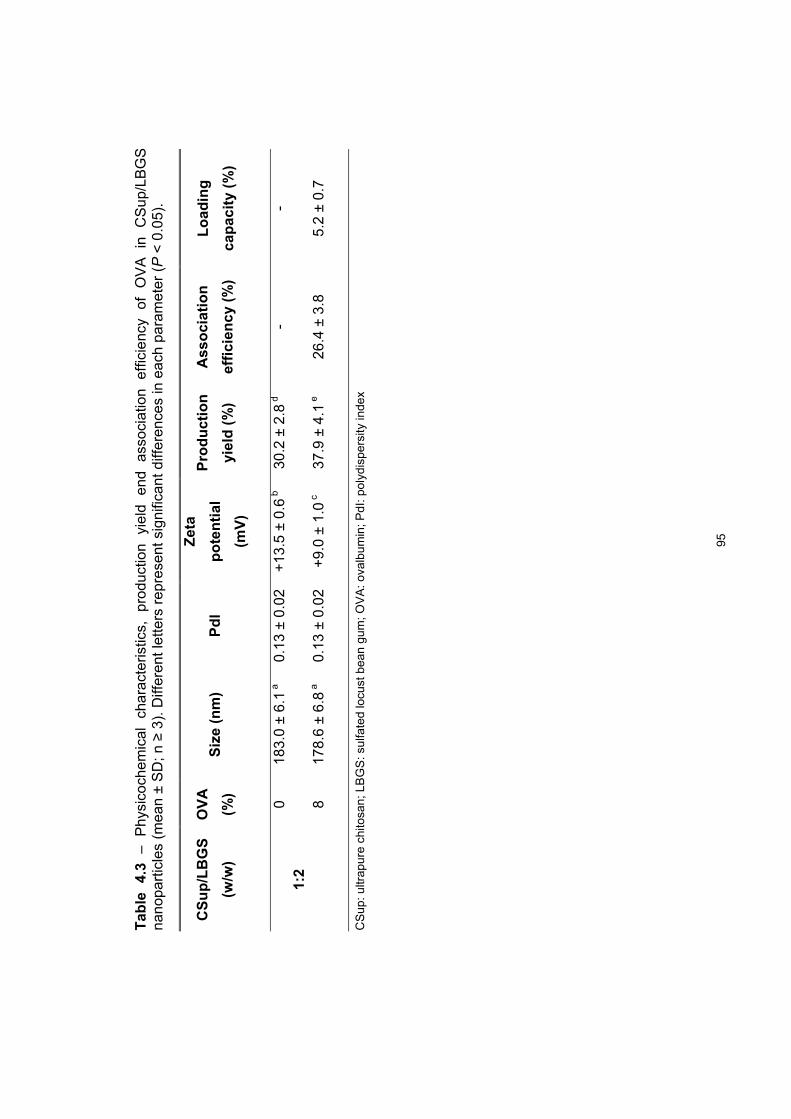
95
Tab
le
4.3
– P
hysi
coch
emic
al
char
acte
ristic
s,
prod
uctio
n yi
eld
end
asso
ciat
ion
effic
ienc
y of
O
VA
in
C
Sup
/LB
GS
na
nopa
rtic
les
(mea
n ±
SD
; n ≥
3).
Diff
eren
t let
ters
rep
rese
nt s
igni
fican
t diff
eren
ces
in e
ach
para
met
er (
P <
0.0
5).
CS
up
/LB
GS
(w/w
)
OV
A
(%)
Siz
e (n
m)
Pd
I
Zet
a
po
ten
tial
(mV
)
Pro
du
ctio
n
yiel
d (
%)
Ass
oci
atio
n
effi
cien
cy
(%)
Lo
adin
g
cap
acit
y (%
)
1:2
0 18
3.0
± 6.
1a
0.13
± 0
.02
+13
.5 ±
0.6
b
30.2
± 2
.8d
- -
8 17
8.6
± 6.
8a
0.13
± 0
.02
+9.
0 ±
1.0 c
37
.9 ±
4.1
e 26
.4 ±
3.8
5.
2 ±
0.7
CS
up: u
ltrap
ure
chito
san;
LB
GS
: sul
fate
d lo
cust
bea
n gu
m; O
VA
: ov
albu
min
; PdI
: pol
ydis
pers
ity in
dex

Locust bean gum nanoparticles: Application in oral immunization
96
4.3.3. In vitro evaluation of Locust Bean Gum-based nanoparticles
4.3.3.1. Evaluation of the structural integrity and antigenicity of the
loaded antigens
As detailed above, the association of either HE or OVA into nanoparticles was performed
by means of a mild ionic interaction. Along with the determination of the ability of the
developed nanoparticles to associate the selected antigens, it is also of utmost
importance to ensure that the particles and the procedure used for their production enable
the preservation of the structural integrity and antigenicity of the encapsulated molecules.
SDS-PAGE analysis followed by immunoblotting was the method used to perform this
evaluation.
The results of this evaluation are depicted in Figure 4.4 and Figure 4.5, for HE and OVA,
respectively. Regarding HE, SDS-PAGE results shown in Figure 4.4-b demonstrate that
the procedure used for HE entrapment did not affect the structural integrity of the
molecules, as no additional fragments are observed in the HE released from nanoparticles
(lane 2) when compared with the control HE solution (lane 1).
Figure 4.4 – Immunoblot (a) and SDS-PAGE (b) analyses of free HE (1) and HE released from HE-loaded CSup/LBGS nanoparticles (2) (PS: standard proteins).
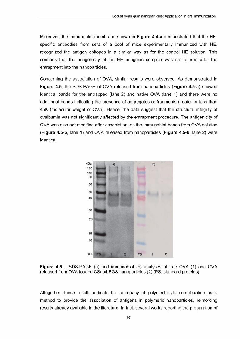
Locust bean gum nanoparticles: Application in oral immunization
97
Moreover, the immunoblot membrane shown in Figure 4.4-a demonstrated that the HE-
specific antibodies from sera of a pool of mice experimentally immunized with HE,
recognized the antigen epitopes in a similar way as for the control HE solution. This
confirms that the antigenicity of the HE antigenic complex was not altered after the
entrapment into the nanoparticles.
Concerning the association of OVA, similar results were observed. As demonstrated in
Figure 4.5, the SDS-PAGE of OVA released from nanoparticles (Figure 4.5-a) showed
identical bands for the entrapped (lane 2) and native OVA (lane 1) and there were no
additional bands indicating the presence of aggregates or fragments greater or less than
45K (molecular weight of OVA). Hence, the data suggest that the structural integrity of
ovalbumin was not significantly affected by the entrapment procedure. The antigenicity of
OVA was also not modified after association, as the immunoblot bands from OVA solution
(Figure 4.5-b, lane 1) and OVA released from nanoparticles (Figure 4.5-b, lane 2) were
identical.
Figure 4.5 – SDS-PAGE (a) and immunoblot (b) analyses of free OVA (1) and OVA released from OVA-loaded CSup/LBGS nanoparticles (2) (PS: standard proteins).
Altogether, these results indicate the adequacy of polyelectrolyte complexation as a
method to provide the association of antigens in polymeric nanoparticles, reinforcing
results already available in the literature. In fact, several works reporting the preparation of

Locust bean gum nanoparticles: Application in oral immunization
98
nanoparticles using methodologies involving electrostatic interaction and using materials
such as chitosan, its derivatives and alginate, have demonstrated to provide protection to
various model antigens including inactivated influenza virus (258), bovine serum albumin
(259), tetanus toxoid (260) and diphtheria toxoid (261).
4.3.3.2. Stability evaluation on storage
Nanoparticles are usually formulated as aqueous suspensions, as in this work, and one of
the most reported limitations of these systems relies on their tendency for aggregation.
Both physical (aggregation/particle fusion) and chemical issues (hydrolysis of polymer and
chemical reactivity) are known to play significant roles in this context, thus contributing for
the low stability that is frequently reported for colloidal drug carriers (262-264). The main
reason for this phenomenon is the higher attractive potential existing between two
particles coming into contact, when comparing with the kinetic energy that could induce
their separation (265). The natural tendency for aggregation during the storage period is,
therefore, one of the most important limitations preventing nanoparticle applications (266,
267). In this regard, the use of charged nanoparticles might prevent aggregation and has
been proposed as strategy to increase nanoparticle stability. Although in this work
nanoparticles do not exhibit a strong surface charge as compared with other chitosan-
based nanoparticle formulations (56, 57, 59, 195, 268), there is still a positive surface
charge that might help on this effect.
In order to study the nanoparticle behavior on storage, the size and zeta potential of HE-
loaded and unloaded CSup/LBGS nanoparticles in the aqueous suspension were
monitored along time. The rationale of conducting this assay in water was the interest in
obtaining information on nanoparticle stability in the resuspension medium. Several
formulations were evaluated for both HE-loaded and unloaded nanoparticles and similar
behaviors were observed. Figure 4.6 represent the results obtained for the formulation
1:2 (w/w) but are representative of the observations performed for the other formulations
(1:1 and 1:1.5). It was observed that, both formulations perfectly maintain the initial
physicochemical characteristics when stored at 4 ºC, no alterations being observed either
on size or zeta potential for up to 6 months. These results demonstrate the physical
stability of the developed nanoparticles, suggesting that the zeta potential is sufficiently
high to induce repulsion on nanoparticles. Chitosan-based nanoparticles have been
reported to exhibit physicochemical stability in similar time intervals (59, 195, 269, 270).

Locust bean gum nanoparticles: Application in oral immunization
99
However, a direct comparison with locust bean gum based nanoparticles is not possible -
as no such system was reported before.
Figure 4.6 – Size (square marks) and zeta potential (triangular marks) evolution as function of time upon storage at 4 ºC of CSup/LBGS 1:2 (w/w) unloaded nanoparticles (empty marks) and HE-loaded nanoparticles (8% w/w; filled marks); (mean ± SD, n ≥ 3).
4.3.3.3. In vitro release in SGF and SIF
Considering the design of these nanoparticles for an oral vaccination approach, it is
adequate to determine the release of the encapsulated antigens in media simulating both
the gastric (SGF) and intestinal (SIF) environments. In this association approach it is
important that the nanoparticles not only provide protection to the antigens regarding the
harsh conditions of the gastric medium, but also prevent the release of the antigens, in
order to maximize the antigen internalization by the M cells mediated by the particle.
Figure 4.7 shows the release profile of HE and OVA in SGF and SIF. As can be
observed, HE is considered to not present significant release in any of the tested media.
In fact, in SGF it releases a maximum amount of 7.5% in 2 h, while releasing 4.3% in SIF
after 4 h. Moreover, although it might not be relevant from a physiological point of view,
HE release at the end of 24 h was 21.6% ± 2.0 and 13.2% ± 4.8 in SGF and SIF,
respectively. In turn, OVA presented a rather different behavior. In this case, the release
in the SGF was of 45.0% at the end of 2 h, and 3.0% in SIF at the end of 4 h.
10
15
20
25
30
130
150
170
190
210
230
250
0 30 60 90 120 150 180 210
Zet
a P
ote
nti
al (
mV
)
Siz
e (n
m)
Time (days)
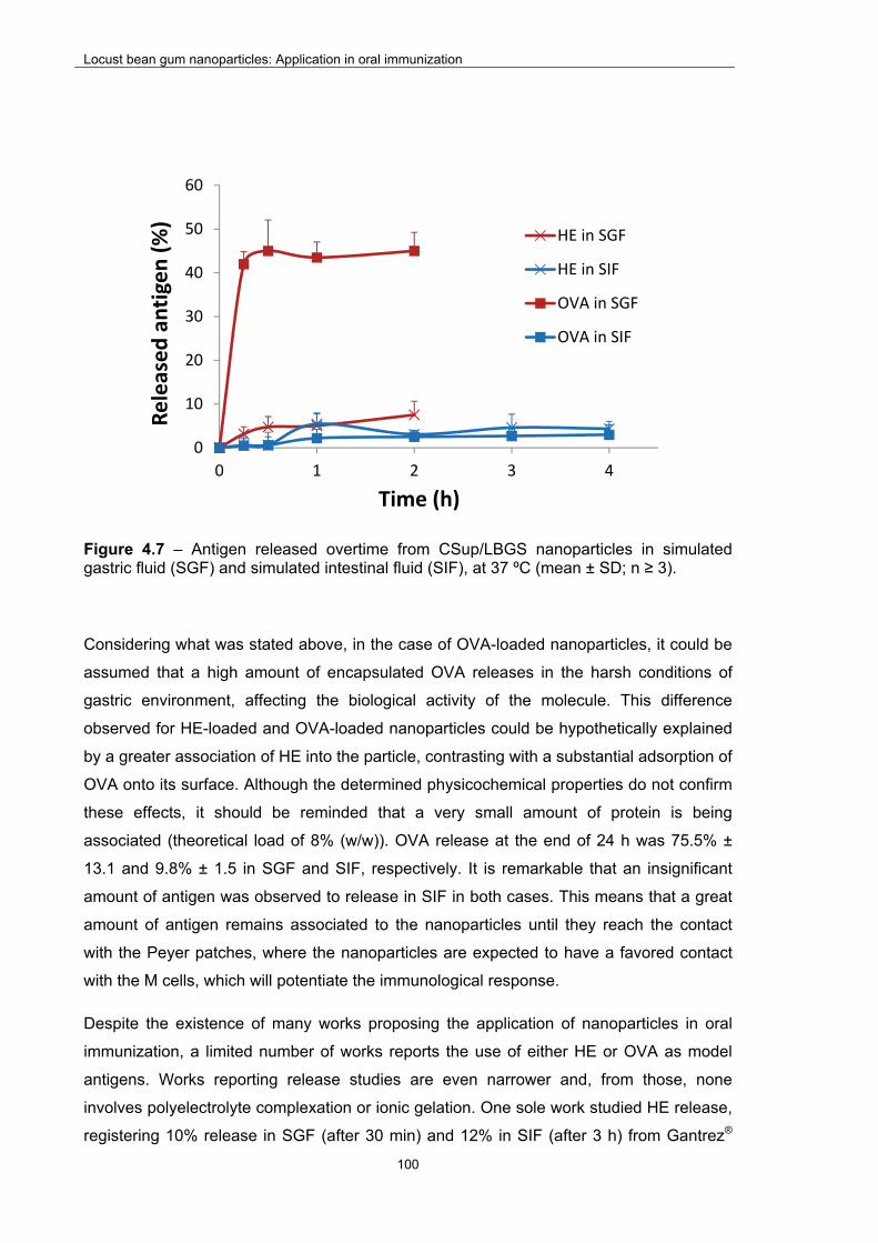
Locust bean gum nanoparticles: Application in oral immunization
100
Figure 4.7 – Antigen released overtime from CSup/LBGS nanoparticles in simulated gastric fluid (SGF) and simulated intestinal fluid (SIF), at 37 ºC (mean ± SD; n ≥ 3).
Considering what was stated above, in the case of OVA-loaded nanoparticles, it could be
assumed that a high amount of encapsulated OVA releases in the harsh conditions of
gastric environment, affecting the biological activity of the molecule. This difference
observed for HE-loaded and OVA-loaded nanoparticles could be hypothetically explained
by a greater association of HE into the particle, contrasting with a substantial adsorption of
OVA onto its surface. Although the determined physicochemical properties do not confirm
these effects, it should be reminded that a very small amount of protein is being
associated (theoretical load of 8% (w/w)). OVA release at the end of 24 h was 75.5% ±
13.1 and 9.8% ± 1.5 in SGF and SIF, respectively. It is remarkable that an insignificant
amount of antigen was observed to release in SIF in both cases. This means that a great
amount of antigen remains associated to the nanoparticles until they reach the contact
with the Peyer patches, where the nanoparticles are expected to have a favored contact
with the M cells, which will potentiate the immunological response.
Despite the existence of many works proposing the application of nanoparticles in oral
immunization, a limited number of works reports the use of either HE or OVA as model
antigens. Works reporting release studies are even narrower and, from those, none
involves polyelectrolyte complexation or ionic gelation. One sole work studied HE release,
registering 10% release in SGF (after 30 min) and 12% in SIF (after 3 h) from Gantrez®
0
10
20
30
40
50
60
0 1 2 3 4
Released antigen (%)
Time (h)
HE in SGF
HE in SIF
OVA in SGF
OVA in SIF

Locust bean gum nanoparticles: Application in oral immunization
101
AN nanoparticles (271), values slightly higher than those obtained in our study. The
release of OVA was studied from poly(d,l-lactide-co-glycolide) (PLGA)-based
nanoparticles. Pegylated PLGA-based nanoparticles which were surface decorated with
RGD molecules to target M cells (OVA association efficiency of 30 – 50%) registered 5%
release after 2 h of incubation in gastric medium (HCl 0.1 M) and 10 – 20% in intestinal
medium (154). In turn, PLGA-lipid nanoparticles conjugated with ulex europaeus
agglutinin-1 and containing the Toll-like receptor agonist monophosphoryl lipid A (95%
OVA association efficiency), released 17% of the protein after 2 h incubation in gastric
acid medium (0.1 M HCl) and 3 h in intestinal medium (PBS pH 6.8) (272). Comparatively,
our work registered higher release of this protein after cumulative contact with the two
media, but we expect that the remaining amount suffices for an adequate immunization
effect.
While no studies report the release of OVA as model antigen from chitosan nanoparticles
proposed for oral immunization, there are some works on chitosan-based microparticles.
The results are however uneven, certainly as a result of different methods of preparation
of microparticles and the use of diverse chitosan molecules. In fact, different works
reported either immediate release of OVA in SGF (273), or only 50% in 2 h (274). The
latter behavior is similar to that registered in our work, where 45% released in 2 h. The
results regarding the release in SIF or PBS pH 7.3 were more coincident, where a
maximum release of 10% - 20% was determined after 1 h (273) (159, 274). This is not far
from the 3% registered in our work at the end of 4 h.
4.3.3.4. Safety evaluation of unloaded nanoparticles
Assessing the biocompatibility of drug delivery systems is a major issue in designing drug
carriers (52, 197, 198). Although the toxic effects of formulations can only be accurately
determined by in vivo assays, several in vitro tests can be performed in adequate cell
lines to give the first indications on the systems cytotoxicity (197, 201). Additionally,
current international guidelines indicate the need to contextualize biocompatibility with a
specific route of administration and dose of the material (198). Moreover, the materials
composing the matrix of the particles and the particle itself should be regarded as different
entities. In line with this assumption, these should be evaluated separately, since the
particle structure, among others, might affect the final toxicological behavior (198, 228).
According to the guidelines issued by the International Organization for Standardization
(ISO), testing biocompatibility implies the performance of a complete set of assays,

Locust bean gum nanoparticles: Application in oral immunization
102
addressing at first cellular morphology, membrane integrity and metabolic efficiency,
among other tests (197, 199-201). In this work we performed two of the most used assays
to test the cytotoxicity of materials, namely the metabolic assay MTT and the membrane
integrity assay based on LDH release. The MTT assay assesses cell metabolic efficiency,
relying on the evaluation of enzymatic function. To do so, after the exposure to the matrix
materials used for the nanoparticles production or the nanoparticles themselves, cells are
incubated with yellow tetrazolium (MTT) salts which are reduced to purple-blue formazan
crystals by active mitochondrial dehydrogenases (197). In this manner, a higher
concentration of the formazan dye corresponds to a higher amount of metabolically active
cells, which is usually interpreted as higher cell viability. The LDH release assay provides
a mean to determine the amount of LDH in cell culture medium upon exposure to
potentially toxic substances. As LDH is a cytoplasmic enzyme, its presence in the cell
culture medium is an indicator of irreversible cell death due to cell membrane damage
(202, 203). The assay thus evaluates cell membrane integrity and complements the
results obtained by the MTT assay.
In this work, Caco-2 cells were used to evaluate the toxicological profile of CSup, LBGS
and the nanoparticles resulting from the combination of these polymers. Figure 4.8 and
Figure 4.9 represent the Caco-2 cell viability obtained after exposure to the mentioned
materials at different concentrations, for a period of 3 h and 24 h. The overall observation
of the results reveals a mild effect on cell viability from both the polymers and
nanoparticles, considered to be devoid of biological relevance. In fact, all samples
resulted in viabilities above 70% after 3 h or 24 h of exposure, when tested at
concentrations varying within 0.1 and 1.0 mg/mL. At 3 h cell viability remained above 88%
in all cases (Figure 4.8), the lower values being observed for the raw material LBGS,
although not to a statistically significant level. As observed in the previous chapter for LBG
and LBG derivatives, prolonging the exposure until 24 h resulted in slight alterations
(Figure 4.9). These were actually considered very mild, as cell viability did not decrease
below 70% in any case. This value is that considered by ISO 10993-5 (201) as the level
below which a toxic effect occurs. The only observation deserving a mention is that the
exposure to the highest concentration tested (1.0 mg/mL) of CSup and the nanoparticle
formulation, decreased cell viability to around 70% and 90%, respectively (P < 0.05).

Locust bean gum nanoparticles: Application in oral immunization
103
Figure 4.8 – Caco-2 cell viability measured by the MTT assay after 3 h exposure to increasing concentrations of Chitosan (CSup), sulfate Locust Bean Gum (LBGS) derivative and CSup/LBGS nanoparticles (NP). Data represent mean ± SEM (n ≥ 3, six replicates per experiment at each concentration). Dashed line indicates 70%. * P < 0.05 compared with DMEM.
A control was used that consists in a mixture of DMEM and H2O in the same ratio used for
the samples, taking into account that both the raw materials and the nanoparticles were
solubilized/suspended in water and diluted with cell culture medium prior to incubation
with the cells. This enables a real evaluation on the contribution of these materials on the
final cell viability. As observed in Figure 4.8 and Figure 4.9, the cell viability induced by
this control varied between 80 and 72% when tested at 3 h and 24 h. While at 3 h CSup
and nanoparticles comparatively induced higher cell viability (100% or more), after 24 h
the incubation with 1.0 mg/mL of CSup was found to decrease cell viability (71%) to a
level comparable to that of this control (DMEM + H2O). Remarkably, focusing on the
concentration of 1.0 mg/mL, the cell viability obtained by exposure to the nanoparticles
was higher than that registered for CSup assessed individually and the control of DMEM +
H2O (P < 0.05).
0
20
40
60
80
100
120
CSup LBGS NP Control
Cell viab
ility (%)
0.1 mg/mL
0.5 mg/mL
1.0 mg/mL
DMEM
DMEM/H2O
*

Locust bean gum nanoparticles: Application in oral immunization
104
Figure 4.9 – Caco-2 cell viability measured by the MTT assay after 24 h exposure to increasing concentrations of Chitosan (CSup), sulfate Locust Bean Gum (LBGS) derivative and CSup/LBGS nanoparticles (NP). Data represent mean ± SEM (n ≥ 3, six replicates per experiment at each concentration). Dashed line indicates 70%. * P < 0.05 compared with DMEM.
LBG-based nanoparticles are now being reported for the first time. Therefore, the
literature does not report any information regarding the cytotoxic effect of the particles,
hindering the establishment of any comparison. In contrast, chitosan-based nanoparticles
have been reported for a long time. As chitosan is one of the most used polymers in drug
delivery, nanocarriers other than nanoparticles have been proposed, such as
nanocapsules and nanoemulsions, for instance. This diversity affects viability results
(108), but even when similar carriers are at play, it is frequently difficult to establish valid
comparisons taking into account the large variety of chitosan molecules (chitosan
base/salt(s), molecular weight, deacetylation degree, etc.) which is also known to have a
role on cell viability (275). Notwithstanding these limitations, the general outcome
regarding chitosan-based nanoparticles is a very mild effect on Caco-2 cells viability.
Attempting to perform a direct and accurate comparison, other works assessing a
nanoparticle concentration of 1.0 mg/mL reported similar results to those found in this
work, both at 3 h (276) and 24 h (277, 278).
As mentioned above, the quantification of LDH released by the cells provides a
complementary indication on the cytotoxicity of materials to which the cells were exposed
to. If the materials affect the integrity of cell membrane, the leaking of this cytoplasmic
enzyme occurs and its quantification is enabled. The loss of intracellular LDH and its
release to the culture medium is therefore an indicator of irreversible cell death due to cell
0
20
40
60
80
100
120
CSup LBGS NP Control
Cell viab
ility (%)
0.1 mg/mL
0.5 mg/mL
1.0 mg/mL
DMEM
DMEM/H2O
**

Locust bean gum nanoparticles: Application in oral immunization
105
membrane damage (202, 203). In this study, the amount of LDH released by Caco-2 cells
exposed to CSup/LBGS nanoparticles was determined, using as control both the
incubation with cell culture medium (negative control of cell death) and the exposure to a
lysis buffer (positive control of cell death, assumed as 100%). The latter corresponds to
the maximum amount of cytoplasmic enzyme that can be released, while the former is the
minimum. As observed in Figure 4.10, the exposure to the raw materials CSup and LBGS
induced the release of an amount of LDH (around 25%) that is comparable to that of the
negative control, as no statistically significant differences were detected. On the contrary,
the contact with CSup/LBGS nanoparticles resulted in an unexpected increased level of
LDH release (43%; P < 0.05), which is indicative of cytotoxicity.
Figure 4.10 – Caco-2 cell viability measured by the LDH release assay after 24 h exposure to 1 mg/mL solutions of Chitosan (CSup), sulfate Locust Bean Gum (LBGS) derivative and CSup/LBGS nanoparticles (NP). Data represent mean ± SEM (n ≥ 3, three replicates per experiment). * P < 0.05 compared with DMEM.
If a direct correlation between LDH release and cell death is assumed, the contact with
the nanoparticles results in approximately 60% cell viability, comparing with the 80%
elicited by DMEM. Surprisingly, this does not correspond with the observations resulting
from the MTT assay, in which a cell viability of 90% was observed for this condition. One
possible explanation is that these nanoparticles act as metabolic enhancers, thus
although with a lower number of available cells (as indicated by the LDH assay), MTT
conversion into formazan is accelerated, resulting in the overestimation of the cell viability.
0
20
40
60
80
100
DMEM CSup LBGS NP
LDH release (%)
*

Locust bean gum nanoparticles: Application in oral immunization
106
This difference in the results of the two assays reinforces the need to perform various and
different tests to conclude on the cytotoxic profile of nanoparticles.
4.3.4. In vivo evaluation of the immune response in BALB/c mice
After verifying the ability of LBG derivatives to produce nanoparticles with capacity to
associate antigens of interest and evaluating the cytotoxicity of the nanoparticles in an
intestinal model, an in vivo assay was designed and setup to evaluate the adjuvant effect
of the particles towards an immunization strategy. Two different model antigens were
used, HE and OVA, and the immunization experiments performed with each antigen were
conducted in different laboratories, applying different methodologies for the analysis of
results. In the former (HE) a serum dilution was selected and the O.D. for each animal
was determined, the data being presented as mean ± SEM. In the latter (OVA), a pool
with the samples of different animals was used and data presented as log2 titers.
4.3.4.1. HE-loaded CSup/LBGS nanoparticles
The immunization of mice was performed by either oral or subcutaneous route, in order to
permit a comparison of effects. Serum and faeces from the mice were collected before the
immunization and at weeks 1-5 post immunization. The collection of both types of
samples enables the determination of immune responses at both the systemic (IgG1 and
IgG2a) and mucosal (IgA) levels. The results obtained upon oral immunization are shown
in Figure 4.11. The transversal observation is that, as expected, unloaded particles did
not induce any type of immune response. Concerning the systemic antibody response
(Figure 4.11 – A and B) no statistical differences were observed between the groups
corresponding to free HE (which is the control) and the NP-HE groups. Moreover, the
results also show that, five weeks after immunization, a similar Th1/Th2-mediated
antibody response for both formulations was elicited. Regarding this, it is important to
refer that the literature reports that to achieve a protective response against Salmonella
Enteritidis infection, a balance between antibody response and cellular mediated immune
response is demanded (279, 280). In this way, a balance between Th1 and Th2 type
responses is required, and Salmonella attenuated vaccines are reported to normally
induce Th1 type responses, being less effective at inducing Th2 type responses (281-
283). In this study, the nanoparticle formulation of HE (NP-HE) induced the required
balanced Th1/Th2 response since the beginning of the study, which can be explained by

Locust bean gum nanoparticles: Application in oral immunization
107
the presence of mannose groups, as was suggested in a study performed with
mannosylated nanoparticles (120, 284). In that work it was verified that mannosylated
nanoparticles induced a more balanced Th1/Th2 response comparing with non-
mannosylated nanoparticles, an effect justified by the authors to be due to the high
tropism of mannosylated particles for uptake by PPs rich in APCs (271). In our work, the
presence of mannose groups in LBG may possibly mediate a stronger interaction of LBG
nanoparticles with the M cells of PPs, contributing to the observed balance in the Th1/Th2
response.

Locust bean gum nanoparticles: Application in oral immunization
108
Figure 4.11 – Immunogenicity of HE after oral administration in mice. Serum A) IgG1 and B) IgG2a systemic response, and C) IgA mucosal response after oral immunization of 5 female BALB/c mice with 200 µg of HE solution (HE), 200 µg of encapsulated HE (NP-HE) and the corresponding mass of blank nanoparticles (NP). In the HE group the results of one mouse were rejected due to the high initial absorbance (week 0) (mean ± SEM; n ≥ 4).
0
0.2
0.4
0.6
0.8
1
0 1 2 3 4 5
O.D.
Week
NP
NP‐HE
HE
0
0.2
0.4
0.6
0.8
1
0 1 2 3 4 5
O.D.
Week
NP
NP‐HE
HE
0
0.2
0.4
0.6
0.8
1
1.2
0 1 2 3 4 5
O.D.
Week
NP
NP‐HE
HE
A
B
C

Locust bean gum nanoparticles: Application in oral immunization
109
The mucosal antibody response (Figure 4.11 – C) was also determined and, between
weeks 2 and 4, it is observed a higher response from the group immunized with free HE
compared with the NP-HE group (P < 0.05). Five weeks after immunization a shift is
observed, with the NP-HE group presenting a higher response than the HE group (P <
0.05).
When the formulations were administered via the subcutaneous route an antibody
response profile was elicited for HE and NP-HE groups (Figure 4.12). It was also
observed that both groups elicited identical Th1/Th2 antibody immune responses from the
first week on (Figure 4.12 – A and B).
Locust bean gum nanoparticles: Application in oral immunization
110
Figure 4.12 – Immunogenicity of HE after S.C. administration in mice. Serum A) IgG1 and B) IgG2a systemic response, and C) IgA mucosal response after S.C. immunization of 5 female BALB/c mice with 40 µg of HE solution (HE), 40 µg of encapsulated HE (NP-HE) and the corresponding mass of blank nanoparticles (NP) (mean ± SEM; n = 5).
0
0.2
0.4
0.6
0.8
1
1.2
0 1 2 3 4 5
O.D.
Week
NP
NP‐HE
HE
0
0.2
0.4
0.6
0.8
1
1.2
0 1 2 3 4 5
O.D.
Week
NP
NP‐HE
HE
0
0.2
0.4
0.6
0.8
1
1.2
0 1 2 3 4 5
O.D.
Week
NP
NP‐HE
HE
A
B
C
Locust bean gum nanoparticles: Application in oral immunization
111
However, free HE induced a much higher IgA response than NP-HE (P < 0.05),
particularly until week 3 (Figure 4.12 – C). It could be argued that free HE is immediately
available to induce a mucosal response, comparing with HE encapsulated into
nanoparticles. Similarly to what was observed for the oral route, unloaded nanoparticles
did not produce an immune response.
A relevant observation regarding the obtained results relies on the confirmation of the
mildness of the nanoparticle production method, as the results indicate that the
association process did not alter the immunogenicity of HE antigens. Nevertheless,
although in fact no differences were generally observed to a statistically significant level,
the trend indicates that free HE has a more favorable performance, when comparing with
encapsulated antigens. Overall, the results suggest that HE antigens are possibly too
robust and capable of inducing a strong immune response when administered in free
form, thus not potentiating the observation of an adjuvant effect by the nanoparticles. This
fact may be explained by the particulate nature of HE (liposome-like), and the high content
of LPS in the HE extract. It is known that LPS is recognized by TLR-4 (150). Considering
the hypothesis that the majority of the HE antigens in the nanoparticle formulation is
entrapped into the matrix, the recognition by DCs will be preferably made by the mannose
receptor, which is a C-type lectin receptor (152). It is reported that the uptake of antigens
by C-type lectin receptors does not necessarily result in the induction of potent effector T-
cells, although it facilitates the antigen-presentation capacity of DCs. Moreover, uptake of
antigen by C-type lectin receptors without any TLR binding may induce antigen-specific
tolerance. In contrast, TLR binding usually leads to DCs maturation and activation
resulting in a robust activation of immune responses and the induction of effector T-cells
(285).
It seems, thus, that the presence of mannose units in LBG nanoparticles, potentially
recognized by the M cells and expected to improve the immune response, was not as
good as the effect of the particulate antigen itself. Therefore, a need was identified to
select and test a second antigen. For this effect, the same formulation of nanoparticles
was associated with OVA and administered under the same protocol.
4.3.4.2. OVA-loaded CSup/LBGS nanoparticles
In order to evaluate the effect of OVA-loaded nanoparticles, these were administered by
oral or subcutaneous routes, as was performed for HE-loaded nanoparticles. Taking into
account the results described above for the unloaded particles, it was considered

Locust bean gum nanoparticles: Application in oral immunization
112
unnecessary to include a group of animals immunized with unloaded particles. Therefore,
only two groups were established, corresponding to OVA-loaded nanoparticles and free
OVA, as a control. Similarly to the previous assay, serum and faeces from the mice were
collected before the immunization and at weeks 1-6 post immunization.
Figure 4.13 and Figure 4.14 show the serum titers of IgG1 and IgG2a (systemic
response), and IgA (mucosal response) after an oral or subcutaneous immunization of
mice by a single dose of OVA-loaded nanoparticles or free OVA. Overall, it is observed
the adjuvant effect of nanoparticle formulations compared to free OVA in both routes.
Focusing on the oral immunization (Figure 4.13), the elicited IgG1 specific response (Th2)
was significant after OVA immunization, either free or encapsulated. Nanoparticles did not
improve the effect of OVA regarding Th2 activation (IgG1, Figure 4.13 – A), but, however,
a strong improvement was observed regarding Th1 activation (IgG2a, Figure 4.13 – B),
as the respective area under the curve (AUC) was five times higher than that determined
for free OVA. Intestinal immune system has a predisposition towards Th2 cell responses
since antigen presentation by DCs from PPs are characterized by the production of IL-4,
IL-6, and IL-10, which inhibit a Th1 response (286). It is also important to refer that soluble
antigens usually elicit high levels of IgG1 antibodies (Th2 response), but very low levels of
IgG2a (Th1 response) (287). Therefore, under these circumstances, the role of the
adjuvant becomes critical in order to achieve a more balanced Th1/Th2 response. A
similar positive effect of association was apparent regarding the mucosal response (IgA),
as shown in Figure 4.13 – C. In this respect, OVA-loaded nanoparticles registered a 3-
fold higher AUC when comparing with free OVA.

Locust bean gum nanoparticles: Application in oral immunization
113
Figure 4.13 – Immunogenicity of OVA after oral administration in mice. Serum anti-OVA A) IgG1; B) IgG2a and C) faecal anti-OVA IgA response in BALB/c mice (n = 6) after oral immunization with 100 µg of OVA solution (OVA) or 100 µg of encapsulated OVA (NP-OVA). Antibody titers were determined in pooled serum samples at days 0, 7, 14, 21 and 28 post-administration.
0
2
4
6
8
10
12
0 1 2 4 6
Log 2
titer
Week
OVA; AUC=31,4
NP‐OVA; AUC=36,8
0
2
4
6
8
10
12
0 1 2 4 6
Log 2
titer
Week
OVA; AUC=0
NP‐OVA; AUC=5,3
0
2
4
6
8
10
12
0 1 2 4 6
Log 2
titer
Week
OVA; AUC=7,0
NP‐OVA; AUC=22,5
A
B
C

Locust bean gum nanoparticles: Application in oral immunization
114
When considering the determined AUC, a comparable mucosal response was elicited
when the formulation was administered by subcutaneous route (Figure 4.14 – C).
However, the pattern registered for the responses was markedly different. In fact, while
the oral immunization induced a response that was detected from week one, the response
obtained upon subcutaneous immunization was only observed starting from week two to
four. In turn, the subcutaneous administration of nanoencapsulated OVA showed a similar
activation of Th2 (IgG1, Figure 4.14 – A) and Th1 (IgG2a, Figure 4.14 – B) to that
described for free OVA.
After oral immunization, OVA-loaded nanoparticles showed a predominant Th2 response
compared to the slightly elicited Th1 one (AUCTh2 was seven times higher than AUCTh1),
consistent with results obtained by others (252, 284, 288-290). On the contrary,
subcutaneous immunization with OVA-loaded nanoparticles elicited a more balanced Th1
and Th2 response. In order to perform a semi-quantitative comparison between the
nanoparticle formulation and the free OVA, the AUC of the titers representing the systemic
immune response (AUCTh1, AUCTh2) were measured and summed. Regarding the oral
immunization, an increase of 34% in the total AUC was accounted for the encapsulated
OVA. In turn, this association only induced a 13% increase in the response after
subcutaneous immunization. This is possibly justified by the presence of mannose units in
LBG nanoparticles, potentiating a stronger interaction with M cells and the mediation of a
stronger immune response when the nanoparticles are administered by the oral route.
The mucosal IgA antibodies obtained after oral immunization with OVA-loaded
nanoparticles were higher than after subcutaneous administration. This phenomenon may
be related to the effective uptake of nanoparticles by gut Peyer’s patches, obviously only
possible after oral delivery, and the passage of the particulate system to lymphocytes
causing an effective generation of mucosal IgA. However, it should be taken into account
that a 5-fold higher dose of antigen was administered by oral route.

Locust bean gum nanoparticles: Application in oral immunization
115
Figure 4.14 – Immunogenicity of OVA after S.C. administration in mice. Serum anti-OVA A) IgG1; B) IgG2a and C) faecal anti-OVA IgA response in BALB/c mice (n = 6) after S.C. immunization with 20 µg of OVA solution (OVA) or 20 µg of encapsulated OVA (NP-OVA). Antibody titers were determined in pooled faecal samples at days 0, 7, 14, 21 and 28 post-administration.
0
2
4
6
8
10
12
0 1 2 4 6
Log 2
titer
Week
OVA; AUC=43,8
NP‐OVA; AUC=47,8
0
2
4
6
8
10
12
0 1 2 4 6
Log 2
titer
Week
OVA; AUC=25,4
NP‐OVA; AUC=30,4
0
2
4
6
8
10
12
0 1 2 4 6
Log 2
titer
Week
OVA; AUC=6,0
NP‐OVA; AUC=15,0
A
B
C
Locust bean gum nanoparticles: Application in oral immunization
116
Considering the in vivo results as a whole, CSup/LBGS nanoparticles did not demonstrate
an extra-adjuvant effect when the self-adjuvant multicomponent HE was used to
immunize; in contrast, a significant adjuvant effect was demonstrated when using OVA as
a model antigen. This is certainly a consequence of the fact that HE is a particulate
antigen and stronger immunological responses are known to be generated to this type of
antigens in comparison with soluble ones, like OVA. Therefore, the suitability of
CSup/LBGS nanoparticles to provide an adjuvant effect in oral immunization is suggested,
although it is strongly dependent on the used antigen. Both the presence of mannose
units in LBG, which target M cells and DC’s, and the mucoadhesive characteristics of
CSup, are thought to have played a role in the improved response mediated by
nanoparticles. Additionally, results obtained by others suggest that targeting to M cells
and/or mucosal DCs should be the way to achieve a better and/or more balanced immune
response (252, 284, 288-290). It is worth to mention that the oral immunization performed
in these in vivo assays, truly evaluates the system capability, since it was made by oral
gavage (not intraduodenally) and no prior administration of sodium bicarbonate solution,
in order to neutralize the acid environment of the stomach, was made. The role of M cells
and mucosal DCs to achieve the adjuvant properties suggested herein should be
demonstrated.
4.4. Conclusions
This work reports for the first time the production of nanoparticles based on locust bean
gum, demonstrating the ability of the produced sulfate derivative of locust bean gum to
form nanoparticulate complexes with chitosan by means of an electrostatic interaction.
The produced nanoparticles were proposed as adjuvants in oral immunization, efficiently
associating two model antigens (HE antigenic complex and OVA) without compromising
their structural integrity. Displaying a size around 180 - 200 nm and a positive zeta
potential, the antigen-loaded nanoparticles were deemed adequate for the proposed
application in oral immunization. These characteristics, along with the mannose content of
LBG and the bioadhesive properties of chitosan, are expected to mediate both a
prolonged contact with the intestinal mucosa and a favored interaction with intestinal M
cells and mucosal DC’s. Nanoparticles were also demonstrated to be stable in suspension
for at least 3 months when stored at 4 ºC. The cytotoxic assessment performed in Caco-2
cells revealed no alterations at the level of cell metabolic activity upon exposure to the
nanoparticles, but a mild negative effect on cell membrane integrity was observed. The in
vivo proof of concept demonstrated the adjuvant effect of the proposed system when OVA

Locust bean gum nanoparticles: Application in oral immunization
117
was used as soluble antigen model, although this effect was not observed when a
particulate antigen like HE was tested. Nanoparticles were found to elicit a balanced
Th1/Th2 immune response, which is a relevant effect towards an effective immunological
protection. Additionally and as expected, nanoparticles were capable of inducing not only
a systemic response, but also a response at the mucosal level.

General conclusions and perspectives
118
This page was intentionally left in blank

General conclusions and perspectives
119
CHAPTER 5
GENERAL CONCLUSIONS AND PERPECTIVES
General conclusions and perspectives
120
This page was intentionally left in blank

General conclusions and perspectives
121
5. General conclusions
The results of the work presented in this thesis indicate the potential of the polymeric
nanoparticulate system based on polysaccharides for an application in oral immunization,
especially for soluble antigens.
In a more detailed manner, the described results permitted the following conclusions:
- Locust bean gum (LBG) is a neutral polysaccharide with a chemical composition
corresponding to a galactomannan. Its high content in OH groups enabled the
performance of chemical modifications to attribute charged functions to the molecule,
resulting in the synthesis of charged derivatives of LBG. Sulfated (LBGS), carboxylated
(LBGC) and ammonium LBG (LBGA) derivatives were synthesized;
- Varied methods were applied for the characterization of LBG and its derivatives, which
demonstrated the effective functionalization of the original polysaccharide. The charged
LBG derivatives permitted the production of LBG-based nanoparticles by a mild method of
polyelectrolyte complexation. Chitosan (CS or CSup)/LBGS, CS/LBGC and LBGA/LBGS
nanoparticles were developed;
- CSup/LBGS nanoparticles were selected to deepen the studies, demonstrating to be
stable up to 180 days when stored at 4 ºC;
- Two model antigens (a particulate cellular extract of Salmonella Enteritidis, HE, and a
soluble antigen, ovalbumin) were effectively associated to CSup/LBGS nanoparticles
without any deleterious effect on their structural integrity, while their antigenicity was
retained. The nanoparticles exhibited adequate physicochemical properties for an
application in oral immunization and demonstrated to restrain the release of the antigens.
In this regard, HE had very limited release in either simulated gastric or intestinal fluids
and ovalbumin released a maximum of 40% in simulated gastric medium;
- The determination of the cytotoxic profile of LBG derivatives revealed severe cytotoxicity
of the ammonium derivative towards a model of the intestinal epithelium (Caco-2 cells),
contrasting to a very mild effect of either original LBG or the negatively charged
derivatives (LBGS and LBGC). This toxicological effect was reverted when LBGA was
converted to nanoparticulate form (LBGA/LBGS nanoparticles). CSup/LBGS nanoparticles
induced cell viabilities around 100% in a test of metabolic activity, although an
assessment of membrane integrity evidenced higher cytotoxicity, thus reinforcing the need
to widen the set of assays used to determine the toxicological profile of the formulations;
General conclusions and perspectives
122
- In vivo studies demonstrated the adjuvant effect of the CSup/LBGS nanoparticles in
obtaining an immunological response (systemic and mucosal) after oral immunization.
This effect was only provided when the soluble antigen ovalbumin was used, contrasting
with an absence of effect when the particulate antigen HE was tested. Nanoparticles were
further found to elicit a balanced Th1/Th2 immune response, which is a relevant
observation regarding an effective immunological protection.
Future perspectives
In the sequence of the studies described in this thesis and the drawn conclusions, there is
great room for further testing and improving. One of the greatest challenges of
nanoparticle design concerns the scaling up of their production. As seen in the description
of results, the final properties of nanoparticles produced by polyelectrolyte complexation
are sensitive to small alterations, such as a slight variation in concentration. The use of a
larger scale does not permit such a good control, comparing with the lab scale, over all
the parameters (stirring speed, addition of one solution over other, etc.) and some of the
desired features of nanoparticles can be lost (291). Interestingly, the controlled scaled up
production of chitosan nanoparticles by ionic gelation with tripolyphosphate was reported
by using a static mixer, indicating the value of the device in this regard (292). It would be
interesting to test and optimize conditions for scaling of the nanoparticle production.
Another important challenge regarding nanoparticle production relies on their stabilization.
In most cases nanoparticles are produced in aqueous medium, as is the case of
CSup/LBGS nanoparticles. Although we have demonstrated that their physicochemical
properties remain unaltered up to 180 days when stored at 4 ºC, longer shelf-life is
needed. Additionally, storage and transport costs need to be reduced as possible,
requiring the preparation of solid forms of nanoparticles. This may be done by freeze-
drying and our group has endeavored several strategies that improve the stability of
protein-loaded nanoparticles, which could be applied in this case. These include not only
freeze-drying in presence of different cryo- and lyoprotectants like sugars (sucrose,
glucose, trehalose, mannitol), cyclodextrins, surfactants (polyvinylalcohol, Pluronic) (26),
but also using annealing as a strategy to optimize lyophilization (293).
One of the limitations identified in the work is the relatively low association efficiency.
Therefore, establishing strategies that could improve that parameter would also be an
important advancement. As the method of nanoparticle production and antigen
association involves electrostatic interactions, an adequate approach would consist in

General conclusions and perspectives
123
testing varied pH values, playing with the exhibited charges and establishing the
conditions of maximized association. Additionally, it would be important to test other
soluble antigens of interest for oral delivery, such as toxins, to demonstrate the adjuvant
effect of the nanoparticles. Distribution in gastrointestinal tract and uptake by Peyer’s
patches using fluorescently labelled antigen/nanoparticle can be studied to unveil the
establishment of a real interaction with the M cells. Moreover, it would be beneficial to
perform in vivo tests in other animals, such as dogs or pigs, which have organic functions
closer to those of humans and permit a more realistic approach of the real effect of
nanoparticles (294).
References
124
This page was intentionally left in blank

References
125
References
1. Rader RA. (Re)defining biopharmaceutical. Nat Biotechnol. 2008;26(7):743-51.
2. Alonso MJ. Nanomedicines for overcoming biological barriers. Biomed
Pharmacother. 2004;58:168-72.
3. Kammona O, Kiparissides C. Recent advances in nanocarrier-based mucosal
delivery of biomolecules. J Control Release. 2012;161(3):781-94.
4. Courrier H, Butz N, Vandamme T. Pulmonary drug delivery systems: recent
developments and prospects. Crit Rev Ther Drug Carrier Syst. 2002;19:425-98.
5. Caon T, Jin L, Simões CMO, Norton RS, Nicolazzo JA. Enhancing the buccal
mucosal delivery of peptide and protein therapeutics. Pharm Res. 2014;32(1):1-21.
6. Antosova Z, Mackova M, Kral V, Macek T. Therapeutic application of peptides and
proteins: parenteral forever? Trends Biotechnol. 2009;27(11):628-35.
7. Casettari L, Illum L. Chitosan in nasal delivery systems for therapeutic drugs. J
Control Release. 2014;190:189-200.
8. de la Fuente M, Csaba N, Garcia-Fuentes M, Alonso MJ. Nanoparticles as protein
and gene carriers to mucosal surfaces. Nanomed. 2008;3:845-57.
9. Li H, Yu Y, Faraji-Dana S, Li B, Lee C, Kang L. Novel engineered systems for oral,
mucosal and transdermal drug delivery. J Drug Target. 2013;21(7):611-29.
10. Cleland JL, Daugherty A, Mrsny R. Emerging protein delivery methods. Curr Opin
Biotechnol. 2001;12(2):212-9.
11. Chiellini F, Piras AM, Errico C, Chiellini E. Micro/nanostructured polymeric systems
for biomedical and pharmaceutical applications. Nanomed. 2008;3(3):367-93.
12. Takeuchi H, Yamamoto H, Kawashima Y. Mucoadhesive nanoparticulate systems
for peptide drug delivery. Adv Drug Deliver Rev. 2001;47(1):39-54.
13. ISO. Nanotechnologies - Vocabulary - Part 2: Nano-objects. International
Organization for Standardization; 2015.
14. Mudshinge SR, Deore AB, Patil S, Bhalgat CM. Nanoparticles: Emerging carriers
for drug delivery. Saudi Pharm J. 2011;19(3):129-41.
15. Singh R, Lillard Jr J. Nanoparticle-based targeted drug delivery. Exp Mol Pathol.
2009;86:215-23.
16. Zhao L, Seth A, Wibowo N, Zhao C-X, Mitter N, Yu C, et al. Nanoparticle vaccines.
Vaccine. 2014;32(3):327-37.
17. das Neves J, Nunes R, Machado A, Sarmento B. Polymer-based nanocarriers for
vaginal drug delivery. Adv Drug Deliver Rev. 2015;92:53-70.
18. Elsabahy M, Wooley KL. Design of polymeric nanoparticles for biomedical delivery
applications. Chem Soc Rev. 2012;41(7):2545-61.

References
126
19. Csaba N, Garcia-Fuentes M, Alonso MJ. The performance of nanocarriers for
transmucosal drug delivery. Expert Opin Drug Deliv. 2006;3(4):463-78.
20. Desai MP, Labhasetwar V, Amidon GL, Levy RJ. Gastrointestinal uptake of
biodegradable microparticles: effect of particle size. Pharm Res. 1996;13(12):1838-45.
21. Jani P, Halbert GW, Langridge J, Florence AT. Nanoparticle uptake by the rat
gastrointestinal mucosa: quantitation and particle size dependency. J Pharm Pharmacol.
1990;42(12):821-6.
22. Yih TC, Al-Fandi M. Engineered nanoparticles as precise drug delivery systems. J
Cell Biochem. 2006;97(6):1184-90.
23. Lopes M, Shrestha N, Correia A, Shahbazi M-A, Sarmento B, Hirvonen J, et al.
Dual chitosan/albumin-coated alginate/dextran sulfate nanoparticles for enhanced oral
delivery of insulin. J Control Release. 2016;232:29-41.
24. Araújo F, Shrestha N, Shahbazi M-A, Fonte P, Mäkilä EM, Salonen JJ, et al. The
impact of nanoparticles on the mucosal translocation and transport of GLP-1 across the
intestinal epithelium. Biomaterials. 2014;35(33):9199-207.
25. Dombu CY, Betbeder D. Airway delivery of peptides and proteins using
nanoparticles. Biomaterials. 2013;34(2):516-25.
26. Fonte P, Reis S, Sarmento B. Facts and evidences on the lyophilization of
polymeric nanoparticles for drug delivery. J Control Release. 2016;225:75-86.
27. Chan H-K, Kwok PCL. Production methods for nanodrug particles using the
bottom-up approach. Adv Drug Deliver Rev. 2011;63(6):406-16.
28. Wais U, Jackson AW, He T, Zhang H. Nanoformulation and encapsulation
approaches for poorly water-soluble drug nanoparticles. Nanoscale. 2016;8(4):1746-69.
29. Wang J, Byrne JD, Napier ME, DeSimone JM. More effective nanomedicines
through particle design. Small. 2011;7(14):1919-31.
30. Fan W, Yan W, Xu Z, Ni H. Formation mechanism of monodisperse, low molecular
weight chitosan nanoparticles by ionic gelation technique. Colloids and Surfaces B:
Biointerfaces. 2012;90:21-7.
31. Canelas DA, Herlihy KP, DeSimone JM. Top-down particle fabrication: Control of
size and shape for diagnostic imaging and drug delivery. Wiley Interdiscip Rev Nanomed
Nanobiotechnol. 2009;1(4):391-404.
32. Pinto Reis C, Neufeld RJ, Ribeiro AJ, Veiga F. Nanoencapsulation I. Methods for
preparation of drug-loaded polymeric nanoparticles. Nanomed: Nanotechnol. 2006;2(1):8-
21.
33. Lima AC, Sher P, Mano JF. Production methodologies of polymeric and hydrogel
particles for drug delivery applications. Expert Opin Drug Deliv. 2012;9(2):231-48.

References
127
34. Calvo P, Remuñán-López C, Vila-Jato JL, Alonso MJ. Novel hydrophylic chitosan-
polyethylene oxide nanoparticles as protein carriers. J Appl Polym Sci. 1997;63:125-32.
35. Bhattarai N, Gunn J, Zhang M. Chitosan-based hydrogels for controlled, localized
drug delivery. Adv Drug Deliv Rev. 2010;62(1):83-99.
36. Poncelet D. Microencapsulation: fundamentals, methods and applications. In: Blitz
J, Gun’ko V, editors. Surface Chemistry in Biomedical and Environmental Science.
Dordrecht: Springer; 2005. p. 23-4.
37. Kissel T, Maretscheck S, Packhäuser C, Schnieders J, Seidel N.
Microencapsulation techniques for parenteral depot systems and their application in the
pharmaceutical industry. In: Benita S, editor. Microencapsulation: Methods and industrial
applications. New York: Taylor & Francis; 2006. p. 99-122.
38. Sarmento B, Martins S, Ribeiro A, Veiga F, Neufeld R, Ferreira D. Development
and comparison of different nanoparticulate polyelectrolyte complexes as insulin carriers.
Int J Pept Res Ther. 2006;12:131-8.
39. Grenha A, Gomes ME, Rodrigues M, Santo VE, Mano JF, Neves NM, et al.
Development of new chitosan/carrageenan nanoparticles for drug delivery applications. J
Biomed Mater Res A. 2010;92A(4):1265-72.
40. Sarmento B, Ribeiro A, Veiga F, Ferreira D. Development and characterization of
new insulin containing polysaccharide nanoparticles. Colloids and Surfaces B:
Biointerfaces. 2006;53:193-202.
41. Agnihotri SA, Mallikarjuna N, Aminabhavi T. Recent advances on chitosan-based
micro- and nanoparticles in drug delivery. J Control Release. 2004;100:5-28.
42. Tiyaboonchai W. Chitosan nanoparticles: a promising system for drug delivery.
Naresuan Univer J. 2003;11:51-66.
43. Carvalho EL, Grenha A, Remunan-Lopez C, Alonso MJ, Seijo B. Mucosal delivery
of liposome-chitosan nanoparticle complexes. Methods Enzymol. 2009;465:289-312.
44. Karewicz A, Bielska D, Nowakowska M. Modified polysaccharides as versatile
materials in controlled delivery of anti-degenerative agents. Curr Pharm Des.
2012;18:2518-35.
45. Anwunobi A, Emeje M. Recent applications of natural polymers in nanodrug
delivery. J Nanomed Nanotechnol. 2011;S4:2-7.
46. Beneke C, Viljoen A, Hamman J. Polymeric plant-derived excipients in drug
delivery. Molecules. 2009;14:2602-20.
47. Pollard M, Kelly R, Fischer P, Windhab E, Eder B, Amadò R. Investigation of
molecular weight distribution of LBG galactomannan for flours prepared from individual
seeds, mixtures, and commercial samples. Food Hydrocoll. 2008;22:1596–606.

References
128
48. Mizrahy S, Peer D. Polysaccharides as building blocks for nanotherapeutics. The
Chemistry Society Reviews. 2012;41:2623-40.
49. d'Ayala GG, Malinconico M, Laurienzo P. Marine derived polysaccharides for
biomedical applications: chemical modification approaches. Molecules. 2008;13:2069-
106.
50. Zhang H, Ma Y, Sun X. Recent developments in carbohydrate-decorated targeted
drug/gene delivery. Med Res Rev. 2010;30(2):270-89.
51. Rodrigues S, Grenha A. Activation of macrophages: Establishing a role for
polysaccharides in drug delivery strategies envisaging antibacterial therapy. Curr Pharm
Des. 2015;21:4869-87.
52. Liu Z, Jiao Y, Wang Y, Zhou C, Zhang Z. Polysaccharides-based nanoparticles as
drug delivery systems. Adv Drug Deliver Rev. 2008;60(15):1650-62.
53. dos Santos MA, Grenha A. Polysaccharide nanoparticles for protein and peptide
delivery: Exploring less-known materials. In: Rossen D, editor. Advances in Protein
Chemistry and Structural Biology. Volume 98: Academic Press; 2015. p. 223-61.
54. Copetti G, Grassi M, Lapasin R, Pricl S. Synergistic gelation of xanthan gum with
locust bean gum: a rheological investigation. Glycoconj J. 1997;14:951-61.
55. Malafaya P, Silva G, Reis R. Natural–origin polymers as carriers and scaffolds for
biomolecules and cell delivery in tissue engineering application. Adv Drug Deliver Rev.
2007;59:207–33.
56. Oyarzun-Ampuero FA, Brea J, Loza MI, Torres D, Alonso MJ. Chitosan–hyaluronic
acid nanoparticles loaded with heparin for the treatment of asthma. Int J Pharm.
2009;381(2):122-9.
57. Goycoolea FM, Lollo G, Remuñán-López C, Quaglia F, Alonso MJ. Chitosan-
alginate blended nanoparticles as carriers for the transmucosal delivery of
macromolecules. Biomacromolecules. 2009;10(7):1736-43.
58. Dionísio M, Braz L, Corvo M, Lourenço JP, Grenha A, da Costa AMR. Charged
pullulan derivatives for the development of nanocarriers by polyelectrolyte complexation.
Int J Biol Macromol. 2016;86:129-38.
59. Dionísio M, Cordeiro C, Remuñán-López C, Seijo B, Rosa da Costa AM, Grenha
A. Pullulan-based nanoparticles as carriers for transmucosal protein delivery. Eur J Pharm
Sci. 2013;50(1):102-13.
60. Kawamura Y. Carob Bean Gum Chemical and Technical Assessment. Joint
FAO/WHO Expert Committee on Food Additives, 2008.
61. Rowe R, Sheskey P, Owen S. Handbook of Pharmaceutical Excipients. 6 ed.
London: Pharmaceutical Press; 2009.

References
129
62. Prajapati VD, Jani GK, Moradiya NG, Randeria NP, Nagar BJ. Locust bean gum: A
versatile biopolymer. Carbohydr Polym. 2013;94(2):814-21.
63. Barak S, Mudgil D. Locust bean gum: Processing, properties and food applications
- A review. Int J Biol Macromol. 2014;66:74-80.
64. Dionísio M, Grenha A. Locust bean gum: exploring its potential for
biopharmaceutical applications. J Pharm Bioall Sci. 2012;4(3):75-85.
65. Andrade C, Azero E, Luciano L, Gonçalves M. Solution properties of the
galactomannans extracted from the seeds of Caesalpinia pulcherrima and Cassia
javanica: comparison with locust bean gum. Int J Biol Macromol. 1999;26:181–5.
66. Rinaudo M. Main properties and current applications of some polysaccharides as
biomaterials. Polym Int. 2008;57:397–430.
67. Picout D, Ross-Murphy S, Jumel K, Harding S. Pressure cell assisted solution
characterization of polysaccharides. 2. Locust bean gum and tara gum.
Biomacromolecules. 2002;3:761-7.
68. Sébastien G, Christophe B, Mario A, Pascal L, Michel P, Aurore R. Impact of
purification and fractionation process on the chemical structure and physical properties of
locust bean gum. Carbohydr Polym. 2014;108:159-68.
69. Mathur V, Mathur N. Fenugreek and other less known legume galactomannan-
polysaccharides: Scope for developments. J Sci Ind Res. 2005;64.
70. Alves MM, Antonov YA, Gonçalves MP. The effect of structural features of gelatin
on its thermodynamic compatibility with locust bean gum in aqueous media. Food
Hydrocoll. 1999;13(2):157-66.
71. Garcia-Ochoa F, Casas J. Viscosity of locust bean (Ceratonia siliqua) gum
solutions. J Sci Food Agr. 1992;59:97-100.
72. Lavazza M, Formantici C, Langella V, Monti D, Pfeiffer U, Galante YM. Oxidation
of galactomannan by laccase plus TEMPO yields an elastic gel. J Biotechnol.
2011;156(2):108-16.
73. Bouzouita N, Khaldi A, Zgoulli S, Chebil L, Chekki R, Chaabouni MM, et al. The
analysis of crude and purified locust bean gum: A comparison of samples from different
carob tree populations in Tunisia. Food Chem. 2007;101(4):1508-15.
74. Dakia P, Blecker C, Robert C, Whatelet B, Paquot M. Composition and
physicochemical properties of locust bean gum extracted from whole seeds by acid or
water dehulling pre-treatment. Food Hydrocoll. 2008;22:807–18.
75. Surana S, Munday D, Cox P, Khan K. Relationship between swelling, erosion and
drug release in hydrophilic natural gum mini-matrix formulations. Eur J Pharm Sci.
1998;6:207-17.
References
130
76. Pollard M, Kelly R, Wahl C, Fischer KP, Windhab E, Eder B, et al. Investigation of
equilibrium solubility of a carob galactomannan. Food Hydrocoll. 2007;21:683-92.
77. Urdiain M, Doménech-Sánchez A, Albertí S, Benedí V, Rosselló J. Identification of
two additives, locust bean gum (E-410) and guar gum (E- 412), in food products by DNA-
based methods. Food Addit Contam A. 2004;21(7):619 - 25.
78. Wang F, Wang YJ, Sun Z. Conformational role of xanthan in its interaction with
locust bean gum. J Food Sci. 2002;67(7):2609-14.
79. Sudhakar Y, Kuotsu K, Bandyopadhyay AK. Buccal bioadhesive drug delivery - A
promising option for orally less efficient drugs. J Control Release. 2006;114:15-40.
80. Alonso-Sande M, Teijeiro-Osorio D, Remuñán-López C, Alonso MJ.
Glucomannan, a promising polysaccharide for biopharmaceutical purposes. Eur J Pharm
Biopharm. 2009;72(2):453-62.
81. Jain A, Gupta Y, Jain S. Perspectives of biodegradable natural polysaccharides for
site-specific drug delivery to the colon. J Pharm Pharm Sci. 2007;10(1):86-128.
82. Bauer A, Kesselhut A. Novel pharmaceutical excipients for colon targeting. STP
Pharmaceutical Sciences. 1995;5:54-9.
83. Malik K, Arora G, Singh I. Locust bean gum as superdisintegrant - Formulation and
evaluation of nimesulide orodispersible tablets. Polim Med. 2011;41(1):17-28.
84. Kurita K. Chitin and chitosan: functional biopolymers from marine crustaceans. Mar
Biothecnol. 2006;8:203-26.
85. Knaul JZ, Bui VT, Creber KAM, Kasaai MR. Characterization of deacetylated
chitosan and chitosan molecular weight review. Can J Chem. 1998;76(11):1699-706.
86. Rinaudo M. Chitin and chitosan: Properties and applications. Prog Polym Sci.
2006;31(7):603-32.
87. Ravi Kumar MNV. A review of chitin and chitosan applications. React Funct Polym.
2000;46(1):1-27.
88. Muzzarelli R. Chitin. In: Aspinall GO, editor. The polysaccharides. Orlando:
Academic Press; 1985. p. 417-50.
89. Singh D, Ray A. Biomedical applications of chitin, chitosan and their derivatives.
Rev Macromol Chem Physic. 2000;C40:69-83.
90. Zalloum HM, Mubarak MS. Antioxidant polymers: Metal chelating agents. In: Cirilo
G, Iemma F, editors. Antioxidant Polymers. New Jersey: John Wiley & Sons, Inc.; 2012. p.
87-114.
91. Sorlier P, Denuziere A, Viton C, Domard A. Relation between the degree of
acetylation and the electrostatic properties of chitin and chitosan. Biomacromolecules.
2001;2(3):765-72.

References
131
92. Dong YM, Qiu WB, Ruan Y, Wu Y, Wang M, Xu CY. Influence of molecular weight
on critical concentration of chitosan/formic acid liquid crystalline solution. Polym J.
2001;33(5):387-9.
93. Yamamoto H, Kuno Y, Sugimoto S, Takeuchi H, Kawashima Y. Surface-modified
PLGA nanosphere with chitosan improved pulmonary delivery of calcitonin by
mucoadhesion and opening of the intercellular tight junctions. J Control Release.
2005;102(2):373-81.
94. Cho Y, Jang J, Park C, Ko S. Preparation and solubility in acid and water of
partially deacetylated chitins. Biomacromolecules. 2000;1(609-614).
95. Fernández-Urrusuno R, Calvo P, Remuñán-López C, Vila-Jato JL, José Alonso M.
Enhancement of nasal absorption of insulin using chitosan nanoparticles. Pharm Res.
1999;16(10):1576-81.
96. De Campos AM, Sánchez A, Alonso MaJ. Chitosan nanoparticles: a new vehicle
for the improvement of the delivery of drugs to the ocular surface. Application to
cyclosporin A. Int J Pharm. 2001;224(1–2):159-68.
97. Al-Qadi S, Grenha A, Carrión-Recio D, Seijo B, Remuñán-López C.
Microencapsulated chitosan nanoparticles for pulmonary protein delivery: In vivo
evaluation of insulin-loaded formulations. J Control Release. 2012;157:383-90.
98. Shrestha N, Araújo F, Shahbazi M-A, Mäkilä E, Gomes MJ, Airavaara M, et al.
Oral hypoglycaemic effect of GLP-1 and DPP4 inhibitor based nanocomposites in a
diabetic animal model. J Control Release. 2016;232:113-9.
99. Fernanda A, Filipa A, Ana Vanessa N, Sara Baptista da S, Jose das N, Domingos
F, et al. Chitosan formulations as carriers for therapeutic proteins. Curr Drug Discov
Technol. 2011;8(3):157-72.
100. Tharanathan RN, Kittur FS. Chitin — The undisputed biomolecule of great
potential. Crit Rev Food Sci Nutr. 2003;43(1):61-87.
101. Hirano S, Seino H, Akiyama Y, Nonaka I. Bio-compatibility of chitosan by oral and
intravenous administrations. Polym Mater Sci Eng. 1988;59:897-901.
102. Dornish M, Hagen A, Hansson E, Peucheur C, Vedier F, Skaugrud O. Safety of
Protasan: ultrapure chitosan salts for biomedical and pharmaceutical use. In: Domard A,
Roberts G, Varum K, editors. Advances in chitin science. Lyon: Jacques Andre Publisher;
1997. p. 664-70.
103. Lehr C-M, Bouwstra JA, Schacht EH, Junginger HE. In vitro evaluation of
mucoadhesive properties of chitosan and some other natural polymers. Int J Pharm.
1992;78:43-8.
104. Artursson P, Lindmark T, Davis SS, Illum L. Effect of chitosan on the permeability
of monolayers of intestinal epithelial cells (Caco-2). Pharm Res. 1994;11:1358-61.

References
132
105. Borchard G, Lueβen HL, de Boer AG, Verhoef JC, Lehr C-M, Junginger HE. The
potential of mucoadhesive polymers in enhancing intestinal peptide drug absorption. III:
Effects of chitosan-glutamate and carbomer on epithelial tight junctions in vitro. J Control
Release. 1996;39(2–3):131-8.
106. Fernández-Urrusuno R, Romani D, Calvo P, Vila-Jato J, Alonso M. Development
of a freeze-dried formulation of insulin-loaded chitosan nanoparticles intended for nasal
administration. STP Pharma Sciences. 1999;9:429–36.
107. Prego C, García M, Torres D, Alonso MJ. Transmucosal macromolecular drug
delivery. J Control Release. 2005;101(1–3):151-62.
108. Prego C, Torres D, Alonso MJ. The potential of chitosan for the oral administration
of peptides. Expert Opin Drug Deliv. 2005;2:843-54.
109. Shrestha N, Shahbazi M-A, Araújo F, Zhang H, Mäkilä EM, Kauppila J, et al.
Chitosan-modified porous silicon microparticles for enhanced permeability of insulin
across intestinal cell monolayers. Biomaterials. 2014;35(25):7172-9.
110. Sogias IA, Williams AC, Khutoryanskiy VV. Why is chitosan mucoadhesive?
Biomacromolecules. 2008;9(7):1837-42.
111. Chen M-C, Mi F-L, Liao Z-X, Hsiao C-W, Sonaje K, Chung M-F, et al. Recent
advances in chitosan-based nanoparticles for oral delivery of macromolecules. Adv Drug
Deliver Rev. 2013;65(6):865-79.
112. Yeh T-H, Hsu L-W, Tseng MT, Lee P-L, Sonjae K, Ho Y-C, et al. Mechanism and
consequence of chitosan-mediated reversible epithelial tight junction opening.
Biomaterials. 2011;32(26):6164-73.
113. Kakutani H, Kondoh M, Fukasaka M, Suzuki H, Hamakubo T, Yagi K. Mucosal
vaccination using claudin-4-targeting. Biomaterials. 2010;31:5463-71.
114. Poland GA, Jacobson RM. Understanding those who do not understand: a brief
review of the anti-vaccine movement. Vaccine. 2001;19(17–19):2440-5.
115. Gangarosa EJ, Galazka AM, Wolfe CR, Phillips LM, Miller E, Chen RT, et al.
Impact of anti-vaccine movements on pertussis control: the untold story. The Lancet.
1998;351(9099):356-61.
116. Murray CJL, Lopez AD. Alternative projections of mortality and disability by cause
1990-2020: global burden of disease study. The Lancet. 1997;349:1498-509.
117. Kieny MP, Excler JL, Girard M. Research and development of new vaccines
against infectious diseases. Am J Public Health. 2004;94:1931-5.
118. Amanna IJ, Slifka MK. Contributions of humoral and cellular immunity to vaccine-
induced protection in humans. Virology. 2011;411:206-15.
119. Chadwick S, Kriegel C, Amiji M. Nanotechnology solutions for mucosal
immunization. Adv Drug Deliver Rev. 2010;62:394-407.
References
133
120. Irache JM, Salman HH, Gomez S, Espuelas S, Gamazo C. Poly(anhydride)
nanoparticles as adjuvants for mucosal vaccination. Front Biosci (Schol Ed). 2010;2:876-
90.
121. Crotty S, Lohman BL, Lu FXS, Tang SB, Miller CJ, Andino R. Mucosal
immunization of cynomolgus macaques with two serotypes of live poliovirus vectors
expressing simian immunodeficiency virus antigens: Stimulation of humoral, mucosal, and
cellular immunity. J Virol. 1999;73:9485-95.
122. Peek LJ, Middaugh CR, Berkland C. Nanotechnology in vaccine delivery. Adv
Drug Deliver Rev. 2008;60:915-28.
123. Putra N. Design, manufacturing and testing of a portable vaccine carrier box
employing thermoelectric module and heat pipe. J Med Eng Technol. 2009;33:232-7.
124. Coffin CS, Saunders C, Thomas CM, Loewen AH, Ghali WA, Campbell NR.
Validity of ICD-9-CM administrative data for determining eligibility for pneumococcal
vaccination triggers. Am J Med Qual. 2005;20:158-63.
125. Corbel MJ, Das RG, Lei D, Xing DK, Horiuchi Y, Dobbelaer R. WHO Working
Group on revision of the Manual of Laboratory Methods for Testing DTP Vaccines-Report
of two meetings held on 20-21 July 2006 and 28-30 March 2007, Geneva, Switzerland.
Vaccine. 2008;26:1913-21.
126. Webster DE, Gahan ME, Strugnell RA, Wasselingh SL. Advances in oral vaccine
delivery options: what is on the horizon? Am J Drug Deliv. 2003;1:227-40.
127. Vogel FR. Adjuvants in perspective. Dev Biol Stand. 1998;92:241-8.
128. Mills KHG. Regulatory T cells: Friend or foe in immunity to infection? Nat Rev
Immunol. 2004;4(11):841-55.
129. Waite J, Skokos D. Th17 response and inflammatory autoimmune diseases. Int J
Inflam. 2012;2012:1-10.
130. Corthay A. How do regulatory T cells work? Scand J Immunol. 2009;70(4):326-36.
131. Garcon N, Segal L, Tavares F, Van Mechelen M. The safety evaluation of
adjuvants during vaccine development: The AS04 experience. Vaccine. 2011;29:4453-9.
132. Mbow ML, De Gregorio E, Valiante NM, Rappuoli R. New adjuvants for human
vaccines. Curr Opin Immunol. 2010;22:411-6.
133. Hammer SM, Sobieszczyk ME, Janes H, Karuna ST, Mulligan MJ, Grove D, et
al. Efficacy trial of a DNA/rAd5 HIV-1 preventive vaccine. N Engl J Med.
2013;369(22):2083-92.
134. Roy A, Eisenhut M, Harris RJ, Rodrigues LC, Sridhar S, Habermann S, et al. Effect
of BCG vaccination against Mycobacterium tuberculosis infection in children: systematic
review and meta-analysis. Br Med J. 2014;349:1-11.

References
134
135. RTS SCTP. Efficacy and safety of RTS,S/AS01 malaria vaccine with or without a
booster dose in infants and children in Africa: final results of a phase 3, individually
randomised, controlled trial. The Lancet. 2015;386(9988):31-45.
136. Lavelle EC, O'Hagan DT. Delivery systems and adjuvants for oral vaccines. Expert
Opin Drug Deliv. 2006;3:747-62.
137. Sarti F, Perera G, Hintzen F, Kotti K, Karageorgiou V, Kammona O, et al. In vivo
evidence of oral vaccination with PLGA nanoparticles containing the immunostimulant
monophosphoryl lipid A. Biomaterials. 2011;32:4052-7.
138. Mann JF, Shakir E, Carter KC, Mullen AB, Alexander J, Ferro VA. Lipid vesicle
size of an oral influenza vaccine delivery vehicle influences the Th1/Th2 bias in the
immune response and protection against infection. Vaccine. 2009;27:3643-9.
139. Ogra PL, Faden H, Welliver RC. Vaccination strategies for mucosal immune
responses. Clin Microbiol Rev. 2001;14:430-45.
140. Baca-Estrada ME, Foldvari M, Babiuk SL, Babiuk LA. Vaccine delivery: lipid-based
delivery systems. J Biotechnol. 2000;83:91-104.
141. Lee VHL, Yang JJ. Oral drug delivery. In: Hillery AM, Lloyd AW, Swarbrick J,
editors. Drug delivery and targeting: for pharmacists and pharmaceutical scientists. 1 ed.
London: Taylor & Francis; 2001. p. 145-83.
142. Srivastava I, Singh M. DNA vaccines: focus on increasing potency and efficacy. Int
J Pharm Med. 2005;19:15-28.
143. des Rieux A, Fievez V, Garinot M, Schneider YJ, Preat V. Nanoparticles as
potential oral delivery systems of proteins and vaccines: a mechanistic approach. J
Control Release. 2006;116:1-27.
144. Jepson MA, Clark MA, Hirst BH. M cell targeting by lectins: a strategy for mucosal
vaccination and drug delivery. Adv Drug Deliver Rev. 2004;56:511-25.
145. Wattendorf U, Merkle HP. PEGylation as a tool for the biomedical engineering of
surface modified microparticles. J Pharm Sci. 2008;97:4655-69.
146. Murphy K. Janeway's Immunobiology. 8 ed. New York: Garland Science; 2012.
147. Pyż E, Marshall ASJ, Gordon S, Brown GD. C-type lectin-like receptors on myeloid
cells. Ann Med. 2006;38(4):242-51.
148. Takeda K, Kaisho T, Akira S. Toll-like receptors. Annu Rev Immunol.
2003;21(1):335-76.
149. Ebner S, Ehammer Z, Holzmann S, Schwingshackl P, Forstner M, Stoitzner P, et
al. Expression of C-type lectin receptors by subsets of dendritic cells in human skin. Int
Immunol. 2004;16(6):877-87.
150. Gi M, Im W, Hong S. Dendritic cells as danger-recognizing biosensors. Sensors.
2009;9(9):6730-51.

References
135
151. Dambuza IM, Brown GD. C-type lectins in immunity: recent developments. Curr
Opin Immunol. 2015;32:21-7.
152. Kerrigan AM, Brown GD. C-type lectins and phagocytosis. Immunobiology.
2009;214(7):562-75.
153. Geijtenbeek TBH, Gringhuis SI. Signalling through C-type lectin receptors: shaping
immune responses. Nat Rev Immunol. 2009;9(7):465-79.
154. Garinot M, Fievez V, Pourcelle V, Stoffelbach F, des Rieux A, Plapied L, et al.
PEGylated PLGA-based nanoparticles targeting M cells for oral vaccination. J Control
Release. 2007;120:195-204.
155. Engering AJ, Cella M, Fluitsma DM, Hoefsmit ECM, Lanzavecchia A, Pieters J.
Mannose receptor mediated antigen uptake and presentation in human dendritic cells. In:
Ricciardi-Castagnoli P, editor. Dendritic Cells in Fundamental and Clinical Immunology. 3.
Boston, MA: Springer US; 1997. p. 183-7.
156. Kapsenberg ML. Dendritic-cell control of pathogen-driven T-cell polarization. Nat
Rev Immunol. 2003;3(12):984-93.
157. Nutt SL, Hodgkin PD, Tarlinton DM, Corcoran LM. The generation of antibody-
secreting plasma cells. Nat Rev Immunol. 2015;15(3):160-71.
158. Shlomchik MJ, Weisel F. Germinal center selection and the development of
memory B and plasma cells. Immunol Rev. 2012;247(1):52-63.
159. van der Lubben IM, Verhoef JC, Borchard G, Junginger HE. Chitosan for mucosal
vaccination. Adv Drug Deliver Rev. 2001;52:139-44.
160. Plapied L, Vandermeulen G, Vroman B, Preat V, des Rieux A. Bioadhesive
nanoparticles of fungal chitosan for oral DNA delivery. Int J Pharm. 2010;398:210-8.
161. Azizi A, Kumar A, Diaz-Mitoma F, Mestecky J. Enhancing oral vaccine potency by
targeting intestinal M cells. PLoS Pathog. 2010;6(11):e1001147.
162. Tyrer PC, Ruth Foxwell A, Kyd JM, Otczyk DC, Cripps AW. Receptor mediated
targeting of M-cells. Vaccine. 2007;25(16):3204-9.
163. Brayden DJ, Jepson MA, Baird AW. Keynote review: intestinal Peyer's patch M
cells and oral vaccine targeting. Drug Discov Today. 2005;10:1145-57.
164. Kraehenbuhl JP, Neutra MR. Epithelial M cells: differentiation and function. Annu
Rev Cell Dev Biol. 2000;16:301-32.
165. Owen RL. Uptake and transport of intestinal macromolecules and microorganisms
by M cells in Peyer's patches--a personal and historical perspective. Semin Immunol.
1999;11:157-63.
166. Maldonado-Contreras AL, McCormick BA. Intestinal epithelial cells and their role in
innate mucosal immunity. Cell Tissue Res. 2011;343:5-12.

References
136
167. Dahan S, Roth-Walter F, Arnaboldi P, Agarwal S, Mayer L. Epithelia: lymphocyte
interactions in the gut. Immunol Rev. 2007;215:243-53.
168. Pukanud P, Peungvicha P, Sarisuta N. Development of mannosylated liposomes
for bioadhesive oral drug delivery via M cells of Peyer’s patches. Drug Deliv.
2009;16(5):289-94.
169. Tomizawa H, Aramaki Y, Fujii Y, Hara T, Suzuki N, Yachi K, et al. Uptake of
phosphatidylserine lipossomes by rat Peyer's patches following intraluminal
administration. Pharm Res. 1993;10(4):549-52.
170. Kadiyala I, Loo Y, Roy K, Rice J, Leong KW. Transport of chitosan-DNA
nanoparticles in human intestinal M-cell model versus normal intestinal enterocytes. Eur J
Pharm Sci. 2010;39:103-9.
171. des Rieux A, Ragnarsson EG, Gullberg E, Preat V, Schneider YJ, Artursson P.
Transport of nanoparticles across an in vitro model of the human intestinal follicle
associated epithelium. Eur J Pharm Sci. 2005;25:455-65.
172. Jung T, Kamm W, Breitenbach A, Hungerer KD, Hundt E, Kissel T. Tetanus toxoid
loaded nanoparticles from sulfobutylated poly(vinyl alcohol)-graft-poly(lactide-co-
glycolide): evaluation of antibody response after oral and nasal application in mice. Pharm
Res. 2001;18:352-60.
173. McClean S, Prosser E, Meehan E, O'Malley D, Clarke N, Ramtoola Z, et al.
Binding and uptake of biodegradable poly-DL-lactide micro- and nanoparticles in intestinal
epithelia. Eur J Pharm Sci. 1998;6:153-63.
174. Fischer S, Merkle HP, Gander B. Micro- and nano-particles for vaccines: exploiting
basic principles of the immune system. In: Ravi Kumar MNV, editor. Handbook of
Particulate Drug Delivery. 2. Valencia: American Scientific Publishers; 2008. p. 311-24.
175. Davitt CJH, Lavelle EC. Delivery strategies to enhance oral vaccination against
enteric infections. Adv Drug Deliver Rev. 2015;91:52-69.
176. Arca HC, Gunbeyaz M, Senel S. Chitosan-based systems for the delivery of
vaccine antigens. Expert Reviews of Vaccines. 2009;8:937-53.
177. Dey P, Sa B, Maiti S. Carboxymethyl ethers of locust bean gum - A review. Int J
Pharm Pharm Sci. 2011;3(2):4-7.
178. Kaity S, Ghosh A. Carboxymethylation of locust bean gum: Application in
interpenetrating polymer network microspheres for controlled drug delivery. Ind Eng Chem
Res. 2013;52(30):10033-45.
179. Sierakowski MR, Milas M, Desbrières J, Rinaudo M. Specific modifications of
galactomannans. Carbohydr Polym. 2000;42:51-7.
180. da Silva Perez D, Montanari S, Vignon MR. TEMPO-mediated oxidation of
cellulose III. Biomacromolecules. 2003;4(5):1417-25.

References
137
181. Cunha PLR, Maciel JS, Sierakowski MR, Paula RCMd, Feitosa JPA. Oxidation of
cashew tree gum exudate polysaccharide with TEMPO reagent. J Braz Chem Soc.
2007;18:85-92.
182. Wang J, Yang T, Tian J, Liu W, Jing F, Yao J, et al. Optimization of reaction
conditions by RSM and structure characterization of sulfated locust bean gum. Carbohydr
Polym. 2014;114:375-83.
183. Maiti S, Chowdhury M, Chakraborty A, Ray S, Sa B. Sulfated locust bean gum
hydrogel beads for immediate analgesic effect of tramadol hydrochloride. J Sci Ind Res.
2014;73:21-8.
184. Yuan H, Zhang W, Li X, Lu X, Li N, Gao X, et al. Preparation and in vitro
antioxidant activity of k-carrageenan oligosaccharides and their oversulfated, acetylated,
and phosphorylated derivatives. Carbohydr Res. 2005;340:685-92.
185. Simkovic I, Yadav MP, Zalibera M, Hicks KB. Chemical modification of corn fiber
with ion-exchanging groups. Carbohydr Polym. 2009;76:250-4.
186. Carmichael J, DeGraff WG, Gazdar AF, Minna JD, Mitchell JB. Evaluation of a
tetrazolium-based semiautomated colorimetric assay: assessment of chemosensitivity
testing. Cancer Res. 1987;47(4):936-42.
187. Braydich-Stolle L, Hussain S, Schlager JJ, Hofmann MC. In vitro cytotoxicity of
nanoparticles in mammalian germline stem cells. Toxicol Sci. 2005;88(2):412-9.
188. Mihai D, Mocanu G, Carpov A. Chemical reactions on polysaccharides: I. Pullulan
sulfation. Eur Polym J. 2001;37(3):541-6.
189. Alban S, Schauerte A, Franz G. Anticoagulant sulfated polysaccharides: Part I.
Synthesis and structure–activity relationships of new pullulan sulfates. Carbohydr Polym.
2002;47(3):267-76.
190. Mähner C, Lechner MD, Nordmeier E. Synthesis and characterisation of dextran
and pullulan sulphate. Carbohydr Res. 2001;331(2):203-8.
191. Rekha MR, Sharma CP. Blood compatibility and in vitro transfection studies on
cationically modified pullulan for liver cell targeted gene delivery. Biomaterials.
2009;30(34):6655-64.
192. Qin C, Xiao Q, Li H, Fang M, Liu Y, Chen X, et al. Calorimetric studies of the action
of chitosan-N-2-hydroxypropyl trimethyl ammonium chloride on the growth of
microorganisms. Int J Biol Macromol. 2004;34(1–2):121-6.
193. Nakanishi K, Goto T, Ohashi M. Infrared spectra of organic ammonium
compounds. Bull Chem Soc Jpn. 1957;30(4):403-8.
194. Grenha A. Chitosan nanoparticles: a survey of preparation methods. J Drug
Target. 2012;20(4):291-300.

References
138
195. Rodrigues S, da Costa AM, Grenha A. Chitosan/carrageenan nanoparticles: effect
of cross-linking with tripolyphosphate and charge ratios. Carbohydr Polym.
2012;89(1):282-9.
196. Crouzier T, Picart C. Ion pairing and hydration in polyelectrolyte multilayer films
containing polysaccharides. Biomacromolecules. 2009;10(2):433-42.
197. Rodrigues S, Dionísio M, Remuñán-López C, Grenha A. Biocompatibility of
chitosan carriers with application in drug delivery. J Funct Biomater. 2012;3:615-41.
198. Gaspar R, Duncan R. Polymeric carriers: Preclinical safety and the regulatory
implications for design and development of polymer therapeutics. Adv Drug Deliver Rev.
2009;61(13):1220-31.
199. ISO. Biological evaluation of medical devices Part 1: Evaluation and testing.
International Organization for Standardization; 2009.
200. ISO. Biological evaluation of medical devices Part 3: Tests for genotoxicity,
carcinogenicity, and reproductive toxicity. International Organization for Standardization;
2003.
201. ISO. Biological evaluation of medical devices Part 5: Tests for in vitro cytotoxicity.
International Organization for Standardization; 2009.
202. Racher AJ, Looby D, Griffiths JB. Use of lactate dehydrogenase release to assess
changes in culture viability. Cytotechnology. 1990;3(3):301-7.
203. Fotakis G, Timbrell JA. In vitro cytotoxicity assays: comparison of LDH, neutral red,
MTT and protein assay in hepatoma cell lines following exposure to cadmium chloride.
Toxicol Lett. 2006;160(2):171-7.
204. Balimane PV, Chong S, Morrison RA. Current methodologies used for evaluation
of intestinal permeability and absorption. J Pharmacol Toxicol Methods. 2000;44(1):301-
12.
205. Fernandes MB, Gonçalves JE, Scotti MT, de Oliveira AA, Tavares LC, Storpirtis S.
Caco-2 cells cytotoxicity of nifuroxazide derivatives with potential activity against
Methicillin-resistant Staphylococcus aureus (MRSA). Toxicol In Vitro. 2012;26(3):535-40.
206. Kamiloglu S, Capanoglu E, Grootaert C, Van Camp J. Anthocyanin absorption and
metabolism by human intestinal caco-2 cells - A review. Int J Mol Sci. 2015;16(9):21555-
74.
207. Shah P, Jogani V, Bagchi T, Misra A. Role of caco-2 cell monolayers in prediction
of intestinal drug absorption. Biotechnol Prog. 2006;22(1):186-98.
208. Sarmento B, Andrade F, Silva SBd, Rodrigues F, das Neves J, Ferreira D. Cell-
based in vitro models for predicting drug permeability. Expert Opin Drug Metab Toxicol.
2012;8(5):607-21.

References
139
209. Sujja-areevath J, Munday DL, Cox PJ, Khan KA. Relationship between swelling,
erosion and drug release in hydrophillic natural gum mini-matrix formulations. Eur J
Pharm Sci. 1998;6:207–17.
210. Coviello T, Alhaique F, Dorigo A, Matricardi P, Grassi M. Two galactomannans and
scleroglucan as matrices for drug delivery: preparation and release studies. Eur J Pharm
Biopharm. 2007;66(2):200-9.
211. Malik K, Arora G, Singh I. Taste masked microspheres of ofloxacin: Formulation
and evaluation of orodispersible tablets. Sci Pharm. 2011;79(3):653-72.
212. Tobyn MJ, Staniforth JN, Baichwal AR, McCall TW. Prediction of physical
properties of a novel polysaccharide controlled release system. I. Int J Pharm.
1996;128:113-22.
213. Sandolo C, Coviello T, Matricardi P, Alhaique F. Characterization of
polysaccharide hydrogels for modified drug delivery. Eur Biophys J. 2007;36(7):693-700.
214. Colombo P, Conte U, Gazzaniga A, Maggi L, Sangalli ME, Peppas NA, et al. Drug
release modulation by physical restrictions of matrix swelling. Int J Pharm. 1990;63(1):43-
8.
215. Conte U, Maggi L. Modulation of the dissolution profiles from Geomatrix® multi-
layer matrix tablets containing drugs of different solubility. Biomaterials. 1996;17(9):889-
96.
216. Syed I, Mangamoori L, Rao Y. Formulation and characterization of matrix and
triple-layer matrix tablets for oral controlled drug delivery. Int J Pharm Pharm Sci.
2010;2(3):137-43.
217. Jana S, Gandhi A, Sheet S, Sen KK. Metal ion-induced alginate–locust bean gum
IPN microspheres for sustained oral delivery of aceclofenac. Int J Biol Macromol.
2015;72:47-53.
218. Dey P, Sa B, Maiti S. Impact of gelation period on modified locust bean-alginate
interpenetrating beads for oral glipizide delivery. Int J Biol Macromol. 2015;76:176-80.
219. Ngwuluka NC, Choonara YE, Kumar P, du Toit LC, Modi G, Pillay V. A co-blended
locust bean gum and polymethacrylate-NaCMC matrix to achieve zero-order release via
hydro-erosive modulation. AAPS PharmSciTech. 2015;16(6):1377-89.
220. Zhao F, Zhao Y, Liu Y, Chang X, Chen C, Zhao Y. Cellular uptake, intracellular
trafficking, and cytotoxicity of nanomaterials. Small. 2011;7(10):1322-37.
221. Fröhlich E. The role of surface charge in cellular uptake and cytotoxicity of medical
nanoparticles. Int J Nanomedicine. 2012;7:5577-91.
222. Turcotte RF, Lavis LD, Raines RT. Onconase cytotoxicity relies on the distribution
of its positive charge. The FEBS journal. 2009;276(14):3846-57.

References
140
223. Ilinskaya O, Dreyer F, Mitkevich V, Shaw K, Pace C, Makarov A. Changing the net
charge from negative to positive makes ribonuclease Sa cytotoxic. Protein Sci.
2002;11(10):2522-5.
224. Bhattacharjee S, de Haan L, Evers N, Jiang X, Marcelis A, Zuilhof H, et al. Role of
surface charge and oxidative stress in cytotoxicity of organic monolayer-coated silicon
nanoparticles towards macrophage NR8383 cells. Part Fibre Toxicol. 2010;7(25):2-12.
225. Platel A, Carpentier R, Becart E, Mordacq G, Betbeder D, Nesslany F. Influence of
the surface charge of PLGA nanoparticles on their in vitro genotoxicity, cytotoxicity, ROS
production and endocytosis. J Appl Toxicol. 2016;36(3):434-44.
226. Huang M, Khor E, Lim L-Y. Uptake and cytotoxicity of chitosan molecules and
nanoparticles: Effects of molecular weight and degree of deacetylation. Pharm Res.
2004;21(2):344-53.
227. Constantin M, Fundueanu G, Cortesi R, Esposito E, Nastruzzi C. Aminated
polysaccharide microspheres as DNA delivery systems. Drug Deliv. 2003;10(3):139-49.
228. Aillon KL, Xie Y, El-Gendy N, Berkland CJ, Forrest ML. Effects of nanomaterial
physicochemical properties on in vivo toxicity. Adv Drug Deliver Rev. 2009;61(6):457-66.
229. Perestrelo AR, Grenha A, Rosa da Costa AM, Belo JA. Locust bean gum as an
alternative polymeric coating for embryonic stem cell culture. Mater Sci Eng C Mater Biol
Appl. 2014;40:336-44.
230. Caetano LA, Almeida AJ, Gonçalves LMD. Approaches to tuberculosis mucosal
vaccine development using nanoparticles and microparticles: A review. J Biomed
Nanotechnol. 2014;10(9):2295-316.
231. Yamamoto M, Pascual DW, Kiyono H. M cell-targeted mucosal vaccine strategies.
In: Kozlowski AP, editor. Mucosal vaccines: Modern concepts, strategies, and challenges.
Berlin, Heidelberg: Springer Berlin Heidelberg; 2012. p. 39-52.
232. Kristensen M, Nielsen HM. Cell-penetrating peptides as carriers for oral delivery of
biopharmaceuticals. Basic Clin Pharmacol Toxicol. 2016;118(2):99-106.
233. Truong-Le V, Lovalenti PM, Abdul-Fattah AM. Stabilization challenges and
formulation strategies associated with oral biologic drug delivery systems. Adv Drug
Deliver Rev. 2015;93:95-108.
234. Sharma R, Agrawal U, Mody N, Vyas SP. Polymer nanotechnology based
approaches in mucosal vaccine delivery: Challenges and opportunities. Biotechnol Adv.
2015;33(1):64-79.
235. Gamazo C, Martín-Arbella N, Brotons A, Camacho AI, Irache JM. Mimicking
microbial strategies for the design of mucus-permeating nanoparticles for oral
immunization. Eur J Pharm Biopharm. 2015;96:454-63.

References
141
236. Convention TUSP. The United States Pharmacopeia XXXII/National Formulary
XXVII. Rockville: The United States Pharmacopeial Convention; 2009.
237. Fernandez-Megia E, Novoa-Carballal R, Quiñoá E, Riguera R. Optimal routine
conditions for the determination of the degree of acetylation of chitosan by 1H-NMR.
Carbohydr Polym. 2005;61(2):155-61.
238. Rodrigues S, Cardoso L, da Costa A, Grenha A. Biocompatibility and stability of
polysaccharide polyelectrolyte complexes aimed at respiratory delivery. Materials.
2015;8(9):5647-70.
239. Li Z, Gu L. Effects of mass ratio, pH, temperature, and reaction time on fabrication
of partially purified pomegranate ellagitannin−gelatin nanoparticles. J Agric Food Chem.
2011;59(8):4225-31.
240. Müller M, Keßler B, Fröhlich J, Poeschla S, Torger B. Polyelectrolyte complex
nanoparticles of poly(ethyleneimine) and poly(acrylic acid): Preparation and applications.
Polymers. 2011;3(2):762-78.
241. Starchenko V, Müller M, Lebovka N. Growth of polyelectrolyte complex
nanoparticles: Computer simulations and experiments. J Phys Chem C.
2008;112(24):8863-9.
242. de la Fuente M, Seijo B, Alonso MJ. Novel hyaluronan-based nanocarriers for
transmucosal delivery of macromolecules. Macromol Biosci. 2008;8(5):441-50.
243. Teijeiro-Osorio D, Remunan-Lopez C, Alonso MJ. New generation of hybrid
poly/oligosaccharide nanoparticles as carriers for the nasal delivery of macromolecules.
Biomacromolecules. 2009;10(2):243-9.
244. Ochoa-Repáraz J, Sesma B, Álvarez M, Renedo MJ, Irache JM, Gamazo C.
Humoral immune response in hens naturally infected with Salmonella Enteritidis against
outer membrane proteins and other surface structural antigens. Vet Res. 2004;35(3):291-
8.
245. Bonafonte MA, Solano C, Sesma B, Alvarez M, Montuenga L, García-Ros D, et al.
The relationship between glycogen synthesis, biofilm formation and virulence in
Salmonella enteritidis. FEMS Microbiol Lett. 2000;191(1):31-6.
246. Ochoa J, Irache JM, Tamayo I, Walz A, DelVecchio VG, Gamazo C. Protective
immunity of biodegradable nanoparticle-based vaccine against an experimental challenge
with Salmonella Enteritidis in mice. Vaccine. 2007;25(22):4410-9.
247. Werling D, Jungi TW. TOLL-like receptors linking innate and adaptive immune
response. Vet Immunol Immunopathol. 2003;91(1):1-12.
248. Calvo P, Remunan-Lopez C, Vila-Jato JL, Alonso MJ. Chitosan and
chitosan/ethylene oxide-propylene oxide block copolymer nanoparticles as novel carriers
for proteins and vaccines. Pharm Res. 1997;14(10):1431-6.

References
142
249. Grenha A, Seijo B, Remuñán-López C. Microencapsulated chitosan nanoparticles
for lung protein delivery. Eur J Pharm Sci. 2005;25:427-37.
250. Le Buanec H, Vetu C, Lachgar A, Benoit MA, Gillard J, Paturance S, et al.
Induction in mice of anti-Tat mucosal immunity by the intranasal and oral routes. Biomed
Pharmacother. 2001;55:316-20.
251. Borges O, Tavares J, de Sousa A, Borchard G, Junginger HE, Cordeiro-da-Silva
A. Evaluation of the immune response following a short oral vaccination schedule with
hepatitis B antigen encapsulated into alginate-coated chitosan nanoparticles. Eur J Pharm
Sci. 2007;32:278-90.
252. Slütter B, Plapied L, Fievez V, Sande MA, Rieux Ad, Schneider Y-J, et al.
Mechanistic study of adjuvant effect of biodegradable nanoparticles in mucosal
vaccination. J Control Release. 2009;138:113-21.
253. Farhadian A, Dounighi NM, Avadi M. Enteric trimethyl chitosan nanoparticles
containing hepatitis B surface antigen for oral delivery. Hum Vaccin Immunother.
2015;11(12):2811-8.
254. Abkar M, Lotfi AS, Amani J, Eskandari K, Ramandi MF, Salimian J, et al. Survey of
Omp19 immunogenicity against Brucella abortus and Brucella melitensis: influence of
nanoparticulation versus traditional immunization. Vet Res Commun. 2015;39(4):217-28.
255. Malik B, Goyal AK, Markandeywar TS, Rath G, Zakir F, Vyas SP. Microfold-cell
targeted surface engineered polymeric nanoparticles for oral immunization. J Drug Target.
2012;20(1):76-84.
256. Mishra N, Khatri K, Gupta M, Vyas SP. Development and characterization of LTA-
appended chitosan nanoparticles for mucosal immunization against hepatitis B. Artif Cells
Nanomed Biotechnol. 2014;42(4):245-55.
257. Harde H, Agrawal AK, Jain S. Tetanus toxoids loaded glucomannosylated chitosan
based nanohoming vaccine adjuvant with improved oral stability and immunostimulatory
response. Pharm Res. 2015;32(1):122-34.
258. Dehghan S, Kheiri MT, Tabatabaiean M, Darzi S, Tafaghodi M. Dry-powder form of
chitosan nanospheres containing influenza virus and adjuvants for nasal immunization.
Arch Pharm Res. 2013;36(8):981-92.
259. Li XY, Li X, Kong XY, Shi S, Guo G, Zhang J, et al. Preparation of N-trimethyl
chitosan-protein nanoparticles intended for vaccine delivery. J Nanosci Nanotechnol.
2010;10(8):4850-8.
260. Sayın B, Somavarapu S, Li XW, Thanou M, Sesardic D, Alpar HO, et al. Mono-N-
carboxymethyl chitosan (MCC) and N-trimethyl chitosan (TMC) nanoparticles for non-
invasive vaccine delivery. Int J Pharm. 2008;363(1–2):139-48.
References
143
261. Sarei F, Dounighi NM, Zolfagharian H, Khaki P, Bidhendi SM. Alginate
nanoparticles as a promising adjuvant and vaccine delivery system. Indian J Pharm Sci.
2013;75(4):442-9.
262. Abdelwahed W, Degobert G, Stainmesse S, Fessi H. Freeze-drying of
nanoparticles: Formulation, process and storage considerations. Adv Drug Deliver Rev.
2006;58(15):1688-713.
263. Chacón M, Molpeceres J, Berges L, Guzmán M, Aberturas MR. Stability and
freeze-drying of cyclosporine loaded poly(D,L lactide–glycolide) carriers. Eur J Pharm Sci.
1999;8(2):99-107.
264. Kumar G, Shafiq N, Malhotra S. Drug-loaded PLGA nanoparticles for oral
administration: Fundamental issues and challenges ahead. Crit Rev Ther Drug Carrier
Syst. 2012;29(2):149-82.
265. Ikeda S, Zhong Q. Polymer and colloidal models describing structure-function
relationships. Annu Rev Food Sci Technol. 2012;3(1):405-24.
266. Sameti M, Bohr G, Ravi Kumar MNV, Kneuer C, Bakowsky U, Nacken M, et al.
Stabilisation by freeze-drying of cationically modified silica nanoparticles for gene delivery.
Int J Pharm. 2003;266(1–2):51-60.
267. Wu L, Zhang J, Watanabe W. Physical and chemical stability of drug
nanoparticles. Adv Drug Deliver Rev. 2011;63(6):456-69.
268. Borges O, Borchard G, Verhoef JC, de Sousa A, Junginger HE. Preparation of
coated nanoparticles for a new mucosal vaccine delivery system. Int J Pharm.
2005;299:155-66.
269. Hafner A, Lovrić J, Voinovich D, Filipović-Grčić J. Melatonin-loaded
lecithin/chitosan nanoparticles: Physicochemical characterisation and permeability
through Caco-2 cell monolayers. Int J Pharm. 2009;381(2):205-13.
270. Morris GA, Castile J, Smith A, Adams GG, Harding SE. The effect of prolonged
storage at different temperatures on the particle size distribution of tripolyphosphate (TPP)
– chitosan nanoparticles. Carbohydr Polym. 2011;84(4):1430-4.
271. Salman HH, Gamazo C, Campanero MA, Irache JM. Salmonella-like bioadhesive
nanoparticles. J Control Release. 2005;106:1-13.
272. Ma T, Wang L, Yang T, Ma G, Wang S. M-cell targeted polymeric lipid
nanoparticles containing a toll-like receptor agonist to boost oral immunity. Int J Pharm.
2014;473(1–2):296-303.
273. Hori M, Onishi H, Machida Y. Evaluation of Eudragit-coated chitosan
microparticles as an oral immune delivery system. Int J Pharm. 2005;297(1-2):223-34.

References
144
274. Maculotti K, Tira EM, Sonaggere M, Perugini P, Conti B, Modena T, et al. In vitro
evaluation of chondroitin sulphate-chitosan microspheres as carrier for the delivery of
proteins. J Microencapsul. 2009;26(6):535-43.
275. Prego C, Torres D, Alonso MJ. Chitosan nanocapsules as carriers for oral peptide
delivery: Effect of chitosan molecular weight and type of salt on the in vitro behaviour and
in vivo effectiveness. J Nanosci Nanotechnol. 2006;6:1-8.
276. da Silva LC, Garcia T, Mori M, Sandri G, Bonferoni MC, Finotelli PV, et al.
Preparation and characterization of polysaccharide-based nanoparticles with
anticoagulant activity. Int J Nanomedicine. 2012;7:2975-86.
277. Loh JW, Saunders M, Lim L-Y. Cytotoxicity of monodispersed chitosan
nanoparticles against the Caco-2 cells. Toxicol Appl Pharmacol. 2012;262(3):273-82.
278. Zaki NM, Hafez MM. Enhanced antibacterial effect of ceftriaxone sodium-loaded
chitosan nanoparticles against intracellular Salmonella typhimurium. AAPS
PharmSciTech. 2012;13(2):411-21.
279. Harrison JA, Villarreal-Ramos B, Mastroeni P, Demarco de Hormaeche R,
Hormaeche CE. Correlates of protection induced by live Aro- Salmonella typhimurium
vaccines in the murine typhoid model. Immunology. 1997;90(4):618-25.
280. Sztein MB, Wasserman SS, Tacket CO, Edelman R, Hone D, Lindberg AA, et al.
Cytokine production patterns and lymphoproliferative responses in volunteers orally
Immunized with attenuated vaccine strains of Salmonella typhi. J Infect Dis.
1994;170(6):1508-17.
281. Okahashi N, Yamamoto M, VanCott JL, Chatfield SN, Roberts M, Bluethmann H,
et al. Oral immunization of interleukin-4 (IL-4) knockout mice with a recombinant
Salmonella strain or cholera toxin reveals that CD4+ Th2 cells producing IL-6 and IL-10
are associated with mucosal immunoglobulin A responses. Infect Immun.
1996;64(5):1516-25.
282. VanCott JL, Staats HF, Pascual DW, Roberts M, Chatfield SN, Yamamoto M, et al.
Regulation of mucosal and systemic antibody responses by T helper cell subsets,
macrophages, and derived cytokines following oral immunization with live recombinant
Salmonella. J Immunol. 1996;156(4):1504-14.
283. Yang DM, Fairweather N, Button LL, McMaster WR, Kahl LP, Liew FY. Oral
Salmonella typhimurium (AroA-) vaccine expressing a major leishmanial surface protein
(gp63) preferentially induces T helper 1 cells and protective immunity against
leishmaniasis. J Immunol. 1990;145(7):2281-5.
284. Salman HH, Irache JM, Gamazo C. Immunoadjuvant capacity of flagellin and
mannosamine-coated poly(anhydride) nanoparticles in oral vaccination. Vaccine.
2009;27:4784-90.

References
145
285. van Kooyk Y. C-type lectins on dendritic cells: Key modulators for the induction of
immune responses. Biochem Soc Trans. 2008;36(6):1478-81.
286. Jump RL, Levine AD. Murine Peyer’s patches favor development of an IL-10-
secreting, regulatory T cell population. J Immunol. 2002;168(12):6113-9.
287. Gutierro I, Hernández RM, Igartua M, Gascón AR, Pedraz JL. Size dependent
immune response after subcutaneous, oral and intranasal administration of BSA loaded
nanospheres. Vaccine. 2002;21(1–2):67-77.
288. Fievez V, Plapied L, Rieux Ad, Pourcelle V, Freichels H, Wascotte V, et al.
Targeting nanoparticles to M cells with non-peptidic ligands for oral vaccination. Eur J
Pharm Biopharm. 2009;73(1):16-24.
289. Salman HH, Gamazo C, Agueros M, Irache JM. Bioadhesive capacity and
immunoadjuvant properties of thiamine-coated nanoparticles. Vaccine. 2007;25:8123-32.
290. Salman HH, Gamazo C, de Smidt PC, Russell-Jones G, Irache JM. Evaluation of
bioadhesive capacity and immunoadjuvant properties of vitamin B(12)-Gantrez
nanoparticles. Pharm Res. 2008;25:2859-68.
291. Paliwal R, Babu RJ, Palakurthi S. Nanomedicine scale-up technologies:
Feasibilities and challenges. AAPS PharmSciTech. 2014;15(6):1527-34.
292. Dong Y, Ng WK, Shen S, Kim S, Tan RBH. Scalable ionic gelation synthesis of
chitosan nanoparticles for drug delivery in static mixers. Carbohydr Polym.
2013;94(2):940-5.
293. Fonte P, Lino PR, Seabra V, Almeida AJ, Reis S, Sarmento B. Annealing as a tool
for the optimization of lyophilization and ensuring of the stability of protein-loaded PLGA
nanoparticles. Int J Pharm. 2016;503(1–2):163-73.
294. Sjögren E, Abrahamsson B, Augustijns P, Becker D, Bolger MB, Brewster M, et al.
In vivo methods for drug absorption – Comparative physiologies, model selection,
correlations with in vitro methods (IVIVC), and applications for formulation/API/excipient
characterization including food effects. Eur J Pharm Sci. 2014;57:99-151.