synthesis of magnetite nanoparticles from optimized iron
TRANSCRIPT

Int.J.Curr.Microbiol.App.Sci (2014) 3(8) 408-417
408
Original Research Article
Synthesis of magnetite nanoparticles from optimized iron reducing bacteria isolated from iron ore mining sites
C.D.Elcey*, Alphons Tess Kuruvilla and Dayon Thomas
Department of Life sciences, Kristu Jayanti College, Bangalore, Karnataka, India
*Corresponding author
A B S T R A C T
Introduction
Nanotechnology is a progressive technology interdisciplinary with physics, chemistry, biology, material science and medicine. Nanoparticles are usually 0.1 to 100 nm in each spatial dimension and chemical as well as biological process supports their synthesis. The usage of toxic chemicals used in the physical and chemical synthesis of nanoparticles is a factor which limits its
biomedical applications. Hence, methods for the synthesis of nanoparticles that are reliable, nontoxic and eco-friendly are of vital importance. Nanoparticle synthesis using microorganisms is one of the possibilities to attain this goal (Narayanan et al., 2010). Microbial synthesis is one of such processes, a green chemistry approach that interlinks nanotechnology and microbial
ISSN: 2319-7706 Volume 3 Number 8 (2014) pp. 408-417 http://www.ijcmas.com
K e y w o r d s
Nano-technology, Magnetite nanoparticles, Rybotyping, Magnetosomes, Iron reducing bacteria, Protein coating, Tween -80
Organisms which survive under iron- rich environments obtain their energy through the reduction of ferric ions. The partial reduction of ferric ions results in the formation of magnetosomes which has a characteristic protein-phospholipid bilayer as its membrane. This magnetosomes helps the bacteria to orient towards the external magnetic field. Bacterial strains isolated from iron ore mining sites were characterized for their ability to impart magnetic properties under laboratory conditions. The strain was identified as Thiobacillus thioparus by rybotyping. Growth of the organism and the magnetite production was optimized under different ranges of pH, temperature and substrate concentrations. Magnetic nanoparticles were purified after the growth and lysis of the bacteria and the magnetic properties were identified by empirical observations under magnetic field. These nanoparticles were analyzed in SDS-PAGE to confirm the protein coating in comparison with the co-precipitated magnetite nanoparticles alone, as well as the particles coated with bacterial protein. The result proved that the purified particles synthesized by isolated bacterial strains have a protein coating as evidenced on the stained polyacrylamide gel. The glowing property of the solution under magnetic field and aggregation of the particles at the edge of the wells in the absence of protein coat showed the presence of mono-dispersive magnetite nanoparticles in the preparation.
Int.J.Curr.Microbiol.App.Sci (2014) 3(8) 408-417
409
biotechnology (Li et al., 2011). The biological synthesis of nanoparticles includes both intracellular and extracellular methods. Magnetite is one among the naturally occurring iron oxide (Fe3O4) nanoparticle (Kumar et al., 2013). Due to its fundamental scientific interest and technological applications in biology the magnetite nanoparticle synthesis has been investigated by various researchers. Magnetite is commonly used in several areas as in recording material, cancer treatment, magnetic resonance imaging, detection of pathogens, magnetic probes, etc (Mohapatra et al., 2010). Magnetite nanoparticles with its membrane coating constitute a unique structure called a magnetosome. Magnetosome membrane comprises of a protein-phospholipid bilayer (Gorby et al., 1988). It has been observed that the magnetosome-specific proteins have particular functions in iron accumulation, nucleation of minerals, redox and pH control, even though its exact role has not been studied (Faivre et al., 2008; Grünberg et al., 2001). The bacteria which inhabit the iron rich conditions have the capability of magnetosome formation due to its Fe ion uptake as source of energy. The external parameters like pH, salinity and oxygen concentrations can affect the magnetosome formation. The presence of certain extra substrates in the standard growth medium can affect the whole cell morphology of the bacterium as well as the magnetosome properties. Therefore the modification of growth medium for high yield or to increase the cell concentration itself is a difficult task (Kundu et al., 2010). In the present study mono-dispersive magnetite nanoparticle were isolated from the optimized iron-reducing bacterium and investigated for its magnetic properties viz include the magnetic alignment and glowing property. The isolated magnetite nanoparticles were studied for their coating properties in
comparison with the co-precipitated nanoparticles. The SDS PAGE analysis was carried out to confirm the presence of protein coating on nanoparticle.
Materials and Methods
Isolation, cultivation and characterization of bacterial strains
The bacteria that are capable of synthesizing magnetite nanoparticles were isolated from the soil of different iron ore mining sites in India. One gram each of the soil samples were inoculated in 1% FeSO4 containing Luria broth and incubated at 300C. After the incubation for 48 hours, 10ml of the suspended broth was transferred to freshly prepared FeSO4 (1% w/v) containing Luria broth. Growth of the organisms was analyzed by measuring the optical densities and the log phase was determined. The cells at log phase were harvested by centrifugation at 6000 × g and were used for further studies. The bacterial strains were characterized using the staining techniques and biochemical characteristics. (Bartholomew and Mittwer, 1952), (Hussain et al., 2013). The organisms were identified using 16srRNA sequencing. The sequence was compared with the existing NCBI database to establish the identity (Vlasceanu et al., 1997).
Synthesis of biological magnetite nanoparticles
The production and optimization of the magnetite nanoparticles were carried out using modified acidic 9K medium [(NH4)2SO4 (3.0 g), KCl (0.1 g), K2HPO4
(0.5 g), MgSO4.7H2O (0.5 g),Ca(NO3)2
(0.01 g),and (10g FeSO4)] dissolved in one liter of distilled water (Khan et al., 2010). The medium was sterilized at 15 lbp and 1210C. The harvested cells were transferred
Int.J.Curr.Microbiol.App.Sci (2014) 3(8) 408-417
410
into the media and incubated at 300C. The log phase of growth was observed after 96 hours and the cells were centrifuged and washed with 0.085% saline and tested for any magnetic attraction (Sparks et al., 1990). The cells were again transferred into the modified 9k medium containing 2% FeSO4. Magnetic property of each culture was analyzed under different culture conditions in the medium with graded doses of FeSO4. Growth of the isolated organisms was optimized and highest number of cells that showed maximum alignment under the magnetic field were selected and subjected to lysis. The cells were harvested by centrifugation at 6000 × g for 10 minutes, washed and macerated with phosphate buffer (pH-7). The suspension was centrifuged at 4000 × g for 4 minutes. The supernatant was collected and verified for its magnetic alignment and glowing property under magnetic field (Kazemzadeh et al., 2010).
Isolation of bacterial protein: Tris-Sucrose-EDTA (TSE) method of protein extraction
The iron reducing bacteria were inoculated in the modified 9K broth and incubated till the mid-log phase. Bacterial cells were harvested by centrifugation at 3,000 × g for 20 min at 4°C. The supernatant was discarded and the pellet was re-suspended in 1 ml TSE buffer and incubated on ice for 30 min. The cell suspension was centrifuged at 12,000 × g for 30 min at 4°C. The supernatant was transferred to a new micro-centrifuge tube which contains the bacterial proteins (Quan et al., 2013). The proteins were precipitated using 30% ammonium sulphate solution.
Estimation of isolated protein by Lowry s method
The quantitation of isolated protein was
carried out by Lowry s method of protein estimation (Lowry et al., 1951). The protein was calculated against a standard protein solution of Bovine Serum Albumin. A standard graph was prepared from different aliquots of working standard ranging from 0.2ml to 1ml using BSA (1mg/ml). The bacterial protein was estimated from 0.2ml of extract and the values were extrapolated from the standard graph.
Tween 80 mediated protein coating
A positive control was prepared by procured co-precipitated nanoparticles with different biocompatible surface coating. The isolated bacterial protein was used for the coating. The co-precipitated nanoparticles procured as formaldehyde suspension, was replaced by sterile water, as the formaldehyde is toxic and non-biocompatible. The isolated bacterial protein was dissolved in 1ml phosphate buffer and a combination of protein solution (200 l) and Tween 80 (20 l) was mixed with the nanoparticles which were suspended in water. The suspension was mixed on a cyclomixer, aligned to the magnet and the magnetite nanoparticles were separated (Sun et al., 2004). The aligned particles were washed with 1 ml distilled water and observed for glowing property with magnetic field rotation.
SDS-PAGE analysis for the determination of protein coating
Protein coating on nanoparticles, both chemically co-precipitated and biosynthesized, were analyzed using SDS-PAGE. The 20 g of each samples of nanoparticles were mixed with electrophoresis buffer containing 2% (wt/wt) SDS, 5% (wt/v) 2-mercaptoethanol and Bromophenol blue.Chemically synthesized co-precipitated nanoparticle alone as negative control and the bacterial protein
Int.J.Curr.Microbiol.App.Sci (2014) 3(8) 408-417
411
coated co-precipitated nanoparticles as positive control were maintained. The samples were loaded on SDS-PAGE with 10% stocking and 15% separating gel (Grunberg et al., 2004).
Results and Discussion
Identification and characterization of magnetite nanoparticle synthesizing bacteria
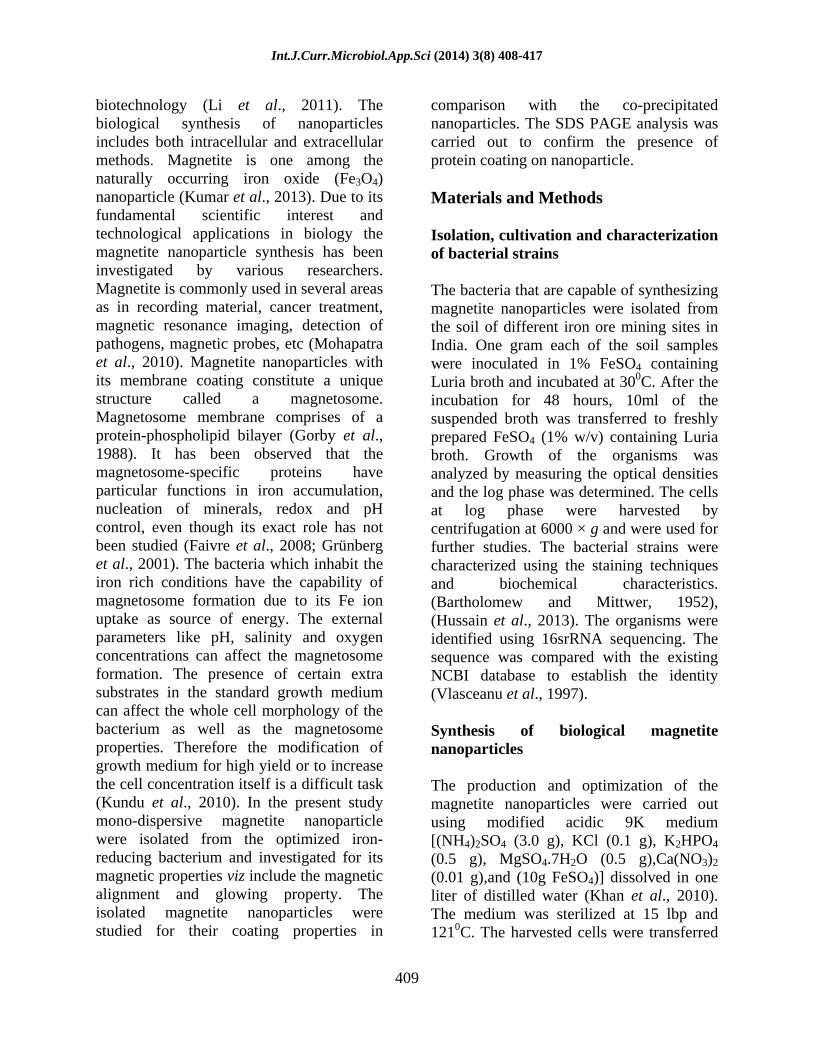
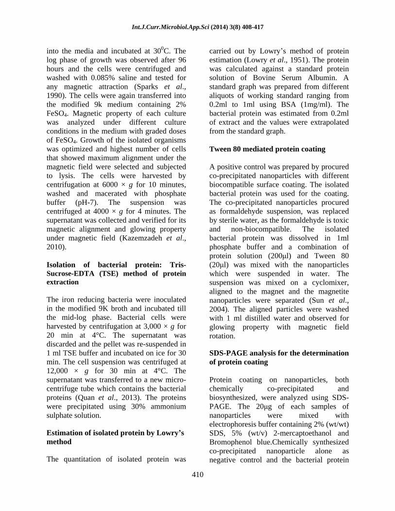
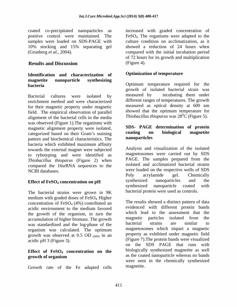
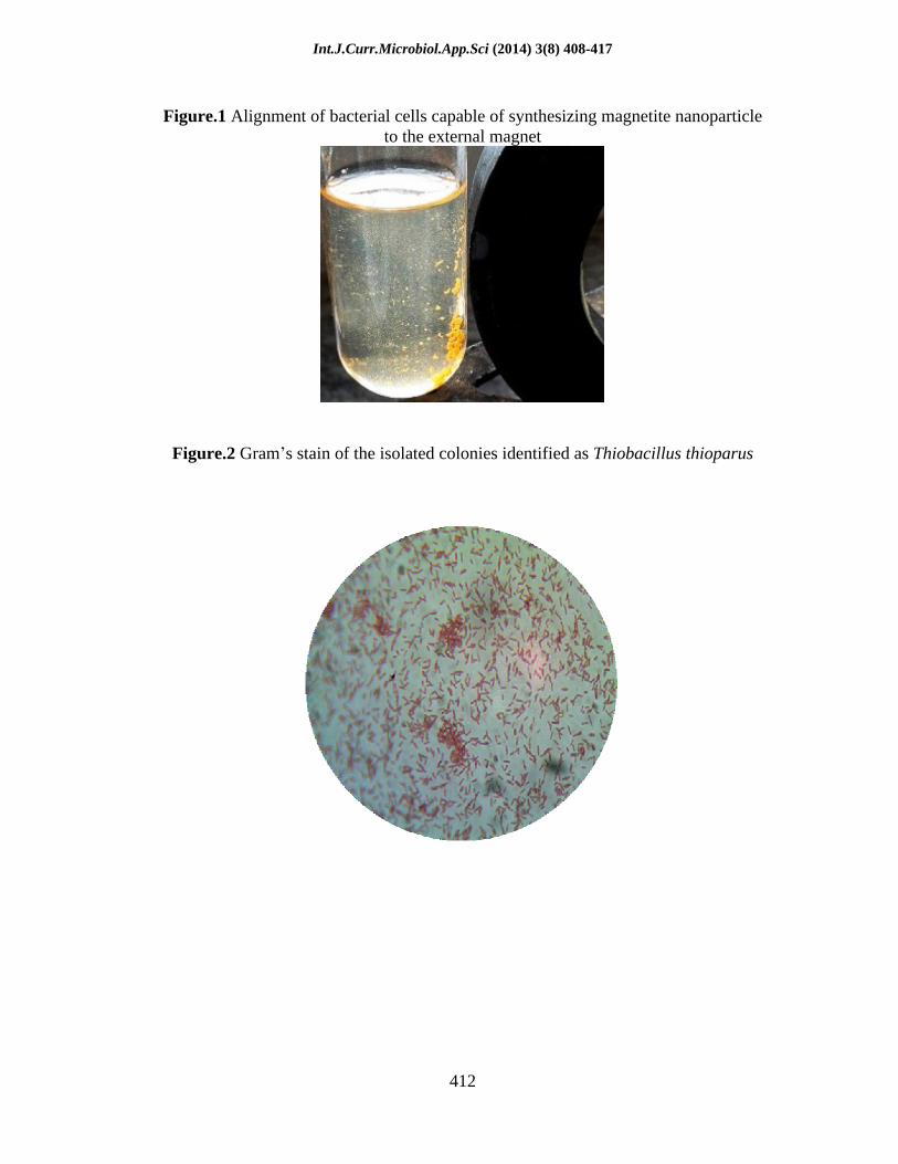
Bacterial cultures were isolated by enrichment method and were characterized for their magnetic property under magnetic field. The empirical observation of parallel alignment of the bacterial cells in the media was observed (Figure 1).The organisms with magnetic alignment property were isolated, categorized based on their Gram s staining pattern and biochemical characteristics. The bacteria which exhibited maximum affinity towards the external magnet were subjected to rybotyping and were identified as Thiobacillus thioparus (Figure 2) when compared the 16srRNA sequences to the NCBI databases.
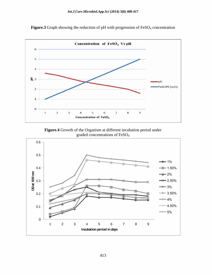
Effect of FeSO4 concentration on pH
The bacterial strains were grown in 9K medium with graded doses of FeSO4. Higher concentration of FeSO4 (4%) contributed an acidic environment to the medium favored the growth of the organism, in turn the accumulation of higher biomass. The growth was standardized and the log-phase of the organism was calculated. The optimum growth was observed at 0.5 OD (600) in an acidic pH 3 (Figure 3).
Effect of FeSO4 concentration on the growth of organism
Growth rate of the Fe adapted cells
increased with graded concentration of FeSO4. The organisms were adapted to the culture condition on acclimatization, as it showed a reduction of 24 hours when compared with the initial incubation period of 72 hours for its growth and multiplication (Figure 4).
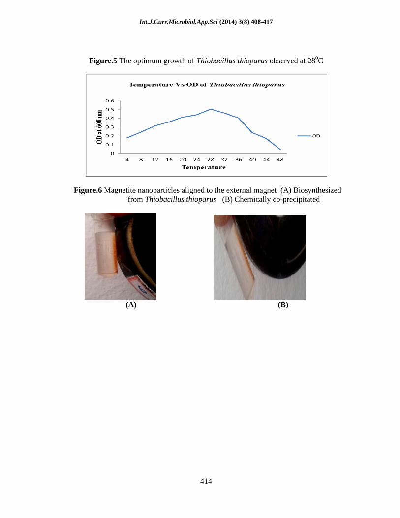
Optimization of temperature
Optimum temperature required for the growth of isolated bacterial strain was measured by incubating them under different ranges of temperatures. The growth measured as optical density at 600 nm showed that the optimum temperature for Thiobacillus thioparus was 280C (Figure 5).
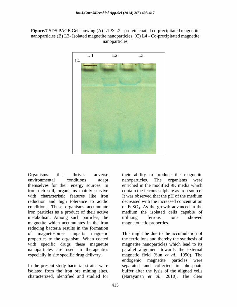
SDS- PAGE determination of protein coating on biological magnetite nanoparticles
Analysis and visualization of the isolated magnetosomes were carried out by SDS PAGE. The samples prepared from the isolated and acclimatized bacterial strains were loaded on the respective wells of SDS Poly acrylamide gel. Chemically synthesized nanoparticles and the synthesized nanoparticle coated with bacterial protein were used as controls.
The results showed a distinct pattern of data evidenced with different protein bands which lead to the assessment that the magnetic particles isolated from the bacterial strains are similar to magnetosomes which impart a magnetic property as exhibited under magnetic field (Figure 7).The protein bands were visualized on the SDS PAGE that runs with biologically synthesized magnetite as well as the coated nanoparticle whereas no bands were seen in the chemically synthesized magnetite.
Int.J.Curr.Microbiol.App.Sci (2014) 3(8) 408-417
412
Figure.1 Alignment of bacterial cells capable of synthesizing magnetite nanoparticle
to the external magnet
Figure.2 Gram s stain of the isolated colonies identified as Thiobacillus thioparus
Int.J.Curr.Microbiol.App.Sci (2014) 3(8) 408-417
413
Figure.3 Graph showing the reduction of pH with progression of FeSO4 concentration
Figure.4 Growth of the Organism at different incubation period under graded concentrations of FeSO4
0
0.1
0.2
0.3
0.4
0.5
0.6
1 2 3 4 5 6 7 8 9
OD
at 6
00 n
m
Incubation period in days
Int.J.Curr.Microbiol.App.Sci (2014) 3(8) 408-417
414
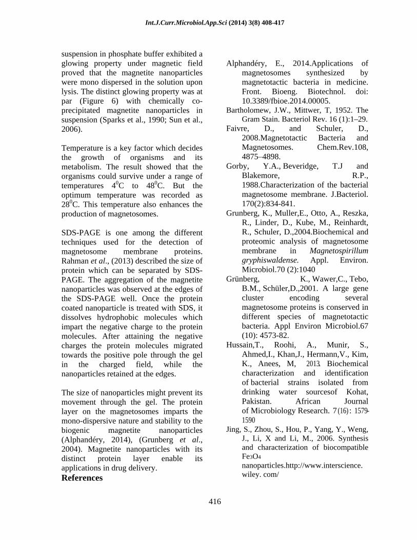
Figure.5 The optimum growth of Thiobacillus thioparus observed at 280C
Figure.6 Magnetite nanoparticles aligned to the external magnet (A) Biosynthesized from Thiobacillus thioparus (B) Chemically co-precipitated
(A) (B)
Int.J.Curr.Microbiol.App.Sci (2014) 3(8) 408-417
415
Figure.7 SDS PAGE Gel showing (A) L1 & L2 - protein coated co-precipitated magnetite nanoparticles (B) L3- Isolated magnetite nanoparticles, (C) L4 - Co-precipitated magnetite
nanoparticles
Organisms that thrives adverse environmental conditions adapt themselves for their energy sources. In iron rich soil, organisms mainly survive with characteristic features like iron reduction and high tolerance to acidic conditions. These organisms accumulate iron particles as a product of their active metabolism. Among such particles, the magnetite which accumulates in the iron reducing bacteria results in the formation of magnetosomes imparts magnetic properties to the organism. When coated with specific drugs these magnetite nanoparticles are used in therapeutics especially in site specific drug delivery.
In the present study bacterial strains were isolated from the iron ore mining sites, characterized, identified and studied for
their ability to produce the magnetite nanoparticles. The organisms were enriched in the modified 9K media which contain the ferrous sulphate as iron source. It was observed that the pH of the medium decreased with the increased concentration of FeSO4. As the growth advanced in the medium the isolated cells capable of utilizing ferrous ions showed magnetotactic properties.
This might be due to the accumulation of the ferric ions and thereby the synthesis of magnetite nanoparticles which lead to its parallel alignment towards the external magnetic field (Sun et al., 1990). The endogenic magnetite particles were separated and collected in phosphate buffer after the lysis of the aligned cells (Narayanan et al., 2010). The clear
L 1 L2 L3 L4
Int.J.Curr.Microbiol.App.Sci (2014) 3(8) 408-417
416
suspension in phosphate buffer exhibited a glowing property under magnetic field proved that the magnetite nanoparticles were mono dispersed in the solution upon lysis. The distinct glowing property was at par (Figure 6) with chemically co-precipitated magnetite nanoparticles in suspension (Sparks et al., 1990; Sun et al., 2006).
Temperature is a key factor which decides the growth of organisms and its metabolism. The result showed that the organisms could survive under a range of temperatures 40C to 480C. But the optimum temperature was recorded as 280C. This temperature also enhances the production of magnetosomes.
SDS-PAGE is one among the different techniques used for the detection of magnetosome membrane proteins. Rahman et al., (2013) described the size of protein which can be separated by SDS-PAGE. The aggregation of the magnetite nanoparticles was observed at the edges of the SDS-PAGE well. Once the protein coated nanoparticle is treated with SDS, it dissolves hydrophobic molecules which impart the negative charge to the protein molecules. After attaining the negative charges the protein molecules migrated towards the positive pole through the gel in the charged field, while the nanoparticles retained at the edges.
The size of nanoparticles might prevent its movement through the gel. The protein layer on the magnetosomes imparts the mono-dispersive nature and stability to the biogenic magnetite nanoparticles (Alphandéry, 2014), (Grunberg et al., 2004). Magnetite nanoparticles with its distinct protein layer enable its applications in drug delivery. References
Alphandéry, E., 2014.Applications of magnetosomes synthesized by magnetotactic bacteria in medicine. Front. Bioeng. Biotechnol.
doi:
10.3389/fbioe.2014.00005. Bartholomew, J.W., Mittwer, T, 1952. The
Gram Stain. Bacteriol Rev. 16 (1):1 29. Faivre, D., and Schuler, D.,
2008.Magnetotactic Bacteria and Magnetosomes. Chem.Rev.108, 4875 4898.
Gorby, Y.A., Beveridge, T.J and Blakemore, R.P., 1988.Characterization of the bacterial magnetosome membrane. J.Bacteriol. 170(2):834-841.
Grunberg, K., Muller,E., Otto, A., Reszka, R., Linder, D., Kube, M., Reinhardt, R., Schuler, D.,2004.Biochemical and proteomic analysis of magnetosome membrane in Magnetospirillum gryphiswaldense. Appl. Environ. Microbiol.70 (2):1040
Grünberg, K., Wawer,C., Tebo, B.M., Schüler,D.,2001. A large gene cluster encoding several magnetosome proteins is conserved in different species of magnetotactic bacteria. Appl Environ Microbiol.67 (10): 4573-82.
Hussain,T., Roohi, A., Munir, S., Ahmed,I., Khan,J., Hermann,V., Kim, K., Anees, M, 2013. Biochemical characterization and identification of bacterial strains isolated from drinking water sourcesof Kohat, Pakistan. African Journal of Microbiology Research. 7 (16) : 1579-1590
Jing, S., Zhou, S., Hou, P., Yang, Y., Weng, J., Li, X and Li, M., 2006. Synthesis and characterization of biocompatible Fe3O4
nanoparticles.http://www.interscience. wiley. com/
Int.J.Curr.Microbiol.App.Sci (2014) 3(8) 408-417
417
Kazemzadeh, H., A. Ataie., and F.
Rashchi., 2012. Synthesis of magnetite nano-particles by reverse co-precipitation. International Journal of Modern Physics: Conference Series.5, 160 167. doi: 10.1142/s2010194512001973
Khan, M.R., Saha, M.L., Begum,N., Islam, M. N., and Hoque, S., 2010.Isolation and characterization of bacteria from rusted iron materials. J.bot.39 (2):185- 191.
Kumar, R., Sen, S., 2013.Biogenic Magnetite Nanoparticle.Research Journal of Pharmaceutical, Biological and Chemical Sciences. 4(3):1037-1043.
Kundu,S., and Kulkarni,G.R., 2010. Enhancement of magnetotactic bacterial yield in a modified MSGM medium without alteration of magnetosomes properties. IJEB.48, 518-523.
Li, X., Xu, H., Chen, Z., and Chen, G., 2011. Biosynthesis of Nanoparticles by Microorganisms and Their Applications.Journal of Nanomaterials.2011, 16 pages Article ID 270974. http://dx.doi.org /10.1155/ 2011/270974
Lowry, O.H., Rosebrough N.J., Farr A.L., Randall R.J., 1951. Protein measurement with the Folin- phenol reagent. J. Biol. Chem. 193 (1): 26575.
Mohapatra, M and Anand,S.,2010. Synthesis and applications of nano-structured iron oxides/hydroxides a review. International Journal of Engineering, Science and Technology. 2(8):127-146.
Narayanan, K.B., Sakthivel, N., 2010. Biological synthesis of metal nanoparticles by microbes. Advances in Colloid and Interface Science. 156, 1 13.
Quan, S., Hinike, A., Collet, J.F., Bardwell, J.C, 2013. Isolation of bacteria envelope proteins. Methods Mol Biol.966, 359-66.
Rahman, M., Laurent, S., Tawil, N.,
Yahia, L.H., Mahmoudi, M, 2013.Protein- Nanoparticle Interactions: The Bio-Nano Interface. Springer-Verlag Berlin Heidelberg.
Sparks,N.H.C., Mann, S., Bazylinski, D.A., Lovley, D.R., Jannasch, H.W and Frankel,R.B.,1990.Structure and morphology of magnetite anaerobically-produced by a marine magnetotactic bacterium and a dissimilatory iron-reducing bacterium. Earth and Planetary Science Letters. 98,14-22.
Sun, W., Xiea, C., Wang, H.,Yu Hu., 2004. Specific role of polysorbate 80 coating on the targeting of nanoparticles to the brain. Biomaterials 25, 3065 3071.
Vlasceanu, L., Popa, R., and Kinkle, B.K., 1997.Characterization of Thiobacillus thioparus LV43 and its distribution in a chemoautotrophically based groundwater ecosystem. Appl Environ Microbiol.63 (8):3123 3127.