synthesis of positional-scanning libraries of fluorogenic peptide substrates to define the extended...
TRANSCRIPT

NATURE BIOTECHNOLOGY VOL 18 FEBRUARY 2000 http://biotech.nature.com 187
RESEARCH ARTICLES
Synthesis of positional-scanning librariesof fluorogenic peptide substrates to define
the extended substrate specificity ofplasmin and thrombin
Bradley J. Backes1,2†, Jennifer L. Harris3†, Francesco Leonetti1, Charles S. Craik3*, and Jonathan A. Ellman1*
1Chemistry Department, University of California Berkeley, Berkeley, CA 94720. 2Current Address: Chemistry Department, Genomics Institute of the Novartis ResearchFoundation, 3015 Merryfield Row, San Diego, CA 92121. 3Department of Pharmaceutical Chemistry, Program in Chemistry and Chemical Biology, University of
California San Francisco, San Francisco, CA 94143. †These authors contributed equally to this work. *Corresponding authors.
We have developed a strategy for the synthesis of positional-scanning synthetic combinatorial libraries(PS-SCL) that does not depend on the identity of the P1 substituent. To demonstrate the strategy, we syn-thesized a tetrapeptide positional library in which the P1 amino acid is held constant as a lysine and theP4-P3-P2 positions are positionally randomized. The 6,859 members of the library were synthesized onsolid support with an alkane sulfonamide linker, and then displaced from the solid support by condensa-tion with a fluorogenic 7-amino-4-methylcoumarin-derivatized lysine. This library was used to determinethe extended substrate specificities of two trypsin-like enzymes, plasmin and thrombin, which areinvolved in the blood coagulation pathway. The optimal P4 to P2 substrate specificity for plasmin was P4-Lys/Nle (norleucine)/Val/Ile/Phe, P3-Xaa, and P2-Tyr/Phe/Trp. This cleavage sequence has recently beenidentified in some of plasmin’s physiological substrates. The optimal P4 to P2 extended substratesequence determined for thrombin was P4-Nle/Leu/Ile/Phe/Val, P3-Xaa, and P2-Pro, a sequence found inmany of the physiological substrates of thrombin. Single-substrate kinetic analysis of plasmin and throm-bin was used to validate the substrate preferences resulting from the PS-SCL. By three-dimensional struc-tural modeling of the substrates into the active sites of plasmin and thrombin, we identified potentialdeterminants of the defined substrate specificity. This method is amenable to the incorporation of diversesubstituents at the P1 position for exploring molecular recognition elements in proteolytic enzymes.
Keywords: Serine protease, proteinase, blood coagulation, molecular recognition, combinatorial libraries
Proteases play essential roles in numerous biological processes.Substrate specificity, or the ability to discriminate among manypotential substrates, is central to the function of proteases.Knowledge of a protease’s substrate specificity may not only givevaluable insights into its biological function but also provide thebasis for potent substrate and inhibitor design. Synthetic substratesare typically used to define substrate specificity. However, the syn-thesis and assay of single substrates is tedious for proteases withspecificity beyond P1 and often results in a limited substrate speci-ficity profile.
Combinatorial approaches have recently been used to address theidentification of several substrate recognition sites in proteases. Allof these combinatorial methods involve the generation of libraries ofsubstrates, proteolysis of the substrates, and identification of theoptimal substrate sequence. Substrate libraries can be broken downinto two categories: those that are biologically generated and thosethat are synthetically generated. Biological library methods includethe display of peptide libraries on filamentous phage1,2, the random-ization of amino acids at physiological cleavage sites3, and the identi-fication of macromolecular cleavage sites of in vitro trans-cription/translation cDNA libraries4,5. The diversity of these librariesis often constrained by the transformation efficiency of the hostorganism and can only contain naturally occurring amino acids.This limitation can be circumvented by use of synthetic substratelibraries. Although combinatorial synthesis allows for the creation of
millions of compounds, these methods are only useful when cou-pled with powerful analytical assays that allow for the identificationof the preferred substrate. Discontinuous analysis of the cleavageproducts through Edman degradation6,7, mass spectrometry8,9, andchromatography10 has proved useful for qualitative assessment ofoptimal substrates from soluble or support-bound peptides.
Substrate consensus sequences have also been obtained usingsupport-bound fluorescence-quenched substrate libraries preparedby the process of split-synthesis, which results in single substratesequences on each of the resin beads11. Partial proteolysis of the sup-port-bound libraries and subsequent sequence determination of thesubstrates on the most fluorescent beads provides the consensussequences12,13. Unfortunately, this method suffers from two majorlimitations: first, the kinetics of support-bound substrates can differgreatly from soluble substrates 14,15, and second, as with the othermethods previously mentioned, identification of the substrateoccurs after the cleavage event, making the kinetic analysis morecumbersome. A method that avoids these limitations, and gives aquantitative assessment of protease substrate preference, is the useof positional-scanning synthetic combinatorial libraries (PS-SCL)16.
Positional-scanning 17 synthetic combinatorial libraries (PS-SCL) of fluorogenic peptide substrates are potentially powerful toolsfor determining protease specificity. In contrast to other combinato-rial libraries, this library format provides rapid and continuousinformation on each of the varied substituents in the substrate. A
© 2000 Nature America Inc. • http://biotech.nature.com©
200
0 N
atu
re A
mer
ica
Inc.
• h
ttp
://b
iote
ch.n
atu
re.c
om

positional-scanning library with the general structure Ac-X-X-X-Asp-AMC was prepared previously by Rano et al. to rapidly andaccurately assess the P4-P2 specificity for caspases that requireaspartic acid (Asp) in the P1 position18,19. (Nomenclature for thesubstrate amino acid preference is Pn, Pn-1, . . .P2, P1, P1′, P2′, . . . ,Pm-1′, Pm′. Amide bond hydrolysis occurs between P1 and P1′. Sn,Sn-1, . . . , S2, S1, S1′, S2′, . . . , Sm-1′, Sm′ denotes the correspondingenzyme binding sites20). Specific cleavage of the amide bond after theAsp residue liberates a fluorescent 7-amino-4-methylcoumarin(AMC) leaving group, thus allowing for the simple determination ofcleavage rates for a library of substrates. The P1 Asp-coumarin sub-strate was conveniently linked to an insoluble polymer through theAsp carboxylic acid side chain, which allowed for library synthesis bystandard peptide synthesis techniques. However, the method used tosynthesize the library was specific for an aspartic acid at P1. Theemployment of strategies to link P1 amino acid-coumarin deriva-tives through side chain functionality may prove viable for someresidues. However, linkage through hydrophobic side chain func-tionalities (leucine, phenylalanine, valine, etc.) will prove difficult,limiting the use of this strategy.
By developing a general strategy toincorporate all 20 proteinogenic aminoacids at the P1 position of a PS-SCL, theextended specificity of virtually any pro-tease could be rapidly determined. Withthis aim in mind, we have developed ageneral method for the preparation andscreening of positional-scanning synthet-ic combinatorial substrate libraries. Thisdesign is free from the limitations of theprevious approach, which requires link-age to the solid support through the sidechain of the P1 substituent and allows forcomplete randomization at the P1 posi-tion.The resulting enzymatic profilesfrom the library resemble the knownphysiological cleavage sites of theseenzymes and were verified by single-sub-strate kinetic analysis. Potential substraterecognition determinants on the enzymeswere identified through the three-dimen-sional modeling of the substrates in theactive sites of the enzymes.
ResultsLibrary design and synthesis. To incor-porate diversity at the P1 position, wecondensed fluorogenic AMC P1-aminoacid derivatives with a support-boundPS-SCL to provide library compounds in
solution (Fig. 1). Three support-bound sublibraries were prepared(P2, P3, P4) using an alkane sulfonamide linker21 and solid-phasepeptide synthesis. The properties of the linker allow for the incorpo-ration of a fluorogenic leaving group through the nucleophilic addi-tion of an AMC-derivatized amino acid. Each sublibrary consisted of19 resins (one unnatural amino acid, norleucine, was included; cys-teine and methionine were excluded) for which a single position wasspatially addressed by the coupling of a single amino acid. The tworemaining positions of each resin were supplied by the coupling ofisokinetic mixtures22 to give a resin-bound mixture of 361 aminoacids. The 57 resins comprising the entire PS-SCL were put in indi-vidual wells and cleaved from the resin with a P1-amino acid-coumarin derivative. Filtration, side chain deprotection, and con-centration provided a PS-SCL of 57 wells containing 361 tetrapep-tide-coumarin derivatives (total of 6,859 peptides). Thus, analysis ofthe three libraries identifies the enzyme’s preferences for amino acidsat P4, P3, and P2. A P1-Lys library was prepared and used to eluci-date the specificity of plasmin and thrombin.
Profiling of plasmin with the positional scanning P1-Lyslibrary. The preferred tetrapeptide substrate recognition sequencefor plasmin was determined to be P4-Lys, P3-Xaa (a nonspecificamino acid), P2-Tyr/Phe/Trp, and P1-Lys (Fig. 2A). To validate theresults from the PS-SCL and to quantitate dependence and use ofextended interactions, kinetic parameters were determined for sev-eral single AMC substrates (Table 1). As indicated from the library,the majority of plasmin’s extended substrate specificity resides inP2 and P4. Hydrolysis of the suboptimal P2 substrate, Ac-Lys-Thr-Ser-Lys-AMC, is up to 340% disfavored when compared to sub-strates that possess an aromatic amino acid at P2: Ac-Lys-Thr-Tyr-Lys-AMC, Ac-Lys-Thr-Phe-Lys-AMC, and Ac-Lys-Thr-Trp-Lys-AMC (Table 1), 0.020 µM-1 s-1 in kcat/KM versus 0.544 µM-1 s-1, 0.677µM-1 s-1, and 0.601 µM-1 s-1, respectively. The subtle preference forlysine over phenylalanine at P4 is also verified by single substrates:Ac-Phe-Thr-Tyr-Lys-AMC retains 63% of the activity of Ac-Lys-Thr-Tyr-Lys-AMC, 0.342 µM-1 s-1 in kcat/KM and 0.544 µM-1 s-1 in
188 NATURE BIOTECHNOLOGY VOL 18 FEBRUARY 2000 http://biotech.nature.com
RESEARCH ARTICLES
Figure 1. Three sublibraries (P2, P3, P4), each made up of 19 wellscontaining 361 compounds, are prepared by a segment condensationreaction with a P1 fluorogenic amino acid substrate. Individualproteinogenic amino acids are used to incorporate spatiallyaddressed positions “Z,” whereas an isokinetic mixture ofproteinogenic amino acids is used to incorporate varied positions “X.”
Figure 2. (A) Activity of plasmin in a P1-Lys positional-scanning synthetic combinatorial library. (B)Activity of thrombin in a P1-Lys positional-scanning synthetic combinatorial library. y-axis ispicomolar of fluorophore released per second. x-axis indicates the amino acid held constant ateach position, designated by the one-letter code (with n representing norleucine).
A B
© 2000 Nature America Inc. • http://biotech.nature.com©
200
0 N
atu
re A
mer
ica
Inc.
• h
ttp
://b
iote
ch.n
atu
re.c
om

NATURE BIOTECHNOLOGY VOL 18 FEBRUARY 2000 http://biotech.nature.com 189
kcat/KM, respectively.Structural determinants of P4-lysine and P2-aromatic substrate
specificity in plasmin. The structure of plasmin was solved in theabsence of a substrate or inhibitor in the active site23. Because of this,analysis of enzyme–substrate interactions required the molecularmodeling of the optimal substrate, Lys-Thr-Phe-Lys, into the activesite. The resulting model reveals potential structural determinantsfor substrate recognition. As is appreciated for trypsin, the majordeterminant for P1-basic specificity lies in Asp189 (according tochymotrypsinogen numbering), a residue at the base of the S1 pock-et (Fig. 3A). The (δ+) ring hydrogens from P2-Phe can interact withthe carboxylate group of Glu60 that protrudes from above the activesite and toward the P2-Phe ring edge. The positively charged (δ+)amino group of Gln192 could then make contact with the (δ-) π-electrons of the P2-Phe face (Fig. 3A). Significant interactionsbetween plasmin and the P3-amino acid side chain are not readilyapparent from the structural model. This is a result of the P3-aminoacid side chain being directed away from the enzyme and into bulksolvent. A substrate with a P4-Lys could make contact with Glu180,with the aliphatic portion of P4-Lys packing against Trp215 (Fig.3A). Position 180 is normally occupied by a hydrophobic amino acidin other chymotrypsin-like serine proteases. The additional, thoughlesser, preference for P4-Nle/Val/Ile/Phe could in part be due tofavorable interaction with Trp215.
Profiling of thrombin with the positional scanning P1-Lyslibrary. Profiling of thrombin with the PS-SCL revealed that the pre-ferred P4-P2 extended substrate specificity is for large aliphatic
amino acids at P4, such as norleucine,leucine, and isoleucine, negligible discrimi-nation at P3, and narrow specificity for pro-line at P2 (Fig. 2B). These preferences werevalidated through single-substrate kineticanalysis (Table 1). Replacement of the opti-mal P2 amino acid proline with the subop-timal amino acid leucine results in a 45-folddecrease in activity, 3.83 µM-1 s-1 in kcat/KM
for Ac-Nle-Thr-Pro-Arg-AMC versus 0.085µM-1 s-1 in kcat/KM for Ac-Nle-Thr-Leu-Arg-AMC. The requirement for aliphatic aminoacids at P4 has a less pronounced effect thanthe requirement for proline at P2, as reflect-ed by the relative activities in the PS-SCL.However, upon replacement of the pre-ferred leucine at P4 for the suboptimal
glycine, there is a 23-fold decrease in specific activity, 0.154 µM-1 s-1
in kcat/KM for Ac-Leu-Gly-Val-Arg-AMC versus 0.007 µM-1 s-1 inkcat/KM for Ac-Gly-Gly-Val-Arg-AMC.
Structural determinants of P4-aliphatic and P2-proline sub-strate specificity in thrombin. The coordinates used forenzyme–substrate analysis of thrombin were that of thrombin com-plexed with the tripeptide inhibitor, D-Phe-Pro-Arg-chloromethylketone24. The P3 side chain was converted to a threo-nine of the L-enantiomer, and a P4-Val was added to the N terminusof the inhibitor. As suggested from the original structural analysis byBode et al., the preference for P1-basic amino acids is determined byAsp189 and the preference for proline arises from the insertion, rela-tive to the digestive serine proteases, of seven amino acids in the 60’sloop24. This loop is above the active site Ser195 and creates a rigidpocket for P2-Pro interaction (Fig. 3B). The specificity for P3 aminoacids is less well understood from the original structure. The use ofP3-D-Phe allows the side chain to point into the enzyme, occupying,in part, the S4 pocket. When the P3-D-Phe is replaced with P3-L-Thr, the side chain is pointed away from the enzyme with signifi-cantly fewer interactions (Fig. 3B). The S4 pocket on thrombin isvery clearly hydrophobic, with Ile174 making significant interac-tions with P4-Val modeled into the pocket. Additional hydrophobicdeterminants for this pocket include Trp215 at the floor of the pock-et, Met180 at the end of the pocket, and Leu99 near the top of thepocket (Fig. 3B).
DiscussionThe rapid discovery of new proteases presents the need for general-ized assays to aid in the elucidation of their biological functions. Theuse of synthetic positional-scanning combinatorial libraries offersthe ability to rapidly test and evaluate the extended substrate speci-ficity of a protease. The major limitation of previous synthetic meth-
RESEARCH ARTICLES
Figure 3. (A) Three-dimensional model of plasmin bound to the tetrapeptide substrate Lys-Thr-Phe-Lys. (B) Three-dimensional model of thrombin bound to the tetrapeptide substrate Leu-Thr-Pro-Arg.The Connolly surface of the enzyme is shown in white mesh. The enzyme backbone is shown inwhite, with the side chains shown in atom colors (white, carbon; blue, nitrogen; red, oxygen; yellow,sulfur) with the catalytic triad (His57, Asp102, Ser195) in orange. The substrate is shown in magenta.
BA
Table 1. Kinetic constants for plasmin and thrombin on single AMCsubstrates.
Substrate kcat (s-1) KM (µM) kcat/KM (µM-1 s-1)
Plasmin
Ac-Lys-Thr-Tyr-Lys-AMC 11.3 ± 0.4 20.8 ± 3.3 0.544 ± 0.071Ac-Lys-Thr-Phe-Lys-AMC 20.1 ± 0.6 29.7 ± 3.6 0.677 ± 0.069Ac-Lys-Thr-Trp-Lys-AMC 11.9 ± 0.3 19.9 ± 2.2 0.601 ± 0.054Ac-Lys-Thr-Ser-Lys-AMC 8.8 ± 1.3 440 ± 100 0.020 ± 0.002Ac-Phe-Thr-Tyr-Lys-AMC 17.5 ± 0.9 51.0 ± 10.2 0.342 ± 0.054Ac-Leu-Thr-Phe-Lys-AMC 33.2 ± 3.9 296 ± 70 0.112 ± 0.015Ac-Leu-Glu-Phe-Lys-AMC 5.5 ± 0.3 74.6 ± 9.9 0.073 ± 0.006
Thrombin
Ac-Nle-Thr-Pro-Arg-AMC 45.0 ± 1.1 11.3 ± 1.3 3.83 ± 0.35Ac-Val-Thr-Pro-Arg-AMC 30.8 ± 1.3 29.6 ± 4.5 1.04 ± 0.13Ac-Nle-Thr-Leu-Arg-AMC 5.8 ± 0.3 67.4 ± 10.7 0.085 ± 0.009Ac-Leu-Gly-Val-Arg-AMC 15.6 ± 1.8 101.5 ± 29.5 0.154 ± 0.029Ac-Gly-Gly-Val-Arg-AMC 1.2 ± 0.1 180.7 ± 55.3 0.007 ± 0.001
Table 2. AMC substrate mass spectral data.
Substrate Mass calculated Mass found(g/mol) (g/mol)
Ac-Lys-Thr-Tyr-Lys-AMC 738.5 738.9Ac-Lys-Thr-Phe-Lys-AMC 722.5 723.0Ac-Lys-Thr-Trp-Lys-AMC 761.5 761.3Ac-Lys-Thr-Ser-Lys-AMC 662.5 662.8Ac-Phe-Thr-Tyr-Lys-AMC 757.5 758.0Ac-Leu-Thr-Phe-Lys-AMC 707.5 707.8Ac-Leu-Glu-Phe-Lys-AMC 735.5 735.9Ac-Nle-Thr-Pro-Arg-AMC 685.5 685.8Ac-Val-Thr-Pro-Arg-AMC 671.5 671.8Ac-Nle-Thr-Leu-Arg-AMC 701.5 702.0Ac-Leu-Gly-Val-Arg-AMC 643.5 643.8Ac-Gly-Gly-Val-Arg-AMC 587.4 587.8
© 2000 Nature America Inc. • http://biotech.nature.com©
200
0 N
atu
re A
mer
ica
Inc.
• h
ttp
://b
iote
ch.n
atu
re.c
om
ods is the inability to permit complete diversity at the P1 position.The development of the synthetic strategy presented in this papersurmounts this limitation and allows for complete diversity of anynaturally occurring amino acid at the P1 position. In fact, a varietyof nucleophiles, including unnatural amino acid derivatives, couldbe incorporated in the cleavage step.
The use of a positional scanning library with P1 lysine held con-stant was employed to determine the extended specificity of plasminand thrombin, proteases involved in the regulation of hemostasis.Plasmin has been traditionally characterized as a protease withbroad substrate specificity. Results from the current study show that,on the contrary, plasmin demonstrates a distinct preference for aro-matic amino acids at P2 and a moderate preference for lysine andhydrophobic amino acids at P4. Molecular modeling of a substratebound into the active site of plasmin can aid in the identification ofpotential structural interactions between enzyme and substrate.Plasmin has an insertion in the 60’s loop, relative to the digestiveprotease trypsin, that could allow for the creation of a S2 pocket.Plasmin’s S2 pocket is not simply a hydrophobic pocket, but is spe-cific for aromatic amino acids. Experimental crystallographic evi-dence exists for structural determinants used to achieve discrimina-tion between aromatic and aliphatic amino acids, including aromat-ic ring interactions with oxygen25 and amide nitrogens26. Likewise,modeling of a P2-Phe into the putative S2 pocket shows that Glu60amay contribute to ring hydrogen bonding, whereas the polar aminogroup of Gln192 may interact with the π-electrons of P2-Phe (Fig.3A). The P3-amino acid side chain is pointed out into solvent andmakes few interactions with the enzyme. The moderate P4 prefer-ence for lysine may be driven by an electrostatic interaction withGlu180 (Fig. 3A). The aliphatic portion of the lysine side chain aswell as other aliphatic amino acids (Nle/Val/Ile) could form favor-able interactions with Trp215. Preference for both charged and aro-matic amino acids is also known for the S2 pocket of the cysteineprotease cruzain, where a glutamate side chain adjusts to accommo-date the disparate side chains27.
Plasmin is an important enzyme in attenuating blood coagulationand restoring blood flow through its degradation of fibrin and proco-agulant factors28,29. Several nonfibrin substrates of plasmin haverecently been demonstrated30–32. Although the cleavage sites of thephysiological substrates have not all been determined, many of thosethat have been identified resemble the optimal site determined in thecurrent study, especially in the conservation of a P2-aromatic aminoacid (Fig. 4). It has been proposed that plasmin may function in accel-erating fibrinolysis and arresting coagulation through the cleavage ofthe normally procoagulant factor X into an anticoagulant cofactor33. Itwas determined that plasmin cleaves Factor X after the arginine in thesite Ile-Thr-Phe-Arg. In a similar vein, plasmin may act in the desensi-tization of the protease activated receptor PAR1. PAR1 is a transmem-brane receptor that when cleaved by thrombin results in the activationof platelets; however plasmin cleaves PAR1 at sites that not only do notresult in activation but also remove the thrombin activation site34. Oneof the plasmin cleavage sites in PAR1 was determined as Thr-Glu-Tyr-Arg. Plasmin may attenuate its own production through the cleavageof vitronectin, a protein that binds both plasminogen activatorinhibitor 1 (PAI-1) and extracellular matrix (ECM)35,36. The cleavage
of vitronectin by plasmin may result in the release of PAI-1 from theECM to inhibit plasminogen activators and subsequent inhibition ofplasmin formation. The cleavage site of vitronectin is Lys-Gly-Tyr-Arg, a site resembling the optimal site determined in this study.Plasmin may also play a role in regulating bone resorption through thecleavage of osteocalcin at the site Glu-Ala-Tyr-Arg37.
Thrombin has been shown to be more restrictive in its substratecleavage profile than trypsin, a protease with the chief function ofdigestion rather than regulation38,39. This is due, in part, to prefer-ences exhibited in the S3-S2’ subsites40,41. Here, for the first time, thesequence space for thrombin’s P4, P3, and P2 substrate preferencehas been completely sampled. As with previous results38,41–43, the PS-SCL shows that thrombin has a pronounced preference for prolineat the P2 position of the substrate. The structure of thrombinbound to the tripeptide inhibitor, D-Phe-Pro-Arg-chloromethylke-tone, reveals that the constraints for proline in the P2 position mostprobably arise from the insertion in the 60’s loop of thrombin24.When a P3-L-amino acid was modeled into the active site, ratherthan the P3-D-Phe solved in the structure, the side chain had mini-mal interaction with the enzyme and was pointed out into solvent(Fig. 3B). This structural analysis of the P3–S3 interaction supportsthe lack of defined P3 specificity seen in the substrate library.However, a P4-aliphatic amino acid modeled into the active site canmake significant interactions with a hydrophobic pocket of throm-bin. The walls of this pocket are formed predominantly by Ile174,other amino acids such as Met180, Leu99, and Trp215 also con-tribute to this hydrophobic environment (Fig. 3B). The interactionbetween Ile174 and a P4-aliphatic amino acid was also observed inthe structure of thrombin bound to the peptide fragment Leu-Asp-Pro-Arg44. The structural analysis was supported by the substratelibrary analysis that showed thrombin to have a definite preferencefor P4-aliphatic amino acids.
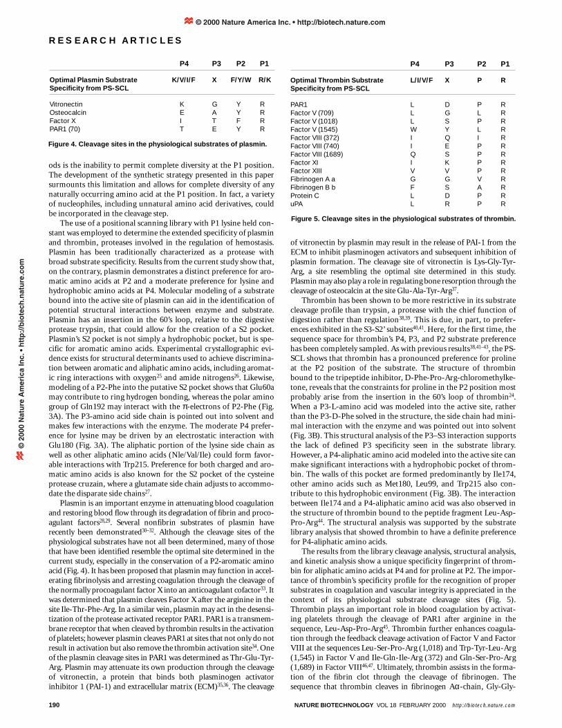
The results from the library cleavage analysis, structural analysis,and kinetic analysis show a unique specificity fingerprint of throm-bin for aliphatic amino acids at P4 and for proline at P2. The impor-tance of thrombin’s specificity profile for the recognition of propersubstrates in coagulation and vascular integrity is appreciated in thecontext of its physiological substrate cleavage sites (Fig. 5).Thrombin plays an important role in blood coagulation by activat-ing platelets through the cleavage of PAR1 after arginine in thesequence, Leu-Asp-Pro-Arg45. Thrombin further enhances coagula-tion through the feedback cleavage activation of Factor V and FactorVIII at the sequences Leu-Ser-Pro-Arg (1,018) and Trp-Tyr-Leu-Arg(1,545) in Factor V and Ile-Gln-Ile-Arg (372) and Gln-Ser-Pro-Arg(1,689) in Factor VIII46,47. Ultimately, thrombin assists in the forma-tion of the fibrin clot through the cleavage of fibrinogen. Thesequence that thrombin cleaves in fibrinogen Aα-chain, Gly-Gly-
190 NATURE BIOTECHNOLOGY VOL 18 FEBRUARY 2000 http://biotech.nature.com
RESEARCH ARTICLES
Figure 5. Cleavage sites in the physiological substrates of thrombin.
P4 P3 P2 P1
Optimal Thrombin Substrate L/I/V/F X P RSpecificity from PS-SCL
PAR1 L D P RFactor V (709) L G L RFactor V (1018) L S P RFactor V (1545) W Y L RFactor VIII (372) I Q I RFactor VIII (740) I E P RFactor VIII (1689) Q S P RFactor XI I K P RFactor XIII V V P RFibrinogen A a G G V RFibrinogen B b F S A RProtein C L D P RuPA L R P R
Figure 4. Cleavage sites in the physiological substrates of plasmin.
P4 P3 P2 P1
Optimal Plasmin Substrate K/V/I/F X F/Y/W R/KSpecificity from PS-SCL
Vitronectin K G Y ROsteocalcin E A Y RFactor X I T F RPAR1 (70) T E Y R
© 2000 Nature America Inc. • http://biotech.nature.com©
200
0 N
atu
re A
mer
ica
Inc.
• h
ttp
://b
iote
ch.n
atu
re.c
om

NATURE BIOTECHNOLOGY VOL 18 FEBRUARY 2000 http://biotech.nature.com 191
Val-Arg, is suboptimal by the current analysis. However, it has beendemonstrated that the fibrinogen Aα-chain supplements its specificbinding energy through the use of thrombin’s distal anion-bindingexosite 1 (ref. 48). Fibrinogen Bβ-chain has a more optimal throm-bin cleavage site, Phe-Ser-Ala-Arg, and has not been shown to usethe anion-binding exosite 1. As thrombin moves away from thewound site, it aids in the attenuation of blood clotting through theactivation of protein C at the site, Leu-Asp-Pro-Arg49. Althoughthrombin plays several roles in blood coagulation, the commontheme is the requirement for proper recognition elements in thephysiological substrates. Information from the P1-Lys PS-SCL facil-itates the identification of recognition elements for productivethrombin–substrate interaction.
Use of this method was demonstrated for the serine proteases,plasmin and thrombin, two enzymes that require P1-basic aminoacids. This library can have broad application to other enzymesbecause proteases that cleave P1-basic amino acids are well repre-sented in the serine and cysteine protease families. The definedextended substrate specificity for plasmin and thrombin was inagreement with the cleavage sites in known physiological substrates.Substrate specificity information can aid in the discovery of newphysiological substrates and the cleavage sites within substrates. Adirect outcome from the library analysis is the creation of sensitivesubstrates to monitor activity. This information can also be used as astarting point in the design and synthesis of potent and selectiveinhibitors.
Experimental protocolMaterials. Unless otherwise noted, chemicals were obtained from commer-cial suppliers and used without further purification. Aminomethyl Merrifieldresin was purchased from Novabiochem (San Diego, CA), and the substitu-tion concentration of the resin was determined (0.84 mEq/g) by using a spec-trophotometric 9-fluorenylmethoxycarbonyl (Fmoc) quantitation assay50.Alkane sulfonamide resin (0.75 mmol/g) was prepared by the method ofBackes and Ellman21 or purchased from Novabiochem. Fmoc-amino acidswere purchased from Novabiochem. Iodoacetonitrile and anhydrous NMP(N-methylpyrrolidone) were purchased from Aldrich. Iodoacetonitrile wasfiltered through a small plug of basic alumina immediately before use.Anhydrous, low amine content dimethylformamide (DMF) was purchasedfrom EM Science (Cincinnati, OH). High-loading tosyl chloride resin (PS-TsCl, 1.55 mmol/g) was purchased from Argonaut Technologies (San Carlos,CA). An Argonaut Quest 210 Organic Synthesizer was used for library syn-thesis. Chromatography was carried out using Merck 60 230–240 mesh silicagel according to the procedure of Still51. Thin-layer chromatography was car-ried out on Merck 60 F254 250 µm silica gel plates. Infrared (IR) spectra wererecorded neat (for oils) and as films from CH2Cl2 or CHCl3 (for crystallinecompounds) and only partial data is reported. NMR chemical shifts arereported in p.p.m. downfield from an internal solvent peak, or trimethylsi-lane, and J values are in hertz. Elemental analyses were done by M-H-W Labs(Phoenix, AZ). A Savant Speed Vac Plus was used for concentrating single-substrate solutions in vials and library member solutions configured inmicrotiter plates. The human enzymes, thrombin and plasmin were pur-chased from Haematologic Technologies. (Essex Junction, VT) and used asreceived.
Library synthesis. Fmoc-amino acids (Fmoc-Ala-OH, Fmoc-Arg(Pbf,2,2,4,6,7-pentamethyldihydrobenzofuran-5-sulfonyl)-OH, Fmoc-Asn(Trt,trityl)-OH, Fmoc-Asp(O-t-Bu, t-butyl)-OH, Fmoc-Glu(O-t-Bu)-OH,Fmoc-Gln(Trt)-OH, Fmoc-Gly-OH, Fmoc-His(Boc, tert-butoxycarbonyl)-OH, Fmoc-Ile-OH, Fmoc-Leu-OH, Fmoc-Lys(Boc)-OH, Fmoc-Nle, Fmoc-Phe-OH, Fmoc-Pro-OH, Fmoc-Ser(O-t-Bu)-OH, Fmoc-Thr(O-t-Bu)-OH,Fmoc-Trp(Boc)-OH, Fmoc-Tyr(O-t-Bu)-OH, Fmoc-Val-OH) were coupledto the alkane sulfonamide resin21, and the amino acid loading concentrationswere determined using a spectrophotometric Fmoc quantitation assay50. Forthe preparation of the P2 sublibrary, 0.10 mmol (∼ 200 mg) of each of the 19Fmoc-amino acid resins were added to 19 reaction vessels (one Fmoc-aminoacid resin/vessel) of the Argonaut Quest 210 Organic Synthesizer and solvat-ed with DMF (2 ml/vessel). After agitating 20 min, the DMF was drained anda solution of 20% piperidine in DMF (2 ml/vessel) was added. The resin wasagitated for 25 min, filtered, and washed with DMF (3 × 2 ml/vessel). To
install the randomized P3 position, 10 equivalents (∼ 1.0 mmol/well, 19mmol) of an isokinetic mixture22 of Fmoc-amino acids (Fmoc-amino acid,mole%: Fmoc-Ala-OH, 3.4; Fmoc-Arg(Pbf)-OH, 6.5; Fmoc-Asn(Trt)-OH,5.3; Fmoc-Asp(O-t-Bu)-OH, 3.5; Fmoc-Glu(O-t-Bu)-OH, 3.6; Fmoc-Gln(Trt)-OH, 5.3; Fmoc-Gly-OH, 2.9; Fmoc-His(Boc)-OH, 3.5; Fmoc-Ile-OH, 17.4; Fmoc-Leu-OH, 4.9; Fmoc-Lys(Boc)-OH, 6.2; Fmoc-Nle, 3.8;Fmoc-Phe-OH, 2.5; Fmoc-Pro-OH, 4.3; Fmoc-Ser(O-t-Bu)-OH, 2.8; Fmoc-Thr(O-t-Bu)-OH, 4.8; Fmoc-Trp(Boc)-OH, 3.8; Fmoc-Tyr(O-t-Bu)-OH,4.1; Fmoc-Val-OH, 11.3) was preactivated with DICI (diisopropylcarbodi-imide)(3.0 ml, 19 mmol), and HOBt(1-hydroxybenzotriazole) (2.6 mg, 19mmol) in DMF (57 ml) in a 100 ml round bottom flask. After the 2 min pre-activation period, 3 ml of the solution was added to each of the 19 reactionvessels. The resin was agitated for 3 h, filtered, and washed with DMF (3 × 2ml/vessel). After Fmoc removal (treatment with 20% piperidine in DMF (2ml/vessel), agitation for 25 min, filtration, and washing with DMF (3 × 2ml/vessel)) the randomized P4 position was incorporated in an identicalmanner. The Fmoc removal step was followed by filtration and washing withDMF (3 × 2 ml/vessel). A capping solution of AcOH (7.6 mmol), DICI (1.2ml, 7.6 mmol), HOBt (2.3 g, 7.6 mmol), and DMF (38 ml) was premixed in a100 ml round bottom flask, and 2 ml was added to each of the 19 reaction ves-sels. After agitating for 3 h, each resin was filtered, washed (DMF, 3 × 2ml/vessel; THF, 3 × 2 ml/vessel; MeOH, 3 × 2 ml/vessel), and dried overnightunder high vacuum with P2O5.
To install the P1 residue, the 19 resins with a fixed P2 residue (0.020 mmol,∼ 40 mg) were added to the reaction vessels (one fixed P2 residue/vessel),swollen with NMP (1 ml/vessel), agitated for 20 min, and filtered. A solutionof ICH2CN (1.4 ml, 19 mmol), i-Pr2EtN (0.65 ml, 3.8 mmol), and NMP (19ml) was prepared and added to each of the 19 reaction vessels (1 ml/vessel).The vessels were shielded from light with aluminum foil. After agitating 24 h,each resin was filtered, washed with NMP (5 × 2 ml/vessel), and DMF (5 × 2ml), and filtered again. Resin washes were agitated for 5 min/wash. A solutionof coumarin-Lys(Boc)-NH2 (760 mg, 1.9 mmol) in DMF (9.5 ml) was pre-pared and 0.5 ml (5 equivalents) of the solution was added to each vessel. Theresin was agitated at 80°C for 12 h to liberate the coumarin-tetrapeptidederivatives. The 19 reaction mixtures were brought to room temperature(RT), and filtered into 19 individual scintillation vials, each containing high-loading tosyl chloride resin (125 mg, 0.300 mmol), Et3N (41 µl, 0.35 mmol),and DMF (1 ml). Each of the 19 resins was washed with DMF (3 × 0.5 ml),and again, the supernatants were filtered into the 19 tosyl chloride resin-con-taining vials. The vials were agitated with orbital stirring for 3–4 h. The tri-ethylamine salts produced were free-based by adding K2CO3 (200 mg, 1.5mmol) to each vial followed by agitation over 2 h. The 19 reaction mixtureswere filtered into 19 scintillation vials and concentrated. Side chain deprotec-tion was accomplished by adding 1 ml of a TFA (trifluoroaceticacid)/H2O/triisopropylsilane mixture (95:2.5:2.5) to each vial. After aging for1 h, the reaction mixtures were concentrated, and ethanol (1 ml) was addedto each vial followed by concentration. Ethanol (1 ml) was again added toeach vial followed by concentration. The contents of each of the 19 vials werelyophilized after the addition of 1:5 acetonitrile/H2O (1 ml/well). The synthe-sis of individual substrates prepared by these methods provided products in50–60% yield based upon the loading of the P2 support-bound Fmoc-aminoacid. The yield of coumarin-peptide compounds in each vial was thereforeestimated to be ∼ 0.01 mmol.
The P3 sublibrary and P4 sublibrary were prepared in a similar fashionwith the exception that the randomized P2 position was incorporated byhand-mixing the preloaded and quantified Fmoc-amino acid resins. Theresin was then transferred to the 19 vessels (0.10 mmol/vessel). To supply thefixed positions, each of the 19 Fmoc-amino acids (0.5 mmol, 5 equivalents)were individually premixed with DICI (78 ml, 0.5 mmol) and HOBt (68 mg,0.5 mmol) in DMF (2 ml) in a vial and added to the designated resin-con-taining vessel. The resin was agitated for 3 h, filtered, and washed with DMF(3 × 2 ml/vessel).
Synthesis of Lys(Boc)-7-amino-4-methylcoumarin. To a 100 ml roundbottom flask were added 7-amino-4-methylcoumarin (2.00 g, 11.4 mmol),Fmoc-Lys-OH (5.62 g, 12.0 mmol), and DMF (40 ml). After stirring for 5 min,HATU (4.56 g, 12.0 mmol) and collidine (3.2 ml, 24 mmol) were added. Thereaction mixture was stirred overnight at RT, diluted with ethyl acetate (500ml), and extracted with 2N HCl (3 × 300 ml). The organic layer was washedwith brine (3 × 300 ml), dried (Na2SO4), and concentrated. Purification oversilica gel (5 × 20 cm eluted with 96:4 CHCl3/MeOH) provided 5.0 g (70%) ofFmoc-Lys(Boc)-7-amino-4-methylcoumarin as a colorless solid: m.p.189–191°C; IR 3305, 1734, 1686, 1663, 1615; 1H NMR (300 MHz) δ 1.31 (s, 9),
RESEARCH ARTICLES
© 2000 Nature America Inc. • http://biotech.nature.com©
200
0 N
atu
re A
mer
ica
Inc.
• h
ttp
://b
iote
ch.n
atu
re.c
om

1.35–1.38 (m, 6), 1.62–1.66 (m, 2), 2.36 (s, 3), 2.88–2.90 (m, 2), 6.23 (s, 1),6.70 (bt, 1), 7.29–7.36 (m, 2), 7.38–7.42 (m, 2), 7.44 (d, 1, J = 8.7) 7.70–7.80(m, 5), 7.85 (d, 2, J = 7.5), 10.50 (s, 1); 13C (101 MHz) δ 17.9, 23.0, 28.2, 29.2,31.3, 46.6, 55.6, 65.7, 77.3, 105.6, 112.3, 115.0, 115.2, 120.1, 125.3, 125.9,127.1, 127.6, 140.7, 142.2, 143.7, 143.8, 153.1, 153.6, 155.6, 156.2, 160.0, 172.0.Anal. Calcd for C36H39N3O7: C, 69.10; H, 6.23; N, 6.71. Found: C, 69.11; H,6.21; N, 6.71. To a 100 ml round bottom flask were added Fmoc-Lys(Boc)-7-amino-4-methylcoumarin (5.0 g, 8.0 mmol), DMF (40 ml), and Et2NH (1.7ml, 16 mmol). After stirring for 1 h, the reaction mixture was concentratedand purified over silica gel (5 × 20 cm eluted with 95:5 CH2Cl2/MeOH) to pro-vide 3.0 g (93%) of NH2-Lys(boc)-AMC isolated as a solid: m.p. 113–116°C; IR1698, 1690, 1617; 1H NMR (300 MHz) δ 1.30–1.40 (m, 15), 1.51–1.58 (m, 2),2.36 (d, 3, J = 1.10), 2.85 (d, 2, J = 5.9), 3.28–3.30 (m, 1), 6.21 (d, 1, J = 1.10),6.75 (t, 3, J = 5.5), 7.50 (dd, 1, J = 2.0, J = 9.5), 7.66 (d, 1, J = 9.5), 7.80 (d, 1, J= 2.0), 10.50 (s, 1); 13C (101 MHz) δ 18.4, 23.2, 28.7, 29.9, 35.0, 56.2, 72.2,77.8, 106.1, 112.6, 115.4, 115.7, 126.3, 142.8, 153.6, 154.1, 156.0, 160.5, 175.8.Analysis calculated for C21H29N3O5 was C, 62.50; H, 7.19; N, 10.41. Analysisfound was C, 62.46; H, 7.28; N, 10.31.
To a 100 ml round bottom flask were added 7-amino-4-methylcoumarin(780 mg, 4.5 mmol), Fmoc-Arg(Pbf)-OH (4.42 g, 6.7 mmol), and DMF (10ml). After stirring for 5 min, HATU (2.0 g, 6.7 mmol) and collidine (1.8 ml,13 mmol) were added. The reaction mixture was stirred overnight at RT, andthen concentrated. The viscous oil was dissolved in hot ethyl acetate (25 ml)and allowed to cool to RT. The precipitate that had formed upon standingwas filtered and washed with ethyl acetate (3 × 5 ml) to provide 2.2 g (55%)of Fmoc-Arg(Pbf)-7-amino-4-methylcoumarin as a gray solid: m.p.224–225°C; IR 3305, 1719, 1692, 1619; 1H NMR (300 MHz) δ 1.10 (t, 2, J =7.0), 1.35 (s, 6), 170–175 (m, 2), 1.80–1.85 (m, 2), 1.98 (s, 3), 2.34 (s, 6), 2.40(s, 3), 2.88 (s, 2), 3.01–3.05 (m, 2), 3.99–4.02 (m, 1), 4.17–4.25 (m, 2),4.30–4.35 (m, 2), 6.3 (s, 1), 6.40 (s, 1), 6.66–6.68 (m, 1), 7.28–7.30 (m, 2),7.38 (d, 2, J = 7.4), 7.47 (d, 1, J = 8.8), 7.50–7.53 (m, 1), 7.70–7.73 (m, 3),7.75–7.78 (m, 2), 7.86 (d, 2, J = 7.4), 10.50 (s, 1); 13C (101 MHz) δ 12.2, 14.1,17.6, 18.0, 18.9, 20.8, 28.2, 30.0, 42.4, 46.7, 55.3, 59.8, 65.7, 86.2, 105.7,112.3, 115.1, 116.2, 120.1, 124.3, 125.3, 125.9, 127.1, 127.6, 131.4, 137.3,140.7, 142.2, 143.7, 143.8, 153.1, 153.6, 156.1, 157.4, 160.0, 169.0, 171.8.Analysis calculated for C45H48N5SO8 was C, 65.56; H, 5.83; N, 8.68. Analysisfound was C, 65.28; H, 5.57; N, 8.42. To a 100 ml round bottom flask wereadded Fmoc-Arg(Pbf)-7-amino-4-methylcoumarin (2.0 g, 2.4 mmol),DMF (12 ml) and Et2NH (500 µl, 4.8 mmol). After stirring for 1 h, the reac-tion mixture was concentrated and purified over silica gel (5 × 20 cm elutedwith 90:10 CHCl3/MeOH) to provide 1.3 g (90%) of NH2-Arg(Pbf)-AMCisolated as a colorless solid: m.p. 134–137°C; IR 3305, 1719, 1692, 1619; 1HNMR (300 MHz) δ 1.05 (t, 2, J = 7.0), 1.35 (s, 6), 1.41–1.52 (m, 3), 1.58–1.60(m, 1), 1.94 (s, 3), 2.36 (s, 3), 2.38 (s, 3), 2.43 (s, 3), 2.89 (s, 2), 3.03–3.06 (m,2), 3.30–3.38 (m, 1), 3.40–3.43 (m, 2), 4.33 (s, 1), 6.23 (s, 1), 6.36 (s, 1), 6.71(s, 1), 7.50–7.60 (m,1), 7.68 (d, 1, J = 8.7), 7.81 (d, 1, J = 1.8), 10.08 (s, 1); 13C(101 MHz) δ 12.2, 17.6, 17.9, 18.6, 18.9, 26.0, 28.3, 32.1, 42.4, 55.4, 56.0,86.3, 105.6, 112.2, 114.9, 115.3, 116.2, 124.3, 125.9, 131.4, 137.3, 142.3,153.1, 153.6, 156.1, 157.4, 160.1, 175.1.
Synthesis of single substrates. Single substrates for kinetic analysis wereprepared by the previously described methods, except that single amino acidswere used in place of mixtures. The P1 residue was introduced usingLys(Boc)-7-amino-4-methylcoumarin or Arg(Pbf)-7-amino-4-methyl-coumarin. After side chain deprotection, the unpurified products were sub-jected to C18 reverse-phase high-pressure liquid chromatography (HPLC)with a 10–40% gradient of 0.1% TFA and 0.08% TFA/95% acetonitrile. Thepurified products were subsequently lyophilized. All coumarin tetrapeptideswere ∼ 95% pure and displayed appropriate molecular masses as determinedby liquid chromatography-mass spectrometry (LC-MS; Hewlett-Packard1100, Palo Alto, CA) (Table 2).
Enzymatic assay of library. The protein concentrations of the enzymeswere determined by absorbance measured at 280 nm. Plasmin’s and throm-bin’s extinction coefficients are 1.70 ml mg-1 cm-1 (ref. 52), 1.83 ml mg-1 cm-1
(ref. 53), respectively. The proportion of catalytically active protein wasquantitated by active site titration with MUGB (4-methylumbelliferyl-p-guanidinobenzoate)54. Briefly, fluorescence was monitored, with excitation at360 nm and emission at 450 nm, upon addition of enzyme to MUGB. Theconcentration of enzyme was determined from the increase in fluorescencebased on a standard concentration curve.
Substrates from the PS-SCL were dissolved in DMSO. Approximately 2.5 ×10⁻⁹ mol of each sublibrary (361 compounds) were added to 57 wells of a 96-well Microfluor White “U” bottom plate (Dynex Technologies, Chantilly,
VA). Final substrate concentration was ∼ 0.25 µM, making the hydrolysis ofthe AMC group directly proportional to the specificity constant, kcat/KM.Hydrolysis reactions were initiated by the addition of enzyme (0.5–10 nM)and monitored fluorometrically with a Perkin-Elmer LS50B LuminescenceSpectrometer 96-well plate reader, with excitation at 380 nm and emission at460 nm (ref. 55). Assays were done in a buffer containing 50 mM Tris (tris-(hydroxymethyl)-amino-methane), pH 8.0, 100 mM NaCl, 5mM CaCl2, 1%DMSO (from substrates), and either 1mg ml-1 BSA (bovine serum albumin)or 0.01% Tween-20.
Single-substrate kinetic assays. Enzyme activity was monitored at 25°C inassay buffer containing 50 mM Tris pH 8.0 and 100 mM NaCl, 5 mM CaCl2,
and 0.01% Tween-20. Substrate stock solutions were prepared in DMSO. Thefinal concentration of substrate ranged from 0.005 to 2 mM, the concentra-tion of DMSO in the assay was less than 5%. Enzyme concentrations rangedfrom 5 to 50 nM. Hydrolysis of AMC substrates was monitored fluorometri-cally with an excitation wavelength of 380 nm and emission wavelength of460 nm on a Fluoromax-2 spectrofluorimeter.
Molecular modeling of thrombin–substrate and plasmin–substrate com-plex. The coordinates for thrombin bound to D-Phe-Pro-Arg-chloromethylketone (1PPB) (ref. 24) and plasmin complexed with streptoki-nase (1BML) (ref. 23) were obtained from the Protein Data Bank56. TheBiopolymer module of the Insight II (Molecular Simulations, San Diego, CA)molecular modeling package was used to build and model substrates into theactive sites of the proteases. Briefly, the P3-D-Phe in the thrombin structurewas deleted and P3-L-Thr and P4-L-Leu were added. The preferred sidechain rotamers were explored manually to maximize interaction with throm-bin. The substrate for plasmin was built into the active site by superpositionof the catalytic triad of plasmin (1BML) with the catalytic triad of the pep-tidyl-chloromethylketone inhibited thrombin (1PPB). The coordinates forthe thrombin protein were deleted, leaving the peptidyl-chloromethylketoneinhibitor docked to plasmin. The side chains from P3-D-Phe, P2-Pro, andP1-Arg were then deleted from the inhibitor and replaced with P4-Lys, P3-Thr, P2-Phe, and P1-Lys side chains. The side chains were manually rotatedto maximize interactions with plasmin.
AcknowledgmentsWe thank Shaun R. Coughlin and Mark Lipton for insightful discussions. Weextend our appreciation to Keith W. Burdick for helpful discussion and assistancewith the figures and to Matthew Trammel for assistance with the preparationand analysis of single-substrate kinetics. This work was supported in part byNational Science Foundation Grant MCB9604379 and National Institutes ofHealth Grant CA72006 (to C.S.C.), National Institutes of Health Grant GM54051 (to J.A.E.) and National Institutes of Health Biotechnology TrainingGrant Fellowship (to J.L.H.).
1. Matthews, D.J. & Wells, J.A. Substrate phage: selection of protease substratesby monovalent phage display. Science 260, 1113–1117 (1993).
2. Ding, L. et al. Origins of the specificity of tissue-type plasminogen activator.Proc. Natl. Acad. Sci. USA 92, 7627–7631 (1995).
3. Bevan, A., Brenner, C. & Fuller, R.S. Quantitative assessment of enzyme speci-ficity in vivo: P2 recognition by Kex2 protease defined in a genetic system. Proc.Natl. Acad. Sci.USA 95,10384–10389 (1998).
4. Lustig, K.D. et al. Small pool expression screening: identification of genesinvolved in cell cycle control, apoptosis, and early development. MethodsEnzymol. 283, 83–99 (1997).
5. Kothakota, S. et al. Caspase-3-generated fragment of gelsolin: effector of mor-phological change in apoptosis. Science 278, 294–298 (1997).
6. Petithory, J.R., Masiarz, F.R., Kirsch, J.F., Santi, D.V. & Malcolm, B.A. A rapidmethod for determination of endoproteinase substrate specificity: specificity ofthe 3C proteinase from hepatitis A virus. Proc. Natl. Acad. Sci. USA 88,11510–11514 (1991).
7. Birkett, A.J. et al. Determination of enzyme specificity in a complex mixture ofpeptide substrates by N-terminal sequence analysis. Anal. Biochem. 196,137–143 (1991).
8. Berman, J. et al. Rapid optimization of enzyme substrates using defined sub-strate mixtures. J. Biol. Chem. 267, 1434–1437 (1992).
9. McGeehan, G.M. et al. Characterization of the peptide substrate specificities ofinterstitial collagenase and 92-kDa gelatinase. Implications for substrate opti-mization. J. Biol. Chem. 269, 32814–32820 (1994).
10. Schellenberger, V., Turck, C.W., Hedstrom, L. & Rutter, W.J. Mapping the S’ sub-sites of serine proteases using acyl transfer to mixtures of peptide nucleophiles.Biochemistry 32, 4349–4353 (1993).
11. Lam, K.S. & Lebl, M. Synthesis of a one-bead one-compound combinatorial pep-tide library. Methods Mol. Biol. 87, 1–6 (1998).
12. Meldal, M., Svendsen, I., Breddam, K. & Auzanneau, F.I. Portion-mixing peptidelibraries of quenched fluorogenic substrates for complete subsite mapping ofendoprotease specificity. Proc. Natl. Acad. Sci.USA 91, 3314–3318 (1994).
13. Meldal, M. et al. Inhibition of cruzipain visualized in a fluorescence quenchedsolid-phase inhibitor library assay. D-amino acid inhibitors for cruzipain, cathep-
192 NATURE BIOTECHNOLOGY VOL 18 FEBRUARY 2000 http://biotech.nature.com
RESEARCH ARTICLES
© 2000 Nature America Inc. • http://biotech.nature.com©
200
0 N
atu
re A
mer
ica
Inc.
• h
ttp
://b
iote
ch.n
atu
re.c
om
NATURE BIOTECHNOLOGY VOL 18 FEBRUARY 2000 http://biotech.nature.com 193
sin B and cathepsin L. J. Peptide Sci. 4, 83–91 (1998).14. St. Hilaire, P.M., Willert, M., Juliano, M.A., Juliano, L. & Meldal, M. Fluorescence-
quenched solid phase combinatorial libraries in the characterization of cysteineprotease substrate specificity. J. Combinatorial Chem. VI, 509–523 (1999).
15. Del Nery, E. et al. Characterization of the substrate specificity of the major cys-teine protease (cruzipain) from Trypanosoma cruzi using a portion-mixing combi-natorial library and fluorogenic peptides. Biochem. J. 323, 427–433 (1997).
16. Dooley, C.T. & Houghten, R.A. Synthesis and screening of positional scanningcombinatorial libraries. Methods Mol. Biol. 87, 13–24 (1998).
17. Pinilla, C., Appel, J.R., Blanc, P. & Houghten, R.A. Rapid identification of highaffinity peptide ligands using positional scanning synthetic peptide combinatori-al libraries. Biotechniques 13, 901–905 (1992).
18. Rano, T.A. et al. A combinatorial approach for determining protease specificities:application to interleukin-1beta converting enzyme (ICE). Chem.Biol. 4, 149–155(1997).
19. Thornberry, N.A. et al. A combinatorial approach defines specificities of mem-bers of the caspase family and granzyme B. Functional relationships establishedfor key mediators of apoptosis. J. Biol. Chem. 272, 17907–17911 (1997).
20. Schechter, I., Berger, A. On the size of the active site in proteases. I. Papain.Biochem. Biophys. Chem. Commun. 27, 157–162 (1968).
21. Backes, B.J. & Ellman, J.A. An alkane sulfonamide “safety-catch” linker for solid-phase synthesis. J. Org. Chem. 64, 2322–2330 (1999).
22. Ostresh, J.M., Winkle, J.H., Hamashin, V.T. & Houghten, R.A. Peptide libraries:determination of relative reaction rates of protected amino acids in competitivecouplings. Biopolymers 34, 1681–1689 (1994).
23. Wang, X., Lin, X., Loy, J.A., Tang, J. & Zhang, X.C. Crystal structure of the cat-alytic domain of human plasmin complexed with streptokinase. Science 281,1662–1665 (1998).
24. Bode, W. et al. The refined 1.9 Å crystal structure of human α-thrombin: interac-tion with D-Phe-Pro-Arg chloromethylketone and significance of the Tyr-Pro-Pro-Trp insertion segment. EMBO J. 8, 3467–3475 (1989).
25. Thomas, K.A., Smith, G.M., Thomas, T.B. & Feldmann, R.J. Electronic distribu-tions within protein phenylalanine aromatic rings are reflected by the three-dimensional oxygen atom environments. Proc. Natl. Acad. Sci.USA 79,4843–4847 (1982).
26. Burley, S.K. & Petsko, G.A. Amino-aromatic interactions in proteins. FEBS Lett.203, 139–143 (1986).
27. Gillmor, S.A., Craik, C.S. & Fletterick, R.J. Structural determinants of specificity inthe cysteine protease cruzain. Protein Sci. 6, 1603–1611 (1997).
28. Omar, M.N. & Mann, K.G. Inactivation of factor Va by plasmin. J. Biol. Chem. 262,9750–9755 (1987).
29. McKee, P.A., Andersen, J.C. & Switzer, M.E. Molecular structural studies ofhuman factor VIII. Ann. NY Acad. Sci. 240, 8–33 (1975).
30. Gundersen, D., Traan-Thang, C., Sordat, B., Mourali, F. & Reuegg, C. Plasmin-induced proteolysis of tenascin-C: modulation by T lymphocyte-derived uroki-nase-type plasminogen activator and effect on T lymphocyte adhesion, activa-tion, and cell clustering. J.Immunol. 158, 1051–1060 (1997).
31. Campbell, P.G. & Andress, D.L. Plasmin degradation of insulin-like growth factor-binding protein-5 (IGFBP-5): regulation by IGFBP-5-(201-218). Am. J. Physiol.273, E996–1004 (1997).
32. Tsirka, S.E., Bugge, T.H., Degen, J.L. & Strickland, S. Neuronal death in the cen-tral nervous system demonstrates a non-fibrin substrate for plasmin (publishederratum appears in Proc Natl Acad Sci USA 26, 14976, 1997). Proc. Natl. Acad.Sci. USA 94, 9779–9781 (1997).
33. Pryzdial, E.L., Lavigne, N., Dupuis, N. & Kessler, G.E. Plasmin converts factor Xfrom coagulation zymogen to fibrinolysis cofactor. J. Biol. Chem. 274,8500–8505 (1999).
34. Kuliopulos, A. et al. Plasmin desensitization of the PAR1 thrombin receptor:kinetics, sites of truncation, and implications for thrombolytic therapy.
Biochemistry 38, 4572–4585 (1999).35. Kost, C., Benner, K., Stockmann, A., Linder, D. & Preissner, K.T. Limited plasmin
proteolysis of vitronectin. Characterization of the adhesion protein as morpho-regulatory and angiostatin-binding factor. Eur. J. Biochem. 236, 682–688 (1996).
36. Chain, D., Kreizman, T., Shapira, H. & Shaltiel, S. Plasmin cleavage of vitronectin.Identification of the site and consequent attenuation in binding plasminogen acti-vator inhibitor-1. FEBS Lett. 285, 251–256 (1991).
37. Novak, J.F., Hayes, J.D. & Nishimoto, S.K. Plasmin-mediated proteolysis ofosteocalcin. J.Bone Mineral Res. 12, 1035–1042 (1997).
38. Pozsgay, M. et al. Study of the specificity of thrombin with tripeptidyl-p-nitroanilide substrates. Eur. J. Biochem. 115, 491–495 (1981).
39. Bode, W., Turk, D. & Karshikov, A. The refined 1.9-Å X-ray crystal structure of D-Phe-Pro-Arg chloromethylketone-inhibited human alpha-thrombin: structureanalysis, overall structure, electrostatic properties, detailed active-site geometry,and structure-function relationships. Protein Sci. 1, 426–471 (1992).
40. Le Bonniec, B.F. et al. Characterization of the P2’ and P3’ specificities of throm-bin using fluorescence-quenched substrates and mapping of the subsites bymutagenesis. Biochemistry 35, 7114–7122 (1996).
41. Vindigni, A., Dang, Q.D. & Di Cera, E. Site-specific dissection of substrate recog-nition by thrombin. Nat. Biotechnol. 15, 891–895 (1997).
42. Lottenberg, R., Hall, J.A., Blinder, M., Binder, E.P. & Jackson, C.M. The action ofthrombin on peptide p-nitroanilide substrates. Substrate selectivity and exami-nation of hydrolysis under different reaction conditions. Biochim. Biophys. Acta742, 539–557 (1983).
43. Kawabata, S. et al. Highly sensitive peptide-4-methylcoumaryl-7-amide sub-strates for blood-clotting proteases and trypsin. Eur. J. Biochem. 172, 17–25(1988).
44. Mathews, I.I. et al. Crystallographic structures of thrombin complexed withthrombin receptor peptides: existence of expected and novel binding modes.Biochemistry 33, 3266–3279 (1994).
45. Vu, T.K., Wheaton, V.I., Hung, D.T., Charo, I. & Coughlin, S.R. Domains specifyingthrombin-receptor interaction. Nature 353, 674–677 (1991).
46. Pittman, D.D., Tomkinson, K.N., Michnick, D., Selighsohn, U. & Kaufman, R.J.Posttranslational sulfation of factor V is required for efficient thrombin cleavageand activation and for full procoagulant activity. Biochemistry 33, 6952–6959(1994).
47. Keller, F.G., Ortel, T.L., Quinn-Allen, M.A. & Kane, W.H. Thrombin-catalyzed acti-vation of recombinant human factor V. Biochemistry 34, 4118–4124 (1995).
48. Stubbs, M.T. & Bode, W. A model for the specificity of fibrinogen cleavage bythrombin. Semin. Thrombosis Hemostasis 19, 344–351 (1993).
49. Ehrlich, H.J. et al. Recombinant human protein C derivatives: altered response tocalcium resulting in enhanced activation by thrombin. EMBO J. 9, 2367–2373(1990).
50. Bunin, B.A. The combinatorial index. xvii, 322 (Academic, San Diego; 1998).51. Still, W.C., Kahn, M. & Mitra, A. Rapid chromatographic technique for preparative
separations with moderate resolution. J. Org. Chem. 43, 2923–2925 (1978).52. Robbins, K.C., Summaria, L. & Wohl, R.C. Human plasmin. Methods Enzymol. C
80, 379–387 (1981).53. Fenton, J.W.d., Fasco, M.J. & Stackrow, A.B. Human thrombins. Production,
evaluation, and properties of alpha-thrombin. J. Biol. Chem. 252, 3587–3598(1977).
54. Jameson, G., Roberts, D.V., Adams, R.W., Kyle, W.S., & Elmore, D.T.Determination of the operational molarity of solutions of bovine alpha-chy-motrypsin, trypsin, thrombin and factor Xa by spectrofluorimetric titration.Biochem. J. 131, 107–117 (1973).
55. Zimmerman, M., Ashe, B., Yurewicz, E. & Patel, G. Sensitive assay for trypsin,elastase, and chymotrypsin using fluorogenic substrates. Anal. Biochem. 78,47–51 (1977).
56. Bernstein, F.C. et al. The Protein Data Bank: a computer-based archival file formacromolecular structures. J. Mol. Biol. 112, 535 (1977).
RESEARCH ARTICLES
© 2000 Nature America Inc. • http://biotech.nature.com©
200
0 N
atu
re A
mer
ica
Inc.
• h
ttp
://b
iote
ch.n
atu
re.c
om