synthesis, spectra, delivery and potentiometric responses ... · synthesis, spectra, delivery and...
TRANSCRIPT

Journal of Neuroscience Methods 151 (2006) 200–215
Synthesis, spectra, delivery and potentiometric responses of new styryldyes with extended spectral ranges
Joseph P. Wuskella, David Boudreaua, Mei-de Weia, Lei Jina, Reimund Engla,Ravikrishna Chebolua,1, Andrew Bullena,2, Kurt D. Hoffackera,3, Josef Kerimoa,4,
Lawrence B. Cohenb, Michal R. Zochowskib,5, Leslie M. Loewa,∗a Department of Cell Biology, Center for Cell Analysis and Modeling, University of Connecticut Health Center, MC-1507, Farmington, CT 06030, USA
b Department of Cellular & Molecular Physiology, Yale University School of Medicine, New Haven, CT 06520, USA
Received 20 June 2005; accepted 18 July 2005
Abstract
Styryl dyes have been among the most widely used probes for mapping membrane potential changes in excitable cells. However, their utilityhas been somewhat limited because their excitation wavelengths have been restricted to the 450–550 nm range. Longer wavelength probes canm in tissue. Int nd emissions dent spectralc nce of manyo the dyes areo promise toe©
K
afamtri
I
C
6
0
U
g theski
the-ein,le
velopthe
02;eenonsesets., Inc.nd
ed onen antribu-y to a
0d
inimize interference from endogenous chromophores and, because of decreased light scattering, improve recording from deep withhis paper we report on our efforts to develop new potentiometric styryl dyes that have excitation wavelengths ranging above 700 nm apectra out to 900 nm. We have prepared and characterized dyes based on 47 variants of the styryl chromophores. Voltage-depenhanges have been recorded for these dyes in a model lipid bilayer and from lobster nerves. The voltage sensitivities of the fluorescef these new potentiometric indicators are as good as those of the widely used ANEP series of probes. In addition, because some offten poorly water soluble, we have developed cyclodextrin complexes of the dyes to serve as efficient delivery vehicles. These dyesnable new experimental paradigms for in vivo imaging of membrane potential.2005 Published by Elsevier B.V.
eywords: Dye; Indicator; Action potential; Spectroscopy; Fluorescence
In the mid 1970s, a systematic screen of commercially avail-ble dye molecules to search for potentiometric optical signals
rom the stained squid giant axon (Cohen et al., 1974; Gupta etl., 1981) led to the establishment of optical methods as a way toeasure the electrical activity of cells for many situations where
raditional microelectrode methods are not applicable. Opticalecording methods have been of great utility to neuroscientistsnterested in mapping patterns of electrical activity in complex
∗ Corresponding author. Tel.: +1 860 679 3568; fax: +1 860 679 1039.E-mail address: [email protected] (L.M. Loew).
1 Present address: Jubilant Organosys, C-26, Sector-59, Noida, U.P. 201301,ndia.
2 Present address: Pharmaceutical Research Institute, Bristol-Myers Squibbo., 5 Research Parkway, Wallingford, CT 06492, USA.3 Present address: Luminex, Inc., 12212 Technology Blvd., Austin, TX 78727-115, USA.4 Present address: HORIBA Jobin Yvon, Inc., 3880 Park Avenue, Edison, NJ8820, USA.5 Present address: Department of Physics and Biophysics Research Division,niversity of Michigan, Ann Arbor, MI 48109, USA.
neuronal preparations with numerous examples spanninpast 20 years (Grinvald et al., 1988; Wu et al., 1998; Zochowet al., 2000). In addition, the dyes have been used to mapspatial (Bedlack et al., 1992, 1994; Gross et al., 1985) and temporal (Antic et al., 2000; Antic, 2003; Shrager and Rubinst1990; Zecevic, 1996) patterns of electrical activity along singcell membranes.
There has recently been a resurgence in activity to deimproved potentiometric dyes. The most recent dyes fromlaboratory of Grinvald (Shoham et al., 1999; Slovin et al., 20Spors and Grinvald, 2002) are in the oxonol class and have bused for in vivo studies on awake animals. The relative resp(�F/F) to electrical activity in mammalian brains for thedyes is ca. 10−3, sufficient to allow these difficult experimenThe dyes have become available through Optical ImagingTsien and collaborators (Cacciatore et al., 1999; Gonzalez aTsien, 1997) have developed dual dye systems that are basthe change in fluorescence resonance energy transfer whanionic acceptor dye undergoes potential dependent redistion across a membrane and thereby changes its proximit
165-0270/$ – see front matter © 2005 Published by Elsevier B.V.
oi:10.1016/j.jneumeth.2005.07.013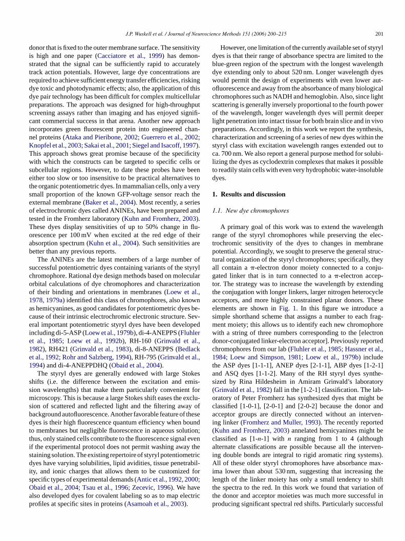
J.P. Wuskell et al. / Journal of Neuroscience Methods 151 (2006) 200–215 201
donor that is fixed to the outer membrane surface. The sensitivityis high and one paper (Cacciatore et al., 1999) has demon-strated that the signal can be sufficiently rapid to accuratelytrack action potentials. However, large dye concentrations arerequired to achieve sufficient energy transfer efficiencies, riskingdye toxic and photodynamic effects; also, the application of thisdye pair technology has been difficult for complex multicellularpreparations. The approach was designed for high-throughputscreening assays rather than imaging and has enjoyed signifi-cant commercial success in that arena. Another new approachincorporates green fluorescent protein into engineered chan-nel proteins (Ataka and Pieribone, 2002; Guerrero et al., 2002;Knopfel et al., 2003; Sakai et al., 2001; Siegel and Isacoff, 1997).This approach shows great promise because of the specificitywith which the constructs can be targeted to specific cells orsubcellular regions. However, to date these probes have beeneither too slow or too insensitive to be practical alternatives tothe organic potentiometric dyes. In mammalian cells, only a verysmall proportion of the known GFP-voltage sensor reach theexternal membrane (Baker et al., 2004). Most recently, a seriesof electrochromic dyes called ANINEs, have been prepared andtested in the Fromherz laboratory (Kuhn and Fromherz, 2003).These dyes display sensitivities of up to 50% change in flu-orescence per 100 mV when excited at the red edge of theirabsorption spectrum (Kuhn et al., 2004). Such sensitivities arebetter than any previous reports.
r ofs tyryc culao ationo ,1 wna es bc Sev-e opedie ,1e ,1
kess mis-s t form excls y ofb thesd oundt utiont l evei thes tricd bil-i fors ;Oa ectrip
However, one limitation of the currently available set of styryldyes is that their range of absorbance spectra are limited to theblue-green region of the spectrum with the longest wavelengthdye extending only to about 520 nm. Longer wavelength dyeswould permit the design of experiments with even lower aut-ofluorescence and away from the absorbance of many biologicalchromophores such as NADH and hemoglobin. Also, since lightscattering is generally inversely proportional to the fourth powerof the wavelength, longer wavelength dyes will permit deeperlight penetration into intact tissue for both brain slice and in vivopreparations. Accordingly, in this work we report the synthesis,characterization and screening of a series of new dyes within thestyryl class with excitation wavelength ranges extended out toca. 700 nm. We also report a general purpose method for solubi-lizing the dyes as cyclodextrin complexes that makes it possibleto readily stain cells with even very hydrophobic water-insolubledyes.
1. Results and discussion
1.1. New dye chromophores
A primary goal of this work was to extend the wavelengthrange of the styryl chromophores while preserving the elec-trochromic sensitivity of the dyes to changes in membranepotential. Accordingly, we sought to preserve the general struc-t theya ju-g -t ndingt yclea hesee as frag-m orew trond rtedc l.,1t -1]a he-s ory( b-o ht bec anda ven-i d( bec ha rven-i ms).A max-i g thel hiftt n oft ful inp ssful
The ANINEs are the latest members of a large numbeuccessful potentiometric dyes containing variants of the shromophore. Rational dye design methods based on molerbital calculations of dye chromophores and characterizf their binding and orientations in membranes (Loew et al.978, 1979a) identified this class of chromophores, also knos hemicyanines, as good candidates for potentiometric dyause of their intrinsic electrochromic electronic structure.ral important potentiometric styryl dyes have been devel
ncluding di-5-ASP (Loew et al., 1979b), di-4-ANEPPS (Fluhlert al., 1985; Loew et al., 1992b), RH-160 (Grinvald et al.982), RH421 (Grinvald et al., 1983), di-8-ANEPPS (Bedlackt al., 1992; Rohr and Salzberg, 1994), RH-795 (Grinvald et al.994) and di-4-ANEPPDHQ (Obaid et al., 2004).
The styryl dyes are generally endowed with large Stohifts (i.e. the difference between the excitation and eion wavelengths) that make them particularly convenienicroscopy. This is because a large Stokes shift eases the
ion of scattered and reflected light and the filtering awaackground autofluorescence. Another favorable feature ofyes is their high fluorescence quantum efficiency when b
o membranes but negligible fluorescence in aqueous solhus, only stained cells contribute to the fluorescence signaf the experimental protocol does not permit washing awaytaining solution. The existing repertoire of styryl potentiomeyes have varying solubilities, lipid avidities, tissue penetra
ty, and ionic charges that allows them to be customizedpecific types of experimental demands (Antic et al., 1992, 2000baid et al., 2004; Tsau et al., 1996; Zecevic, 1996). We havelso developed dyes for covalent labeling so as to map elrofiles at specific sites in proteins (Asamoah et al., 2003).
lr
e-
u-
e
;n
c
ural organization of the styryl chromophores; specifically,ll contain a�-electron donor moiety connected to a conated linker that is in turn connected to a�-electron accep
or. The strategy was to increase the wavelength by extehe conjugation with longer linkers, larger nitrogen heteroccceptors, and more highly constrained planar donors. Tlements are shown inFig. 1. In this figure we introduceimple shorthand scheme that assigns a number to eachent moiety; this allows us to identify each new chromophith a string of three numbers corresponding to the [eleconor-conjugated linker-electron acceptor]. Previously repohromophores from our lab (Fluhler et al., 1985; Hassner et a984; Loew and Simpson, 1981; Loew et al., 1979b) include
he ASP dyes [1-1-1], ANEP dyes [2-1-1], ABP dyes [1-2nd ASQ dyes [1-1-2]. Many of the RH styryl dyes syntized by Rina Hildesheim in Amiram Grinvald’s laboratGrinvald et al., 1982) fall in the [1-2-1] classification. The laratory of Peter Fromherz has synthesized dyes that miglassified [1-0-1], [2-0-1] and [2-0-2] because the donorcceptor groups are directly connected without an inter
ng linker (Fromherz and Muller, 1993). The recently reporteKuhn and Fromherz, 2003) annelated hemicyanines mightlassified as [1-n-1] with n ranging from 1 to 4 (althouglternate classifications are possible because all the inte
ng double bonds are integral to rigid aromatic ring systell of these older styryl chromophores have absorbance
ma lower than about 530 nm, suggesting that increasinength of the linker moiety has only a small tendency to she spectra to the red. In this work we found that variatiohe donor and acceptor moieties was much more successroducing significant spectral red shifts. Particularly succe

202 J.P. Wuskell et al. / Journal of Neuroscience Methods 151 (2006) 200–215
Fig. 1. Summary and notation of the building blocks used to design the styryl chromophores. The styryl dyes are constructed by choosing one structure from eachcolumn and linking them together as [donor–linker–acceptor]. The R1 groups are generally hydrocarbon chains to anchor the probes in the membrane. The R2 groupsare generally polar moieties that orient the acceptor portion of the dye toward the aqueous interface and can reduce the rate of membrane permeation. We have alsodeveloped R2 groups for covalent labeling applications (Asamoah et al., 2003; De Lorimier et al., 2002).
red-shifting donors had the amino nitrogen in the aniline tiedback via one or two saturated cyclic rings (i.e. 3-, 4- and 7- fromFig. 1). Particularly good acceptor moieties were the acridinium(-4) and indolenium (-5) heterocycles. A summary of spectralabsorbance and emission maxima in both ethanol and lipid vesi-cle membranes for all the styryl dyes in our library, includingpreviously available blue-absorbers, is provided inTable 1. Thesyntheses of the dyes follow elaborations of the aldol conden-sation and palladium-catalyzed coupling schemes that we havedeveloped previously (Hassner et al., 1984) and are detailed inSection3.
As can be seen fromTable 1, we have succeeded in produc-ing styryl dyes with absorbance maxima as high as 820 nm and
emission maxima that extend above 900 nm. As with the clas-sical styryl chromophores, these dyes usually display Stokesshifts of 100 nm or more. Also similar to our previous experi-ence with these dyes, the spectra in lipid vesicle membranes aresignificantly blue shifted compared to the spectra in ethanol. Weattribute this effect to a differential solvation of the two ends ofthe molecule that stabilizes the positive charge in the ground statenear the membrane surface, but destabilizes the charge in theexcited state at its new location deeper within membrane amongthe non-polar lipid acyl chains (Loew et al., 1979b). Table 1doesnot display data on the brightness of these dyes as this was notmeasured quantitatively for most of them. Generally, however,and again as was the case for the older styryl dyes, they display

J.P. Wuskell et al. / Journal of Neuroscience Methods 151 (2006) 200–215 203
Table 1Wavelength ranges of absorbance and emission maxima for styryl dyechromophores
Chromophore Numberof Dyes
λABS
(EtOH)λEM
(EtOH)λABS (lip) λEM (lip)
[1-1-1] 18 490± 9 617± 8 468± 7 600± 10[1-1-2] 2 470 610± 10 441± 2 590± 30[1-1-3] 3 564± 5 684± 7 533± 5 680± 70[1-1-4] 3 564± 5 684± 7 533± 5 680± 70[1-1-5] 1 567 599 562 601[1-1-6] 2 590± 1 679 578 662[1-2-1] 5 510± 10 680± 50 468± 7 620± 20[1-2-3] 2 560± 30 675± 7 540± 20[1-2-4] 3 700± 20 800± 50 611± 10 771± 1[1-2-5] 1 650 700 638[1-3-1] 1 475 610 450 584[1-4-1] 1 485 715 463 690[1-5-4] 2 630± 10 720± 40 580± 10 660± 10[1-6-1] 1 482 461 670[1-7-3] 3 640± 20 890± 70 580± 10 740± 30[1-8-3] 1 607 579 646[1-8-5] 1 626 639 634 647[1-9-3] 4 590± 20 800± 100 548± 5 720± 50[1-9-5] 2 690± 30 870 640± 10 757[1-10-3] 1 626 664 616 650[1-11-1] 1 440 641 420[1-11-4] 1 391 692 404 552[2-1-1] 47 500± 10 710± 10 470± 10 640± 20[2-1-7] 1 486 464 627[2-2-3] 1 584 812 526 702[2-2-6] 1 609[3-1-3] 3 580± 6 689± 1 556± 5 680[3-1-4] 1 681 770 658[4-1-3] 1 608 833 589 760[4-1-4] 3 705± 7 833± 612± 1 716[4-1-6] 1 610 610[4-7-3] 1 592 873 597 715[5-1-3] 1 514 620 500 714[6-1-1] 1 474 656 432 630[6-1-3] 2 520± 20 714± 9 480± 20 671± 8[6-1-5] 1 616 636 620 643[7-1-1] 1 523 640[7-1-3] 3 620± 10 720± 10 579± 1 673± 4[7-1-4] 4 726± 10 810± 40 678± 7 770± 60[7-1-5] 1 588 612 594 618[7-1-6] 1 600 708 618[7-2-4] 1 825 880 674 790[7-8-3] 1 663 687 668 680[7-9-3] 2 620± 30 559[8-1-1] 1 440 588 421 566[8-1-3] 1 500 648 486 640[9-1-1] 1 472 672 457 622[9-1-2] 2 540± 20 760± 40 530± 30 681± 8[9-1-3] 2 540± 20 760± 40 530± 30 681± 8
quite strong fluorescence intensities when membrane-bound, balmost negligible emission in aqueous solution. This is an impor-tant advantage for imaging dye-stained cell membranes.
1.2. Voltage dependent spectra
To characterize the dye responses to membrane potentiawe measured the modulation by the voltage of transmitted anfluorescent light signals as a function of wavelength. This is
Fig. 2. Hemispherical bilayer transmittance response spectra for three represen-tative dyes. Response to 100 mV steps. JPW-4023, RE-66 and di-4-ANEPPS are,respectively, [1-7-3], [1-1-4] and [2-1-1] chromophores (Fig. 1 dye structuresare shown inTable 2). These biphasic spectra are characteristic of the styryldyes and are an indicator of an electrochromic mechanism for the sensitivity ofthe spectrum to voltage.
achieved with a voltage-clamped hemispherical bilayer appa-ratus (Dragsten and Webb, 1978; Loew et al., 1979a, 1992a;Loew and Simpson, 1981) that has been modified to allow fornear infrared fluorescence detection with an avalanche photodi-ode. In this apparatus, the voltage is applied as a 40 Hz squarewave and the modulation of transmitted or fluorescence light isdetected through a lockin amplifier.Fig. 2shows transmittanceresponse spectra for three dyes obtained with this apparatus for100 mV changes in membrane potential. The spectrum for di-4-ANEPPS, a widely used and relatively sensitive older 2-1-1 dye,is shown for comparison. RE66 is a 1-1-4 dye and JPW4023has a 1-7-3 chromophore. As can be seen, all three of thesedyes display a biphasic modulation of the transmitted light sig-nal as a function of wavelength with an increased transmittance(decreased absorbance) on the red wing of each spectrum anda decreased transmittance on the respective blue wings. Thisarises from a voltage dependent shift of the absorbance spectraof these dyes corresponding to a blue shift for depolarizationof the membrane. The maximum amplitude of response is seenfor di-4-ANEPPS at about 505 nm, for JPW 4023 at 650 nm andfor RE66 at 630 nm. All of these wavelengths are to the red ofthe respective absorbance maxima of the dyes. This is becausethe optimal voltage sensitivity for dyes with voltage-dependentspectral shifts will occur at the wavelength of maximum slopeof the absorbance spectrum, not at the absorbance maximum.One might be tempted to say that di-4-ANEPPS is the mosts f thec iningo ranei canb d nota ofd nityo
ge-d et cien-
ut
l,d
ensitive of these three dyes. However, the amplitude ohange in transmitted light also depends on the level of staf the hemispherical bilayer, as the more heavily the memb
s stained, the larger the fraction of the incident light thate absorbed and therefore affected by the voltage. We dittempt to control for the staining level which is a functionye solubility and concentration in water and the binding affif the dye for the membrane.
Theoretically, the staining level should not affect the voltaependent relative fluorescence change,�F/F. This is becaus
hese dyes have extremely low fluorescence quantum effi

204 J.P. Wuskell et al. / Journal of Neuroscience Methods 151 (2006) 200–215
Fig. 3. Excitation wavelength dependence of the relative fluorescence changes (�F/F) for 4 new dyes. DB1-195 and JPW-5026 were applied to the inside of thepipet that supports the hemispherical bilayer; all the other experiments were performed with the dye applied from the external solution.
cies in aqueous solution. Therefore, only dye bound to themembrane contributes significantly to the fluorescence, causingboth the numerator and denominator of�F/F to change propor-tionately with the level of staining. However, to the extent thatredistribution mechanisms contribute to the voltage-dependenceof the fluorescence, the dye concentration in the membrane willaffect the sensitivity at the low or high ends of the membrane-binding isotherm. We believe that most of our new dyes respondprimarily with an electrochromic mechanism that should ren-der �F/F relatively insensitive to dye concentration.Fig. 3shows the relative fluorescence response to a 100 mV depolar-izing pulse for four of the new dyes as a function of excitationwavelength. The emission was collected through a longpass fil-ter that was chosen in each case to collect most of the emissionspectrum while rejecting most scattered light. However, somefluorescence from dye in the aqueous solution bathing the bilayerwill contribute to the denominator of�F/F, thus rendering theseresponses somewhat lower than could be achieved if the dye werewashed out after staining. There is also always scattered highwavelength stray light that is not fully blocked by the excitationmonochromator and that contributes to the denominator; this isespecially significant for weakly fluorescent or poorly bounddyes. The spectra of DB2-039 and JPW-5019, both applied tothe external bathing solution, display a decrease for the depo-larizing pulse at the high wavelength wings, corresponding toa blue shift of the excitation spectrum and consistent with thes .D thed theo dyea lectfi face
These response spectra do not show inversion symmetry aroundthe wavelength at which they cross zero; this is primarily becausethe use of a long pass emission filter superimposes the red partof the emission spectrum response on the excitation response.As has been noted previously (Kuhn et al., 2004; Loew, 1982;Loew and Simpson, 1981) the�F/F response is maximal at thered edges of the excitation spectra. The explanation for this isthat while�F is maximal at the wavelength of greatest slopein the excitation spectrum,�F/F will show a maximum at alonger wavelength where�F may still be significantand F isminimized. In our experiment with a tungsten/halogen excitingsource, the response spectra are somewhat noisy at the edgesbecause the fluorescence signal that is being collected is lowand the background scattered light becomes significant as theexciting wavelength approaches the emission filter; therefore,for some dyes the reported optimal sensitivity may actually be anunderestimate. Strong monochromatic laser excitation sourcesminimize scattered light and can overcome such signal to noiselimitations that might preclude excitation at the extreme rededge of the spectra. The optimal excitation wavelength for thefluorescence response of JPW-5026 is approximately 710 nmwith a 780 nm longpass emission filter. This corresponds to aca. 180 nm red shift compared to the optimal responses of the2-1-1 (ANEP) class of dyes.
To test the ability of the dyes to follow action potentials ina neuronal preparation, we applied a representative selection oft entsp lowa ablev s witht twon a
hift for the transmitted light signal as noted forFig. 2, aboveB1-195 and JPW-5026 have the opposite phase becauseyes were applied to the inner pipet solution rather thanuter bathing solution; as expected, the response of there reversed because the dye molecules experience an eeld of opposite polarity to the dyes applied to the outer sur
se
sric.
he dyes to the lobster walking-leg nerve. These experimermit us to determine if the dyes are able to faithfully folfast electrical signal. All of the dyes that gave measur
oltage-dependent signals did show fluorescence changehe same kinetics as the action potential. Experiments withewly synthesized dyes are shown inFig. 4. The dye RH-1692,

J.P. Wuskell et al. / Journal of Neuroscience Methods 151 (2006) 200–215 205
Fig. 4. Representative optical signals during the action potential of dye-stained lobster walking-leg nerve. (A simultaneously recorded extracellular recording of thecompound action potential is shown in (A).).
red absorbing oxonol dye from the laboratory of Amiram Grin-vald (Shoham et al., 1999), was examined for comparison. Inaddition to�F/F, the signal to noise ratio (S:N) was also mea-sured in these experiments. The S:N provides an indication ofthe practical utility of a dye as it incorporates the overall bright-ness of the fluorescence. However, the S:N is not an intrinsicproperty of the dye being tested, as it also depends on the sen-sitivity of the instrumentation at the wavelengths being used,the efficiency of staining, the level of background staining ofnon-electrically active cells or intracellular organelles, etc. Thesensitivities of the dyes on the lobster nerve as measured by�F/F are generally more than an order of magnitude lower thanthe sensitivities determined on the hemispherical bilayer. Thisis primarily because fluorescence from non-electrically activeglial cells and other extracellular material in this preparationcontributes a large background that inflates the denominator of
�F/F. The dye JPW-3067 gave the best S:N of all the new dyeseven though its sensitivity on the hemispherical bilayer systemwas only moderate. Its S:N and�F/F on the lobster nerve wereas good as the best of the earlier [2-1-1] styryl dyes includingdi-4-ANEPPS. The low sensitivity on the hemispherical bilayersystem is due to the large contribution of non-membrane fluores-cence to the total fluorescence signal for JPW-3067, which hasa chromophore and spectral properties that differ significantlyfrom the other styryl dyes. With several dyes we were unableto detect a voltage-dependent signal; the limit of detection forS:N was more than two orders of magnitude smaller than thelargest signals previously obtained for the [2-1-1] dyes. In manyinstances where no signal was detected, we performed a posi-tive control to show that the nerve was functional by restainingwith a known [2-1-1] dye and confirming the expected voltage-dependent fluorescence change.

206 J.P. Wuskell et al. / Journal of Neuroscience Methods 151 (2006) 200–215
Table 2summarizes the results from both the hemispher-ical bilayer and the lobster walking nerve for 20 of the newdyes. As can be seen, the spectra of these dyes range into thenear infrared region, with the longest emission wavelength inethanol extending above 900 nm. The wavelength maxima ofthe dyes bound to lipid vesicle membranes (data not shown) arealways significantly blue shifted compared to the ethanol spec-tra. The best voltage-dependent fluorescence changes are in therange of 10%/100 mV, comparable to those of the original [2-1-1] dyes such as di-4-ANEPPS. The relative fluorescence changeson the hemispherical bilayer are not optimized for emissionwavelength as long pass filters were used instead of attempt-ing to determine the optimal wavelength with a bandpass filteror monochromator; pushing the excitation further to the red edgeand use of narrow emission bandpass filter could easily double�F/F for many of the dyes, but at the expense of the overall flu-orescence signal. Furthermore, the hemispherical bilayer datacould not be perfectly corrected for background light scatter-ing and fluorescence from the aqueous dye that both contributeto the denominator of�F/F, especially at the spectral wings.These background signals would not be expected to contributeas significantly to a high resolution microscope image, where theoptical field will be much more highly restricted to the cell(s)of interest and the residual dye in the staining solution can bewashed away. While these factors could lead to significantlyincreased dye sensitivity in a real experimental preparation, flu-o duca
1
r thdi e ana en-t alizo shiftf ongs inings thec fors
ithh le toe culea thea nesw rietyo eryo i-c rents ech-n andPn , 2-h� The
efficacy of binding to the CD is related to the relative size of theguest dye with respect to the size of the cavity. Because CDs aresomewhat heterogeneous, compounds with the same name butobtained from different supplier were also compared. The CDstested were:�-CD,�-CD, CE-�-CD, 2HP-�-CD, SBE-4-�-CD,SBE-7-�-CD,�-CD, CE-�-CD, 2HP-�-CD and 2HP-�-CD (2.7variate). Additionally, a selection of hydroxyl acids (i.e., citricacid and ascorbic acid) were tested with each CD to determinetheir impact on solubility. In each case, an ethanolic stock of di-8-ANEPPS (3 mM) was incrementally added to a chilled (4◦C)solution of each candidate CD (normally 10 mM) with (1 or5 mM) and without a hydroxyl acid. This solution was vigor-ously stirred and repeatedly sonicated. Addition of di-8 stockto this mixture was continued until maximum solubility wasreached as judged by the presence of particulates or precipitants.Water and ethanol were then removed by vacuum evaporationto produce a crystalline solid (with some volume expansion) orin some cases a viscous paste. The water solubility of each com-plex was tested by reconstituting the CD/di-8-ANEPPS solid instandard saline.
The effectiveness of the different CDs as vehicles for dyestaining was evaluated using three criteria: level of dye encap-sulation, relative water solubility and staining efficacy. Themolar ratio of dye to cyclodextrin varied depending on thecyclodextrin used, the presence or absence of hydroxyl acidsand complexing conditions (especially pH). In the best case thes ter-e Dt edi tudeo di-8 effi-c erea ther sol-u tiona thatc thec hS ig-n i-8-A is-t . Ther ublea . Too hingf tar-g finityf tra-c hC di-8 iths com-m 74;L dfl CDe
rescence from non-excitable cells such as glia might prodecrease in�F/F.
.3. Cyclodextrin for delivery
The sidechains on the chromophores are used to tailoyes for different applications. Longer R1 alkyl chains (Fig. 1)
ncrease the strength of binding of the dyes to the membranlso slow the rate of internalization. Internalization is detrim
al to the potentiometric response because as the dye equn either side of the bilayer the voltage-dependent spectral
rom the two leaflets will cancel each other. However, lidechains decrease the solubility of the dye, making stalow and inefficient. In addition, the large size of some ofhromophores themselves led to lowered solubility evenhort alkyl sidechains.
Cyclodextrins (CDs,Fig. 5) are large cyclic saccharides wydrophobic cavities and hydrophilic exteriors. They are abncapsulate medium sized water insoluble organic molend thereby effectively solubilize them. We investigatedbility of cyclodextrins to catalyze staining of cell membraith voltage-sensitive dyes. We empirically screened a vaf CDs to identify the “best” carrier molecule for the delivf di-8-ANEPPS, where R1 ofFig. 1 is octyl, as a prototypal hydrophobic dye. Sixteen different CDs from three diffeuppliers were tested (Sigma, St Louis, MO; Cyclodextrin Tology Development Inc., High Springs, FL; CyDex, Overlark, KS). These CDs differed in their type (�, �, or �) andature of chemical modification (i.e., methyl, hydroxyethylydroxypropyl, sulfobutylether, etc.). As shown inFig. 5, the, �, and� CDs have, respectively increasing cavity sizes.
e
e
d
ess
s
toichiometry of dye to CD was between 1:5 and 1:10. Instingly,�-CDs exhibited higher stoichiometry of dye to C
han �-CDs. Additionally, complex formation often resultn a blue shift in di-8-ANEPPS fluorescence. The magnif this spectral shift was essentially the same for each/CD combination and was not predictive of the relativeiency of dye binding. Almost all cyclodextrins tested wble to solublize di-8-ANEPPS to some extent; howeveresulting complexes varied considerably in their waterbility. All complexes required some degree of sonicand/or mechanical agitation to produce a clear solutionould be filtered. The best results were obtained witharboxyethyl-gamma-CD (CE-�-CD, obtained from CTD, Higprings, FL) (Fig. 6). In most instances, hydroxyl acids sificantly enhanced the inclusiveness of CD hosts for dNEPPS; however it was difficult to make quantitative d
inctions between the efficacies of the different acids usedesulting CD/di-8-ANEPPS complexes are very water-solnd can be easily loaded into cells via a patch pipetteptimize dye unloading from the carrier we were searc
or cases where the relative affinity between carrier andet were matched (i.e. membrane had higher relative af
or di-8-ANEPPS than the CD). The best results for both inellular and extracellular staining (Fig. 6) were obtained witE-�-CD. Both extra- and intra-cellular staining with CD/-ANEPPS is improved over di-8-ANEPPS solubilized wolvents (e.g. ethanol or DMSO) and detergents (mostonly Pluronic F127 (Bedlack et al., 1992; Cohen et al., 19ojewska and Loew, 1986)). Conveniently, the backgrounuorescence of di-8-ANEPPS is significantly reduced byncapsulation.

J.P. Wuskell et al. / Journal of Neuroscience Methods 151 (2006) 200–215 207
Table 2Summary of spectral and potentiometric characteristics of a selection of new long wavelength styryl dyes
Name [chromophore] ABS (nm) EM (nm)�T/T HB Best T� �F/F, HB Best Ex/EM F S/N, Nerve Nerve Ex/Em� Structure
JPW-3012 [7-1-3] 630 709 1.5E−004 600 0.10 630/>780
KDH-160 [3-1-3] 583 690 3.0E−005 580 0.07 590/>645
JPW-3066 [1-9-3] 602 924 8.0E−005 620 0.09 640/>695 3.0 620/>695
JPW-3067 [1-9-5] 670 870 6.7E−005 660 0.05 650/>715 40.0 660/>715
JPW-3080 [1-7-3] 630 896 6.0E−005 640 0.11 660/>715 26.0 630/>695
JPW-4012 [1-7-3] 620 824 3.2E−004 655 0.05 685/>780 5.0 660/>715
JPW-4023 [1-7-3] 660 964 1.4E−004 650 0.08 685/>780 36.0 660/>715
RE-66 [1-1-4] 663 744 1.0E−004 630 0.04 630/>695 7.0 630/>695
RE-136 [1-2-4] 696 820 8.0E−005 660 0.13 680/>780 2.0 660/>715
RK-57 [7-1-4] 713 760 8.0E−005 660 0.05 660/>780 0.0 660/>715
JPW-4090 [2-2-3] 584 812 1.6E−004 590 0.06 640/>715 2.0 630/>715
JPW-5019 [1-9-3] 602 691 3.2E−005 620 0.06 640/>695
JPW-5021 [1-7-3] 632 833 6.5E−006 650 0.04 670/>715
JPW-5020 [1-9-5] 704 878 2.4E−004 660 0.05 680/>780
JPW-5028 [1-7-3] 646 817 2.4E−005 650 0.07 680/>780

208 J.P. Wuskell et al. / Journal of Neuroscience Methods 151 (2006) 200–215
Table 2 (Continued )
Name [chromophore] ABS (nm) EM (nm)�T/T HB Best T� �F/F, HB Best Ex/EM F S/N, Nerve Nerve Ex/Em� Structure
JPW-5026 [1-9-5] 706 873 2.1E−004 680 0.08 720/>780
DB1-195 [1-1-4] 664 1.0E−004 630 0.09 660/>715
JPW-5031 [2-2-5] 623 828 2.2E−005 620 0.02 630/>695
JPW-5034 [1-9-5] 710 880 2.0E−004 670 0.05 730/>780
DB2-039 [7-1-3] 624 724 1.3E−004 620 0.04 625/>695
Abs and Em are the respective absorbance and fluorescence emission maxima in ethanol; the maximum values of the relative transmittance,�T/T and fluorescence,�F/F responses are shown with the wavelengths at which these were acquired. The signal to noise of some of the dyes on the lobster walking nerve (F S/N Nerve)is provided adjacent to the corresponding excitation and emission wavelengths that were employed for these measurements.
Fig. 5. Cyclodextrin structures. These images were generated with the Chime software plugin (Elsevier MDL Inc., San Leandro, CA) for Internet Explorer. Theunderlying atomic coordinates were derived from the public Cambridge Structural Database. These structures show relative molecular dimensions between the CDsof different sizes.

J.P. Wuskell et al. / Journal of Neuroscience Methods 151 (2006) 200–215 209
Fig. 6. CE-�-CD effectively delivers a hydrophobic voltage-sensitive dye to hippocampal neurons both extracellularly and intracellularly. Top: Hippocampalneuronstained externally with di-8/CD (10�M dye) displayed as overlay of green (505–550 nm) and red (>570 nm) emission channels. This image was obtained witha Zeiss (Thornwood, NY) LSM510 confocal microscope using 488 nm laser excitation. Bottom: Single cells were continuously dialyzed via a patch pipette withdi-8-ANEPPS/CD 100�M (left) vs. di-2-ANEPEQ 5 mM (right) using an OG590 emission filter. This data was obtained with a Zeiss Axiovert microscope. Imageswere recorded at 490 nm excitation wavelength and were captured with a Pixera (Los Gatos, CA) color CCD camera. (For interpretation of the references to color inthis figure legend, the reader is referred to the web version of the article.)

210 J.P. Wuskell et al. / Journal of Neuroscience Methods 151 (2006) 200–215
We also found that application of dye/CD complexes througha patch pipet results in much more selective staining of theplasma membrane compared to a water soluble styryl dye thathad been previously used for intracellular application, di-2-ANEPEQ (Antic et al., 2000; Zecevic, 1996). As shown in thelower panels ofFig. 6, the di-8-ANEPPS/CD complex displaysselective staining of the outer membrane, while fluorescencefrom di-2-ANEPEQ is distributed throughout the cytoplasm,presumably from stained endoplasmic reticulum. The cytoplas-mic fluorescence is undesirable because it will produce a largebackground that will reduce the�F/F measured in an experi-ment. Also noteworthy is the lower level of dye/CD complexrequired to stain the cells.
In general terms, these results can be explained on the basisof relative molecular size and binding efficiency. Di-8-ANEPPSlikely fits snugly into the smaller binding pocket of�-CDs but itsrelative binding affinity is probably higher than the target mem-brane. In contrast, the binding pocket of�-CDs is much largerand its corresponding binding affinity less than the target mem-brane, resulting in more effective unloading/membrane-staining.One potential side effect of using the CDs as a dye vehicle isthe possibility of perturbing the cell membrane as the ß-CDs arecommonly used to extract cholesterol from the cells. It shouldbe noted, however that the�-CD is not particularly efficient atextracting sterols like cholesterol and, further, that the levels ofCD and incubation times used for staining are each typicallya teroe herl ona t side sedf r fora ryt sol-uo velyd
2
ss ov d nei lobs arabt tinga willb s. Wh ryo -c sizea
3
3
hasb l.,
1979a), but has been upgraded to allow for computer con-trol of excitation monochromator, data acquisition and analysis,with a PCI-MIO-16E-4 multifunction board running under Lab-Windows software from National Instruments (Austin, Texas),and enhanced sensitivity for fluorescence up to 1000 nm witha model S/N136 cooled large area avalanche photodiode fromAdvanced Photonix (Carmallito, CA).
3.2. Cyclodextrin complexes
As described above, a large number of CDs were screenedto optimize solubilization and delivery of the dyes with di-8-ANEPPS being used as a prototype for a hydrophobic insolubledye. The results of the optimization led to the conclusion thatCE-�-CD (catalog no. TRCEG from CTD, Inc., High Springs,FL) was the best choice. Optimized CE-�-CD/dye complexeswere prepared as follows.
A 600�l solution of 1 mM dye in ethanol was added drop bydrop to 10 ml of a stirring solution of 20 mM CD; care was takenthat each drop diffused evenly in the solution and the solutionwas clear before the addition of the next drop. It is helpful butnot necessary to do this procedure at 4◦C, as the cyclodextrinsolution appears to have a higher affinity for dye under low tem-perature. The dye/cyclodextrin ratio could be changed, as longas after the procedure the solution attains clarity. The resultingsolution was aliquoted into microcentrifuge tubes at 0.5 ml/tubea tratoro tablea ure.B uffers e dis-sc ndf n forsa used.S e for5 heh ences
3
4 d ofF r them twoa atercM Md s ofp gapsw rcedt lularr ith al t of1 til ther peak
n order of magnitude lower than those used for cholesxtraction. On the other hand, for intracellular staining hig
evels and long exposures to�-CD are unavoidable, so cautind appropriate controls should be performed to assure thaffects are negligible.�-CD-encapsulated dyes were also u
or staining the oxidized cholesterol hemispherical bilayelmost all the dyes reported inTable 2, as this was necessa
o achieve sufficient staining levels with these generally inble compounds. Treatment of the bilayer with�-CDs had nobvious deleterious effect on the longevity of these relatielicate membranes.
. Conclusion
We have presented data that indicate that the styryl claoltage sensitive dyes has been extended to the red annfrared spectral range. On our hemispherical bilayer andter nerve screens, the dye sensitivities to voltage are compo the ANEP class of styryl dyes. However, significant tesnd optimization of the dyes in experimental preparationse necessary and could lead to even further improvementave also developed the�-CDs as new vehicles for the delivef hydrophobic voltage sensitive dyes. The�-CDs will be espeially useful for the new near infrared dyes as their largernd longer length reduce their water solubility.
. Materials and methods
.1. Hemispherical bilayer
The voltage-clamped hemispherical lipid bilayer systemeen previously described (Fluhler et al., 1985; Loew et a
l
e
far-le
e
nd the solvent removed with a centrifuge vacuum concenvernight. The resultant colored powders or pastes were st 4◦C, indefinitely, but should be protected from light exposefore use, the solid in each tube was dissolved with 1 ml bolution. Sonication with a bath sonicator helps to acceleratolution. This resultant 10× solution has 30�M dye and 10 mMyclodextrin. This 10× solution could be further aliquoted arozen. Thus, the recommended final working concentratiotaining either the hemispherical bilayer or cells is 3�M dyend 1 mM CD although higher concentrations can also betaining cells was typically carried out at room temperaturmin followed by washing with fresh buffer or medium. Temispherical bilayer was stained until a stable fluorescignal developed – typically also within 5 min.
.3. Lobster walking-leg nerve
Walking leg nerves from Lobsters,Homarus americanus,50–600 g, were obtained using the pulling-out methourusawa (1929). We did not use the whole leg nerve foeasurements; the nerve was divided length-wise intopproximately equal sections. The lobster artificial sea wontained 457 mM NaCl, 13 mM KCl, 14 mM CaCl2, 10 mMgCl2, 14 mM Na2SO3, 6 mM Hepes, pH 7.8 and 2 mextrose. Stimulation and recording was done with pairlatinum electrodes in contact with the nerves. Vaselineere used so that all of the extracellular currents were fo
o flow in the extracellular space in the nerve. The extracelecordings were made using a Tektronix 5A22 amplifier wow pass filter of 1.0 KHz and an AC coupling time constan00 ms. The stimulus strength was increased gradually unecorded compound action potential reached 95% of its

J.P. Wuskell et al. / Journal of Neuroscience Methods 151 (2006) 200–215 211
value. For the optical measurements we further increased thestimulus by a factor of 2 with the aim of using a suprathresholdstimulus. In several tests a further increase in stimulus voltageresulted in little or no change in the optical signal. The prepa-ration was illuminated using a 12 V to 50 W tungsten–halogenbulb. The light was passed through a heat filter and the incidentwavelength was chosen using an interference filter with abandwidth of 30–90 nm (full width at half height). The incidentlight was focused on the nerve with a 3× microscope objective.The incident wavelengths were removed from the fluorescencepathway using a secondary filter (Schott Optical Glass; RG-610to RG-715). The fluorescent light was measured using anEG&G UV 444 photodiode (1 cm diameter) at 90◦ to theincident light and 1.2 cm from the nerve. The output of thephotodiode was measured using a current-to-voltage converter(Teledye-Philbrick 102601) with a 10 mOhm feedback resistor.The amplifier low pass filter had a time constant of 1.0 ms; thehigh pass filter had a time constant of 200 ms. The optical andelectrode amplifier outputs were digitized with 12 bit accuracyat 4000 KHz using the BNC-only option in NeuroPlex (RedShir-
tImaging, LLC, Fairfield, CT). The data were analyzed using theNeuroPlex software. We measured both the signal-to-noise ratioand the fractional fluorescence change (�F/F) for most dyes.
3.4. Dye synthesis
As in our earlier dye synthesis (Hassner et al., 1984), twoalternate strategies were employed for the key chromophoreassembly step: aldol condensation or Heck coupling. Mass spec-trometry on dye samples and intermediates was provided bythe Washington University Mass Spectrometry Resource withsupport from the NIH National Center for Research Resources(Grant No. P41RR0954). The full synthesis of three of the morecomplex new dyes, JPW-5019, DB1-195 and DB2-039, are pre-sented here.
3.5. Synthesis of JPW-5019
The synthetic details presented below are referenced toScheme 1.
Scheme
1.
212 J.P. Wuskell et al. / Journal of Neuroscience Methods 151 (2006) 200–215
3.5.1. p-(Di-n-octylamino)benzaldehyde (1)To 20 ml of anhydrousNN-dimethylformamide cooled in an
ice-bath at 0–5◦C was added drop wise with stirring 3.0 ml(32.6 mmol) of phosphorus oxychloride over a period of 3 min.The mixture was stirred for another 20 min then 7.0 g ofN,N-di-n-octyl aniline was added drop wise with ice-bath coolingover a period of 13 min. When the addition was complete, thecooling bath was removed and a heating mantel was appliedand the mixture heated with stirring at 100◦C for a period of2.5 h, then allowed to stand at room temperature over night.The mixture was then poured onto ca. 100 g of crushed icewhich produced a brown precipitate. The slurry was neutral-ized by the addition of a saturated solution of ca. 20 g of sodiumacetate to pH 6–8. The mixture was then extracted twice withportions of ethyl acetate. The combined extracts were washedwith saturated NaCl solution, dried over anhydrous MgSO4, andconcentrated to dryness by rotary evaporator to leave the crudeproduct as a clear brown oil. TLC analysis (CHCl3/hexane,1:1)showed one spot,Rf = 0.25. The aldehyde was further purified bycolumn chromatography on silica-gel by gradient elution withhexane–chloroform. This afforded 6.1 g of aldehyde (1) (80.3%of theo.) sufficiently pure for the following Wittig reaction.
3.5.2. p-(Di-n-octylamino)styrene (2)To a stirred slurry of 5.1 g (14.1 mmol) of methyltriph-
enylphosphonium bromide in 25 ml of anhyd. THF under argona Mn edt d int 44 g(T tionf ther2 Them y thd wast thee overM oduca lysisbc phyo inha ),3 d,J
3c
.72( l)5 0 mgt urewa eact ,
the aqueous phase extracted twice with portions of CHCl3, thecombined extracts washed with saturated brine solution, driedover MgSO4, and concentrated to dryness by rotary evaporator toleave 2.85 g of crude product as an orange-brown oil. Flash chro-matography on silica-gel, eluting with 40–70% dichloromethanein hexane afforded 1.33 g of (3) (58.6% of theo.) as a dark orangefluorescent oil. TLC analysis (CHCl3–hexane, 1:1) showedone orange fluorescent spot,Rf = 0.140.λmax(EtOH) = 446 nm,Emax= 666 nm. 1H NMR(CDCl3): 0.942–1.578 (m, 30 H),3.27–3.31 (m, 4H), 6.55–7.62 (m, 8H), 9.79 (s, CHO, 1H) ppm.
3.5.4. 1-(Propyl-3-trimethylammonium)4-[2-(p-di-n-octylamino)styryl-thiophyl-5-vinyl]quinolinium dibromide(JPW-5019)
A mixture of 0.45 g (1.0 mmol) of aldehyde (3) and 0.41 g(1.0 mmol) 1-(propyltrimethyl-ammonium)lepidinium dibro-mide (4) in 4.0 ml of acetic anhydride was heated with stirringunder nitrogen in a 110◦C oil bath for a period of 46 min. Thereaction mixture was cooled to ambient temperature, 8.0 ml ofisopropanol was added, and the mixtue refrigerated over night.The reaction mixture was concentrated to dryness under reducedpressure and the residue taken up in chloroform and charged to acolumn of silica-gel for flash chromatography. Gradient elutionwith 30–60% methanol in chloroform afforded 0.400 g (47.6%of theo.) ofJPW-5019 as a dark blue-green hygroscopic solid.TLC analysis (MeOH-CHCl, 1:3) showed one spot,R = 0.143.λ
3
d toS
3
at0 df na1 rateda ct wase andd hedc allc ctlyf
3(
lde-h dis-s derA uredi ctedw withs rude
tmosphere and cooled to 0◦C was added 8.0 ml of 2.5-butyl lithium in hexane via syringe. (All solids dissolvo form a clear orange solution.) The solution was stirrehe cooling bath for another 15 min, then a solution of 4.12.85 mmol) of di-octylaminobenzaldehyde (1) in 10 ml ofHF was rapidly added with stirring by means of an addi
unnel. The turbid mixture was stirred in ice-bath for ano0 min then allowed to warm to room temp. overnight.ixture was then cooled in an ice bath and quenched brop wise addition of 40 ml of water. The aqueous mixture
hen extracted twice with portions of ether. The combined extracts were washed twice with sat’d NaCl solution, driedgSO4, and concentrated to dryness to leave the crude prs a tan solid containing triphenyl phosphine oxide. Anay TLC (CHCl3–hexane, 1:4) showed one spot,Rf = 0.50. Therude product was further purified by flash chromatogran a silica-gel column. Elution with 20% dichloromethaneexane afforded 3.44 g (78% of theo.) of the styrene, (2) aslight yellow oil. 1H NMR (CDCl3): 0.69–2.18 (m, 30H
.1–3.34 (m, 4H), 4.8–5.03 (dd,J = 10 Hz, 1H), 5.30–5.62 (d= 17 Hz, 1H), 6.3–7.32 (complex m, 5H) ppm.
.5.3. 2-[p-(Di-n-octylamino)styryl]thiophene-5-arbaldehyde (3)
Into a heavy walled pyrex pressure tube was placed 15.0 mmol) of the dialkylaminostyrene (2), 0.955 g (5.00 mmo-bromo-2-thiophenaldehyde, 20 mg Palladium Acetate, 4
ri-o-tolylphosphine, and 5.0 ml dry triethylamine. The mixtas capped under nitrogen, then stirred in a 114◦C oil bath forperiod of 72 h. Upon cooling to room temperature, the r
ion mixture was partitioned with 50 ml CHCl3, 50 ml water
e
r
t
g
-
3 f
max(EtOH) = 602 nm,Emax(MLV) = 744 nm.
.6. Synthesis of DB1-195
The synthetic details presented below are referencecheme 2.
.7. 4-Dibutylamino benzaldehyde (5)
To a dry two-neck RB flask containing dry DMF (10 ml)◦C, 3.8 ml (6.25 g, 40.1 mmol) of POCl3 was added and stirre
or 10 min. 4.53 g (22.1 mmol) ofN,N-dibutylaniline was thedded and the contents of the flask were heated to 75◦C forh. The reaction mixture was cooled and 40 ml of concentqueous Sodium acetate solution was added. The produxtracted with dichloromethane, washed with water, brineried with sodium sulphate. Evaporation of solvent furnisrude aldehyde5 in 76% yield, which was passed through smolumn of Silica gel (elution with Chloroform) and used direor the next step.
.8. N-{4-[2-Acridin-9-yl-vinyl]phenyl}-N,N-dibutylamine6)
Six hundred and seventy milligrams (2.87 mmol) of ayde5 and 500 mg (2.59 mmol) of 9-methylacridine wereolved in 5 ml of Acetic anhydride and refluxed for 2 h unrgon atmosphere. The reaction mixture was cooled, po
nto cold solution of aqueous 5% NaOH and then extraith dichloromethane, washed with water, brine and driedodium sulphate. Evaporation of solvent furnished a c

J.P. Wuskell et al. / Journal of Neuroscience Methods 151 (2006) 200–215 213
Scheme 2.
residue which was purified by column chromatography (elutingwith 10% ethylacetate–chloroform) to furnish pure6 as orangecolored solid in 65% yield.
3.9. 3-{9-[2-(4-Dibutylamino-phenyl)-vinyl]-acridin-10-yl}-propane-1-sulfonate(DB1-195)
Seventy milligrams (0.17 mmol) of6 and 209 mg(1.71 mmol) of freshly distilled 1,3-propane sultone weremixed in a dry 10 ml RB flask and heated to 130◦C underArgon for 10 min. The flask was cooled, the residue wasdissolved in dichloromethane and purified by silica-gel columnchromatography (using CHCl3, 10% MeOH–CHCl3 then20% MeOH–CHCl3 as eluent), Fractions blue in color werecombined and the solvent removed to afford a blue-black oilwhich was triturated with diethyl ether to furnish pureDB1-19522% yield as blue solid.
3.10. Synthesis of DB2-039
3.10.1. 4-Methyl-1-(3-propyl trimethylammonium)quinolinium dibromide (JPW-4008) (Scheme 3)
To a 100 ml round bottom flask equipped with a magnetic stir-rer, oil bath was charged 10.44 g (0.04 mol) of (3-bromopropyl)trimethylammonium bromide and 5.72 g (0.04 mol) lepidine.
The flask was flushed with argon and heated to 115◦C for 22 hthen allowed to cool to room temperature. Twenty milliliters ofmethanol was added to the solid reaction mixture and warmed to50◦C to dissolve. Cooled to room temperature and added 130 mlether with stirring resulting in the dissolution of a pale purple oil.Chilled to−20◦C for 2 days giving a purple-grey solid whichwas washed several times with hexane then dried in a vacuumdesiccator. Yield = 12.99 g (80.4%).
3.10.2. 1-Benzotriazol-1-yl-2-butyl-3-penty-l-2,3,6,7-tetrahydro-1H,5H-benzo[ij]quinolizine (DB2-003a)(Scheme 4)
Reference (related synthesis): Katritzky AR, Rachwal B,Rachwal S, Abboud KA. Convenient synthesis of julo-lidines using benzotriazole methodology, J Org Chem1996;61:3117–26.
To a flame dried 50 ml round bottom flask equipped with aDean Stark trap, magnetic stirrer and argon inlet was charged1,2,3,4-tetrahydroquinoline (1.33 g, 10 mmol), 1-hexanal(3.12 g, 31 mmol), 1H-benzotriazole (1.23 g, 10.3 mmol),p-toluenesulfonic acid monohydrate (20 mg, 0.1 mmol), andtoluene (17 ml). Flushed the system with argon and refluxedfor 1.5 h with stirring. Cooled to room temperature androtovaped down to give a turbid yellow-orange oil. The crudereaction product was then used immediately for the nextstep.
eme
eme
Sch
Sch
3.
4.

214 J.P. Wuskell et al. / Journal of Neuroscience Methods 151 (2006) 200–215
Scheme 5.
Scheme 6.
Scheme 7.
3.10.3. 2-Butyl-3-pentyl-julolidine (DB2-003b) (Scheme 5)To the reaction flask containingDB2-003a, was charged
20 ml of dry anisole and 500 mg (13 mmol) of lithium aluminumhydride. The system was flushed with argon after attaching areflux condenser and magnetic stirrer. Slowly heated to reflux(160◦C) over a period of 45 min in order to minimize the rateof effervescence. Stirred at reflux for 1 h then cooled to roomtemperature, resulting in a grey slurry. Excess LiAlH4 was neu-tralized with the slow addition of methanol then water. Extractedwith 8 ml× 20 ml of ether. Combined organics and dried overMgSO4. Filtered and rotovaped down (30◦C) giving a clearsemi-crystalline glass which yellowed over time. The crude reac-tion product was then used immediately for the next step.
3.10.4. 2-Butyl-3-pentyl-julolidine-9-carbaldehyde(DB2-005) (Scheme 6)
Ten milliliters of dry DMF was charged to a flame dried100 ml round bottom flask equipped with a magnetic stir-rer, addition funnel, and argon inlet and cooled to 0◦C. 5 ml(53.6 mmol) of phosphorous oxychloride was then charged dropwise via addition funnel with stirring. After complete addition,the solution was stirred an additional 10 min at 0◦C. Warmed toroom temperature and charged the crudeDB2-003b drop wisevia addition funnel after dissolving in 20 ml dry DMF. Reactionmixture turned a turbid olive green. Warmed mixture to 80◦C for1 d nor ls s intm th tha withm and
dried over Na2SO4, then filtered and evaporated down to a brownoil. Impurities were removed via flash column chromatographyusing silica and hexanes as the eluent. 2.6 g of light yellow oilwas collected (80% yield).
3.10.5. 4-(2-Butyl-3-pentyl-julolidine-9-yl-vinyl)-1-(3-propyl trimethylammonium) quinolinium dibromide(DB2-039) (Scheme 7)
To a 25 ml round bottom flask equipped with a magnetic stir-rer, argon inlet and oil bath was charged 100 mg ofDB2-005(0.3 mmol) and 5 ml of 200 proof ethanol. To this, 112 mg ofJPW-4008 (0.27 mmol) was added. The color changed fromyellow to blue within 30 s. Flushed the flask with argon andwarmed to 60◦C with stirring for 2 h. Cooled to room tempera-ture and evaporated to dryness giving a blue paste. Dissolved in25 ml H2O and extracted unreactedDB2-005 and byproducts bywashing 2 ml× 10 ml ether. Rotovaped off water, thereby givinga wet paste. Solvated in 20 ml of 200 proof ethanol, then roto-vaped down to dryness. Repeated three times. Placed in vacuumdesiccator for 1 week yielding 140 mg blue paste (72% yield).
Acknowledgements
This work was supported through NIH Grants EB001963,HL071635 and DC05259.
R
A cel-
.5 h then cooled to room temperature. TLC (ether) showeemainingDB2-003b. Neutralized reaction mixture with 40 maturated Sodium Acetate solution, and extracted organicethylene chloride. The aqueous layer was made basic widdition of Sodium Bicarbonate and was further extractedethylene chloride. The organic fractions were combined
oeeferences
ntic S, Loew L, Wuskell J, Zecevic D. Voltage-sensitive dyes for intralular application. Biol Bull 1992;183:350–1.

J.P. Wuskell et al. / Journal of Neuroscience Methods 151 (2006) 200–215 215
Antic S, Wuskell JP, Loew L, Zecevic D. Functional profile of the giantmetacerebral neuron of Helix aspersa: temporal and spatial dynamics ofelectrical activity in situ. J Phys 2000;527:55–69.
Antic SD. Action potentials in basal and oblique dendrites of rat neocorticalpyramidal neurons. J Physiol (Lond) 2003;550:35–50.
Asamoah OK, Wuskell JP, Loew LM, Bezanilla F. A fluorometric approach tolocal electric field measurements in a voltage-gated ion channel. Neuron2003;37:85–97.
Ataka K, Pieribone VA. A genetically targetable fluorescent probe of channelgating with rapid kinetics. Biophys J 2002;82:509–16.
Baker BJ, Cohen LB, Kosmidis E. Expression of the GFP-voltage sensorSPARC in HEK 293 cells. Biophys J 2004;86:425a [meeting abstract].
Bedlack RS, Wei M-d, Fox SH, Gross E, Loew LM. Distinct electric poten-tials in soma and neurite membranes. Neuron 1994;13:1187–93.
Bedlack RS, Wei M-d, Loew LM. Localized membrane depolarizations andlocalized intracellular calcium influx during electric field-guided neuritegrowth. Neuron 1992;9:393–403.
Cacciatore TW, Brodfuehrer PD, Gonzalez JE, Jiang T, Adams SR, TsienRY, et al. Identification of neural circuits by imaging coherent electricalactivity with FRET-based dyes. Neuron 1999;23:449–59.
Cohen LB, Salzberg BM, Davila HV, Ross WN, Landowne D, Waggoner AS,et al. Changes in axon fluorescence during activity: molecular probes ofmembrane potential. J Membr Biol 1974;19:1–36.
De Lorimier RM, Smith JJ, Dwyer MA, Looger LL, Sali KM, PaavolaCD, et al. Construction of a fluorescent biosensor family. Protein Sci2002;11:2655–75.
Dragsten PR, Webb WW. Mechanism of the membrane potential sensitiv-ity of the fluorescent membrane probe merocyanine 540. Biochemistry1978;17:5228–40.
Fluhler E, Burnham VG, Loew LM. Spectra, membrane binding andpotentiometric responses of new charge shift probes. Biochemistry
F hilicActa
G t use.
G g ofhys
G nal
G eadopti-
rosci
G cententia
G nt of
G ash:ically3607
G S,es in
H oten-
K raneteins
Kuhn B, Fromherz P. Anellated hemicyanine dyes in a neuron membrane:molecular stark effect and optical voltage recording. J Phys Chem B2003;107:7903–13.
Kuhn B, Fromherz P, Denk W. High sensitivity of stark-shift voltage-sensingdyes by one- or two-photon excitation near the red spectral edge. BiophysJ 2004;87:631–9.
Loew LM. Design and characterization of electrochromic membrane probes.J Biochem Biophys Methods 1982;6:243–60.
Loew LM, Bonneville GW, Surow J. Charge shift optical probes of membranepotential. Theory Biochem 1978;17:4065–71.
Loew LM, Cohen LB, Dix J, Fluhler EN, Montana V, Salama G, et al. Anaphthyl analog of the aminostyryl pyridinium class of potentiometricmembrane dyes shows consistent sensitivity in a variety of tissue, celland model membrane preparations. J Membr Biol 1992a;130:1–10.
Loew LM, Cohen LB, Dix J, Fluhler EN, Montana V, Salama G, et al. Anaphthyl analog of the aminostyryl pyridinium class of potentiometricmembrane dyes shows consistent sensitivity in a variety of tissue, cell,and model membrane preparations. J Membr Biol 1992b;130:1–10.
Loew LM, Scully S, Simpson L, Waggoner AS. Evidence for a charge-shift electrochromic mechanism in a probe of membrane potential. Nature1979a;281:497–9.
Loew LM, Simpson L. Charge shift probes of membrane potential. A prob-able electrochromic mechanism for ASP probes on a hemispherical lipidbilayer. Biophys J 1981;34:353–65.
Loew LM, Simpson L, Hassner A, Alexanian V. An unexpected blue shiftcaused by differential solvation of a chromophore oriented in a lipidbilayer. J Am Chem Soc 1979b;101:5439–40.
Lojewska Z, Loew LM. Pluronic F127: an effective and benign vehiclefor the insertion of hydrophobic molecules into membranes. Biophys J1986;49:521.
Obaid AL, Loew LM, Wuskell JP, Salzberg BM. Novel naphthylstyryl-anal-
R icro-issue:) in
309.S cter-
ur J
S t al.with
S ingle
S rane
S iveeuro-
S ns in
T Dyeltage-
W ge-NS.
Z by
Z etBull
1985;24:5749–55.romherz P, Muller CO. Voltage-sensitive fluorescence amphip
hemicyanine dyes in neuron membrane. Biochim Biophys1993;1150:111–22.
onzalez JE, Tsien RY. Improved indicators of membrane potential thafluorescence resonance energy transfer. Chem Biol 1997;4:269–77
rinvald A, Fine A, Farber IC, Hildesheim R. Fluorescence monitorinelectrical responses from small neurons and their processes. Biop1983;42:195–8.
rinvald A, Frostig RD, Lieke E, Hildesheim R. Optical imaging of neuroactivity. Physiol Rev 1988;68:1285–366.
rinvald A, Lieke EE, Frostig RD, Hildesheim R. Cortical point-sprfunction and long-range lateral interactions revealed by real-timecal imaging of macaque monkey primary visual cortex. J Neu1994;14:2545–68.
rinvald AS, Hildesheim R, Farber IC, Anglister J. Improved fluoresprobes for the measurement of rapid changes in membrane potBiophys J 1982;39:301–8.
ross D, Loew LM, Webb WW. Spatially resolved optical measurememembrane potential. Biophys J 1985;47:270.
uerrero G, Siegel MS, Roska B, Loots E, Isacoff EY. Tuning flredesign of the dynamics, voltage range, and color of the genetencoded optical sensor of membrane potential. Biophys J 2002;83:18.
upta RK, Salzberg BM, Grinvald A, Cohen LB, Kamino K, Lesheret al. Improvements in optical methods for measuring rapid changmembrane potential. J Membr Biol 1981;58:123–37.
assner A, Birnbaum D, Loew LM. Charge shift probes of membrane ptial. Synthesis. J Org Chem 1984;49:2546–51.
nopfel T, Tomita K, Shimazaki R, Sakai R. Optical recordings of membpotential using genetically targeted voltage-sensitive fluorescent proMethods 2003;30:42–8.
J
l.
–
.
pyridinium potentiometric dyes offer advantages for neural networkysis. J Neurosci Methods 2004;134:179–90.
ohr S, Salzberg BM. Characterization of impulse propagation at the mscopic level across geometrically defined expansions of excitable tmultiple site optical recording of transmembrane voltage (MSORTVpatterned growth heart cell cultures. J Gen Physiol 1994;104:287–
akai R, Repunte-Canonigo V, Raj CD, Knopfel T. Design and charaization of a DNA-encoded, voltage-sensitive fluorescent protein. ENeurosci 2001;13:2314–8.
hoham D, Glaser DE, Arieli A, Kenet T, Wijnbergen C, Toledo Y, eImaging cortical dynamics at high spatial and temporal resolutionnovel blue voltage-sensitive dyes. Neuron 1999;24:791–802.
hrager P, Rubinstein CT. Optical measurement of conduction in sdemyelinated axons. J Gen Physiol 1990;95:867–90.
iegel MS, Isacoff EYIN. A genetically encoded optical probe of membvoltage. Neuron 1997;19:735–41.
lovin H, Arieli A, Hildesheim R, Grinvald A. Long-term voltage-sensitdye imaging reveals cortical dynamics in behaving monkeys. J Nphysiol 2002;88:3421–38.
pors H, Grinvald A. Spatio-temporal dynamics of odor representatiothe mammalian olfactory bulb. Neuron 2002;34:301–15.
sau Y, Wenner P, O’Donovan MJ, Cohen LB, Loew LM, Wuskell JP.screening and signal-to-noise ratio for retrogradely transported vosensitive dyes. J Neurosci Methods 1996;170:121–9.
u J-Y, Lam Y-W, Falk C, Cohen LB, Fang J, Loew L, et al. Voltasensitive dyes for monitoring mult-neuronal activity in the intact CHistochem J 1998;30:169–87.
ecevic D. Multiple spike-initiation zones in single neurons revealedvoltage-sensitive dyes. Nature 1996;381:322–5.
ochowski M, Wachowiak M, Falk CX, Cohen LB, Lam YW, Antic S,al. Imaging membrane potential with voltage-sensitive dyes. Biol2000;198:1–21.