systematic analysis of the role of cd19 cytoplasmic ... · pdf filefor y330 and y391 and...
TRANSCRIPT

of November 7, 2017.This information is current as
Tyrosinesof Grb2 and Sos with Different CD19Clustering of Phospholipase C and Vav and Activation in Daudi Human B Cells:Cytoplasmic Tyrosines in Enhancement of Systematic Analysis of the Role of CD19
Robert H. CarterStephen R. Brooks, Xiaoli Li, Emmanuel J. Volanakis and
http://www.jimmunol.org/content/164/6/3123doi: 10.4049/jimmunol.164.6.3123
2000; 164:3123-3131; ;J Immunol
average*
4 weeks from acceptance to publicationSpeedy Publication! •
Every submission reviewed by practicing scientistsNo Triage! •
from submission to initial decisionRapid Reviews! 30 days* •
?The JIWhy
Referenceshttp://www.jimmunol.org/content/164/6/3123.full#ref-list-1
, 26 of which you can access for free at: cites 37 articlesThis article
Subscriptionhttp://jimmunol.org/subscription
is online at: The Journal of ImmunologyInformation about subscribing to
Permissionshttp://www.aai.org/About/Publications/JI/copyright.htmlSubmit copyright permission requests at:
Email Alertshttp://jimmunol.org/alertsReceive free email-alerts when new articles cite this article. Sign up at:
Print ISSN: 0022-1767 Online ISSN: 1550-6606. Immunologists All rights reserved.Copyright © 2000 by The American Association of1451 Rockville Pike, Suite 650, Rockville, MD 20852The American Association of Immunologists, Inc.,
is published twice each month byThe Journal of Immunology
by guest on Novem
ber 7, 2017http://w
ww
.jimm
unol.org/D
ownloaded from
by guest on N
ovember 7, 2017
http://ww
w.jim
munol.org/
Dow
nloaded from

Systematic Analysis of the Role of CD19 CytoplasmicTyrosines in Enhancement of Activation in Daudi Human BCells: Clustering of Phospholipase C and Vav and of Grb2and Sos with Different CD19 Tyrosines1
Stephen R. Brooks,† Xiaoli Li,* Emmanuel J. Volanakis,* and Robert H. Carter 2*†‡
CD19 is a coreceptor on B cells that enhances the increase in cytoplasmic calcium and ERK2 activation when coligated with theB cell Ag receptor. Constructs containing point mutations and truncations were expressed in Daudi human B lymphoblastoid cellsto systematically determine the requirement for individual CD19 cytoplasmic tyrosines in these responses. Evidence for activitywas found for Y330, Y360, and Y421 as well as that previously published for Y391. Precipitates formed with phosphopeptidesconsisting of CD19 sequences flanking these residues were used to screen for cytoplasmic proteins that mediate signaling. Phos-phopeptide Y330 precipitated Grb2 and Sos, whereas phosphopeptides Y391 and Y421 both precipitated Vav and phospholipaseC-g2. These molecules also were found associated with native CD19. In mapping studies with altered constructs, CD19 Y330and/or Y360 were necessary for binding Grb2 and Sos. Vav associated with CD19 constitutively in unstimulated cells by atyrosine-independent mechanism requiring the portion of CD19 encoded by exons 9–12. After B cell Ag receptor stimulation, Vavassociation was tyrosine-dependent, but binding was influenced by multiple residues. However, when maximally phosphorylatedby pervanadate, Y391 and, to a lesser extent, Y421 were sufficient. CD19 Y391 was also both necessary and sufficient for bindingphospholipase C-g2. Thus, different tyrosines along the CD19 cytoplasmic domain provide scaffolding for the formation of com-plexes of different signaling molecules. The Journal of Immunology,2000, 164: 3123–3131.
CD19 is expressed on all B cells until late differentiation.Several lines of evidence suggest that CD19 acts as anenhancer of B cell Ag receptor (BCR)3 signaling. CD19-
deficient mice exhibit a loss of B-1 cells, decreased serum Ig lev-els, a lowered response to protein Ags, and defective germinalcenter reaction (1–3). Mice which overexpress human CD19 haveincreased numbers of B-1 cells, have increased levels of serum Igin spite of decreased numbers of conventional B cells, are hyperre-sponsive to T-dependent Ags and exhibit skewed isotype switching(1, 4). Ex vivo, CD19-deficient B cells have increased expression ofsurface IgM, yet they are hyporesponsive to IgM cross-linking. Bcells which overexpress human CD19 are hyperresponsive to IgMcross-linking (1). In vitro, CD19 costimulates (with the BCR) in-creases in proliferation, intracellular free Ca21 concentration([Ca21]i), extracellular signal-regulated kinase 2 (ERK2), c-JunN-terminal kinase and p38 activation, inositol 1,4,5 triphosphate
production, and Ab secretion (5–11). These findings point to a rolefor CD19 in modulating BCR signaling.
Phosphorylation of CD19 cytoplasmic tyrosines provides amechanism for enhancement of BCR signaling by CD19 (8, 10).The phosphorylated CD19 tyrosines provide docking sites for Srchomology 2 (SH2) domain-containing proteins, serving as a scaf-fold for the assembly of complexes of signaling molecules. Vavand phosphatidylinositol 3-kinase (PI3K) are easily demonstrablein immunoprecipitates of CD19 (12–14). Their binding is reducedby mutation of CD19 tyrosine (Y)391 (numbering as per Ref. 15)and Y482 and Y513, respectively (10, 12, 16). Phosphopeptidescorresponding to CD19 Y403 and Y443 bound to Fyn SH2 do-mains, although coprecipitation of the intact molecules was notshown (16). An association of Lyn and CD19 has been reportedbut not mapped (17, 18). A phospholipase (PLC)-g1 SH2-Ig fusionprotein precipitated a 95-kDa phosphoprotein thought to beCD19 (16).
Coligation of CD19 and membrane IgM (mIgM) on B cell lineshas served as a model of the enhancement of B cell downstreamsignaling pathways by CD19 (19, 20). Although CD19 may haveindependent functions, cross-linking is required for CD19 to havea positive effect on c-Jun N-terminal kinase activation or Bcl-2expression in murine B cells and on ERK2 activation, DNA syn-thesis, or Ab secretion in mature human B cells (6, 8–10, 21, 22).In addition, C3d-opsonized Ag has been postulated to coligate theBCR and the CD21/CD19/CD81 complex, and covalently linkedC3d is a potent adjuvant to hen egg lysozyme (23, 24).
We and others have used the coligation of CD19 and mIgM inDaudi B cells to study the function of the CD19 cytoplasmic do-main (8, 20, 25–27). Chimeric molecules containing the extracel-lular region of CD4 linked to the transmembrane and cytoplasmicportions of CD19 mimic the signaling functions of native CD19(25). Using such chimeric molecules expressed in Daudi cells, we
Departments of *Medicine and†Microbiology, University of Alabama, Birmingham,AL 35294; and‡Birmingham Veterans Affairs Medical Center, Birmingham, AL35233
Received for publication September 16, 1999. Accepted for publication December27, 1999.
The costs of publication of this article were defrayed in part by the payment of pagecharges. This article must therefore be hereby markedadvertisementin accordancewith 18 U.S.C. Section 1734 solely to indicate this fact.1 This work was supported by the National Institutes of Health (RO1 AI42265) andthe Office of Research and Development, Medical Research Service, Department ofVeterans Affairs. The University of Alabama Multipurpose Arthritis and Musculo-skeletal Disease Center Flow Cytometry Core Facility is supported by National In-stitutes of Health Grant P60 AR20614.2 Address correspondence and reprint requests to Dr. Robert H. Carter, 409 LHRB,701 South 19th Street, Birmingham, AL 35294. E-mail address: [email protected] Abbreviations used in this paper: BCR, B cell Ag receptor; [Ca21]i, intracellularfree calcium concentration; ERK, extracellular signal-regulated kinase; SH2, Src ho-mology 2; mIg, membrane Ig; PI3K, phosphatidylinositol 3-kinase; mIgM, membraneIgM; WT, wild type; ex, exon; PLC, phospholipase C; WCL, whole-cell lysate.
Copyright © 2000 by The American Association of Immunologists 0022-1767/00/$02.00
by guest on Novem
ber 7, 2017http://w
ww
.jimm
unol.org/D
ownloaded from
previously demonstrated that CD19 Y391 was required for normalfunction but that Y482 and Y513 were not in this system (8, 10).In CD45-transfected plasmacytoma cells, mutation of Y482 andY513 in cotransfected CD19 reduced the magnitude of the increasein [Ca21]i induced by ligation of the BCR alone (28, 29). How-ever, CD19 has six additional cytoplasmic tyrosines of unknownfunction.
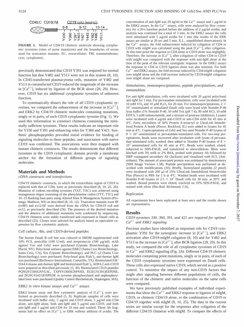
To systematically dissect the role of all CD19 cytoplasmic ty-rosines, we compared the enhancement of the increase in [Ca21]iand ERK2 by CD4/19 chimeric molecules containing mutations,singly or in pairs, of each CD19 cytoplasmic tyrosine (Fig. 1). Weused this information to construct chimeras containing the mini-mally sufficient tyrosines. These results indicate an important rolefor Y330 and Y391 and enhancing roles for Y360 and Y421. Syn-thetic phosphopeptides provided initial evidence for binding ofsignaling molecules to these peptides, and association with nativeCD19 was confirmed. The associations were then mapped withmutant chimeric constructs. The results demonstrate that differenttyrosines in the CD19 cytoplasmic domain provide a membraneanchor for the formation of different groups of signalingmolecules.
Materials and MethodscDNA constructs and transfections
CD4/19 chimeric constructs, in which the extracellular region of CD19 isreplaced with that of CD4, were as previously described (8, 10, 25, 26).Mutation of codons encoding tyrosines (TAT, TAC) was achieved usingmutagenesis oligos consisting of the phenylalanine codon (TTT, TTC) and10–15 flanking base pairs using Altered Sites II Mutagenesis System (Pro-mega, Madison, WI) as described (8, 10, 12). Truncation mutants exon 8F(ex8F) and ex12-6F were derived from the cDNA for CD4/19 ex8 andex12, as previously described (26). The presence of the desired mutationand the absence of additional mutations were confirmed by sequencing.CD4/19 chimeras were stably transfected and expressed in Daudi cells asdescribed (25). Clones were selected for analysis based on equivalent ex-pression by flow cytometric analysis.
Cell culture, Abs, and CD19-derived peptides
The human Daudi B cell line was cultured in IMDM supplemented with10% FCS, penicillin (100 U/ml), and streptomycin (100mg/ml). mAbagainst Vav and Grb2 were purchased (Upstate Biotechnology, LakePlacid, NY). Polyclonal antisera against ERK2 (Santa Cruz Biotechnology,Santa Cruz, CA), Sos (Upstate Biotechnology), and PLC-g2 (Santa CruzBiotechnology) were purchased. Polyclonal goat F(ab9)2 anti-human IgMwas purchased (BioSource International, Camarillo, TX). Biotinylated Fab9DA4.4 mouse anti-human IgM and biotinylated F(ab9)2 ADF4.2 anti-CD19were prepared as described previously (5, 30). Biotinylated CD19 peptidesPQNQY(330)GNVLSL, TAPSY(360)GNPSSD, EGEGY(391)EEPDSE,and DGSGY(421)ENPEDE in tyrosine phosphorylated andunphosphory-lated form were purchased (Quality Control Biochemicals, Hopkington, MA).
ERK2 in vitro kinase assays and Ca21 assays
ERK2 kinase assay and flow cytometric analysis of [Ca21]i were per-formed as previously described (5, 8). Replicate samples of cells wereincubated with buffer only, 5mg/ml anti-CD19 alone, 5mg/ml anti-CD4alone, anti-IgM alone, both anti-IgM and 5mg/ml anti-CD19, and bothanti-IgM and 5mg/ml anti-CD4 for 10 min and washed. These Ab frag-ments had no effect on [Ca21]i or ERK without addition of avidin. The
concentration of anti-IgM was 20 ng/ml in the Ca21 assays and 1mg/ml inthe ERK2 assays. In the Ca21 assays, cells were analyzed by flow cytom-etry for a 20-s baseline period before the addition of 2mg/ml avidin, andanalysis was continued for a total of 3 min. In the ERK2 assays the cellswere stimulated with 5mg/ml avidin for 1 min (the results of the ERKassays are similar at 30 sec and 5 min, X.L., unpublished observations). Inthe Ca21 assays, the fold enhancement induced by coligation of CD4 orCD19 with mIgM was calculated using the peak [Ca21]i after coligation.At this time point the response to CD4 alone or CD19 alone was negligible.Therefore, the increase in [Ca21]i after coligation of either CD4 or CD19with mIgM was compared with the response with anti-IgM alone at thetime of the peak of the relevant synergistic response. In the ERK2 assaysthe response to CD4 or CD19 ligation alone was also minimal. For bothCa21 and ERK2 assays, the fold increase induced by CD4/mIgM coligationover mIgM alone and the fold increase induced by CD19/mIgM coligationover mIgM alone are compared.
Stimulations, immunoprecipitations, peptide precipitations, andimmunoblots
For anti-IgM stimulation, cells were incubated with 20mg/ml polyclonalanti-IgM for 1 min. For pervanadate stimulation, cells were incubated with10 mM VO4 and 10mM H2O2 for 20 min. For immunoprecipitations, 23107 unstimulated or stimulated Daudi cells were lysed with Nonidet P-40lysis buffer (1% Nonidet P-40, 50 mM Tris (pH 7.4), 80 mM KCl, 10 mMEDTA, 5 mM iodoacetamide, and a mixture of protease inhibitors). Lysateswere incubated with 4mg/ml anti-CD19 or anti-CD4 mAb for 45 min at4°C. Forty microliters of 50% Protein A-trisacryl or UltraLink Immobi-lized Protein A beads (Pierce, Rockford, IL) were added to lysates for 45min at 4°C. Coprecipitations of Grb2 and Sos used Nonidet P-40 lysates of8 3 107 unstimulated or pervanadate-stimulated cells. For two-step pre-cipitations, beads were recovered after incubation with lysate of pervana-date-stimulated cells, washed, then added to Nonidet P-40 lysates of 83107 unstimulated cells for 45 min at 4°C. Beads were washed, eluted,subjected to SDS-PAGE, and transferred to nitrocellulose. Blots wereblocked with 3% milk or 2% BSA, probed with primary Ab followed byHRP conjugated secondary Ab (Jackson) and visualized with ECL (Am-ersham). The amount of associated protein was estimated by densitometry(NIH Image Version 1.58). Peptide precipitation was performed as de-scribed with modifications (10). Briefly, biotinylated peptides (2 pmol)were incubated with 200ml of 25% UltraLink Immobilized NeutrAvidinPlus (Pierce) in PBS for 1 h at 4°C. Washed beads were incubated withNonidet P-40 lysates of 2.53 107 Daudi cells for 30 min. at 20°C andwashed. Bound proteins were eluted, resolved on 10% SDS-PAGE, andstained with silver (Bio-Rad, Richmond, CA).
Replication
All experiments have been replicated at least once and the results shownare representative.
ResultsCD19 tyrosines 330, 360, 391, and 421 are required for optimalCa21 and ERK2 signaling
Previous studies have identified an important role for CD19 cyto-plasmic Y391 for the synergistic increase in [Ca21]i and ERK2activation after CD19-mIgM coligation (8, 10) and for Y482 andY513 in the increase in [Ca21]i after BCR ligation (28, 29). In thisstudy, we compared the role of all cytoplasmic tyrosines of CD19in Ca21 and ERK2 signaling (Fig. 2A). Different CD4/19 chimericmolecules containing point mutations, singly or in pairs, of each ofthe CD19 cytoplasmic tyrosines were expressed on Daudi cells.These cells also expressed native CD19, which served as a positivecontrol. To minimize the impact of any non-CD19 factors thatmight alter signaling between different populations of cells, thefunction of the chimeric and native molecules on the same cellswere compared.
We have previously published examples of individual experi-ments that show the Ca21 and ERK2 response to ligation of mIgM,CD19, or chimeric CD4/19 alone, or the combination of CD19 orCD4/19 together with mIgM (8, 10, 25). The data in the currentseries are similar except for the responses after coligation of thedifferent CD4/19 chimeras with mIgM. To compare the effects of
FIGURE 1. Model of CD4/19 chimeric molecule showing cytoplas-mic tyrosines (sites of point mutations) and the boundaries of exonsused for truncation mutants. EC, extracellular; TM, transmembrane;cyto, cytoplasmic.
3124 CD19 TYROSINES: FUNCTION AND BINDING OF Grb2/Sos AND PLC/Vav
by guest on Novem
ber 7, 2017http://w
ww
.jimm
unol.org/D
ownloaded from

the systematic mutations, we calculated the fold enhancement, rel-ative to native CD19, for each chimera, as described inMaterialsand Methods. The results are reported as the fold increase inducedby CD4/mIgM coligation divided by the fold increase induced byCD19/mIgM coligation. For efficiency, in addition to Y482 andY513, the sites of association with PI3K, we also grouped Y403and Y443 on the basis of homology (YEND and YENE) and as theputative sites of association with Fyn (16) and Y330 and Y360 onthe basis of homology (YGNV and YGNP).
Mutation of Y391 indeed had the most profound effect on boththe increase in [Ca21]i and ERK2 activation (Fig. 2,A andB). Theadditional experiments performed here also confirm the lack ofeffect of mutation of Y482 and Y513 in this system. However,other CD19 tyrosines also were required for optimal signaling inthe Ca21 assays. Mutation of Y421 and of Y330 and Y360 alsoresulted in decreases in responses that were significantly differentfrom the unmutated chimera. Mutation of Y490 or of Y403 andY443 had no statistically significant effect. In the ERK2 assays,mutation of Y391 was the only change that resulted in a significantloss of activity. Mutation of Y330 and Y360 resulted in a small but
statistically significant increase in response. The discrepancy in therequirement of Y330 and Y360 for the increase in [Ca21]i but notfor ERK2 suggests that there are differences in the signaling path-ways linked to CD19 that enhance the increase in [Ca21]i andERK2 activation.
These experiments suggested that Y330, Y360, Y391, and Y421play a role in CD19 signaling in this system. We found that chi-meric molecules containing only these four tyrosines, with pointmutations of the other five, were sufficient for synergistic enhance-ment of the increase in [Ca21]i and ERK2 activation (Fig. 2,C andD). Chimeras containing substitutions in the remaining four ty-rosines were constructed and tested. In these experiments, the ef-fects of the mutations were similar in both Ca21 and ERK2 assays.In both types of analysis, any further mutation reduced the en-hancement of signaling, suggesting that, in the absence of othertyrosines, Y330, Y360, Y391, and Y421 all can contribute toCD19 signaling. Single further mutation of either Y330 or Y391resulted in a.70% reduction in enhancement, suggesting a crucialrole for these tyrosines. Mutation of either Y360 (Y330, Y391, andY421 remaining) or of Y421 (Y330, Y360, and Y391 remaining)
FIGURE 2. Effect of mutation of CD19 tyrosines on synergistic enhancement of the increase in [Ca21]i (A andB) or of ERK2 activation (CandD).Daudi B cells were transfected with CD4/19 chimeric constructs containing phenylalanine substitutions of the CD19 cytoplasmic tyrosines.A and C,Numbers shown on thex-axis refer to the tyrosine residues replaced with phenylalanine in each construct; all other tyrosines in these constructs are wildtype. B and D, Numbers refer to the unmutated tyrosines present in the constructs tested; all other tyrosines are mutated to phenylalanine. Cells werestimulated by cross-linking mIgM alone, CD19 alone, or CD4 alone or by co-cross-linking mIgM with either CD19 or CD4. Changes in [Ca21]i weremonitored by flow cytometry. ERK2 activation was measured by incorporation of32P into myelin basic protein. The data are presented for each constructas mean6 SD in replicate experiments of the fold enhancement induced by CD4/mIgM coligation divided by the fold enhancement induced by CD19/mIgM coligation on replicate samples of cells. InA andC, the asterisks indicate those CD4/19 chimera whose response is statistically significantly differentfrom those of unmutated chimera (p, 0.05). InB andD, all other chimera were statistically significant from the construct containing Y330, Y360, Y391,Y421 (first column). The responses with chimera containing Y330, Y391 and Y421 (second column), or Y330, Y360, and Y391 (fourth column) were notstatistically different, but all others shown were different from these two (p, 0.05). WT, unmutated CD19 cytoplasmic domain.
3125The Journal of Immunology
by guest on Novem
ber 7, 2017http://w
ww
.jimm
unol.org/D
ownloaded from

resulted in 40% and 32% reduction in enhancement in the Ca21
assays and 33% and 51% reduction in enhancement in the ERK2assays, respectively. Further mutation of Y360 from Y330, Y360,and Y391 (Y330 and Y391 remaining) or of Y421 from Y330,Y391, and Y421 (Y330 and Y391 remaining) resulted in furtherloss of function. These data suggest a cooperative role for Y360and Y421 with Y330 and Y391 in the synergistic increase in[Ca21]i and ERK2 activation after CD19-mIgM coligation. Thedifferential requirement for Y330 and Y360 in ERK2 activation inthe presence or absence of additional tyrosines suggests some re-dundancy when other tyrosines are present.
CD19-derived phosphopeptides associate with proteins ofapparentMr of 24, 113, 139, and 149
Phosphorylation of CD19 tyrosines provides docking sites for SH2domain-containing proteins, and these associations are likely tomediate CD19 function. We searched for proteins associating withthe tyrosines shown above to play a role in synergistic increase in[Ca21]i and ERK2 activation after CD19-mIgM coligation. Bio-tinylated 11-mer peptides corresponding to unphosphorylated andphosphorylated Y330, Y360, Y391, and Y421 and flanking se-quences were bound to avidin-coated beads and incubated withDaudi lysates. Bound proteins were eluted, resolved by SDS-PAGE, and silver stained. Phospho-Y330 bound two proteins withapparentMr of 24 and 149, whereas nonphosphorylated Y330 didnot (Fig. 3A, filled arrows). Phospho-Y391 and, to a lesser extent,phospho-Y421 bound two proteins with apparentMr of 113 and139 (Fig. 3B), whereas nonphosphorylated Y391 and Y421 pep-tides did not. No specific associations with phospho-Y360 wereidentified. The homology with Y330 raises the question of a sec-ondary role in binding of similar molecules, below the limits ofdetection here.
CD19 associates with Grb2 and Sos and association with Grb2requires Y330 and/or Y360
To identify the proteins of apparentMr of 24 and 149 that wereassociated with phospho-Y330, precipitates were formed fromDaudi lysates as above, transferred to nitrocellulose, and probedwith Abs specific for Grb2 and Sos. Phospho-Y330 was able tocoprecipitate Grb2 and Sos, whereas nonphosphorylated Y330could not (data not shown). To determine whether native CD19could coprecipitate Grb2, CD19 was precipitated from lysates of8 3 107 unstimulated or pervanadate-stimulated cells. ReplicateCD19 precipitates from stimulated cells were also subjected to anadditional incubation with lysates of 83 107 unstimulated cells.CD19 could coprecipitate Grb2 in stimulated but not unstimulatedlysates (Fig. 4A). More Grb2 could be coprecipitated when a sec-ond incubation with unstimulated lysates was performed. This sug-gests that other proteins are competing with phospho-CD19 forbinding Grb2 in stimulated lysates. An anti-phosphotyrosine probeof the same blot confirms that tyrosine phosphorylation was in-duced by pervanadate. A CD19 probe shows a slightly lower re-covery of CD19 from pervanadate-stimulated cells. CD19 couldcoprecipitate Sos from 83 107 cells of pervanadate-stimulatedDaudi lysates, whereas UPC10, a control Ab, could not (Fig. 4B).Again, a two-step precipitation first using pervanadate-stimulatedand then unstimulated lysates was able to precipitate more Sosthan a single-step precipitation from pervanadate-stimulated cells.The same blot was probed with anti-phosphotyrosine to demon-strate that pervanadate stimulation induced tyrosine phosphoryla-tion. The CD19 probe shows equivalent recovery. To map thetyrosines required for association of CD19 and Grb2, chimeric
CD4/19 was precipitated from stably transfected Daudi cell lines(Fig. 4C). A chimera with an intact CD19 cytoplasmic domain(wild type (WT)) was able to coprecipitate Grb2 from lysates of8 3 107 pervanadate-stimulated cells but not from those of un-stimulated cells. Mutation of all nine tyrosines to phenylalanine(Y9F) abolishes this association, demonstrating the requirementfor cytoplasmic phosphotyrosines of CD19 in mediating this as-sociation. Mutation of 330 and 360 (330,360F) ablates the abilityof the chimera to coprecipitate Grb2, establishing a requirement of330 and/or 360 for Grb2 association. An anti-phosphotyrosineprobe of the same blot demonstrates phosphorylation of the WTand 330,360F chimeras and an absence of tyrosine phosphoryla-tion in Y9F. The CD4 probe demonstrates equivalent recovery ofchimera from each sample. In preliminary experiments, a constructcontaining only Y330 and Y360 failed to precipiate Grb2, but thesignificance of this is as yet unclear, given the ability of the phos-pho-Y330 peptide to do so.
FIGURE 3. Proteins associating with 11-mer peptide fragments con-taining either tyrosine or phosphotyrosine and flanking peptide sequencecorresponding to CD19 Y330, Y391, and Y421. Proteins from Daudi ly-sates were adsorbed to peptides, eluted, resolved by SDS-PAGE, and silverstained.A, Phosphotyrosine 330 (pY330) associates with proteins with ap-parentMr of 24 and 149 (filled arrows), whereas unphosphorylated Y330does not. pY391 associates with proteins with apparentMr of 113 and 139(open arrows).B, Both pY391 and (more weakly) pY421 but not unphos-phorylated Y391 or Y421 associate with proteins with apparentMr of 113and 139 (arrows).
3126 CD19 TYROSINES: FUNCTION AND BINDING OF Grb2/Sos AND PLC/Vav
by guest on Novem
ber 7, 2017http://w
ww
.jimm
unol.org/D
ownloaded from

Mapping of CD19 tyrosines required for BCR-induced Vavassociation
Previous studies demonstrated activation-induced association ofCD19 and Vav and that CD19 cytoplasmic Y391 is important inthis interaction (8, 10, 14). The protein with apparentMr of 113that coprecipitated with peptides phospho-Y391 and phospho-Y421 (Fig. 3B) comigrated with Vav, as demonstrated by immu-noblotting (data not shown). The requirement for other CD19 cy-toplasmic tyrosines in the activation-induced association of CD19and Vav was investigated by precipitating the CD4/19 chimerafrom stably transfected Daudi cells before and after stimulationwith polyclonal anti-IgM. The precipitates were probed with anti-Vav (shown on the left in Fig. 5). The amount of coprecipitatingVav was estimated by densitometry. For each chimera, the fold
date-treated Daudi cells (VO4). Replicate precipitates formed from lysatesof pervanadate-treated cells were recovered and further incubated withlysates of unstimulated cells (VO4 . U). Precipitates and WCL fromunstimulated and stimulated cells were probed first with anti-Sos (top) andthen sequentially stripped and reprobed with anti-phosphotyrosine (middle)and anti-CD19 (bottom).C, Requirement for Y330 and Y360. Daudi cellsexpressing the CD4/19 chimeric constructs WT (unmutated CD19 cyto-plasmic domain), 330,360F (phenylalanine substitution oftyrosines Y330and Y360), or Y9F (phenylalanine substitution of all nine tyrosines) werestimulated with either buffer only or pervanadate and were lysed. Precipitatesformed with anti-CD4 from the lysates were probed with anti-Grb2 (top) andthen sequentially stripped and reprobed with anti-phosphotyrosine (middle)and anti-CD19 (bottom).
FIGURE 4. Association of Grb2 and Sos with native CD19 and chi-meric CD4/19 constructs.A, Grb2 and native CD19. Precipitates wereformed with anti-CD19 from either lysis buffer only (2) or from lysates ofunstimulated (U) or pervanadate-treated (VO4) untransfected Daudi cells.A replicate precipitate formed from lysate of pervanadate-treated cells wasrecovered and further incubated with a lysate of unstimulated cells (VO4 .U). Precipitates and whole-cell lysate (WCL) from unstimulated and stim-ulated cells were probed first with anti-Grb2 (top) and then sequentiallystripped and reprobed with anti-phosphotyrosine (middle) and anti-CD19(bottom).B, Sos and native CD19. Precipitates were formed with eitheranti-CD19 or control Ab (UPC10) from lysates of untransfected, pervana-
FIGURE 5. Daudi cells expressing the CD4/19 constructs WT (unmu-tated CD19 cytoplasmic domain); 330,360F, 391F, 403,443F, 421F, or490F (phenylalanine substitutions in one or two tyrosines only); ex12(truncated at the end of exon 12); Y5F (phenylalanine substitution of Y403,Y443, Y482, Y490, and Y513 (Y330, Y360, Y391, and Y421 are intact));or Y9F (phenylalanine substitution of all cytoplasmic tyrosines) were stim-ulated either with buffer only or with polyclonal anti-IgM and were lysed.Anti-CD4 precipitates formed from the lysates were probed with anti-Vav,and the density of the Vav band was measured. The change in the densityof the Vav band after anti-IgM stimulation, relative to unstimulated cells,was calculated for each construct. Equivalent loading was confirmed byblotting with anti-CD4. The data are presented for each construct asmean6 SD in replicate experiments. The previously published experimentwith 391F (8) is typical of the primary data.
3127The Journal of Immunology
by guest on Novem
ber 7, 2017http://w
ww
.jimm
unol.org/D
ownloaded from

increase in Vav association after stimulation over the basal asso-ciation in unstimulated cells is reported (shown on the right in Fig.5). The WT cytoplasmic domain exhibits a low basal associationand an increase in Vav association on stimulation. Y9F, a constructin which all nine tyrosines are mutated to phenylalanine, shows anequivalent basal association that decreases on stimulation. Whentyrosines 330 and 360 are mutated to phenylalanine (330,360F),the increase in Vav association on stimulation is not significantlydifferent from WT. There is a similar increase when the cytoplas-mic domain is truncated at the end of ex12. However, if any oftyrosines 391, 403/443, 421, or 490 are mutated to phenylalanine,after stimulation there is more Vav coprecipitated than with Y9Fbut less than with WT. This suggests a role for each of thesetyrosines in maximal association of Vav with CD19 after stimu-lation with anti-IgM.
Constitutive association of CD19 and Vav is mediated by theCD19 cytoplasmic region encoded by exons 9–12
The ability of Y9F to coprecipitate Vav in unstimulated but notstimulated lysates suggested that there is a constitutive associationof CD19 and Vav that is independent of tyrosine phosphorylation.To determine the site of this interaction, CD4/19 chimeras that aretruncations of Y9F were tested for their ability to associate withVav in a constitutive manner (Fig. 6). Y9F is able to coprecipitateVav before but not after pervanadate stimulation, similar to whatis seen with anti-IgM stimulation. A chimera that is truncated atthe end of exon 8 with all tyrosines mutated to phenylalanine(ex8F) is unable to coprecipitate Vav in either unstimulated orstimulated cells. However, a chimera that is truncated at the end ofexon 12 with all tyrosines mutated to phenylalanine (ex12-6F) isable to coprecipitate Vav, suggesting that the constitutive associ-ation of CD19 and Vav is mediated by the CD19 cytoplasmicregion encoded by exons 9–12. The blot was reprobed with anti-CD4 Ab to demonstrate equivalent recovery of the chimera.
CD19 associates with PLC-g2 and tyrosines 330, 360, 391, and421 are sufficient for the BCR-induced association
To identify the protein of apparentMr of 139 that bound to pep-tides phospho-Y391 and phospho-Y421 (Fig. 3B), precipitatesformed with these peptides were probed with Abs specific forPLC-g2. Phospho-Y391 and phospho-Y421 were able to copre-cipitate PLC-g2, whereas nonphosphorylated Y391 and Y421could not (data not shown). To determine whether native CD19
could coprecipitate PLC-g2, precipitates were formed from lysatesof 2 3 107 unstimulated and pervanadate-stimulated cells withanti-CD19 Ab. Immune complexes were eluted, resolved by SDS-PAGE, transferred to nitrocellulose, and probed with anti-PLC-g2Ab (Fig. 7A). CD19 is able to coprecipitate PLC-g2 in stimulatedbut not unstimulated cells. The blot was stripped and reprobed withanti-phosphotyrosine to demonstrate tyrosine phosphorylation ofCD19 after pervanadate stimulation. The blot was further reprobedwith anti-CD19 to show equivalent recovery. To determine therequirement for CD19 cytoplasmic tyrosines in the anti-IgM-induced association, anti-CD4 immunoprecipitates wereformed from unstimulated and anti-IgM-stimulated Daudi cellsthat were either untransfected or that expressed CD4/19 chimerascontaining either the WT cytoplasmic domain or a cytoplasmicdomain containing mutations in all nine tyrosines (Fig. 7B). Prob-ing for PLC-g2 reveals a low-level, non-CD4-dependent, nonspe-cific precipitation that was observed consistently in untransfectedcells and in cells expressing the Y9F chimera. The slight increaseobserved in the latter relative to untransfected cells was variablyobserved but is unlikely to be meaningful given the absence ofcoprecipitation with native CD19 from untransfected cells. How-ever, the non-CD4-dependent precipitation precluded analysis ofconstitutive association of PLC-g2. An increase (2.4-fold) in as-sociation after anti-IgM stimulation is observed only with the in-tact cytoplasmic domain (WT), suggesting that the activation-in-duced association between CD19 and PLC-g2 is dependent on
FIGURE 6. Constitutive association of Vav and CD19. Daudi cells ex-pressing the CD4/19 constructs Y9F (phenylalanine substitution of all cy-toplasmic tyrosines), ex12-6F (truncated at the end of exon 12 with phe-nylalanine substitution of all tyrosines), or ex8F (truncated at the end ofexon 8 with phenylalanine substitution of all tyrosines) were stimulatedwith buffer only or with pervanadate and were lysed. Precipitates formedwith anti-CD4 or WCL (from Y9F Daudi cells) were probed first withanti-Vav, stripped, and reprobed with anti-CD4.
FIGURE 7. Association of PLC-g2 with CD19.A, Untransfected Daudicells were treated with buffer only or with pervanadate and were lysed.Precipitates formed with anti-CD19 or a WCL were probed first with anti-PLC-g2 and then sequentially stripped and reprobed with anti-phosphotyrosine and anti-CD19.B, Daudi cells that were either untras-fected (Untx) or expressing the CD4/19 chimeric constructs WT(unmutated CD19 cytoplasmic domain) or Y9F (phenylalanine substitutionof all cytoplasmic tyrosines) were stimulated with buffer only or with anti-IgM and were lysed. Precipitates formed with anti-CD4 from the lysateswere probed first with anti-PLC-g2 and then were sequentially stripped andreprobed with anti-phosphotyrosine and anti-CD4.
3128 CD19 TYROSINES: FUNCTION AND BINDING OF Grb2/Sos AND PLC/Vav
by guest on Novem
ber 7, 2017http://w
ww
.jimm
unol.org/D
ownloaded from

tyrosine phosphorylation. In other experiments, activation with anti-IgM induced an increase in PLC-g2 association with a construct inwhich all but four tyrosines (Y330, Y360, Y391, and Y421) aremutated to phenylalanine (Y5F), suggesting that these tyrosinesbind PLC-g2 (data not shown). Additional experiments were con-ducted to map this association.
CD19 Y391 is sufficient for association of Vav and PLC-g2 inpervanadate-stimulated cells
The data in Fig. 5 for Vav and similar experiments with PLC-g2suggest that multiple CD19 tyrosines had an effect on bindingwhen cells were stimulated with anti-IgM. This suggested that cer-tain CD19 cytoplasmic tyrosines are required to induce maximalphosphorylation of other tyrosines, possibly by recruiting and/oractivating the tyrosine kinases Lyn (17, 18) or Fyn (16) or others.To eliminate this variable, pervanadate stimulation was used tomaximally phosphorylate tyrosines in CD4/19 chimeras to map therequirements for CD19 cytoplasmic tyrosines in Vav and PLC-g2association (Fig. 8). A WT cytoplasmic domain is able to associatewith Vav and PLC-g2 in a stimulation-dependent manner. Muta-tion of Y391, Y421, and Y490 (Y3F) eliminates stimulation-in-duced association of PLC-g2, as does mutation of Y391 and Y421(Y2F). A chimera in which all tyrosines except Y391 are mutated(391Y) associates with PLC-g2 as well as WT, whereas Y421alone (421Y) does not show any PLC-g2 association above back-ground although the chimera is well phosphorylated. A light bandobserved in the PLC-g2 probe in stimulated lanes Y3F, Y2F, Y9F,and 421Y represents an increase in non-CD4-dependent precipi-tation (as above) in pervanadate-treated cells and is insignificantcompared with the increase observed in WT or 391Y. The sameblot was stripped and reprobed for Vav. Vav association exhibitsa similar pattern of association except that there is a constitutiveassociation in unstimulated cells. 391Y exhibits a stimulation-in-ducedincrease in Vav association comparable to that observedwith WT. Y9F again exhibits a decrease in Vav association onstimulation. Y3F and Y2F have lost the stimulation-inducedincrease in association seen in WT but do not show the loss ofassociation observed with Y9F. This suggests that other CD19tyrosines can mediate weaker binding of Vav in the absence ofY391, perhaps through indirect association. The blot wasstripped and reprobed with anti-phosphotyrosine. Chimeraswith fewer tyrosines show a proportionally lower but still sub-stantial level of tyrosine phosphorylation after pervanadate
stimulation. Blots were reprobed with anti-CD4 to demonstrateequivalent recovery of chimera.
DiscussionCD19 is an important modulator of B cell activation. Most signal-ing functions of CD19 described to date are dependent on tyrosine-mediated interactions. We sought to determine which of the nineCD19 cytoplasmic tyrosines are involved in the Ca21 and ERK2signals. We used Daudi cells primarily because a cell line wasnecessary for expression of the many mutant constructs required.This cell also reflects an environment in which CD19 is normallyexpressed, and we could compare the mutant and native CD19molecules in the same cells. In addition, this system has proven auseful tool for dissecting CD19 function in previous studies (5, 7,8, 10, 20, 22, 25, 26, 31).
Our functional studies suggest that Y330, Y360, Y391, andY421 can each interact with downstream signaling pathways. Inthe presence of the other tyrosines, Y330 and Y360 are dispensablefor ERK2 but not Ca21 signaling. However, Y330 and Y360 arerequired for both ERK2 and Ca21 signaling in the absence ofY403, Y443, Y482, Y490, and Y513. We suspect that these dif-ferences may be due to alternative binding sites for effectors oractivation of different effectors that may converge into these path-ways. Introduction of structural changes by the tyrosine mutationsare difficult to exclude, but current evidence suggests that this isnot the case. Phosphorylation of each of these constructs (includ-ing those with mutations of Y403 and Y443, the putative sites ofFyn association) is proportional to the number of remaining ty-rosines (data not shown). Association with PI3K is intact if Y482and Y513 are present (data not shown). Mutation of Y330 andY360 by themselves did not reduce ERK2 activation (Fig. 2C).These observations suggest that the tyrosine to phenylalanine sub-stitutions do not result in gross structural changes that disrupt thebinding at the intact residues.
Previous studies have identified a role for Y391 in ERK2 andCa21 signaling and in Vav association. Vav can enhance Ca21
signaling via activation of phosphatidylinositol 4-phosphate 5-ki-nase and increased production of phosphatidylinostol 4,5-bisphos-phate (8, 10), the substrate for PLC-g2. We now show that PLC-g2also associates with CD19 via Y391. Therefore, CD19 may play arole in membrane localization of PLC-g2. We have demonstratedby coprecipitation that Vav and PLC-g2 inducibly associate in
FIGURE 8. Role of Y391 and Y421. Daudi cells expressing the CD4/19 chimeric constructs WT (unmutated CD19 cytoplasmic domain), Y3F (phe-nylalanine substitution of Y391, Y421, and Y490), Y2F (phenylalanine substitution of Y391 and Y421), Y9F (phenylalanine substitution of all cytoplasmictyrosines), 391Y (phenylalanine substitution of all tyrosines except Y391), or 421Y (phenylalanine substitution of all tyrosines except Y421) were treatedwith buffer only or with pervanadate and were lysed. Precipitates formed with anti-CD4 were probed with anti-PLC-g2 and then were sequentially strippedand reprobed with anti-Vav, anti-phosphotyrosine, and anti-CD4. The higher m.w. band observed in that anti-Vav blot in the WT and 391Y lanes is likelya residual signal after blotting with anti-PLC.
3129The Journal of Immunology
by guest on Novem
ber 7, 2017http://w
ww
.jimm
unol.org/D
ownloaded from

pervanadate stimulated Daudi cells (data not shown). Together,this suggests a model in which, upon activation, a Vav/PLC-g2complex binds phospho-CD19 and coordinately regulates the ac-tivation of PLC-g2 and the pathway which produces its substrate,phosphatidylinostol 4,5-bisphosphate.
We previously had interpreted our results with mutation ofY391 (n which the amount of associated Vav was equivalent inprecipitates of the CD4/19 chimera containing mutation of Y391from unstimulated and stimulated cells, as opposed to the increaseobserved with WT CD19 cytoplasmic domain) as reflecting anabsence of activation-induced association when Y391 was mutated(8). However, our current results show that when all tyrosines aremutated, there is a dramatic reduction in the amount of associatedVav after stimulation (Fig. 5, 6, and 8). Thus, the residual binding,after activation, to CD4/19 with mutation of Y391 represents par-tial, tyrosine-based binding. Other tyrosines also can mediate Vavbinding after mutation of Y391, although at lower levels thanY391 (Fig. 8). The complete loss of postactivation binding aftermutation of all tyrosines suggests that the basal association of Vavwith CD19 is of relatively low affinity and that, after activation,Vav shifts to SH2-mediated, tyrosine-dependent binding to CD19or an associated molecule (e.g., PLC-g2). CD19 may provide areservoir of Vav near the site of postactivation binding.
BLNK/SLP-65, an adaptor protein that binds effectors/adaptorsincluding PLC-g2, Vav, and Grb2 (32–34), has been shown to becrucial for activation of PLC-g2 in DT40 cells (35, 36). We havebeen unable to detect BLNK associated with CD19 in stimulatedDaudi cells, although such precipitates contain Vav, PLC-g2,Grb2, and Sos. BLNK/SLP-65 is detectable in Daudi whole celllysates (data not shown). CD19 may provide an alternative path-way for membrane localization and activation of PLC-g2.
The enhanced association of Grb2 and Sos when phosphorylatedCD19 is recovered from activated lysates and then incubated withunactivated lysates suggests that, under conditions in which phos-phorylation of tyrosine residues on intracellular proteins is maxi-mal, the affinity of binding to phospho-CD19 is less than that ofother phosphoproteins. This suggests the hypothesis that, underconditions in which CD19 is preferentially phosphorylated such asafter ligation of CD19 alone (12), CD19 might serve as a sink forGrb2 and Sos and play a negative role (6, 37, 38). Such an effectmight explain the small increase in ERK2 activity observed withthe chimera with mutations in Y330 and Y360. The mutationalanalysis does not permit conclusions as to whether the proteinsidentified as associating with particular residues mediate the func-tional role demonstrated for those residues. The tyrosine residuesmay play other roles in addition to SH2-mediated protein binding,such as forming part of the motif for phosphorylation of nearbyserine/threonine residues. In addition, we suspect that mutation ofsome tyrosines may affect the phosphorylation of other CD19 ty-rosines or of CD19-associated molecules. In the studies of asso-ciation after stimulation with anti-IgM, alterations of different ty-rosines reduced the level of Vav association, even when Y391,shown in the pervanadate studies to be sufficient by itself for max-imal Vav association, was intact.
Native CD19 is present on these cells, and thus these experi-ments only address the question of which tyrosines are required onthe CD19 molecule that is cross-linked with the BCR. This mightexplain the lack of effect of mutation of Y482 and Y513. Buhl etal. (28) have shown that these residues are important for optimalsignaling after ligation of the BCR alone. The native CD19 in thetransfected Daudi cells could serve this function in our system. Inother studies we have found that wortmannin suppresses the Ca21
and ERK signals after CD19-mIgM coligation in Daudi cells(X. Li and R. H. Carter, unpublished observations).
This is the first report to demonstrate a function for the upstreamCD19 tyrosines and the association between CD19 and the effec-tors PLC-g2, Grb2, and Sos and to map associations with CD19Y330 and Y421. Additional studies will be required to furtherelucidate the relationship of Vav and PLC-g2 in binding CD19, therole of the association of Grb2/Sos with CD19, and the conditionsunder which different cytoplasmic tyrosines of CD19 are phos-phorylated. However, identification of the rich array of effectorsthat associate with CD19 suggests a model in which CD19 acts asa point of convergence for multiple signaling pathways. Combin-ing this and previous studies, we can now assign association formost CD19 tyrosines: Y330 with Grb2/Sos, Y391 and Y421 withVav/PLC-g2, Y403/Y443 with Fyn, and Y492/Y513 with PI3K.Tyrosine phosphorylation of these residues could occur on a con-certed basis or individual tyrosines may be phosphorylated inde-pendently, depending on stage of development, strength of signal-ing through the BCR, complement activation, stimulation bycoreceptors, anatomical location, or other factors. By such a mech-anism, CD19 could play a modulated role in multiple signalingpathways that are central to BCR-mediated B cell activation.
AcknowledgmentsWe thank Larry Freeberg for technical assistance in selection and mainte-nance of the transfected cells and Marion Spell and Tina Rogers for flowcytometric analysis of Ca21. We also thank Drs. Amnon Altman andDouglas Fearon for helpful discussions.
References1. Engel, P., L.-J. Zhou, D. C. Ord, S. Sato, B. Koller, and T. F. Tedder. 1995.
Abnormal B lymphocyte development, activation, and differentiation in mice thatlack or overexpress the CD19 signal transduction molecule.Immunity 3:39.
2. Rickert, R. C., K. Rajewsky, and J. Roes. 1995. Impairment of T-cell-dependentB-cell responses and B-1 cell development in CD19-deficient mice.Nature 376:352.
3. Sato, S., D. A. Steeber, and T. F. Tedder. 1995. The CD19 signal transductionmolecule is a response regulator of B-lymphocyte differentiation.Proc. Natl.Acad. Sci. USA 92:11558.
4. Sato, S., N. Ono, D. Steeber, D. Pisetsky, and T. F. Tedder. 1996. CD19 regulatesB lymphocyte signaling thresholds critical for the development of B-1 lineagecells and autoimmunity.J. Immunol. 157:4371.
5. Carter, R. H., D. A. Tuveson, D. J. Park, S. G. Rhee, and D. T. Fearon. 1991. TheCD19 complex of B lymphocytes: activation of phospholipase C by a proteintyrosine kinase-dependent pathway that can be enhanced by the membrane IgMcomplex. J. Immunol. 147:3663.
6. Carter, R. H., and D. T. Fearon. 1992. CD19: lowering the threshold for antigenreceptor stimulation of B lymphocytes.Science 256:105.
7. Bradbury, L. E., G. S. Kansas, S. Levy, R. L. Evans, and T. F. Tedder. 1992. TheCD19/CD21 signal transducing complex of human B lymphocytes includes thetarget of antiproliferative antibody-1 and Leu-13 molecules.J. Immunol. 149:2841.
8. Li, X., D. Sandoval, L. Freeberg, and R. H. Carter. 1997. Role of CD19 tyrosine391 in synergistic activation of B lymphocytes by coligation of CD19 and mem-brane Ig.J. Immunol. 158:5649.
9. Callard, R. E., K. P. Rigley, S. H. Smith, S. Thurstan, and J. G. Shields. 1992.CD19 regulation of human B cell responses: B cell proliferation and antibodysecretion are inhibited or enhanced by ligation of the CD19 surface glycoproteindepending on the stimulating signal used.J. Immunol. 148:2983.
10. O’Rourke, L. M., R. Tooze, M. Turner, D. M. Sandoval, R. H. Carter,V. L. J. Tybulewicz, and D. T. Fearon. 1998. CD19 as a membrane-anchoredadaptor protein of B lymphocytes: costimulation of lipid and protein kinases byrecruitment of Vav.Immunity 8:635.
11. Tooze, R. M., G. M. Doody, and D. T. Fearon. 1997. Counterregulation by thecoreceptors CD19 and CD22 of MAP kinase activation by membrane immuno-globulin. Immunity 7:59.
12. Tuveson, D. A., R. H. Carter, S. P. Soltoff, and D. T. Fearon. 1993. CD19 of Bcells as a surrogate kinase insert region to bind phosphatidylinositol 3-kinase.Science 260:986.
13. Chalupny, N. J., S. B. Kanner, G. L. Schieven, S. F. Wee, L. K. Gilliland,A. Aruffo, and J. A. Ledbetter. 1993. Tyrosine phosphorylation of CD19 in pre-Band mature B cells.EMBO J. 12:2691.
14. Weng, W.-K., L. Jarvis, and T. W. LeBien. 1994. Signaling through CD19 ac-tivates Vav/mitogen-activated protein kinase pathway and induces formation of aCD19/Vav/phosphatidylinositol 3-kinase complex in human B cell precursors.J. Biol. Chem. 269:32514.
15. Zhou, L. J., D. C. Ord, A. L. Hughes, and T. F. Tedder. 1991. Structure anddomain organization of the CD19 antigen of human, mouse, and guinea pig B
3130 CD19 TYROSINES: FUNCTION AND BINDING OF Grb2/Sos AND PLC/Vav
by guest on Novem
ber 7, 2017http://w
ww
.jimm
unol.org/D
ownloaded from

lymphocytes: conservation of the extensive cytoplasmic domain.J. Immunol.147:1424.
16. Chalupny, N. J., A. Aruffo, J. M. Esselstyn, P. Y. Chan, J. Bajorath, J. Blake,L. K. Gilliland, J. A. Ledbetter, and M. A. Tepper. 1995. Specific binding of Fynand phosphatidylinositol 3-kinase to the B cell surface glycoprotein CD19through their src homology 2 domains.Eur. J. Immunol. 25:2978.
17. van Noesel, C. J. M., A. C. Lankester, G. M. W. van Schijndel, and R. A. W. Lier.1993. The CR2/CD19 complex on human B cells contains the src-family kinaseLyn. Int. Immunol. 5:699.
18. Fujimoto, M., J. C. Poe, P. J. Jansen, S. Sato, and T. F. Tedder. 1999. CD19amplifies B lymphocyte signal transduction by regulating Src-family protein ty-rosine kinase activation.J. Immunol. 162:7088.
19. Fearon, D. T., and R. H. Carter. 1995. The CD19/CR2/TAPA-1 complex of Blymphocytes: linking natural to acquired immunity.Annu. Rev. Immunol. 13:127.
20. Bradbury, L. E., V. S. Goldmacher, and T. F. Tedder. 1993. The CD19 signaltransduction complex of B lymphocytes.J. Immunol. 151:2915.
21. Roberts, T., and E. C. Snow. 1999. Recruitment of the CD19/CD21 coreceptor toB cell antigen receptor is required for antigen-mediated expression of Bcl-2 byresting and cycling hen egg lysozyme transgenic B cells.J. Immunol. 162:4377.
22. Li, X., and R. H. Carter. 1998. Convergence of CD19 and B cell antigen receptorsignals in the ERK2 activation cascade.J. Immunol. 161:5901.
23. Dempsey, P. W., M. E. Allison, S. Akkaraju, C. C. Goodnow, and D. T. Fearon.1996. C3d of complement as a molecular adjuvant: bridging innate and acquiredimmunity. Science 271:348.
24. Villiers, M.-B., C. L. Villiers, A.-M. Laharie, and P. N. Marche. 1999. Ampli-fication of the antibody response by C3b complexed to antigen through an esterlink. J. Immunol. 162:3647.
25. Matsumoto, A. K., D. R. Martin, R. H. Carter, L. B. Klickstein, J. Ahearn, andD. T. Fearon. 1993. Functional dissection of the CD21/CD19/TAPA-1/Leu-13complex of B lymphocytes.J. Exp. Med. 178:1407.
26. Carter, R. H., G. M. Doody, J. B. Bolen, and D. T. Fearon. 1997. MembraneIgM-induced tyrosine phosphorylation of CD19 requires a CD19 domain thatmediates association with components of the B cell antigen receptor complex.J. Immunol. 158:3062.
27. Doody, G. M., P. W. Dempsey, and D. T. Fearon. 1996. Activation of B lym-phocytes: integrating signals from CD19, CD22 and FcgIIb1.Curr. Opin. Im-munol. 8:378.
28. Buhl, A. M., C. M. Pleiman, R. C. Rickert, and J. C. Cambier. 1997. Qualitativeregulation of B cell antigen receptor signaling by CD19: selective requirement forPI3-kinase activation, inositol-1,4,5-trisphosphate production and Ca21 mobili-zation.J. Exp. Med. 186:1897.
29. Buhl, A. M., and J. C. Cambier. 1999. Phosphorylation of CD19 Y484 and Y515,and linked activation of phosphatidylinositol 3-kinase, are required for B cellantigen receptor-mediated activation of Bruton’s tyrosine kinase.J. Immunol.162:4438.
30. Carter, R. H., M. O. Spycher, Y. C. Ng, R. Hoffman, and D. T. Fearon. 1988.Synergistic interaction between complement receptor type 2 and membrane IgMon B lymphocytes.J. Immunol. 141:457.
31. Doody, G. M., L. B. Justement, C. C. Delibrias, R. J. Matthews, J. Lin,M. L. Thomas, and D. T. Fearon. 1995. A role in B cell activation for CD22 andthe protein tyrosine phosphatase SHP.Science 269:242.
32. Fu, C., and A. C. Chan. 1997. Identification of two tyrosine phosphoproteins,pp70 and pp68, which interact with phospholipase Cg, Grb2, and Vav after B cellantigen receptor activation.J. Biol. Chem. 272:27362.
33. Fu, C., C. W. Turck, T. Kurosaki, and A. C. Chan. 1998. BLNK: a central linkerprotein in B cell activation.Immunity 9:93.
34. Wienands, J., J. Schweikert, B. Wollscheid, H. Jumaa, P. J. Nielsen, and M. Reth.1998. SLP-65: a new signaling component in B lymphocytes which requiresexpression of the antigen receptor for phosphorylation.J. Exp. Med. 188:791.
35. Ishiai, M., H. Sugawara, M. Kurosaki, and T. Kurosaki. 1999. Cutting edge:association of phospholipase C-g 2 Src homology 2 domains with BLNK iscritical for B cell antigen receptor signaling.J. Immunol. 163:1746.
36. Ishiai, M., M. Kurosaki, R. Pappu, K. Okawa, I. Ronko, C. Fu, M. Shibata,A. Iwamatsu, A. C. Chan, and T. Kurosaki. 1999. BLNK required for couplingSyk to PLCg2 and Rac1-JNK in B cells.Immunity 10:117.
37. Pezzutto, A., B. Dorken, P. S. Rabinovitch, J. A. Ledbetter, G. Moldenhauer, andE. A. Clark. 1987. CD19 monoclonal antibody HD37 inhibits anti-immunoglobulin-induced B cell activation and proliferation.J. Immunol. 138:2793.
38. Pezzutto, A., T. B. Barret, L. Ellingsworth, B. Dorken, and E. A. Clark. 1989.Down-regulation of B-cell activation by CD19 mAb. InLeucocyte Typing IV.W. Knapp, ed. Oxford Univ. Press, Oxford, pp. 39–40.
3131The Journal of Immunology
by guest on Novem
ber 7, 2017http://w
ww
.jimm
unol.org/D
ownloaded from