systemic pathology - respiratory, musculoskeletal systems of fish
TRANSCRIPT

Course;Systemic Pathology(2+1)
Topics: Musculo skeletal systemRespiratory systemHemopoietic Tissue
ByAbisha Juliet Mary.S.J

THE MUSCULOSKELETAL SYSTEM •The fusiform shape of the typical fish is determined by the requirements of swimming. •The streamlined exterior minimises drag

Teleost fish have Axial skeletonAppendicular Skeletons Of true bone with same basic components of mammalian skeletons.
The main muscle blocks (myomeres) are arranged on either side of the axial skeleton in order to bend the body laterally to generate propulsive forces by oscillation of body and tail
ANATOMY


AXIAL SKELETON

SkullThe skull consists of a rigid cranium the bones of the jaws and branchial and opercular apparatus

VertebraeThe number of vertebrae is not constant in a given species and is affected by environmental conditions during larval development
Each vertebral centrum is a simple cylinder, the ‘ cross ’ seen on radiographs.
The edges of adjacent centra are connected by ligaments and the whole column is held together by longitudinal elastic ligaments which run dorsal and ventral to the vertebrae.

All the vertebrae have a neural arch and a neural spine
The caudal vertebrae also having a ventral haemal arch and haemal spine

In thoracic region it has pleural ribs


Finsin lower teleosts (e.g. salmonids)- The pelvic girdle - embedded in the ventral body musculature. In more advanced types, it is in a more anterior position resting against the pectoral girdle.
The pectoral girdle is suspended immediately behind the opercular region of the skull.
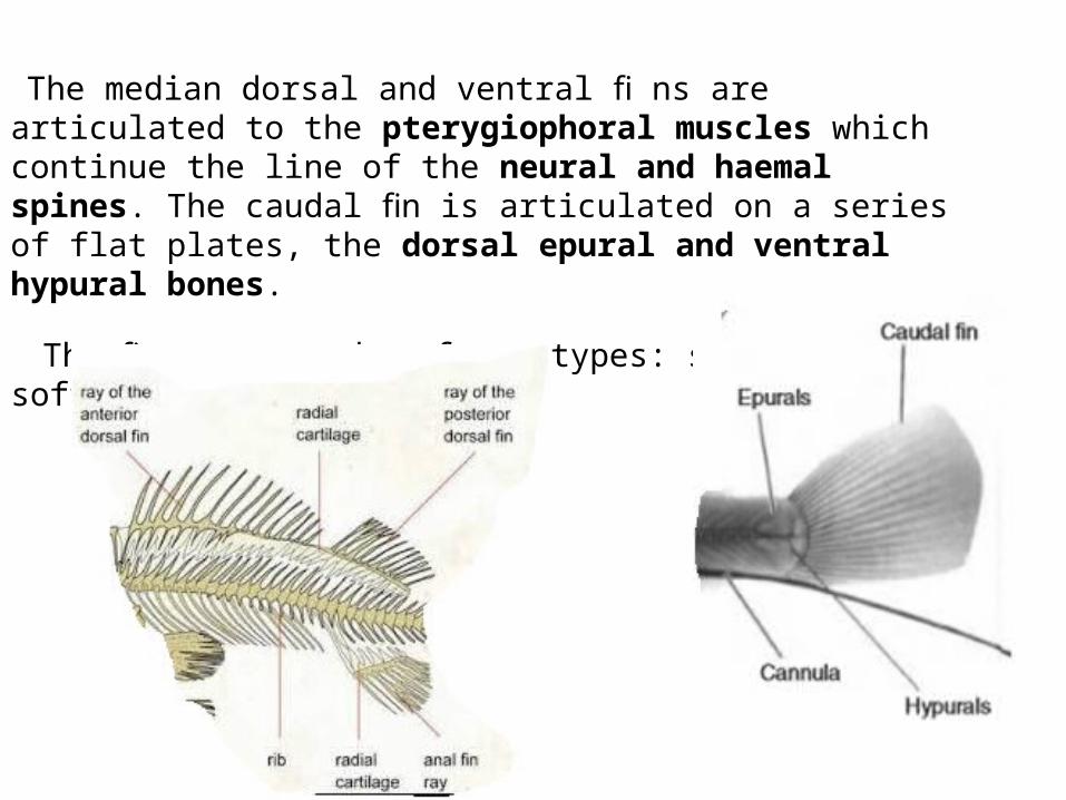
The median dorsal and ventral fi ns are articulated to the pterygiophoral muscles which continue the line of the neural and haemal spines. The caudal fin is articulated on a series of flat plates, the dorsal epural and ventral hypural bones.
The fin rays can be of two types: spiny or soft.

The spiny rays are simple single bones as in the first dorsal fin of Perciformes.
The caudal fin rays in all teleosts are of the soft kind, as are all the other fins in Isospondylii (e.g. Salmonidae and Clupeidae).
The soft rays are •segmented,• often branched and •formed of two identical lateral components on either side of the midline.
Whereas the spiny fin rays are rigid,
The soft fin rays are capable of bending by the activity of muscles at the base of each fin ray pulling on ligaments running along the column of bones. In this way subtle movements are possible.

BONE The microscopic structural elements of fish bones are similar to those of other vertebrates and generally two types of bone are found, cellular and acellular.
The former contains osteocytes and is confi ned to lower orders, such as Clupeidae, Salmonidae and Cyprinidae.
Acellular bone is unique in vertebrates; it contains no osteocytes and is found in advanced teleosts such as Percidae and Centrarchidae, often having a solid featureless matrix
The lack of cells has been shown to preclude resorption of calcium from the bones so that acellular bones cannot function as a calcium reserve.

MUSCLESThe wave is generated by sequential contraction from head to tail of the muscle blocks or myomeres – Angulliform movement
In shorter bodied, more typical fish, the mechanism is the same, but during swimming the flexure of the body shows less than a complete wave and only the oscillation of the tail is really apparent -carangiform locomotion

The most obvious feature of the muscle of a round fish is the folding and interlocking of the myomeres (Figure )
Externally, the body muscles occupy the quadrants of the body, separated from each other by the median septum and the transverse horizontal septum.
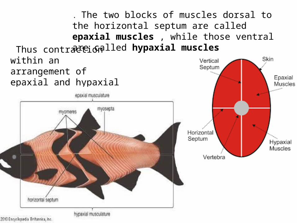
. The two blocks of muscles dorsal to the horizontal septum are called epaxial muscles , while those ventral are called hypaxial muscles
Thus contraction within an arrangement of epaxial and hypaxial muscles causes the body to bend

In most teleosts there are two main subdivisions: the muscularis lateralis superficialis, consisting of the so called red muscle fibres, and the muscularis lateralis profundus , which consists of white fibres
From mechanical, electrophysiological and biochemical differences it has been shown that the
red fibres are aerobic, slow - contracting fibres white are anaerobic, fast - contracting and fast fatiguing fibres
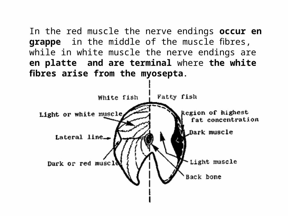
In the red muscle the nerve endings occur en grappe in the middle of the muscle fibres, while in white muscle the nerve endings are en platte and are terminal where the white fibres arise from the myosepta.

Difference between dark and light muscles
The dark or red muscle in trout and other fusiform fishes is located just beneath the skin
In trout – this is a middle band of muscle In tunas and other fishes – much more extensive
Dark muscle involved in sustained swimmingHas high lipid content

TISSUE SYSTEMLike terrestrial vertebrates Fish have 3 major histological classification of muscle tissue
Involuntary smooth muscle2 Striated form of muscle
The muscular tissue is composed of cells that specialize in mechanical work. Three chief types are classically described : smooth, cardiac and skeletal.
The last two types are frequently referred to as striated because of the cross-striations on the cells, which are lacking in smooth muscle.

Smooth muscle unbranchedLong taperingMononucleated cells Devoid of striations(similar to more familiar land animals)
Striated muscle
Cardiac Skeletal muscle

Cardiac•Similar to terrestrial sp•Has striated•Branched•Multi nucleated cells with nuclei at the centre of cell
Skeletal muscle in fish is UnbranchedMultinucleated with nuclei just under sarcolemma or cell membrane
Skeletal muscle
Dark muscle light muscle
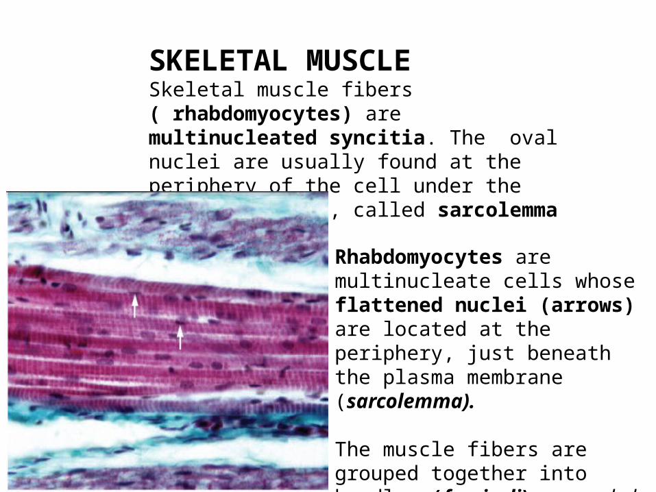
SKELETAL MUSCLESkeletal muscle fibers ( rhabdomyocytes) are multinucleated syncitia. The oval nuclei are usually found at the periphery of the cell under the plasma membrane, called sarcolemma
Rhabdomyocytes are multinucleate cells whose flattened nuclei (arrows) are located at the periphery, just beneath the plasma membrane (sarcolemma).
The muscle fibers are grouped together into bundles (fasciculi) surrounded by loosecollagenous tissue .

Myomeres•Refers to seperate muscle bundle with parallel fibres•Running along the axis of the body of the fish connected by myosepta to a vertebrae
Epaxial muscles- Transverse sec on through the axial trunk musculature. In fish this musculature is dividedinto a series of myomeres (here in grey and taking up 90 % of the picture) separated by collagenous sheets called myosepta (in blue - arrows).
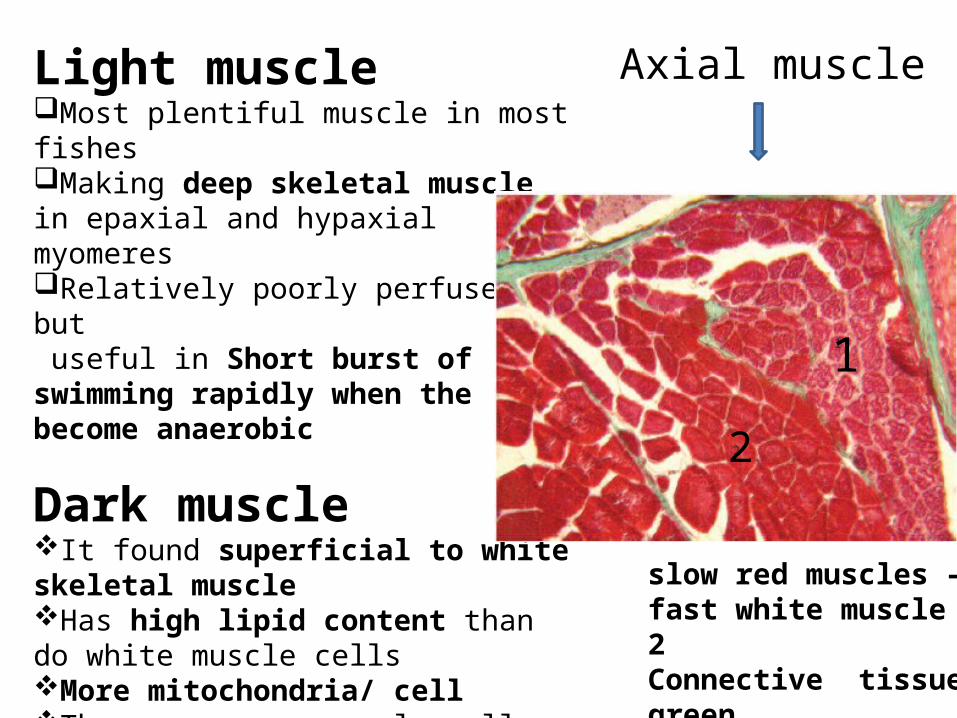
Light muscleMost plentiful muscle in most fishesMaking deep skeletal muscle in epaxial and hypaxial myomeresRelatively poorly perfused but useful in Short burst of swimming rapidly when the become anaerobic
Dark muscleIt found superficial to white skeletal muscleHas high lipid content than do white muscle cellsMore mitochondria/ cellThese narrower muscle cells supported by a better vascular supply than that of white muscle
slow red muscles – 1fast white muscle - 2 Connective tissue-green
1
2
Axial muscle
Dark muscle Most prominent in areas of underlying fins of major propulsion
Fish species positionSalmonids under the lateral line in configuration to move the tailPerch & others Utilize their pectoral fins for locomotion So abundant in pectoral finsSea horses Use caudal fins for continuous propulsion So more prominent under caudal fins
Thought to be utilized for continuous slower swimming

Sonic fibers Sonic fibers are thicker than red and thinner than white epaxial fibers
Sonic fibers and myofibrils exhibit an unusual helicoidal organization
Sound production in many fish species results from the action of extrinsic muscles that insertonto the gas bladder.
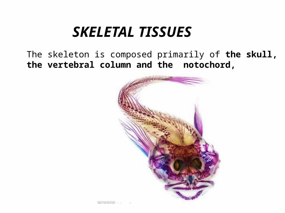
SKELETAL TISSUES The skeleton is composed primarily of the skull, the vertebral column and the notochord,
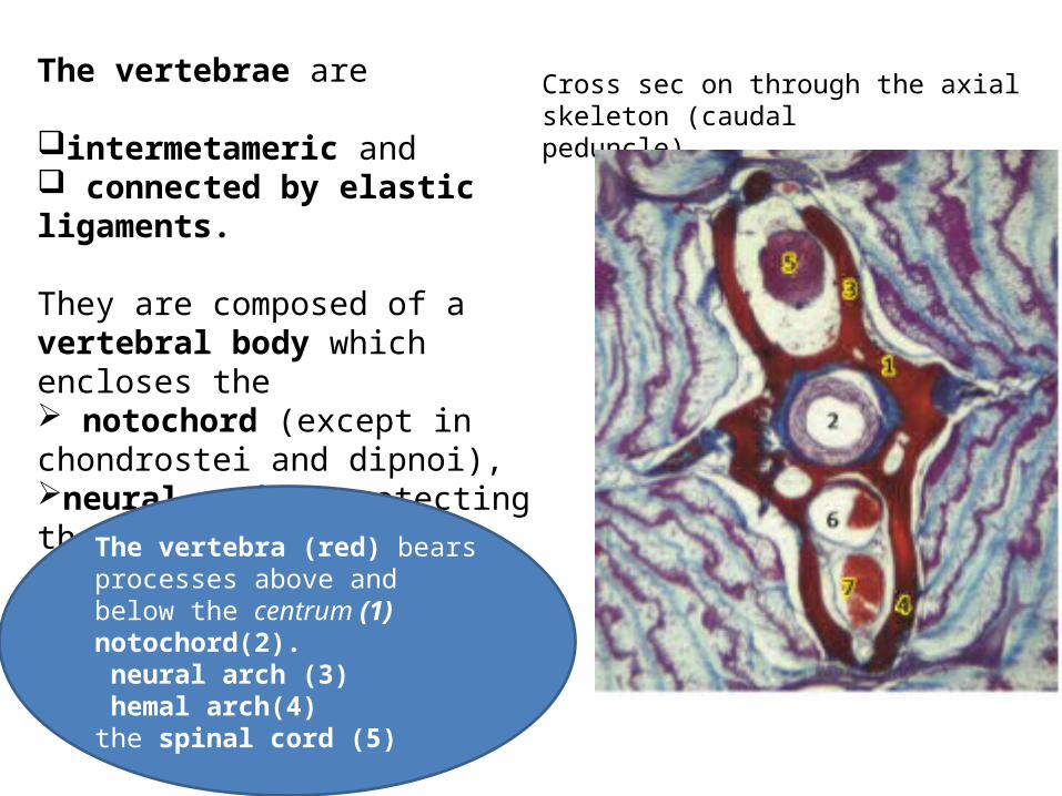
The vertebrae are
intermetameric and connected by elastic ligaments.
They are composed of a vertebral body which encloses the notochord (except in chondrostei and dipnoi), neural arches protecting the spinal cord and ventral arches
Cross sec on through the axial skeleton (caudalpeduncle)
1
The vertebra (red) bears processes above and below the centrum (1) notochord(2). neural arch (3) hemal arch(4)the spinal cord (5)
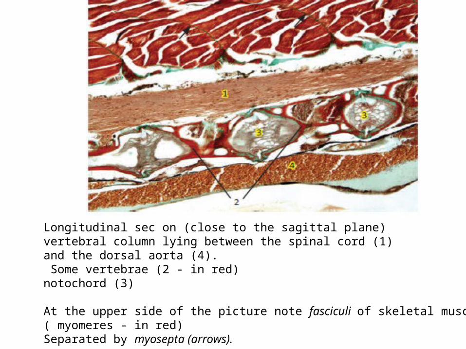
Longitudinal sec on (close to the sagittal plane)vertebral column lying between the spinal cord (1)and the dorsal aorta (4). Some vertebrae (2 - in red)notochord (3)
At the upper side of the picture note fasciculi of skeletal muscular fibers ( myomeres - in red) Separated by myosepta (arrows).

In Chondrichthyesendoskeletal tissues are mainly of two types :• notochord•and cartilage (no bone). Large vacuolated cells (1) fill the central core of the notochord, andtogether with the connective tissue sheath (2) givethe notochord its characteristic flexibility. The vertebral centrum (3) encircles the notochord. Typically the spinal cord (4) is surrounded by the neural arches(5). The hemal processes are present only in the caudalregion. Myomeres (6) separated by myosepta (7)In reddish : blood vessels

(1) neural arches; (2) hemal arches; (3) transverse processes(4) Nerves of spinal ganglia as well as skeletal muscular tissue (5) are illustrated. In the vertebra Differences of colors (green to red) are probably explained by varia on in mineral deposits.
Transverse sec on of the caudal region.

Fin rays of elasmobranchsare slender, unsegmented, of a horny aspect and are called actinotrichia (orange/red).
These rays are disposed on both sides of the fins. Connective tissue, stainedgreen, is covered by the epidermis (arrow)
Cross sec on of the caudal fin

Actinotrichia are long horny, acellular, cylindrical, flexible and unsegmented rays made up of elastoidin,supporting the fins of cartilaginous fish.Two placoid scales are seen at the picture.
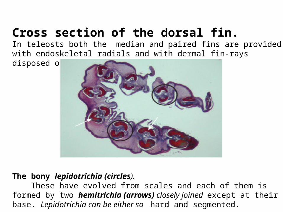
Cross section of the dorsal fin. In teleosts both the median and paired fins are provided with endoskeletal radials and with dermal fin-rays disposed on both sides of the fin.
The bony lepidotrichia (circles). These have evolved from scales and each of them is formed by two hemitrichia (arrows) closely joined except at their base. Lepidotrichia can be either so hard and segmented.
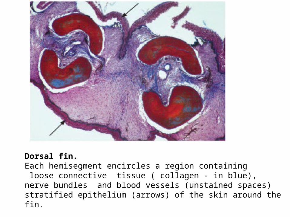
Dorsal fin. Each hemisegment encircles a region containing loose connective tissue ( collagen - in blue), nerve bundles and blood vessels (unstained spaces)stratified epithelium (arrows) of the skin around the fin.

CartilageThey are expected in sharks and raysBut skeletons of many young fish consists of Hyaline cartilage
In addition fish have variety of specialized cartilages(that are some what different from those found in terrestrial sp)
Eg; Cartilage supporting gill filament

CARTILAGE Cartilage is a firm, resistant tissue but is neither as hard nor as brittle as bone. It is found in all classes of vertebrates.
There are three main types of cartilage : hyaline, elastic and fibrous.
Hyaline-cell cartilage is typically an avascular tissue that contains tightly-packed cells with abundant cytoplasm.
Its cellularity approaches that of an epithelium.
The matrix is poorly developed and close to the plasma membranes

In hyaline cartilage the fibers are collagenous but the refractive indices of fibers and matrix are such that the fibers are invisible in ordinary histological preparations.
In elastic cartilage the elastic fibers are visible in histological sections by means of a special stain (orcein).
Fibrous cartilage contains a heavy mesh work of collagenous fibers which makes it very tough.

The resident cell of cartilage, the chondrocyte, is the main constituent cell of cartilage. It is isolated within a voluminous extracellular matrix which is neither vascularized nor innervated
This micrograph illustrates hyaline-cell cartilage containingclose-packed cells (chondrocytes - pink with dark nuclei).
The extracellular matrix (orange and blue) ismore visible.
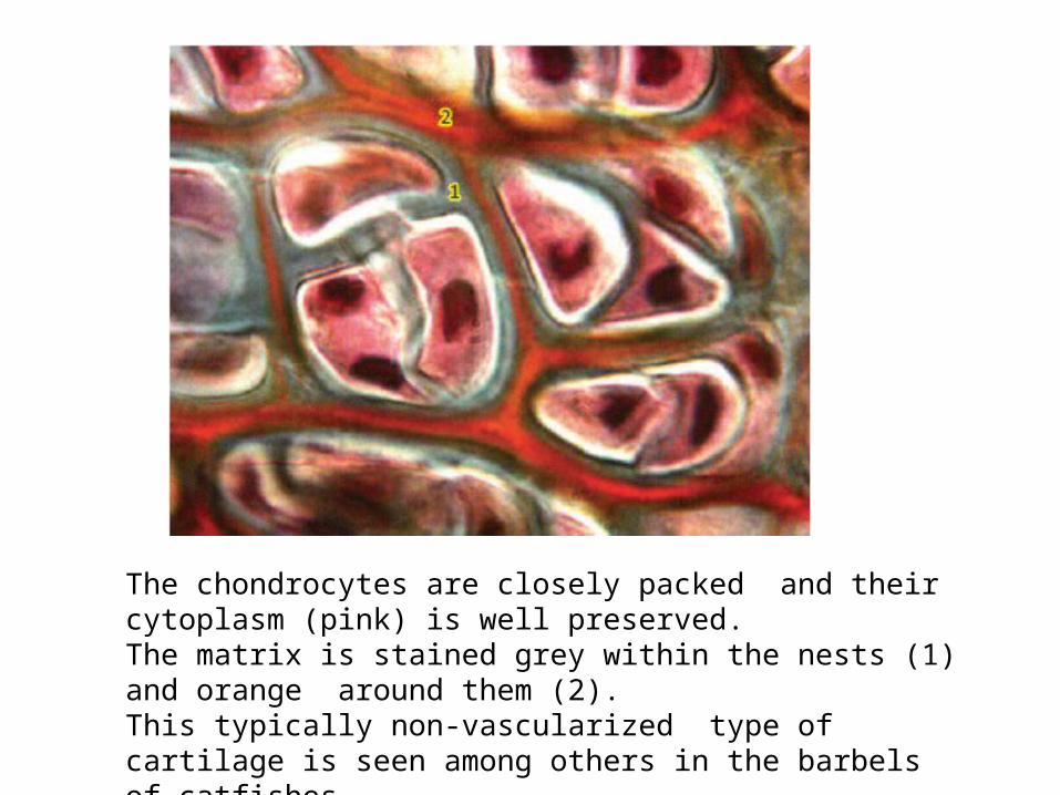
The chondrocytes are closely packed and their cytoplasm (pink) is well preserved. The matrix is stained grey within the nests (1) and orange around them (2). This typically non-vascularized type of cartilage is seen among others in the barbels of catfishes.
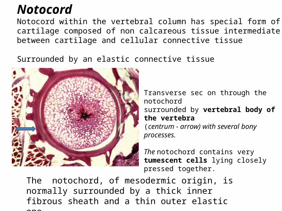
NotocordNotocord within the vertebral column has special form of cartilage composed of non calcareous tissue intermediate between cartilage and cellular connective tissue
Surrounded by an elastic connective tissue
Transverse sec on through the notochordsurrounded by vertebral body of the vertebra(centrum - arrow) with several bony processes.
The notochord contains very tumescent cells lying closely pressed together.
The notochord, of mesodermic origin, is normally surrounded by a thick inner fibrous sheath and a thin outer elastic one.

Spinal cord (1) vertebra (2) notochord (large unstained vacuolized cells – (3).
The notochord is a rather flexible rod partially persisting throughout life in lower vertebrates.
Transverse sec on of the notochord sheath. This perichordal tube is composed of a thick fibrous inner layer (1) a thin outer elastic one (2). The notochord sheath surrounds vacuolized cells (3). hyaline cartilage (4)
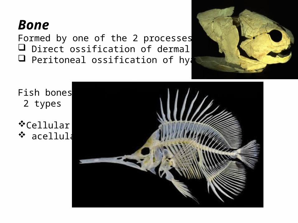
BoneFormed by one of the 2 processes Direct ossification of dermal bone Peritoneal ossification of hyaline cartilage
Fish bones 2 types
Cellular acellular
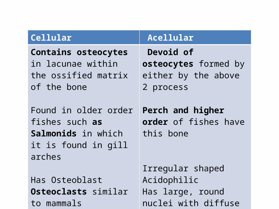
Cellular Acellular
Contains osteocytes in lacunae within the ossified matrix of the bone
Found in older order fishes such as Salmonids in which it is found in gill arches
Has OsteoblastOsteoclasts similar to mammals
Devoid of osteocytes formed by either by the above 2 process
Perch and higher order of fishes have this bone
Irregular shapedAcidophilicHas large, round nuclei with diffuse chromatin

Cellular bone (orange) and cartilage (1)are side by side within connective tissue (2) supporting a stratified epithelium (3).
Numerous osteocytes (the cellsresponsible for maintenance of bone matrix) with densely stained nuclei are visible (arrowheads).
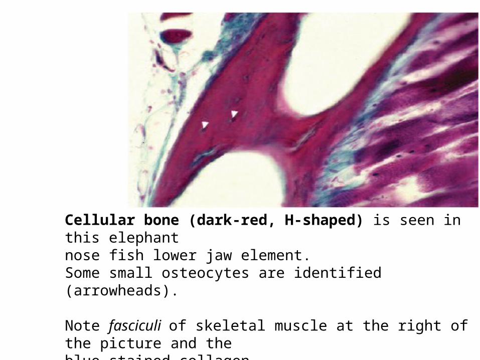
Cellular bone (dark-red, H-shaped) is seen in this elephantnose fish lower jaw element. Some small osteocytes are identified (arrowheads).
Note fasciculi of skeletal muscle at the right of the picture and theblue stained collagen.
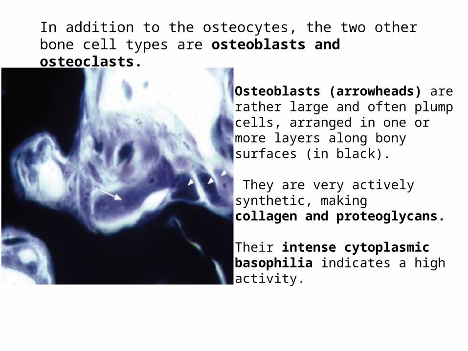
Osteoblasts (arrowheads) are rather large and often plump cells, arranged in one or more layers along bony surfaces (in black).
They are very actively synthetic, makingcollagen and proteoglycans.
Their intense cytoplasmicbasophilia indicates a high activity.
Osteoclast (arrow) is a very large, multinucleate cell, closely applied to the surface of a bony spicule and often lying in a depression.
In addition to the osteocytes, the two other bone cell types are osteoblasts and osteoclasts.

Acellular bone.
The osseous endoskeleton of higher teleosts (Perciformes.) is devoid of osteocytes.
This micrograph shows the typical appearance of such acellular bone (dark blue) surrounding a canal.
The unstained cells at the right bottom corner are adipocytes

Mixed boneResult of both Dermal and chondral bone formationMay have Hyaline cartilage associated with it Commonly Acellular bone in areolar or dense white connective tissue
Spongy bone Sub type of mixed boneForms the jaw bones, gill arches ,some other head bones
Laminated with spaces that give it a spongy like appearances
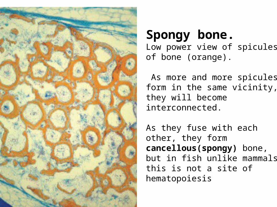
Spongy bone. Low power view of spicules of bone (orange).
As more and more spiculesform in the same vicinity, they will become interconnected.
As they fuse with each other, they form cancellous(spongy) bone, but in fish unlike mammalsthis is not a site of hematopoiesis
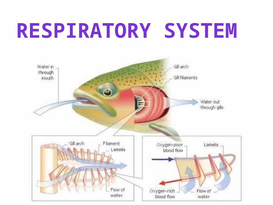
RESPIRATORY SYSTEM

In trout and most teleost Pharynx
Buccal cavity 2Opercular cavities
Opercular cavities –Guarded by mandibular and maxillary valves
ANATOMY

In sharks – •single Orobranchial cavity •5 or more Parabranchial cavity on each sides•Presence or absence of maxillary and mandible valve is very important
Have mouth inorder to open the valves and allow water to ram over gills
Most chondrichthyean species have five pairs of slits, but six or seven pairs occur in Hexanchiformes (frilled and cow sharks).
Gill slits are external openings leading to the orobranchial cavity and allowing water to exit after passing over the gilllamellae.

THE STRUCTURE OF THE GILLS Gills4 gill arches with holobranch1 gill arches with hemibranch(pseudobranch)
These arches were supported by cartilaginous rods extending from floor to roof of opercular cavities of pharynx
Cranial aspects of gill arches are modified into Gill rakers - prevents food and debris from reaching the respiratory components of the gills

Taxonomic features1. Structure of gill rakers2. Arrangement and form of the actual
gill
Abductor muscle- allow the fish toAdductor muscle – spread and collect hemibranch on individual gill arch
Gill filamentsEach arch has – 2 rows of of gill filamentsEach row called hemibranch2hemibranch forms holobranch
Each hemibranch – consists of gill filament (like comb)

Gill filamentsPrimary lamellae ( formed at the caudal edges of gill arches)Secondary lamellae (respiratory platelets) (run perpendicular to the axis of primary lamellae)
New Secondary lamellae formed at the tip of the gill filaments on bonyfishOlder secondary lamellae formed at the base
Gills – supplied by Dual artery System1 artery providing for oxygenation of blood by gillsOther supplying the arch & lamellae
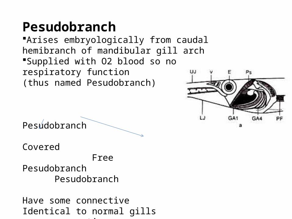
PesudobranchArises embryologically from caudal hemibranch of mandibular gill archSupplied with O2 blood so no respiratory function(thus named Pesudobranch)
Pesudobranch
Covered Free Pesudobranch Pesudobranch
Have some connective Identical to normal gills tissue eg: Herring, Flounder,blenniesFound in individual sp In no of orders

Fish species Pesudobranch
In some air breathing fishes district from carotoid labrynith Trout located on underside of each operculumStripped bass attached to hyoid arch and resembled as a gill Carp lies deep in the roof of opercular cavity
As well as having a respiratory function, the gills are responsible for regulating the exchange of salt and water and play a major role in the excretion of nitrogenous waste products.

TISSUE SYSTEM

Respiratory tissues
Epithelium covering of gill arches continuous with epithelium of pharynx and buccal cavity
Often has Taste buds mucous cells
Abductor muscles of striated muscles can often found along the lateral sides of gill arch and between membranes
Supporting gill arch boneCartilage changes with age & species
Adult fish have less cartilage & more bone

1. primary lamella 2. secondary lamella 3. epithelial cell4. mucous cell 5. pillar cell 6. lacuna (capillary lumen) 7. erythrocyte
within capillary lumen 8. undifferentiated basal cell 9. central venous sinus

Gill filaments
Primary lamellae - support by central core of cartilage
Covered by epithelium that is continuous with that of the secondary lamellae
Secondary lamellae covered with squamous epithelium, usually atleast 2 layers thick
Epithelium is thickest in air breathing fishes.
Layers of epithelium are ocassionally seperated by intracellular space containing macrophage (Rather than its normal it is an indicator of inflammation)

The gills of the Osteichthyes are quite similar to those of the cartilaginous fish except for two major differences : the Osteichthyes always have an operculum and the gill septa are very reduced (aseptal gills). Their lamellae are free in the opercular cavity.
Osseous fish have four pairs of cartilaginous or bony gill arches (gill arch in cross sec on – 1) onto which two rows of gill filaments (2) are arched. Each filamentis well vascularized (afferent - 3 and efferent bloodvessels – 4) and bears many platelike secondary lamellae(arrowheads). In 5, gill arch cartilage in 6 branchial muscles.

Elasmobranchs have five pairs of gills. Muscular interbranchial septa separate gill slits.
Each septum bears branchial arch, gillrays, one afferent blood-vessel and paired efferent blood-vessels.
Elasmobranchs present well-visible gill slits on both sides of the head in sharks, or underneath in skates and rays.
Two gill slits are separated by a long andmuscular interbranchial septum (*) which supports the gill filament(arrows).
Skin is seen in 1 ( dermis in green) and branchial cartilage in 2. Erythrocytes are stained in orange

Sagittal section on through a gill filament. Numerous parallel threadlike secondary lamellae are obvious (1)and arranged nearly at right angles to the filament.
The cartilaginous skeleton, which supports the primary lamella, is also evident (chondrocytes – 2 ; extracellular matrix – 3).

The vacuolated mucus-secreting cells (PAS+, arrowheads) are located principally within the epithelium of the primary lamellae near the base of secondary lamellae.
The mucous cells produce a thin mucous coating which protects against bacterial infection and abrasion.
1 : gill filament or primary lamella 2 : secondary lamellae 3 : cartilaginous support
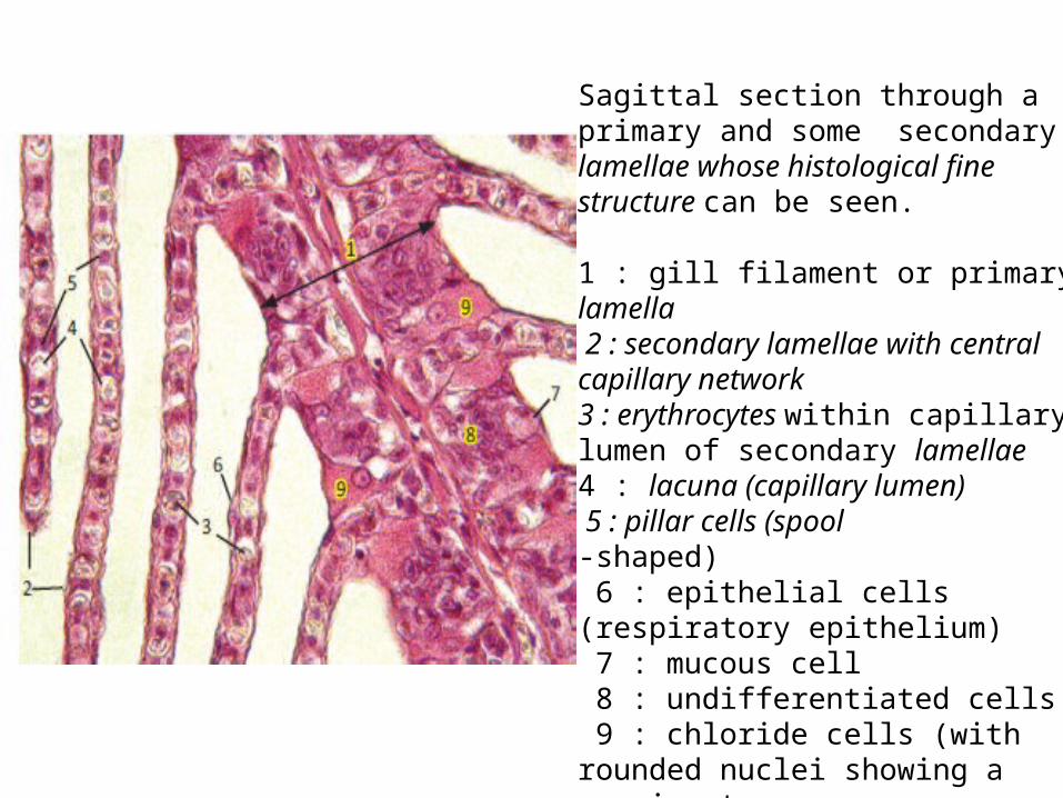
Sagittal section through a primary and some secondary lamellae whose histological fine structure can be seen.
1 : gill filament or primary lamella 2 : secondary lamellae with central capillary network 3 : erythrocytes within capillary lumen of secondary lamellae4 : lacuna (capillary lumen) 5 : pillar cells (spool-shaped) 6 : epithelial cells (respiratory epithelium) 7 : mucous cell 8 : undifferentiated cells 9 : chloride cells (with rounded nuclei showing a prominentnucleolus)

Gill rakers. Gill rakers are variously shaped bony or cartilaginous projections which point forward and inward from the gill (or branchial) arch(*). The common carp possesses about twenty relatively short gill rakers.
The shape and number of these structures are a good indication of the diet of the fish.
They are thinner, longer and more numerous in species eating small prey
The plankton feeders have the longest and a number that can exceed 150 per arch.
This very low magnification (8x) of a teleost aseptal gill displays in the same document the gill lamellae (short arrows) and the gill rakers (long arrows).

Two gill rakers cut at different levels.
These comb-like projections on the inner edge of the gill arches filter solid material from the water and serve to retain food particles in the buccal cavity.
The gill rakers are lined with the mucous pharyngeal strafitied epithelium (long arrows) supported by loose connective tissue (*)
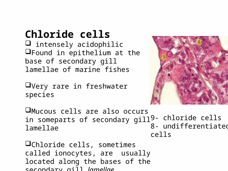
Chloride cells intensely acidophilicFound in epithelium at the base of secondary gill lamellae of marine fishes
Very rare in freshwater species
Mucous cells are also occurs in someparts of secondary gill lamellae
Chloride cells, sometimes called ionocytes, are usually located along the bases of the secondary gill lamellae.
9- chloride cells8- undifferentiated cells
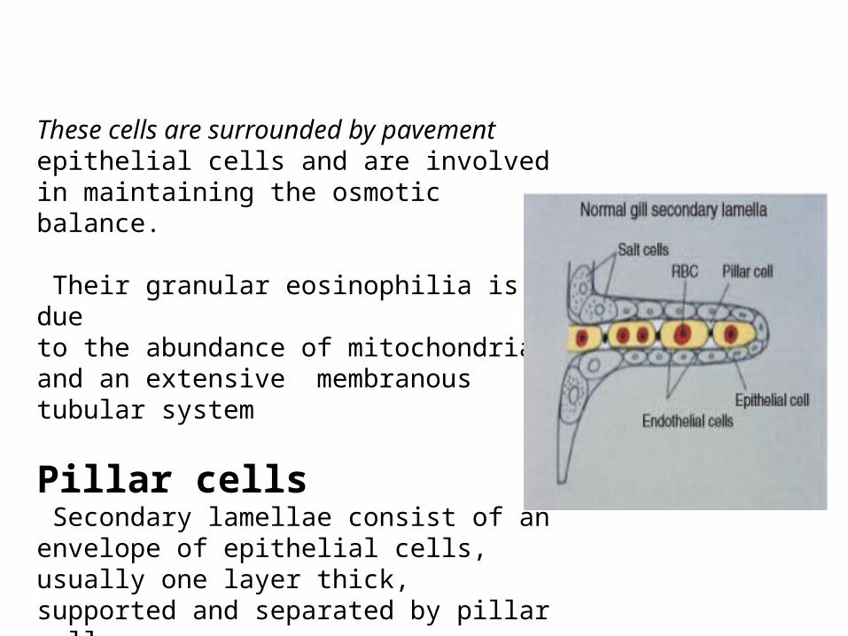
These cells are surrounded by pavementepithelial cells and are involved in maintaining the osmotic balance.
Their granular eosinophilia is dueto the abundance of mitochondria and an extensive membranous tubular system
Pillar cells Secondary lamellae consist of an envelope of epithelial cells, usually one layer thick, supported and separated by pillar cells.
They are used to control lamellar perfusion

Thankyou