systems neuroscience - wordpress.com
TRANSCRIPT

1
Systems Neuroscience
Basic Anatomy

2
Motor Control
Spinal control of movement The motor system consists of all our muscles and the neurons that control them. There are:
o Efferent components, which command muscles to contract
o Afferent components, which report body status (somatosensation)
The spinal cord contains motor programs for the generation of coordinated movements, which
can work in isolation (e.g. reflexes).
These programs are accessed, executed, and modified by descending commands from the brain.
Thus, motor control can be divided into two parts: (1) the spinal control of coordinated muscle
contraction and (2) the brain’s control of the motor programs sent to the spinal cord.
The neurons in the spinal cord that innervate muscles and lead to muscle contraction are called
alpha motor neurons or motoneurons.
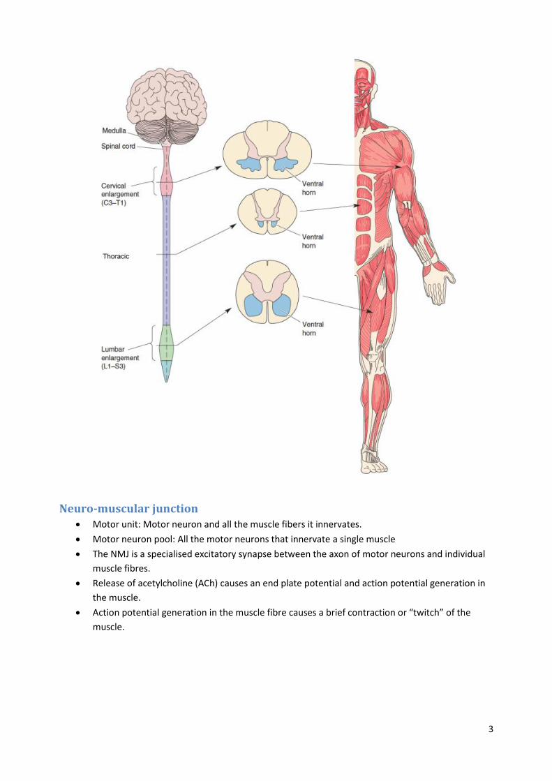
Motor neurons are located in a part of the spinal cord called the ventral horn, and send out
axons via ventral roots.
The spinal cord can be divided in different regions: Cervical, thoracic, lumbar and sacral.
o Motoneurons in the cervical region project to the arms.
o Motoneurons in the thoracic region project to the body.
o Motoneurons in the lumbar region project to the legs.
A myotome is the group of muscles innervated by motoneurons from a single ventral root.
3
Neuro-muscular junction Motor unit: Motor neuron and all the muscle fibers it innervates.
Motor neuron pool: All the motor neurons that innervate a single muscle
The NMJ is a specialised excitatory synapse between the axon of motor neurons and individual
muscle fibres.
Release of acetylcholine (ACh) causes an end plate potential and action potential generation in
the muscle.
Action potential generation in the muscle fibre causes a brief contraction or “twitch” of the
muscle.

4
Excitation-contraction coupling 1. An action potential travels down the alpha motor neuron axon.
2. ACh is released at the axon terminal at the neuromuscular junction.
3. ACh binds and opens nicotinic (ACh) receptor channels on muscle fibres causing depolarization
leading to an end-plate potential.
4. Voltage-gated sodium channels in the muscle fibre open and an action potential is generated in
the muscle fibre, which sweeps down the fibre and into the T tubules.
5. Depolarization of the T tubules causes Ca 2+ release from the sarcoplasmic reticulum (SR) and
muscle contraction.
6. At rest myosin cannot interact with actin because the myosin attachment sites on actin are
covered by tropomyosin and troponin.
7. Following excitation Ca 2+ binds to troponin.
8. Tropomyosin shifts its position exposing myosin binding sites on actin.
9. Myosin heads bind actin.
10. The myosin heads pivot causing the filaments to move and the muscle to contract.
11. ATP binds to the myosin head and it disengages from actin.
12. The cycle continues as long as Ca 2+ and ATP are present.
5
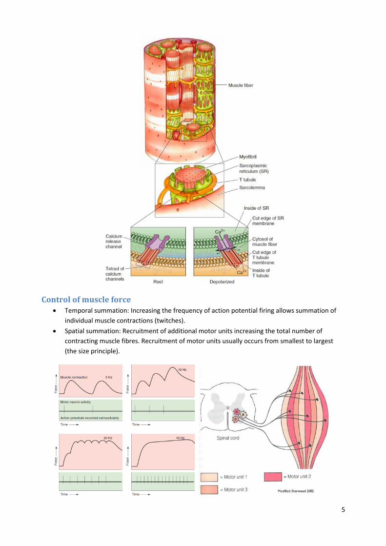
Control of muscle force Temporal summation: Increasing the frequency of action potential firing allows summation of
individual muscle contractions (twitches).
Spatial summation: Recruitment of additional motor units increasing the total number of
contracting muscle fibres. Recruitment of motor units usually occurs from smallest to largest
(the size principle).
6
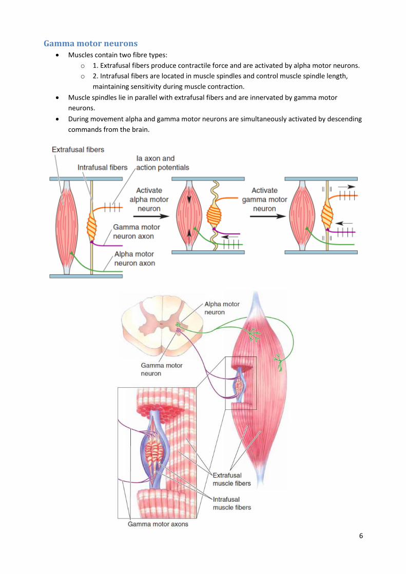
Gamma motor neurons Muscles contain two fibre types:
o 1. Extrafusal fibers produce contractile force and are activated by alpha motor neurons.
o 2. Intrafusal fibers are located in muscle spindles and control muscle spindle length,
maintaining sensitivity during muscle contraction.
Muscle spindles lie in parallel with extrafusal fibers and are innervated by gamma motor
neurons.
During movement alpha and gamma motor neurons are simultaneously activated by descending
commands from the brain.
7
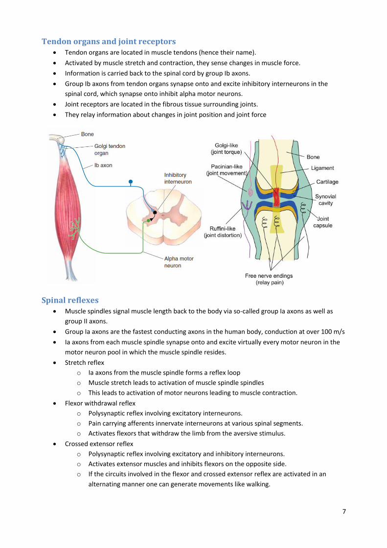
Tendon organs and joint receptors Tendon organs are located in muscle tendons (hence their name).
Activated by muscle stretch and contraction, they sense changes in muscle force.
Information is carried back to the spinal cord by group Ib axons.
Group Ib axons from tendon organs synapse onto and excite inhibitory interneurons in the
spinal cord, which synapse onto inhibit alpha motor neurons.
Joint receptors are located in the fibrous tissue surrounding joints.
They relay information about changes in joint position and joint force
Spinal reflexes Muscle spindles signal muscle length back to the body via so-called group Ia axons as well as
group II axons.
Group Ia axons are the fastest conducting axons in the human body, conduction at over 100 m/s
Ia axons from each muscle spindle synapse onto and excite virtually every motor neuron in the
motor neuron pool in which the muscle spindle resides.
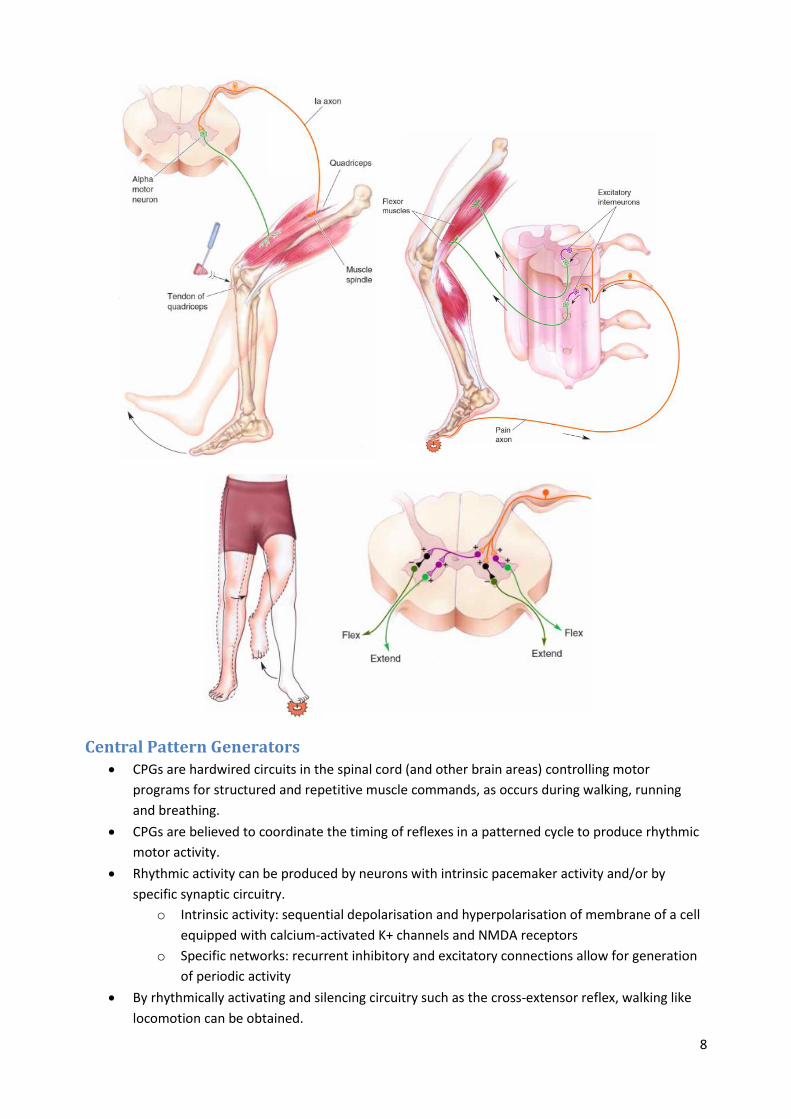
Stretch reflex
o Ia axons from the muscle spindle forms a reflex loop
o Muscle stretch leads to activation of muscle spindle spindles
o This leads to activation of motor neurons leading to muscle contraction.
Flexor withdrawal reflex
o Polysynaptic reflex involving excitatory interneurons.
o Pain carrying afferents innervate interneurons at various spinal segments.
o Activates flexors that withdraw the limb from the aversive stimulus.
Crossed extensor reflex
o Polysynaptic reflex involving excitatory and inhibitory interneurons.
o Activates extensor muscles and inhibits flexors on the opposite side.
o If the circuits involved in the flexor and crossed extensor reflex are activated in an
alternating manner one can generate movements like walking.
8
Central Pattern Generators CPGs are hardwired circuits in the spinal cord (and other brain areas) controlling motor
programs for structured and repetitive muscle commands, as occurs during walking, running
and breathing.
CPGs are believed to coordinate the timing of reflexes in a patterned cycle to produce rhythmic
motor activity.
Rhythmic activity can be produced by neurons with intrinsic pacemaker activity and/or by
specific synaptic circuitry.
o Intrinsic activity: sequential depolarisation and hyperpolarisation of membrane of a cell
equipped with calcium-activated K+ channels and NMDA receptors
o Specific networks: recurrent inhibitory and excitatory connections allow for generation
of periodic activity
By rhythmically activating and silencing circuitry such as the cross-extensor reflex, walking like
locomotion can be obtained.

9
The motor cortex Control of voluntary movement engages most of the neocortex.
The cortical area important for execution of movement is the primary motor cortex or M1
Activation of M1 leads directly to movement of muscles on the opposite side of the body.
Adjacent to M1 are the premotor area (PMA) and supplementary motor area (SMA). Activation
of these areas evokes more complex movements at higher stimulation intensities.
SMA sends axons to innervate distal motor units directly, PMA connects primarily with neurons
that innervate proximal motor units.
There is a spatial map of the body in the motor cortex, similar to in somatosensory cortex, called
the motor homunculus.
This was discovered by a neurosurgeon who performed electrical stimulation of the cortex in
patients who were undergoing surgery to remove the parts of their brain causing seizures.
These stimulation experiments indicated that activation of M1 surprisingly can also lead to
inhibition of movement.
Motor maps in the cortex are plastic, and can change the muscles they control with learning of
tasks like fine motor skills.
Activation of neurons in M1 code two aspects of the movement: Force and direction.
Different cells are “tuned” for movements in different directions.
Control of movement uses a “population code”, whereby groups of neurons broadly tuned for
the movement are recruited together.
10
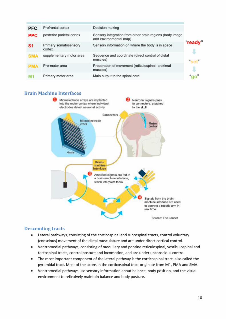
Brain Machine Interfaces
Descending tracts Lateral pathways, consisting of the corticospinal and rubrospinal tracts, control voluntary
(conscious) movement of the distal musculature and are under direct cortical control.
Ventromedial pathways, consisting of medullary and pontine reticulospinal, vestibulospinal and
tectospinal tracts, control posture and locomotion, and are under unconscious control.
The most important component of the lateral pathway is the corticospinal tract, also called the
pyramidal tract. Most of the axons in the corticospinal tract originate from M1, PMA and SMA.
Ventromedial pathways use sensory information about balance, body position, and the visual
environment to reflexively maintain balance and body posture.
11
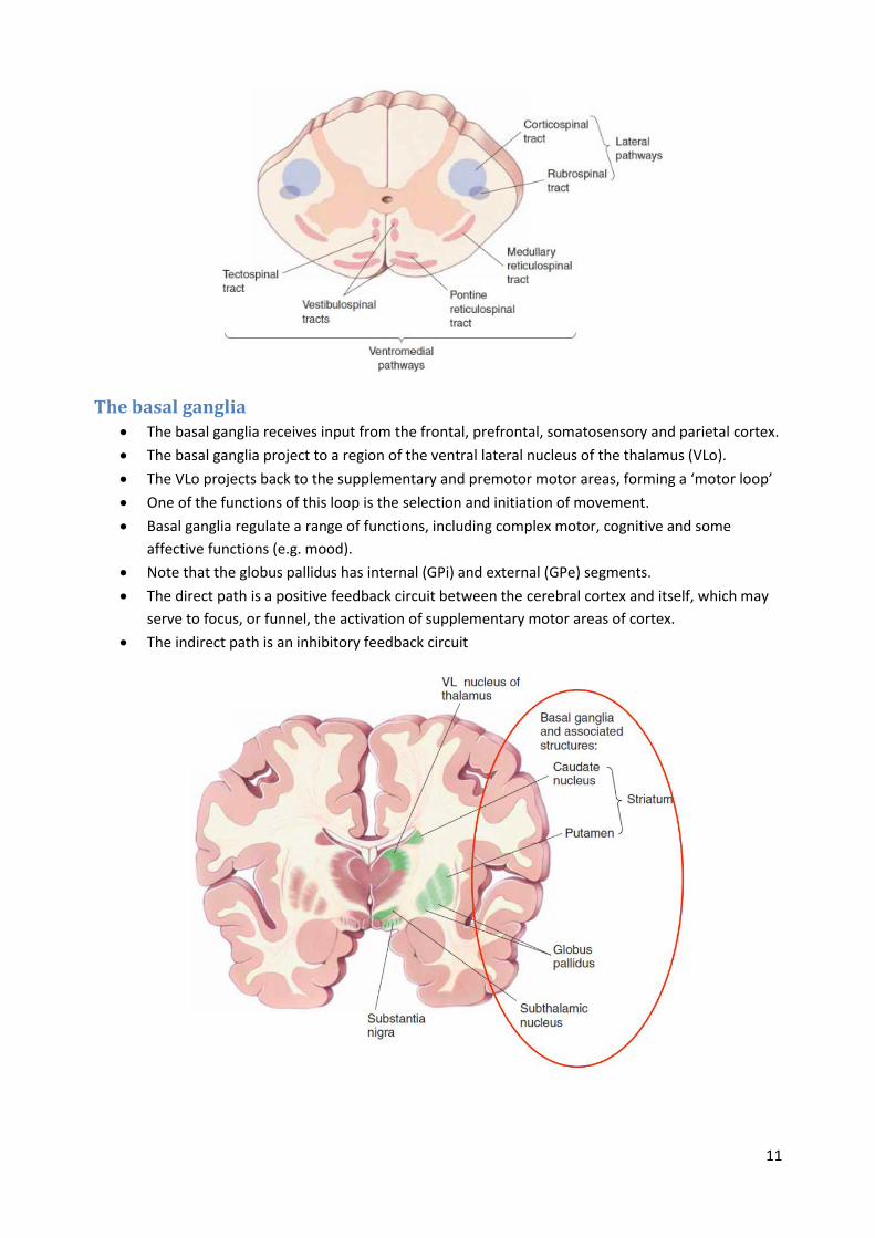
The basal ganglia The basal ganglia receives input from the frontal, prefrontal, somatosensory and parietal cortex.
The basal ganglia project to a region of the ventral lateral nucleus of the thalamus (VLo).
The VLo projects back to the supplementary and premotor motor areas, forming a ‘motor loop’
One of the functions of this loop is the selection and initiation of movement.
Basal ganglia regulate a range of functions, including complex motor, cognitive and some
affective functions (e.g. mood).
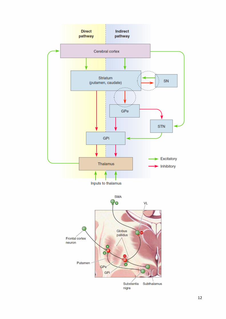
Note that the globus pallidus has internal (GPi) and external (GPe) segments.
The direct path is a positive feedback circuit between the cerebral cortex and itself, which may
serve to focus, or funnel, the activation of supplementary motor areas of cortex.
The indirect path is an inhibitory feedback circuit
12

13
Basal ganglia disorders Studies of several human diseases support the view that the direct motor loop functions to
facilitate the initiation of willed movements.
According to one model, increased inhibition of the thalamus by the basal ganglia underlies
hypokinesia (loss of movement), whereas decreased basal ganglia output leads to hyperkinesias.
Parkinson’s disease exemplifies a disease with hypokinesia. Its symptoms include slowness of
movement (bradykinesia), difficulty in initiating willed movements (akinesia), increased muscle
tone (rigidity) and tremors.
The cellular basis of Parkinson’s disease is a degeneration of dopaminergic substantia nigra
neurons and their inputs to the striatum. Dopamine can enhance the direct motor loop by
activating cells in the putamen.
Huntington’s disease is a hereditary disease characterized by hyperkinesia and dyskinesias
(abnormal movements). It is associated with a profound loss of neurons in the caudate nucleus,
putamen and globus pallidus.
The cerebellum The cerebellum is critical for coordination of movement. Damage to the cerebellum leads to
uncoordinated and inaccurate movements (ataxia).
Its primary function is to detect the difference, or “motor error,” between an intended
movement and the actual movement, and through its projections to motor (and premotor)
cortex to reduce this error.
The cerebellum receives input via “mossy fibers” from the pons, which receives massive input
from frontal areas such as premotor areas (PMA and SMA) as well as from somatosensory and
posterior parietal cortex.
The lateral cerebellum projects back to motor (and premotor) cortex via a relay in the ventral
lateral nucleus of the thalamus (VLc).
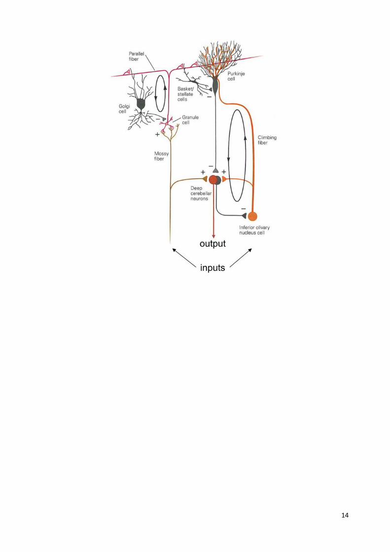
Cerebellar circuitry Mossy fibers synapse onto granule cells, whose axons bifurcate and run in parallel beams
perpendicular to Purkinje cell dendrites. These axons are called “parallel fibers”. They make
direct excitatory contact onto Purkinje dendrites
Purkinje cells receive input from 100,000 to 200,000 parallel fiber inputs, but only one climbing
fiber input.
The climbing fiber wraps itself around the Purkinje cell dendrites making multiple (100’s) of
synapses. The climbing fibre synapse is so powerful it always leads to a burst of APs in the
Purkinje cell.
Output of Purkinje cells is also regulated by local inhibitory interneurons (Golgi and basket cells).
Purkinje cells are the sole output of the cerebellum and project to the deep cerebellar nuclei.
Purkinje cells are inhibitory, so their activation inhibits cells in the deep cerebellar nuclei.
Purkinje cells and deep cerebellar nuclear cells “recognize” potential movement errors by
comparing patterns of convergent activity.
Deep nuclear cells then send corrective (error) signals to the cortex to maintain or improve
movement accuracy.
14

15
Motor feedback summary diagram
16
Somatosensory System
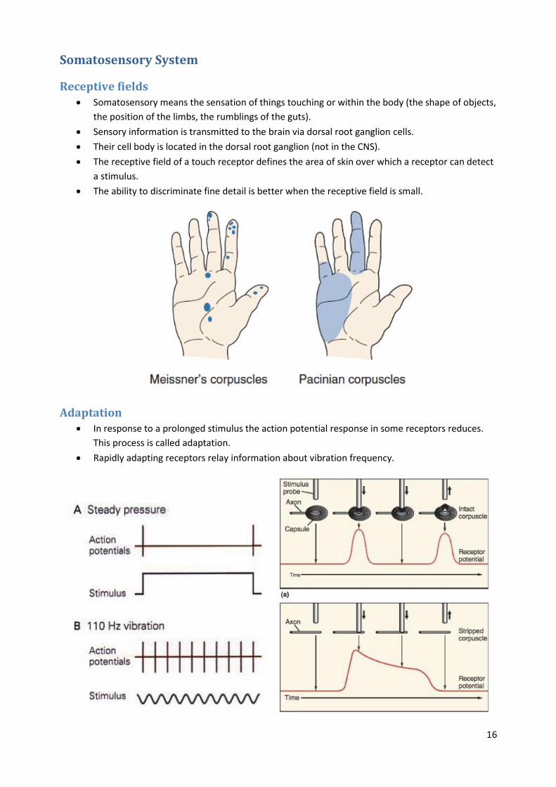
Receptive fields Somatosensory means the sensation of things touching or within the body (the shape of objects,
the position of the limbs, the rumblings of the guts).
Sensory information is transmitted to the brain via dorsal root ganglion cells.
Their cell body is located in the dorsal root ganglion (not in the CNS).
The receptive field of a touch receptor defines the area of skin over which a receptor can detect
a stimulus.
The ability to discriminate fine detail is better when the receptive field is small.
Adaptation In response to a prolonged stimulus the action potential response in some receptors reduces.
This process is called adaptation.
Rapidly adapting receptors relay information about vibration frequency.

17
Touch receptors Touch receptors with small receptive fields tend to be more superficial, whereas receptors
located deeper in the skin have larger receptive fields.
The extent of specialisation of touch receptor nerve endings influences whether they “adapt” or
not to a constant stimulus.
Stretch receptors
The proteins “Piezo1” and “Piezo2” have been identified as candidate sensors of mechanical
stimuli in cells.
These proteins code for a cation-selective channel that is sensitive to mechanical stretch with a
reversal potential close to zero mV.

18
Coding of somatosensory information Stimulus intensity is encoded by the frequency of action potentials in sensory nerves.
Increasing stimulus intensity also activates a greater number of receptors, hence stimulus
intensity is also encoded by the size of the activated receptor population (population code)
Receptors for joint and muscle position Detection of joint and muscle position is called proprioception.
Receptors detecting the position of the body’s limbs and muscles are called proprioceptors.
There are 3 main types: Muscle spindles, tendon organs and joint receptors.
Muscle spindles: detect muscle length, contractile elements (driven by the gamma motor fibres)
keep the central sensory portion within its optimal range during muscle shortening.
Tendon organs: detect muscle tension, located in series with the muscle, within the muscle
tendon.
Joint receptors: endings of the Ruffinin type and are located in the joint capsules. Their function
is to signal the joint angle.
19
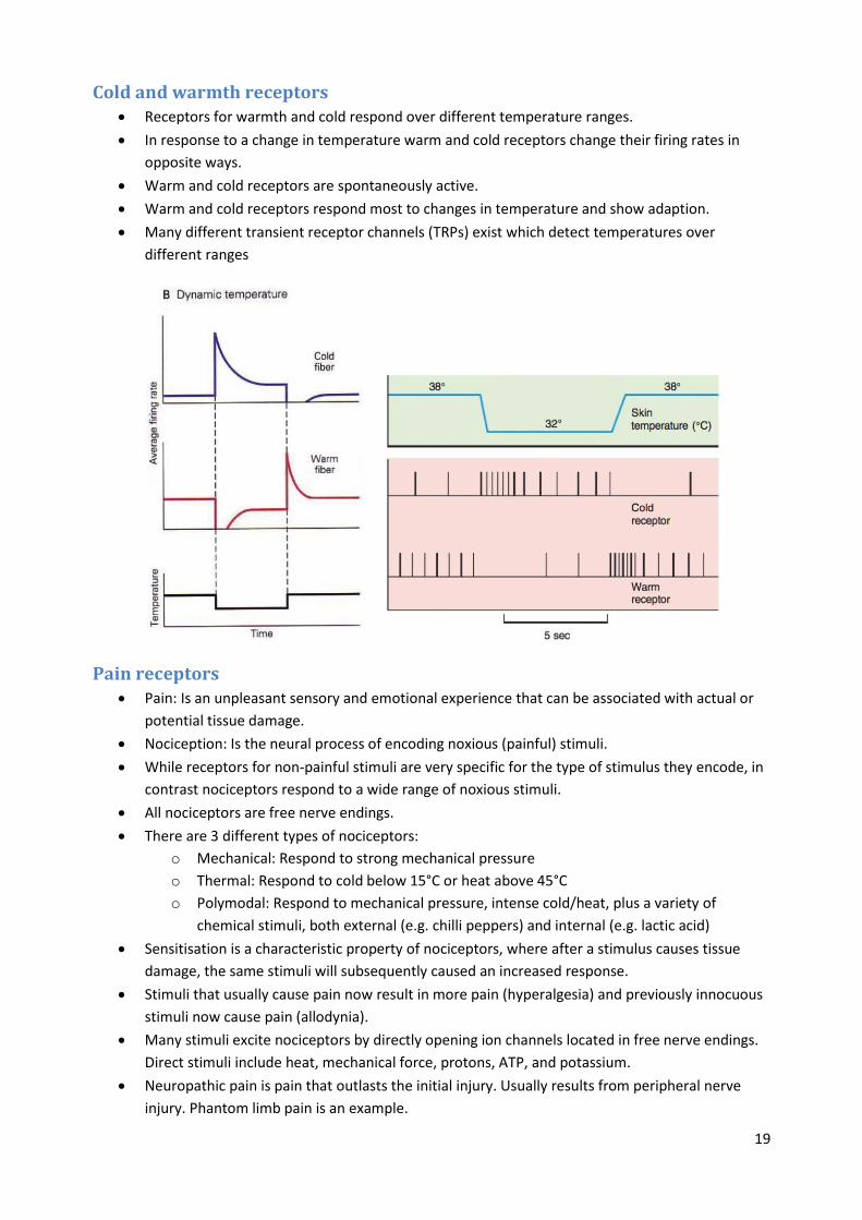
Cold and warmth receptors Receptors for warmth and cold respond over different temperature ranges.
In response to a change in temperature warm and cold receptors change their firing rates in
opposite ways.
Warm and cold receptors are spontaneously active.
Warm and cold receptors respond most to changes in temperature and show adaption.
Many different transient receptor channels (TRPs) exist which detect temperatures over
different ranges
Pain receptors Pain: Is an unpleasant sensory and emotional experience that can be associated with actual or
potential tissue damage.
Nociception: Is the neural process of encoding noxious (painful) stimuli.
While receptors for non-painful stimuli are very specific for the type of stimulus they encode, in
contrast nociceptors respond to a wide range of noxious stimuli.
All nociceptors are free nerve endings.
There are 3 different types of nociceptors:
o Mechanical: Respond to strong mechanical pressure
o Thermal: Respond to cold below 15°C or heat above 45°C
o Polymodal: Respond to mechanical pressure, intense cold/heat, plus a variety of
chemical stimuli, both external (e.g. chilli peppers) and internal (e.g. lactic acid)
Sensitisation is a characteristic property of nociceptors, where after a stimulus causes tissue
damage, the same stimuli will subsequently caused an increased response.
Stimuli that usually cause pain now result in more pain (hyperalgesia) and previously innocuous
stimuli now cause pain (allodynia).
Many stimuli excite nociceptors by directly opening ion channels located in free nerve endings.
Direct stimuli include heat, mechanical force, protons, ATP, and potassium.
Neuropathic pain is pain that outlasts the initial injury. Usually results from peripheral nerve
injury. Phantom limb pain is an example.

20
21
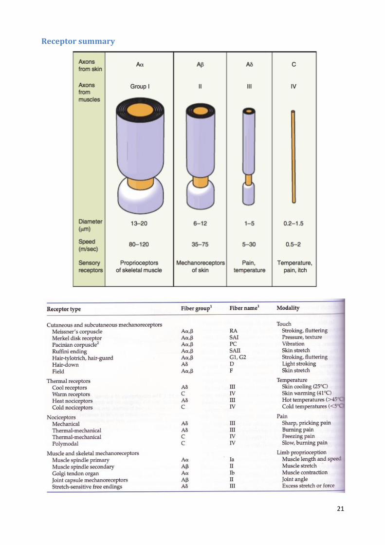
Receptor summary
22
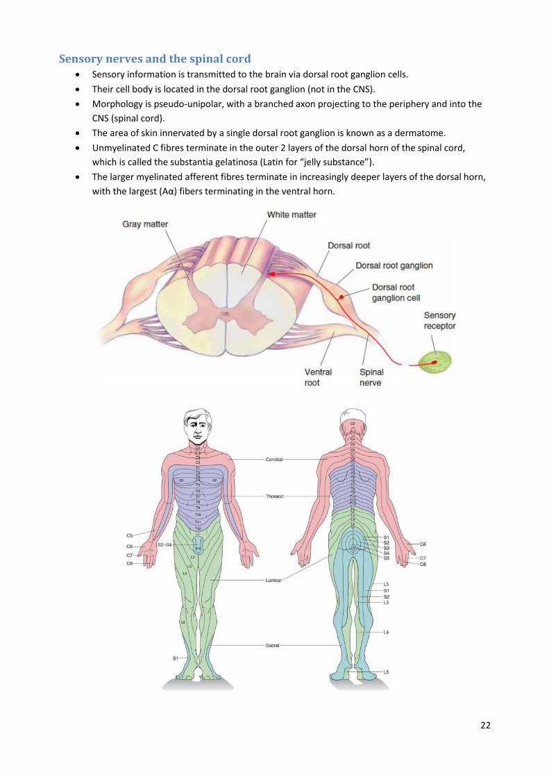
Sensory nerves and the spinal cord Sensory information is transmitted to the brain via dorsal root ganglion cells.
Their cell body is located in the dorsal root ganglion (not in the CNS).
Morphology is pseudo-unipolar, with a branched axon projecting to the periphery and into the
CNS (spinal cord).
The area of skin innervated by a single dorsal root ganglion is known as a dermatome.
Unmyelinated C fibres terminate in the outer 2 layers of the dorsal horn of the spinal cord,
which is called the substantia gelatinosa (Latin for “jelly substance”).
The larger myelinated afferent fibres terminate in increasingly deeper layers of the dorsal horn,
with the largest (Aα) fibers terminating in the ventral horn.

23
Ascending pathways Touch and proprioceptive information ascend to the cortex via the dorsal column-medial
lemniscal pathway or via the trigerminal pathway.
Temperature and pain information ascend to the cortex via the spino-thalamic pathway.

24
Somatosensory cortex In addition to S1, pain pathways also project to the anterior cingulate cortex (ACC) and the
insula.
The ACC is part of the limbic system and is thought to be responsible for the emotional element
of pain.
Insular cortex processes information on the internal state of the body and thus contributes to
the autonomic component of the overall pain response.
Area 3b is primary somatic sensory cortex (or S1). Most thalamic fibres terminate in this area.
Area 3b receives information from receptors in the skin (touch), whereas areas 3a processes
information from muscles and joints (body position).
Areas 1 and 2 receive input for 3b and process texture information (area 1) and size and shape
information (area 2).
Areas of the body are represented in the cortex in proportion to their peripheral innervation
density, forming a sensory homunculus.

25
Regulation of pain transmission Descending regulation of sensory input to the cortex can occur in the spinal cord, the dorsal
column nuclei, or the thalamus.
Inhibition can come from feedforward, feedback, and descending neurons.
Many peripheral receptors converge onto a single second order neuron.
The receptive field of second order neurons can have a central excitatory field surrounded by an
inhibitory region (we call this “surround inhibition”).
Surround inhibition improves two-point discrimination.
The “gate control” theory attempts to explain how transmission of the sensation of pain can be
regulated by local spinal cord circuitry.
The idea is that painful input (red) can be “gated” by non-noxious input (blue) via activation of
local inhibitory interneurons that inhibit dorsal horn neurons transmitting pain to the brain.
The periaqueductal gray (PAG) is the main descending system involved in modulating pain.
Activation of the PAG leads to enkephalin release in the dorsal horn.
Electrical stimulation of the PAG can produce sufficient analgesia to perform abdominal surgery
without the need for anaesthesia. Other, non‐painful, sensations are left intact.
Release of serotonin (5HT) from the raphe actives interneurons in the dorsal horn of the spinal
cord, blocking transmission from the nerve terminals of pain fibres via presynaptic inhibition.

26
Visual System
Eye anatomy Cornea: transparent external surface
Pupil: allows light entry
Iris: contains muscles that control pupil size
Lens: transparent structure located behind the iris, suspended by ligaments (zonule fibers)
Vitreous humor: the clear gel that fills the space between the lens and the retina
Retina: light-sensing tissue of the eye
Accommodation is the process whereby ciliary muscles and zonule fibers change the shape of
the lens for focusing on near or distant objects.
Pupillary Light Reflex: Continual adjusting of pupil size for varying light levels, based on feedback
between retina and neurons in the brainstem.
Visual field - approximately 150 degrees per eye
27
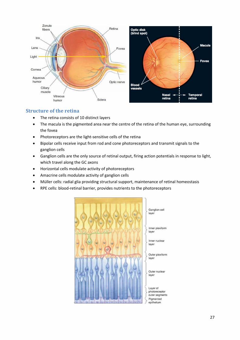
Structure of the retina
The retina consists of 10 distinct layers
The macula is the pigmented area near the centre of the retina of the human eye, surrounding
the fovea
Photoreceptors are the light-sensitive cells of the retina
Bipolar cells receive input from rod and cone photoreceptors and transmit signals to the
ganglion cells
Ganglion cells are the only source of retinal output, firing action potentials in response to light,
which travel along the GC axons
Horizontal cells modulate activity of photoreceptors
Amacrine cells modulate activity of ganglion cells
Müller cells: radial glia providing structural support, maintenance of retinal homeostasis
RPE cells: blood-retinal barrier, provides nutrients to the photoreceptors

28
Photoreceptors Opsins are photo-pigments that absorb light
The human retina contains about 92 million rods and 5 million cones
Cones contain fewer disks of opsins but allow for our ‘useful’ central vision (high visual acuity)
Cones are only found in the central fovea and rods are absent
Phototransduction
A process by which light is converted into electrical signals in the rod cells, cone cells and
photosensitive ganglion cells of the retina of the eye.
Rhodopsin is the photopigment in the membrane of stacked disks in the rod outer segments. It
consists of opsin (receptor protein) and retinal (a small molecule derived from vitamin A).
In the dark retinal is inactive, but in light, retinal undergoes a change in conformation after
absorbing the light, thereby activating the opsin.
In darkness the membrane potential of the photoreceptor outer segment is depolarised to
around -30mV, resulting in regular discharge of glutamate neurotransmitter.
This process is known as bleaching because it changes the wavelengths absorbed by rhodopsin.
Bleaching stimulates transducin, a G-protein, which activates the effector enzyme
phophodiesterase (PDE).
PDE hydrolyzes cGMP, forming GMP. This lowers the intracellular concentration of cGMP and
therefore the sodium channels close.
Closure of the sodium channels causes hyperpolarization of the cell due to the ongoing efflux of
potassium ions.
Hyperpolarization of the cell causes voltage-gated calcium channels to close.
As the calcium level in the photoreceptor cell drops, the amount of the neurotransmitter
glutamate that is released by the cell also drops.
In bright light, cGMP levels fall to the paoint where all Na+ channels are closed causing
saturation, meaning that further addition light has no further effect of hyperpolarisation.

29
Light and dark adaptation Adaptation to the dark involves a dilation of the pupils and also regeneration of rhodopsin into
unbleached form.
Rods are more sensitive to light and so take longer to fully adapt to the change in light than
cones.
When adapting from dark to light, at first all the rods will be hyperpolarised. To depolarise the
membrane so that continued stimulation is possible, Ca2+ enters through Na+ channels and
inhibits the production of cGMP, causing channels to close.
Receptive fields The receptive field is a portion of sensory space that can elicit neuronal responses when
stimulated.
Bipolar cells in the retina have a circular receptive field with ‘ON’ and ‘OFF’ regions.
Ganglion cells have basically the same receptive field, however unlike bipolar cells they fire
action potentials.

30

31
M and P pathways The two min classes of ganglion cells are called M and P cells
P-type constitute 90% of the population of ganglion cells, while 5% are M-type cells, and the
remainder are nonM-nonP type cells.
M-type cells have larger receptive fields, conduct action potentials more rapidly, adapt more
quickly to changing stimuli, and are more sensitive to low-contrast stimuli.
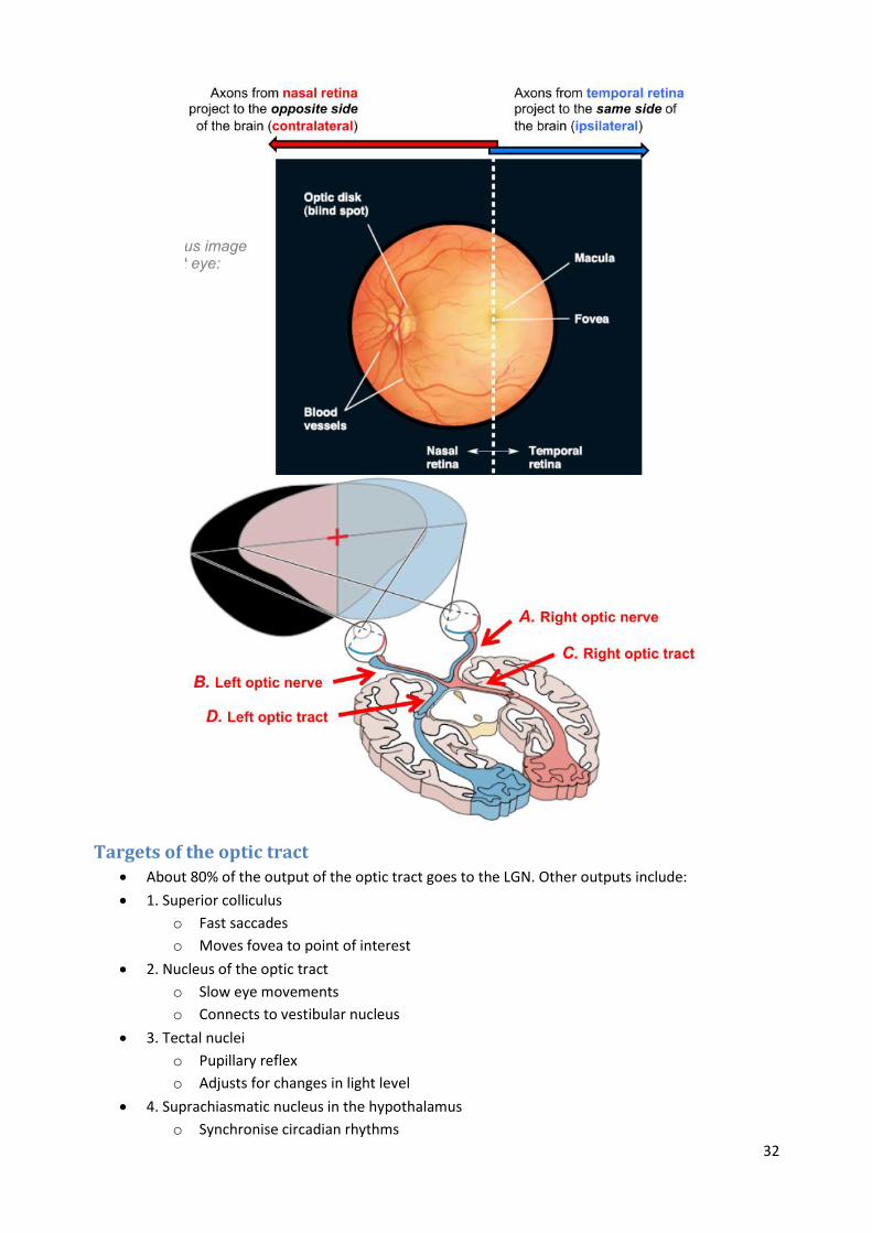
Retinofugal projection Axons from retinal ganglion cells form their first synapses in the brain stem & midbrain, after
passing through the optic nerve, chiasm, and tract.
Left part of left visual hemifield is seen by left nasal retina, while the right part of left visual
hemifield is seen by right temporal retina.
32
Targets of the optic tract About 80% of the output of the optic tract goes to the LGN. Other outputs include:
1. Superior colliculus
o Fast saccades
o Moves fovea to point of interest
2. Nucleus of the optic tract
o Slow eye movements
o Connects to vestibular nucleus
3. Tectal nuclei
o Pupillary reflex
o Adjusts for changes in light level
4. Suprachiasmatic nucleus in the hypothalamus
o Synchronise circadian rhythms

33
Lateral geniculate nucleus LGN is the ‘gateway’ or ‘relay station’ for the visual cortex
Like a stack of 6 pancakes, bent into a knee shape (seen in cross-section)
Principal layers 1 & 2:
o Large cells
o Magnocellular LGN layers
Principal layers 3-6:
o Small cells
o Parvocellular LGN layers
Layers K1-K6 (in-between layers):
o Tiny cells
o Koniocellular LGN layers
The LGN receives massive feedback from the visual cortex, and so there seems to be ‘top-down’
modulation of the information passing through the LGN.
Primary visual cortex Also called the striate cortex, because of its striped appearance in some animals, due to
myelinated axons within some layers.
Located at the occipital lobe of the brains of many animals.
Neighbouring cells in retina project to neighbouring locations in LGN, V1 in a retinotopic map.
Magnification in the retinotopic map: central 10 o of eccentricity maps to 50% of V1.
M-cell and P-cell inputs remain largely segregated in V1.
Organisation of V1 In addition to the retinotopic map, there are several other kinds of maps or organisational
patterns found in V1:
o Ocular dominance columns
o Cytochrome oxidase ‘blobs’
o Orientation columns
Autoradiography reveals alternating bands of inputs from the two eyes.

34
Stain for cytochrome oxidase, a mitochondrial enzyme for cell metabolism reveals ‘blobs’,
effecitvely pillars of staining across layers 2/3, 5 & 6.
Blobs seem to be associated with the K-cell (colour) pathway, interblobs with the P-cell (acuity)
pathway. Blobs are thought to be important for the analysis of object colour.
Neurons with the same orientation preference are arranged in orientation columns.
Orientation-selective neurons are found in all layers except layer 4C. Cells in this layer show
mostly centre-surround receptive fields, and some are responsive to colour.
Direction-selective neurons in V1 are a subset of orientation-selective neurons. Typically a
property of neurons receiving magnocellular input.
A complete set of orientation and ocular dominance columns, along with associated blobs, is
called a hypercolumn or cortical module.
Dorsal and ventral streams ‘Action stream’: Fast-response analysis of visual motion
Almost all neurons are direction selective with large receptive fields
35
Area MT (middle temporal):
o Arranged in direction-of-motion columns (analogous to orientation columns in V1)
o Very sensitive to linear movement of spots of light
o Neurons responsive to more complex motions e.g. radial, circular
Area MST (medial superior temporal):
o Navigation
o Directing eye movements
o Perception’ stream: Visual attributes other than motion
o Many neurons are both colour and orientation selective
Area V4:
o Have larger receptive fields than neurons in V1
o Seems to be important for shape and colour perception
o Visual recognition
o Visual memory
o Neurons responsive to colours and shapes
Area IT (inferior temporal lobe):
o Contains regions sensitive to faces
Olfaction and Taste
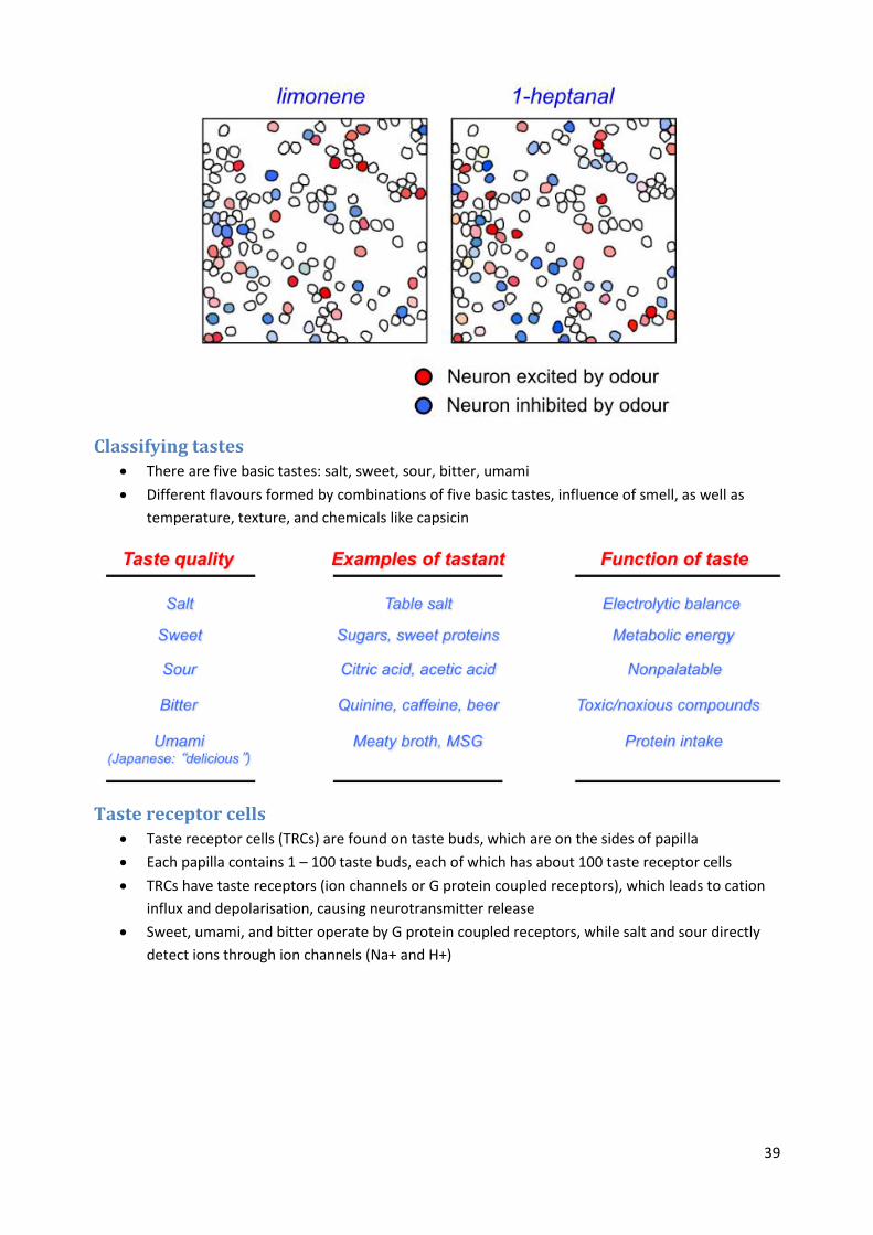
Multidimensionality of odour space Humans can distinguish 10,000 different odours
Not clear what the ‘odour primitives’ are, like we have for hearing and vision
Humans generally cannot recognise single components, but recognise blends (synthetic)
Humans can detect <0.01 nM (1 molecule in a billion, a few drops of odorant in an Olympic pool)
Many connections between olfaction and the limbic system (e.g. hippocampus, amygdala),
which means there is a close connection between olfaction and memory
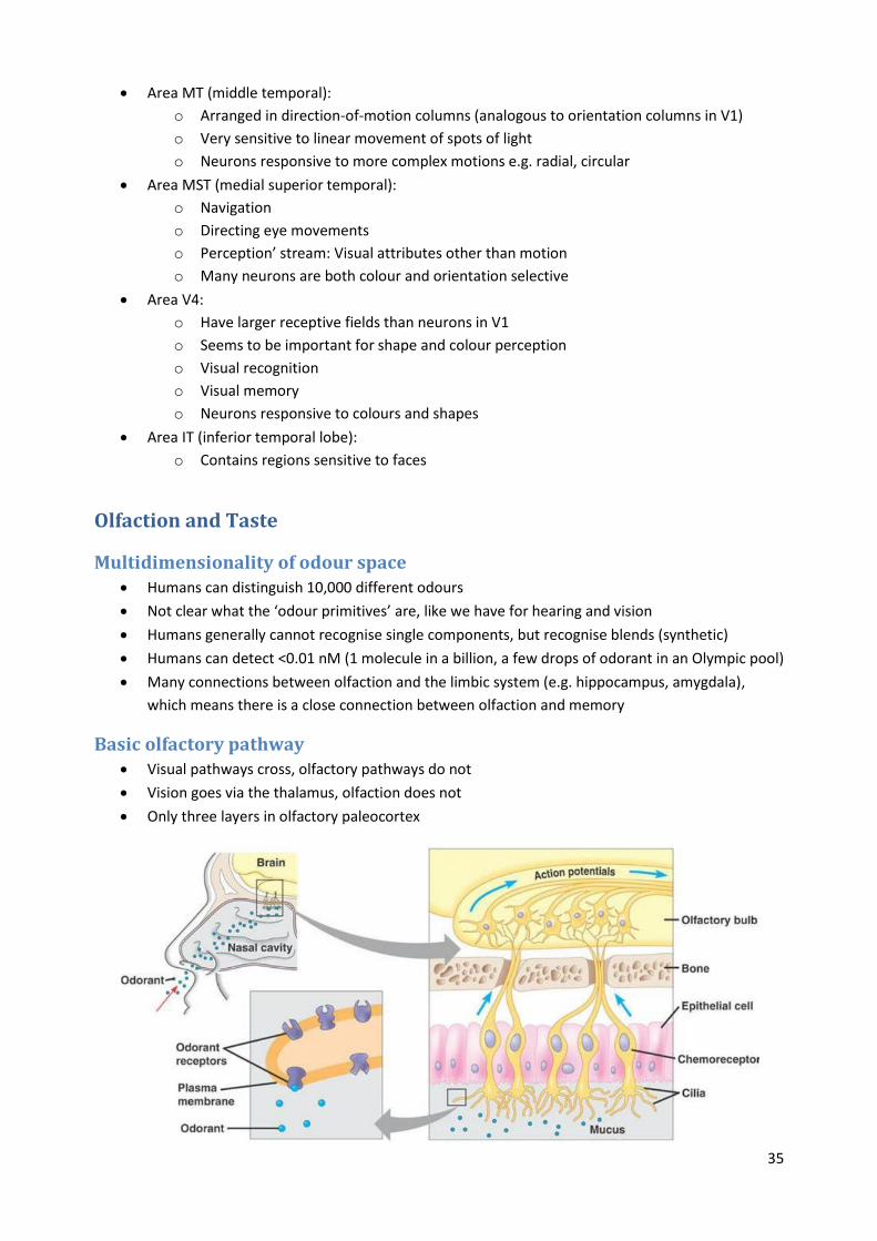
Basic olfactory pathway Visual pathways cross, olfactory pathways do not
Vision goes via the thalamus, olfaction does not
Only three layers in olfactory paleocortex

36
Olfactory epithelium Olfactory receptor neurons (ORNs) are regularly replaced on a 4-8 week cycle, from stem cells in
olfactory epithelium
Unlike photoreceptors and hair cells, ORNs have axons and fire action potentials
The mucus includes binding proteins to shuttle odors to the cilia
Scavenger molecules clean up odorants over time to reduce olfactory response
Complex series of transduction mechanisms for generation of action potentials
o Odour molecule binds selectively to olfactory receptor protein
o G-protein activates Adenyl cyclase III to generate cAMP
o This opens cAMP-gated channel, causing Na+ and Ca2+ influx and depolarisation
o Ca2+ also opens Ca-gated Cl- channel, causing Cl- efflux and further depolarisation
o After firing of action potentials, ion exchangers restore the ion gradients

37
Combinatorial code for odours ORNs have overlapping responses to different odours
An odour is identified by a characteristic pattern of action potentials from different ORNs
This is a “combinatorial code”, which depends on the higher-level “decoder” being able to keep
track of firing of each separate ORN
Requires each ORN to have different responses (H, M, Z) to at least several odours
These ORNs are encoded for by a large (~1000) multigene family of G-protein-coupled receptors
expressed largely in the olfactory epithelium
ORNs expressing the same gene (one receptor gene each) – and hence the same odorant
receptor – are randomly distributed within the olfactory epithelium

38
Olfactory bulb All ORNs expressing the same odorant receptor send their axons to 1 - 2 glomeruli per olfactory
bulb (they are a bilateral structure)
There are about 25 mitral cells per glomerulus
High convergence ensures that only a few ORNs need to be active to stimulate mitral cells in
each glomerulus
Different odorants produce distinctive patterns of glomerular activation, the ‘odotopic map’
Rather few glomeruli are active, indicating that the olfactory code is quite sparse
The olfactory bulb seems to identify odour components, while the olfactory cortex generates
‘odour images’
Olfactory cortex Primary olfactory cortex is tri-laminar, and lacks the cortical columns found in other sensory
cortices
Different odours are ‘coded’ by different spatial patterns of excited and inhibited neurons in the
olfactory cortex
39
Classifying tastes There are five basic tastes: salt, sweet, sour, bitter, umami
Different flavours formed by combinations of five basic tastes, influence of smell, as well as
temperature, texture, and chemicals like capsicin
Taste receptor cells Taste receptor cells (TRCs) are found on taste buds, which are on the sides of papilla
Each papilla contains 1 – 100 taste buds, each of which has about 100 taste receptor cells
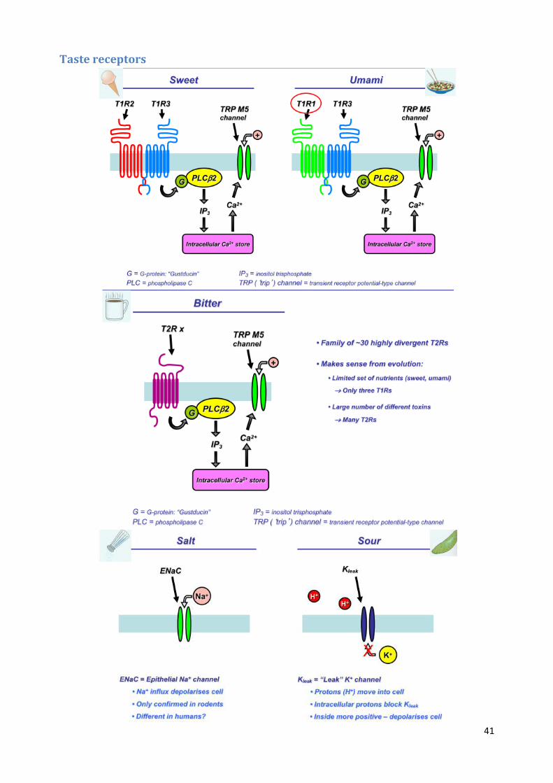
TRCs have taste receptors (ion channels or G protein coupled receptors), which leads to cation
influx and depolarisation, causing neurotransmitter release
Sweet, umami, and bitter operate by G protein coupled receptors, while salt and sour directly
detect ions through ion channels (Na+ and H+)

40
41
Taste receptors
42
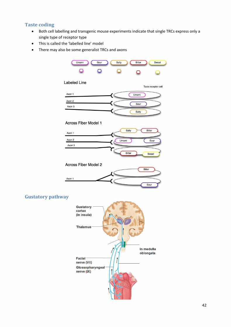
Taste coding Both cell labelling and transgenic mouse experiments indicate that single TRCs express only a
single type of receptor type
This is called the ‘labelled line’ model
There may also be some generalist TRCs and axons
Gustatory pathway
43
Neural Coding
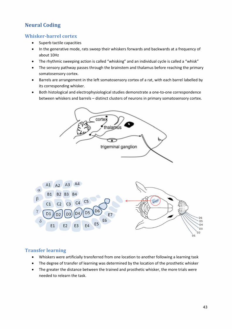
Whisker-barrel cortex Superb tactile capacities
In the generative mode, rats sweep their whiskers forwards and backwards at a frequency of
about 10Hz
The rhythmic sweeping action is called “whisking” and an individual cycle is called a “whisk”
The sensory pathway passes through the brainstem and thalamus before reaching the primary
somatosensory cortex.
Barrels are arrangement in the left somatosensory cortex of a rat, with each barrel labelled by
its corresponding whisker.
Both histological and electrophysiological studies demonstrate a one-to-one correspondence
between whiskers and barrels – distinct clusters of neurons in primary somatosensory cortex.
Transfer learning Whiskers were artificially transferred from one location to another following a learning task
The degree of transfer of learning was determined by the location of the prosthetic whisker
The greater the distance between the trained and prosthetic whisker, the more trials were
needed to relearn the task.

44
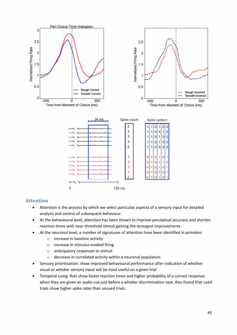
Sensory coding • Generative mode: texture coding
o Contacts with the rough texture evoked significantly higher firing rates in barrel cortex
than did contact with the smooth texture.
o During a texture discrimination task firing rate of barrel cortex neurons correlated with
the animal’s choice.
• Receptive mode: vibration coding
o Barrel cortex neurons encode the product of frequency and amplitude which is
proportional to the mean speed of the vibration.
o Behavioural experiments confirmed these electrophysiological findings by
demonstrating that rats perceive vibrations as the speed of whisker motion.
Spike train behaviour On each trial, the rat approached the texture plate (Rough or Smooth) which signalled the
reward spout.
After touching the texture the rat made a behavioural choice by turning towards one of the
spouts and collected the water reward if the choice was correct.
In a rough versus smooth texture discrimination task, contacts with the rough texture evoked
significantly higher firing rates in barrel cortex than did contact with the smooth texture.
This firing-rate code was reversed on error trials (lower for rough than for smooth).
45
Attention Attention is the process by which we select particular aspects of a sensory input for detailed
analysis and control of subsequent behaviour.
At the behavioural level, attention has been shown to improve perceptual accuracy and shorten
reaction times with near-threshold stimuli gaining the strongest improvements.
At the neuronal level, a number of signatures of attention have been identified in primates:
o increase in baseline activity
o increase in stimulus-evoked firing
o anticipatory responses to stimuli
o decrease in correlated activity within a neuronal population
Sensory prioritisation: show improved behavioural performance after indication of whether
visual or whisker sensory input will be most useful on a given trial
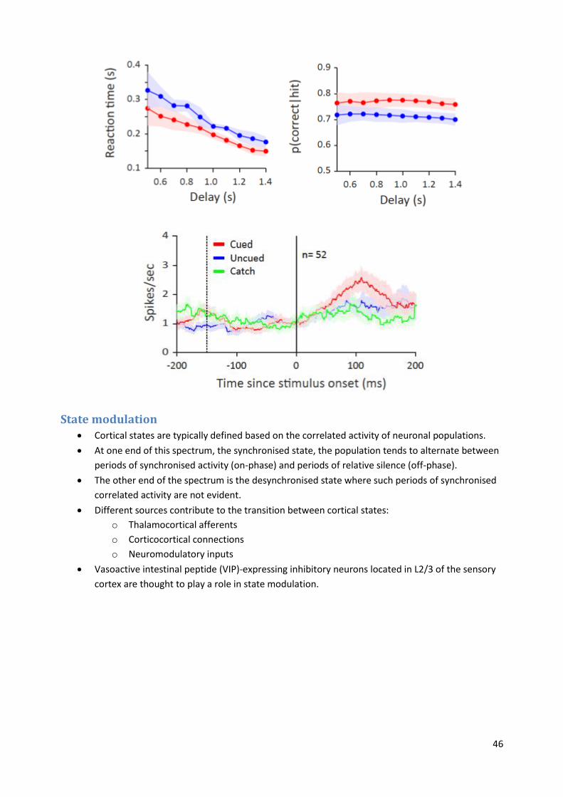
Temporal cuing: Rats show faster reaction times and higher probability of a correct response
when they are given an audio cue just before a whisker discrimination task. Also found that cued
trials show higher spike rates than uncued trials.
46
State modulation Cortical states are typically defined based on the correlated activity of neuronal populations.
At one end of this spectrum, the synchronised state, the population tends to alternate between
periods of synchronised activity (on-phase) and periods of relative silence (off-phase).
The other end of the spectrum is the desynchronised state where such periods of synchronised
correlated activity are not evident.
Different sources contribute to the transition between cortical states:
o Thalamocortical afferents
o Corticocortical connections
o Neuromodulatory inputs
Vasoactive intestinal peptide (VIP)-expressing inhibitory neurons located in L2/3 of the sensory
cortex are thought to play a role in state modulation.

47
Reward systems Midbrain dopamine neurons show phasic excitatory responses following primary food and liquid
rewards, visual, auditory and somatosensory reward-predicting stimuli.
Dopamine neurons acquire responses to reward-predicting visual and auditory stimuli.
The responses covary with the expected value of reward, irrespective of spatial position, sensory
stimulus and arm, mouth and eye movements (generalisation).
Auditory System
The nature of sound Sound is a longitudinal wave of pressure variations in a medium

48
Sound is measured in terms of its pressure component rather than displacement
The loudness of a sound is measured in decibels (dB) which expresses how much bigger it is
(logarithmically) relative to the average human hearing threshold.
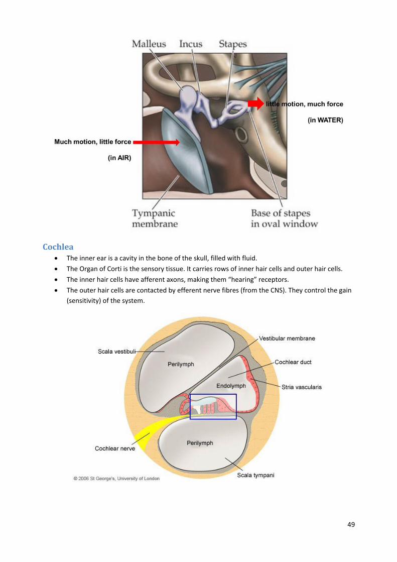
Outer and middle ear Outer ear: pinna to ear drum (tympanic membrane)
Middle ear: the 3 ossicles (middle ear bones) in an air space
o 1. Malleus (hammer)
o 2. Incus (anvil)
o 3. Stapes (stirrup)
Inner ear: Cochlea and semicircular canals
Middle ear The middle ear muscles are the smallest striated muscles in the human body
The bones in the middle ear provide impedance matching between the eardrum (which is acted
upon by vibrations in air) and the fluid-filled inner ear.
This protects the cochlea from loud sounds
49
Cochlea The inner ear is a cavity in the bone of the skull, filled with fluid.
The Organ of Corti is the sensory tissue. It carries rows of inner hair cells and outer hair cells.
The inner hair cells have afferent axons, making them “hearing” receptors.
The outer hair cells are contacted by efferent nerve fibres (from the CNS). They control the gain
(sensitivity) of the system.

50
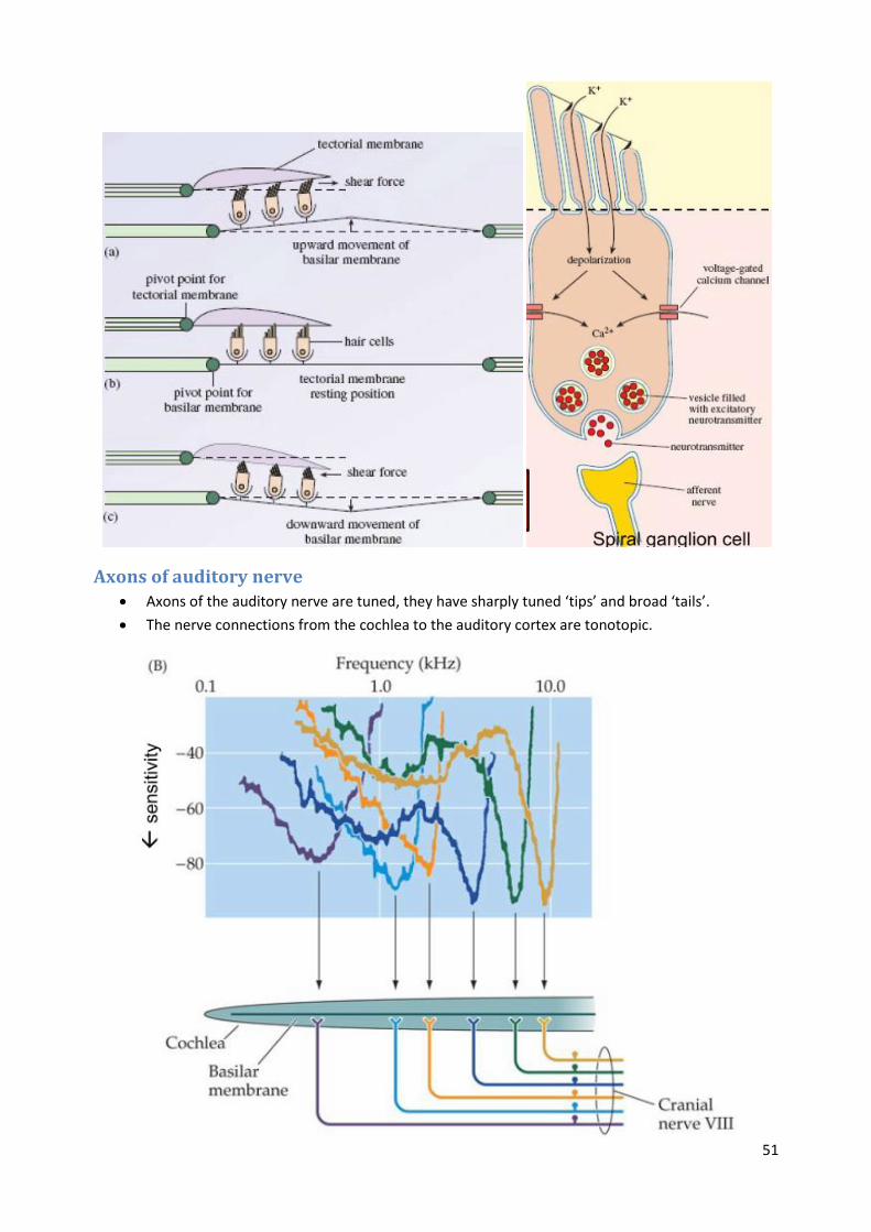
Hair cells Displacement of the basilar membrane by sound ultimately leads to bending of hair bundles on
top of hair cells.
The base of the basilar membrane is tuned to high frequencies, while the apex is tuned to low
frequencies.
Bending of stereocilia on the hair cell depolarises the cell and sends a signal to the auditory
nerve.
Mechano-electrical transduction channels open in response to bending of hair channels, causing
K+ ion entry and depolarisation.
Depolarization opens voltage dependent calcium channels (CaV1.3 L-type) in the inner hair cell
membrane.
Calcium entry causes glutamate release at the inner hair cell–spiral ganglion cell synapse.
Released glutamate activates receptors (mostly AMPA) in the spiral ganglion cell, which results
in depolarization and action potential firing of the spiral ganglion cell.
Action potentials are carried by the auditory nerve to the brain.
51
Axons of auditory nerve Axons of the auditory nerve are tuned, they have sharply tuned ‘tips’ and broad ‘tails’.
The nerve connections from the cochlea to the auditory cortex are tonotopic.

52
Otoacoustic emissions The outer hair cells (OHCs) are active – they move when stimulated and produce a faint sound
of their own.
An otoacoustic emission (OAE) is a sound which is generated from within the inner ear.
OAEs are thought to be related to the amplification of the cochlea. In the absence of external
stimulation, the activity of the cochlear amplifier increases, leading to production of sound.
Four different sorts of otoacoustic emissions:
o 1. Spontaneous OAEs (no stimulus required)
o 2. Evoked OAEs (echoes in response to clicks or tone bursts)
o 3. Distortion-product OAEs (2 continuous tones in; a faint third tone out)
o 4. Stimulus frequency OAEs (a faint tone out in response to a single stimulus tone)
Conductive hearing loss Caused by loss in transmission of sound from the outer ear to the inner ear.
Blockage of the outer ear by wax.
Fluid in the middle ear caused by middle ear infections (otitis media).
Otosclerosis, in which the ossicles of the middle ear harden and stiffen (surgery can be helpful).
Damage to the ossicles, for example by serious infection or head injury.
A perforated eardrum, perhaps from infection or injury.
Sensorineural hearing loss
Due to damage somewhere in the pathway from the hair cells to the cortex.
Age-related hearing loss (presbyacusis) – the natural loss in hearing that most people
experience as they get older. It is due to the loss of hair cells
In general, the loss starts at high frequencies (10 kHz), and progressively moves down to lower
frequencies (1 kHz and below).
Acoustic trauma caused by loud noise can damage hair cells.
Some infections such as mumps or meningitis can lead to loss of hair cells or damage to the
auditory nerve.

53
Ménière’s disease (of unknown cause), which gives rise to bouts of dizziness, tinnitus, and
hearing loss.
Certain drugs, such as powerful antibiotics, can cause permanent hearing loss.
Acoustic neuroma. This is a benign (non-cancerous) tumour affecting the auditory nerve.
Congenital abnormalities (e.g. dysfunctional hair cells)
Tinnitus Tinnitus is the hearing of sound when no external sound is present. While often described as a
ringing, it may also sound like a clicking, hiss or roaring.
Affects about 10% of the general population. For 1–3% it seriously affects their quality of life
and they seek medical treatment.
Cause of tinnitus is unknown. It’s largely in the brain, not the ear.
Currently no direct therapy. Treatments involve noise makers and cognitive therapy.
Auditory pathway

54
The Auditory Nerve Made up of around 30,000 fibres
Each afferent nerve fibre contacts an inner hair cell from a specific region of the cochlear
Each fibre is sensitive to a specific frequency – the “characteristic frequency”
But they cannot fire at the same frequency as the basilar membrane is detecting; the maximum
action potential rate is about 100 times too slow (200 Hz vs 20 kHz).
Instead they use “Phase Locking”, wherein each one of a group of nerves responds in phase with
stimulus, but not on every cycle.
Tonotopy is also preserved in the auditory nerve, up to the level of the cortex.
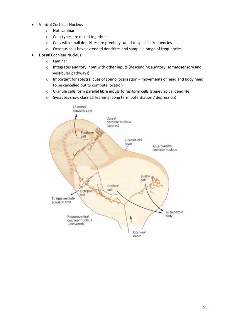
Cochlear nucleus The cochlear nuclear (CN) complex comprises two cranial nerve nuclei in the human brainstem,
the ventral cochlear nucleus (VCN) and the dorsal cochlear nucleus (DCN).
The ventral cochlear nucleus is unlayered whereas the dorsal cochlear nucleus is layered.
55
Ventral Cochlear Nucleus
o Not Laminar
o Cells types are mixed together
o Cells with small dendrites are precisely tuned to specific frequencies
o Octopus cells have extended dendrites and sample a range of frequencies
Dorsal Cochlear Nucleus
o Laminar
o Integrates auditory input with other inputs (descending auditory, somatosensory and
vestibular pathways)
o Important for spectral cues of sound localisation – movements of head and body need
to be cancelled out to compute location
o Granule cells form parallel fibre inputs to fusiform cells (spiney apical dendrite)
o Synapses show classical learning (Long term potentiation / depression)
56
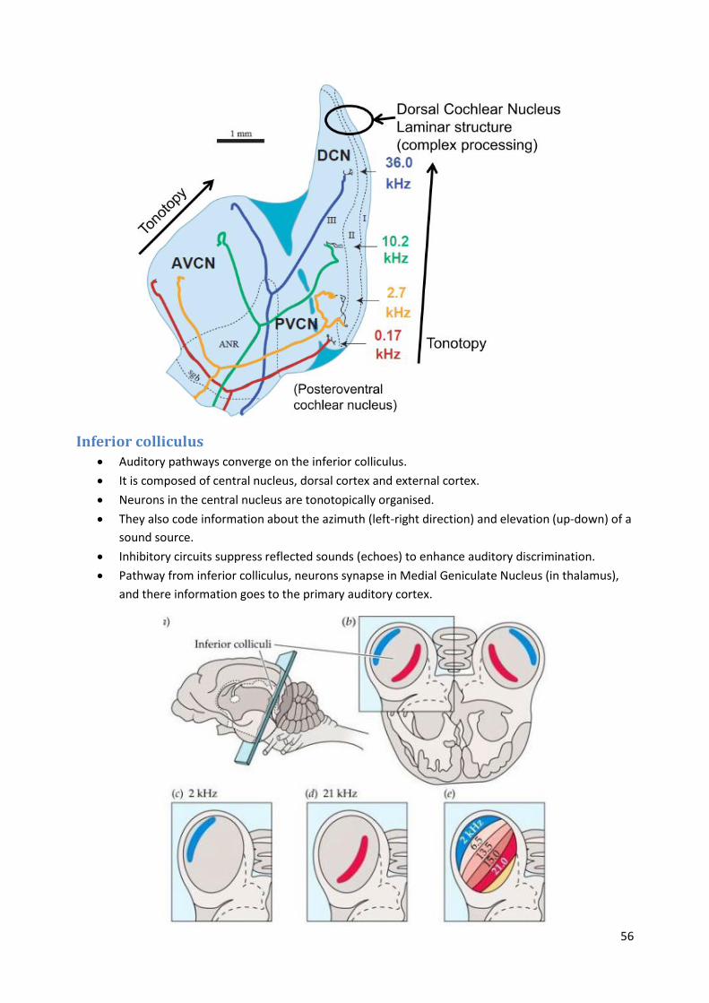
Inferior colliculus Auditory pathways converge on the inferior colliculus.
It is composed of central nucleus, dorsal cortex and external cortex.
Neurons in the central nucleus are tonotopically organised.
They also code information about the azimuth (left-right direction) and elevation (up-down) of a
sound source.
Inhibitory circuits suppress reflected sounds (echoes) to enhance auditory discrimination.
Pathway from inferior colliculus, neurons synapse in Medial Geniculate Nucleus (in thalamus),
and there information goes to the primary auditory cortex.
57
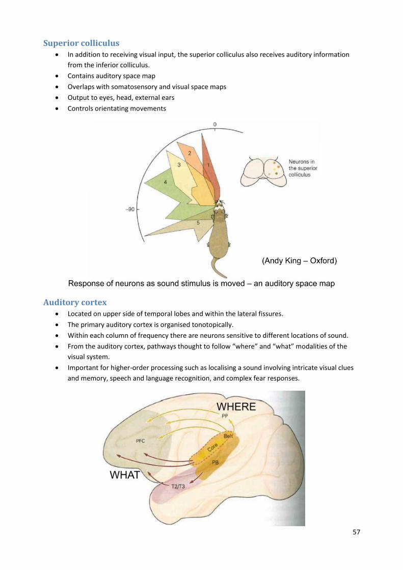
Superior colliculus In addition to receiving visual input, the superior colliculus also receives auditory information
from the inferior colliculus.
Contains auditory space map
Overlaps with somatosensory and visual space maps
Output to eyes, head, external ears
Controls orientating movements
Auditory cortex Located on upper side of temporal lobes and within the lateral fissures.
The primary auditory cortex is organised tonotopically.
Within each column of frequency there are neurons sensitive to different locations of sound.
From the auditory cortex, pathways thought to follow “where” and “what” modalities of the
visual system.
Important for higher-order processing such as localising a sound involving intricate visual clues
and memory, speech and language recognition, and complex fear responses.
58
The McGurk Effect A perceptual phenomenon that demonstrates an interaction between hearing and vision in
speech perception.
The visual information a person gets from seeing a person speak changes the way they hear the
sound.
The sound is “ba-ba” but she is mouthing “ga-ga”.
Even if you know, auditory information is still altered by visual system
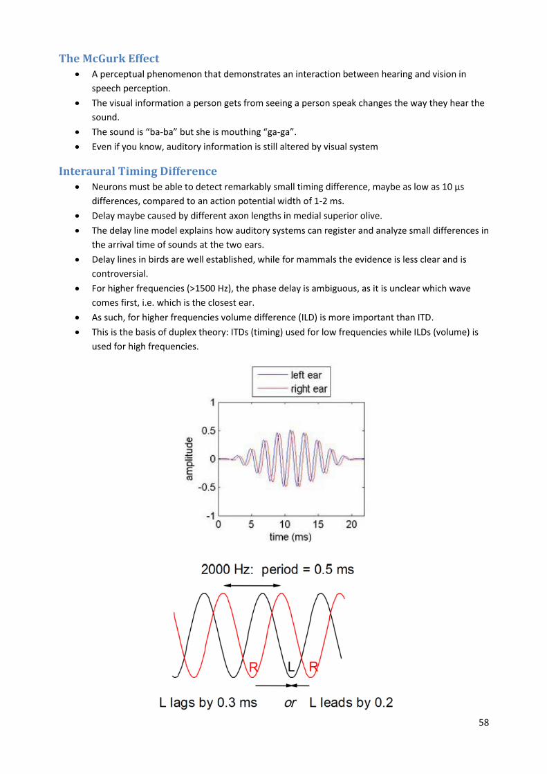
Interaural Timing Difference Neurons must be able to detect remarkably small timing difference, maybe as low as 10 µs
differences, compared to an action potential width of 1-2 ms.
Delay maybe caused by different axon lengths in medial superior olive.
The delay line model explains how auditory systems can register and analyze small differences in
the arrival time of sounds at the two ears.
Delay lines in birds are well established, while for mammals the evidence is less clear and is
controversial.
For higher frequencies (>1500 Hz), the phase delay is ambiguous, as it is unclear which wave
comes first, i.e. which is the closest ear.
As such, for higher frequencies volume difference (ILD) is more important than ITD.
This is the basis of duplex theory: ITDs (timing) used for low frequencies while ILDs (volume) is
used for high frequencies.

59
Interaural Volume (Level) Difference Head Shadow makes sound in further ear quieter
Frequency dependent – most pronounced for > 4000 Hz
60
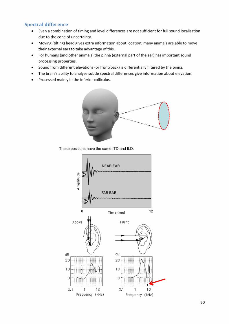
Spectral difference Even a combination of timing and level differences are not sufficient for full sound localisation
due to the cone of uncertainty.
Moving (tilting) head gives extra information about location; many animals are able to move
their external ears to take advantage of this.
For humans (and other animals) the pinna (external part of the ear) has important sound
processing properties.
Sound from different elevations (or front/back) is differentially filtered by the pinna.
The brain’s ability to analyse subtle spectral differences give information about elevation.
Processed mainly in the inferior colliculus.

61
Plasticity of localisation Localisation calibration is based on experience and is continually learnt. Can be modified by
experimental intervention.
After 42 days learning, sounds map shifts to realign multimodal map.
When prisms removed, visual location returns to correct position quickly.
The visual and auditory space maps in the optic tectum (mamalian superior colliculus) change
location over several weeks.
Juveniles adapt quickly, while older animals do not. However old animals that have experinece
of changing pattens when younger can however re-learn the patterns from the past.
62
Autonomic Nervous System
Hypothalamus Part of the limbic system.
A collection of small nuclei, adjacent to 3rd ventricle.
Links nervous system to endocrine system via the pituitary gland.
Synthesises and secretes neurohormones (hypothalamic hormones; releasing hormones).
Controls body temperature, thirst, hunger and sleep etc.
Periventricular zone lies next to third ventricle. This region contains neurosecretory neurons
that extend axons into the pituitary gland.
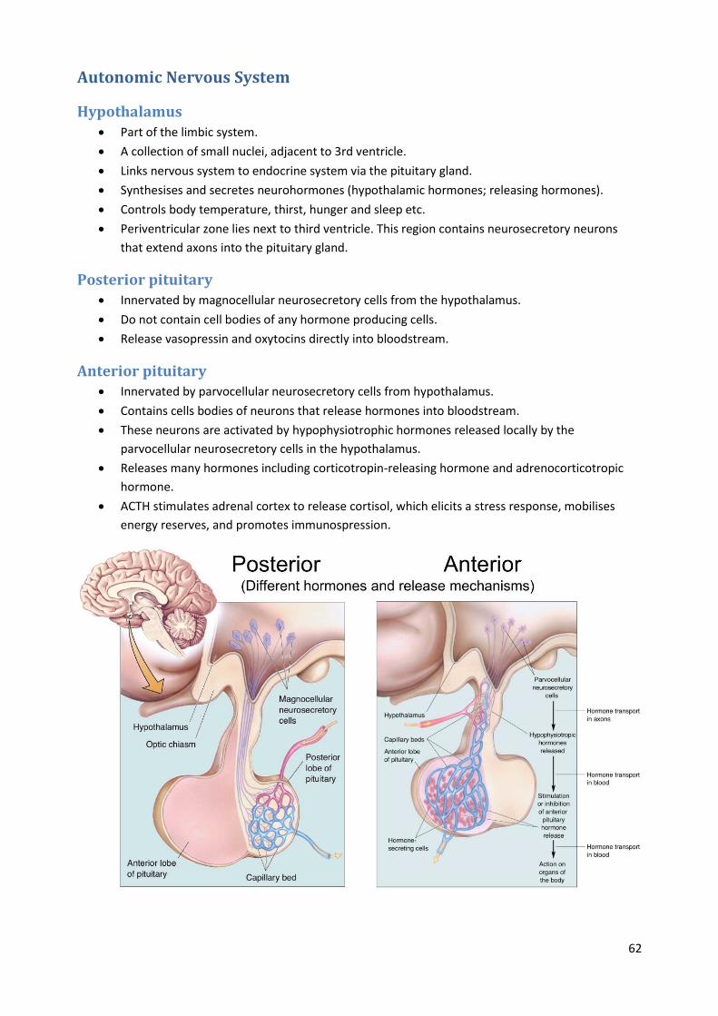
Posterior pituitary Innervated by magnocellular neurosecretory cells from the hypothalamus.
Do not contain cell bodies of any hormone producing cells.
Release vasopressin and oxytocins directly into bloodstream.
Anterior pituitary Innervated by parvocellular neurosecretory cells from hypothalamus.
Contains cells bodies of neurons that release hormones into bloodstream.
These neurons are activated by hypophysiotrophic hormones released locally by the
parvocellular neurosecretory cells in the hypothalamus.
Releases many hormones including corticotropin-releasing hormone and adrenocorticotropic
hormone.
ACTH stimulates adrenal cortex to release cortisol, which elicits a stress response, mobilises
energy reserves, and promotes immunospression.
63
Regulation of thirst Volumetric Thirst: decrease in blood volume (Hypovolemia)
Osmometric Thirst: increase in blood tonicity (Hypertonicity)
In response to a decrease in blood volume, the kidneys release the hormone renin.
This converts angiotensinogen to angiotensin I then to angiotensin II, which acts on
magnocellular neurosecretory cells in the hypothalamus to release vasopressin (anti-diuretic
hormone). This ADH acts on the kidneys to conserve water.
Mechanoreceptors in blood vessels and the heart detect reduced blood pressure, and signal this
via vagus nerve to the hypothalamus, causing the release of vasopressin.
This causes sympathetic activity to increase blood pressure, and also produces a drive to drink.
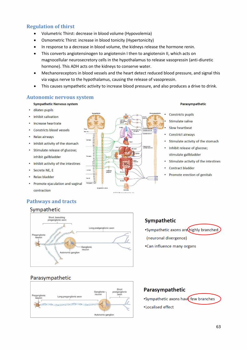
Autonomic nervous system
Pathways and tracts
64
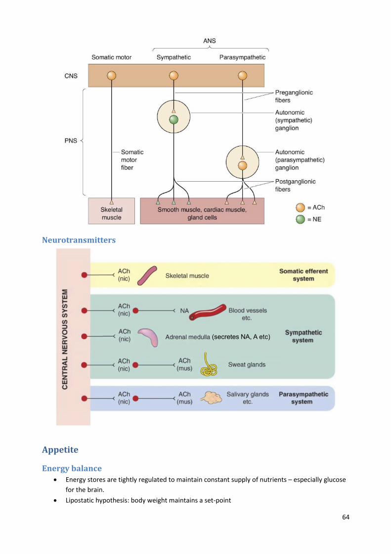
Neurotransmitters
Appetite
Energy balance
Energy stores are tightly regulated to maintain constant supply of nutrients – especially glucose
for the brain.
Lipostatic hypothesis: body weight maintains a set-point

65
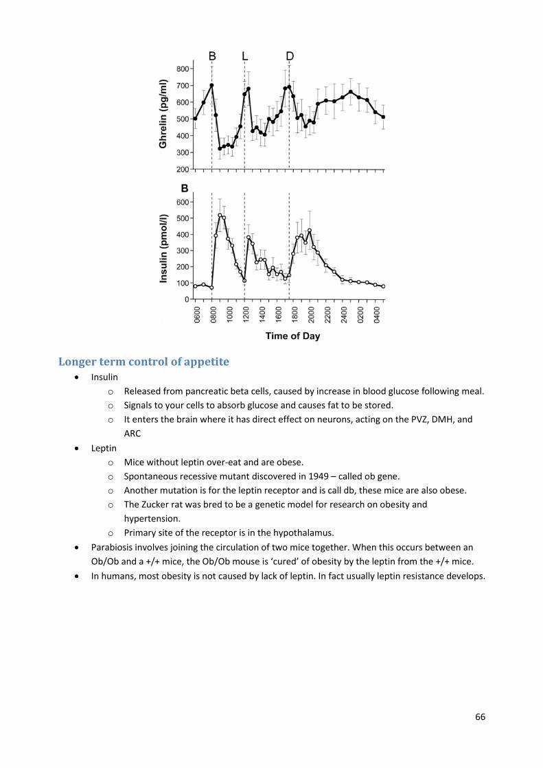
Short term control of appetite Hypothalamus is central in controlling appetite, particularly the periventricular zone (PVZ).
Lesions of the ventromedial hypothalamus lead to obesity in mice.
Ghrelin
o A hormone released by the stomach (mainly) and the hypothalamus.
o Highest levels just before a meal and lowest just after.
o Ghrelin causes a feeling of intense hunger and initiates feeding via action in arcuate
nucleus of hypothalamus (ARC) and via input to nucleus tractus solitarius (NTS).
Cholecystokinin
o A peptide hormone that is released from the small intestine in response to food.
o Stimulates secretion from pancreas and bile duct to digest fats and proteins.
o Signals satiety to the brain, and stops feeding by activating vagal input to NTS and also
acting on the hypothalamus.
o Injection of CCK reduces meal size and duration, but half life is very short.
o CCK receptor agonists have no major effect on weight in humans but further clinical
trials are being conducted.
66
Longer term control of appetite Insulin
o Released from pancreatic beta cells, caused by increase in blood glucose following meal.
o Signals to your cells to absorb glucose and causes fat to be stored.
o It enters the brain where it has direct effect on neurons, acting on the PVZ, DMH, and
ARC
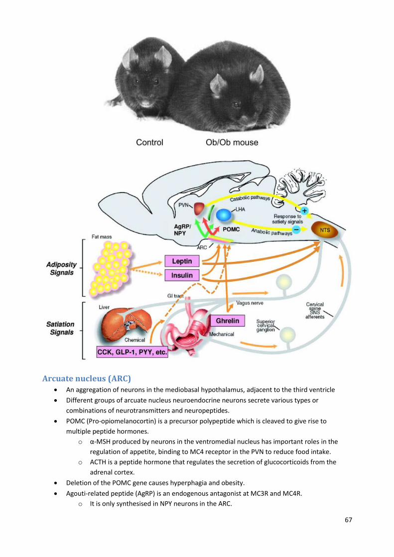
Leptin
o Mice without leptin over-eat and are obese.
o Spontaneous recessive mutant discovered in 1949 – called ob gene.
o Another mutation is for the leptin receptor and is call db, these mice are also obese.
o The Zucker rat was bred to be a genetic model for research on obesity and
hypertension.
o Primary site of the receptor is in the hypothalamus.
Parabiosis involves joining the circulation of two mice together. When this occurs between an
Ob/Ob and a +/+ mice, the Ob/Ob mouse is ‘cured’ of obesity by the leptin from the +/+ mice.
In humans, most obesity is not caused by lack of leptin. In fact usually leptin resistance develops.
67
Arcuate nucleus (ARC) An aggregation of neurons in the mediobasal hypothalamus, adjacent to the third ventricle
Different groups of arcuate nucleus neuroendocrine neurons secrete various types or
combinations of neurotransmitters and neuropeptides.
POMC (Pro-opiomelanocortin) is a precursor polypeptide which is cleaved to give rise to
multiple peptide hormones.
o α-MSH produced by neurons in the ventromedial nucleus has important roles in the
regulation of appetite, binding to MC4 receptor in the PVN to reduce food intake.
o ACTH is a peptide hormone that regulates the secretion of glucocorticoids from the
adrenal cortex.
Deletion of the POMC gene causes hyperphagia and obesity.
Agouti-related peptide (AgRP) is an endogenous antagonist at MC3R and MC4R.
o It is only synthesised in NPY neurons in the ARC.

68
o It is a potent appetite stimulator.
Polymorphisms in MC4R are associated with increased body weight; 1-2.5% of people with BMI
> 30 harbour a MC4R polymorphism, making it the most common susceptibility gene.
ARC is primary area in appetite control, with other hypothalamic nuclei referred to as secondary
areas.
Insulin and leptin both activate POMC cells in the ARC, while inhibiting AgRP cells; thus they
signal satiety.
Ghrelin has the opposite effect, activating AgRP cells and inhibiting POMC cells, thereby causing
hunger and feeding.
Integration in the NTS The solitary nucleus is a series of nuclei in the medulla.
It is central to detecting satiety based on inputs from the hypothalamus and gut hormones.

69
Modulation by 5-HT 5-HT neurons in the raphe nuclei project widely, and are engaged in a wide range of functions.
Depletion of brain serotonin increases food intake and promotes obesity.
5-HT neurons provide input to activate POMC and inhibit NPY/AgRP cells, thereby reducing
feeding.
Drugs to treat obesity Reduce fat absorption, by lipase inhibitors that prevents fat absorption.
Increase levels of amine neurotransmission (5-HT, noradrenaline, dopamine).
Reduce cannabinoid neurotransmission.
Weight loss treatments are moderately effective.
70