t argeting s phingosine k inase 1 and a poptosis by m etformin to d ecrease t umor r esistance to a...
TRANSCRIPT
Targeting Sphingosine Kinase 1 and Apoptosis by Metformin to Decrease Tumor
Resistance to Adriamycin
ByBy
Dr. Ahmed Mohamed KabelPharmacology Department, Faculty of Medicine, Tanta University, EgyptPharmacology Department, College of Pharmacy, Taif University, KSA
• Adriamycin (ADR, Doxorubicin) is an anthracycline antibiotic that is frequently
used as a treatment for many types of cancer such as leukemia, lymphoma, breast,
ovarian and lung cancer. • It damages DNA by intercalation into DNA and inhibition of topoisomerase II resulting
in DNA strand breaks.
• The major limiting factor for the use of ADR in cancer therapy is the development of resistance.
• The mechanisms of this resistance may include increased activity of sphingosine kinase-1 (SphK1)
enzyme which inhibits penetration and intercalation of ADR into DNA and significantly
abolishing the anticancer effect of ADR.
• This makes it essential to combine ADR with agents that inhibit these mechanisms to decrease the incidence of resistance of cancer cells to ADR.
• Metformin is an antidiabetic agent that decreases intestinal absorption of glucose, increases its
anaerobic metabolism and improves insulin sensitivity.
• Studies on animal models had demonstrated that metformin prevents tumor development and
inhibits cell proliferation. This effect may be mediated through its regulatory role on the
hormonal, metabolic and immune functions.
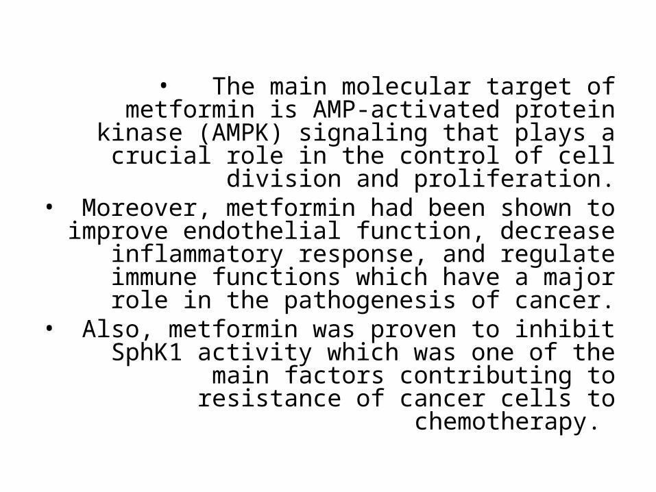
• The main molecular target of metformin is AMP-activated protein kinase (AMPK) signaling that
plays a crucial role in the control of cell division and proliferation.
• Moreover, metformin had been shown to improve endothelial function, decrease
inflammatory response, and regulate immune functions which have a major role in the
pathogenesis of cancer.• Also, metformin was proven to inhibit SphK1
activity which was one of the main factors contributing to resistance of cancer cells to
chemotherapy.
So, The aim of this work was to study
the effect of targeting sphingosine kinase 1
and apoptosis by metformin on tumor
resistance to adriamycin using a
transplantable tumor model in mice.
• In this study, we used a model of solid Ehrlich
carcinoma (SEC), where Ehrlich carcinoma cells
(ECCs) were implanted subcutaneously into the
right thigh of the hind limb of mice. A palpable
solid tumor mass (about 100 mm3) was
developed within 10 days.
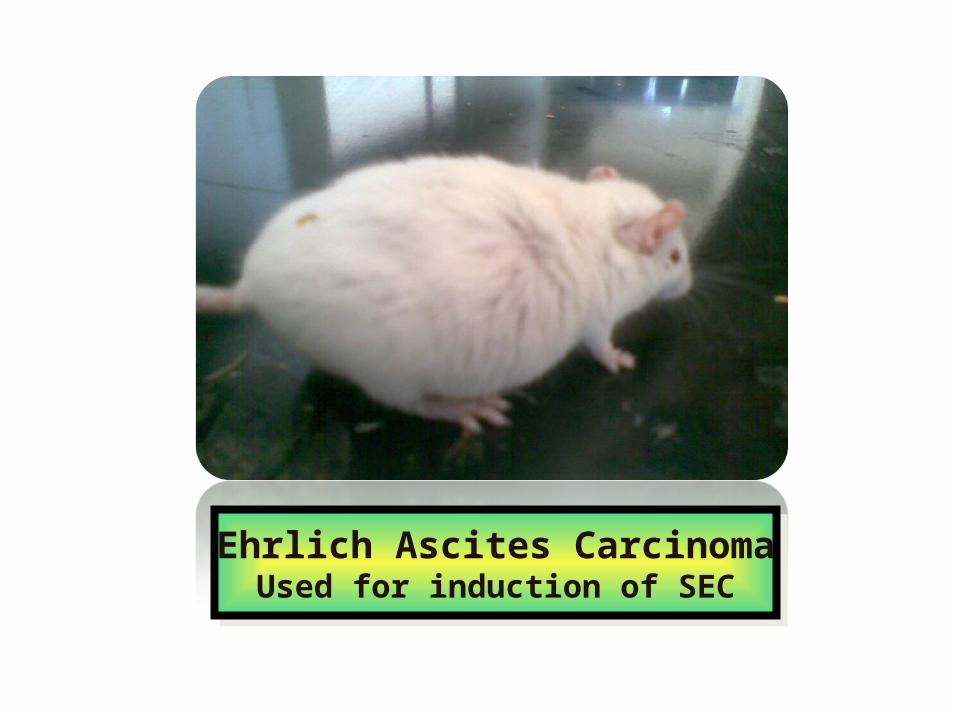
Ehrlich Ascites CarcinomaUsed for induction of SEC
Ehrlich Ascites CarcinomaUsed for induction of SEC
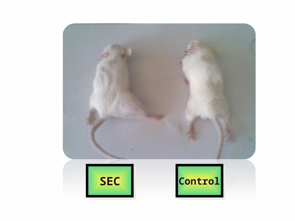
ControlControlSECSEC
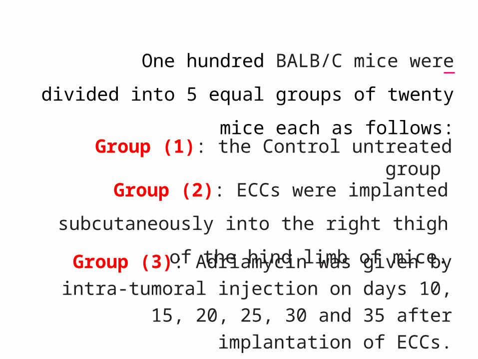
One hundred BALB/C mice were divided into
5 equal groups of twenty mice each as follows:
Group (1): the Control untreated group
Group (2): ECCs were implanted subcutaneously
into the right thigh of the hind limb of mice.
Group (3): Adriamycin was given by intra-tumoral
injection on days 10, 15, 20, 25, 30 and 35 after
implantation of ECCs.
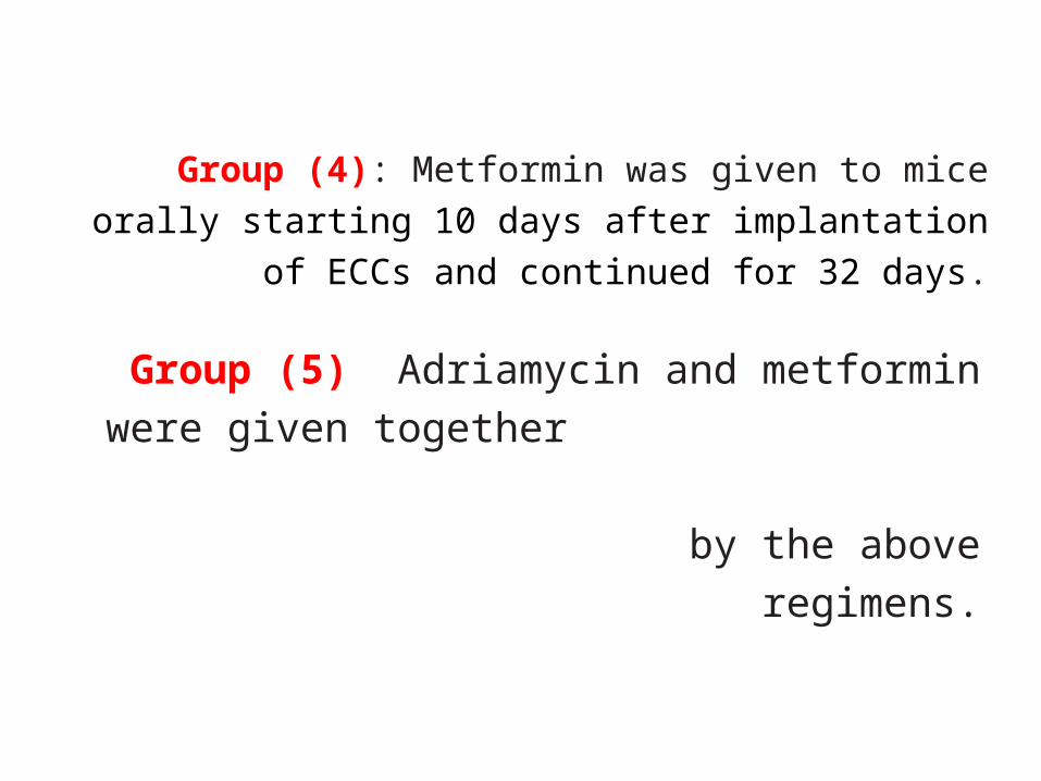
Group (4): Metformin was given to mice orally
starting 10 days after implantation of ECCs and
continued for 32 days.
Group (5): Adriamycin and metformin were given
together starting 10 days after implantation of
ECCs and continued for 32 days by the above
regimens.
• Tumor volume was measured on days 15, 20, 25, 30, 35 and 40 after implantation of ECCs.
• In the 42nd day after implantation of ECCs, the animals were killed and the tumor was excised and divided into two portions: one for homogenization
and the other for histopathological examination.
• The tumor tissue was homogenized for determination of tissue catalase, glutathione reductase, malondialdehyde, sphingosine kinase 1 activity, caspase 3 activity and tumor necrosis factor alpha.
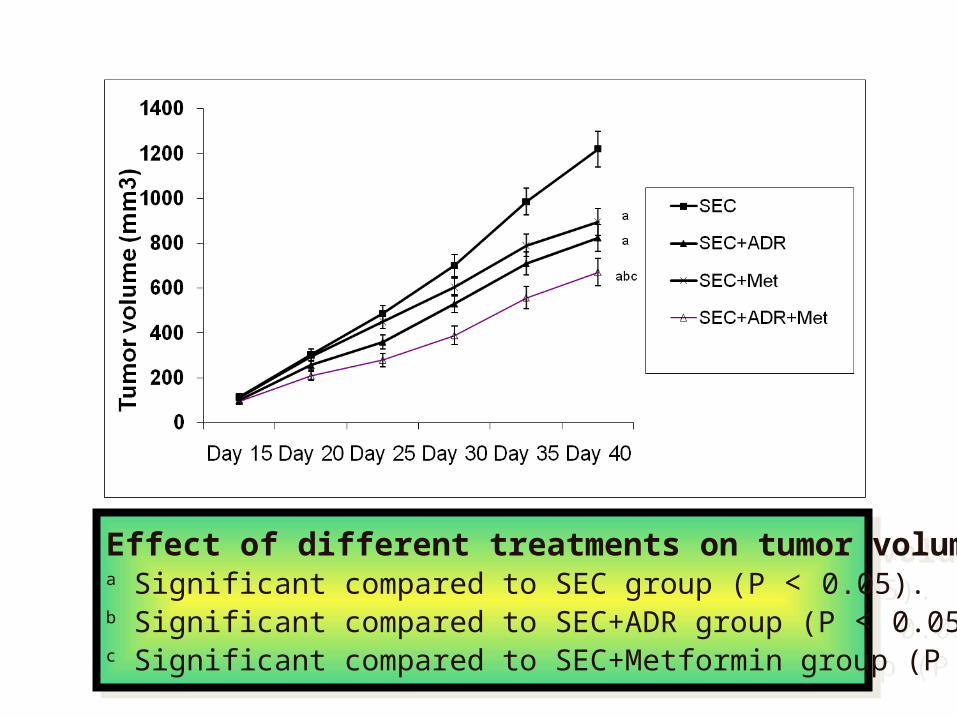
Effect of different treatments on tumor volume a Significant compared to SEC group (P < 0.05).b Significant compared to SEC+ADR group (P < 0.05).c Significant compared to SEC+Metformin group (P < 0.05).
Effect of different treatments on tumor volume a Significant compared to SEC group (P < 0.05).b Significant compared to SEC+ADR group (P < 0.05).c Significant compared to SEC+Metformin group (P < 0.05).
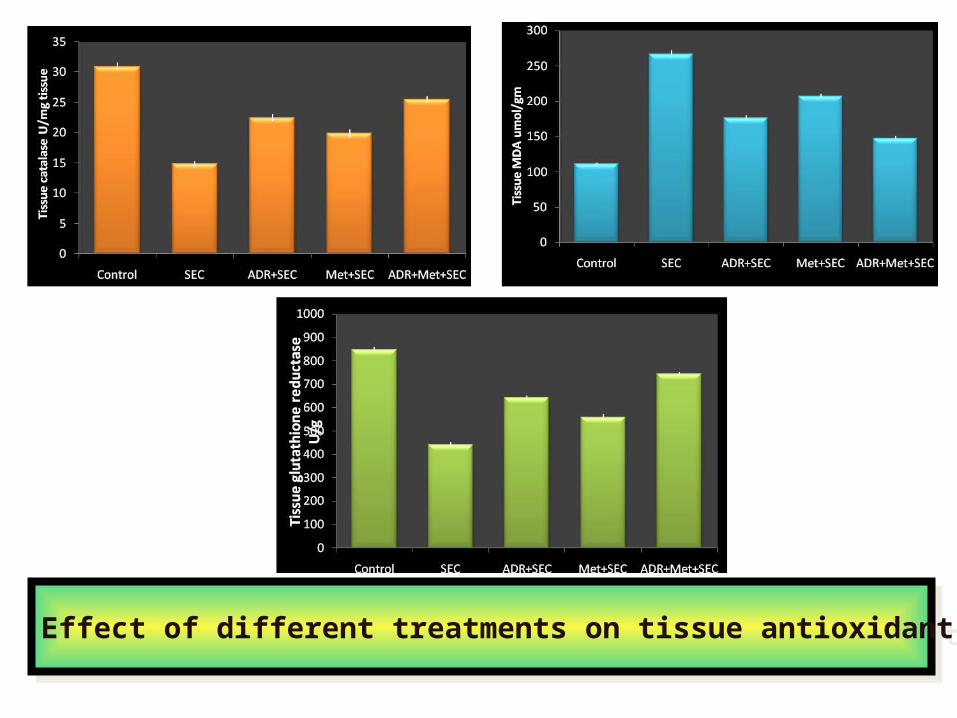
Effect of different treatments on tissue antioxidant parametersEffect of different treatments on tissue antioxidant parameters
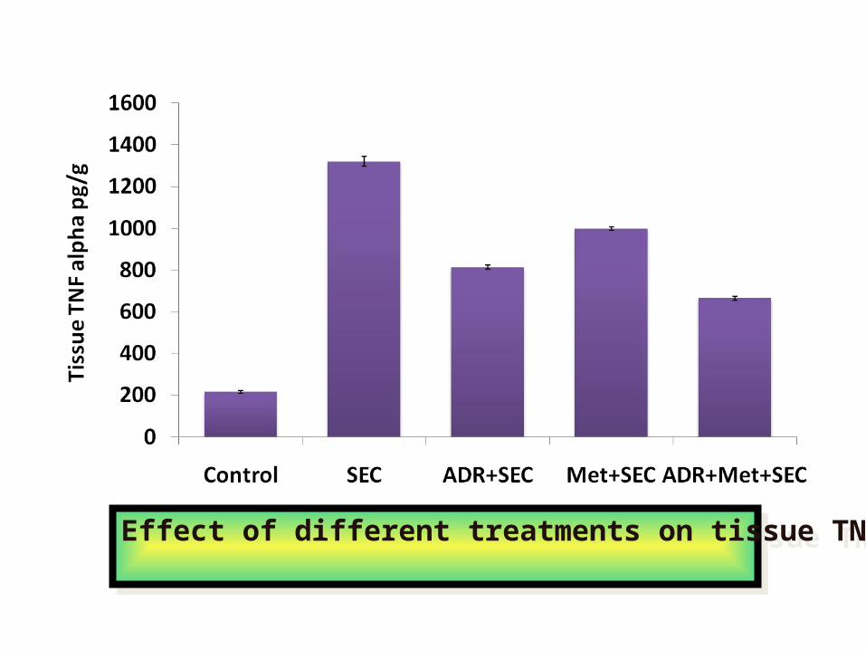
Effect of different treatments on tissue TNF-αEffect of different treatments on tissue TNF-α
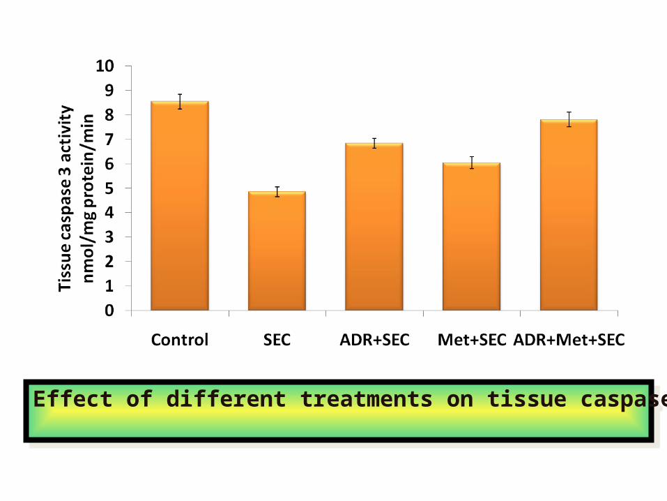
Effect of different treatments on tissue caspase 3 activityEffect of different treatments on tissue caspase 3 activity
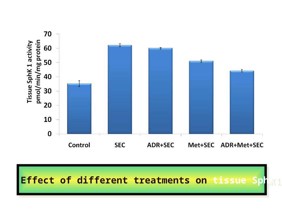
Effect of different treatments on tissue SphK1 activityEffect of different treatments on tissue SphK1 activity
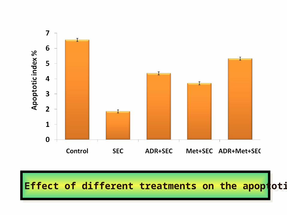
Effect of different treatments on the apoptotic indexEffect of different treatments on the apoptotic index
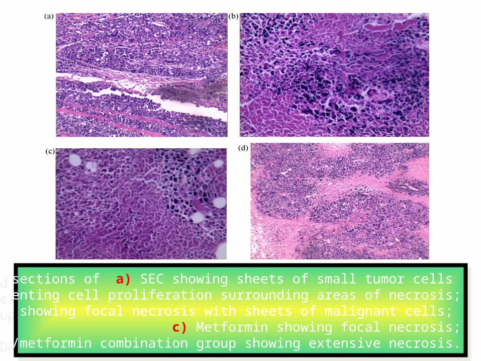
H&E stained sections of a) SEC showing sheets of small tumor cells representing cell proliferation surrounding areas of necrosis;
b) ADR group showing focal necrosis with sheets of malignant cells; c) Metformin showing focal necrosis;
d) ADR/metformin combination group showing extensive necrosis.
H&E stained sections of a) SEC showing sheets of small tumor cells representing cell proliferation surrounding areas of necrosis;
b) ADR group showing focal necrosis with sheets of malignant cells; c) Metformin showing focal necrosis;
d) ADR/metformin combination group showing extensive necrosis.
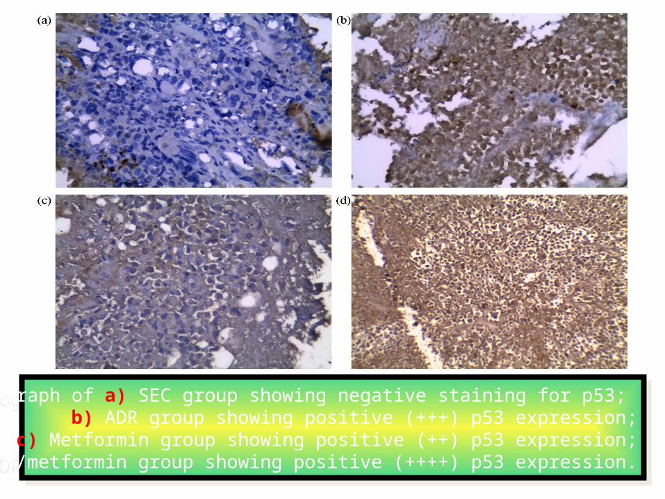
A photomicrograph of a) SEC group showing negative staining for p53; b) ADR group showing positive (+++) p53 expression;
c) Metformin group showing positive (++) p53 expression;d) ADR/metformin group showing positive (++++) p53 expression.
A photomicrograph of a) SEC group showing negative staining for p53; b) ADR group showing positive (+++) p53 expression;
c) Metformin group showing positive (++) p53 expression;d) ADR/metformin group showing positive (++++) p53 expression.
• The combination of ADR and metformin had a better effect than each of these drugs alone against transplantable tumor model in mice.
Conclusions
• This might be due to the combined antioxidant and anti-inflammatory properties of both drugs
together with their ability to induce apoptosis of cancer cells.
• Moreover, metformin was able to decrease SphK1 enzyme activity which potentiates the
effect and decreases resistance of cancer cells to ADR.

• So, it is recommended to add metformin to
the anti-cancer regimens containing ADR to
decrease resistance of cancer cells to ADR.