table of contents - · pdf file · 2017-02-20table of contents acromegaly ......
TRANSCRIPT

1
1
Table of Contents Acromegaly ........................................................................................................................................................ 3 Adrenal insufficiency (addison’s) ............................................................................................................. 5 Alcoholic cirrhosis/ascites/varicies ........................................................................................................ 6 Alpha 1 anti-trypsin deficiency .................................................................................................................. 9 Ankylosing spondylitis .............................................................................................................................. 11 Arrhythmias including long QT, VT, defibrillators and pacing ................................................. 13 Asthma .............................................................................................................................................................. 15 Atrial fibrillation/flutter ............................................................................................................................... 16 Bronchiectasis ............................................................................................................................................... 18 Cardiomyopathy ............................................................................................................................................ 21 Chronic obstructive pulmonary disease ............................................................................................ 23 Chronic renal failure .................................................................................................................................... 26 Cirrhosis ........................................................................................................................................................... 29 Complex regional pain syndrome ......................................................................................................... 32 Congenital heart disease........................................................................................................................... 34 Cushings syndrome .................................................................................................................................... 38 Cystic Fibrosis ............................................................................................................................................... 40 Diabetes ............................................................................................................................................................ 43 Fibrosing alveolitis ...................................................................................................................................... 46 Guillain-Barre ................................................................................................................................................. 48 Haemodialysis ................................................................................................................................................ 50 Haemophilia .................................................................................................................................................... 52 Heart failure ..................................................................................................................................................... 54 Hemochromatosis ........................................................................................................................................ 57 Hepatitis C ....................................................................................................................................................... 58 Hypertrophic obstructive cardiomyopathy ....................................................................................... 60 Hyperthyroidism ........................................................................................................................................... 62 Hypothyroidism ............................................................................................................................................. 64 Infective Endocarditis ................................................................................................................................. 67 Inflammatory bowel disease .................................................................................................................... 70 Interstitial lung disease .............................................................................................................................. 72 Ischaemic heart disease ............................................................................................................................ 74 Katagener’s syndrome ............................................................................................................................... 76 Liver transplant ............................................................................................................................................. 77 Lung carcinoma ............................................................................................................................................ 79 Marfan’s Syndrome ...................................................................................................................................... 81 Multiple sclerosis.......................................................................................................................................... 82 Muscular dystrophies ................................................................................................................................. 84 Myasthenia gravis ........................................................................................................................................ 86 Myotonic dystrophy ..................................................................................................................................... 88 Obstructive sleep apnoea ......................................................................................................................... 92 Peripheral neuropathy ................................................................................................................................ 95 Polycystic kidney disease ........................................................................................................................ 97 Post cardiac transplantation.................................................................................................................... 99 Post lung transplantation ........................................................................................................................ 102 Primary sclerosing cholangitis: ........................................................................................................... 104

2
2
Primary biliary cirrhosis .......................................................................................................................... 104 Pulmonary fibrosis ..................................................................................................................................... 105 Pulmonary hypertension ......................................................................................................................... 106 Raynaud’s Disease .................................................................................................................................... 109 Renal transplantation ................................................................................................................................ 110 Rheumatic heart disease ......................................................................................................................... 112 Rheumatoid arthritis ................................................................................................................................. 113 Sarcoidosis ................................................................................................................................................... 116 Scleroderma (systemic sclerosis) ....................................................................................................... 118 Systemic Lupus Erythematosus .......................................................................................................... 121 Systemic vasculitis .................................................................................................................................... 124 TB ...................................................................................................................................................................... 125 Thrombophilia .............................................................................................................................................. 127 Valvular heart disease: Mitral stenosis ............................................................................................. 129 Valvular heart disease: Mitral regurgitation .................................................................................... 131 Valvular heart disease: Aortic regurg ................................................................................................ 133 Valvular heart disease: Aortic Stenosis............................................................................................ 135 Valvular heart disease: Tricuspid regurg ......................................................................................... 137 Valvular heart disease: Pulmonary stenosis .................................................................................. 139

3
3
Acromegaly Overproduction of growth hormone from the anterior pituitary adenoma. GH stimulates the liver and other tissues to produce somatomedins which in turn promote growth. GH also acts as anti-insulin effect in muscel and increases hepatic glucose release.
Questions:
x How was it diagnosed? x Why was it suspected? (Usually a very gradual onset of symptoms and headache) x Mass lesion effects?
o Headache o Vision (field) effects
x Growth hormonal effects? o Overgrowth of tissues and bones
� Cardiac effects – arrhythmia, failure � Sleep apnoea and associated complications (pulmonary hypertension and RHF)
x Other hormonal effects o Diabetes o ACTH – blood pressure and fluid balance o Thyroid problems?
x Voice changes?
x What investigations have they had? x Current and past treatment? x Medical treatment is available, but surgical is the treatment of choice
o Drugs � Somatostatin analogues (octreotide [subcut 3xday], lanreotide) may cause
vomiting/diarrhoea. Bromocriptine (decreases growth hormone levels) – severe postural hypotension
� The drugs may reduce the size of the tumor preop, or be used post op if incomplete resection
o Surgery � Transphenoidal pituitary surgery – resects benign adenoma, and may preserve
pituitary function. 10% have hypopituitarism � Can cause diabetes insipidus, CSF rhinnorhea, stroke
o Radiation � usually second line after surgery ineffective
EXAM:
x General Inspection o Characteristic facies, thickening of the skin o ?scoliosis
x Hands o Spade-like shape o Increased sweating and warmth o Carpal tunnel – prayer sign
x Arms o Palpate underneath medial epicondyle – thickened ulnar nerve o BP o Proximal myopathy
x Axillae o Skin tags

4
4
o Acanthosis nigrans x Face
o Large supraorbital ridge o Frontal bossing o Square shaped, macrognathia, prognathia o Large, thickend lips o Teeth gapping from increased jaw size o Macroglossia
x Eyes o Visual fields – may cause bitemporal hemianopia if optic chiasm involved. o Cranial nerves III, IV, VI o Ophthalmoscopy for papilloediema from increased ICP, also look for diabetic and
hypertensive changes x Neck
o Thyroid may be enlarged x Chest
o Coarse hair o Gynaecomastia from prolactin
x Heart o Cardiac failure o Cardiomyopathy o Valvular lesions o Arrhythmia
x Abdo o Organomegally
x Back o kyphoscoliosis
x Lower limbs o Hips/knees – osteoarthritis o Entrapment neuropathy – foot drop from common peroneal entrapment
Investigations
x Diagnostic o Insulin like growth factor – unlike GH – doesn‟t fluctuate o With a glucose tolerance test the GH suppression is measured in response to a
glucose load. In Acromegaly GH fails to be suppressed in response to glucose. x ECHO
o Heart function o Valvular disease o Diastolic dysfunction from LVH
x MRI/CT – mass effects; MRI = modality of choice x Blood sugars x Other hormones
o Prolactin level often raised (25%) with galatorrhoea o Hyperthyroidism o Cushing‟s from excess ACTH and cortisol o There may be hypo function of other pituitary hormones – baseline tests should be
performed: Prolactin, cortisol, thyroxine, FSH, LH

5
5
Adrenal insufficiency (addison’s) Establish cause
y Primary (failure of adrenal cortex to produce cortisol or aldosterone) o TB o Autoimmune adrenal disease o Following heparin therapy o Haemochromatosis
y Secondary o Pituitaty or hypothalamic disease
o Tumour o Raditaion o Surgery o Drgus: etomidate
Addisonian crisis:
y Back/leg/abdominal pain, vomiting, diarrhea, dehydration, hypotension and loss of consciousness
During surgery, may require “stress” dose glucocorticoids History/Questions
y Symptoms o Faitgue o Anorexia o Weight loss o Orthostatic dizziness o Nausea/vomiting o Muscle weakness o Hyperpigmentation (from excess ACTH in primary insufficiency)
y Treatment o Corticosteroid o Fludrocorticoid
Exam
y Hyperpigmentation: palmar creases, elbow, gums and scars y Postural drop
Investigations
y Electrolyte – hyponatraemia, hyperkalaemia y Serum cortisol (low); ACTH high – Secondary y ACTH low – secondary y Renal function test

6
6
Alcoholic cirrhosis/ascites/varicies Questions:
x How did it first present? How was it diagnosed? o Jaundice o Acsites o Abdominal pain o Acute bleeding o Encephalopathy o Incidental
x Length of history of liver disease x Causes:
o Alcohol intake (quantity and duration) o Drug intake: legal/illegal, IV drugs o Hepatitis (duration and treatment) o Transfusions o Tattoos o Hemochromatosis
� Diabetes, cardiac failure, arthropathy, family history o Wilsons (Young patients)
x Progression of the disease o Hospitalisation o Bleeds – varices, malena, haematemesis, etc o Encephalopathy – memory loss o Coexisting cardiac disease and exercise tolerance o Pulmonary hypertension as part of portopulmonary hyepertension o Pulmonary involvement as part of hepatopulmonary syndrome o Renal impairment as part of hepatorenal syndrome o General
� Weight loss x Treatment
o Operations � TIPS
o Medications � Octreotide, pindolol for portal hypertension � Terlipressin for variceal bleed � Hep B: interferon, lamivudine � Hep C: oral ribavirin and weekly interferon
Exam: Liver failure: General
x Cachexia and muscle wasting Hands
x CLUBBING x Red palms x Hepatic flap, asterixis x Bruising x Jaundice

7
7
x Dupuytrens contracture x Anaemia
Chest
x Spider nevi Heart
x Cardiomyopathy x Hypertension
Abdomen (GI)
x Hepatomegaly x Splenomegaly x Ascites (shifting dullness and fluid thrill)
o Percussion starting in the midline with the finger pointing towards the feet; the percussion note is tested out towards the flanks on each side
o If dullness detected, percuss out to the left flank until dullness is reached. This point is marked and the patient rolled towards the examiner (R hand side). Wait 30 seconds to 1 minute. Then repeat the percussion and mark the dullness. Shifting represent presence of ascites.
o Fluid thrill: patient put hand in midline, and examiner flick one side and the other feeling for pulsation.
x Caput medussa Exclude severe right heart failure, tricuspid regurg or constrictive pericarditis clinically in all patients Investigations:
x Full blood count o megaloblastic anaemia o thrombocytopenia o iron studies (increased transferrin and ferritin – hemochromatosis)
x U/E o Renal impairment (hepatorenal syndrome) o Hypoglycaemia o Urea may be low due to decreased production
x LFTS o Pattern of obstructive/intrahepatic
x Albumin/INR/bilirubin o For Childs Pugh score
x Viral hepatitis screen x ABG
o Hepatopulmonary shunts – particularly if the patient complains of dyspnoea. x ECG: increased cardiac risk
o Consider echo if cardiac failure
x Ultrasound/CT scan: may exclude biliary obstruction/infiltration. Causes of Cirrhosis in Adults:
x Alcohol x Post viral x NASH (Non-alcoholic steatohepatitis) x Drugs
8
8
x Autoimmune x Hemochromatosis x Wilsons x Primary sclerosing cholangitis x Primary biliary cirrhosis x Cystic fibrosis x Budd-Chiari
Causes of portal hypertension
x Cirrhosis of the liver x Presinusoidal
o Portal vein compression (lymphoma, carcinoma) o Intravascular clotting o Umbilical vein phlebitis
x Intrahepatic o Sarcoid o Lymphoma o Congenital hepatic fibrosis
x Post-sinusoidal o Hepatic vein outflow obstruction (Budd-chiari syndrome) o Cancer o Veno-occlusive disease o Constrictive pericarditis o Chronic cardiac failure
Other alcohol problems CVS – Arrhythmias (incidence similar to CHF patients); cardiomyopathy CNS – wernicke‟s encephalopathy, peripheral neuropathy GI – cirrhosis, pancreatitis, malnutrition Endo/metabolism – hypoglycaemia, increased triglyceride Haematological – thrombocytopaenia, anaemia, leucopaenia

9
9
Alpha 1 anti-trypsin deficiency Inherited disorder – involving lung, liver and rarely skin Imbalance of neutrophils elastase which destroys elastin and A1AT which is protective. History/Questions: Pulmonary:
x COPD – especially emphysema x Risk factor for early onset emphysema (ie in 40s) x Other risk factors include
o Cigarette smoking o Dusty occupational exposure o Parental exposure COPD o Personal hx of asthma, bronchitis, pneumonia
x Symptoms: Dyspnoea, phlegm, wheezing x Exercise capacity x Drugs and inhalers x Previous lung function test x Previous Anaesthetics
Extrapulmonary:
x Liver disorders (especially in first 2 decades) – ie neonatal hepatitis, cirrhosis in children and adults, Hepatocellular carcinoma. (Intrahepatic accumulation of alpha 1 antitrypsin)
Drugs:
x Intravenous pooled human AAT o Can cause fever, thrombocytopenia
x Supportive therapy: Not smoking, bronchodilators/glucocorticoids/nutritional support/vaccination
x Lung volume reduction surgery Exam: General appearance
x Look around for oxygen masks, inhalers and other medications x Presence of sputum mug x Dyspnoea at rest x Respiratory rate (Normal: 16-25) x Saturation x Use of accessory muscles (SCM, platysma, scalene muscles) x Contraction of abdominal muscels may occur in expiration with obstruction of airways. x Indrawing of the muscles x Pursed-lip breathing x Cyanosis x Cough, sputum, hoarseness (lung cancer impinging on recurrent laryngeal nerve)
Hand
x Staining of fingers x Tachycardia
o Side-effect of b-agonist

10
10
x Flapping tremor (asterixis) o Due to CO2 retention o Can also occur with liver and renal failure
Face
x Mouth: Central cyanosis Chest
x Inspection o Barrel chest
x Chest expansion o Diminished chest wall movement o Thumbs should separate >5cm in normal. Reduced in COPD
x Vocal (tactile fremitus) o Reduced in COPD
x Percussion o Hyper-resonant
x Auscultation o Early inspiratory crackels due to opening of airways o Vocal resonance - reduced
Abdomen:
x Liver ptosis Investigations
x ABG x CXR: bullous changes are more prominent at the bases than the apices x Lung function tests: obstructive pattern x Liver function tests/ultrasound (cirrhosis)
11
11
Ankylosing spondylitis Ankylosing spondylitis is a chronic, usually progressive, inflammatory disease involving the articulations of the spine and adjacent soft tissues. Spinal disease begins in the sacroiliac joints and moves cranially. The degree of spinal disease can range from just sacroiliac involvement to complete ankylosis of the spine. Hip involvement occurs in approximately one third of patients. Back pain characterized by morning stiffness that improves with activity and exercise plus radiographic evidence of sacroiliitis is highly suggestive of this diagnosis. Systemic involvement can manifest as weight loss, fatigue, and low-grade fever. Conjunctivitis and uveitis occur in approximately 40% of patients. The uveitis is usually unilateral and presents as visual impairment, photophobia, and eye pain. Distinctive pulmonary abnormalities associated with ankylosing spondylitis include apical cavitary lesions and pleural thickening that mimic tuberculosis. Cardiovascular involvement, such as aortic regurgitation or bundle branch block, is observed in 40% of patients. Arthritic involvement of the thoracic spine and costovertebral articulations can result in a decrease in chest wall compliance and, consequently, a decrease in vital capacity. History/Questions: Articular:
x When was the onset? (peak 20-30) x Sacroileitis – low back/buttock pain. Inflammatory spinal pain improves with exercise, but not
with rest. x Hip arthritis is common x Bamboo spine – kyphosis and fixation x Atlantoaxial subluxation is common (20% anterior, 2% posterior)
Non articular:
x Fatigue x Uveitis x Increased ischaemic heart disease/CHF/PVD/hyperlipidaemia/Diabetes x Aortic regurgitation x Cervical fracture x Cauda equina
Exam: Non articular: Hand
x Anaemia Face
x Uveitis x Anaemia x Mouth opening x Limited neck flexion and extension x Tracheostomy scar
Chest x Reduced chest movement due to costochondral osteoarthritis
Heart x Aortic regurgitation x Heart failure
Articular:

12
12
x Sacroileitis: springing the ASIS with patient lying supine. Loss of lumbar lordosis. x Bamboo spine – kyphosis and fixation. Inability to touch toes. Test lateral flexion also (running
hand down lateral thigh) x Mouth opening x Neck extension/flexion x Achilles tendinitis
Drugs:
x NSAIDS/DMARDS x TNF-D (Infliximab) x Sulphasalazine (Leukpenia/neutropenia) x Steroids x Palmidronate
Investigations
x FBC o Anaemia of chronic disease from inflammation
x U/E o Renal impairment from amyloid (5%) and drugs
x HLA B27 o For diagnosis
x Flexion/extension of C-spine

13
13
Arrhythmias including long QT, VT, defibrillators and pacing History/Questions:
x Congenital/acquired? o Family history of arrhythmias or sudden cardiac death
x Type of arrhythmia o Bradyarrhythmia o Tachyarrhythmia o Life-threatening vs non-life threatening
x Triggers of arrhythmias (frights, cold water, exercise) x Symptoms
o Palpitations o Syncope o Seizures o Cardiac arrests o Symptoms of failure: dyspnoea, swelling, PND, orthopnoea
x Some have associated sensorineural deficits, hearing loss x What investigations have they had?
o Have they had EP studies? x Cause of arrhythmia
o Ischaemia o Cardiomyopathy o Primary arrhythmia o Acute abnormalities
x Past medical problems x Drugs
Exam: General appearance Hand Pulses (radial, carotid, radio-radial delay, radio-femoral delay): rate, rhythm, characteristics, volume BP Face Neck: JVP Heart:
x Inspection o Scar from Prev ICD insertion, sternotomy, etc o Any prosthetic valve clicking noise
x Palpation o Apex beat o Heave o Thrills
x Auscultation: HS, murmur, manouvres Chest
x Presence of pulmonary oedema Abdomen
x Enlarged liver from RHF x Pulsatile liver from TR x AScites x Splenomegaly
Lower limbs

14
14
x Pitting oedema Drugs:
x Beta blockers x Cardiac sympathetic denervation (Left) x Pacemakers x Mexilitine x Flecanide
Investigations
x ECG: long QT x Echo if indicated x Pacemaker report: type, rate, ICD function, dependence, firing frequency, magnet response x Letters from cardiologists

15
15
Asthma History/Questions:
• Duration • Admissions/ICU admissions • Control
o How often gets exacerbation? o How often needs inhalers? o PFR (morning dip – suboptimal control) o What is their normal? o How often do they test it?
• Use of inhalers: o Beclamethasone (steroid) o Flixotide (steroid) o Salbutamol/salmeterol o Ipratropium (anticholinergic)
• Other drugs o Use of nebulisers o Oral steroids o Phosphodiesterase inhibitors (theophylline or aminophylline)
• Functional capability – Does asthma limit activities? • Nutrition – losing weight? General health? • Smoking • Aspirin sensitivity
Exam:
• Rule out other problems o Concurrent infection o OSA o Obesity o GORD
• Hyperinflation • Prolonged expiratory phase • Wheeze
Drugs: See above. Consider leucotriene antagonists (monteleukast) Investigations
• PEFR • Spirometry • ABG • CXR: hyperinflation
16
16
Atrial fibrillation/flutter History/Questions:
• Duration: How long have you had it for? • How was it diagnosed? What was the initial symptoms? • What was and is the predisposing cause?
o Valvular lesions (especially mitral valve) o Rheumatic heart disease o MI o Thyrotoxicosis o Electrolyte o Chest pathology
• Is it paroxysmal? Or continuous and rate controlled? • Complications
o Stroke or emboli • Other medical problems
o IHD o Cardiomyopathies o Heart failures o Hyperthyroidism o Hepatic and Renal pathologies
• Drugs and treatment o Rate control o Rhythm control? (medical and electrical) o Previous EP studies/MAZE
• Exercise tolerance • EP studies/Maize procedures • Smoking
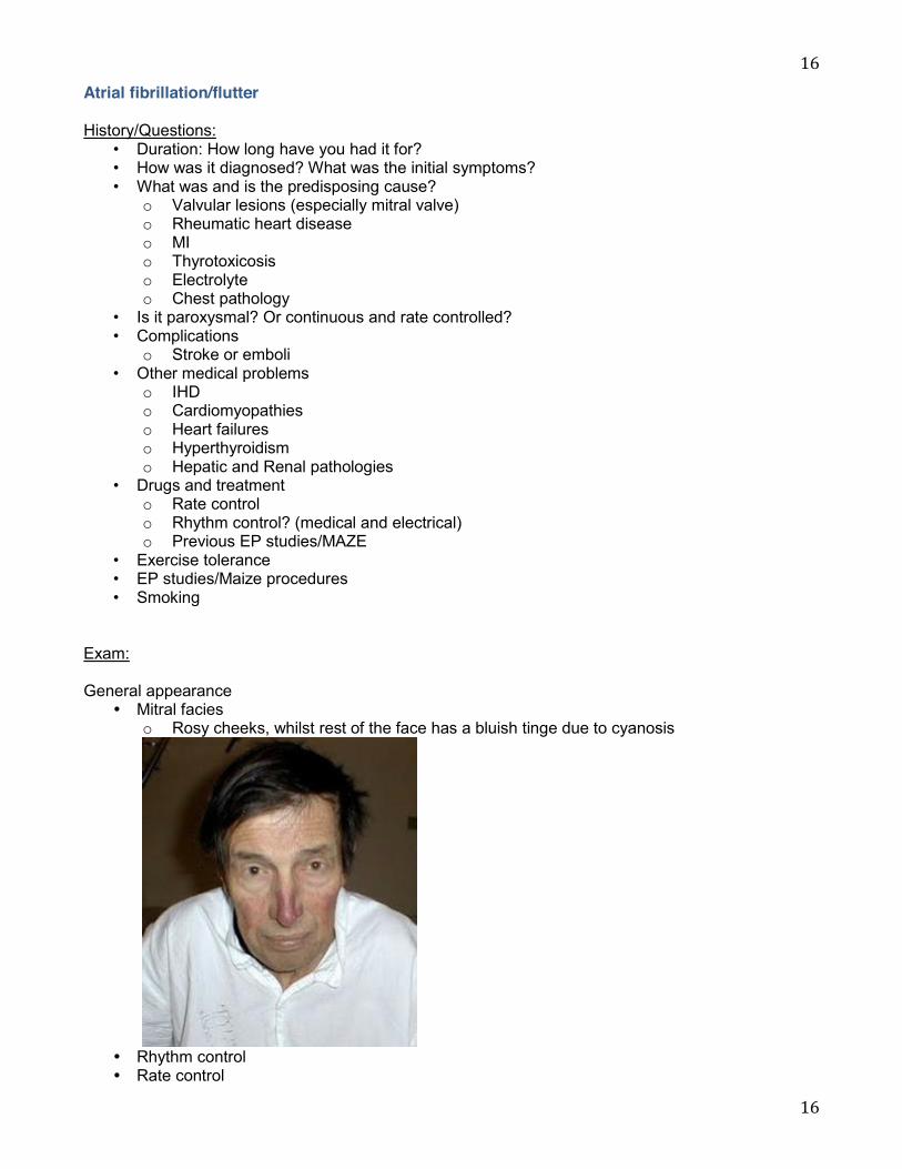
Exam: General appearance
y Mitral facies o Rosy cheeks, whilst rest of the face has a bluish tinge due to cyanosis
y Rhythm control y Rate control

17
17
y Thyrotoxicity o Tremor o Weigh loss o Sweaty, warm hands o Tachycardia o Arrhythmia o Enlarged thyroid
y Amiodarone related issues o Skin changes – hyperpigmentation to blue-gray due to photosensitivity o Corneal deposits o Pulmonary fibrosis o Thyroid dysfunction
y Blood pressure y Murmurs
Drugs:
y Amiodarone o side effects of long term treatment: Pulmonary fibrosis, ocular, skin changes, thyroid
dysfunction, sinus Bradycardia, elevated LFTs y Beta blockers y Digoxin
o vomiting, confusion, delirium, hallucintions, blurred vision, disturbed color perception (yellow color), photophobia, arrhythmias, AV block, decreased QT interval, reverse tick on ECG.
y Warfarin
Investigations y FBC (anaemia as a cause of AF) y Electrolytes (esp K and Mag) y ECG y Pacemaker investigations y CXR (failure) y ECHO (diastolic dysfunction, valvular abnormalities)

18
18
Bronchiectasis
History/Questions:
x How was it diagnosed? x What was the cause?
o Congenital � Cystic fibrosis (see below) � Primary ciliary dyskinesia � Congenital hypogammaglobulinaemia � Katagener‟s syndrome
o Acquired � Recurrent chest infections as a child � Whooping cough � Measles � Necrotising bacterial infections, TB, HIV
x Duration? x Symptoms
o Productive cough o Recurrent haemoptysis o Chronic sinusitis o Recurrent pneumonia and pleurisy o Dyspnoea o Symptoms of RHF o Often a component of asthma
x Clinical progress o Recurrent infection o Exercise capacity o Nutrition o Chest physiotherapy, frequency o Treatments: inhalers, drugs and vaccines
� Mainstay: physiotherapy, antibiotics, postural drainage, bronchodilators and lung resections as necessary
Exam: General inspection
x Look around for oxygen masks, inhalers and other medications x Presence of sputum mug x Dyspnoea at rest x Respiratory rate (Normal: 16-25) x Saturation x Use of accessory muscles (SCM, platysma, scalene muscles) x Contraction of abdominal muscels may occur in expiration with obstruction of airways. x Indrawing of the muscles x Pursed-lip breathing x Cyanosis
Hand
x Staining of fingers x Clubbing x Tachycardia
19
19
o Side-effect of b-agonist x Flapping tremor (asterixis)
o Due to CO2 retention Face
x Mouth: Central cyanosis Chest
x Inspection o Barrel chest
x Chest expansion o Diminished chest wall movement o Thumbs should separate >5cm in normal. Reduced in COPD
x Vocal (tactile fremitus) o Reduced in COPD and bronchiectasis
x Percussion o Hyper-resonant
x Auscultation o Early inspiratory crackels due to opening of airways o Vocal resonance – reduced o Crackles
Heart
x Apex beat o Dextrocardia – Kartagener‟s syndrome
Consider complications and look for them
x Pneumonia x Pleurisy x Empyema x Lung abscess x Cor-pulmonale x Cerebral abscess
Drugs:
x Bronchodilators x Steroids x Postural drainage x Chest physio x Antibiotics: not prophylactic x Vaccines x Immunoglobulin (if deficient) x Smoking cessation x +/- transplant
Investigations
x Arterial blood gas o Respiratory failure
� Type1: PaO2 <60mmHg, or PaCO2 <50mmHg � Type2: PaO2 <60mmHg, or PaCO2 >50mmHg

20
20
x Immunoglobulin levels x CXR: cystic lesions, thickened bronchial walls (tram tracking) x Sputum microscopy: H influ, pseudomonas, e coli x Spitometry x HRCT

21
21
Cardiomyopathy Hypertrophic cardiomyopathy
x Abnormal hypertrophy of the muscle in LV or RV outflow tract or both. It can obstruct outflow from the LV late in systole when the hypertrophied area contracts. Systolic displacement of the mitral valve apparatus into the LVOT also occurs causing MR and SAM. Although there is LVOT obstruction from hypertrophied septum, primary contribution is SAM
x Aetiology unknown Dilated cardiomyopathy
x Abnormal global reduction in cardiac function. x CAD is excluded as a cause by definition. Ischaemic cardiomyopathy is a term often used to
describe severe myocardial dysfunction secondary to recurrent ischaemic events. x Ventricular arrhythmias are common. It is a common indication for cardiac transplantation. x Cause: idiopathic, alcohol, viral, post-partum, IVDU, dystrophia myotonica, haemochromatosis
Restrictive cardiomyopathy
x Causes diastolic dysfunction x Causes: idiopathic, eosinophilic endomyocardial disease, endomyocardial fibrosis, infiltrative
disease (amyloid), granulomas (sarcoma) History/Questions:
x How was it diagnosed? o Age o Cause
� Congenital � Pregnancy � Viral � Alcohol � Wilsons � Haemochromatosis � Idiopathic
x Duration? x Symptoms
o Angina (if hypertrophic cardiomyopathy) o Dyspnoea o Orthopnoea from LHF o Peripheral swelling from RHF o Liver pathology o Syncope o Exercise tolerance
x Other associated problems o Palpitation from Arrhythmia o Stroke from emboli
Hypertrophic cardiomyopathy
x Exam o Sharp, rising and jerky pulse – rapid ejection by hypertrophied ventricles early in
systole followed by obstruction caused by the displacement of the mitral valve into the outflow tract.
o Raised a-wave in a-wave due to forceful atrial contraction against stiff Right ventricle
22
22
o Apex beat: double or triple due to presystolic expansion of ventricle o Auscultation: late systolic murmur at the lower left sternal edge and apex (due to
obstruction) and a pansystolic murmur at the apex due to MR o Manouvre: murmur increased by valsalva manouvre (decreased filling and increased
contraction, leading to increased obstruction). Decreased by squatting (increased filling and increased SVR relieving obstruction)
x Investigation o ECG: LVH o Echo: Assess systolic and diastolic function, LVH; SAM; degree of MR
x Management o Inotropes are contraindicated o Normal to high volume o Beta-blockers or verapamil to maintain low contractility and HR o Maintain high SVR o May need dual chamber pacing if severely impaired
Restrictive: Rare. Ie myocardial infiltration by amyloid. Stiff ventricles that impair filling. Right heart failure is often prominent. ECHO: diastolic dysfunction. Want to increase myocardial contraction – ketamine is good. Dilated cardiomyopathy: Cardiac failure with an enlarged heart, poorly contractile. Functional MR and TR – exacerbates heart failure. Common problems – heart failure, arrhythmias, emboli. Failure Rx with diuretics, ACEi, vasodilators. Amiodarone used commonly. Dual chamber pacing may be used.
23
23
Chronic obstructive pulmonary disease Stages of COPD
x Stage I: Mild COPD o Mild airflow limitation (FEV1 >80% predicted; FEV1/FVC <70%) o Occasional chronic cough and sputum production
x Stage II: moderate COPD o Worsening airflow limitation (50% < FEV1 < 80%; FEV1/FVC <70%) o SOB developing on exertion o This is the stage at which patients typically seek medical attention because of chronic
respiratory symptoms or an exacerbation of their disease x Stage III: severe COPD
o Further worsening of airflow limitation (30% < FEV1 <50%) o SOB o Reduced exercise capacity and repeated exacerbations which have an impact on
patients‟ quality of life x Stage IV: very severe COPD
o Severe airflow limitation (FEV1 <30% predicted; FEV1/FVC <70%) o Exacerbation may be life threatening
History/Questions:
x How was it diagnosed? x What symptoms do you get?
o Wheeze o Cough o Sputum production o Infective exacerbations? How often? o Nutrition? Weight loss? o Haemoptysis
x Exercise tolerance and functional ability? x DO you measure PEFR? x Component of chronic bronchitis/emphysema
o Productive cough >2 weeks 3 consecutive years x History of smoking
o Pack year o Age when started? o When quit?
x Other occupational dust exposure? x Current treatment
o Inhalers: b-agonist, anticholinergics, steroids o Antibiotics o Steroids o Home oxygen o CPAP?
x Other co-mobidity o Pulmonary hypertension and RHF
x Clinical letter o Progress o Spirometry test
24
24
o Any reversible element (>15% improvement and at least 200ml increase in FEV1 or FVC)
Drugs:
x Regular bronchodilators/anticholinergics/steroids x Antibiotics for infections x Influenza vaccine x Pulmonary rehabilitation programs x Alpha 1 antitrypsin – weekly/monthly IV injection. Expensive. Only if demonstrated low levels
and lung disease x CPAP/BiPAP (acute exacerbations) x Treatment of cor pulmonale – ie spirinolactone/diuretics. Although treatment of the lung
disease will improve the heart disease. Exam General appearance
x Look around for oxygen masks, inhalers and other medications x Presence of sputum mug x Dyspnoea at rest x Respiratory rate (Normal: 16-25) x Saturation x Use of accessory muscles (SCM, platysma, scalene muscles) x Contraction of abdominal muscles may occur in expiration with obstruction of airways. x Indrawing of the muscles x Pursed-lip breathing x Cyanosis x Cough, sputum, hoarseness (lung cancer impinging on recurrent laryngeal nerve)
Hand
x NO CLUBBING unlike bronchiectasis x Staining of fingers x Tachycardia
o Side-effect of b-agonist x Flapping tremor (asterixis)
o Due to CO2 retention o Can also occur with liver and renal failure
Face
x Mouth: Central cyanosis x Smokers faces
Chest
x Inspection o Barrel chest
x Chest expansion o Diminished chest wall movement o Thumbs should separate >5cm in normal. Reduced in COPD
x Vocal (tactile fremitus) o Reduced in COPD
x Percussion o Hyper-resonant
25
25
x Auscultation o Early inspiratory crackels due to opening of airways o Vocal resonance - reduced
Abdomen:
x Liver ptosis Signs of RHF
Investigations
x FBC: polycythemia x ABG: respiratory failure at rest. PaCo2 >50mmHg.
o Respiratory failure � Type1: PaO2 <60mmHg, or PaCO2 <50mmHg � Type2: PaO2 <60mmHg, or PaCO2 >50mmHg
x Check spirometry to clarify diagnosis and assess severity x ECG
o P-pulonale: right heart disease o Cor pulmonale o Consider Echo
x CXR o Sign of infection o Hyperinflation o Bullae
x Consider sputum culture x Alpha 1 antitrypsin if suspected
Differential diagnosis x Asthma: Non smoker, onset in childhood, family history of allergy, episodic attacks and
nocturnal symptoms, rapid response to treatment (especially steroids), reversibility of obstruction
x Bronchiectasis: Daily sputum production +/- hemoptysis, onset in childhood, recurrent chest infection, clubbing

26
26
Chronic renal failure
y Normal GFR >90ml/minute y Failure = <15ml/min y Cockcroft-Gault GFR = (140-age) * (Wt in kg) * (0.85 if female) / (72 * Cr)
Causes
y Nephrotic syndrome: o Proteinuria: >3.5g/24 hours, hypoalbuminaeima, odema, hyperlipidaemia o Primary (80%): focal gomerulosclerosis, membranoproliferative, minimal change o Secondary: SLE, diabetes, hep B, HIV, gold, penicillamine
y Glomerulonephritis: o Ask if there is a history of proteinuria, heamturia, oliguria, oedema, sore throat, sepsis,
rash, hemoptysis, renal biopsy o PRIMARY
� Diffuse: minimal change, membranous � Focal: IgA, focal glomerulonephritis
o SECONDARY: SLE, wegners, goodpastures, henoch-schonlein purpura, diabetes y Analgesic nephropathy y Polycystic kidneys
o Family history, how was it diagnosed, heamturia, polyuria, loin pain, hypertension, renal calculi, headache, subarachnoid, visual disturbance.
y Alports: deafness and persistent hematuria (Nephritis) y Reflux Nephropathy y Diabetic nephropathy: make sure they are on an ACE/A2RB y Hypertensive nephropathy y Connective tissue disease: SLE and scleroderma
History/Questions:
x How was it diagnosed? x Duration of the disease? x What is the cause?
o Diabetes o Hypertensive o Glomerulonephritis o Amyloid o Drug induced o Lupus o PCKD ?biopsy
x Current treatment o Dialysis
� Type � Frequency � Problems associated with it
o Fistula formation o Still passing urine or completely dialysis dependent o Is patient awaiting transplantation
x Associated problems o Increased risk of IHD o HTN o Anaemia o Bone pains from osteitis fibrosa cystica due to secondary hyperparathyroidism

27
27
x If diabetic o Ask about eyes and sensory changes o Autonomic neuropathy: postural dizziness, reflux, sweating abnormality (gustatory
sweating), impotence o Heart, PVD and stroke
Drugs
x Dialysis prescription x Vitamin D and calcium x EPO x Diabetic medications x Cardiac medications
Exam: General inspection
x Sallow complexion (a dirty brown appearance or „uraemic tinge‟) due to decreased excretion of urinary pigments (urochromes) combined with anaemia
x Hyperventilation: which may indicate metabolic acidosis x Myoclonic jerks due to neuromuscular irritability or a low serum calcium level in late renal
failure Hand and arm
x Nail o Leuconychia from hypoalbuminaemia (nephritic syndrome)
x Anaemia: pale palmar crease x Asterixis: in terminal renal failure x Fistula formation x Scratch marks and excoriations due to uraemic pruritus x BP
Face
x Eye: look for anaemia x Mouth: uraemic fetor, ammoniacal, musty odour
Neck
x JVP for fluid assessment x Look for jugular vein puncture due to previous vascular access x Parathyroidectomy performed for management of tertiary hyperparathyroidism
Chest
x Basal crepitus if pulmonary oedema present Heart
x Routine exam Abdo
x Tenckhoff catheter for peritoneal dialysis x Nephrectomy scars? Transplant? x Ascites from nephritic syndrome or dialysis fluid x Balloting of the kidneys (mass or enlarged kidney)
28
28
x Auscultation o For bruits above umbilicus 2cm to left or right of the midline
Back
x Scars x Percuss with fist against vertebrae (osteodystrophy) and flank for pains
Legs
x Oedema Investigations FBC: anaemia UE: Sodium (normal), potassium (high), creatinie, MGanesium (low), Calcium (low), Phosphate (high) ECG: q waves, hypertension, arrhythmias Coags: Should be normal, but platelet function may be reduced - (consider DDAVP) Platelet function

29
29
Cirrhosis Liver function impairment Coagulopathy Encephalopathy Portal hypertension Hepatopulmonary syndrome with shunts Portopulmonary hypertension with pulmonary hypertension Hepatorenal syndrome with renal impairment Questions:
x How did it first present? How was it diagnosed? o Jaundice o Acsites o Abdominal pain o Acute bleeding o Encephalopathy o Incidental
x Length of history of liver disease x Causes:
o Alcohol intake (quantity and duration) o Drug intake: legal/illegal, IV drugs o Hepatitis (duration and treatment) o Transfusions o Tattoos o Hemochromatosis
� Diabetes, cardiac failure, arthropathy, family history o Wilsons (Young patients)
x Progression of the disease o Hospitalisation o Bleeds – varices, malena, haematemesis, etc o Encephalopathy – memory loss o Coexisting cardiac disease and exercise tolerance o Pulmonary hypertension as part of portopulmonary hyepertension o Pulmonary involvement as part of hepatopulmonary syndrome o Renal impairment as part of hepatorenal syndrome o General
� Weight loss x Treatment
o Operations � TIPS
o Medications � Octreotide, pindolol for portal hypertension � Terlipressin for variceal bleed � Hep B: interferon, lamivudine � Hep C: oral ribavirin and weekly interferon
Exam: Liver failure: General
x Cachexia and muscle wasting

30
30
Hands
x CLUBBING x Red palms x Hepatic flap, asterixis x Bruising x Jaundice x Dupuytrens contracture x Anaemia
Chest
x Spider nevi Heart
x Cardiomyopathy x Hypertension
Abdomen (GI)
x Hepatomegaly x Splenomegaly x Ascites (shifting dullness and fluid thrill)
o Percussion starting in the midline with the finger pointing towards the feet; the percussion note is tested out towards the flanks on each side
o If dullness detected, percuss out to the left flank until dullness is reached. This point is marked and the patient rolled towards the examiner (R hand side). Wait 30 seconds to 1 minute. Then repeat the percussion and mark the dullness. Shifting represent presence of ascites.
o Fluid thrill: patient put hand in midline, and examiner flick one side and the other feeling for pulsation.
x Caput medussa Exclude severe right heart failure, tricuspid regurg or constrictive pericarditis clinically in all patients Who to transplant liver?
x Child pugh score >6 (Bilirubin, albumin, INR, encephalopathy, ascites) x MELD (model for end-stage liver disease) score
o Bilirubin, creatinine, INR to predict survival. o 3.78(ln bilirubin mg/dl) + 11.2 (ln INR) + 9.57 (ln Creatinine mg/dl) + 6.43 o Score >10
x King‟s college criteria: separate criteria for acetaminophen and non-acetaminophen. Looks at age, coagulation, renal function, and mental status.
Child pug score and perioperative mortality

31
31

32
32
Complex regional pain syndrome
x CRPS 1: Tissue injury (no known nerve injury). Spontaneous pain or allodynia/hyperalgesia occurs beyond single peripheral nerve and is disproportionate to the event. There is or has been evidence of skin blood flow/oedema/abnormal sudomotor activity
x CRPS 2: Following known nerve injury (otherwise similar to CRPS I). Can be divided into sympathetically mediated or independent pain
x Diagnosis: Spontaneous pain or allodynia (pain resulting from a stimulus which would not normally provoke pain, such as a light touch of the skin) is not limited to the territory of a single peripheral nerve, and is disproportionate to the inciting
o There is a history of edema, skin blood flow abnormality, or abnormal sweating in the region of the pain since the inciting event.
o No other conditions can account for the degree of pain and dysfunction History/Questions:
x What was the injury x How long after the injury did they notice sx x What sx do they experience x What makes it better/worse/radiating/relieving/medications/intensity/site x Burning/shooting/aching x Element of allodynia and hyperalgesia x Temperature and proprioception x Treatment
o Medication and associated side effects o Counselling and psychology involvement o Physio involvement
x Social o Sleeping o Eating o Bowel motions o Occupation o Functional capabilities, etc.
Drugs:
x What are they on at the moment x What have they been on x Pain programs
Exam:
x Clinical TRIAD: Pain, vasomotor, trophic changes x Vasomotor: Changes in color/temperature x Sudomotor: Hyper or hypohydrosis and oedema. x Trophic: Hair/skin/nail changes, osteoporosis x Brief neuro
o Sensory changes: allodynia o Motor functions
x FUNCTIONAL IMPAIRMENT Investigations
x Thermography – vasomotor instability x Quantitative sweat test x Radiography – bone density scan; bone scan for osteoporosis

33
33
Management x Multimodal approach x Drugs x Local anaesthetic block x Spinal cord stimulation x Sympathectomy x Psychology and counseling x Physiotherapy x Mirror therapy, motor imagery training x Amputation

34
34
Congenital heart disease
History/Questions: x What is the lesion?
o Is it acyanotic lesion or cyanotic lesion? o R to L shunt lesion
� Tetralogy of fallot � Pulmonary atresia � Tricuspid atresia � Ebstein‟s anomaly
o L to R shunt lesion � Increased pulmonary blood flow +/-failure � ASD � VSD � PDA � AP window
o Complex shunts � Mixing of pulmonary and systemic blood with cyanosis � Transposition � Truncus arteriosus � TAPVD � HLHS
o Obstructive lesions � AS � MS � PS coarctation
x What procedures have they had? o Biventricular repair
� VSD, ASD, ToF, Transposition, truncus arteriosus o Fontan palliation
� Tricuspid atresia � Hypoplastic left heart � Pulmonary atresia � Double inlet left ventricle � Double outlet right ventricle
x Are future procedures planned? x How often are they followed up? x Symptoms
o Heart failure � Dyspnoea � Swelling � Decreased exercise tolerance
o Arrhythmia or palpitations o Thromboembolic events and bleeding issues o Cyanosis o Dizziness and syncope o Nutrition o Growth
x Other problems o Liver o Renal o Pulmonary, etc
x Clinic letters and previous anaesthetic records

35
35
How does blood get to the lungs? How does blood get to the systemic circulation Is pulmonary or systemic flow duct dependent Is the circulation balanced? – what will happen with variation in SVR and PVR What is the Qp:Qs which determines the saturation. Drugs:
x PDE inhibitors x Diuretics x Anti-arrhythmics x B-blockers x Digoxins
Exam: General inspection
x General build – malnutrition x Plethoric appearance – polycythaemia x Cyanosis
Hand and Arm
x Clubbing – cyanotic congenital heart disease x Capillary refill x Warthm x Anaemia x SPO2 x HR, rhythm, character, volume x Radioradial delay, rediofemoral delay x BP
Face and neck
x Central cyanosis x JVP – volume status
Chest and heart
x Inspection o Pectus carinatum, excavatum o Scars – sternotomy, ICD insertion
x Palpation o Apex beat o Palpable thrill or heave
x Auscultation of heart o Murmur o Manouvre
x Auscultation of chest o Pulmonary oedema
Abdo
x Hepatosplenomegaly

36
36
Peripheral pulses Investigations
x FBC/U&E (Polycythemia) x CXR x ECG x ECHO x ? pulmonary function tests
Specific lesions: VSD:
x Small – often Asx. x L-> R shunt x Moderate size may have CHF. Increased pulmonary blood flow x Hyperkinetic displaced apex if the defect is large and thrill at the left sterna edge x Harsh pansystolic murmur maximal at lower left sterna edge with a third or fourth heart sound.
ASD
x Often Asx x Usually results in left to right shunt x Can be closed surgically or transcatheter (ostium secundum <3cm). x Fixed splitting of S2. Pulmonary hypertension (late) x Systolic murmur at pulmonary valve area due to increased flow x ECG: RAD, RBBB, RVH, p-pulmonale x CXR: Increased pulmonary vasculature, enlarged atrium and ventricle x ECHO: paradoxical septal motion, bubble study
PDA
x Moderate L to right shunt. May result in elevated pulmonary vascular resistance. x Reverse of the shunt if eisenmengers. x Low diastolic pressure due to rapid collapsing of the aorta x Collapsing pulse with sharp upstroke x Hyperkinetic apex beat x Murmur – continuous machinery murmur maximal at left 1st intercostals space. Flow murmur of
the left ventricle may be heard. (systolic murmur, mitral mid-diastolic murmur). Reversed splitting of the S2 may occur if LV loading increases significantly.
TOF
x Defects o Pulmonary stenosis o RVH o Large VSD o overriding aorta
x Prior to surgical Rx may be treated with E blockade (to relieve RVOT obstruction) or BT shunt. x In patients without a BT shunt, and prior to definitive treatment the ratio of SVR:PVR
determines systemic blood flow and oxygenation. Cyanosis should be treated with hyperventilation, IV fluid and vasoconstrictors.
x Surgery involves pulmonary valve annulus enlargement and right ventriculotomy. This may result in pulmonary regurgitation which eventually lead to exertional dyspnoea. Ventriculotomy

37
37
leaves a scar that can be associated with cardiac rhythm abnormalities in later life presenting with palpitations or syncope.
x Signs (in adult): median sternotomy scar, long diastolic murmur of PR, signs of RVH and later of tricuspid regurgitation.
Eisenmengers
x High morbidity and mortality x Abnormal and irreversible elevation in PVR resulting in cyanosis and R to left shunting. Avoid
reductions in SVR or increases in PVR x Signs: central cyanosis, clubbing, polycythaemia, signs of pulmonary hypertension (RHF,
peripheral oedema, dyspnoea, hepatomegaly) Fontan repair
x Palliative procedure, typically for patients with tricuspid atresia, but also hypoplastic left heart) x Indications
o Tricuspid atresia o Hypoplastic left heart o Pulmonary atresia o Double inlet left ventricle o Double outlet right ventricle
x Leads to elevated systemic venous pressures, liver congestion, protein loosing enteropathy, fluid overload, Ascites, pleural effusions. Hypovolaemia poorly tolerated. IPPV decreases cardiac output (aim for spont vent or short I time)
Adults with congenital heart disease:
x Discuss with cardiologist! x Uncorrected disease: VSD/ASD may be small and have no Sx/effects. Consider endocarditis
prophylaxis. x If eisenmongers -> perform in a spciealist centre

38
38
Cushings syndrome
y Cushings disease: Pituitary ACTH release y Cushings syndrome: Appearances secondary to increased cortisol y Need to differentiate the source of the corticosteroid excess
o Pituitary (Cushings disease) o Adrenal o Paraneoplastic o Exogenous (oral steroids)
y Increased incidence of OSA y Increased sensitivity to catecholamines y Increased infection and poor wound healing y LVH with systolic and diastolic dysfunction
History/Questions
y How was it diagnosed? y When was it diagnosed? y Symptoms
o Often complain of weight gain – around face, upper back o Snoring and somnolence o Changes in skin – stretch marks (striae), thinning, easy bruising o Progressive muscel weakness o Osteoporosis and fractures o Cataract o Psychosis o Symptoms of pituitary tumor
o Headache o Bitemporal hemianopia o Gynaecomastia o Lactation o Thirst (DI) with polyuria
y Investigation y Treatment y Other co-morbidities
o HTN o Diabetes o Cardiac
Exam General inspection
y Moon facies y Striae y Central obesity y Limb appear thin y Bruising y ?excessive pigmentation due to Excess ACTH release y Buffalo hump y Psychosis
Face and neck

39
39
y Acne y Hirsutism y Visual fields
Arms
y BP - ?HTN Abdomen
y Purple striae Legs
y Myopathy y Swelling y Bruising
Investigations
y Blood count: secondary ploycythemia y Electrolytes
o Hypernatraemia o Hypokalaemia o Hyperglycaemia
y Cortisol levels (morning and evening): loss of diurnal rhythm – but not a great test y 24 hour urinary cortisol y Overnight dexamethasone suppression test (1mg at midnight): suppression in normal people.
No Suppression in Cushings syndrome y Definitive tests:
o 2mg dexamethasone – no suppression of cushings, but does suppress in obese, depressed
o 8mg dexamethasone: suppresses Cushings DISEASE, but not if adrenal adenoma or carcinoma or ectopic ACTH
o ACTH level o Petrosal sinus ACTH sampling

40
40
Cystic Fibrosis Auosomal recessive, common in Caucasians – 1/2000, chromosome 7 Mutation in single gene on chromosome 7. Causes defective chloride ion transport in epithelial cells in lungs, pancreass, liver, GIT, and reproductive organs. Decreased Cl- transport is accompanied by decreased Sodim and water transport resulting in dehydrated viscous secretions that causes luminal obstruction as well as destruction and scarring of various exocrine glands.
x Chronic pulmonary infection with bronchiectasis x Pancreatic insufficiency x Meconium ileus at birth x Diabetes mellitus x Obstructive hepatobiliary tract disease x Azoospermia
Diagnosis x Sweat chloride concentration >80mEq/L x Clinical presentation (cough, chronic purulent sputum production, exertional dyspnoea) x Family history x Malabsorption with a response to pancreatic enzyme treatment is an evidence of exocrine
insufficiency. x Obstructive azoospermia confirmed by testicular biopsy. x Bronchoalveolar lavage with large neutrophil numbers x COPD is present in virtually all adult patients
Treatment x As for Bronchiectasis
o Bronchodilators o Steroids o Postural drainage o Chest physio o Antibiotics: not prophylactic o Vaccines o Immunoglobulin (if deficient) o Smoking cessation o +/- transplant
x Correction of organ dysfunction (pancreatic enzyme replacement) History/Questions:
x How was it diagnosed? o Sweat test o Bronchoalveolar lavage o Family history o Growth problem or weight gain problems
x How long have you had it for? x What are the problems associated with the problem?
o Pulmonary � Recurrent chest infection � Bronchiectasis and COPD � Cough, sputum, hemoptysis, whezze, and dyspnoea � Sinusitis are common
o Cardiac problems � Late sign due to pulmonary pathology
o GIT � Diarrhea and steatorrhoea
41
41
� Constipation and bowel obstruction � Are you on pancreatic enzyme replacement
o Liver � Jaudice and variceal bleeding from cirrhosis and portal hypertension
o Diabetes x Hospital admissions in the past 12 months x Family history x Smoking x Current treatment
o Chest physio o Antibiotics o Vaccine o Immunoglobulin o Smoking cessation o On transplant list? o Pancreatic enzyme replacement o Human recombinant DNAase – Good for decreasing sputum viscosity o Diabetic medications o Cardiac meds
� Diuretics, spironolactone, vasodilators Exam: General appearance
x Look around for oxygen masks, inhalers and other medications x Presence of sputum mug x Dyspnoea at rest x Respiratory rate (Normal: 16-25) x Saturation x Use of accessory muscles (SCM, platysma, scalene muscles) x Contraction of abdominal muscles may occur in expiration with obstruction of airways. x Indrawing of the muscles x Pursed-lip breathing x Cyanosis
Hand
x CLUBBING x Staining of fingers x Tachycardia
o Side-effect of b-agonist x Flapping tremor (asterixis)
o Due to CO2 retention o Can also occur with liver and renal failure
Face
x Mouth: Central cyanosis Chest
x Inspection o Barrel chest
x Chest expansion o Diminished chest wall movement o Thumbs should separate >5cm in normal. Reduced in COPD
42
42
x Vocal (tactile fremitus) o Reduced in COPD
x Percussion o Hyper-resonant
x Auscultation o Early inspiratory crackels due to opening of airways o Vocal resonance - reduced
Abdomen:
x Liver ptosis Signs of RHF Signs of liver failure Drugs:
x Antibiotics – what/how often x Pancreatic enzymes x Double lung transplant x Human recombinant DNAase seems effective in degrading the concentration of DNA in
sputum, reducing sputum viscosity and improving the patients ability to clear secretions Investigations
x Sputum culture: H influenzae, S aureus, Pseudomonas x FBC: anaemia, malabsorption and chronic disease x Electrolytes from malabsortion x LFTS and albumin x Coagulation – vit AEDK malabsorption x CXR: increased lung markings in 98% of patients x Spirometry may fluctuate and lung function test
43
43
Diabetes
x Metabolic disorder associated with microvascular (retinopathy, nephropathy) and macrovascular complications (cardio and cerebrovascular)
x Cause o Type 1 – insulin deficiency (autoimmune)
� Risk of DKA o Type 2 – Insulin resistance, altered insulin secretion
� Risk of Hyperosmolar hyperglycemia syndrome � Associated with metabolic syndrome
x Obesity x Dyslipidaemia x Hypertension x Procoagulant state x OSA
x Perioperative hyperglycaemia associated with wound infection. DM may increase morbidity in certain surgical population (CABG and vascular cases)
x Risk of hypoglycaemia o Weakness, fatigue, confusion, seizures, sweating, tachycardia, hunger
History/Questions x How was it diagnosed?
o Incidental o DKA
x Age at diagnosis x Type of diabetes x Current control?
o Diet o Oral medication o Insulin o Frequency of BSL checks? o BSL range o Frequency of hypoglycaemia
x Previous hospital admissions, DKA. x Other complications
o Eye o Kidney o IHD o PVD o Stroke o PVD o Peripheral neuropathy o Symptoms of autonomic neuropathy: abnormal sweating, reflux, postural dizziness
x Other associated co-morbidity o Obesity o Hypercholesterol o Hypertension o Smoking o Kidney impairment o Cardiovascular disease
x Exercise tolerance

44
44
Exam
x General o Weight (obesity) o Hydration o Pigmentation (hemochromatosis)
x Hands o Neuropathy o Infections
x Arm o BP – lying/standing o HR – valsalva for HR changes o fistula
x Neck o Carotids – palpate and auscultate o Stiff neck movement from glycosylation
x Heart o Murmurs, strain
x Abdomen: ?tenckoff, insulin injections x Legs:
o Inspect: hair loss, infection, ulcers, venous stasis, muscle wasting, charcots joints o Palpate: cold/blue, decreased peripheral pulses o Peripheral neuropathy -> feet -? Ulcers, amputations o Peripheral pulses: femoral (auscultate), popliteal, posterior tibial, dorsalis pedis
Drugs x Metformin
o Biguanide o Decrease hepatic gluconeogenesis o Increased peripheral insulin sensitivity o Can cause diarrhoea, anorexia, nausea o Inhibit pyruvate dehydrogenase and cause lactic acidosis in renal impairment
x Sulphonylurea o Increase insulin secretion and peripheral insulin action o Can cause hypoglycaemia o Weight gain o Interaction with other drugs due to high plasma protein binding
x Glitazone o Thiozolindinediones o Decrease insulin resistance o May precipitate heart failure
x Acabose o Inhibits instetinal alpha glucosidase and slows absorption o May cause flatulence and diarrhoea
x Insulin
Investigations
x Bloods o FBC o Electrolyte: glucose and Cr

45
45
o Protein/Cr ratio o HbA1c: aim < 7%
x ECG x Urinalysis

46
46
Fibrosing alveolitis
Chronic inflammatory disease of alveoli, later leading to fibrosis of alveoli. It occurs in advanced rheumatoid arthritis and other autoimmune diseases. Honeycomb lung appearance on CT. Restrictive changes. Remember 3Cs of ILD - Cough (dry), clubbing and crackles
History/Questions:
x How was it diagnosed? x What symptoms do you get?
o Dry Cough o Dyspnoea o Cyanosis o Wheeze is uncommon o Infective exacerbations? o Nutrition? Weight loss?
x Exercise tolerance and functional ability? x History of smoking
o Pack year o Age when started? o When quit?
x Other occupational dust exposure? x Current treatment
o Inhalers: b-agonist, anticholinergics, steroids o Antibiotics o Steroids o Home oxygen o CPAP?
x Other co-mobidity that causes ILD o Upper lobe: SCART
� Sarcoidosis � Coal worker‟s pneumoconiosis � Ankylosing spondylitis � Radiation � Tuberculosis � Also cystic fibrosis
o Lower lobe: RASIO � Rheumatoid � Asbestosis � Scleroderma � Idiopathic � Other (drugs: amiodarone, belomycin, methotrexate)
x Clinical letter o Progress o Spirometry test o Diffusion capacity
Exam:
x Respiratory examination x CLUBBING

47
47
x Tachypnoea, dysnpnoea, cyanosis, low saturation x Chest expansion reduced x Fine (Velcro-like late inspiratory or pan-inspiratory crackles heard over the affected lobes) x Signs of other associated connective tissue
o Rheumatoid arthritis o Scleroderma o SLE o Sjogren‟s o Polymyositis o Dermatomyositis
Drugs:
x Steroids? x Inhalers? x Immunosuppressive agents? – ie cyclophosphamide x Home O2 therapy x Unilateral lung transplantation
Investigations
x FBC: Polycythemia x CXR: Bibasilar reticular pattern thin, well defined linear densities, honeycomb arrangement) x CT: reticular abnormality (ground glass appearance) x Spirometry: Restrictive pattern with decreased TLC, decreased FRC, decreased RV, slightly
increased FEV1 for given lung volume x Decreased DLCO x ABG: CO2 retention – end stage disease x CPEX: exercise induced hypoxaemia (ie may be normoxaemic at rest)

48
48
Guillain-Barre
x Acute inflammatory polyneuritis caused by an immunologic reaction x Etiology is unknown x In many cases a timely association with a viral (influenza-like) or bacterial infection or even
lymphomatous disease can be demonstrated. Begin 7-10 days after an infective illness. x Symmetric peripheral flaccid muscle weakness and sensory loss develop. The lower
extremities are affected first, after which the disease progresses to the upper extremities and cranial nerve–innervated muscles in some cases.
x May also have autonomic involvement that could lead to sudden fatal cardiac and circulatory collapse. The diagnosis is made after careful neurologic examination, clinical electrophysiology, and CSF analysis.
x CSF analysis may show a typical increase in CSF protein in combination with a normal cell count, which is a classic sign of the disease.
x Treatment focuses on respiratory support, nutritional support, and early initiation of plasmapheresis and IVIG.
x Need to avoid suxamethonium - hyperkalaemia History/Questions:
x How was it diagnosed? x Initial presentation
o Muscle weakness which is ascending o Time course o Any problem with breathing o Vision changes and facial weakness (can get cranial nerve palsies, but not I, II, VIII) o Sensory changes or paraesthesia or pain o Back pain is common
x Triggers o Respiratory infection o GI infections o Any operation, vaccines or malignancy
x Course o Ventilation required? o Motor recovery and rehabilitation under way? o Pain issues? o Will eventually recover fully, but may take several months
x Autonomic neuropathy o Urinary retention o Hypertension or hypotension o Arrhythmia o Diarrhoea
Exam: General appearance
x Respiratory distress x Tracheostomy scar
Complete neuro-examination
x Test orientation and note any speech defect x Cranial nerve

49
49
o Ask about any noticed problem with the sense of smell (I) o Visual acuity, visual fields, the fundi (II) o The pupils and eye movements (III, IV, VI) o Testing sensation and pain over the face (V) o Strength of upper and lower facial muscles (VII) o Whispered voice hearing (VIII) o Palatal movement (say Ah~) (IX, and X) o Poking out tongue (XII) o Rotaion of the hand and lifting shoulder (XI)
x Upper limb o Look for wasting and fasciculation in the upper limbs o Test tone, power and reflex
� Biceps C5/6 � Brachioradial C5/6 � Triceps C6, 7, 8
o Assess finger-nose movement (cerebella) o Sensation – pinprick and discriminative (deltoid, inner and outer arm, radial, ulnar,
median nerve distribution) x Lower limb
o Gait – walk, then turn briskly and return on heel-toe walking (cerebellum) o Stand on toes (S1, S2) and heels (L4,5) o Squatting (proximal muscles) o Tone, power and reflex
� Patella reflex (L2, 3, 4) � Ankle jerk reflex (S1 and s2) � Babinski‟s reflex – UMN abnormality
o Heel-on-shin test o Sensation – fine and pinprick
x Autonomic nervous system
o BP: lying and sitting/standing, BP change after 5min hand-grip o HR: valsalva, deep inspiration and expiration, standing
x Cranial nerve lesions (lower more common, bulbar palsy) x Strength: predominantly distal muscle weakness x Decreased tendon reflexes/areflexic x Autonomic dysfunction x Forced expiratory time
Drugs: Plasmapheresis, immunoglobulin infusions Investigations Spirometry (FVC <1L = ventilatory) ECG: arrhythmias CSF: increased protein, normal cells Differential: Ascending motor paralysis: diptheria, polio, botulism, acute intermittent porphyria
50
50
Haemodialysis Essentially asking about Chronic renal failure History/Questions:
x How was it diagnosed? x Duration of the disease? x What is the cause?
o Diabetes o Hypertensive o Glomerulonephritis o Amyloid o Drug induced o Lupus o PCKD ?biopsy
x Current treatment o Dialysis
� Type � Frequency � Problems associated with it
o Fistula formation o Still passing urine or completely dialysis dependent o Is patient awaiting transplantation
x Associated problems o Increased risk of IHD o HTN o Anaemia o Bone pains from osteitis fibrosa cystica due to secondary hyperparathyroidism
x If diabetic o Ask about eyes and sensory changes o Autonomic neuropathy: postural dizziness, reflux, sweating abnormality (gustatory
sweating), impotence o Heart, PVD and stroke
Drugs
x Dialysis prescription x Vitamin D and calcium x EPO x Diabetic medications x Cardiac medications
Exam: General inspection
x Sallow complexion (a dirty brown appearance or „uraemic tinge‟) due to decreased excretion of urinary pigments (urochromes) combined with anaemia
x Hyperventilation: which may indicate metabolic acidosis x Myoclonic jerks due to neuromuscular irritability or a low serum calcium level in late renal
failure
51
51
Hand and arm x Nail
o Leuconychia from hypoalbuminaemia (nephritic syndrome) x Anaemia: pale palmar crease x Asterixis: in terminal renal failure x Fistula formation x Scratch marks and excoriations due to uraemic pruritus x BP
Face
x Eye: look for anaemia x Mouth: uraemic fetor, ammoniacal, musty odour
Neck
x JVP for fluid assessment x Look for jugular vein puncture due to previous vascular access x Parathyroidectomy performed for management of tertiary hyperparathyroidism
Chest
x Basal crepitus if pulmonary oedema present Heart
x Routine exam Abdo
x Tenckhoff catheter for peritoneal dialysis x Nephrectomy scars? Transplant? x Ascites from nephritic syndrome or dialysis fluid x Balloting of the kidneys (mass or enlarged kidney) x Auscultation
o For bruits above umbilicus 2cm to left or right of the midline Back
x Scars x Percuss with fist against vertebrae (osteodystrophy) and flank for pains
Legs
x Oedema Investigations FBC: anaemia UE: Sodium (normal), potassium (high), creatinie, MGanesium (low), Calcium (low), Phosphate (high) ECG: q waves, hypertension, arrhythmias Coags: Should be normal, but platelet function may be reduced - (consider DDAVP) Platelet function

52
52
Haemophilia
x Type A and B clinically indistinguishable o X-linked recessive
x Type A – Factor VIII deficiency o Very large gene on the X chromosome o Degree of severity
� Factor VIII activity <1%: diagnosed during childhood. Frequent spontaneous hemorrhage into joints, muscles, and vital organs. Require frequent treatment with factor VIII replacement
� Factor VIII activity 1-5%: enough to reduce the severity of hemophilia. Increased risk of bleed during surgery or trauma. Much less spontaneous hemarthroses or hematomas.
� Factor VIII activity >5%: mild disease. Undiagnosed well into adult life. Risk of excessive bleeding when undergoing a major surgical procedure.
� Female carriers of hemophilia A can also be at risk of bleed. ~10% female carriers have factor VIII activity of less than 30%.
o Management � Factor VIII level must be brought to near normal (100%) for the procedure. � Requires an infusion of factor VIII concentrate. � T1/2 is ~12 hours, so repeated infusions every 8~12 hours will bee needed. � Therapy must be continued for upto 2 weeks or longer if bone surgery. � 30% of patient who has factor VIII concentrate will develop inhibitor antibodies
(high vs low titres, high and low responders). Low titre and low responders can be management with administration of Factor VIIa bypassing factor VIII pathway.
x Type B – Factor IX deficiency o Degree of severity similar to type A o Management similar to type A. Can infuse recombinant product of factor IX or
prothrombin complex. o T1/2 – 18-24 hours. Infusion every 12-24hours is suffice o Development of Factor IX inhibitor antibodies is much less at 3-5%.
History/Questions:
x What type is it? x When was it diagnosed?
o Childhood – likely severe form x Symptoms
o Easy bruising o Haemathroses o Spontaneous or after trauma? o Soft tissue or muscle haematoma? o Organ – kidney, liver, GI bleed o Previous surgery – any issues?
x Any transfusion history o Any associated complications – HIV and Hepatitis C
x Family history x Treatment
o Factor VIII o Facotr IX o Have above infusions been effective. If not suspect development of inhibitor antibodies.

53
53
x Co-morbidity o HIV o Hepatitis C o Chronic pain o Arthropathy o HTN
Exam:
x Routine exam x Hemarthrosis, joints, jaundice
Drugs:
x Prophylactic factor infusions? x Mild Haemophilia A: respond to DDAVP?
Investigations
x FBC: Normal platelet count x Coags: Normal INR, increased APTT, normal fibrinogen x Factor levels x Hepatitis serology x Inhibitors (annual screening)

54
54
Heart failure Cardiac failure defined as a reduction cardiac function such that cardiac output is reduced relative to the metabolic demands of the body and compensating mechanisms have occurred. History/Questions:
x How was it diagnosed x Duration x Symptoms
o LHF: Dyspnea, orthopnoea, PND, fatigue, pleural effusions, exercise intolerance o RHF: swelling of ankles/legs, oedema, nausea, anorexia
x NYHA classification and functional capacity o I: No symptoms and no limitations o II: mild symptoms and slight limitation during ordinary activity (mild SOB) o III: Marked limitation in activity even during less than ordinary activity e.g. walking short
distance o IV: experience symptoms even while at rest
x Cause of LVF o Myocardial infarction o Cardiomyopathy
� Obstructive (HOCM) � Restrictive (amyloid) � Dilated (alcohol)
o Valvular disease o HTN o Precipitating cause
� MI � Anaemia � Thyroitoxicosis � Arrhythmia
x Cause of RVF o COPD o LVF and cor-pulmonale o Volume overload (ASD, TR) o Pressure overload (PS, pulmonary HTN) o Myocardial disease
� MI � Cardiomyopathy
x Ask about o Other medical problem that may have caused CHF (MI, Arrhythmia, thyroid problems,
anaemia, valvular problem) o Coronary risk factors: Hyperlipidaemia, hypertension, diabetes, OCP, obesity, physical
inactivity o Smoking o Alcohol intake o IVDU o Occupation
x Treatments o Diuretics o ACEI o B-blockers o Digoxin o Pacing
55
55
Exam: LVF: General appearance
x Tachypnoea x Central cyanosis from pulmonary oedema x Peripheral cyanosis from low output
Hand and arm
x Cool x Poor capillary refill x HR: tachycardia (increased sympathetic tone), Pulsus alternans x BP: Hypotension, low pulse pressure
Heart
x Dispaced apex beat x S3 +/- pansystolic murmur from functional MR
Chest
x Basal inspiratory crackles (pulmonary congestion) x Mayy hear wheezes throughout the lung fields due to rasie venous pressure.
RVF General appearance
x Peripheral cyanosis due to low output Hand and arm
x Cool x Poor capillary refill x HR: tachycardia (increased sympathetic tone), weak x BP: Hypotension, low pulse pressure
Neck
x Raised JVP Heart
x Right ventricular heave x RV S3 +/- pansystolic murmur from functional TR
Abdomen
x Tender hepatomegaly x Pulsatile liver from TR
Oedema
x Sacral x Ankle x Ascites
Drugs:

56
56
x Beta blockers x Diuretics x ACEi (remodeling) x Digoxin (improves symptoms and be used to control ventricular rate) x Anticoagulation
Investigations
x FBC: Anaemia – worsening heart failure x U/E: End organ disease x BNP x TFTs x ECG: ischaemia, hypertrophy, rhythm, BBB
o V4,5,6 >26mm o LVH: S in V1 + R in V5/6 (whichever greater) > 35mm o Any lead >45 mm
x CXR: prominent upper lobe vasculature, bats wings, pleural effusions, kerly B lines, cardiomegaly
x ECHO: Systolic (EF <50%) vs diastolic dysfunction (low LVEDV with normal EF) x 40-50% mild impairment, 30-40% moderate impairment, <30% - severe impairment x Cardiac catheterization studies x Right ventricular biopsy – may help determine the etiology in selected patients

57
57
Hemochromatosis
x Iron overload x Cause
o Primary: Hereditary haemochromatosis o Secondary:
� Transfusional iron overload from repeated transfusion � Chronic haemolysis of any cause � Parenteral iron supplement � Excess dietary iron
x Clinical presentation o Organs commonly affected are liver, heart, and endocrine glands o May present with following clinical syndromes
� Cirrhosis � Diabetes due to pancreatic islet cell failure � Cardiomyopathy � Testicular failure � Tanning of the skin � Joint pain and bone pain
x Diagnosis o Iron study
� Raised Serrum ferritin � Low total iron binding capacity
o Liver biopsy o Positive HFE o MRI
� Alternative to liver biopsy � Measures liver iron concentration
o Other investigation � FBC � Electrolyte and kidney function � Liver function test � BNP � Echo and ultrasound abdo
x 1/3 of those untreated develop hepatocellular carcinoma x Treatment
o Treat the cause if secondary o Regular phlebotomies o Deferoxamine – chelating agent
58
58
Hepatitis C
x About 80% of those exposed to the virus develop a chronic infection. x About 10–30% of people develop cirrhosis over 30 years. x Cirrhosis is more common in those co-infected with hepatitis B or HIV, alcoholics, and those of
male gender. x Those who develop cirrhosis have a 20-fold greater risk of hepatocellular carcinoma, a rate of
1–3% per year. If this is complicated by excess alcohol the risk becomes 100 fold greater. x Hepatitis C is the cause of 27% of cirrhosis cases and 25% of hepatocellular carcinoma
worldwide Liver function impairment Coagulopathy Encephalopathy Portal hypertension Hepatopulmonary syndrome with shunts Portopulmonary hypertension with pulmonary hypertension Hepatorenal syndrome with renal impairment Questions:
x How did it first present? How was it diagnosed? x What is the cause of hepatitis C
o Vertical transmission o Blood transfusion – why transfused?
x Length of history of liver disease x Progression of the disease
o Hospitalisation o Bleeds – varices, malena, haematemesis, etc o Encephalopathy – memory loss o Coexisting cardiac disease and exercise tolerance o Pulmonary hypertension as part of portopulmonary hyepertension o Pulmonary involvement as part of hepatopulmonary syndrome o Renal impairment as part of hepatorenal syndrome o Hepatic cancer o General
� Weight loss x Treatment
o Operations � TIPS
o Medications � Hep C: oral ribavirin and weekly interferon
Exam: Liver failure: General
x Cachexia and muscle wasting x Saturation – orthodeoxia – desaturation on standing
Hands
x CLUBBING

59
59
x Red palms x Hepatic flap, asterixis x Bruising x Jaundice x Dupuytrens contracture x Anaemia
Chest
x Spider nevi x Pulmonary oedema
Heart
x Cardiomyopathy x Hypertension
Abdomen (GI)
x Hepatomegaly x Splenomegaly x Ascites (shifting dullness and fluid thrill)
o of ascites. o Fluid thrill: patient put hand in midline, and examiner flick one side and the other
feeling Percussion starting in the midline with the finger pointing towards the feet; the percussion note is tested out towards the flanks on each side
o If dullness detected, percuss out to the left flank until dullness is reached. This point is marked and the patient rolled towards the examiner (R hand side). Wait 30 seconds to 1 minute. Then repeat the percussion and mark the dullness. Shifting represent presence for pulsation.
x Caput medussa Who to transplant liver?
x Child pugh score >6 (Bilirubin, albumin, INR, encephalopathy, ascites) x MELD (model for end-stage liver disease) score
o Bilirubin, creatinine, INR to predict survival. o 3.78(ln bilirubin mg/dl) + 11.2 (ln INR) + 9.57 (ln Creatinine mg/dl) + 6.43 o Score >10
x King‟s college criteria: separate criteria for acetaminophen and non-acetaminophen. Looks at age, coagulation, renal function, and mental status.
Child pug score and perioperative mortality

60
60
Hypertrophic obstructive cardiomyopathy Hypertrophic cardiomyopathy
x Abnormal hypertrophy of the muscle in LV or RV outflow tract or both. It can obstruct outflow from the LV late in systole when the hypertrophied area contracts. Systolic displacement of the mitral valve apparatus into the LVOT also occurs causing MR and SAM. Although there is LVOT obstruction from hypertrophied septum, primary contribution is SAM
x Aetiology unknown History/Questions:
x How was it diagnosed? o Age o Cause
x Duration? x Symptoms
o Angina (if hypertrophic cardiomyopathy) o Dyspnoea o Orthopnoea from LHF o Peripheral swelling from RHF o Liver pathology o Syncope o Exercise tolerance
x Other associated problems o Palpitation from Arrhythmia o Stroke from emboli
x Exercise capability o NYHA classification
x Family history x Other co-morbidities
Exam
x Sharp, rising and jerky pulse – rapid ejection by hypertrophied ventricles early in systole followed by obstruction caused by the displacement of the mitral valve into the outflow tract.
x Raised a-wave in a-wave due to forceful atrial contraction against stiff Right ventricle x Apex beat: double or triple due to presystolic expansion of ventricle x Auscultation: late systolic murmur at the lower left sternal edge and apex (due to obstruction)
and a pansystolic murmur at the apex due to MR x Manouvre: murmur increased by valsalva manouvre (decreased filling and increased
contraction, leading to increased obstruction). Decreased by squatting (increased filling and increased SVR relieving obstruction)
x Signs of failure? Especially Diastolic dysfunction o Pulmonary oedema and crackles
Drugs:
x Beta blockers x Calcium channel blockers
Investigation
x ECG: LVH x Echo: Assess systolic and diastolic function, LVH; SAM; pulmonary hypertension

61
61
Management
x Inotropes are contraindicated x Normal to high volume x Beta-blockers or verapamil to maintain low contractility and HR x Maintain high SVR x May need dual chamber pacing if severely impaired
62
62
Hyperthyroidism
x Hyperfunctioning of the thyroid glands with excessive secretion of active thyroid hormones x Majority of cases of hyperthyroidism result from
o Graves‟ disease o Toxic multinodular goiter o Toxic adenoma
History/Questions:
x How was it diagnosed? x What were the symptoms when diagnosed? x What is the cause?
o Graves? o Cancer? o Nodule?
x What is the current symptoms? o Hormonal
� Heat intolerance � Weight loss � Palpitation � Hypertension � Anxiety � Diarrhoea � Tremor � Increased sweating � Weakness and fatigue � Irregular menses
o Space occupying effect � Breathing � Swallowing?
Exam: General inspection
x Weight loss x Anxiety x Frightened facies x Thyroid stare
Hands and arms
x Tremor x Onycholysis (plummer‟s nails, where there is separation of the nail from its bed) due to
sympathetic overactivity x Clubbing rarely seen in Graves, but not with other causes x Warm and sweaty hands x Pulse: sinus tachycardia or AF x Proximal myopathy and brisk reflexes – ask patient to lift arms for proximal myopathy
Faces
x Exophthalmos in Graves‟ x Complications of proptosis
o Chemosis
63
63
o Conjunctivitis o Corneal ulceration o Optic atrophy
x Lid lag Neck
x Goitre x Thyroidectomy scar x If enlarged, do pemberton
Heart and lung
x Sinus tachycardia x Systolic flow murmurs x Signs of failure
o Elevated JVP o Migrated apex beat o Oedema o S3
Drugs:
x Propanolol o Symptom control o Added benefit of inhibition of T4 to T3 conversion
x Carbimazole o Inhibits Thyroid peroxidase o Agranulocytosis o Rsh o Headache o Alopecia o Pruritus
x Propothyouracil o Inhibits thyroid peroxidase o Prevents peripheral conversion of T4 to T3
x Radioactive iodine x surgery
Investigations
x FBC: anaemia chronic disease x TFTS: Decreased TSH, increased T4/T3 x Flow volume curves – not much evidence for looking at obstruction x CT scan looking at the airway and metastasis

64
64
Hypothyroidism
x Deficiency of thyroid hormone cause by o Iodine deficiency o Congenital defect o Autoimmune thyroiditis (Hashimoto) o Acquired deficiency due to
� Thyroid surgery � Amiodarone � Lithium
o CNS tumour – pituitary/hypothalamus x Myxoedema implies a more severe form of hypothyroidism. x In myxoedema, hydrophilic mucopolysaccharides accumulate in the ground substance of
tissue including the skin. This results in excessive interstitial fluid, which is relatively immobile, causing skin thickening and doughy induration.
History/Questions:
x How was it diagnosed x What is the cause?
o Previously hyperthyroid? o Cancer? o Cardiac drugs? o Autoimmune disease
x Duration of the disease x What are the symptoms
o Hormonal � Cold intolerance � Weight gain � Fatigue and weakness � Dry skin � Constipation � Low blood pressure � Anaemia � Mental slowness, memory?
o Space occupying effect � Mass in neck � Swallowing and breathing?
o Severe hypothyroidism � CHF � Respiratory failure � Hypothermia � Hyponatraemia � Hypoglycaemia � Ileus � Adrenal insufficiency � Coagulopathy � Seizure � Coma
x Other associated disease o Hypercarotenaemia – yellow skin o Hypercholesterolaemia
65
65
o Cardiac problems x Drug treatment
o Thyroxine? o Amiodarone (cause for hypothyroid)
Exam: General inspection
x Mental slowness x Physical slowness x ?obesity
Hand and Arm
x Peripheral cyanosis due to reduced CO x Cool, dry hand x Anaemia from chronic disease or folate deficiency or iron deficiency due to menorrhagia x Poor capillary refill x Carpal tunnel syndrome: check sensory of median nerve distribution as tunnel thickened in
myxoedema x Pulse: bradycardic and weak x Hypotension x Weaness x Delayed muscle relaxation following reflex test (hung-up reflex)
Face
x Skin may appear yellow from carotenaemia (not sclera) x Thickened skin x Periorbital oedema x Xanthelasma due to associated hypercholestrolaemia x Coarse hair x Swelling of the tongue x Hoarse and slow speech
Neck
x Goitre o A primary decrease in thyroid hormone results in a compensatory oversecretion of TSH.
This will result in goiter if there is viable thyroid tissue. o If enlarged – perform pemberton for SVC obstruction
x Thyroidecctomy scar Heart and lungs
x Bradycardia x Pericardial effusion may be present x Pleural effusion may be present
Legs
x Non-pitting oedema x Decreased reflex x Weakness
Drugs:
x Past/current

66
66
x Thyroxine? Investigations FBC: anaemia of chronic disease/VitB12 deficiency TFTS: Increased TSH, low T4/T3 ? thyroid antibodies (little relevance to us though) CT scan looking at the airway

67
67
Infective Endocarditis Classification
y By duration o Acute
� Fulminant illness over days to weeks � More likely due to S. aureus
o Subacute � Often due to streptococci of low virulence and mild to moderate illness which
progresses slowly over weeks and months y Bu culture results
o Culture-positive o Culture-negative
� Common cause is prior antibiotic administration y By heart side
o Right side – usually IVDU o Left side – patient without IV drug exposures
y By valve type o Native-valve endocarditis o Prosthetic-valve endocarditis
� Early (<60 days) � Intermediate (60d-1year) � Late (>1year)
Diagnosis by Duke criteria (2 major or 1 major 3 minor or 5 minor)
y Major o Positive blood culture with typical organisms o Evidence of endocardial involvement with positive echocardiogram
y Minor o Predisposing factor: known cardiac lesion, recreational drugs injection o Fever >38 o Evidence of embolism o Immunological problems: glomerulonephritis, osler‟s nodes o Positive blood culture (atypical)
Organisms: strep viridans, strep faecealis/bovis, staph aureus (drug addicts) Valve replacement: resistant organisms, valve dysfunction causing heart failure, recurrent embolic phenomenon History/Questions
� How was it diagnosed? � When was it diagnosed? � Presenting symptoms
o Malaise o Fever o Anaemia o Murmur o Symptoms of embolic phenomenon
� Brain � Viscera with abdominal pain and SBO � Kidney with loin pain and haematuria
o Affecting physical capability from acute valvular pathology

68
68
� Risk factors o Previous rheumatic fever o Valve operation o Congenital heart disease o HOCM o Previous IE o IVDU
� Current treatment � Complications
o Septic emboli � Investigations
o TTE/TOE o Blood cultures
� Surgical plan? Drugs
y Antibiotics Exam General
y Fever y Weight loss y Pallor
Hand
y Splinter haemorrhages y Clubbing y Osler‟s nodes (rare) y Janeway lesions (rare)
Arms
y Evidence of IVDU if right sided IE Eyes
y Anaemia y Conjunctival haemorrhages
Heart
y Signs of underlying disease o Murmur o Signs of failure o Congenital?
Abdomen
y Splenomegaly Investigations
y Blood cultures: at least 3 sets y FBC/ESR

69
69
y Renal function y CXR – RVH/failure y ECG: AF y ECHO: vegetations must be >2mm to be detected y

70
70
Inflammatory bowel disease Ulcerative colitis
y Only large bowel affected. y Terminal ileum can be secdondarily involved (backwash ileitis) y Disease almost always involves the rectum and may extend without skip area y Symptoms
o Typically young adults with relapsing abdominal pain, bloody diarrhoea, malaise, fever, weight loss
y Signs o If proctitis only, no abnormal external finding; Occasionally anal fissure o With colitis, uncomplicated case the abdominal examination may be normal or there may
be tenderness and guarding over the affected colon y Complications
o Toxic dilatation (megacolon): dstension, generalised guiarding and rigidity, pyrexia and tachycardia
o Massive bleeding ot perforation o Carcinoma – increased incidence of colonic cancer in UC
y Systemic complication o Chronic liver disease: primary sclerosing cholangitis or cirrhosis o Anaemia – due to chronic disease o Arthritis / ankylosing spondylitis o Skin manifestation: erythemia nodosum, pyoderma gangrenosum: mouth ulcers; finger
clubbing; conjunctivitis, iritis and epislceritis Crohn‟s Disease
y Whole GI tract may be affected from mouth to anus y Most commonly terminal ileum y Presemtatopm
o Obstruction o Fistulae o Megacolonc o Liver disease (PSC less common); Gall stones o Renal disease o Malabsorption
y Signs o If only terminal ileum, there are often no abnormal findings, although tenderness fullness
or a mass in the RIF may be present o Sign of abdominal abscess o High swinging fever, localised tenderness, palpable mass and evidence of bowel
obstruction and onstipation with dehydration o Anal disease is common, including skin tags, fissures, fistulae and abscesses o Colonic involvement produces the same signs as UC o Mouth aphthous
y Complications o As per UC; PSC less common than UC
Investigations
y Bloods: FBC, ESR, CRP, U+E, LFT, blood culture, Serum iron, B12 and folate if anaemic y Stool microscopy and culrue to exclude infectious diarrhoea y Sigmoidoscopy and rectal biopsy y Small bowel enema: to detect ileal disease y Colonoscopy: if indicated if barium enema is equivocal

71
71
Management y Prednisolone y IV steroids y IV hydration y Nil by mouth y Rectal topical steroid y Other immunosuppresants
o Azathioprine o Sulfasalazine o Methotrexate o Infliximab

72
72
Interstitial lung disease Remember 3Cs of ILD - Cough (dry), clubbing and crackles Pulmonary fibrosis has a very slow onset.
History/Questions:
x How was it diagnosed? x What symptoms do you get?
o Dry Cough o Dyspnoea o Cyanosis o Wheeze is uncommon o Infective exacerbations? o Nutrition? Weight loss?
x Exercise tolerance and functional ability? x History of smoking
o Pack year o Age when started? o When quit?
x Other occupational dust exposure? Asbestos x Current treatment
o Inhalers: b-agonist, anticholinergics, steroids o Antibiotics o Steroids o Home oxygen o CPAP?
x Other co-mobidity that causes ILD o Upper lobe: SCART
� Sarcoidosis � Coal worker‟s pneumoconiosis � Ankylosing spondylitis � Radiation � Tuberculosis � Also cystic fibrosis
o Lower lobe: RASIO � Rheumatoid � Asbestosis � Scleroderma � Idiopathic � Other (drugs: amiodarone, belomycin, methotrexate)
o Ask about symptoms of above x Clinical letter
o Progress o Spirometry test o Diffusion capacity
Exam:
x Respiratory examination x CLUBBING x Tachypnoea, dysnpnoea, cyanosis, low saturation

73
73
x Chest expansion reduced x Fine (Velcro-like late inspiratory or pan-inspiratory crackles heard over the affected lobes) x Signs of other associated connective tissue
o Rheumatoid arthritis o Scleroderma o SLE o Sjogren‟s o Polymyositis o Dermatomyositis
Drugs:
x Steroids? x Inhalers? x Immunosuppressive agents? – ie cyclophosphamide x Home O2 therapy x Unilateral lung transplantation
Investigations
x FBC: Polycythemia x CXR: Bibasilar reticular pattern thin, well defined linear densities, honeycomb arrangement) x CT: reticular abnormality (ground glass appearance) x Spirometry: Restrictive pattern with decreased TLC, decreased FRC, decreased RV, slightly
increased FEV1 for given lung volume x Decreased DLCO x ABG: CO2 retention – end stage disease x CPEX: exercise induced hypoxaemia (ie may be normoxaemic at rest)

74
74
Ischaemic heart disease
History/Questions:
x How was it diagnosed? x Symptoms?
o Chest pain � Location � Radiation � Nature � When does it occur? with exercise or on rest? � Does it settle with rest? or GTN? how often needed? � Other associated symptoms
o SOB o Dizziness with sweats o Palpitation
x Have you previously had MI o When? o What was the treatment?
� Medical � Thrombolysis � PCI – how many, where? � CABG
o What was the outcome? o Still CP? o Still need GTN?
x What is the exercise capacity? o CP or SOB o NYHA classification
x Any symptoms of failure? o SOB/Orthopnoea o Leg swelling
x Other risk factors o HTN o Hypercholesterolaemia o Diabetes o PVD o Arrhythmia o renal o Smoking alcohol o Family history
x Current treatment o B-blockers o ACEI o Aspirin o Other antithrombotic agents or anticoagulation for stents? o Statins o Nitrates
Exam: Routine cardiovascular examination Look for

75
75
x Anaemia x Xanthalasma – cholesterol deposits x Central and peripheral cyanosis x Arrhythmia x Signs of failure (or ischaemic cardiomyopathy)
o Displaced apex beat o High JVP o Murmur o Pulmonary oedema o Oedema
x Signs of PVD Investigations
x FBC x U/E: end organ damage x ECG
o Q waves, conduction defects x Angio
o levels of stenosis x ECHO
o RWMA, EF o LV and RV functions
LIMA grafts have a higher long term patency than saphenous vein grafts Very early angina following CABG: technical problem

76
76
Katagener’s syndrome
x Generic with autosomal recessive inheritance pattern. x Triad of
o Situs inversus o Chronic sinusitis o Bronchiectasis
x Basic defect of primary ciliary dyskinesia (PCD) x PCD leads to recurrent upper and lower respiratory tract infections and bronchiectasis. x It is very important to recognize this defect in mucociliary clearance and manage the patients
with early antibiotics, mucolytics, bronchodilators and physiotherapy to prevent further lung damage.
x HS are heard best on the right side of the chest. Extremities may exhibit digital clubbing x Differential diagnosis: a1-antitrypsin deficiency, immunosuppression, CF
Presentation
x Chronic upper and lower respiratory tract disease resulting from ineffective mucociliary clearance
x Rhinorreha x Mucopurulent discharge since birth x Immotile spermatozoa result in male sterility x Recurrent pneumonia, bronchiectasis x COPD with wheeze
Work-up
x ABG x Spirometry and bronchodilator responsiveness (variable) x Radiology
o CXR � Situs inversus (50% of patients) � Bronchial wall thickening � Hyperinflation � Atelectasis � Bronchiectasis
o CT of sinus and HRCT of chest x Nasendoscopy for nasal polyposis x Mucosal biopsy of respiratory tract
o Ciliary beat frequency on histology <11Hz x Semen analysis showing abnormal sperm motility
Treatment
x Inhalers x Mucolytics and chest physio x Human DNAse to loosen secretion x Early antibiotics x Immunisation x Chest physio x Smoking cessation

77
77
Liver transplant See Cirrhosis History/Questions:
y May be pre or post transplant y What liver disease? Diagnosis? Duration?
o Cirrhosis o Primary sclerosing cholangitis o Autoimmune chronic hepatitis o Chronic portal-systemic encephalopathy o Budd-Chiari syndrome o Wilsons disease o Alpha-1 antitrypsin deficiency o Haemochromatosis o Acute or subacute hepatic failure
y Symptoms and signs of liver failure and cirrhosis pre-transplant y Complications from their liver disease
o Encephalopathy o Bleeds with varices o Ascites
y Who to transplant o Child Pugh >6 o MELD > 10 o Kings college criteria
y Who not to transplant? o Sepsis outside the liver o Metastatic disease o Continuing alcohol consumption o Advances cardiopulmonary or renal disease o Localised portal vein thrombosis o Severe hepatopulmonary syndrome o Prior complex biliary surgery o HIV
y Pre-transplant tests: o ECG o Echo o Stress Echo o Pulmonary function tests o Renal function
y Post-transplant o Early complications
� Rejection � Primary graft failure � Thrombosis � Biliary leak � Renal failure � Pulmonary complication
o Late � Rejection � Infection � Biliary strictures � Bone disease

78
78
� Nutrition � Renal carcinoma and skin cancer from immunosuppressnats
y Current medications and problems o Immunocompromised
� Chest infections � Candida
o Hypertension cyclosporin
79
79
Lung carcinoma
x Adenocarcinoma: 32% x Squamous – 29% x Small cell – 18% (very poor 5 year survival)
Staging: Small cell:
x Limited (lung primary with ipsilateral and contralateral hilar/mediastinal/supraclavicular nodes) x Extensive disease (contralteral lung/distant mets)
Non small cell: TNM Many patients have no signs. History/Questions:
x How was the diagnosis made/suspected o Weight loss o Haemoptysis o SOB o Incidental o unresolved pneumonia, pleural effusion, lung abscess
x Duration of illness and respiratory symptoms o SOB o Pleural effusion o Rib involvement
x Other symptoms o Weight loss o SVC obstruction o Horner syndrome and hoarseness (pancoast tumour with recurrent laryngeal n
damage) o Metastases
� Brain – neurological symptoms (weakness, vision, sensory changes) � Liver problems � Bone pains
x Risk factors o Smoking o Family history
x Other endocrine features o Hypercalcaemia o SIADH o Eaton-Lambert Syndrome with progressive muscle weakness(assoc. small-cell lung
carcinoma) o Carcinoid syndrome (small cell carcinoma) o Hypoglycaemia (insulin-like peptide from squamous cell carcinoma) o Polymyositis, clubbing, hypertrophic pulmonary osteoarthropathy, ELS, polymyositis etc
Exam:
x Clubbing x Lymphadenopathy – supraclavicular or axillary lymphadenopathy!! x Recurrent laryngeal nerve palsy – hoarseness x Fixed insipiratory wheeze over a large bronchus x Pleural effusion

80
80
o Decreased BS with decreased vocal fremitus x Horners
o Ptosis, miosis, anhydrosis, stuffinosis Investigations
x Sputum cytology x CXR/CT x Pleural aspirate x Biopsy x VATS/Mediastinoscopy
81
81
Marfan’s Syndrome Marfan's syndrome, a connective tissue disorder, is inherited as an autosomal dominant trait.
History/Questions:
x Family history – autosomal dominant x Any problems?
o Heart rhythms o Have they been told they had a murmur? o Have they had problems with their eyes (lens dislocation) o Have they had an aortic dissection (chest pain radiating to the back) Have they had
surgery for this if so. o Spontaneous PTX o Any breathing difficulties (pectus excavatum)
Exam:
x Tall and thin x Long fingers (arachnodactyly), joint hypermobility x Pectus excavatum x Scoliosis x High arched palate x Dental crowding x Aortic regurgitation (1% bicuspid aortic valve also) x Pulse – rhythm x Face: cataract/lens x Arm span>height
Investigations ECG ECHO Spirometry

82
82
Multiple sclerosis
y Disease of axonal inflammation and demyelination in the central nervous system y Common triggers: infection, stress, trauma, hyperthermia y Occasional respiratory impairment due to decreased inspiratory and expiratory strength y Decreased rate of relapse during pregnancy, but 3 fold increase in first 3 months of post-
partum y Anaesthetic implication
o Neuraxial anaesthesia implicated in post-operative relapse (spinall >>epidural) o Avoid sux if motor neuron involved!!!! o Autonomic neuropathy can occur
History/Questions
y How was it diagnosed? y When was the first incidence? y How often to you get relapses?
o What are the triggers? y What are the symptoms?
o Weakness: where? o Sensory changes? Numbness or tinglings o Respiratory problem?
� Ability to cough? Clear secretions o Visual disturbances o Gait o Autonomic neuropathy symptoms
� Postural dizziness � Urinary problems? Retension or incontinence � Abnormal sweating � Impotence
y Admissions to hospital? y Family history
Drugs
y Steroids y Baclofen y Cyclophosphamide (pancytopenia, pulmonary fibrosis, myocarditis) y Other symptomatic reliefs
Exam Full neuro examination Test orientation and note any speech defect Cranial nerve
y Change in sense of smell (I) y Visual acuity, visual field (II) y Eye movements (III, IV, VI) y Facial sensation and corneal reflex (V) y Facial strength (VII) y Hearing test (VIII), tuning fork to differentiate conduction vs sensoryneural defect y Opening mouth and say „ah~‟ to observe soft palate movement (IX and X) y Turning head side ways and shrugging shoulder (XI) y Tongue protrusion and push against cheek (XII)

83
83
Upper limb x Find out if patient is right or left handed
General inspection x Wasting x Fasciculations x Ulcers and bruise, abrasions from sensory loss
Tone Power (shoulder, elbows, wrists and fingers) Reflex: Biceps and brachioradialis (C5-6), triceps (C6, 7, 8) Assess co-ordination: finger-nose movements (Cerebellum) Sensation
x Pin-prick sensation x Fine sensation x Proprioception of thum and wrist
Functional ability x Button and unbutton
Lower limbs General inspection Test gait: walk, turn briskly, heel-toes walking (Cerebellum), tip-toe (S1) and heel walking (L4,5) Squatting (proximal muscles) Tone Power: Hip, knee, ankle Refelx: knee (L2, 3, 4), ankle (S1, 2) Heel-on-shin coordination (cerebellum) Sensation
x Pinprick x Fine sensation x Proprioception of toe and ankle
Investigation
y MRI showing multifocal lesions in CNS

84
84
Muscular dystrophies Duchenne muscular dystrophy
x Most common x 1 :3500 live birth x X-linked recessive ; hence male only x Progressive wasting and weakness of proximal muscle usually. x Fatal by late adolescnece from respiratory and cardiac failure x Characteristics
o Psudohypertrophy of calf muscles between age 3-5 years o Waddlgin gait o Wheelchair bound before teens and suffer from contractures, marked scoliosis,
restrictive lung function and cardiomyopathy o Dilated cardiomyopathy by 15
x Pathophysiology o Lack dystrophin which helps to anchor muscel cells to the extra-cellular matrix
Becker‟s muscular dystrophy
x Dystrophin protein is only partially absent x 1 :30000 birth x Males only (X-linked recessive) x Symptoms and sign milder than Duchenne muscular dystrophy x Initial presentation in teenage years x Death from cariorespiratory failure occurs in 4th or 5th dacade
Anaesthetic consideration
x Cardiac insufficiency and arrhythmia x Respiratory failure x Sux is contraindicated due to fatal hyperkalaemia x Increased sensitivity to non-depolarising NMBD x Volatile is relatively contraindicated, may lead to rhabdomyolysis. x Blood loss is increased due to abnormal smooth muscle and platelet integrity x Positioning from contractures x Temperature control may be difficult
History/Questions
x How was it diagnosed ? x Age at diagnosis ? x Symptoms
o Cardiac failure � Previously investigated ? � What was the finding ?
o Palpitations ? � Any current treatment � ICD ?
o Respiratory failure � Previously investigated ?
o Contractures ? o Wheele-chair bound ?
x Current treatment and management ? o Physio o Management at home ?

85
85
x Exercise tolerance x Any family history ? x Anaesthetic history ?
o Any problems with muscle relaxants or volatiles ? Examination
x Wheel-chair bound ? x Significant wasting ? x Pseudohypertrophied muscles ? x Scoliosis x Restrictive lung disease
o Reduced lung expansion x Cardiac failure
o Raised JVP o Displaced apex beat o Heave o Murmur o S3
Investigation
x FBC x U/E x LFT especially albumin for nutrition x CXR
o Failure o Cardiomegaly with pulmonary oedema/pleural effusion
x Echo x Spirometry
86
86
Myasthenia gravis
x Autoimmune disease targeting post-junctional acetylcholine receptors, causing weakness. x Associated with thymoma and anterior mediastinal mass x There is little muscle wasting and no sensory change. x Hallmark: fatigability with repeated use of the muscle (c.f ELS – improve with exercise)
o Potential respiratory failure o Potential bulbar involvement and aspiration risk o Potential for both myasthenic crisis or cholinergic crisis, both of which can cause acute
respiratory failure x Altered response to neuromuscular blockers
o Sensitive to NDMR o Resistant to Sux
x Increased risk for post-op ventilation if: o Duration >6 years o Pyridostigmine dose > 750mg o Co-existing disease: pulmonary disease such as COPD o Surgery of major body cavity o FVC < 2.9L o Bulbar dysfunction
History/Questions:
x How was it diagnosed? o Weakness?
� Characteristics. Fatigability � Double vision from ocular weakness � Bulbar weakness: choking, swallowing, fatigue on mastication � Aspiration and pneumonia � Arm and leg weakness with exercises � Previous anaesthetic problems?
o ICU admission with respiratory failure? x When was it diagnosed? x Have you ever had myasthenic crisis requiring ICU admission? x What is the current treatment?
o Steroid? o Anticholinesterases? What is the dose? o IVIG
x Exercise tolerance? Does it limit physical capability? x Other co-morbidity
o COPD o Thymoma – had surgery? o Presence of orthopnoea – anterior mediastinal mass o Other autoimmune disease
� Diabetes � Thyroid (Grave‟s, Hashimoto) � Rheymatoid � Scleroderma � SLE
Drugs:

87
87
x Symptomatic o Anticholinesterase (Pyridostigmine) o Avoid drugs that interfere with nerve transmission – ie glycosides
x Disease suppressing o Steroids: indicated for generalized severe disease when anticholinesterase is
inefficient. May initially aggravate disease (need close monitoring) o Immunosuppression: if failed steroids: ie azathioprine o Plasmapheresis: ie preop, peripartum
Exam:
x Thymectomy scar x Steroid effects x Ptosis x Testing for fatigability
o Sustained upward gaze: looking for fatigue and ptosis o Peek sign – ask them to close their eyes (within 30 seconds orbicularis oculi weakens
and lower sclera is visible) o Abduct arm above the head – pushing the arm repeatedly. Looking for fatigability
x Neurological o No muscle wasting o Reflexes are preserved and no sensory loss
Investigations
x U/E o Hypokalaemia may worsen the weakness
x Acetylcholine receptor antibodies o Titre not related to disease severity
x Tensilon (edrophonium test) o Improves muscle weakness
x CXR o Thymoma
x Spirometry o Decreased FVC/FEV1
x EMG tests x Associated conditions
o TFTS o Glucose o Rheumatoid factor

88
88
Myotonic dystrophy
x Myotonic dystrophy designates a groups of hereditary degenerative diseases of skeletal muscles characterized by persistent contracture (myotonia) after voluntary contraction of a muscle or following electrical stimulation.
x Peripheral nerves and the NMJ are not affected. This is from abnormal calcium metabolism. Ca fails to return to sarcoplasmic reticulum.
x Electromyographic findings are diagnostic and are characterized by prolonged discharges of repetitive muscle action potentials.
x Quinine (300 to 600mg IV) has been reported to be effective in some case. x Increasing the ambient temperature of the operating room decreases the severity of myotonia
and shivering. Classification of myotonic dystrophies
x Myotonic dystrophy x Myotonia congenital x Paramyotonia congenital, etc
Myotonic dystrophy (dystrophica)
x Most common and most serious form x Autosomal dominant x Onset during second and third decade x Is a multi-system disease, unlike other myotonic dystrophies.
o Skeletal muscles affected the most o Pneumonia o Heart failure o Other smooth mucle affected
x Treatment is symptomatic and include use of phenytoin, quinine, and procainamide (but these may worsen cardiac conduction)
x Symptoms and signs o Facial weakness o Wasting and weakness of SCM, ptosis, dysarthria, dysphagia, inability to relax the hand
grip o Triad of mental retardation, frontal baldness and cataracts o CVS: cardiac dysrhythmia and conduction abnormality (e.g. 1st degraa AV block); MV
prolapse; cardiomyopathy o RS: Pharyngeal and thoracic muscle weakness resulting in aspiration; Central sleep
apnoea o GI: delayed emptying and intestinal pseudoobstruction o Endocrine: DM, hypothyroidism and adrenal insufficiency. o GU: uterine atony and retained placenta often complicate
x Issues with anaesthetics o Acknowledge multi-system involvement o Sux should not be administered due to prolonged skeletal muscle contraction.
Response to nondepolarising NMBD is normal. o Sensitive to respiratory depressant effect due to central respiratory depression
� Ensure extubation occurs when fully awake!! o Myotonic contraction during surgical manipulation or use of diathermy may interfere
with surgical access. o Maintenance of normothermia and avoidance of shivering very important
History/Questions:

89
89
x How was it diagnosed? x Duration? x What are the symptoms?
o Myotonia? � Difficult to release hand grip? � Difficult to relax muscles
o Any weakness � Difficulty swallowing � Breathing problems
o Cardiac problems � Arrhythmia � Heart block � Heart failure
o Respiratory � Aspiration pneumonia � Central apnoea at night time
o GI � Pseudo-obstruction � Reflux
o Other � Baldness � Memory problem (mental retardation) � Cataracts
x Hospital Admissions for respiratory support? x Other co-morbidity
� Diabetes � Thyroid problems � Adrenal problems
x What treatment are you getting? o Phenytoin o Quinine o Procainamide o Are you getting any cardiac side-effects?
x Exercise capacity x Family history of weakness
o Myotonica congenital (autosomal dominant) Drugs:
x Quinine, phenytoin, procainamide x Cardiac drugs x Diabetic drugs x Thyroid hormone replacement x PPI
Exam: General Inspection
x Frontal baldness (patient may be wearing a wig) x Expressionless triangular facies (temporalis, masseter and SCM atrophy) x Partial ptosis
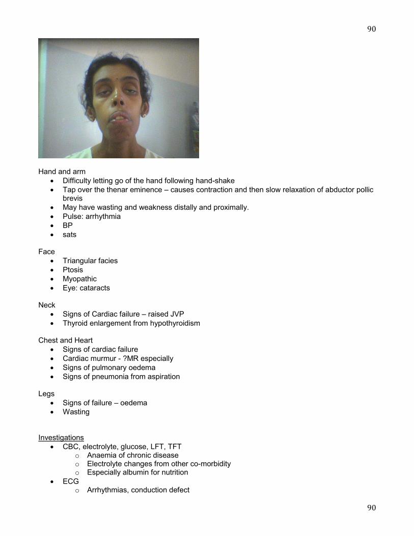
90
90
Hand and arm
x Difficulty letting go of the hand following hand-shake x Tap over the thenar eminence – causes contraction and then slow relaxation of abductor pollic
brevis x May have wasting and weakness distally and proximally. x Pulse: arrhythmia x BP x sats
Face
x Triangular facies x Ptosis x Myopathic x Eye: cataracts
Neck
x Signs of Cardiac failure – raised JVP x Thyroid enlargement from hypothyroidism
Chest and Heart
x Signs of cardiac failure x Cardiac murmur - ?MR especially x Signs of pulmonary oedema x Signs of pneumonia from aspiration
Legs
x Signs of failure – oedema x Wasting
Investigations
x CBC, electrolyte, glucose, LFT, TFT o Anaemia of chronic disease o Electrolyte changes from other co-morbidity o Especially albumin for nutrition
x ECG o Arrhythmias, conduction defect

91
91
x Echo o If cardiomyopathy suspected o Mitral regurgitation
x Spirometry o If respiratory system affected from weakness and repeated aspiration pneumonia
NB: nerve blockade will not prevent the myotonic reflex. Direct administration of LA to muscle somewhat effective.

92
92
Obstructive sleep apnoea
STOP BANG x Snore Loudly? x Tired/fatigued during the day? Daytime-somnolence x Observed apnoea while asleep? x Pressure – Presence of High blood pressure? x BMI >35 x Age >50 x Neck circumference >40cm x Gender – Male x If yes to >3 = Increased risk
Physiological changes
x Aterial hypoxaemia x Polycythaemia x Hypercarbia x Systemic Hypertension with LVF x Pulmonary hypertension with RVF
Anaesthetic implications
x Indicator for difficult airway x Increased sensitivity to respiratory depressants x Increased early (1st 24hours) and late (due to REM catch-up day 2-4 post-op) desaturation
following anaesthesia x Associated with arrhythmia and AV block.
History/Questions
x When was it diagnosed? x How was it diagnosed? x Symptoms
o Daytime somnolence o End organ damage
� Hypertension � Breathing problems � Heart failure – orthopnoea, PND, Swelling � Pulmonary hypertension � Headache
x STOPBANG x Previously investigated?
o Sleep study (i.e. polysomnography), what did it show? � AHI 5-15 = mild � AHI 15-30 = moderate � AHI >30 = severe
x Treatment o CPAP or BiPAP o Tolerance?
x Other associated medical problems? o Obesity o Hypothyroidism o Acromegaly o Diabetes

93
93
x Clinical letter from respiratory service x Drug use
o Alcohol o Smoking
Drugs:
x CPAP/BiPAP o Compliance o Improvement in symptoms
x Antihypertensives x Diabetic medications x Cardiac meds
Exam
x Obesity o Height and weigh o Calculation of BMI
x Signs of o Acromegaly o Hypothalamus look
x Cyanosis x Neck circumference x Signs of R or L heart failure
Investigations
x FBC o polycythemia
x U/E o End organ damage
x ABG o Hypoxaemia o Hypercapnia
x TFTS o Exclude hypothyroidism as a cause
x ECG o LVH o RVH/cor pulmonale
x ECHO o LVF o Diastolic dysfunction o RVH/F o Pulmonary hypertension
x Sleep study o Apnoea (> 10 seconds is significant): apnoea/hypopnoea index: o 5-15 = mild, moderate 6-30, >30 = severe
Polysomnography results Assessment of sleep stages requires 3 studies
x Electroencephalography (EEG) x Electrooculography (EOG) x Electromyography (EMG)
94
94
Two channels are used for monitoring airflow. One thermistor channel (oral and/or nasal) is used to evaluate the presence or absence of airflow. Any change in temperature as a patient inhales and exhales leads to a normal signal, so this channel is insensitive for partial flow obstruction. Thermistor is the recommended channel for evaluation of apneas. Nasal pressure transducer channel is a more sensitive measure of airflow restriction. Normal breathing has a rounded pattern, while resistance to airflow leads to a squaring off of the flow signal. Pressure transducer is the recommended channel for evaluating hypopneas. It is also used for airflow resistance in upper airway resistance syndrome. Other parameters that can be monitored in a sleep study include the following:
x Electrocardiography x Pulse oximetry x Respiratory effort (thoracic and abdominal) x End tidal or transcutaneous CO2 x Sound recordings to measure snoring x Surface EMG monitoring of limb muscles (to detect limb movements, periodic or other) x Continuous video monitoring

95
95
Peripheral neuropathy
Distal parts of the nerves are usually involved fist because of their distance from the cell bodies, causing a distal loss of sensation or motor function or both in the limbs. A typical sensory change is a symmetrical glove and stocking loss to all modalities. This is unlike the pattern found with individual nerve or nerve root disease, which should be suspected if sensory loss is asymmetrical. Ocasionally motor neuropathy may occur without sensory change. Causes
x Alcoholism x Autoimmune disease
o SLE o RA o Guillain-Barre syndrome
x Diabetes x Infection
o HIV o Hepatitis o Singles
x Vitamin deficiencies o Vitamine B
x Inherited disorder o Charcot-Marie-Tooth disease: peripheral neuromuscular denervation. Atrophy of the
muscels leading to spinal and limb deforemities. Muscel weakness as well as sensory disturbances. Orthopaedic intervention is often required for foot deforemities (pes cavus). Spinal deformities can result in restrictive lung dysfunction.
o Amyloid polyneuropathy x Idiopathic
History/Questions
x How was it diagnosed? x What is the distribution?
o Symmetrical o Asymmetrical o Upper limb or lower limb or both
x Sensory changes o To pain and temperature o Fine sensation
x Any motor weakness? x Duration of the disease?
o Course of the disease: rapid vs. chronic x What is the cause?
o Idiopathic o Part of other co-morbidity
� Diabetes � SLE � RA � Hepatitis � HIV
o Alcohol o Smoking o Peripheral vascular disease

96
96
o Nerve damage? Trauma, surgery, etc. o Family history?
x Functional ability Exam:
x Find out if patient is right or left handed Upper limb General inspection
x Wasting x Fasciculations x Ulcers and bruise, abrasions from sensory loss
Tone Power (shoulder, elbows, wrists and fingers) Reflex: Biceps and brachioradialis (C5-6), triceps (C6, 7, 8) Assess co-ordination: finger-nose movements (Cerebellum) Sensation
x Pin-prick sensation x Fine sensation x Proprioception of thum and wrist
Functional ability x Button and unbutton
Lower limbs General inspection Test gait: walk, turn briskly, heel-toes walking (Cerebellum), tip-toe (S1) and heel walking (L4,5) Squatting (proximal muscles) Tone Power: Hip, knee, ankle Refelx: knee (L2, 3, 4), ankle (S1, 2) Heel-on-shin coordination (cerebellum) Sensation
x Pinprick x Fine sensation x Proprioception of toe and ankle
Drugs: Previous (precipitators) Current Control of disease Investigations Depends on the cause
x Diabetes: FBC, U+E, HbA1c, Glucose x Hepatitis: Haptitis serology, LFT, FBC, U+E, coag x HIV: CD4 count, lyomphocyte, PCR x Alcohol: FBC, U+E, LFT, albumin, Vitamin, folate x SLE, RA: rheumatoid factors, CRP, ESR, etc

97
97
Polycystic kidney disease
y Genetic disorder most commonly inherited as an autosomal dominant trait y Cysts in kidney as well as in other organs such as the liver and pancreas y Intracranial aneurysms and cardiac valve abnormalities (esp. Mitral prolapse) may be present y Mild systemic HTN, haematuria, kidney stones, UTI are common y Progress slowly until renal failure during middle age y Haemodialysis or renal transplantation is eventually necessary in most patients
History/Questions:
x How was it diagnosed? x Duration of the disease? x Symptoms
o Abdominal Pain o Haematuria o Infections? o Kidney stones
x Progress o What is renal function?
� Passing urine? � On dialysis?
o On transplant list? o Received transplant
x Associated problems o Increased risk of IHD o Cardiac pathology common 50%
� HTN � Mitral prolapse
o Anaemia o Bone pains from osteitis fibrosa cystica due to secondary hyperparathyroidism o Cysts elsewhere
� Liver � Brain / Intracranial aneurysm
Drugs
x End-stage renal failure meds o Dialysis prescription o Vitamin D and calcium o EPO
x Cardiac medications x Diabetic meds from pancreatic cyst and insufficiency
Exam: General inspection
x Sallow complexion (a dirty brown appearance or „uraemic tinge‟) due to decreased excretion of urinary pigments (urochromes) combined with anaemia
x Hyperventilation: which may indicate metabolic acidosis x Myoclonic jerks due to neuromuscular irritability or a low serum calcium level in late renal
failure x Pruritus - uraemia

98
98
Hand and arm
x Nail o Leuconychia from hypoalbuminaemia (nephritic syndrome)
x Anaemia: pale palmar crease x Asterixis: in terminal renal failure x Fistula formation x Scratch marks and excoriations due to uraemic pruritus x BP
Face
x Eye: look for anaemia x Mouth: uraemic fetor, ammoniacal, musty odour
Neck
x JVP for fluid assessment x Look for jugular vein puncture due to previous vascular access x Parathyroidectomy performed for management of tertiary hyperparathyroidism
Chest
x Basal crepitus if pulmonary oedema present Heart
x Routine exam Abdo
x Tenckhoff catheter for peritoneal dialysis x Nephrectomy scars? Transplant? x Ascites from nephritic syndrome or dialysis fluid x Balloting of the kidneys (mass or enlarged kidney) x Auscultation
o For bruits above umbilicus 2cm to left or right of the midline Back
x Scars x Percuss with fist against vertebrae (osteodystrophy) and flank for pains
Legs
x Oedema Investigations U/E: renal failure ECG: Hypertensive changes MRI brain: aneurysms

99
99
Post cardiac transplantation Cardiac transplant suitability assessment
y Cardiac assessment o Echo o Coronary angiography if due to IHD o Right heart catheterisation to measure PVR and its response to vasodilators (fixed
Pulmonary hypertension is a contraindication) o 24 hour holter
y CXR and spirometry y HLA y Biochemical profile including LFT, U+E, kidney function y Bacteriology and virology
o Hepatitis o CMV o HIV
y Psychiatric work-up y Contraindications: alcoholism, Chronic renal disease, Pulonary parenchymal disease,
continued tobacco use, advanced liver disease
y Cardiac transplant for end-stage heart failure o Ischaemic o Dilated o Congenital o Cardiomyopathy o Cystic fibrosis o Pulmonary pathology
y Physiology of transplanted heart o Denervated but instrinsic mechanisms preserved
� No baroreceptor reflex on heart – hence no tacycardiac or contractility change with hypotension, therefore,
o Sensitive to changes in preload o High resting HR due to denervated from parasympathetic o Higher risk of arrhythmias or conduction abnormalities
� RA conduction pathway may be severed during plantation especially if atrial anastomoses rather than bicaval anastomoses
o Re-innervation may be possible y Direct acting drugs should be used
o Isoprenaline for HR o Phenylephrine or Noradrenaline for vasoconstrictor (but NA may cause increased
contractility and chronotropy due to denervation supersensitivity) o Adrenaline for increased inotropy o Ephedrine may be used but will show increased effect due to increased sensitivity.
y Immunosuppresants o Cyclosporine
� Renal impairment � HTN � Generalised seizures � Hyperkalaemia and hypomagnesaemia may be observed � Interaction with drugs: may prolong non-depolarising NMBD
o Azathioprine y Bone marrow suppression: anaemia, leucocytopaenia, thrombocytopaenia

100
100
y May cause prolonged duration of action of non-depolarising muscle relaxant. But not clinically significant un human
o Steroids y Porlonged use results in steroid-related side effects
y Complications o Rejection o Infection o Malignancy o Immunosuppresants o Arrhythmia: Pacemaker present
History/Questions:
y What was the indication? o What was the cause of the failure? o What was the symptoms prior to transplantation and exercise tolerance? o What was the rhythm prior to surgery? o What was the treatment before surgery
� Drugs � Pacemaker � ICD
y When was the transplant o Any post-operative complications
� Rejection: any history of biopsy � Failure � GVHD � Re-operation
o Duration of stay in ICU y Do you have pacemaker? y How is the heart functioning?
o Exercise tolerance o NYHA class o Echocardiography o Cardiology follow-up
Drugs
y Immunosuppresants o Cyclosporine
� Renal impairment � HTN � Generalised seizures � Hyperkalaemia and hypomagnesaemia may be observed � Interaction with drugs: may prolong non-depolarising NMBD
o Azathioprine y Bone marrow suppression: anaemia, leucocytopaenia, thrombocytopaenia y May cause prolonged duration of action of non-depolarising muscle relaxant. But not
clinically significant un human o Steroids
y Porlonged use results in steroid-related side effects Exam
y Denervated heart o Increased resting heart beat o Loss of HR variability with valsalva manoeuvre and standing

101
101
y Cardiopulmonary examination o Arrhythmia o Look for failure and murmur
Investigations
y FBC y U/E: renal dysfunction y ECG: hypertension, 2 x P waves, sinus tachycardia y ECHO: valve, diastolic/systolic function y Angiography y Cardiac biopsy if rejection suspected

102
102
Post lung transplantation
y General considerations: the transplanted lung is denervated – mucosal sensitivity and the cough reflex are suppressed
y Hypoxic vasoconstriction is not impaired y Lymphatic drainage – severed, but reestablishes after 2-4 weeks
History/Questions
y Why was the transplant undertaken? o What was the condition?
� Cystic Fibrosis � Emphysema � Alpha-1 antitrypsin deficiency � Primary pulmonary hypertension � Pulmonary fibrosis
o What was the functional status at that time? y When was the transplant? y Bilateral vs unilateral? y How successful was the transplant?
o Any issues post-op? � Rejection � Recurrent infection � Bronchiolitis obliterans: 20% decrease in FEV1 with dyspnoea, wheeze, non-productive
cough, fever (non-reversible obstructive lung disease) o Improvement in exercise tolerance o Lung function test recently? What did it show?
y What is your exercise tolerance? y Clinical letter y Recent investigations
Drugs:
y Immunsupressants o Bone marrow suppression o Kidney impairment o HTN o Sometimes cardiac impairment
y Steroids y Rejection
o Usually treated with boost doses of prednisone
Exam:
y Cardiopulmonary examination y Other signs of associated disease
o See cystic fibrosis, a1-antitrypsin deficiency, pulmonary hypertension, CODP, ILD
Investigations o FBC: polycythaemia o U/E: problems with immunosuppressant‟s o Spirometry
o Changes from post transplant

103
103
o Bronchiolitis obliterans o Lung transbronchial biopsy o Echo if previous pulmonary hypertension
104
104
Primary sclerosing cholangitis: Primary sclerosing cholangitis (PSC) is a chronic liver disease caused by progressive inflammation and scarring of the bile ducts of the liver. The inflammation impedes the flow of bile to the gut, which can ultimately lead to liver cirrhosis, liver failure and liver cancer. The underlying cause of the inflammation is believed to be autoimmunity. The definitive treatment is liver transplantation. 80% ANCA positive Associated with cholangiocarcinoma, ulcerative colitis.
Primary biliary cirrhosis Autoimmune disease characterized by slow destruction of small bile ducts. This leads to choleastasis causing tissue damage. Female:male = 9:1. Bilirubin – proportional to prognosis. Liver transplant possible, but recurrence as high as 30% at 10 year. 95% antimitochondrial antibody positive (AMA)

105
105
Pulmonary fibrosis See interstitial lung disease
106
106
Pulmonary hypertension Present when mean pulmonary artery pressure exceeds 25mmHg at rest and 30mmHg with exercise Causes
y Pulmonary ARTERIAL hypertension o Idiopathic o Familial o Part of disease process
� Collagen vascular disease � Systemic-pulmonary shunts � Cirrhosis � Sickle cell disease with recurrent pulmonary infarction
o Pulmonary circulation pressure and volume overload � VSD � ASD � PDA
y Pulmonary hypertension with LEFT HEART disease o Left sided ventricular or atrial disease o Left sided valvular disease
y Pulmonary hypertension with LUNG disease/HYPOXAEMIA o COPD o Interstitial lung disease o OSA o Alveolar hypoventilation o Chronic altitude
y Pulmonary hypertension due to CHRONIC THROMBOTIC/EMBOLIC disease y Other: sarcoidosis, fibrosing mediastinitis
History/Questions:
y How was it diagnosed? y What is the cause?
o Idiopathic o Familial o Part of other disease: liver cirrhosis, collagen vascular disease o Cardiac problem
� Previous MI � Cardiac failure � Valvular problem
o Pulmonary disease � COPD � OSA � ILD
o Thromboembolic disease o Other: sarcoidosis
y What is the symptoms o Dyspnoea o Fatigue o Angina o Syncope o Oedema o Reduced exercise tolerance
y What is your exercise tolerance
107
107
o NYHA class y What kind of investigations have you had?
o Echo o Right heart catheterization o Other investigations associated with other disease
y What treatment are you receiving o Anticoagulation o Vasodilators? o Helpful? Side effects?
y Home oxygen? Drugs
y Treatment of underlying cause: o COPD: bronchodilators, steroids, oxygen o ILD: oxygen
y Anticoagulations y Vasodilators
o Sildenafil o Inhaled prostacyclin analogue – iloprost o Endothelin receptor antagonist – bosentan for class 3 symptoms
y Preload reducers: diuretics, spironolactone y Right heart support and reduction of PVR
o Digoxin (only improve contraction) o Milrinone
Exam:
y Dyspnoea o Tachypnoea o Use of accessory muscles o Pulsed-lip breathing (suggestive of COPD) o Can they walk to the door?
y Polycythaemia? y Cyanosis y Signs of low cardiac output (mottling, poor capillary refill, hypotension) y Signs of LHF
o Displaced apex beat o Functional MR o Pulmonary oedema
y Signs of RHF o Raised JVP o Heave, S3, loud P2 o Enlarged liver (cirrhosis) pulsatile liver (from TR) o Pitting oedema (sacral, ankle)
y Signs of COPD, ILD Investigations
y FBC – polycythaemia y Electrolytes, LFT and coagulation
o End organ damage o Cardiac, renal, hepatic failure
y ABG y CXR

108
108
y Lung function test o COPD o Restrictive lung pathology
y Echo o LV, RV functions, PA pressure o Valvular functions
y Cardiac catheterization o Gold standard for assessment o Performed if other study inconclusive
y Other o CTPA for PE and CTEPH o 6 minute walk test – looking for reduction in saturation >10% or inability to get to 332m
(1000feet) in 6 minutes

109
109
Raynaud’s Disease Categorised as either primary (called Raynaud‟s disease) or secondary when it is associated with other disease (SLE, Scleroderma, Rheumatoid, Sicca syndrome).
y Raynaud‟s phenomenon is episodic vasospastic ischemia of the digits o It affects women more often than men o Characterised by digital blanching or cyanosis in associated with cold exposure or
sympathetic activation o Vasodilation with hyperemia is often seen after rewarming and reestablishment of blood
flow o It may appear as part of CREST syndrome (calcinosis, Raynaud‟s, Esophageal
dysmotility, sclerodactyly (scleroderma limited to the fingers), and telangiectasia. y Raunaud‟s disease is typically bilateral and occurs most frequently as a mild condition in many
young adult women y Secondary Raynaud‟s phenomenon tends to be unilateral and may be the first symptom in
patients who develop scleroderma or SLE, although the systemic disease may not become apparent until years later
Diagnosis is based on history and physical examination. Angiography is not necessary to diagnose this disorder but may be iseful if digital ischaemia is due to artherosclerosis or thrombosis and revascularization is being considered. When clinical diagnosis of Raynaud‟s phenomenon is made, it may lead to workup of associated inflammatory diseases.
y ESR y ANA (antinuclear antibodies) y Rheumatoid factors y Cryoglobulins y Cold agglutinins
Treatment is usually conservative by protecting hands and feet from exposure to cold. Pharmacologic intervention include calcium channel blockade or a-blockade. Surgical sympathectomy may be considered if persistent severe digital ischaemia. See Scleroderma, Rheumatoid arthritis, SLE

110
110
Renal transplantation History/Questions:
x What was the reason for transplant o Diabetes o Hypertensive o Glomerulonephritis o Amyloid o Drug induced o Lupus o PCKD ?biopsy o Sepsis
x When was the transplant x Any complication?
o Rejection o Hypertension
x Current kidney function o Normal o Impaired o On dialysis
x Current treatment o Immunosuppressants
� Side effects? o Dialysis
� Type � Frequency � Problems associated with it
o Fistula formation o Still passing urine or completely dialysis dependent
x Associated problems with chronic kidney disease o Increased risk of IHD o HTN o Anaemia o Bone pains from osteitis fibrosa cystica due to secondary hyperparathyroidism
x Other medical problems o Diabetes
� Ask about contro � Eyes and sensory changes � Autonomic neuropathy: Postural dizziness, reflux, abnormal sweating
o Hypertension o SLE o Rheumatoid o PCKD
Drugs
x Immunosuppressants o Bone marrow suppression o Increased risk of cancer – lymphoma, squamous cell carcinoma, renal impairment
x Chronic kidney impairment meds o Dialysis prescription o Vitamin D and calcium o EPO

111
111
x Diabetic medications x Cardiac medications
Exam and investigation (see chronic renal failure)

112
112
Rheumatic heart disease Rheumatic fever causes valvular lesions. Mitral valve is affected the most often, followed by the aortic valve. Right sided heart manifestations are rare. As the inflammation subsides, it leaves scar tissue. Chronic insidious onset of symptoms and sign. Ask about
y When patient had Rheumatic Fever (RF)? y Was it recurrent? y Any evidence of murmur noted by GP or any other clinician? y Had echo?
o What did it show? o Which valve involved?
y What are the symptoms o Dyspnoea o Reduced exercise tolerance o Orthopnoea o Swelling o Arrhythmia o Syncope o Angina
y Exercise tolerance y Current treatment
o Surgery planned? Or already had surgical intervention o Valvotomy o Diuretics o B-blockers o ACEI/A2RB
y Other co-morbidity from end-organ damage See individual valvular pathology for exam and investigation
113
113
Rheumatoid arthritis
y Most common chronic inflammatory arthritis y Women:Men = 3:1 y Aetiology unknown (complex interaction between genetic and environmental) y Characterised by symmetrical polyarthropathy
o Proximal interphalangeal and metacarpophalangeal joints of the hands and feet which is different from OA (distal IP joints and large joints).
o TMJ, criocoarytenoid involvement o Atalantoaxial subluxation o Extra-articular features
� Skin: Reynaud‟s, rheumatoid nodules � CVS: pericarditis, myocarditis, coronary arteritis, conduction abnormalities, AR � Resp: pleural effusion, pulmonary fibrosis, pulmonary rheymatoid nodules, restrictive
disease from costochondral involvement � CNS: cerebral vasculitis, peripheral neuropathy (esp ulnar nerve) � GI: hepatosplenomegaly � Effects of drugs
y Complications of treatment: o Aspirin and NSAIDs: bleeding, peptic ulcer, renal impairment o Methotrexate: Orally or IM. Has hepatic (cirrhosis) and ILD. Bone marrow suppression o Leflunomide: Hepatotoxicity, weight loss, hypertension, diarrhoea, alopecia o Cyclosporine: renal impairment, hypertension o Azathioprine: cholestatic hepatitis, bone marrow suppression o Hydroxychloroquine: Nausea, pigmentation, retinopathy, cardiomyopathy o Sulphasalazine: rash, nausea, hematological abnormalities, abnormal LFTS, fibrosing
alveolitis o Steroids: hypertension, electrolyte imbalance, easy bruising, osteoporosis, myopathy o Penicillamine: nephrotic syndrome, thrombocytopenia, rashes, mouth ulcers, alteration in
taste, rarely o Cyclophosphamide: Leucopenia, hemorrhagic cystitis, inhibition of
pseudocholinesterase o Mycophenolate: nausea, vomiting, diarrhoea o Anti TNF D: ie infliximab, reactivation of TB, positive ANA, lymphoma; etanercept: flu
like symptoms, blood disorders, demyelinating CNS disorder o Gold: Fatal blood disorders, pulmonary fibrosis, nephrotic syndrome
History/questions:
y How was it diagnosed? y When was the diagnosis? y How often does it flare-up? y Hospital admissions? y What are the symptoms?
o Fatigue o Anorexia o Joint pains? Distribution? o Neck involved? o Morning stiffness which improves with activity o Extra-articular symptoms
� Skin: nodule? � CVS: arrhythmia, failure � RS: pleural effusion, ILD � CNS: numbness or tingling
114
114
� GI: liver or spleen enlargement o Weight loss
y Disease progress y Exercise tolerance and physical capability y Functional limitation y Treatment and side effects
Drugs:
� Analgesics � DMARDS � Joint replacements
EXAM General inspection
y Cushingoid appearance due to steroid treatment y Signs of weight loss which may indicate active disease
Hand and arm
y Symmetrical small joint synovitis (distal IPJ are spared) y Ulnar deviation of the fingers y Splinter-like vasculitic changes y Wasting of small muscles y Signs of ulnar nerve palsy (fingers spread apart) and median nerve palsy (break ring) y Phalen‟s sign for carpal tunnel y Look around elbow for rheumatoid nodules which suggest seropositive disease
Face
y Eyes o Dryness which may suggest sjogren‟s syndrome o Nodular scleritis (elevated white or purple-red lesion)
y Enlargement of parotid gland – occur with sjogren‟s syndrome y Dryness of mouth and dental caries (sjogren‟s syndrome) and ulcer from MTX y TMJ – feel for crepitus
Neck
y C-spine tenderness and range of motion y Cervical lymphadenopathy
Chest
y Look for pleural effusion y Look for pulmonary fibrosis
Heart
y Listen for pericardial rub (pericarditis) y Murmur – indicating valvular regurgitation usually aortic valve
Abdomen
y Feel for liver and spleen (hepatosplenomegaly) Lower limb

115
115
y Limitation of joint movement y Look for foot drop – peroneal nerve entrapment or vasculitis) y Feel Achilles tendon for nodules – sign of seropositive disease
Investigations
y Bloods o FBC: anaemia of chornic disease
� Felty syndrome: rheymatoid arthritis, splenomegaly and neutropenia o U/e: renal impairment (NSAIDS) o ECG: cardiac disease, inclusind RHF secondary to Lung disease o Serology
� Rheumatoid factor (70% positive) � Anti CPP – more specific 97% � ESR/CRP
y Radiological o CXR – respiratory involvement with restrictive pattern o C-spine – flexion and extension view.
� Anterior arch and dens distance >3mm = C-spine instability o Joint X-ray: eronsions, soft tissue swelling
y ECG - pericarditis y Pulmonary function test y Echo: effusions, valvular involvement
116
116
Sarcoidosis A systemic disease characterized by formation of non-caseating granulomata which occur in ANY body tissue and heal with fibrosis. This occurs due to an exaggerated T-helper lymphocyte response that occurs for unknown reasons. Diagnosis is suspected due to abnormal CXR or lymphadenopathy.
y Pulmonary involvement (50% of causes) o Airway granulomas causing bronchial obstruction and distal atelectasis o Restrictive +/- obstructive disease o Alveolar fibrosis o Decreased diffusion capacity
y Airway involvement o Laryngeal o Nasal mucosal
y CNS involvement o Dementia o Encenphalopathy o Seizures o Headache o Facial nerve neuropathy (Bell‟s palsy) o Uveitis
y Myocardial involvement o Heart block o Pericardial disease o Restrictive cardiomyopahy o RHF from lung disease o Valvular granulomata are rare
y Skin o Erythema nodosum
y GI o Dysphagia
History/Questions:
y How was it diagnosed? y When was it diagnosed? y Relapses?
o Frequency y What are the symptoms?
o Respiratory: SOB, dry cough, Stridor, wheeze o Cardiac: Arrhythmia, AV block, failure (SOB, swelling?) o CNS: Seizures, dementia (memory problems), facial weakness o Other: eye redness, vision changes, dysphagia, kidney problems o General
� Fatigue � Weight loss � Fever � Palpable nodes
y What is exercise capacity y What investigations have you had?
o Lung function test o Echo o EP study o CT o Bloods: ESR, CRP, etc.

117
117
y W treatment are you receiving?
Drugs:
y NSAIDs y Prednisolone: 12 month treatment y If longer treatment required: steroid sparing drugs: methotrexate, azathioprine y Hydroxychloroquine for skin disease y Infliximab: TNF D monoclonal antibody: improves lung function if already on steroids and
cytotoxics.
Exam Hand and arm
y Erythema nodosum – painful red lesion on skin (not necessarily on hands) y Maculopapular eruption y Laymphadenopathy
Face and neck
y Uveitis y Cervical lymphadenopathy y Facial nerve palsy
Signs of restrictive lung disease Signs of heart failure (left and right) Abdomen
y Hepatosplenomegaly Neuro-exam for nerve involvement Investigations
y Bloods o FBC - lymphopenia o U/E – elevated Ca2+ o Elevated ESR/CRP o Serum ACE elevated (MCQ) o Raised Immunoglobulin
y ECG o AV blocks o Other conduction blocks and arrhythmia
y CXR o Bilateral hilar lymphadenopathy o Pulmonary infiltration
y CT chest o Ground glass appearance of the chest
y Spirometry o Restricitve pattern
118
118
Scleroderma (systemic sclerosis) Autoimmune disease Scleroderma is characterized by inflammation and fibrosis of the skin and viscera. Scleroderma can be classified as limited or diffuse.
y Limited o Limited to face, and skin upto elbow witout chest, abdominal or internal organ (except
oesophagus) o Evolve into CREST syndrome (calcinosis, Reynaud‟s phenomenon, Esophageal
dysmotility, sclerodactyly, and telengiectasia) y Diffuse
o Internal organ involvement o Often associated with pulmonary fibrosis and pulmonary hypertension o As well as cardiac and renal impairment
Skin changes y Joint contractures y Reduced mouth opening y Telangiectasia
Pulmonary y ILD y Pulmonary HTN
CVS y Cardiac fibrosis with CHF y Conduction system defects y Sclerosis of coronary arteries y HTN y Pericarditis
CNS y Impaired nerve conduction with peripheral and/or cranial neuropathy
GI y Dysmotility
Renal y Renal failure from Renal artery obstruction from intimal proliferation
History/Question
y How was it diagnosed? y When was it diagnosed? y What are the symptoms?
o Affects face, hand arms only? Or more diffuse? o Skin
� Tight � Reynaud;s � Sclerodactyly � Telangiectasia
o Pulmonary involvement � SOB � Dry cough � Home oxygen? � Inhalers?
o Cardiac involvement � Failure – SOB, swelling
o Reflux o Weakness of muscles and nerve entrapment: carpal tunnel
119
119
o Dry eyes! y Other associated disease
o Lung o Cardiac o Renal o Neurologic
y What is your exercise tolerance and degree of disability y What is current treatment
o BP control to prevent renal impairment o ACEi/A2RB o Cyclophosphamide for lung disease o Endothelin-1 receptor blockade )bosentan) if pulmonary hypertension and renal crisis o Prostacycline analogue (iloprost) for pulm HTN o Any S/E
Drugs
y BP control to prevent renal impairment y ACEi/A2RB y Cyclophosphamide for lung disease y Endothelin-1 receptor blockade )bosentan) if pulmonary hypertension and renal crisis y Prostacycline analogue (iloprost) for pulm HTN y Any S/E
Exam General Appearance
y Bird-like facies y Cachexia from malnutrition (dysphagia and oesophgeal motility) y Skin changes y Anaemic
Hand
y Calcinosis (palpable nodules due to clcific deposits in the subcutaneous tissue of the fingers) y Raynaud‟s phenomenon sometimes cause atrophy of the finger pulps due to ischaemia y Sclerodactyly (tightening of the skin of the fingers leading to tapering) y Multiple large telangiectasia on the fingers (red “spots”) y Contracture deformity of the fingers y Anaemia
Arms
y If skin tightening and thickening extends beyond wrist and elbow the disease is DIFFUSE rather than CREST.
y BP Face
y Loss of normal wrinkles and skinfolds as well as of the eyebrows y Inability to close eyes fully y Dry eyes
Heart and lungs
y Look for failure (RHF) – heave, S3, TR, Elevated JVP, peripheral swelling y Look for restrictive lung disease – reduced chest expansion, crackles
120
120
Legs
y Signs of vasculitis y Ulceration and skin involvement
Investigations
y FBC – anaemia y Electrolytes and LFT– renal impairment, CHF, liver impairment y Coags – malabsorption and malnutrition (ADEK) y ECG – for cardiac conduction abnormality y CXR, Echo, ABG as necessary especially if diffuse scleroderma y RF/ANA for exclusion of RA or SLE

121
121
Systemic Lupus Erythematosus y Multisystemic chronic inflammatory disease of unknown origin. y Women are more affected than men y Associated with complement deficiencies y Diagnosis requires at least 4 of the 11 published criteria (serially or simnultaneously), either
current of in the past. o Malar rash (butterfly rash) o Discoid rash o Photosensitive rash o Oral ulcers o Arthritis – NON-EROSIVE, 2 peripheral joints o Seositis: Pleurisy or pericarditis – with audible rub, ECG changes or effusion o Renal disorder: persistent protienuria or casts o Neurological disorder: seizures or psychosis without other causes o Haematological disorder: Haemolytic anaemic, leucophaemia, lymphopenia,
thrombocytopaenia o Immunological disorder: Anti-DNA antibodies, anti-smooth muscle antibody or positive
anti-phospholipid antibodies, abnormal ANA titre y System affected
o Cardiac: � Conduction abnormalities, myocarditis, effusion, noninfective vegetations, CAD
o Pulmonary: pleuritis/effusion, restrictive lung disease, pulmonary HTN o Renal: lupus nephritis o CNS: seizures, psychosis, peripheral neuropathy o Heme: anaemia, leucopaenia, thrombocytophaemia, antiphospholipid antibody, recurrent
thrombosis, MI, PE, fetal loss y Meds
o Often on steroids, NSAIDs, heparin, cyclophosphamide, antimalarials
History/Questions
y How was it diagnosed? y When was it diagnosed? y What are the presenting symptoms?
o Malaise o Weight loss o Musculoskeletal symptoms: arthralgia, arthritis, myositis o Dermatological: skin rash, alopecia, ulcers, photosensitivity o Fever o CNS: neuropsychiatric o Renal: haematuria, oedema, renal failure o RS: pleurisy, CP, SOB o Cardiac: pericarditis, myocarditis, valvular lesions o Recurrnet thromboembolic event, recurrent miscarriage
y Activity level and functional ability y Current treatment and S/E
Drugs
y Avoid sunlight y Arthralgias – NSAIDS y Raynauds: calcium channel antagonists y Steroids y Hypercoaguability: warfarin

122
122
y Hydroxychloroquine: arthritis and skin rash. Watch for retinal changes and cardiomyopathy y Azathioprine – steroid sparing agent y Mycophenolate
Exam General inspection
y Weight loss (due to chronic inflammation) y Cushingoid apperarance (steroid treatment) y Any mental problems
Hands and arm
y Vasculitic appearing lesions y Rash from photosensitivity y Raynaud‟s phenomenon y Synovitis of the proximal and metacapophalangeal joints y Livedo reticularis: skin discoloration in the form of a small net y Proximal myopathy
Head and neck
y Alopecia: important diagnostic clue y Scleritis and episcleritis y Anaemia y Mouth ulcers y Butterfly rash y Cranial nerve lsions
Chest
y Pericarditis y Pleural effusion y Plereurisy y Pulmonary fibrosis, collapse or infection
Abdomen
y Hepatosplenomegaly y Tenderness
Legs
y Red soles y Rash y Ulcers over the malleoli (e.g. antiphospholipid syndrome with venous ulcer) y Proximal myopathy y Cerebellar ataxia y Hemiplegia
Investigations
y Multiple antibodies are associated with SLE. Includes: anti-dsDNA, anti-sm o Most are ANA positive
y FBC o Anaemia o Leucopenia
y U+E

123
123
o Renal impairment y Coagulation may be difficult to interpret as aPTT may be abnormally elevated y CXR
o Pleural effusion y CT head to rule out other cause of neurological abnormality y X-ray of joint: non-erosive
124
124
Systemic vasculitis y Affects multiple body systems. y Systemic features (fever, malaise) are common y Ask about renal involvement (common)
Wegners
y Pulmonary renal syndrome. Fatal if not treated. Symptoms include: nasal congestion, rhinorrhoea, cough, breathlessness
Giant cell arteritis y Bitemporal headache, visual disturbance, jaw/tongue claudication
Polyarteritis nodosa y Coronary arteries, mesenteric. Ie multipl systems. May rpesent with foot drop and abdominal
pain Churg-Strauss vasculitis: usually have eiosinophilia/asthma first Normally look unwell Check for livedo reticularis (net like pattern) Investigations: biopsy, ernal function, wegners – c-ANCA, others p-ANCA Treatment: steroids/immunosuppressants

125
125
TB Primary TB
y Initial Infection is usually pulmonary y Primary TB is often asymptomatic or there is fever, lassitude, sweats, anorexia, cough, sputum,
yellow-grey nodules near the lumbus. y Acid-fast bacilli may be found in sputum y CXR may help y The commonest non-pulmonary primary infection is GI, typically affecting the ileocaecal
junction and associated lymph nodes Post-primary TB
y Any immunocompromise may allo reactivation o Malignancy o Diabetes o Steroids o Debilitation (HIV or old age)
y Lung lesion (usually upper lobe) progress and fibrose Pulmonary TB
y May be silent or present with cough, sputum, malaise, weight loss, night sweats, pleurisy, haemoptysis, pleural effusion, superimposed pulmonary infection.
Miliary TB
y Occurs following haematogenous dissemination y Non specific symptoms. y CXR shows characteristic reticulonodular shadowing y Biopsy of lung, liver, lymp nodes, or marrow may yield AFB
Other
y Meningeal TB y Genitourinary TB: symptoms of UTI, sterile pyuria y Bone TB: vertebral collapse y Skin TB: jelly-like nodule y Peitoneal TB: Abdo pain and GI upset y Acute TB Pericarditis y Chornic pericardial effusion and constrictive pericarditis
History/Questions
y How was it diagnosed? y When was it diagnosed? y Currently being treated? y What site has been involved?
o Lung o Brain o Abdomen o Skin o Bone
y What are the symptoms? o Fatigue o Night sweats

126
126
o Weight loss o Fever o Cough and sputum o Haemoptysis
y Any investigations o Mantoux o CXR or CT
y What treatment are you having? And for how long? y How did you get the disease?
o Recent travel? o Family or relative with TB o Immigrants? o Immunocompromised?
� HIV � Steroids, etc
Drugs
y Compliance is important y Rifampicin: hepatitis y Isoniazid: Hepatitis, neuropathy y Pyranzinamide: hepatitis, arthralgia y If resistance possible, add ethambutol (ocular toxicity)
Exam:
y Cardiopulmonary exam y Look for lymphadenopathy
Investigations
y FBC, Liver and renal function prior to treatment as may cause impairment y Microbiology
o Multiple sputum sample for AFB o Bronchoalveolar lavage may be helpful o Biopsy of any suspicious lesions
y TB PCR y Radiology
o CXR o CT
y Immunological o Mantoux test: test posive if produces >10mm induration and negative if <5mm o TB Gold

127
127
Thrombophilia Prothrombotic pathologies
y Factor 5 Leiden o Abnormal Factor V molecule o Resistant to neutralization by activated protein C o Also called activated protein C resistance
y Antithrombin III deficiency o Thrombotic risk is unpredictable, but if it occurs = life long warfarin
y Proteins C and S deficiency o Associated with recurrent DVT and PE o Testing must occur 2 weeks after warfarin has stopped as warfarin inhibits its synthesis
y Prothrombin gene mutation o Requires DNA PCR
y Homocysteine levels o also a risk factor for coronary artery disease
y Antiphospholipid syndrome o ie lupus anticoagulant, anticardiolipin antibodies.
Indications for thrombophilia investigations:
y Recurrent venous thrombosis y Venous thombosis before age 45 y Thrombosis at unusual site y Adverse family history of thrombosis
Qustions Nature of thrombotic episodes
y Arterial y Venous y Provoked y Unprovoked
Ask whether a thrombotic tendency has been identified? y Recurrence y Thrombosis at unusual site y Family history
What anticoagulant therapy is currently being used? y Heparin, warfarin, rivaroxaban, dabigatran
INR testing – current and target Other risk factors:
y Smoking y Oestrogen OCP y Pregnancy y Smoking y Immobilization y Cancer
In women y ?Previous unexplained miscarriages – ie antiphospholipid antibodies: includes:
o Lupus anticoagulant o Anticardiolipin antibodies o Can also occur in women with pre-eclampsia, eclampsia

128
128
On examination:
y Heparin infusion? y Obesity y Oedema, venous ulceration y Abdominal wall bruising
Management: In general: an initial episode of thrombus is treated with low molecular weight heparin or IV non fractionated heparin then 6 months warfarin Patients with APC only get long term anticoagulant if they have a second DVT Pregnant women with a history of DVT require LMWH

129
129
Valvular heart disease: Mitral stenosis
y Normal valve area: 4-6cm2 o Mild 1.5-2.5 o Moderate 1.0-1.5 o Severe < 1.5 o Severe if transvalvular gradient 15mmHg
y If transvalvular gradient >25 mmHg, enough to cause pulmonary hypertension y As valve area decreases, chronic pressure gradient leads to elevated LA pressures, pulmonary
oedema, pulmonary hypertension and RHF. y Reduced flow limits LV output y Causes
o Rheumatic heart disease o IE o SLE o RA o Congenital abnormality
y Consider infective endocarditis antibiotics antibiotics y Tachycardia and arrhythmias poorly tolerated y Management
o Treat arrhythmia o Rate control if not sinus o Treat HTN o Symptomatic : diuretics if failure o Maintain normovolaemia o Maintain SVR – fixed CO o Contractility – maintain, do not over-do it o Definitive
� Valvotomy � Valve repair or replacement
History/Questions
y How was it diagnosed? y When was it diagnosed? y What is the cause? y Symptoms?
o Asymptomatic o Palpitation o Dyspnoea, clear frothy sputum, exercise tolerance o Signs of RHF with pitting oedema o LV low output symptoms
� Angina � Syncope
y Associated AF y What investigations have you had? y Treatment plan?
Drugs
y HR control y Anticoagulation y Pulmonary HTN drugs

130
130
Exam y Mitral facies y Irreugular pulse y Signs of MS
o Reduced pulse pressure o Increased S1 with opening snap o Mid-diastolic murmur
y Signs of pulmonary oedema o Dyspnoea o Wheeze o Hypoxaemia o Cyanosis o Bibasal Crackles
y Signs of pulmonary HTN and RHF o RV heave o Increased P2 o Functional TR o Hepatomegaly o Ascites
Investigation
y ECG o Large P wave o Rhythm o RAD/RVH or P-pulmonale
y CXR o Mitral valve calcification o Big left atrium o Pulmonary oedema or effusion
y Echo o Severity o LA thrombus o Pulmonary HTN o RV function
y Exercise or pharmacological stress o IHD o Cardiac catheterization as necessary

131
131
Valvular heart disease: Mitral regurgitation Severe MR = regurgitant fraction > 0.6 Acute MR Cause
y Infective endocarditis y MI y Surgery y Trauma
Chronic MR cause
y Degenerative y Rhematic heart disease y Mitral valve prolapsed y Papillary muscle dysfunction y CTD: rheumatoid, ankylosing spondylitis y Congenital
Acute
y Sudden rise in LA pressure and pulmonary venous pressure leading to acute fulminant pulmonary oedema
Chronic y LV volume overload y LVH and reduction in EF. Pulmonary oedema does not occur at this stage. This is buffered by
dilated LA y Eventually leads to LVF and increased LA pressure and pulmonary oedema (late)
Indications for replacement/repair
y Acute MR with pulmonary oedema y LV dysfunction with EF <0.6 y Symptomatic MR even though LV function Normal
Management
y Maintain normovolaemia y HR – high @ 80-100 y SVR – normal to low y Contractility - Normal
History/Questions
y How was it diagnosed? y When was it diagnosed? y What is the cause?
o History of Rheumatic fever o Endocarditis o CTD o Previous MI and cardiac failure?
y Symptoms? o Dyspnoea o Arrhythmia (AF common) o Fatigue o Reduced exercise tolerance
y Current Treatment

132
132
y Exercise tolerance? y Other co-morbidities
Exam
y Signs of pulmonary oedema y Signs of LVF and MR
o Displaced apex o Pansystolic murmur at apex radiating to axilla o Louder on exhalation
y Signs of pulmonary HTN and RHF (late)
Investigations
y ECG o Atrial fibrillation o Signs of LVH
y CXR o Cardiomegaly
y Echo o Severity o LVH o LA diameter o Cause of MR
y BNP

133
133
Valvular heart disease: Aortic regurg Severe AR = regurgitant fraction > 0.6 Acute AR Cause
y Infective endocarditis y Dissection y Trauma
Chronic MR cause
y Degenerative y Rhematic heart disease y Marfans y CTD: rheumatoid, ankylosing spondylitis
Acute
y Sudden rise in LV pressure y Decreased perfusion y LV failure with acute pulmonary oedema
Chronic
y LV volume overload y LVH and reduction in EF. Pulmonary oedema does not occur at this stage. This is buffered by
dilated LA y Eventually leads to LVF and increased LA pressure and pulmonary oedema (late)
Indications for replacement/repair
y Acute AR with pulmonary oedema y LV dysfunction with EF <0.6
Management
y Maintain normovolaemia or high y HR – high @ 80-100 y SVR – normal to low y Contractility – Normal to high
History/Questions
y How was it diagnosed? y When was it diagnosed? y What is the cause?
o History of Rheumatic fever o Endocarditis o CTD
y Symptoms? o Dyspnoea o Angina o Fatigue o Reduced exercise tolerance
y Current Treatment y Exercise tolerance? y Other co-morbidities

134
134
Exam
y Wide pulse pressure y Chronic AR tachycardia y Signs of pulmonary oedema y Signs of LVF and AR
o Displaced apex o Diastolic murmur at aortic valve o Louder on exhalation
y Signs of pulmonary HTN and RHF (late) Investigations
y ECG o Signs of LVH
y CXR o Cardiomegaly
y Echo o Severity o Co-existing AS o LVH o LA diameter o Cause of AR - ?thickening ?vegetation, etc
y Exercise stress test
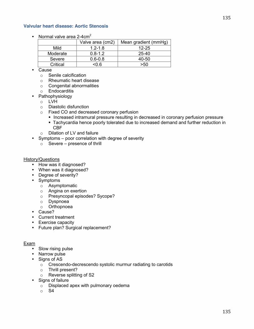
135
135
Valvular heart disease: Aortic Stenosis
y Normal valve area 2-4cm2 Valve area (cm2) Mean gradient (mmHg)
Mild 1.2-1.8 12-25 Moderate 0.8-1.2 25-40 Severe 0.6-0.8 40-50 Critical <0.6 >50
y Cause o Senile calcification o Rheumatic heart disease o Congenital abnormalities o Endocarditis
y Pathophysiology o LVH o Diastolic disfunction o Fixed CO and decreased coronary perfusion
� Increased intramural pressure resulting in decreased in coronary perfusion pressure � Tachycardia hence poorly tolerated due to increased demand and further reduction in
CBF o Dilation of LV and failure
y Symptoms – poor correlation with degree of severity o Severe – presence of thrill
History/Questions
y How was it diagnosed? y When was it diagnosed? y Degree of severity? y Symptoms
o Asymptomatic o Angina on exertion o Presyncopal episodes? Sycope? o Dyspnoea o Orthopnoea
y Cause? y Current treatment y Exercise capacity y Future plan? Surgical replacement?
Exam
y Slow rising pulse y Narrow pulse y Signs of AS
o Crescendo-decrescendo systolic murmur radiating to carotids o Thrill present? o Reverse splitting of S2
y Signs of failure o Displaced apex with pulmonary oedema o S4

136
136
Investigations y ECG
o LVH o Ischaemia
y CXR o LVH o calcification of the valve o Pulmonary oedema
y Echo o Severity o Aetiology o LVH, LV function
y Exercise stress test
137
137
Valvular heart disease: Tricuspid regurg
y Tricuspid regurgitation is usually functional, caused by annular dilation secondary to RV dilation or pulmonary hypertension.
y Other causes o Infective endocarditis (associated with IVDU) o Carcinoid syndrome o Rheymatic heart disease (rare) o Tricuspid valve prolapsed o Ebstein‟s anomaly
y Tricuspid valve disease is often associated with mitral or aortic valve disease y Mild MR can be normal finding at any age and is very commonly seen in highly trained athletes y Pathophysiology
o Chronic RV volume overload o Right sided congestive heart failure o Manifested by raised JVP, hepatic congestion, peripheral oedema and ascites
y Management of anaesthesia o Preload – high normal
� Limit vasodilator drugs as it will decreased VR and significantly hinder CO o Avoid rise in PVR
� Acidosis � Hypoxaemia � Hypercapnia � PEEP � Positive ventilation
History/Questions
y How was it diagnosed? y When was it diagnosed? y Degree of severity? y Symptoms
o Asymptomatic o Decreased exercise tolerance o Peripheral oedema o Enlargement of liver o Distended abdomen from ascites o Symptoms of LVF and pulmonary HTN other valvular pathology
y Cause? y Current treatment y Exercise capacity y Future plan? Surgical replacement?
Exam
y Low cardiac output state if severe TR y Elevated JVP with large V wave y Signs of RHF and TR
o RV heave o Pansystolic murmur maximal at the lower end of the sternum and increased on inspiration
y Abdomen o Hepatomegaly
138
138
o Pulsating liver o Ascites o Peripheral oedema
Investigations
y CXR o RV enlargement o Biventricular enlargement if TR is secondary to LHF
y Echo
139
139
Valvular heart disease: Pulmonary stenosis
y Usually diagnosed in childhood y May be related to congenital heart disorders y Other cause: carcinoid y Pathophysiology
o RVH o +/-TR o RA pressure increases which may open foramen ovale leading to R to L shunt causing
cyanosis y Treatment of choice is percutaneous balloon valvuloplasty and is done when a resting peak
gradient is seen to be >60mmHg or a mean >40mmHg History/Questions
y How was it diagnosed? y When was it diagnosed? y Degree of severity? y Symptoms
o Asymptomatic o Decreased exercise tolerance o Peripheral oedema o Enlargement of liver o Distended abdomen from ascites o Symptoms of LVF and pulmonary HTN other valvular pathology
y Cause? y Current treatment y Exercise capacity y Future plan?
Exam
y Peripheral cyanosis – low cardiac output y Central cyanosis – PFO with R to L shunt y Pulse – Reduced due to low cardiac output y JVP: giant a wave due to stiff RVH y Signs of RV failure and PS
o RV heave o Functional TR o Systolic murmur over PV ?thrill present o Peripheral oedema
y Abdomen o Hepatomegaly o Pulsating liver o Ascites o Peripheral oedema
Investigations
y CXR o RV enlargement
y Echo