tachycardia-induced cardiomyopathy: case presentation … · tachycardia-induced cardiomyopathy:...
TRANSCRIPT

Ralph J. Damiano, Jr., MDEvarts A. Graham Professor of Surgery
Chief of Cardiothoracic SurgeryVice Chairman, Department of Surgery
Barnes-Jewish HospitalWashington University School of Medicine
St. Louis, MO USA
Tachycardia-Induced Cardiomyopathy:Case Presentation and Clinical Management
Department of SurgeryDivision of Cardiothoracic Surgery
DISCLOSURE
Speaker for AtriCure, Edwards Lifesciences, LivaNova Consultant for Medtronic Research and educational grants over the last 2 years: AtriCure Edwards

Department of SurgeryDivision of Cardiothoracic Surgery
Case Presentation:History of Present Illness
• 64 yo M engineer, an active marathon runner until he was firstdiagnosed with atrial fibrillation (AF).
• Presented to hospital on multiple occasions with progressivedyspnea on exertion, decreased endurance and increasedpalpitation.
• The patient remained in symptomatic AF (+/- RVR) despitetrials with several anti-arrhythmic drugs and multiplecardioversions.

Department of SurgeryDivision of Cardiothoracic Surgery
Case Presentation:History of Present Illness
• Three failed attempts at catheter ablation on 2/17/07 (PVI),6/29/07 (repeat PVI), and on 11/20/07 (repeat PVI, left andright atrial lesions).
• Noted to have progressive worsening LV dysfunction (LVEF:10-20%).
• Referred to our clinic for surgical ablation.

Department of SurgeryDivision of Cardiothoracic Surgery
Case PresentationPast Medical History:
• GI bleed due to diverticulosisHome Medication:
• Coumadin, Propafenone, Aspirin, Digoxin, MetoprololPhysical Exam:
• Vitals - BP: 97/65, HR: 101 irregular, O2 Sat: 96%, BMI: 23.2• Cardiac exam: tachycardia, irregularly irregular, no murmur• Otherwise normal exam

Department of SurgeryDivision of Cardiothoracic Surgery
Case Presentation:Preoperative evaluation - ECG

Department of SurgeryDivision of Cardiothoracic Surgery
Case Presentation:Preoperative evaluation - TTE
• Echocardiogram 4/08/2008 (Patient in Atrial Fibrillation)• Four-chamber dilation• Severe LV systolic dysfunction, EF 15%• Moderate to severe RV systolic dysfunction • Normal diastolic function • Moderate MR• Mild TR

Department of SurgeryDivision of Cardiothoracic Surgery
Case Presentation:Preoperative evaluation - TTE

Department of SurgeryDivision of Cardiothoracic Surgery
Case Presentation:Preoperative evaluation - TTE

Department of SurgeryDivision of Cardiothoracic Surgery
Case Presentation:Preoperative evaluation - TTE post cardioversion
• Echocardiogram 4/15/2008)• Upper normal LV size• Severe LV dysfunction, EF 25%• Moderate to severe RV systolic dysfunction • Mild MR• Normal myocardial contractile function and diastolic function by
tissue Doppler imaging

Department of SurgeryDivision of Cardiothoracic Surgery
Case Presentation:Preoperative evaluation - TTE post cardioversion

Department of SurgeryDivision of Cardiothoracic Surgery
Case PresentationPreoperative evaluation - Cardiac MRI
• Cardiac MRI 4/15/2008 (Post cardioversion to NSR)• Right atrial and ventricular enlargement• Left atrium and ventricle appeared normal• Global left ventricular hypokinesis• Gadolinium enhancement:
• No evidence of myocardial infarction, fibrosis

Department of SurgeryDivision of Cardiothoracic Surgery
Case PresentationPreoperative evaluation - Cardiac MRI

Department of SurgeryDivision of Cardiothoracic Surgery
Case PresentationPreoperative evaluation - Cardiac Cath
• Cardiac catheterization • Right dominant• No coronary artery disease• Impaired global ejection fraction with normal hemodynamics• Normal right-sided pressures

Department of SurgeryDivision of Cardiothoracic Surgery
Case PresentationPreoperative evaluation - Cardiac Cath

Department of SurgeryDivision of Cardiothoracic Surgery
Case PresentationPreoperative evaluation - Cardiac Cath

Department of SurgeryDivision of Cardiothoracic Surgery
Case PresentationPreoperative evaluation - Cardiac Cath

Department of SurgeryDivision of Cardiothoracic Surgery
Operative Approach• Median sternotomy• Stand alone Cox-Maze IV Procedure

Department of SurgeryDivision of Cardiothoracic Surgery
Case PresentationPostoperative evaluation - TTE
• Echocardiogram 12/28/2015 (NSR) - 7 years later!• Normal LV size and systolic function, EF 67%• Normal LV wall thickness/mass• Normal RV size with normal function• Mild MR + TR

Department of SurgeryDivision of Cardiothoracic Surgery
Case PresentationPostoperative evaluation - TTE

Department of SurgeryDivision of Cardiothoracic Surgery
Case PresentationPostoperative Follow-up
• Office visit 7/18/2017 – 9 years later!• Doing well, running regularly (1-6 miles daily), lifting weights
regularlyHome Medication:
• AspirinPhysical Exam:
• Vitals - BP: 113/71, HR: 63 regular

Department of SurgeryDivision of Cardiothoracic Surgery
Case Presentation:Postoperative Holter Rhythm Monitoring
• Annual Holter recordings have shown no recurrent atrial tachyarrhythmias.
• Last Holter 7/18/2017 – NSR - 9 years later!• Predominant rhythm: Sinus with average HR of 69• 60 episodes of SVT with maximum duration of 11 beats• No sustained episodes of AF

Department of SurgeryDivision of Cardiothoracic Surgery
What is Tachycardia-Induced Cardiomyopathy?• Unexplained REVERSIBLE systolic dysfunction AND any form of
long-standing tachycardia• Must exclude secondary causes of cardiomyopathy (e.g., ischemic and
dilated cardiomyopathy)• Definitive diagnosis is ONLY made after cardiac function restoration
following rate and/or rhythm control
Jeong YH, Choi KJ, Song JM, Hwang ES, Park KM, Nam GB, Kim JJ, Kim YH. Diagnostic approach and treatment strategy in tachycardia-induced cardiomyopathy. Clin Cardiol. 2008 Apr;31(4):172-8.
Stulak JM, Dearani JA, Daly RC, Zehr KJ, Sundt TM 3rd, Schaff HV. Left ventricular dysfunction in atrial fibrillation: restoration of sinus rhythm by the Cox-maze procedure significantly improves systolic function and functional status. Ann Thorac Surg. 2006 Aug;82(2):494-500

Department of SurgeryDivision of Cardiothoracic Surgery
Mueller AL, et al.J Am Coll Cardiol 2017;69:2160-2172
Tachycardia-induced CardiomyopathyDefinition
• Sustained heart rate > 100 beats/min• Exclusion of other causes of heart failure
• echo, cardiac cath, cardiac MRI• Partial or complete recovery of LV function after
restoration of sinus rhythm• preop cardioversion/rate control

Department of SurgeryDivision of Cardiothoracic Surgery
Jeong et al.Clin Cardiol 2008;314:172-178
Tachycardia-induced Cardiomyopathy:How To Differentiate From Dilated Cardiomyopathy?
• LVEF ≤ 45% and LVEDD ≤ 61 mm was predictive of TIC with a sensitivity of 100% and a specificity of 71%
• LVEF < 30% and LVEDD ≤ 66 mm was predictive of TIC with a sensitivity of 100% and a specificity of 83%

Department of SurgeryDivision of Cardiothoracic Surgery
Mueller AL, et al.J Am Coll Cardiol 2017;69:2160-2172
Tachycardia-induced CardiomyopathyHow To Differentiate From Dilated Cardiomyopathy?
• Endomyocardial biopsy can be useful• Less fibrosis• Less T cells and microphages• Enhanced myocyte size

Department of SurgeryDivision of Cardiothoracic Surgery
Department of SurgeryDivision of Cardiothoracic Surgery
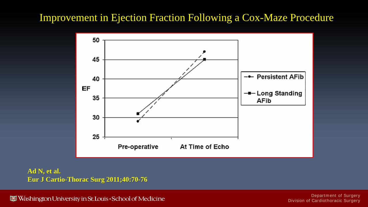
Ad N, et al.Eur J Cartio-Thorac Surg 2011;40:70-76
Improvement in Ejection Fraction Following a Cox-Maze Procedure

Department of SurgeryDivision of Cardiothoracic Surgery
Tachycardia Induced Cardiomyopathy:The Washington University Experience
• Between January 2002 and January 2017, 34 consecutive patients with tachycardia-induced cardiomyopathy underwent CMP IV
• Mean age was 56 ± 11 years• Twenty-four patients (70%) had long-standing persistent AF• The remainder had paroxysmal (7/34, 21%) or persistent (3/34,
9%) AF

Department of SurgeryDivision of Cardiothoracic Surgery
Tachycardia Induced Cardiomyopathy:The Washington University Experience
• Preoperative mean ejection fraction was 32% ± 8% (range, 18% - 40%)
• Ejection fraction improved to 55% ± 8% (95% CI [51%, 58%], P<0.001) at a median 22 months
• Of the 11 patients with NYHA class III/IV symptoms, 8 patients were in class I/II at last follow up, P = 0.02

Department of SurgeryDivision of Cardiothoracic Surgery
Tachycardia Induced Cardiomyopathy:The Washington University Experience
01020304050607080
Preoperative Postoperative
LVEF
(%)
Left Ventricular Ejection Fraction in Patients Undergoing Cox-Maze Procedure
P < 0.001

Department of SurgeryDivision of Cardiothoracic Surgery
Tachycardia-induced CardiomyopathyConclusions
• Tachycardia-induced cardiomyopathy is a known complication of AF/flutter.
• The definition of TIC is one of exclusion.• Cardiac MRI and occasionally myocardial biopsy can be used to help
differentiate it from other cardiomyopathies.• The presence of TIC is a strong indication for interventional therapy in
patients who have failed medical management.• Surgical results have been excellent in this subgroup of patients.

Department of SurgeryDivision of Cardiothoracic Surgery

Department of SurgeryDivision of Cardiothoracic Surgery

Department of SurgeryDivision of Cardiothoracic Surgery
Stulak JM, et alAnn Thorac Surg 2006;82:494-501