taking control early - cloud object storage | store ...abdomen... · “app = map minus iap = map...
TRANSCRIPT

Best Practices in Managing Patientswith an Open Abdomen
Sponsored by North American Center for Continuing Medical Education, LLC, An HMP Communications Holdings Company
Supported by an educational grant from KCI
Taking Control Early:
Faculty
Mark J. Kaplan, MD, FACSChief, Division Trauma and Surgical Critical Care
Albert Einstein Medical Center Philadelphia, Pennsylvania
Learning Objectives
• Employ early intervention for managing open abdominal wounds
• Review nontrauma surgical scenarios with an open abdomen, including sepsis, peritonitis, transplant, appendicitis, and abdominal aortic aneurism repair
• Implement best practices to reduce the occurrence of fistula

Open Abdomen
Ivatury RR. World J Surg. 2009;33(6):1150-1153.
Open Abdomen
Ivatury RR. World J Surg. 2009;33(6):1150-1153.
We just madea mess of it
until now
Management Goals
• Planning-is the key element in managing a patient with an open abdomen
• Management of IAH/ACS
• Resuscitation- of crystalloids/MTP adoption
• Adoption of Damage Control Mentality
• Early enteral nutrition
• Effective temporary abdominal closure techniques
DSL#12‐0169 (3/12) 6

The Problem
• Open abdomen use has increased because of risks of abdominal hypertension and abdominal compartment syndrome
• Studies have clearly shown that without adequate stabilization and control of systemic derangements, prolonged time in the operating room has negative effect on outcome
• Therefore, damage control was developed to increase outcomes based on rapid evaluation and stabilization
• Significant morbidity with this procedure including: fistulas, renal failure, acute respiratory distress syndromes, ACS, and giant abdominal wall hernias
ACS = abdominal compartment syndrome.DeWale JJ, et al. American Surgeon. 2011;77(Suppl 1):S46-S50.
Paradigm Change inManaging the Open Abdomen
• Stopping the intestines from falling to the floor is not a goal of therapy
• To minimize complications and improve outcomes, the entire local and systemic effects of the open abdomen must be treated
• The goal is a change from TAC to TMOA
TAC = temporary abdominal closure; TMOA = total management of the open abdomen.
Elements inManaging the Open Abdomen
• Understanding the concepts of ACS/IAH
• Understanding the intra-abdominal inflammatory response
• Concepts in managing sepsis and trauma
• Concepts in TAC/TMOA
IAH = intra-abdominal hypertension.

Most significant pitfallin managing the open abdomen
is the lack of understandingof the pathophysiology, treatment,
and prevention of ACS/IAH
Considerations with the Open Abdomen
11
IAP
“IAP is the steady-state pressureconcealed within the abdominal cavity.”
• Elevated IAP is a common finding in the ICU
• IAP increases and decreases with respiration
• IAP is directly affected by:
– Solid organ or hollow viscera volume
– Space occupying lesions
• Ascites, blood, fluid, tumors
– Conditions that limit expansion of the
– abdominal wall
– Burn eschars, third-space edema
IAP = intra-abdominal pressure; ICU = intensive care unit.Malbrain ML, et al. Intensive Care Med. 2006;32(11):1722-1732.

APP
“APP = MAP minus IAP = MAP – IAP”
• The critical IAP that leads to organ failure varies by patient
• A single threshold IAP cannot be globally applied to all patients
• Analogous to cerebral perfusion pressure, APP assesses not only the severity of IAP, but also the relative adequacy of abdominal blood flow
• APP is superior to IAP, arterial pH, base deficit, and arterial lactate in predicting organ failure and patient outcome
• Failure to maintain APP >60 mm Hg by day 3 predicts survival
APP = abdominal perfusion pressure; MAP = mean arterial pressure.Malbrain ML, et al. Intensive Care Med. 2006;32(11):1722-1732.
IAH
“IAH is defined by a sustained orrepeated pathological elevation in IAP ≥12 mm Hg”
• Not a new concept and was recognized 150 years ago
• Graded clinical pressure change
• The definition of IAH has varied over the years with thresholds as high as 40 mm Hg being previously advocate
• Most clinicians are therefore concerned only when IAP exceeds 20 mm Hg to 25 mm Hg; this is well above the IAP that can cause organ dysfunction and failure
• Failure to intervene when IAP rises above 25 mm Hg is associated with poorer outcome
Malbrain ML, et al. Intensive Care Med. 2006;32(11):1722-1732.
Key Elements of IAH
• There is a direct correlation between IAH and outcome
• Sugrue et al showed that IAP >18 mm Hg was an independent predictor of renal failure
• Ivatury et al showed intervention to reduce IAH/ACS leads to a reduction in death, raging from 36% to 11%
• Joseph et al showed a direct correlation with intractable ICP and elevated IAP
ICP = intracranial pressure.Sugrue M, et al. Arch Surg. 1999;134:1082-1085. Ivatury RR, et al. J Trauma. 1998;44:1016-1021. Joseph DK, et al. J Trauma. 2004;57:687-695.

Impact of IAH on Outcomes
• IAH is the engine that leads to ACS; it is an important graded clinical entity
• There is a direct effect on outcomes using IAH as a physiologic monitor of organ hypoperfusion and exacerbation of the post-injury inflammatory state
• Lower grades can be managed nonoperatively, but pressures over 25 mm Hg need decompression
Shock: An Overview. http://www.surgicalcriticalcare.net/Lectures/shock_overview.pdf. Accessed February 22, 2013. Cheatham ML, et al. Acta Clin Belg Suppl. 2007:98-112.
IAH Grading
• IAH is graded as follows:
– Grade I IAP 12 mm Hg to 15 mm Hg
– Grade II IAP 16 mm Hg to 20 mm Hg
– Grade III IAP 21 mm Hg to 25 mm Hg
– Grade IV IAP >25 mm Hg
• Grade III strong consideration for decompression
• Grade IV decompression essential to prevent permanent organ damage
Malbrain ML, et al. Intensive Care Med. 2006;32(11):1722-1732.
Realization of the Risks of IAH/ACS
18
Real-World Mentality

Risk Factors For IAH
“Despite a diverse range of associated conditions…the unifying feature of IAH appears to be the presence of shock requiring aggressive resuscitation with crystalloid fluids”
Kirkpatrick. J Am Coll Surg. 2006.
ACS
“ACS is defined as a sustained IAP >20 mm Hg(with or without an APP <60 mm Hg)
that is associated with new organ dysfunction/ failure.”
• ACS = IAH + organ dysfunction
• The most common organ dysfunction/failure(s):
– Metabolic acidosis despite resuscitation
– Oliguria despite volume repletion
– Elevated peak airway pressures
– Hypercarbia refractory to increased ventilation
– Hypoxemia refractory to oxygen and positive end-expiratory pressure
– Intracranial hypertension
Malbrain ML, et al. Intensive Care Med. 2006;32(11):1722-1732.
Conclusions from Malbrainas Far Back as 2004
“Our study suggests that there isno specific type of patient or disease or treatment that
reliably indicates when IAP needs to be measured,or when measurement is not necessary
in a mixed ICU population.Indeed, it seems that...IAP should be routinely measured.”
Malbrain ML. Intensive Care Med. 2004;30(5):822-829.

Recommendations: Risk Factors and Surveillance for IAH/ACS
• Patients should be screened for IAH/ACS risk factors upon ICU admission and in the presence of new or progressive organ failure (Level I evidence)
• Independent risk factors for IAH/ACS include
– Large volume fluid resuscitation (>3.5 L/24 hours)
– Acidosis
– Hypothermia
– Coagulopathy/polytransfusion
– Pulmonary, renal, and hepatic dysfunction
– Ileus
– Abdominal surgery/primary fascial closure
Malbrain ML, et al. Intensive Care Med. 2006;32(11):1722-1732.
Post-Traumatic Inflammatory State
• Growing body of evidence to show that the post-traumatic inflammatory state plays an important role in outcomes in trauma patients
• Secretion of cytokines and other inflammatory substances are stimulated by the over use of crystalloid; over use of blood leads to edema, decreased blood flow, ischemia, worsening of the inflammatory state and as a result
– Compartment syndromes develop in any part of the body with potential systemic effects if not controlled locally
Yao YM, et al. Inflamm Res. 1998;47(5):201-210.
Post-Traumatic Inflammatory State
• Growing body of evidence to show that the post-traumatic inflammatory state plays an important role in outcomes in trauma patients
• Secretion of cytokines and other inflammatory substances are stimulated by the over use of crystalloid; over use of blood leads to edema, decreased blood flow, ischemia, worsening of the inflammatory state and as a result
– Compartment syndromes develop in any part of the body with potential systemic effects if not controlled locally
TOTAL BODY COMPARTMENTSYNDROME
Yao YM, et al. Inflamm Res. 1998;47(5):201-210.

Critical Values in IAH/ACS Monroe-Kellie Doctrine
• Pressure/volume relationship:
• At a critical volume, pressure rises dramatically with any additional edema
• Result
– Reduced perfusion pressure
– Reduced blood flow
– End-organ ischemia
Malbrain ML, et al. Intensive Care Med. 2006;32(11):1722-1732.
Org
an
Dys
fun
cti
on
Intra-abdominal Pressure (mmHg)
10 25 400 15 355 3020
Not a constant slope to ACS
NormalAbdominalPressure
AbdominalCompartment
Syndrome
Ab
do
min
al H
yper
ten
sio
n
Risk Factors for IAH/ACS
1. Diminished abdominal wall compliance
– Acute respiratory failure, especially with elevated intrathoracic pressure
– Abdominal surgery with primary fascial or tight closure
– Major trauma / burns
– Prone positioning, head of bed >30 degrees
– High body mass index (BMI), central obesity
2. Increased intra-luminal contents– Gastroparesis
– Ileus
– Colonic pseudo-obstruction
3. Increased abdominal contents– Hemoperitoneum / pneumoperitoneum
– Ascites / liver dysfunction
4. Capillary leak / fluid resuscitation– Acidosis (pH <7.2)
– Hypotension
– Hypothermia (core temperature <33°C)
– Polytransfusion (>10 units of blood / 24 hrs)
– Coagulopathy (platelets <55000 / mm3
OR prothrombin time [PT]) >15 seconds OR partial thromboplastin time [PTT]>2 times normal OR international standardised ratio [INR] >1.5)
– Massive fluid resuscitation (>5L / 24 hrs)
– Pancreatitis
– Oliguria
– Sepsis
– Major Trauma / burns
– Damage control laparotomy
Cheatham ML, et al. Intensive Care Med. 2007;33(6):951-962.
So How Is the Pathophysiology ofIAH Related to
Current Resuscitation Guidelines?
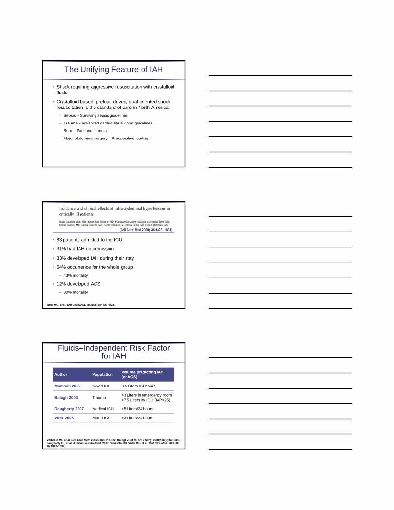
The Unifying Feature of IAH
• Shock requiring aggressive resuscitation with crystalloid fluids
• Crystalloid-based, preload driven, goal-oriented shock resuscitation is the standard of care in North America
– Sepsis – Surviving sepsis guidelines
– Trauma – advanced cardiac life support guidelines
– Burn – Parkland formula
– Major abdominal surgery – Preoperative loading
• 83 patients admitted to the ICU
• 31% had IAH on admission
• 33% developed IAH during their stay
• 64% occurrence for the whole group
– 43% mortality
• 12% developed ACS
– 80% mortality
Vidal MG, et al. Crit Care Med. 2008;36(6):1823-1831.
Fluids–Independent Risk Factorfor IAH
Author PopulationVolume predicting IAH (or ACS)
Malbrain 2005 Mixed ICU 3.5 Liters /24 hours
Balogh 2003 Trauma>3 Liters in emergency room>7.5 Liters by ICU (IAP>25)
Daugherty 2007 Medical ICU +5 Liters/24 hours
Vidal 2008 Mixed ICU +3 Liters/24 hours
Malbrain ML, et al. Crit Care Med. 2005;33(2):315-322. Balogh Z, et al. Am J Surg. 2003;186(6):602-608. Daugherty EL, et al. J Intensive Care Med. 2007;22(5):294-299. Vidal MG, et al. Crit Care Med. 2008;36(6):1823-1831.

Where DoesResusitation Fluid Go?
Right Here!
Clinical Reality
With the clinical information availableabout the relationship of IAH and organ failure
to allow a patient to progress to ACSis the equivalent of finding a
tension pneumothorax on a chest x-ray
Compartment Syndromes
• There is a clear relationship to over aggressive fluid resuscitation with crystalloids and the development of compartment syndromes
• While there may be isolated development of compartment syndromes; “multiple compartment syndromes can occur as a result of a futile attempt to optimize circulation with preload driven resuscitation with crystalloids”
• Poly-compartment syndrome
Balogh Z. Critical Care Med. 2010;38(Suppl):S445-S451.
Fluid Resuscitation and IAH
• Goal: Balanced resuscitation
– “Enough but not too much”
• Utilizing both IAP and volume measurements allows judgment of when “enough” has been given
– Once IAP and central venous pressure/pulmonary capillary wedge pressure starts to rise, total body fluids are sufficient (or excessive) and you need to try something else –step out of the futile crystalloid preloading cycle
• Still proceed towards early, goal-directed treatment, but utilize IAP to assist in decision making

Pitfall: Lack of a Resuscitation Protocol
• Establish a protocol to include:
– Minimize use of crystalloids with increased use of 1:1:1; Blood: FFP:PLTS
– Use of end points of resuscitation as a guide to fluid management: BD, SVo2, Lactate, RVEDVI
• Mandatory serial bladder pressure monitoring in high-risk patients
• Indiscriminate use of crystalloids in resuscitation is the leading cause of IAH/ACS
Back to the Question: When Is Decompression TOO Late
• When ACS develops:
– ACS is preventable and is due to uncontrolled IAP or lack of monitoring
• Not recognizing at-risk patients for IAH
• Patients that have had large volumes of crystalloid with a rapidly increasing IAP that require decompression before the consequences of ACS
• Not monitoring patients at risk for secondary ACS and allowing progression to organ failure
Chovanes J, et al. Surg Clinc N Am. 2012;92(4):859-875.
Management Failures in ACS/IAH
• The reasons for failure of management is an out of control inflammatory response that is “self-sustaining”
• Current practices exacerbate the inflammatory/pressure state
• Established an equilibrium between inflammation and maintenance of organ perfusion

Survival Triad Acute Abdominal Injuries and Sepsis
Wound Biology
Patient Survival
Complications:hernia, fistula, ACS,
infection
InflammatoryState
Inflammatory Response
• Key to understanding the inter-relationship of IAH/ACS
• Root cause of most complications seen in septic and traumatic states
• Mimimizing the effect of IAH/ACS and control of sepsis and mimimizing crystalloid is the goal of TMOA
Effects of Increased IAP
• Increased IAP produces
– Decreases in bowel submucosal TPO2 without similar changes in extra-abdominal TPO2
– Severe intestinal ischemia
– Decreases in cardiac index
– Increases in pulmonary artery occlusion
TPO2 = bowel tissue oxygenation.Bongard F, et al. J Trauma. 1995;39(3):519-522. Diebel LN, et al. J Trauma. 1992;33(1):45-49. Ridings PC, et al. J Trauma. 1995;39(6):1071-1075.

Ischemia/Reperfusion Injury
Hemorrhagic Shock
Resuscitation
Global Ischemia/Reperfusion Injury
Increased Capillary Permeability
Hydrostatic Edema
Mesenteric VenousHypertension(Secondary to bdominal Packing)
Resuscitation
IncreasedHydrostaticPressures and Decreased Oncotic Pressures
HydrostaticIntestinalEdema
Pathophysiology of ACS
Injury
∆ MicroQ
IAPVas Perm
LymphQ
Edema &Ascites
More Edema& Ascites
Inflammation
ACS

Are Lymph and Ascites Toxic?
• Toxic lymph
– Ligate mesenteric lymph duct
Lung injury
Neutrophil activation
Mortality
– Mesenteric lymph EN apoptosis and perm
• Toxic ascites
– Peritoneal NPT (remove ascites)
• Reduced systemic inflammation
• Reduced organ injury (MODS)
• Reduced mortality
NPT = negative pressure therapy; MODS = multiple organ dysfunction syndrome.Barlos D, et al. Am J Physiol Lung Cell Mol Physiol. 2009;296(3):L404-L417. Kubiak BD, et al. Shock. 2010;34(5):525-534.
Peritoneal and Systemic Inflammation
Plasma TNF
Kubiak, et al. Critical Care. Submitted.
NPT vs PD Survival
83% (4/5)
50% (3/3)
Kubiak, et al. Critical Care. Submitted.

Ivatury RR, et al. J Trauma. 1998;44(6):1016-1021.
IAH, MOF, and Mortality
No IAH(n = 47)
IAH(n = 23)
P
Mortality 4 (8.5%) 10 (43.5%) .006
MODS 0.8 ± 1.9 4.3 ± 3.7 .0001
Raeburn CD, et al. Am J Surg. 2001 Dec;182(6):542-6.
ACS, MOF, and Mortality
No ACS(n = 49)
ACS(n = 28)
P
Mortality 12% 43% .01
MOF 8% 32% . 01
Prevention of ACS
• ACS and complications of the open abdomen is potentially preventable in most cases can be minimized by:
– Closely monitoring of abdominal pressure
– Using an open abdomen technique in high-risk patients
– Controlling IAP in the open abdomen
– Early decompression with trends of IAP leading to ACS
51
Monitoring IAP:The Key to Early Intervention
52
IAP Monitoring
• Clinical judgment
• Homemade pressure transducer technique
• Standard IAP monitoring kit
53
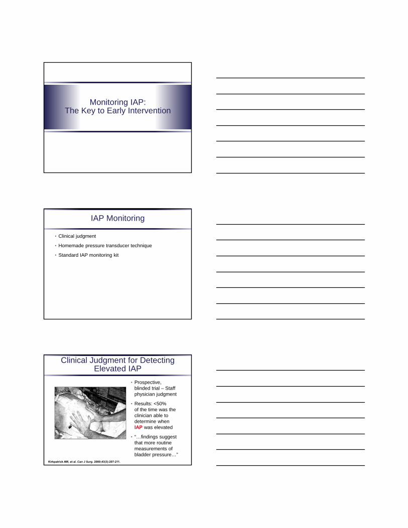
Clinical Judgment for Detecting Elevated IAP
• Prospective,blinded trial – Staff physician judgment
• Results: <50%of the time was the clinician able to determine when IAP was elevated
• “…findings suggest that more routine measurements of bladder pressure…”
Kirkpatrick AW, et al. Can J Surg. 2000;43(3):207-211.

Pitfalls in IAP Monitoring
• Improper placement of the transducer: zeroed at midaxillary line
• Over distention of the bladder
• When using manometric fluid measurements, mm H2O need to be converted to mm Hg for readings consistent with guidelines
55
Pitfall IAH/ACS Management: Positioning
Vasquez DG, et al. J Surg Res. 2007;139(2):280-285.
Stretch out
Head-of-Bed Elevation (degrees)
Overweight
Obese
Normal
30
25
20
15
10
5
0
15 30 30 + 15 tilt
mm
Hg
All
450
Pitfall: Over Distention of Bladder
“The reference standard forintermittent IAP measurement
is via the bladder with amaximal instillation volume of
25 mL sterile saline.”
WSACS.org
Standard IAP Monitoring Kit
Closed system in-line with the foley catheter
• Once attached, it is left in place during entire time IAP is measured
• 30 seconds to measure IAP
• Standardized measurement
• No reproducibility errors
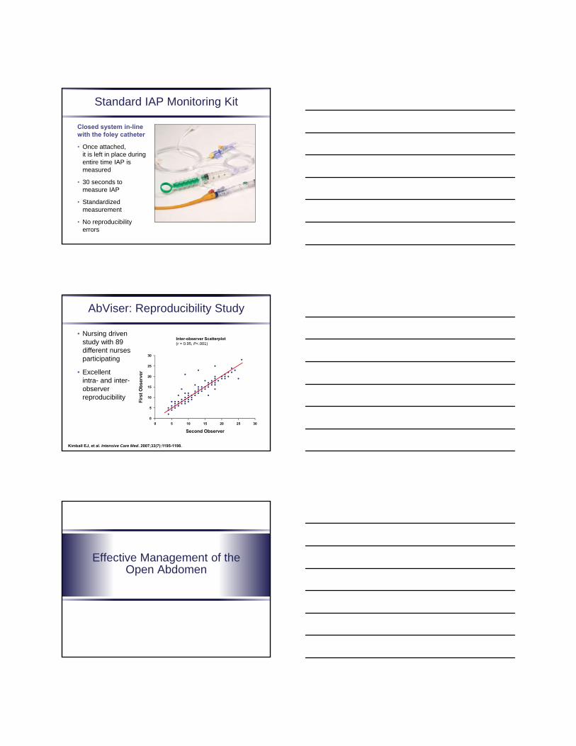
AbViser: Reproducibility Study
• Nursing driven study with 89 different nurses participating
• Excellent intra- and inter-observer reproducibility
59Kimball EJ, et al. Intensive Care Med. 2007;33(7):1195-1198.
Second Observer
Inter-observer Scatterplot (r = 0.95, P<.001)
30
25
20
15
10
5
0
10 20 30
Fir
st
Ob
se
rve
r
0 15 255
Effective Management of theOpen Abdomen

Use of an Effective TAC Protocol
• Low trigger to leave abdomen opened
• Planned early closure
• Use of an effective TAC device that will
– Effectively reduce cytokines
– Minimize IAP to physiologically acceptable levels
– Minimize abdominal retraction
– Minimize bowel fixity
Evolution In Management of theOpen Abdomen
• Initially used to control intra-abdominal contents and were static
• Evolution of devices that are more dynamic with recognition that multiple factors must be controlled while an abdomen is opened
• Including: inflammatory fluid, minimize IAH, and preserve the abdominal wall
• Early closure has been shown to decrease complications and improve overall outcomes
• A standardized approach is the key to controlling the physiology
TMOA
• Various techniques achieve temporary closure
• TMOA manages complex biologic factors observed in the open abdomen
– Inflammatory response
– Wound healing/mechanics of the abdominal wall
– Improve closure and decrease complications
– Resuscitation (minimizing crystalloid)
• Cornerstone of an integrated approach incorporating a comprehensive management guideline

Damage Control Paradigm
Traditional Resuscitation Damage Control
Aggressive crystalloidBalanced resuscitation• Crystalloid/colloid• Vasoactive medications
Surgical management onlyComprehensive medical and surgical management
IAP measurements only to predict decompression
IAP-guided therapy
Emergent decompression Prophylactic decompression
Decompression for IAP >30 mm Hg to 40 mm Hg
Decompression for IAP >25 mm Hg
Late closure Early closure
Cheatham,M.: personal communication
DSL#12‐0169 (3/12) 65J Trauma Acute Care Surg1380 Volume 73, Number 6, 2012
Types of TAC
• Bogota Bag
• Absorbable mesh – Vicryl®
• Nonabsorbable – Marlex®, Gortex®
• “Vac Pac” closure
• ABThera™ Open Abdomen NPT System
66

Methods of TAC
Desired Goal
Keys To Successful Management
1. Open early to avoid IAH/ACS
2. Prevent visceral adherence to the abdominal wall
– Prevention starts at the first operation!
– Maintain your options for subsequent closure
3. Prevent lateralization and loss of abdominal domain
– Actively combat fascial retraction
4. Avoid enteric fistula formation
– Resuscitate early and properly
– Avoid elevated IAP!
5. Start closing as soon as physiologically reasonable

Vacuum Pack Technique(Barker VAC Pac)
• 4-year experience from University of Tennessee College of Medicine in 1997; reviewed 93 patients with vacuum assisted abdominal closure with ultimate STSG
• Reviewed the experience with 112 trauma patients in 2000
• 55% had primary closure
STSG = split-thickness skin graft.Smith LA, et al. Am Surg. 1997;63(12):1102-1107. Barker DE, et al. J Trauma. 2000 ;48(2):201-206.
Hydrostatic BowelEdema
Inflammation Toxic Ascites
AbThera™
Open Abdomen
ACSIAH
Barker “Vacuum-Pack”:Uneven Distribution of Negative Pressure
APPLIED SUCTION = -125 mm Hg
Actual Intra-abdominal Suction
Sammons A, et al. Presented at: Clinical Symposium on Advances in Skin and Wound Care; October 22-25, 2009; San Antonio, Texas. Abstract 78.

ABThera™:Even Distribution of Negative Pressure
APPLIED SUCTION = -125 mm Hg
Actual IAP
Sammons A, et al. Presented at: Clinical Symposium on Advances in Skin and Wound Care; October 22-25, 2009; San Antonio, Texas. Abstract 78.
Pressure at the Bowel Surface during Topical NPT of the Open Abdomen
• Porcine model to study pressure at bowel surface with ABThera™
• Pressures at -50,-75,-100,-125, and -150 mm Hg
• Pressures at outer foam corresponded with applied pressure
• Median pressure at the bowel surface was between -2 mm Hg and -10 mm Hg regardless of the surface pressure
Bjarnason T, et al. World J Surg. 2011;35(4):917-923.
Are Commercial Negative Pressure Systems Worth the Cost in Open Abdomen Management?
• Retrospective review
• 74 patients treated with a TAC
• 37 patients with Barker Vacuum Pack Technique (2009-2010)
• 37 patients with ABThera™ NPT (2010-2011)
• Age and BMI higher ABThera™, all other variable same
• 33/37 (89%) successful midline closure with ABThera™
• 22/37 (59%) successful midline closure with Barker
• P < .05, odds ratio 7.97 favoring ABThera™
Frazee RC, et al. J Am Coll Surg. 2013;216(4):730-743.

Components
• Nonadherentpolyurethane fenestrated inner
– Prevents bowel from adhering to the abdominal wall
– Allows for fluid drainage (improved with ABThera™ Dressing)
– Prevents V.A.C.®
GranuFoam™
Dressing from sticking to bowel
Components
• Blue foam – V.A.C.®
GranuFoam™
Dressing
– Open cell hydrophobic dressing
– Negative pressure manifold
– Manifolds negative pressure, thus facilitating the closure of the abdominal wall
Components
• ABThera™ SensaT.R.A.C.™ Open Abdomen Dressing Compatible with InfoV.A.C.® and V.A.C.ATS® Therapy System
– To maintain constant negative pressure at the wound site not in the canister
• Interface pad is used with ABThera™ open abdomen NPT system
• Adherent drape—water tight seal
• Use of the adherent drape is key to a negative pressure environment

Placement of Visceral Protective Layer
• Full deployment to all gutters
• Can be placed without cutting
• Use of a blunt instrument to help placement
The image part with relationship ID rId4 was not found in the file.
Cutting and Sizing of VPL
• Cut plastic VPL to place around colostomies and feeding tubes
• If cutting the VPL is needed, cut in the center of the square foam and pull off
VPL = visceral protective layer.
Proper Placement of the VPL
81

Placement of Foam
• Fig.1 Placement of entire piece of foam over the VPL and under the fascia
• Fig. 2 Place the second piece of foam to conform to the skin
• Fig. 3 Applied adhesive to skin, place the trac pad and apply suction
The image part with relationship ID rId2 was not found in the file.
Fig.1 Fig. 2
Fig. 3
Fig. 1
Placement of Adhesive Layer
• 2-person placement
• Placed transversely and not pulled tight
• Place in a shingled pattern
Personal Clinical Experience with ABThera™ Open Abdomen NPT
• Simpler than the Barker “vacuum-pack”
– Placement in the gutters is easier
– “VPL” stays in place
– Fixity is less
• The limbs of the ABThera™ nonadherent polyurethane fenestrated inner can be cut to reach specific areas of concern
• The open abdomen is much drier with better fluid removal
• The suction pump is much simpler

Abdomen Allowed to Granulate with Skin Graft
Facilitating Fascial Closing
“If you fail to plan, you plan to fail.”
Begin planning your closure as soon as you open the abdomen
Enteric Fistulas
• The dreaded complication of the open abdomen
• Most likely a complication of
– Inadequate resuscitation
– Visceral malperfusion
– Incorrect TAC management
– Exposed anastomoses and tubes

Enteric Fistula Complication
• Recent studies suggest that TAC is NOT the culprit Prospective, 5-year study of NPT
– 4.7% fistula rate
• Prospective, 6-year study of various TACs
– 5.0% fistula rate
– Fistula rate correlates with
• Timing of abdominal decompression
• Closure type
• Resuscitation algorithm
Shaikh IA, et al. Colorectal Dis. 2010;12(9):931-934. Cheatham ML, et al. Crit Care Med. 2010;38(2):402-407.
Total Open Abdomen Management
Comprehensive abdominal management significantly decreases complications and increases patient survival
Open early, close early!
APACHE = Acute Physiology and Chronic Health Education.Cheatham ML, et al. Crit Care Med. 2010;38(2):402-407.
YearAPACHE
2Days to Closure
FistulaPrimary Closure
STSG Survival
2002 23 ± 9 22 ± 21 16% 59% 13% 50%
2003 20 ± 8 17 ± 19 13% 72% 23% 57%
2004 22 ± 8 17 ± 16 3% 69% 15% 53%
2005 22 ± 8 16 ± 17 9% 72% 19% 63%
2006 20 ± 9 13 ± 16 8% 76% 12% 70%
2007 20 ± 9 10 ± 10 3% 81% 3% 72%
Financial Implications of Closure
Earlier intervention = decreased resource utilization
Cheatham ML, et al. Crit Care Med. 2010;38(2):402-407.
Primary Staged Mesh Skin Only STSG
ICU days 11 ± 12 21 ± 14 24 ± 14 23 ± 14 32 ± 19
Hospital days 25 ± 21 42 ± 21 44 ± 20 49 ± 22 70 ± 39
Ventilator days 9 ± 10 19 ± 12 23 ± 16 20 ± 16 31 ± 23
Days to closure 5 ± 4 19 ± 9 16 ± 11 21 ± 14 39 ± 23
Fistula (%) 1.3 1.3 20 11 36
Charges ($1000)
$227±
206
$378±
209
$491±
279
$459±
274
$598±
335

Philadelphia Bag
• There are clinical situations, because of loss of domain, in which packing and severe bowel edema will not allow the full ABThera™ Open Abdomen NPT system to be applied
• A modification of the system can be used to place only the VPL sutured to the skin with suction applied
• This allows for the application of NPT without exacerbating the extreme bowel and abdominal wall edema
VPL toskin
Bluefoam strips
Interface pad
Adherent layer
Transabdominal GSW
• GSW abdomen
• Hypotensive
• GCS 10
• BP 90 systolic
• Lactate 5.0
GSW = gun shot wound; GCS = Glasgow Coma Scale; BP = blood pressure.

Injuries
The image part with relationship ID rId2 was not found in the file.
Combinedgrade 4 colon injury,grade 3 liver injury,
grade 4 pancreaticodudenalinjury
Damage Control and ABTheraTM
Traumatic Whipple Resection
Patient was taken back to the OR for serial explorations andhad a colectomy with ostomy, Whipple resection and
primary closure of the abdomen in one week.

Shot Gun Blast Abdomen
• Point blank shot gun blast
• Eviscerated bowel on presentation
• Damage control procedure
97
Initial Procedure: Damage Control
Injuries right colon, blast and tissue loss abdominal wall
The image part with relationship ID rId2 was not found in the file.
Blast Effect Silver Foam
99
The image part with relationship ID rId2 was not found in the file.

Return to OR Closure Midline Incision
Patient had midline closed with interrupted sutures and Alloderm underlay to close abdominal wall defect.
Silver foam placed over the repair.
Creation of Skin Flaps and Closure
Secondary ACS

Bowel Ischemia with Increased IAP
48 Hours ABTheraTM
Patient had 48 hours of AbTheraTM placement that reduced the edema and allowed for primary closure of
the fascia
Grade 4 Liver Laceration, DCL, Packing

Second Return Omental Packing
Third Return to the OR
Fourth Return to OR

Closure Abdominal Wall
Summary
• Management of an open abdomen is complex
• Just “keeping the bowel in” is not reflective of the biology of an open abdomen
• The open abdomen is the engine for systemic complications: systemic inflammatory response syndrome, MODS
• TMOA is designed to manage the patient