tamer belal, md,phd lecturer of neurology mansoura university hospitals eeg teaching courses
TRANSCRIPT


Tamer Belal, MD ,PHDLecturer of Neurology
Mansoura University Hospitals
EEG Teaching Courses

Uses of ambulatory EEG
Evaluation of interictal epileptiform activity
Documentation of seizures of which patients are unaware
Evaluation of response to therapy
Evaluation of nocturnal or sleep-related events
Evaluation of syncope
Evaluation of suspected pseudoseizures

Uses of ambulatory EEG

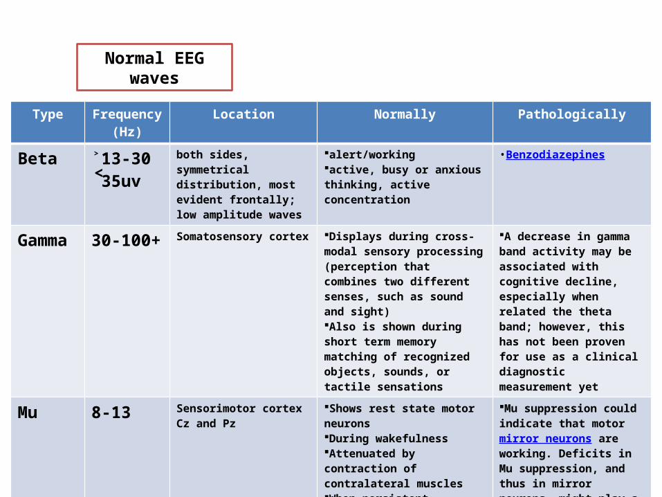
Normal EEG waves

Type Frequency (Hz)
Location Normally Pathologically
Delta 0-4 frontally in adults, posteriorly in children; high amplitude waves
adults slow wave sleep (deep sleep)in babiesHas been found during some continuous attention tasks
subcortical lesionsdiffuse lesionsMetabolic encephalopathy hydrocephalusdeep midline lesions
Theta 4-8 Found in locations not related to task at hand
young childrendrowsiness or arousal in older children and adultsAssociated with inhibition of elicited responses (has been found to spike in situations where a person is actively trying to repress a response or action)
focal subcortical lesionsmetabolic encephalopathydeep midline disorderssome instances of hydrocephalus
Alpha 8-13(5-100uv)
posterior regions of head, both sides, higher in amplitude on non-dominant side. Central sites (c3-c4) at rest
Relaxed/reflectingClosing the eyesAlso associated with inhibition control, seemingly with the purpose of timing inhibitory activity in different locations across the brain.Attenuated by eye opening, attention and mental effort (Alpha block)
Alpha Coma (unresponsive)Paradoxical alphaInterside differences˃50% (lt)Unilateral failure of the alpha rhythm to attenuatereflects an ipsilateral abnormality (Bancaud’s phenomenon
Normal EEG waves
Type Frequency (Hz)
Location Normally Pathologically
Beta ˃13-30˂35uv
both sides, symmetrical distribution, most evident frontally; low amplitude waves
alert/workingactive, busy or anxious thinking, active concentration
•Benzodiazepines
Gamma 30-100+ Somatosensory cortex Displays during cross-modal sensory processing (perception that combines two different senses, such as sound and sight)Also is shown during short term memory matching of recognized objects, sounds, or tactile sensations
A decrease in gamma band activity may be associated with cognitive decline, especially when related the theta band; however, this has not been proven for use as a clinical diagnostic measurement yet
Mu 8-13 Sensorimotor cortexCz and Pz
Shows rest state motor neuronsDuring wakefulnessAttenuated by contraction of contralateral musclesWhen persistent, unreactive, and associated with focal slowing, mu like frequencies are abnormal
Mu suppression could indicate that motor mirror neurons are working. Deficits in Mu suppression, and thus in mirror neurons, might play a role in autism
Normal EEG waves

Normal EEG waves
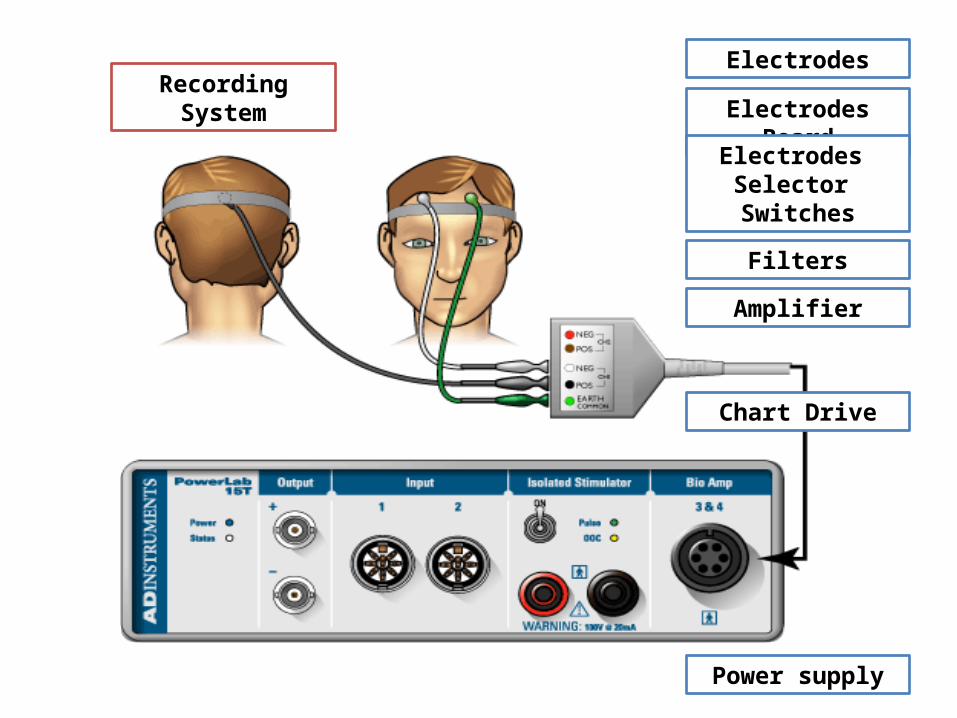
Recording SystemElectrodes
Electrodes Board
Electrodes Selector Switches
Filters
Amplifier
Chart Drive
Power supply

Recording System

Sleep and EEG

EEG and states of Arousal
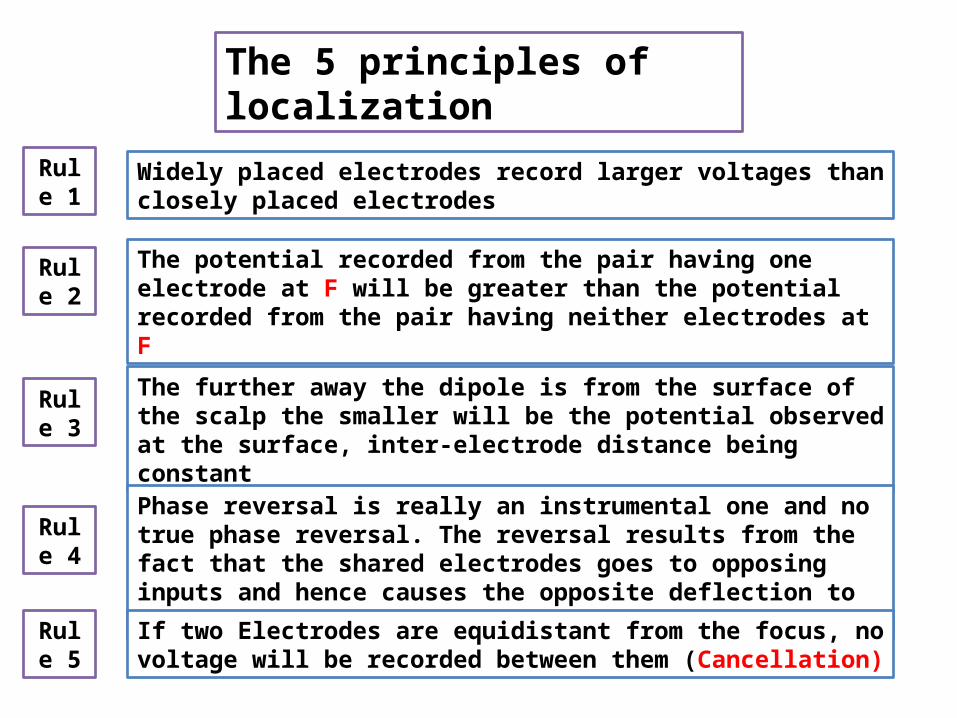
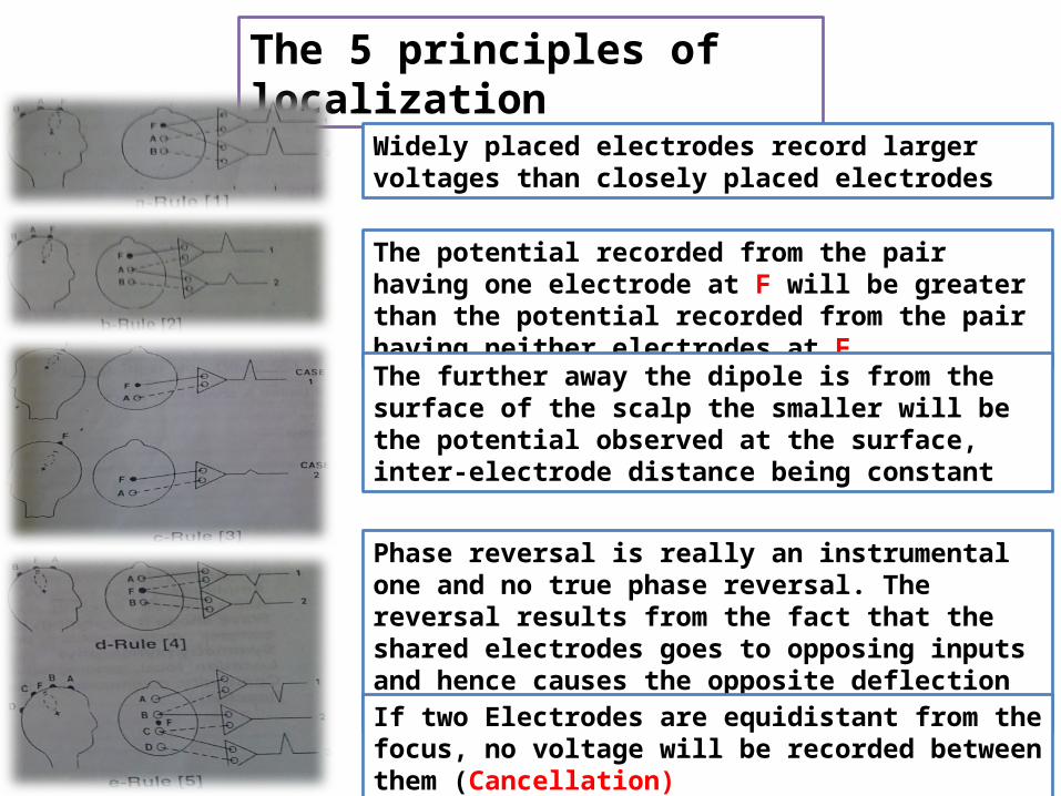
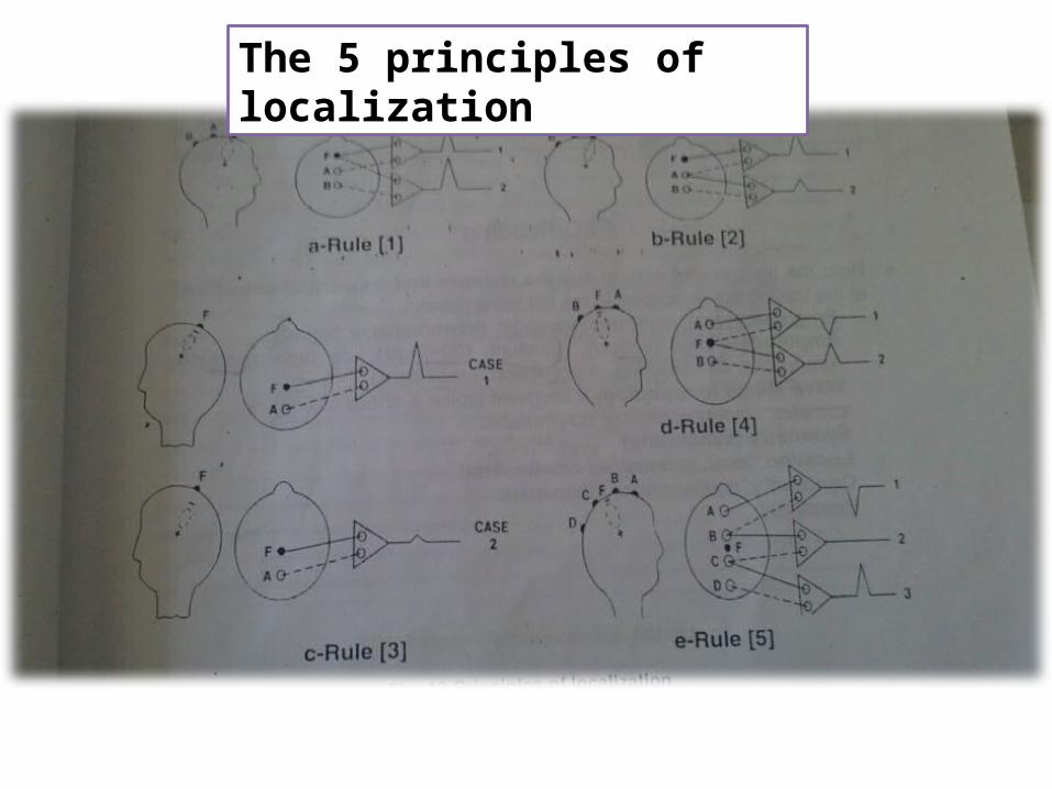
Widely placed electrodes record larger voltages than closely placed electrodes
The potential recorded from the pair having one electrode at F will be greater than the potential recorded from the pair having neither electrodes at F
The further away the dipole is from the surface of the scalp the smaller will be the potential observed at the surface, inter-electrode distance being constant
Phase reversal is really an instrumental one and no true phase reversal. The reversal results from the fact that the shared electrodes goes to opposing inputs and hence causes the opposite deflection to occur
If two Electrodes are equidistant from the focus, no voltage will be recorded between them (Cancellation)
Rule 1
Rule 2
Rule 3
Rule 4
Rule 5
The 5 principles of localization
The 5 principles of localization
Widely placed electrodes record larger voltages than closely placed electrodes
The potential recorded from the pair having one electrode at F will be greater than the potential recorded from the pair having neither electrodes at F
The further away the dipole is from the surface of the scalp the smaller will be the potential observed at the surface, inter-electrode distance being constant
Phase reversal is really an instrumental one and no true phase reversal. The reversal results from the fact that the shared electrodes goes to opposing inputs and hence causes the opposite deflection to occur
If two Electrodes are equidistant from the focus, no voltage will be recorded between them (Cancellation)
The 5 principles of localization
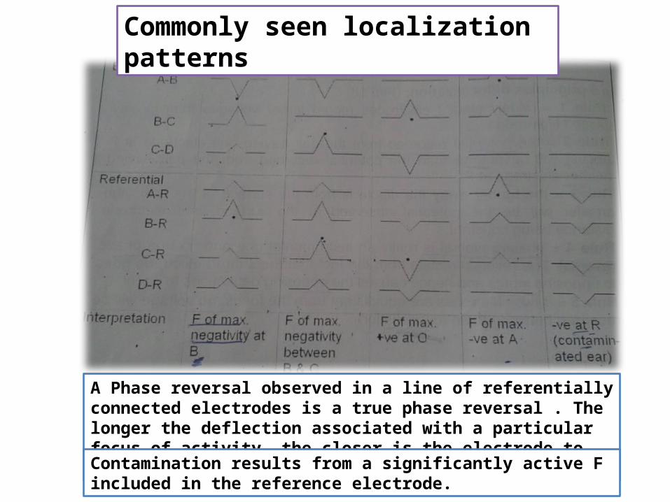
Commonly seen localization patterns
A Phase reversal observed in a line of referentially connected electrodes is a true phase reversal . The longer the deflection associated with a particular focus of activity, the closer is the electrode to the focus
Contamination results from a significantly active F included in the reference electrode.

Commonly seen localization patterns
(A) Bipolar montage demonstrating phase reversal and (B) referentialmontage demonstrating absolute voltage.
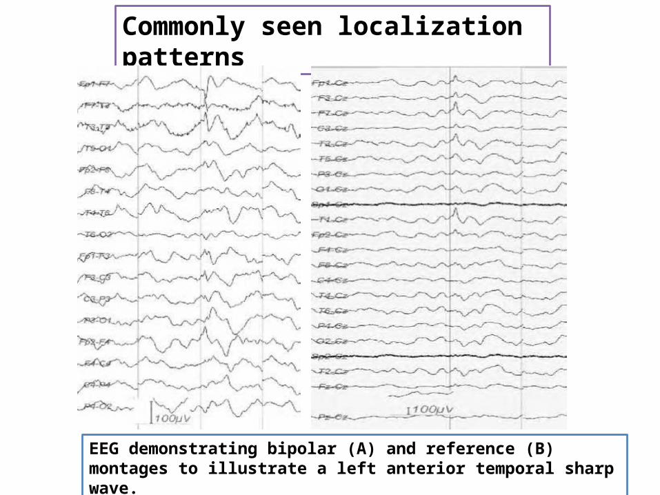
Commonly seen localization patterns
EEG demonstrating bipolar (A) and reference (B) montages to illustrate a left anterior temporal sharp wave.
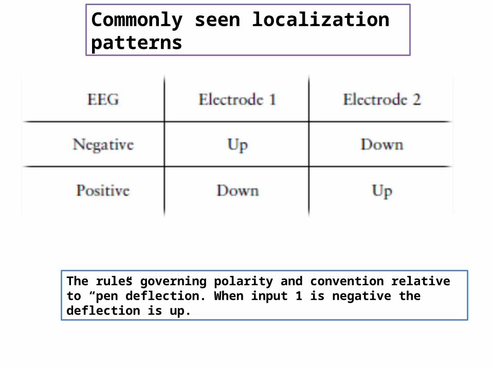
Commonly seen localization patterns
The rules governing polarity and convention relative to “pen”deflection. When input 1 is negative the deflection is up.

EEG Reading
Both the background activity and the changes that appear in the features of the tracing are described in the following terms
Frequency: fast, slow, monomorphic, polymorphic or periodic
Amplitude :low ˂20uv, Medium 20-5-uv, high˃50uvAttenuation and blocking, suppression , paroxysmal
Wave shape (morphology) : transients (sharp, spike) or complex, monomorphic , polymorphic
Symmetry (synchrony)
Location : focal , generalized or lateralized
Continuity : continuous or intermittent
Reactivity

Writing the EEG report
Two parts1- Actual description f the EEG findings and their interpretation2- Clinical correlation that render the report meaningful
Attempt to correlate the EEG with clinical picture
Brief history of the clinical findings today
Mention what the referring physician hope to find out
Descriptive details regarding the testing situation
Describe the state of the patient
Describe the EEG ( just descriptive)
Impression : normal or abnormal and define abnormality
Suggest further study if needed

Writing the EEG report
The EEG was recorded with the standard 10-20 system of electrode placement. The patient was awake and cooperative.
EEG Report : Background activity comprises of alpha activity 9-10 c/s, which is symmetrical in the occipital leads and spreading anteriorly interspersed with fast beta activity. No paroxysmal activity seen. Hyperventilation and photic stimulation is non-contributory.
IMPRESSION: Normal record. No epileptiform activity seen. Clinical correlation advised.
Note: A normal EEG does not rule out the diagnosis of epilepsy, as epileptiform discharges may be paroxysmal.

Abnormal EEG Patterns
Abnormality of background rhythm
Abnormal sleep patterns
Abnormal slow activity:Generalized intermittent slow activityFocal and lateralized intermittent slow activityPersistent slow activity
Paroxysmal epileptogenic abnormalitiesInter-ictal epileptiform discharges( focal, generalized)IctalSecondary bilateral synchronyEpileptiform patterns of doubtful significance
Abnormal periodic paroxysmal patternsGeneralized periodic paroxysmal patterns
SSPE,CJD,Herpes S E, suppression patterns, Triphasic wavesLateralized periodic paroxysmal patterns
PLEDS,BPLEDS

The Normal EEG Patterns
Normal 10-Hz alpha rhythm “blocked” by eye opening and returning on eye closure. Note the faster frequency immediately on eye closure (“squeak”).
Alpha rythm Alpha frequency
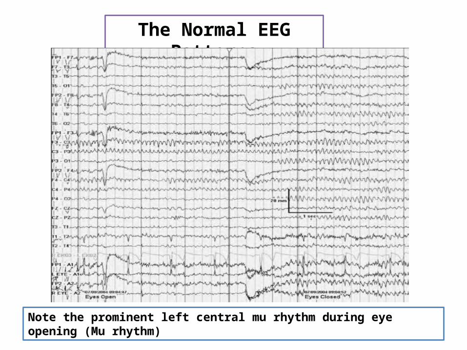
The Normal EEG Patterns
Note the prominent left central mu rhythm during eye opening (Mu rhythm)

The Normal EEG Patterns
Breach rhythm in the right temporal region (maximal at T4) following craniotomy for temporal lobectomy

The Normal EEG Patterns
Normal frontocentral theta rhythm in an 18-year-old patient while awake.
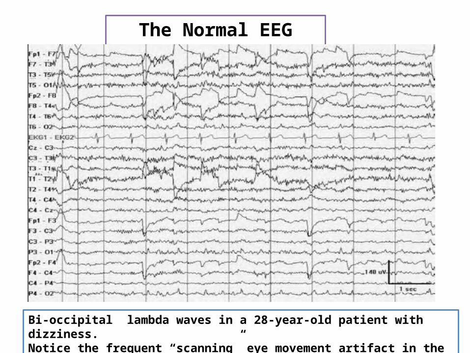
The Normal EEG Patterns
Bi-occipital lambda waves in a 28-year-old patient with dizziness.Notice the frequent “scanning” eye movement artifact in the F7 and T8 derivations.
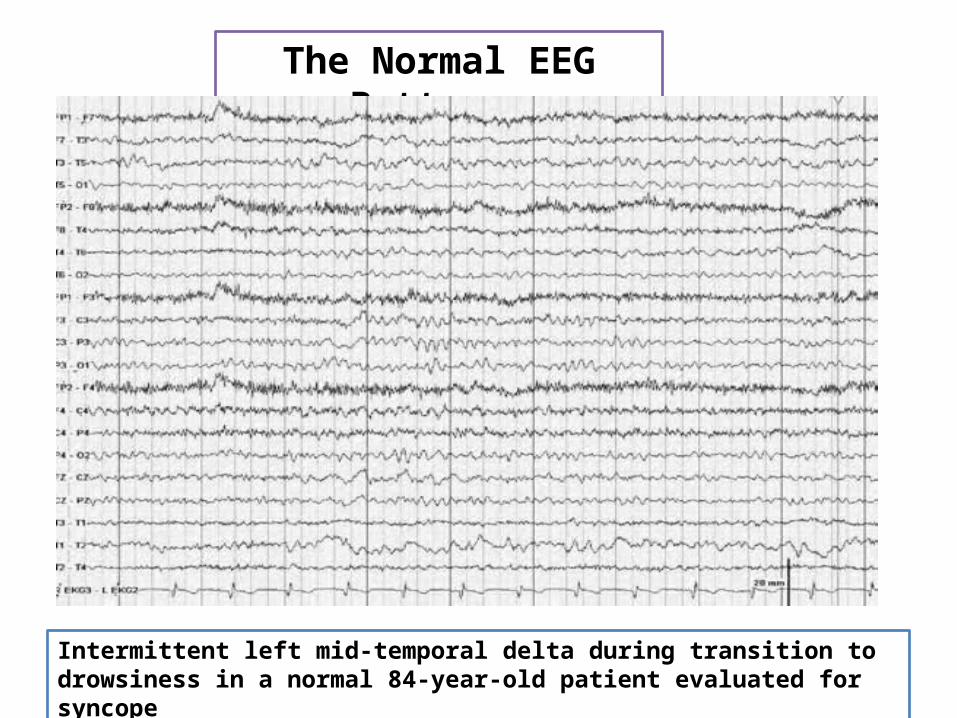
The Normal EEG Patterns
Intermittent left mid-temporal delta during transition to drowsiness in a normal 84-year-old patient evaluated for syncope

The Normal EEG Patterns
POSTS appearing in the lower three channels in a bipolar circle montage demonstrating positive polarity in the occipital region during sleep. Notice the surface negative vertex waves maximal at Cz
NORMAL SLEEP ARCHITECTURE
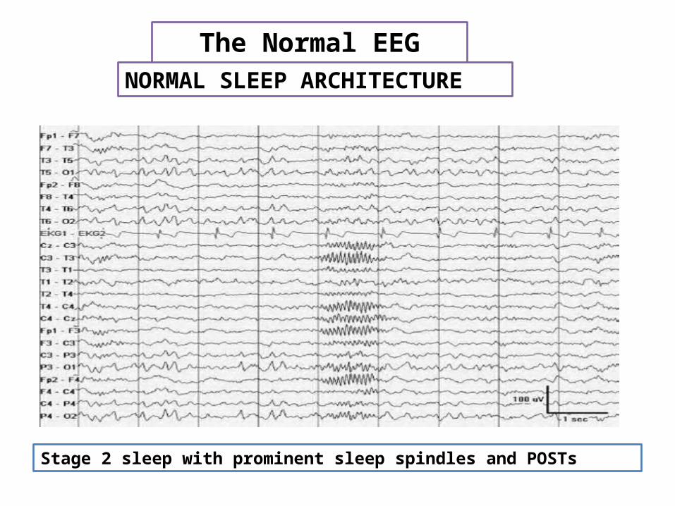
The Normal EEG Patterns
Stage 2 sleep with prominent sleep spindles and POSTs
NORMAL SLEEP ARCHITECTURE

The Normal EEG Patterns
Slow-wave sleep. Note the intermittent POSTs and sleep spindles against the continuous delta background
NORMAL SLEEP ARCHITECTURE

The Normal EEG Patterns
REM sleep with rapid eye movements associated with lateral rectus spikes is noted at the F7 and F8 derivations
NORMAL SLEEP ARCHITECTURE
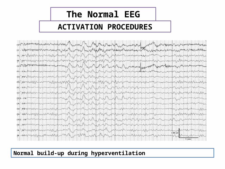
The Normal EEG Patterns
Normal build-up during hyperventilation
ACTIVATION PROCEDURES
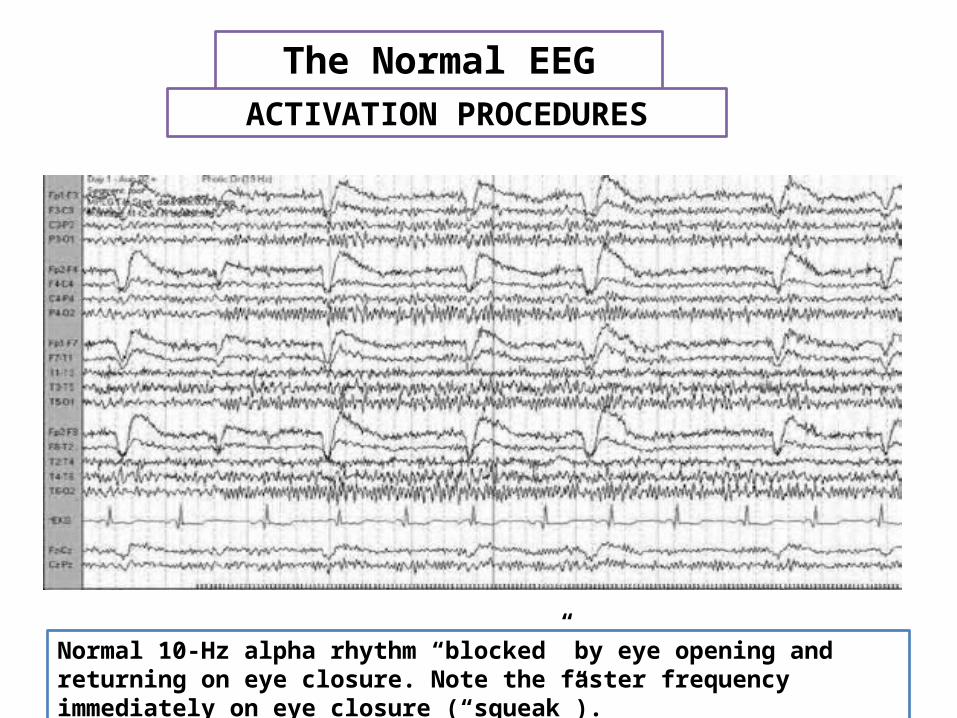
The Normal EEG Patterns
Normal 10-Hz alpha rhythm “blocked” by eye opening and returning on eye closure. Note the faster frequency immediately on eye closure (“squeak”).
ACTIVATION PROCEDURES

The Normal EEG Patterns
Rhythmic temporal theta bursts of drowsiness. Note the sharply contoured morphology.
BENIGN VARIANTS OF UNCERTAIN SIGNIFICANCE

The Normal EEG Patterns
Central theta (maximal at Cz) seen during the awake state in a 35-year-old patient with migraine headaches
BENIGN VARIANTS OF UNCERTAIN SIGNIFICANCE

The Normal EEG Patterns
A 6-Hz (phantom) spike-wave burst with frontal predominance in the 5th second of this EEG in an awake patient with temporal lobe epilepsy.
BENIGN VARIANTS OF UNCERTAIN SIGNIFICANCE
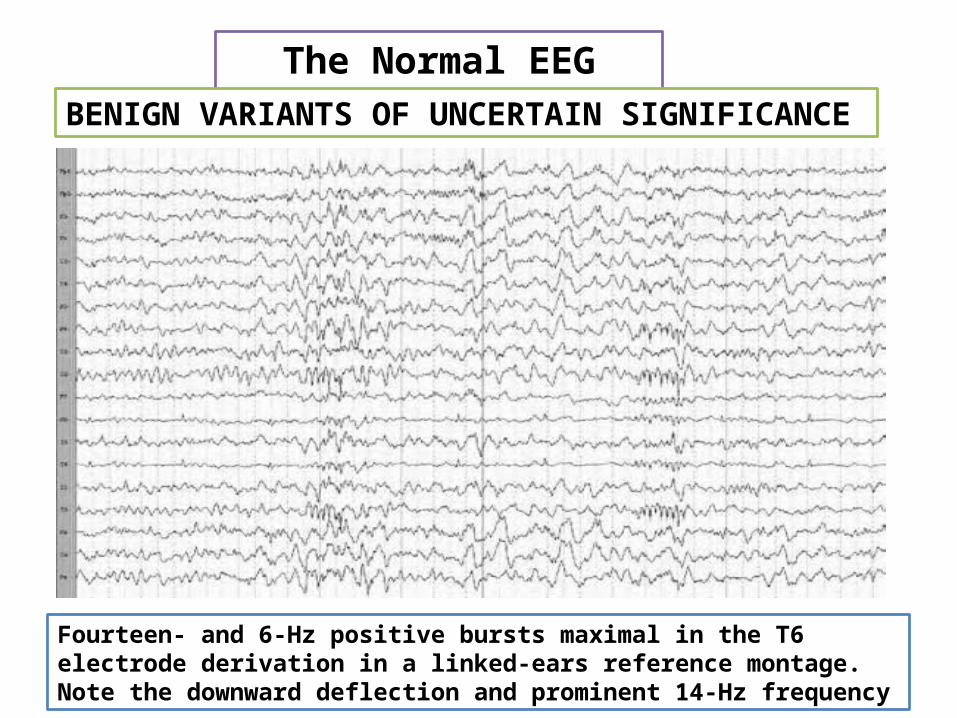
The Normal EEG Patterns
Fourteen- and 6-Hz positive bursts maximal in the T6 electrode derivation in a linked-ears reference montage. Note the downward deflection and prominent 14-Hz frequency
BENIGN VARIANTS OF UNCERTAIN SIGNIFICANCE
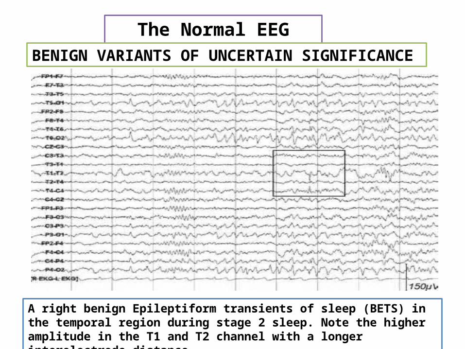
The Normal EEG Patterns
A right benign Epileptiform transients of sleep (BETS) in the temporal region during stage 2 sleep. Note the higher amplitude in the T1 and T2 channel with a longer interelectrode distance
BENIGN VARIANTS OF UNCERTAIN SIGNIFICANCE

The Normal EEG Patterns
Wicket waves maximal at T3 and T4
BENIGN VARIANTS OF UNCERTAIN SIGNIFICANCE

Normal EEG
Variants

Normal EEG Variants
Refer to waves that are rare or unusual but not generally abnormal. They may be unusual in shape or in distribution.
wave mixtures that can appear unusual and can confuse the casual reader (for example, wave harmonics)
They can include
Artifacts or electrical disturbances from structures that are not in or part of the brain and do not affect the brain or its function but appear in the EEG tracing

Psychomotor variant (rhythmic harmonic theta)
14- and 6-Hz waves
Mu (rhythm en arceau or wicket rhythm, arciform rhythm )
Normal EEG Variants
Odd-Looking Waveforms
Small sharp spikes of sleep (SSS) , benign epileptiform transients of sleep (BETS). posterior occipital transients of sleep POSTS
6-Hz spike and wave (phantom spike and wave)
Wicket spikes
Subclinical rhythmic EEG discharges in adults
Rhythmic midline theta

Psychomotor variant (rhythmic harmonic theta)
Asymmetrical runs of theta or delta activity primarily in the mid-temporal regions, lasting for a few seconds or as long as 30-45
occurs in 0.5% to 2.0% of selected normal adults and consists of bursts or runs of 5- to 7-Hz theta waves that may appear sharp, flat, or notched in appearance
It starts suddenly on 1 side and lasts for several seconds before terminating suddenly. This behavior resembles a seizure discharge, hence the name "psychomotor variant."
Generally considered benign, this waveform does not correlate with seizure disorder. It is best seen on a prolonged EEG and tends to be more common in children and young people
Normal EEG Variants
Odd-Looking Waveforms

Psychomotor variant (rhythmic harmonic theta)
Normal EEG Variants
Odd-Looking Waveforms
Rhythmic temporal theta bursts of drowsiness. Note the sharply contoured morphology.
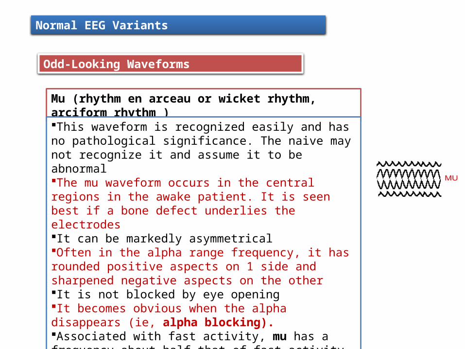
Mu (rhythm en arceau or wicket rhythm, arciform rhythm )
This waveform is recognized easily and has no pathological significance. The naive may not recognize it and assume it to be abnormalThe mu waveform occurs in the central regions in the awake patient. It is seen best if a bone defect underlies the electrodesIt can be markedly asymmetricalOften in the alpha range frequency, it has rounded positive aspects on 1 side and sharpened negative aspects on the otherIt is not blocked by eye openingIt becomes obvious when the alpha disappears (ie, alpha blocking).Associated with fast activity, mu has a frequency about half that of fast activity.The most classical feature of mu waveform is that it blocks with motor activity of the contralateral body (or the thought of such movement).
Normal EEG Variants
Odd-Looking Waveforms

14- and 6-Hz waves
The 2 frequencies are intimately intertwined and the complexes occur in bursts.They generally are thought to be clinically insignificant.They occur in healthy children and adolescents. Some claim that they are best seen in referential recordings during sleep
Normal EEG Variants
Odd-Looking Waveforms
Small sharp spikes of sleep (SSS)
This waveform also is known as benign epileptiform transients of sleep (BETS).These sharp, small waves occur on 1 or both sides (often asynchronously), especially in the temporal and frontal regions.Rarely seen in children, they are seen most often in adults and the elderlyThey can occur in epileptic patients but often are seen in healthy individuals. They can be regarded as a probable normal variant
6-Hz spike and wave (phantom spike and wave)
These occur as bursts of miniature spike and wave complexes or runs of such complexes at 6 Hz rather than the usual 2-4 Hz.Their significance is debated, but generally those occurring in the posterior head regions are regarded as benignSeen at all ages (but especially in adults), they often are confused with 14- and 6-Hz waves and may merge into themThe anterior variety are regarded by some as consistent with epilepsy, but further studies are needed to confirm this
Normal EEG Variants
Odd-Looking Waveforms
Wicket spikes
•Almost exclusively in adults•Like wicket rhythm, (rounded aspects to 1 side and sharp points to the other, giving the appearance of spikes or sharp waves•distinguished by their morphology and at times by their defined background rhythms, which are harmonizing. •Can be seen either in wakefulness or sleep in the anterior or temporal head regions.

Subclinical rhythmic EEG discharges in adults
SREDA consists of theta rhythm occurring in a widespread manner, maximal over the parietal and posterior temporal regions, and lasting for a few seconds to a minute without clinical signs or symptoms. It is described as "not evolving" and appears quite stable for its duration. Mechanism of SREDA is not understood, represent a benign EEG phenomenon that distinguished from seizure dischargesAnother unusual variant (delta rhythm as well as notched waveforms with a frontal distribution and a more prolonged duration that even includes sleep(FRIDA)
Normal EEG Variants
Odd-Looking Waveforms
Rhythmic midline theta
•Rhythm maximal at the midline, most prominently at Cz•It has a frequency of 5-7 Hz and typically has an arciform, spiky, mu like appearance•Waxes and wanes, can appear during wakefulness or drowsiness, and is usually reactive to eye opening or limb movement
Forehead, jaw, and eyelid muscle movements homotor variant (rhythmic harmonic theta)
Sweating produces electrical disturbances by shorting electrode pairs.
Tongue and eyes have their own dipole electric chargeu (rhythm en arceau or wicket rhythm)
Normal EEG Variants
Artifacts
Other sources of artifacts include ambient electrical waves from respirators, intravenous pump machines, televisions, and other electrical equipment.
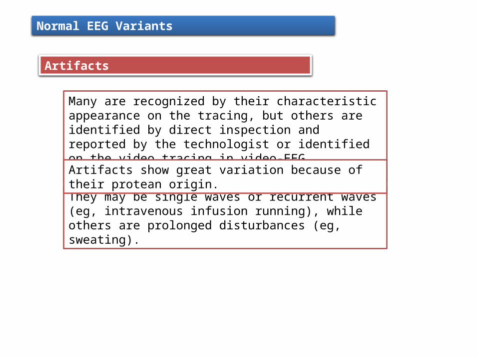
Many are recognized by their characteristic appearance on the tracing, but others are identified by direct inspection and reported by the technologist or identified on the video tracing in video-EEG recording.
They may be single waves or recurrent waves (eg, intravenous infusion running), while others are prolonged disturbances (eg, sweating).
Artifacts show great variation because of their protean origin.
Normal EEG Variants
Artifacts

Artifact produced by tongue movement
EEG artifact of eye blinking.
Example of EEG chewing artifact

Chewing produces spurious spike and wave runs in the frontal and temporal regions from the temporalis muscles
Eye movements occur with blinking and result from the electrical charge of the eye itself (see image below). They are frontal. Nystagmus also produces artifactual waves
Sweating produces very slow waves, because the salt solution shorts out pairs of adjacent electrodes
Normal EEG Variants
Artifacts
ECG and pulse motion produce unusual waveforms. ECG produces small spikes that are recurrent and are especially evident in the monopolar montages.
The following can be regarded as clinically insignificant

Tremor and movement of the head or body may cause electrodes to move
Electrical fields result from electrical devices and televisions.
Normal EEG Variants
Artifacts
ICU special waveforms may result from respirator-induced movements, intravenous drips and drip pumps, electrical fields, or cautery in the operating room or emergency department.
Electrode pops or movements can produce sudden, recurrent, or continuous electrical waves
The following can be regarded as clinically insignificant

Different frequencies sometimes add to or cancel each other, creating odd waveforms or fluctuations of waveforms
Many fascinating patterns have been generated by mixing artificially created computer-generated frequencies. These waveforms have the significance of the basic waveforms that underlie the patterns.
Normal EEG Variants
Harmonics
Pseudospikes or pseudoslow waves may be seen with intermixing of waves.
EEG is a complex summation of many frequencies
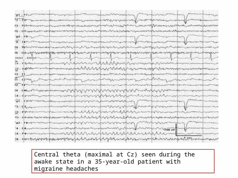
Central theta (maximal at Cz) seen during the awake state in a 35-year-old patient with migraine headaches

A 6-Hz (phantom) spike-wave burst with frontal predominance in the 5th second of this EEG in an awake patient with temporal lobe epilepsy.
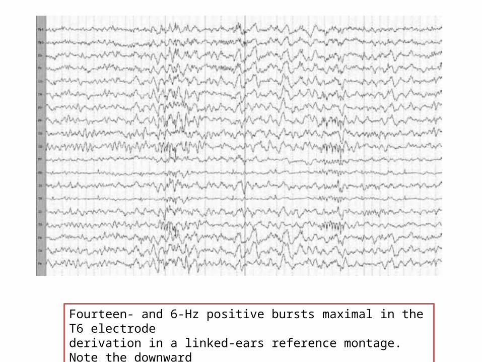
Fourteen- and 6-Hz positive bursts maximal in the T6 electrodederivation in a linked-ears reference montage. Note the downwarddeflection and prominent 14-Hz frequency.

A right benign epileptiform transients of sleep (BETS) in the temporal region during stage 2 sleep. Note the higher amplitude in the T1 andT2 channel with a longer interelectrode distance.

Wicket waves maximal at T3 and T4.

SREDA in a 73-year-old patient during hyperventilation (HV). No clinical signs were present.

Thank you