tandem sumo fusion vectors for improving soluble protein ... · tandem sumo fusion vectors for...
TRANSCRIPT

Protein Expression and Purification 116 (2015) 42–49
Contents lists available at ScienceDirect
Protein Expression and Purification
journal homepage: www.elsevier .com/ locate /yprep
Tandem SUMO fusion vectors for improving soluble protein expressionand purification
http://dx.doi.org/10.1016/j.pep.2015.08.0191046-5928/� 2015 Elsevier Inc. All rights reserved.
Abbreviations: HSQC, heteronuclear single quantum coherence NMR spectrum;RF cloning, restriction free cloning (cloning which is independent of restrictionsites).⇑ Corresponding author.
E-mail address: [email protected] (H. Iwaï).
Fernando Guerrero, Annika Ciragan, Hideo Iwaï ⇑Research Program in Structural Biology and Biophysics, Institute of Biotechnology, University of Helsinki, P.O. Box 65, Helsinki FIN-00014, Finland
a r t i c l e i n f o
Article history:Received 22 June 2015and in revised form 17 August 2015Accepted 18 August 2015Available online 20 August 2015
Keywords:Fusion proteinsSolubility enhancement tagSUMO systemUlp1 proteaseTonBScytovirinProtein ligation
a b s t r a c t
Availability of highly purified proteins in quantity is crucial for detailed biochemical and structuralinvestigations. Fusion tags are versatile tools to facilitate efficient protein purification and to improvesoluble overexpression of proteins. Various purification and fusion tags have been widely used foroverexpression in Escherichia coli. However, these tags might interfere with biological functions and/orstructural investigations of the protein of interest. Therefore, an additional purification step to removefusion tags by proteolytic digestion might be required. Here, we describe a set of new vectors in whichyeast SUMO (SMT3) was used as the highly specific recognition sequence of ubiquitin-like protease 1,together with other commonly used solubility enhancing proteins, such as glutathione S-transferase,maltose binding protein, thioredoxin and trigger factor for optimizing soluble expression of protein ofinterest. This tandem SUMO (T-SUMO) fusion system was tested for soluble expression of theC-terminal domain of TonB from different organisms and for the antiviral protein scytovirin.
� 2015 Elsevier Inc. All rights reserved.
1. Introduction
Although the use of recombinant proteins has been a valuableadvance in recent times, the choice of the appropriate host andexpression system needs to be optimized on a case-by-case basisaccording to the target protein [1,2]. Purification tags likepolyhistidine-tag are indispensable for facilitating efficient proteinpurification of heterogeneous proteins overexpressed in Escherichiacoli [3,4]. In addition, various proteins have been used as fusiontags in combination with purification tags for improving propertieslike solubility and expression levels of target proteins [5]. Glu-tathione S-transferase (GST) [6], thioredoxin (TRX) [7], DsbA [8],maltose binding protein (MBP) [9], trigger factor (TF) [10] andothers have been often used [11]. Various vectors for fusion pro-teins are already commercially available. However, fusion tagsmight interfere with biological assays or structural investigations,making it necessary to remove them before carrying out such stud-ies. Thus, additional steps of proteolytic cleavage and subsequentremoval of the proteolytic enzyme and fusion tag are applied toproduce tag-free target proteins of interest. Widely used and com-
mercially available proteases for the specific cleavages are throm-bin, factor Xa, enterokinase, TEV protease, and precision protease[12]. Due to their lower specificity and instability of the target pro-teins, undesired cleavages have been observed outside theexpected cleavage site [13]. For example, thrombin cannot differ-entiate between Ser and Cys in their recognition [14]. Moreover,these commercial enzymes might not be cost-effective whenlarge-scale protein production is required, restricting their indus-trial scale applications in biotechnology. An alternative might bethe use of thiol-inducible self-cleavable intein tags, which do notrequire additional proteases [15]. Instead, it utilizes an autocat-alytic self-cleavage reaction induced by thiol reagents to avoid thisproblem. However, premature cleavage has been observed, therebyreducing the purification efficiency, and they often require opti-mization of parameters such as expression temperature and junc-tion sequences [16,17]. In addition, the reducing condition used forthe cleavage might not be compatible with some target proteinsbearing disulfide bridges.
Herewe report a set of new vectors in which small ubiquitin-likemodifier (SUMO or SUMO homologue, SMT3) from yeast is used ascleavage tag, in tandemwith other fusion tags such as TRX, TF, MBPand GST for solubility and expression enhancement. These tandemfusion vectors utilize the high specificity of ubiquitin-like protease1 (Ulp1), which recognizes the three-dimensional structure ofSUMO domain and cleaves after di-glycine at the C-terminus[18]. We demonstrated soluble expression and purification of the

F. Guerrero et al. / Protein Expression and Purification 116 (2015) 42–49 43
C-terminal domain (CTD) of TonB protein from three organisms,and a small lectin protein scytovirin for optimal choice of a tandemfusion vector.
2. Materials and methods
2.1. Construction of plasmids
The backbone plasmid used to construct the expression vectorswas pHYRSF53 [19,20]. Four different fusion tags were inserted inframe upstream of the coding sequence of SMT3. Two PCR stepswere necessary to add a hexa-histidine (H6) tag at the N-terminus of the cloned fusion tag. In a first PCR step, the codingsequences of the fusion tags were amplified using syntheticoligonucleotides. In all cases the forward primer contained anoverhang coding part of a H6-tag. The full H6-tag and an NcoI sitefor cloning were added in a second PCR reaction using the firstPCR product as a template and the oligonucleotide HK683: 50-TACCATGGGCAGCAGCCATCATCATCATCATCACGG as a forward primer.The resulting amplicon containing the H6-fusion tag was insertedinto the vector pHYRSF53 using the restriction sites NcoI and SpeIto generate the tandem SUMO-fusion vectors. The backbonepHYRSF53 and the four vectors generated are shown in Fig. 2A.To obtain pLJSRSF3, the glutathione S-transferase (GST) codingregion was amplified with the primers I399: 50-CATCATCATCATCACGGCTCCCCTATACTAGGTTATTG and I398: 50-TTTACTAGTTTTTGGAGGATGGTCGCCACC using the vector pGEX-2TK (GE Healthcare)as a template and cloned into pHYRSF53 as described above. Thevector pLJSRSF7 was generated in the same way, amplifying thegene of maltose binding protein (MBP) from the plasmid pTWIN-MBP1 (New England Biolabs) using the primers I397: 50-CATCATCATCATCACGGCAAAATCGAAGAAGGTAAAC and I395: 50-AAACTAGTACCCGAATTAGTCTGCGCGTC. The plasmid pCARSF85 was createdsimilarly, amplifying trigger factor (TF) directly from E. coli geno-mic DNA using the primers I09: 50-ATCATCATCATCATCACGGTCAAGTTTCAGTTGAAACC and I08: 50-AAACTAGTACCTCCACCCGCCTGCTGGTTCATCAGC. Finally, to generate the plasmid pCARSF63thioredoxin (TRX) was also cloned directly from E. coli genomicDNA using the primers HK682: 50-CATCATCATCATCACGGCAGCGATAAAATTATTCACC and HK684: 50-CCACTAGTTCCCGCCAGGTTAGCGTCGAGG, and inserted into pHYRSF53 after the second
Fig. 1. Overview of the purification procedure of the target protein using the tandem SUMUlp1: protease domain (residues 403–621) of the Saccharomyces cerevisiae ubiquitin-lik
PCR reaction as described above. The plasmids pHYRSF53,pLJSRSF3, pLJSRSF7, pCARSF85 and pCARSF63 are deposited andavailable for academic researchers from Addgene (addgene.org)with the deposit numbers #64696, #64692, #64693, #64694 and#64695, respectively.
The construction of the plasmid pJDJRSF05 carrying SMT3 fusedwith the single chain NpuDnaE intein variant was described previ-ously [21]. The linker region was shortened by one Gly residuecompared to pJDJRSF05 by amplifying the sequence of an inactivevariant of single chain NpuDnaE intein using the synthetic oligonu-cleotides HK202: 50-GTGGATCCGGAGCTCTAAGCTATGAAACG andSK187: 50-ATCAAGCTTAATTAGAAGCTATGAAGCC. The PCR productwas digested with BamHI and HindIII and cloned into the vectorpHYRSF53 to generate the plasmid pJDJRSF04 (Fig. 3). Likewise,to lengthen the linker region by one Gly residue compared topJDJRSF05, a similar approach was carried out using the syntheticoligonucleotides HK204: 50-GTGGATCCGGAGGAGGAGCTCTAAGCTATGAAACG and SK187. The resulting plasmid was pJDJRSF06.These three constructs were used to assess the Ulp1403–621 activity.
The expression vectors constructed allow the possibility to addanother two features downstream of the protein of interest (POI):the N-term of the NpuDnaE split intein, and a chitin bindingdomain. In this work we inserted the POI into the sites BamHIand HindIII to avoid modifications in the C-terminus (Fig. 2A). Fourdifferent proteins were used in this study: the C-terminal domain(CTD) of TonB from three different organisms: E. coli (EcTonB),Pseudomonas aeruginosa PAO1 (PsTonB), and Helicobacter pylori(HpTonB), and the antiviral lectin scytovirin (SVN) from Scytonemavarium. The coding sequences of TonB variants were amplified byPCR from purified genomic DNA using synthetic oligonucleotides.The fragment containing the last 89 residues of EcTonB (EcTonB-89) was cloned using I470: 50-AAGGATCCGGACCACGCGCATTAAGCCG and SK009: 50-TACAAGCTTACTGAATTTCGGTGGTGCCG.The amplified PCR fragment was BamHI-HindIII digested andinserted into the BamHI-HindIII digested expression vectorspHYRSF53, pLJSRSF3, pLJSRSF7, pCARSF85, pCARSF63 to generatethe plasmids pFGRSF01, pFGRSF02, pFGRSF03, pFGRSF04,pFGRSF05, respectively. For PsTonB two versions with differentlengths were generated: the last 77 residues of TonB (PsTonB-77)and the last 96 residues (PsTonB-96). To clone the last 77 residues(PsTonB-77) we used the synthetic oligonucleotides HK062: 50-AAGGATCCCGGATGGCCCAGGCGCGGCG and HK063: 50-TACAAGCT
O vectors. T-SUMO: tandem fusion bearing yeast SMT3 as protease recognition site.e-specific protease 1.
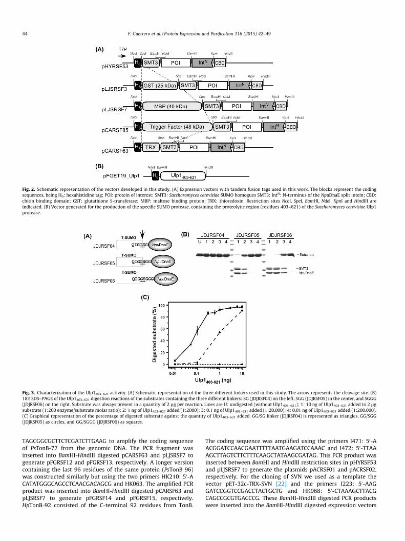
Fig. 2. Schematic representation of the vectors developed in this study. (A) Expression vectors with tandem fusion tags used in this work. The blocks represent the codingsequences, being H6: hexahistidine tag; POI: protein of interest; SMT3: Saccharomyces cerevisiae SUMO homogues SMT3; IntN: N-terminus of the NpuDnaE split intein; CBD:chitin binding domain; GST: glutathione S-transferase; MBP: maltose binding protein; TRX: thioredoxin. Restriction sites NcoI, SpeI, BamHI, NdeI, KpnI and HindIII areindicated. (B) Vector generated for the production of the specific SUMO protease, containing the proteolytic region (residues 403–621) of the Saccharomyces cerevisiae Ulp1protease.
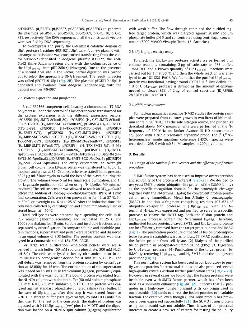
Fig. 3. Characterization of the Ulp1403–621 activity. (A) Schematic representation of the three different linkers used in this study. The arrow represents the cleavage site. (B)18% SDS–PAGE of the Ulp1403–621 digestion reactions of the substrates containing the three different linkers: SG (JDJRSF04) on the left, SGG (JDJRSF05) in the center, and SGGG(JDJRSF06) on the right. Substrate was always present in a quantity of 2 lg per reaction. Lines are U: undigested (without Ulp1403–621); 1: 10 ng of Ulp1403–621 added to 2 lgsubstrate (1:200 enzyme/substrate molar ratio); 2: 1 ng of Ulp1403–621 added (1:2000); 3: 0.1 ng of Ulp1403–621 added (1:20,000); 4: 0.01 ng of Ulp1403–621 added (1:200,000).(C) Graphical representation of the percentage of digested substrate against the quantity of Ulp1403–621 added. GG/SG linker (JDJRSF04) is represented as triangles, GG/SGG(JDJRSF05) as circles, and GG/SGGG (JDJRSF06) as squares.
44 F. Guerrero et al. / Protein Expression and Purification 116 (2015) 42–49
TAGCGGCGCTTCTCGATCTTGAAG to amplify the coding sequenceof PsTonB-77 from the genomic DNA. The PCR fragment wasinserted into BamHI-HindIII digested pCARSF63 and pLJSRSF7 togenerate pFGRSF12 and pFGRSF13, respectively. A longer versioncontaining the last 96 residues of the same protein (PsTonB-96)was constructed similarly but using the two primers HK210: 50-ACATATGGGCAGCCTCAACGACAGCG and HK063. The amplified PCRproduct was inserted into BamHI-HindIII digested pCARSF63 andpLJSRSF7 to generate pFGRSF14 and pFGRSF15, respectively.HpTonB-92 consisted of the C-terminal 92 residues from TonB.
The coding sequence was amplified using the primers I471: 50-AACGGATCCAACGAATTTTTAATGAAGATCCAAAC and I472: 50-TTAAAGCTTAGTCTTCTTTCAAGCTATAAGCGATAG. This PCR product wasinserted between BamHI and HindIII restriction sites in pHYRSF53and pLJSRSF7 to generate the plasmids pACRSF01 and pACRSF02,respectively. For the cloning of SVN we used as a template thevector pET-32c-TRX-SVN [22] and the primers I223: 50-AAGGATCCGGTCCGACCTACTGCTG and HK968: 50-CTAAAGCTTACGCAGCCGCGTGACCCG. These BamHI-HindIII digested PCR productswere inserted into the BamHI-HindIII digested expression vectors
F. Guerrero et al. / Protein Expression and Purification 116 (2015) 42–49 45
pHYRSF53, pLJSRSF3, pLJSRSF7, pCARSF85, pCARSF63 to generatethe plasmids pFGRSF07, pFGRSF08, pFGRSF09, pFGRSF10, pFGRSF11, respectively. The DNA sequences of all the constructed vectorswere verified by DNA sequencing.
To overexpress and purify the C-terminal catalytic domain ofUlp1 protease (residues 403–621, Ulp1403–621), a new plasmid withkanamycine resistance was constructed transferring from the vec-tor pHYRS52 (deposited in Addgene, plasmid #31122) the XbaI-EcoRI Shine-Dalgarno region along with the coding sequence ofthe Ulp1403–621 into pET-28b(+) (Novagen). Due to the presenceof a second XbaI site in the vector, partial digestion was carriedout to select the appropriate DNA fragment. The resulting vectorwas called pFGET19_Ulp1 (Fig. 2B). The plasmid pFGET19_Ulp1 isdeposited and available from Addgene (addgene.org) with thedeposit number #64697.
2.2. Protein expression and purification
E. coli ER2566 competent cells bearing a chromosomal T7 RNApolymerase under the control of a lac operon were transformed forthe protein expression with the different expression vectors:pFGRSF01 (H6-SMT3-EcTonB-89), pFGRSF02 (H6-GST-SMT3-EcTonB-89), pFGRSF03 (H6-MBP-SMT3-EcTonB-89), pFGRSF04 (H6-TF-SMT3-EcTonB-89), pFGRSF05 (H6-TRX-SMT3-EcTonB-89), pFGRSF07(H6-SMT3-SVN), pFGRSF08 (H6-GST-SMT3-SVN), pFGRSF09(H6-MBP-SMT3-SVN), pFGRSF10 (H6-TF-SMT3-SVN), pFGRSF11 (H6-TRX-SMT3-SVN), pFGRSF12 (H6-TRX-SMT3-PsTonB-77), pFGRSF13(H6-MBP-SMT3-PsTonB-77), pFGRSF14 (H6-TRX-SMT3-PsTonB-96),pFGRSF15 (H6-MBP-SMT3-PsTonB-96), pACRSF01 (H6-SMT3-HpTonB-92), pACRSF02 (H6-MBP-SMT3-HpTonB-92), pJDJRSF04 (H6-SMT3-SG-NpuDnaE), pJDJRSF05 (H6-SMT3-SGG-NpuDnaE), pJDJRSF06(H6-SMT3-SGGG-NpuDnaE). For every experiment, an overnightgrown cell colony from LB-agar plates was transferred to liquid LBmedium and grown at 37 �C (unless otherwise stated) in the presenceof 25 lg ml�1 kanamycin to avoid the loss of the plasmid during thegrowth. The volumes were 5ml for small scale purification, and 1 lfor large scale purification (2 l when using 15N labelled M9 minimalmedium). The cell suspension was allowed to reach an OD600 of �0.5before the addition of isopropyl-b-D-thiogalactoside (IPTG) at 1 mMfinal concentration. Cells were then grown either for 4 h at 37 �C, 5 hat 30 �C, or overnight (�16 h) at 25 �C. After the induction time, thecells were collected by centrifugation and either immediately used orstored frozen at �70 �C.
Total cell lysates were prepared by suspending the cells in B-PER reagent (Thermo scientific) and incubated at 25 �C and1000 rpm shaking for 10 min. Soluble and insoluble fractions wereseparated by centrifugation. To compare soluble and insoluble pro-tein fractions, supernatant and pellet were separated and dissolvedin an equal volume of SDS-buffer. The protein content was ana-lyzed in a Coomassie-stained 18% SDS–PAGE.
For large scale purification, whole-cell pellets were resus-pended in wash buffer (50 mM sodium phosphate, 300 mM NaCl,pH 8.0). The cells were lysed either by ultrasonication or in anEmulsiFlex C5 homogenizer device for 10 min at 15,000 PSI. Thecell debris was removed from the protein solution by centrifuga-tion at 18,000g for 45 min. The entire amount of the supernatantwas loaded on a 5 ml HP HisTrap column (Qiagen) previously equi-librated with the wash buffer. The bound protein was eluted fromthe Ni-NTA column with elution buffer (50 mM sodium phosphate,300 mM NaCl, 250 mM imidazole, pH 8.0). The protein was dia-lyzed against standard phosphate-buffered saline (PBS) buffer. Inthe case of Ulp1403–621, after this step it was stored frozen at�70 �C in storage buffer (50% glycerol v/v, 25 mM DTT) until fur-ther use. For the rest of the constructs, the dialyzed protein wasdigested with 0.1% Ulp1403–621 (v/v) plus 1 mM DTT, and the diges-tion was loaded on a Ni-NTA spin column (Qiagen) equilibrated
with wash buffer. The flow-through contained the purified tag-free target protein, which was dialyzed against 20 mM sodiumphosphate buffer pH 6, and concentrated using centrifugal concen-trators (5000 MWCO Vivaspin Turbo 15, Sartorius).
2.3. Ulp1403–621 activity assay
To check the Ulp1403–621 protease activity we performed 5 llvolume reactions containing 2 lg of substrate in PBS buffer,1 mM DTT, and a known quantity of Ulp1403–621. Reactions werecarried out for 1 h at 30 �C, and then the whole reaction was ana-lyzed in an 18% SDS–PAGE. We found that the purified Ulp1403–621protein was functional, having around 1000 U ll�1. Unit definition:1 U of Ulp1403–621 protease is defined as the amount of enzymeneeded to cleave 85% of 2 lg of control substrate (JDJRSF06,28.5 kDa) in 1 h at 30 �C.
2.4. NMR measurements
For nuclear magnetic resonance (NMR) studies the protein sam-ples were prepared from cultures grown in two liters of M9 med-ium containing 15NH4Cl as the sole nitrogen source, and purified asdescribed above. NMR measurements were performed at the 1Hfrequency of 600 MHz on Bruker Avance III HD spectrometerequipped with a triple resonance cryogenic probe. The [1H,15N]-heteronuclear single quantum coherence (HSQC) spectra wererecorded at 298 K with �0.5 mM samples in 200 ll volume.
3. Results
3.1. Design of the tandem fusion vectors and the efficient purificationprocedure
SUMO fusion system has been used to improve overexpressionand solubility of the protein of interest [2,23–25]. We decided touse yeast SMT3 protein (ubiquitin-like protein of the SUMO family)as the specific recognition domain for the proteolytic cleavagetogether with the N-terminal H6-tag for convenient protein purifi-cation using Immobilized Metal Ion Affinity Chromatography(IMAC). In addition, a fragment comprising residues 403–621 ofubiquitin-like-specific protease 1 (Ulp1403–621) with an N-terminal H6-tag was expressed and purified to serve as a specificprotease to cleave the SMT3 tag. Both, the fusion protein andUlp1403–621 protease contain the N-terminal H6-tag. Therefore,undigested fusion protein, cleaved SMT3, and Ulp1403–621 proteasecan be efficiently removed from the target protein in the 2nd IMAC(Fig. 1). The purification procedure of the SMT3 fusion protein/pro-tease system is straightforward as follows: (1) IMAC purification ofthe fusion protein from cell lysate. (2) Dialysis of the purifiedfusion protein in phosphate-buffered saline (PBS); (3) Digestionby Ulp1403–621 protease; (4) Purification of the target protein byIMAC by removing Ulp1403–621 and H6-SMT3 and the undigestedprecursor (Fig. 1).
This purification system has been used in our laboratory to pur-ify various proteins for structural studies and also produced severalhigh-quality crystals without further purification steps [19,26–29].However, in several cases we found that the fusion proteins wereinsoluble even with SMT3 fusion partner, which has often beenused as a solubility enhancer (Fig. 4A) [2]. It seems that T7 pro-moter in a high-copy number plasmid with RSF origin used inour plasmids [30] tends to direct the fusion proteins to insolublefraction. For example, even though E. coli TonB protein has previ-ously been expressed successfully [31], the SUMO fusion proteinusing our plasmid was not soluble. Thus, it was of our practicalinterests to create a new set of vectors for testing the solubility

Fig. 4. (A) Coomassie stained 18% SDS–PAGE gel from the expression in E. coli ER2566 of the different fusion tags fused to the EcTonB-89. The gel shows the separation of theinsoluble fraction (I) and the soluble fraction (S). M: molecular weight marker. (B) Coomassie stained 18% SDS–PAGE gel from the expression in E. coli ER2566 of the differentfusion tags fused to SVN. I: insoluble membrane fraction. S: soluble fraction. M: molecular weight marker.
46 F. Guerrero et al. / Protein Expression and Purification 116 (2015) 42–49
in the same plasmid backbone by fusing several commonly usedfusion tags (e.g. GST, MBP, TF and TRX) to the POI, yet utilizingthe SUMO domain for the cleavage. Fig. 2A depicts five schematicvector maps of the newly constructed tandem-fusion vectors andour previous vector with SMT3 [19].
The vector has been originally designed for protein ligation bysplit NpuDnaE intein by protein trans-splicing (PTS) [19]. The splitfragments required for protein ligations by PTS can be less solublethan the original target protein [26]. The newly developed tandemfusion vectors can be not only useful for protein purification (POIcan be cloned between BamHI and HindIII), but they can also beused for protein ligation by protein trans-splicing with enhancedsolubility due to solubility enhancement tag (POI can be clonedusing BamHI or by restriction-free (RF) cloning) [32] (Fig. 2A).
3.2. Linker length required for Ulp1 protease digestion
The SUMO system relies on highly pure Ulp1 protease to cleavethe SMT3 tag from the POI even though Ulp1 is very specific toSMT3 domain. With the newly constructed plasmid, the expressionand purification of the Ulp1403–621 catalytic domain yielded up to87 mg of the highly pure enzyme per liter. Generally the insertionof various fusion tags (GST, MBP, TF and TRX) at the N-terminus ofSUMO domain did not interfere with the activity of Ulp1403–621
protease for cleavage of our fusion proteins because we coulddigest the fusion proteins equally well when the linker after theC-terminal di-glycine of SMT3 is sufficiently long (data not shown).This is presumably because the N-terminus of SMT3 is distantlylocated from Ulp1403–621 as observed in the crystal structure ofSMT3/Ulp1 complex [18]. Therefore, the SUMO domain could servenot only as a solubility enhancer but also as a general cleavage site.However, the linker after di-glycine peptide of the C-terminus ofSMT3 domain influenced the activity of Ulp1 protease drastically(Fig. 3). We systematically analyzed the activity of Ulp1403–621 withdifferent lengths for the linker connecting SMT3 and the targetprotein. We used an inactive variant of the NpuDnaE intein (C1A)as a POI. We tested three different linker lengths by inserting SG,SGG or SGGG between the C-terminal di-glycine of SMT3 and thefirst residue of NpuDnaE intein (C1A). When only two residueswere inserted at the front of the NpuDnaE intein (C1A), the fusedprotein could not be digested by Ulp1403–621 at all (Fig. 3). Sincethe first residue of NpuDnaE intein is already integral part of thethree-dimensional structure of NpuDnaE intein [33], it is thus likelythat the short linker to di-glycine residue of SMT3 inhibitsUlp1403–621 to access the cleavage site. Extending the linker lengthby glycine residues improved the cleavage considerably, indicating
that Ulp1403–621 requires at least three flexible residues between astructured globular domain and di-glycine peptide of SMT3 forcleaving the protein of interest from SMT3. It might require evena longer linker for larger target proteins, since NpuDnaE intein isa small protein (15.8 kDa).
3.3. Comparison of the five tandem SUMO fusion vectors for solubleprotein expression
Even though the C-terminal domain (CTD) of TonB from E.coli has been previously overexpressed and purified [31,34,35],the fusion protein with SMT3 domain bearing CTD of E. coli TonBconsisting of 89 residues (EcTonB-89) was mostly insoluble usingour vector of a high-copy number plasmid with T7 promoter(Fig. 4A). We compared the solubility of various fusion proteinsusing the tandem SUMO fusion vectors bearing an additionalfusion tag: GST, MBP, TF, or TRX. The open reading frame ofEcTonB-89 was inserted between BamHI and HindIII sites ofthe newly constructed tandem fusion vectors for comparison(Fig. 2A). All EcTonB-89 fusion proteins were highly expressedin E. coli. The fusion proteins in soluble fraction were compared(Fig. 4A).
The improved solubility was particularly observed for TRX andMBP fusion tags. The solubility when using TF as a fusion tagwas slightly improved. Similar results were observed when wetested the expression of scytovirin within the set of the five differ-ent tandem vectors (Fig. 4B), being MBP and TRX as the fusion part-ners that improved more the solubility of the expressed protein.Interestingly, in the case of scytovirin, TRX fusion improved thesolubility more than EcTonB-89.
EcTonB-89 was successfully purified from either MBP-fusion orTRX-fusion after Ulp1 digestion (Fig. 5A). The well-dispersed NMRsignals of Ec-TonB-89 purified from MBP-fusion in the [1H,15N]-HSQC spectrum indicate the well-ordered structure of EcTonB-89(Fig. 5B), which is in agreement with the previously published res-onance assignments [34]. Fig. 5B shows that the majority of thechemical shift positions of EcTonB-89 match with the EcTonB res-onance assignments published earlier ((BMRB ID 6375), suggestingthat EcTonB-89 is identical with that of EcTonB produced withoutany fusion tag [34].
3.4. Self-contained domain of Pseudomonas TonB
Next, we used these vectors for the production of CTD of one ofthe TonB proteins from P. aeruginosa (PsTonB), which is annotatedas TonB in UniProt database with no available structural informa-

Fig. 5. Release of E. coli TonB-89 after digestion with Ulp1. (A) SDS–PAGE gel analysis after purification of the fusion proteins H6-MBP-SMT3-EcTonB-89 (left) and H6-TRX-SMT3-EcTonB-89 (right) before (�) and after (+) the digestion. The solubility tag has been removed by a second affinity chromatography purification. Purified EcTonB-89 (p)fromMBP-fusion has been used for NMR studies. (B) Two-dimensional [1H,15N]-HSQC spectrum of purified 0.5 mM EcTonB-89 in 20 mM sodium phosphate buffer at pH 6.0 at298 K. The spectrum was recorded at 1H frequency of 600 MHz. The crosses show the chemical shift positions of the previously published resonance assignments of EcTonB(BMRB ID 6375) [34].
Fig. 6. Purification of Pseudomonas TonB-96. (A) Coomassie stained 18% SDS–PAGE gel analysis of the purification of PsTonB using the short version (PsTonB-77) or the longversion (PsTonB-96) of the POI fused to the tags H6-TRX-SMT3 (indicated as H6-TRX-SMT3-PsTonB-77 or H6-TRX-SMT3-PsTonB-96, respectively) or to H6-MBP-SMT3 (right,indicated as H6-MBP-SMT3-PsTonB-77 or H6-MBP-SMT3-PsTonB-96, respectively). The gel shows the separation of the insoluble fraction (I) and the soluble fraction (S) atdifferent induction temperatures (indicated below the lanes). (B) Two-dimensional [1H,15N]-HSQC spectrum of purified 1 mM PsTonB-96 in 20 mM sodium phosphate bufferat pH 6.0 at 298 K. The spectrum was recorded at the 1H frequency of 600 MHz.
F. Guerrero et al. / Protein Expression and Purification 116 (2015) 42–49 47
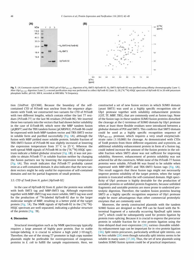
Fig. 7. (A) Coomassie stained 18% SDS–PAGE gel of Ulp1403–621 digestion of H6-SMT3-HpTonB-92. H6-SMT3-HpTonB-92 was purified using affinity chromatography (Lane 1).After Ulp1403–621 digestion (Lane 2), a second purification step was performed to collect HpTonB-92 (lane 3). (B) [1H,15N]-HSQC spectrum of HpTonB-92 in 20 mM potassiumphosphate buffer, pH 6.0, 300 K, recorded at 600 MHz 1H frequency.
48 F. Guerrero et al. / Protein Expression and Purification 116 (2015) 42–49
tion (UniProt: Q51368). Because the boundary of the self-contained CTD of PsTonB was unclear from the sequence align-ments with TonB, we constructed two variants for CTD of PsTonBwith two different lengths, which contain either the last 77 resi-dues (PsTonB-77) or the last 96 residues (PsTonB-96). We insertedthese two variants into the vectors that had shown better solubilityin the case of EcTonB-89, which were the MBP tandem fusion(pLJRSF7) and the TRX tandem fusion (pCARSF63). PsTonB-96 couldbe expressed with both MBP-tandem vector and TRX-SMT3 vectorin soluble form and purified successfully (Fig. 6A), although thefusion with MBP yielded more soluble protein. Soluble fraction ofTRX-SMT3 fusion of PsTonB-96 was slightly increased at loweringthe expression temperature from 37 �C to 25 �C. Whereas thewell-spread NMR signals of PsTonB-96 in the [1H,15N]-HSQC spec-trum indicate a folded globular structure (Fig. 6B), it was not pos-sible to obtain PsTonB-77 in soluble fraction neither by changingthe fusion partners nor by lowering the expression temperature(Fig. 6A). This result indicates that PsTonB-77 probably cannotexist as a self-contained domain. It also indicates that the new tan-dem vectors might be only useful for expression of self-containeddomains and not for partial fragments of small proteins.
3.5. CTD of TonB from H. pylori (HpTonB-92)
In the case of HpTonB-92 from H. pylori the protein was solublewith both SMT3 tag and MBP-SMT3 tag. Although expressiontogether with MBP-SMT3 tag was more soluble, SMT3 tag was usedfor the production of 15N-labeled HpTonB-92 due to the largermolecular weight of MBP, resulting in a better yield of the targetprotein (Fig. 7A). The NMR signals of HpTonB-92 in the [1H,15N]-HSQC spectrum are well separated, indicating a globular structureof the protein (Fig. 7B).
4. Discussion
Structural investigation such as by NMR spectroscopy typicallyrequires a large amount of highly pure protein. Due to stableisotope-labeling, it is crucial to achieve a high yield (>10 mg/l).Therefore, the use of the strong T7 promoter in high copy numberplasmids might be preferable for overexpression of exogenousproteins in E. coli to fulfill the sample requirements. Here, we
constructed a set of new fusion vectors in which SUMO domain(yeast SMT3) was used as a highly specific recognition site ofUlp1 protease together with solubility enhancement proteins(GST, TF, MBP, TRX), that are commonly used as fusion tags. Noneof the fusion tags in these tandem SUMO fusion proteins disturbedthe cleavage at the C-terminus of SUMO domain by Ulp1 proteasewhen at least three flexible residues were introduced between aglobular domain of POI and SMT3. This confirms that SMT3 domaincould be used as a highly specific recognition sequence ofUlp1403–621 protease, which requires a very small enzyme/sub-strate ratio (1:10,000) for cleavage. As demonstrated with CTDsof TonB protein from three different organisms and scytovirin, anadditional solubility enhancement protein in form of a fusion tag,could indeed increase the amount of the fusion protein in the sol-uble fraction when SMT3 alone was not sufficient for improvingsolubility. However, enhancement of protein solubility was notachieved for all the constructs. While none of the PsTonB-77 fusionproteins were soluble, PsTonB-96 was found to be soluble whenexpressed with MBP-SMT3 and TRX-SMT3 fusion tags (Fig. 6A).This result suggests that these fusion tags might not necessarilyimprove protein solubility of the target protein, when the targetprotein is truncated within the self-contained domain. High speci-ficity of Ulp1 protease is highly desirable for the production ofunstable proteins or unfolded protein fragments, because unfoldedfragments and unstable proteins are more prone to undesired pro-teolytic digestion. Therefore, the tandem fusion proteins bearingSMT3 as a highly specific recognition sequence of Ulp1 proteasemight be more advantageous than other commercial proteolyticenzymes that are commonly used.
Moreover, the newly constructed plasmids with the tandemSUMO fusion are designed so that POI can be fused with the N-terminal fragment of a naturally occurring NpuDnaE split intein(IntN), which could be subsequently used for protein ligation byprotein trans-splicing. Because it is crucial to express the precursorprotein in soluble fraction for in vivo protein ligation using thetime-delayed dual over-expression [26], various cleavable solubil-ity enhancement tags can be important for in vivo protein ligation[36]. Split intein precursors, particularly artificial split inteins, canbe less soluble [28] even though naturally split inteins seems to besoluble in many cases [37,38]. Thus, the set of new plasmids usingtandem SUMO fusion system could be of practical importance.

F. Guerrero et al. / Protein Expression and Purification 116 (2015) 42–49 49
5. Conclusions
We demonstrated that tandem-SUMO fusion vectors bearingfive different fusion tags could improve solubility of otherwisepoorly soluble protein fragments. In order to achieve soluble pro-tein expression, SMT3 (yeast SUMO homologue) was used as thehighly specific cleavage site of Ulp1 protease, together with asolubility enhancing fusion tag. The proteolytic activity of Ulp1protease (residue 403–621) remained undisturbed, when athree-residue linker was introduced between SMT3 and the targetprotein. Significant differences regarding the solubility of targetproteins could be observed between the five constructs, provingthat the choice of an ideal solubility tag is essential for solubleexpression of otherwise poorly soluble proteins.
Acknowledgments
This work is supported by the grants from the Academy of Fin-land (137995) and Sigrid Juselius Foundation. A. C. acknowledgesthe National Doctoral Programme in Informational and StructuralBiology (ISB) for financial support. The NMR facility at the Instituteof Biotechnology is supported by Biocenter Finland. The authorsthank C. Albert, L. Sipilä and J. Djupsjöbacka for constructingvectors and Drs. A. Wlodawer and B.R. O’Keefe for providing theplasmid containing SVN.
References
[1] K. Terpe, Overview of bacterial expression systems for heterologous proteinproduction: from molecular and biochemical fundamentals to commercialsystems, Appl. Microbiol. Biotechnol. 72 (2006) 211–222.
[2] J.G. Marblestone, S.C. Edavettal, Y. Lim, P. Lim, X. Zuo, T.R. Butt, Comparison ofSUMO fusion technology with traditional gene fusion systems: enhancedexpression and solubility with SUMO, Protein Sci. 15 (1) (2006) 182–189.
[3] E. LaVallie, J. McCoy, Gene fusion expression systems in Escherichia coli, Curr.Opin. Biotechnol. 6 (1995) 501–506.
[4] D. Esposito, D.K. Chatterjee, Enhancement of soluble protein expressionthrough the use of fusion tags, Curr. Opin. Biotechnol. 17 (2006) 353–358.
[5] M.R. Bell, M.J. Engleka, A. Malik, J.E. Strickler, To fuse or not to fuse: what isyour purpose?, Protein Sci 22 (2013) 1466–1477.
[6] D.B. Smith, K.S. Johnson, Single-step purification of polypeptides expressed inEscherichia coli as fusions with glutathione S-transferase, Gene 67 (1988) 31–40.
[7] E.R. Lavallie, E.A. DiBlasio, S. Kovacic, K.L. Grant, P.F. Schendel, J.M. McCoy, Athioredoxin gene fusion expression system that circumvents inclusion bodyformation in the E. coli cytoplasm, Biotechnology 11 (1993) 187–193.
[8] L.A. Collins-Racie, J.M. McColgan, K.L. Grant, E.A. DiBlasio-Smith, J.M. McCoy, E.R. LaVallie, Production of recombinant bovine enterokinase catalytic subunit inEscherichia coli using the novel secretory fusion partner DsbA, Biotechnology11 (2) (1993) 187–193.
[9] R.B. Kapust, D.S. Waugh, Escherichia coli maltose-binding protein isuncommonly effective at promoting the solubility of polypeptides to whichit is fused, Protein Sci. 8 (1999) 1668–1674.
[10] A. Basters, L. Ketscher, E. Deuerling, C. Arkona, J. Rademann, K. Knobeloch, G.Fritz, High yield expression of catalytically active USP18 (UBP43) using aTrigger Factor fusion system, BMC Biotechnol. 12 (2012). 56-56.
[11] C.L. Young, Z.T. Britton, A.S. Robinson, Recombinant protein expression andpurification: a comprehensive review of affinity tags and microbialapplications, Biotechnol. J. 7 (2012) 620–634.
[12] D.S. Waugh, An overview of enzymatic reagents for the removal of affinitytags, Protein Expr. Purif. 80 (2011) 283–293.
[13] R.J. Jenny, K.G. Mann, R.L. Lundblad, A critical review of the methods forcleavage of fusion proteins with thrombin and factor Xa, Protein Expr. Purif. 31(1) (2003) 1–11.
[14] M. Gallwitz, M. Enoksson, M. Thorpe, L. Hellman, The extended cleavagespecificity of human thrombin, PLoS ONE 7 (2012).
[15] S. Chong, F.B. Mersha, D.G. Comb, M.E. Scott, D. Landry, L.M. Vence, F.B. Perler,J. Benner, R.B. Kucera, C.A. Hirvonen, J.J. Pelletier, H. Paulus, M.Q. Xu, Single-
column purification of free recombinant proteins using a self-cleavable affinitytag derived from a protein splicing element, Gene 192 (2) (1997) 271–281.
[16] S. Chong, G.E. Montello, A. Zhang, E.J. Cantor, W. Liao, M.Q. Xu, J. Benner,Utilizing the C-terminal cleavage activity of a protein splicing element topurify recombinant proteins in a single chromatographic step, vol. 26, pp. 5,Nucleic acids Res. 26 (1998) 5109–5115.
[17] Y. Minato, T. Ueda, A. Machiyama, I. Shimada, H. Iwaï, Segmental isotopiclabeling of a 140 kDa dimeric multi-domain protein CheA from Escherichia coliby expressed protein ligation and protein trans-splicing, J. Biomol. NMR 53 (3)(2012) 191–207.
[18] E. Mossessova, C.D. Lima, Ulp1-SUMO crystal structure and genetic analysisreveal conserved interactions and a regulatory element essential for cellgrowth in yeast, Mol. Cell 5 (2000) 865–876.
[19] M. Muona, A.S. Aranko, H. Iwai, Segmental isotopic labelling of a multidomainprotein by protein ligation by protein trans-splicing, ChemBioChem 9 (2008)2958–2961.
[20] A.S. Aranko, S. Züger, E. Buchinger, H. Iwaï, In vivo and in vitro protein ligationby naturally occurring and engineered split DnaE inteins, PLoS One 4 (2009)e5185.
[21] K. Heinämäki, J.S. Oeemig, K. Pääkkönen, J. Djupsjöbacka, H. Iwaï, NMRresonance assignment of DnaE intein from Nostoc punctiforme, Biomol. NMRAssign. 3 (1) (2009) 41–43.
[22] C. Xiong, B.R. O’Keefe, I. Botos, A. Wlodawer, J.B. McMahon, Overexpressionand purification of scytovirin, a potent, novel anti-HIV protein from thecultured cyanobacterium Scytonema varium, Protein Expr. Purif. 46 (2) (2006)233–239.
[23] C.D. Lee, H.C. Sun, S.M. Hu, C.F. Chiu, A. Homhuan, S.M. Liang, C.H. Leng, T.F.Wang, An improved SUMO fusion protein system for effective production ofnative proteins, Protein Sci. 17 (7) (2008) 1241–1248.
[24] T.R. Butt, S.C. Edavettal, J.P. Hall, M.R. Mattern, SUMO fusion technology fordifficult-to-express proteins, Protein Expr. Purif. 43 (2005) 1–9.
[25] M.P. Malakhov, M.R. Mattern, O.A. Malakhova, M. Drinker, S.D. Weeks, T.Butt, SUMO fusions and SUMO-specific protease for efficient expressionand purification of proteins, J. Struct. Funct. Genomics 5 (1–2) (2004)75–86.
[26] M. Muona, A.S. Aranko, V. Raulinaitis, H. waï, Segmental isotopic labeling ofmulti-domain and fusion proteins by protein trans-splicing in vivo andin vitro, Nat. Protoc. 5 (2010) 574–587.
[27] J.S. Oeemig, D. Zhou, T. Kajander, A. Wlodawer, H. Iwaï, NMR and crystalstructures of the Pyrococcus horikoshii RadA intein guide a strategy forengineering a highly efficient and promiscuous intein, J. Mol. Biol. 421 (1)(2012) 85–99.
[28] A.S. Aranko, J.S. Oeemig, D. Zhou, T. Kajander, A. Wlodawer, H. Iwaï, Structure-based engineering and comparison of novel split inteins for protein ligation,Mol. BioSyst. 10 (5) (2014) 1023–1034.
[29] A.S. Aranko, J.S. Oeemig, T. Kajander, H. Iwaï, Intermolecular domain swappinginduces intein-mediated protein alternative splicing, Nat. Chem. Biol. 9 (2013)616–622.
[30] T. Som, J. Tomizawa, Origin of replication of Escherichia coli plasmid RSF 1030,Mol. Gen. Genet. 187 (3) (1982) 375–383.
[31] J. Ködding, F. Killig, P. Polzer, S.P. Howard, K. Diederichs, W. Welte, Crystalstructure of a 92-residue C-terminal fragment of TonB from Escherichia colireveals significant conformational changes compared to structures of smallerTonB fragments, J. Biol. Chem. 280 (4) (2005) 3022–3028.
[32] F. van den Ent, J. Löwe, RF cloning: a restriction-free method forinserting target genes into plasmids, J. Biochem. Biophys. Methods 67 (1)(2006) 67–74.
[33] J.S. Oeemig, A.S. Aranko, J. Djupsjöbacka, K. Heinämäki, H. Iwaï, Solutionstructure of DnaE intein from Nostoc punctiforme: structural basis for thedesign of a new split intein suitable for site-specific chemical modification,FEBS Lett. 583 (9) (2009) 1451–1456.
[34] R.S. Peacock, A.M. Weljie, S.P. Howard, F.D. Price, H.J. Vogel, The solutionstructure of the C-terminal domain of TonB and interaction studies with TonBbox peptides, J. Mol. Biol. 345 (2005) 1185–1197.
[35] C. Chang, A. Mooser, A. Plückthun, A. Wlodawer, Crystal structure of thedimeric C-terminal domain of TonB reveals a novel fold, J. Biol. Chem. 276 (29)(2001) 27535–27540.
[36] S. Züger, H. Iwai, Intein-based biosynthetic incorporation of unlabeled proteintags into isotopically labeled proteins for NMR studies, Nat. Biotechnol. 23 (6)(2005) 736–740.
[37] H. Wu, Z. Hu, X.-Q. Liu, Protein trans-splicing by a split intein encoded in a splitDnaE gene of Synechocystis sp. PCC6803, Proc. Natl. Acad. Sci. U.S.A. 95 (1998)9226–9231.
[38] S.B. Kim, T. Ozawa, S. Watanabe, Y. Umezawa, High-throughput sensing andnoninvasive imaging of protein nuclear transport by using reconstitution ofsplit Renilla luciferase, Proc. Natl. Acad. Sci. U.S.A. 101 (32) (2004) 11542–11547.