tantalum cranioplasty in fracture of the supra-orbital
TRANSCRIPT

TANTALUM CRANIOPLASTY IN FRACTURE OF THE SUPRA-ORBITAL RIDGE AND FRONTAL SINUS
Report of a Case
WM. A. NOSIK, M.D. Department of Neurosurgery
TANTALUM has proved to be most satisfactory in the closure of cranial
defects, as evidenced by the increasing amount of literature on the subject. Tantalum is nontoxic, noncorrosive, inert in tissue, nonabsorbable, and mal-leable, and is therefore desirable material for alloplastic grafts. This metal has been used with increasing frequency in reparative and restorative surgical procedures about the head and elsewhere in the body. Its value in the recon-struction of traumatic defects involving the periorbital structures and frontal sinuses was demonstrated by Turner1 in a group of 6 patients, though Conley2
had previously described its use in a patient with a postoperative defect in this region. Turner noted that small defects in the supra-orbital ridge resulted in disproportionate deformity, and, while the supraciliary arch could be spared in elective surgical procedures, this was not always possible in the extensive comminuted fractures of traumatic lesions. He used tantalum to repair the defect with excellent cosmetic result in the series reported. Tantalum has been used in similar cases at the Cleveland Clinic.
Case Report
A man, aged 24, was admitted to the Cleveland Clinic on September 9, 1947. He had been in an automobile accident on August 31, 1947, when the car in which he was riding struck a tree. He was thrown from the back seat to the front and knocked unconscious for five or ten minutes. T h e right eye was swollen shut for three or four days, and when it opened the eye was turned downward and outward. He had had moderately severe headaches for several days, which were controlled with aspirin. Roentgenograms had revealed a fracture of the right supra-orbital ridge. The patient was referred to the Cleveland Clinic Hospital for correction of the outward rotation of the eye and the orbital defect.
Past medical history disclosed that at the age of 5 the patient had had a sudden onset of what may have been a right third nerve palsy without other symptoms. He was given corrective glasses, and the eye had improved until it was "almost straight."
General physical examination revealed a well-developed, well-built young man with >light ecchymosis a n d swelling over the right eyelid. A depression was present in the supra-orbital ridge. T h e right eye was turned downward and outward, the entire eye being de-pressed. T h e blood pressure was 150 systolic, 90 diastolic. The remainder of the general physical examination was negative.
Neurologic examination revealed that, except for a possible slight degree of weakness of the right third nerve, the cranial nerves were intact. The right eye, while being rotated, was obviously depressed as compared to the left but still had a good range of movement. T h e deep reflexes were normal. No pathologic reflexes were elicited. There were no sensory-changes.
O n laboratory examination the urinalysis, blood chemistry studies, and blood Wasser-mann reaction were negative. Blood count demonstrated 5.400 ;000 red cells, hemoglobin
1 2 5
only. All other uses require permission. on December 18, 2021. For personal usewww.ccjm.orgDownloaded from
W M . A . N O S I K
15.0 Gm. (97 per cent), and 16,000 white blood cells, 58 per cent neutrophils, 2 per cent eosinophils, and 40 per cent lymphocytes.
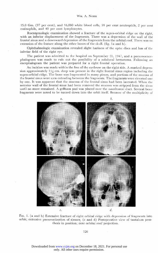
Roentgenologic examination showed a fracture of the supra-orbital ridge on the right, with an inferior displacement of the fragments. There was a depression of the wall of the frontal sinus and a downward depression of the fragments f rom the orbital roof. There was no extension of the feature along the other bones of the skull, (fig. l a and b).
Ophthalmologic examination revealed slight haziness of the optic discs and loss of the inferior field of the right eye.
The patient was admitted to the hospital on September 11, 1947, and a pneumoence-phalogram was made to rule out the possibility of a subdural hematoma. Following an encephalogram the patient was prepared for a right frontal operation.
An incision was made within the line of the eyebrow on the right side. A marked depres-sion approximately lJ/£ cm. deep was present in the right frontal sinus region including the supra-orbital ridge. The bone was fragmented in many pieces, and portions of the mucosa of the frontal sinus were seen extruding between the fragments. The fragments were elevated one by one. It was apparent that the mucosa of the frontal sinus had been lacerated. When the anterior wall of the frontal sinus had been removed the mucosa was stripped from the sinus until no more remained. A gelfoam pad was placed over the nasofrontal duct. Several bone fragments were noted to be turned down into the orbit itself. Because of the multiplicity of
a. b.
c. d.
Fio. 1. (a and b) Extensive fracture of right orbital ridge with depression of fragments into orbit; extensive pneumatization of sinuses, (c and d) Postoperative view of tantalum pros-
thesis in position: note orbital roof projection.
1 2 6
only. All other uses require permission. on December 18, 2021. For personal usewww.ccjm.orgDownloaded from

F R A C T U R E O F S I N U S
fractures it was impossible to fit the fragments together. A tantalum plate .0007 inch thick was molded to conform to the orbital ridge over the frontal defect. A portion of this plate extended posteriorly at right angles to form a new orbital roof and floor of the frontal sinus. It was tightly fitted and required only a single tantalum screw placed in the right lateral aspect of the incision to hold it in place (fig. l c and d). A mixture of sulfadiazine and 100,000 units of penicillin was instilled into the sinus cavity. T h e incision was closed with a double row of buried, inverted, interrupted black silk sutures. T h e right eye, which had previously deviated downward and outward, had again resumed its normal position in the head.
The patient was given penicillin, sulfadiazine, and neosynephrine nasal sprays for ten days and had an uneventful postoperative course. He was discharged from the hospital on September 25, 1947, with instructions to continue the nasal spray.
O n October 31, 1947, he noted a slight swelling over the plated area. A small amount of fluid was aspirated from beneath the skin, cultures and smears of which were negative. He was given penicillin prophylactically and was instructed to use a neosynephrine spray. The patient improved rapidly, and the swelling subsided within a day or two. When last seen seven months later he had resumed his full normal activity without further complica-tions (fig. 2).
FIG. 2. Postoperative appearance of right orbital reconstruction. Lateral and frontal views.
Discussion
In this case the external violence fractured the heavy supraciliary ridge downward into the orbit and the anterior wall of the large frontal sinus into the sinus cavity. Since the tissue overlying the defect appeared in good con-dition the operation was performed early, though Gardner3 believes that the presence of a tantalum implant does not impair the healing of an infected wound. Pneumoencephalography was carried out to reject the possibility of other intracranial disease, a procedure done by Turner before each cranio-plasty.
1 2 7
only. All other uses require permission. on December 18, 2021. For personal usewww.ccjm.orgDownloaded from

W i n . A . N O S I K
It is important to reproduce the convexity of the vertical or frontal limb as well as the convexity of the orbital portion which extended backward at approximately right angles. This approximation was most accurately obtained by comparison with the anterior-posterior and lateral roentgenographic views of the head. Because of the malleability and the lightness of the tantalum sheet used, no difficulty was encountered in obtaining the necessary tight fit. After the plate was molded it was locked in place with only one laterally placed tantalum screw. Bearing in mind the experience of Woodhall and Cramer4 with extradural pneumatocele in tantalum cranioplasty in which a fistulous tract extended from the frontal sinus into a fibrous sac enveloping the tantalum plate, no perforations were made in the plate. The small collection of fluid which appeared one month later might have been avoided had perforation been present, though it seemed to be the lesser of two evils.
References
1. Turner, O. A.: Repair of defects of skull with special reference to periorbital structures and frontal sinus. Arch. Surg. 53:312-326 (Sept.) 1946.
2. Conley, J . J . : Removal of frontal osteoma and correction of defect with tanta lum implanl. Arch. Otolaryng. 40:295-298 (Oct . ) 1944.
3. Gardner, W. J . : Use of tanta lum for repair of cranial defects in infected cases. Cleveland Clin. Quar t . 13:72-87 (April) 1946.
4. Woodhall, B., and Cramer, F. J . : Extradural pneumatocele following tantalum cranio-plasty. J . Neurosurg. 2:524-529 (Nov.) 1945.
1 2 8
only. All other uses require permission. on December 18, 2021. For personal usewww.ccjm.orgDownloaded from