tardir/tiffs/a372718 - dtic
TRANSCRIPT
TNO report PML 1999-A83
Characterisation of Bacteria by Matrix- assisted Laser Desorption/Ionisation and Electrospray Mass Spectrometry
TNO Prins Maurits Laboratory
Lange Kleiweg 137 P.O. Box 45 2280 AA Rijswijk The Netherlands
Phone+31 15 284 28 42 Fax+31 15 284 39 63
Date
December 1999
Author(s)
Dr. B.L.M. van Baar
Classification
Classified by : N.H.W. van Xanten, pharmacist Classification date : 27 August 1999
(This classification will not change)
Title Ongerubriceerd Managementuittreksel Ongerubriceerd Summary Ongerubriceerd Report text Ongerubriceerd Annexes A - B Ongerubriceerd
All rights reserved. No part of this publication may be reproduced and/or published by print, photoprint, microfilm or any other means without the previous written consent of TNO.
In case this report was drafted on instructions, the rights and obligations of contracting parties are subject to either the Standard Conditions for Research Instructions given to TNO, or the relevant agreement concluded between the contracting parties. Submitting the report for inspection to parties who have a direct interest is permitted.
»1999 TNO
Copy no.
No. of copies
No. of pages
No. of annexes
Iff 30
60 (incl. annexes, excl. RDP & distribution list)
2
All information which is classified according to Dutch regulations shall be treated by the recipient in the same way as classified information of corresponding value in his own country. No part of this information will be disclosed to any party.
The classification designation Ongerubriceerd is equivalent to Unclassified.
" DJBPBCIED 1 DISTRIBUTION STATEMENT A
Approved for Public Release Distribution Unlimited
TNO Prins Maurits Laboratory is part of TNO Defence Research which further consists of:
TNO Physics and Electronics Laboratory TNO Human Factors Research Institute
UK» 20000127 098
Netherlands Organization for Applied Scientific Research (TNO)

Characterisation of Bacteria by Matrix-assisted Laser Desorption/lonisation and Electrospray Mass Spectrometry
dr. B.L.M. van Baar december1999
■if) TNOrapport PML 1999-A83 ^
Probleemstelling In verband met bescherming van burgers en militairen en voor versterking van de Biolo- gische Wapen Conventie (1972) is detectie en identificatie van biologische strijdmiddelen gewenst. In het algemeen kunnen detectie en identificatie van biologische strijdmiddelen, typische micro-organismen zoals bacterien en virussen, worden uitgevoerd met biologische of chemische analysemethoden. De meest gangbare biologische methoden, veelal ontwikkeld voor geneeskundige toepassing, zijn bewerkelijk en vergen veel tijd. De ontwikkeling van chemische methoden staat nog in de kinderschoenen. De chemische analysemethoden kunnen in principe snel en met relatief weinig werk worden ingezet, mits die methoden goed ontwikkeld zijn. TNO Prins Maurits Laboratorium (TNO- PML) werkt samen met de Technische Uni- versiteit Delft (TU-Delft) aan de ontwikkeling van een detectiesysteem voor micro- organismen in lucht ('bio-aerosolen'). Dit systeem is gebaseerd op chemische analyse, onder andere door massaspectrometrie van bacterien. Vanuit deze achtergrond werd een Studie gemaakt van het werk dat reeds door anderen was gedaan aan massaspectrometrie van bacterien.
Beschrijving van de werkzaamheden Van eerder werk op het gebied van massa- spectrometrie van bacterien werd een over- zicht met kritische beschouwing samengesteld aan de hand van literatuuronderzoek.
Resultaten en conclusies Bij de resultaten van literatuurstudie is onder- scheid gemaakt naar massaspectrometrische analysemethoden. Het blijkt mogelijk bacterien te herkennen aan massaspectra, door de aanwezigheid van specifieke verbindingen. Al naar gelang de toegepaste methoden kijkt men naar signalen van intacte en behandelde bacterien ('lysaten') of van gescheiden verbindingen (bijvoorbeeld eiwitten). Er heerst nog onduidelijkheid over de effecten van voeding op de aan- of afwezigheid van de gebruikte signalen. Verder weet men nog te weinig over de precieze verbindingen die verantwoordelijk zijn voor de waargenomen signalen. Desalniettemin is de massa- spectrometrie een veelbelovende techniek voor de detectie en identificatie van bacterien.
Toepasbaarheid De resultaten van het onderzoek zullen ge- bruikt worden bij de verdere ontwikkeling van het bio-aerosoldetectiesysteem.
Vervolgonderzoek Vervolgonderzoek dient zieh te richten op de praktische analyse van bacterien. Daarbij moet aandacht worden besteed aan de analyse van specifieke verbindingen en aan de effecten van voedingsstoffen op de waargenomen signalen.
ill Ö
UK« Defensieopdracht A98D420
t

*#
1 1 ' i I >i
1 / i
i 1 I
-)
\ !
1 "* I
— 1
^ j
Projectinformatie
Projecttitel Massaspectrometrisch bio-aerosol alarm. Projectnummer TNO-PML 014.11019 Omschrijving programma Onderzoek aan de ontwikkeling van een op lasergekoppelde massaspectrometrie gebaseerd systeem voor de alarmeringsdetectie van 'mid- spectrum agents' en biologische strijdmiddelen (samen met de TU-Delft). Planning programma (tijdspad) Haalbaarheidsstudie, tot 31-12-1998. Verbe- tering van het TU-Delft-systeem, tot 31-12-2001. Realisatie van verbeterd systeem en tests, tot 31-12-2003. Projectbegeleider defensie drs. N.H.W. van Xanten, apotheker, Ministerie van Defensie, DICO/MGFB/GGB. Projectleider TNO-PML dr. ing. C.E. Kientz, Divisie Toxische Stoffen, researchgroep Ana- lyse van Toxische en Explosieve Stoffen. Communicatie Gedurende de acht maanden van het totstand- komen van dit rapport werd zesmaal overlegd met de projectbegeleider.
TM«

TNO report
PML1999-A83
Contents
Managementuittreksel 2
1 Introduction 5 1.1 General introduction 5 1.2 Scope 5 1.3 Mass spectrometry methods 6
2 Mass spectrometry of bacteria 16 2.1 Analysis of lipids 16 2.2 Analysis of lipopolysaccharides (LPS) and -
oligosaccharides (LOS) 18 2.3 Analysis of proteins 20 2.4 Analysis of DNA 24 2.5 Analysis of other specific compounds 27 2.6 Analysis of whole bacteria and bacteria lysates 29
3 Outlook 38
4 References 41
5 Authentication 57
Annexes A List of abbreviations B Overview of bacteria typing by ES MS and MALDI MS

TNO report
PML1999-A83
Introduction
1.1 General introduction
Until the early 1980's mass spectrometry (MS) was applied mainly in the field of organic chemistry. Information from mass spectra typically encompassed molecu- lar weight, molecular structure, and element composition of compounds. Analysis of compounds from biological origin was severely restricted by their thermolability and limited volatility, because both properties were incompatible with ionisation methods used at that time. The mass spectrometric analysis of biological materials was conducted through MS of vapours generated by pyrolysis of sample material. In addition, derivatisation was widely used to make particular compounds, e.g. fatty acids, amenable to MS analysis. However, general bioanalytical MS remained underdeveloped. Since that time, the field of MS has been revolutionised by the introduction of various new ionisation methods. With the advent of particle bombardment ionisation methods, the field of bioanaly- sis opened up. Particularly plasma desorption (PD; [1, 2, 3]), fast atom bombard- ment (FAB; [4]) and its fast ion bombardment analogue (liquid secondary ion mass spectrometry, LSIMS) were employed to ionise compounds of biological origin and bring them into the gas phase. The scope of MS analysis was extended to peptides, polysaccharides and (oligo)nucleotides. Pioneering work in the analysis of lipids and fatty acids and endotoxin from whole cells or cell lysates with FAB, PDMS and laser desorption ionisation (LDI) was presented from the 1980's on [5, 6,7, 8, 9]. The development of bioanalytical mass spectrometry was further boosted by the interfacing of mass spectrometers to column liquid separation technology, especially by the combination of ES MS with liquid chromatography (LC) or capillary electrophoresis (CE). The subsequent introduction of matrix- assisted LDI (MALDI) provided further capabilities in analysing compounds hitherto not amenable to MS. With these advances, the sequencing of proteins, for example, has come to be a typical routine MS analysis [10,11]. The potential of applying MS in analyses of progressive complexity, to the level of analysis of whole organisms, is clearly demonstrated by the analysis of viruses, a subject recently reviewed by Siuzdak [12]. In the on-going development, efforts have been directed most recently towards direct MS of complicated systems like whole bacte- ria and single cells.
1.2 Scope
The MS characterisation of bacteria provides an interesting possibility for the study of various environmental problems. Rapid distinction of pathogenic and non- pathogenic species is important in occupational and health care, in defence against
TNO report
PML1999-A83
biological warfare (BW) agents and in environmental monitoring. This review gives an overview of the current possibilities of MS characterisation of bacteria with limitations posed by MS instrumentation and by the materials analysed. The scope of the review will be limited to the use of ES MS and MALDI MS. Charac- terisation of bacteria by pyrolysis MS is not discussed here, but the reader is re- ferred to recent publications on this subject [13,14,15,16]. Characterisation by FAB MS of lipids is not discussed either; an excellent overview of this subject is already available [17]. In addition, related methods for the identification of fatty acids from bacterial lipids, either directly, by FAB MS (e.g. [5]), or after derivati- sation to the methyl esters, by gas chromatography (GC) MS (see for example [18]), are not considered. Also, broad studies on highly specific compounds not explicitly used for typing, e.g. lipo-chitin oligosaccharides from Rhizobii [19], are not considered here. Literature on the subjects considered was covered until May 1999; an overview is given in Table 1 (Annex B). A brief introduction is given on ES and MALDI MS methods, in order to outline general possibilities and limitations of these methods. The analysis of complex mixtures obtained from bacteria is considered next, focusing on the compounds most widely used for characterisation: lipids, lipooligo- and lipopolysaccharides (LOS/LPS), proteins, plasmid DNA, polymerase chain reaction (PCR) products of isolated genomic DNA and of other specific compounds. As far as Chromato- graphie (including electrophoretic) sample treatment is concerned in the analysis of these compound mixtures, attention will be limited to on-line methods with the potential of bacteria characterisation. In contrast, isolation and characterisation of single compounds by MS methods are not discussed, because the characterisation potential of the information obtained is low. Next, discussion focuses on the analy- sis of single bacteria and of bacteria lysates without specific identification of compounds. This approach provides the most rapid means of characterisation, and some attention will be given to the information accessible from the mass spectra thus obtained. The review concludes with an outlook on future developments.
1.3 Mass spectrometry methods
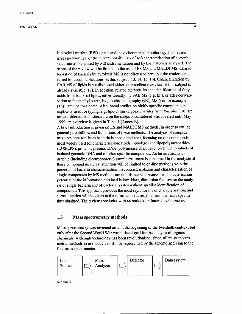
Mass spectrometry was invented around the beginning of the twentieth century, but only after the Second World War was it developed for the analysis of organic chemicals. Although technology has been revolutionised, since, all mass spectro- metric methods in use today can still be represented by the scheme applying to the first mass spectrometer.
Detector
^
Data system
Scheme 1.

TNO report
PML 1999-A83
Essentially, recording of a mass spectrum proceeds through three steps: analytes are ionised, analyte ions are mass separated and then detected. Compounds of biological interest are amenable to ion sources which generate ions through spray- ing, as in ES, or through particle bombardment of sample deposited on a target, as in MALDI. ES and MALDI will be discussed below, because they are presently the most relevant ionisation methods within the context of bioanalytical MS. The limit of detection (LOD) of a single compound is commonly determined by performing experiments on a dilution series. Measurement of the same compound in a mixture may either suppress or enhance ionisation and, therefore, the single compound LOD cannot be translated to a general LOD. The LOD is strongly dependent on the type of compound and a comparison can only be made for classes of compounds; here, peptides, proteins and DNA will be referred to. In ES MS, the LOD of peptides and proteins, with positive ion detection, lies in the low picomole range, whereas the LOD of DNA, with negative ion detection, typically lies in the picomole range. In MALDI MS, the LOD of peptides lies in the low femtomole range, with occasional reports of an LOD in the attomole range (10~18 mole, on- target). The LOD for protein MALDI lies in the low femtomole range, whereas the LOD of DNA lies in the low picomole range, provided that sample preparation is done with sufficient care. In general, these LOD values are sufficiently low to allow successful investigations of biological materials, although lower limits of detection remain ever desirable.
1.3.1 Electrospray Electrospray (ES) was introduced as an ionisation method in MS in the mid 1980's, by Fenn and co-workers [20, 21, 22], although the ES process had already been experimentally studied as early as 1917 [23]. In all forms of ES MS a solution of analytes is nebulised through a capillary into a high voltage electric field region (typically at 2-4 kV; see Figure 1). The nebulisation capillary may have a coaxial flow of gas ('ionspray' [24]) or may be miniaturised for better spray performance and lower sample consumption: micro-ES [25] and nanospray [26, 27]. Nebulisa- tion takes place at atmospheric pressure and, therefore, ES ionisation requires the use of an atmospheric pressure ionisation (API) type mass spectrometer.

TNO report
PML 1999-A83
Figure 1: Simplified scheme of an electrospray LC-MS interface; CV= cone voltage, HV = high voltage, C = cone, S = (capillary) spray needle, M = (quad- rupole) mass separation system; open arrows indicate curtain gas flow.
The spraying process initially generates small, highly charged droplets, which disintegrate into smaller, charged droplets during the flight. Solvent evaporation and droplet fission are aided by collision with air molecules. Upon arrival at the mass analyser system, the droplets have fully evaporated. The excess charge, initially present on the droplets, is now (partly) left on the molecules originally dissolved in the sprayed liquid. Although there is some theoretical debate as to whether droplets produce ions through subsequent evaporation and fission steps [28] or through ejection of ions from the highly charged droplet surface [29], practice shows that ions from various biomolecules are readily obtained. For ex- ample, reviews have appeared covering ES MS of peptides, proteins [10, 30], nucleotides and oligonucleotides [31, 32] and carbohydrates [33]. ES is quite effective in ionising biomolecules, because a solvent aided transfer to the gas phase imparts negligible internal energy on these molecules; thus, involatility and frag- mentation through thermolability are only rarely an issue in ES MS.

TNO report
500 II1I|TMI<| I Fli|
600 700 800 900 1000 1100 1200 1300 1400 1500 1600 1700
100n
%
0 10000
B: 11605.8711.00 B
A:27210.29±1.10
i|i IPI<(II ^^|tti|l|Hiji|J|li
12000 14000 16000 18000 20000 ^iiii|rN>iii"Ht|'n'i')fiiii^['ri^ ftfi>flW*yfrftirf,i«waq
22000 24000 26000 28000 MW (Da)
30000
Figure 2: A) positive ion ES mass spectrum of cholera toxin (from Vibrio cholerae, classical strain 569B), showing the charge state envelopes of A- andB-chain (in labels: A/B corresponds to chain, number corresponds to number of protons), and B) same spectrum in its deconvolutedform (measured MWav
shown; calculated: 27210 and 11605 Da for A- and B-chain, respectively); conditions: 3.8 kVpositive ion ES, with eluent H20/MeCN, 1:1 (v:v) + 0.2% formic acid (data obtained in our laboratory).

TNO report
PML1999-A83 10
ES mass spectra can generally be obtained by positive ion or negative ion detec- tion. The mode of detection is chosen to suit particular classes of compounds studied. As a rule of thumb, an acid, e.g. an oligonucleotide, is detected as the deprotonated base (negative ions), whereas a base, e.g. a peptide, is detected as the protonated molecule (positive ions). Although ES spectra obtained under different conditions or on different mass spectrometers are hardly ever identical, spectra of biomolecules have the common feature of a charge state envelope.
100,
N27klo„ 682 N26
709
MWav:18451.9±1.2
N25 757 N24
768 N23 801
H^WwJwi
N22 838 N21
878 N20 KHQ QOO N19 aj 970
■ i I ■ ' ■ I I ■ ' ' I I '■' ' ' H ' ' ' I
100i
17000 17500 18000 18500 19000 19500 20000
Figure 3. Negative ion ES mass spectrum of a DNA oligonucleotide (A; synthesised; nt 1-60, o/V. cholerae 569B, ctx operon, 5'-ATGGTA...AATGAT-3'), showing the charge state envelope, and corresponding deconvoluted spectrum (B; measured MWav shown; calculated: 18452.1 Da); conditions: 3 kVnegative ion ES, with eluent H20/MeCN, 1:1 (v:v) + 0.05%piperidine (data obtained in our laboratory).
As shown in Figures 2A and 3A, one compound produces various signals, with each signal belonging to a determined m/z ratio; every single signal reflects a distinct state of (de)protonation of the biomolecule. The combination of the ob- served m/z ratios allows the extraction of a single molecular weight from the spec-
TNO report
PML 1999-A83
trum; a result of the software deconvolution of the spectra shown in 2A and 3A is given in Figures 2B and 3B, respectively. The multiple charging of large bio- molecules provides ions with low m/z ratio, typically up to 2000 Da, and allows the use of most types of mass analyser system. ES MS requires sample dissolution in a suitable solvent. Mixtures of water and an organic modifier, typically methanol or acetonitrile, are commonly used. This solvent is often modified by the addition of acid (<1% v:v), typically formic or trifluoroacetic acid, to improve (de)protonation. The acid additive provides a proton donor or acceptor and suppresses the undesired formation of alkali metal adduct ions. The use of solvent implies that ES MS can be used for on-line detec- tion with LC or CE. Interfacing technology for LC-ES MS is commercially avail- able, whereas interfacing of CE and ES MS is often also performed through labo- ratory-made constructions (see for example [34]). The use of solvent also implies that whole cells are not amenable to ES MS. Instead, ES MS is typically applied in the analysis of mixtures of cell components, e.g. lipids, or of cell lysates. Due to the spray technology, ES MS is not very tolerant to salt; in addition to undesirable alkali adduct ion formation, salts may clog the spray capillary. Therefore, desalina- tion of samples by (micro)dialysis or ion exchange is often required; here, microdi- alysis has the advantage of on-line operation (see for example [35, 36]). Although some sample preparation is generally required, the on-line capabilities of ES MS have even been applied to in vivo analysis (see for example [37]).
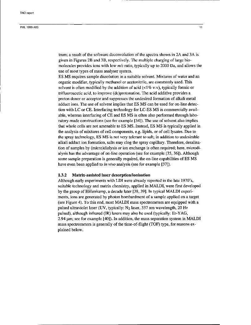
1.3.2 Matrix-assisted laser desorption/ionisation Although early experiments with LDI were already reported in the late 1970's, suitable technology and matrix chemistry, applied in MALDI, were first developed by the group of Hillenkamp, a decade later [38, 39]. In typical MALDI experi- ments, ions are generated by photon bombardment of a sample applied on a target (see Figure 4). To this end, most MALDI mass spectrometers are equipped with a pulsed ultraviolet laser (UV, typically: N2 laser, 337 nm wavelength, 20 Hz pulsed), although infrared (IR) lasers may also be used (typically: Er-YAG, 2.94 um; see for example [40]). In addition, the mass separation system in MALDI mass spectrometers is generally of the time-of-flight (TOF) type, for reasons ex- plained below.
TNO report
PML1999-A83 12
Figure 4. Schematic representation of a MALDITOF mass spectrometer; L = laser, F = fibre optics, 0 = optics, B = laser beam, S = sample spot, A = acceler- ating grids, V = vacuum chamber.
The processes by which MALDI generates ions are still not fully understood. It is known that the mixing of a sample with an excess of a solid organic chemical, the matrix, is required for successful ionisation. This matrix-sample mixture is made to form a crystalline precipitate, through various possible methods of preparation [41]. The matrix is thought to absorb most of the photon energy, converting the mixture locally into a plume of vaporised material. Ions are thought to be formed in this vapour, generally by proton transfer from matrix to analyte (positive ions) or from analyte to matrix (negative ions). In addition, the dried matrix-sample prepa- ration is inhomogeneous, and it has been shown that best ion formation occurs from particular spots in the preparation [42, 43]. Although it is known that specific analytes require the use of selected matrices, matrix selection remains empirical. When applying UV MALDI, proteins are often successfully ionised with a-cyano- 4-hydroxycinnamic acid [44] or sinapinic acid [45], whereas DNA is better ionised with 3-hydroxy-picolinic acid [46]. Furthermore, the laser power is known to be of crucial influence: a lower threshold value for ion formation is generally observed and significant ion formation occurs only within a limited window of the laser power range. A full review of findings is beyond the scope of this review, but it is clear that many experimental parameters are determined empirically or drawn from the quickly expanding amount of literature on MALDI.

TNO report
PML1999-A83 13
•.I.
1500
1000
500
SI
14190 i 28368
L -i 1 1 r 1 1 i "i r— -i 1 r
Figure 5: MALDI mass spectrum o/Staphylococcus aureus enterotoxin B (SEB), showing singly and doubly protonated molecule signals; the inset shows an enlargement of the [M+H]+ region, displaying matrix adduct signals above the molecular mass of SEB (data obtained in our laboratory).
MALDI mass spectra are mostly obtained by positive ion detection, because nega- tive ion formation is often relatively inefficient; only DNA and other acidic com- pounds typically form negative ions more efficiently. Although spectra obtained with different instruments, or even under different ionisation conditions, are rarely identical, spectra of biomolecules have some common features. As shown in Figure 5, spectra typically show singly protonated molecules, [M+H]+, whereas multiply protonated species, [M+nH]n+, and cluster ions, [nM+H]+, may some- times be present. The single protonation of biomolecules provides ions with high m/z ratio, sometimes well over 106 Da. As a consequence, a TOF mass analyser is generally used with MALDI, because it (in principle) has an indefinitely large mass range. Although low molecular mass biomolecules may be mass analysed with other types of analyser system, the low mass range of the TOF analyser (i.e. below ~30 kDa) provides excellent capabilities through the use of a reflectron (in the analyser system [47]), and of 'delayed ion extraction' (in the ion source; [48, 49]).

TNO report
PML1999-A83 14
Thus, MALDI ionisation and TOF mass separation form a most widely used com- bination. MALDI mass spectra typically lack fragment ion signals, because the desorption/ionisation process imparts negligible internal energy on the analyte molecules and, thus, prevents in-source fragmentation. Nevertheless, spontaneous ('metastable') fragmentation of initially formed ions occurs during ion flight and is a source of signal broadening in TOF MS, particularly notorious with oligonucleo- tides. In contrast, fragmentation can be induced by specifically selected ionisation conditions; for example, it was observed that oligonucleotides fragment extensively with 266 nm but not with 337 nm laser wavelength [50]. Given the low tendencies for multiple (de)protonation and fragmentation, MALDI mass spectra of pure compounds are generally simple. MALDI MS requires deposition of material on a target and mixing with a suitable matrix compound. The sample volume applied on-target is typically less than one microlitre, containing less than one picomole of the analyte. Common sample targets are stainless steel, but more exotic materials can also be used as they are particularly useful in the combination of MALDI and other analytical methods, e.g. with surface plasmon resonance (SPR; [51, 52]) or with polyacrylamide gel elec- trophoresis (PAGE; [53, 54, 55]). Alternatively, the application of polymeric coatings to sample targets can be used to improve MALDI performance (see for example [56, 57]). Sample application can be performed straightforwardly by the 'dried drop' method [38], but more elaborate methods are generally required to obtain low detection limits and good mass resolution (see for example. [41]). Sample preparation can also be performed on the target; for example, desalination with ion exchange resin is widely applied. The fact that only a fraction of the applied analyte is consumed by MALDI opens up the possibility for further ex- periments, for example: MALDI remeasurement after on-target enzymatic digest (see for example [41, 53]), and analysis by methods like immunochemical detec- tion after protein recovery [58]. Another interesting application of MALDI MS concerns the measurement of peptides directly from single whole cells; after a first demonstration with neurones [59], this has recently culminated in a two- dimensional cell imaging technique [60]. Thus, MALDI is often compatible with analytical methods more common to a biochemical or biological laboratory than ES MS. Of course, there are some drawbacks to MALDI as well. Although the ionisation method is more tolerant to salt than ES, alkali ion adducts are frequently observed. In addition, matrix adduct ions are often formed, either through covalent bonding of the matrix to the analyte or as a non-covalent ion-molecule complex. These adduct ions generally cause a deteriorated mass resolution; the adduct ion m/z ratio lies relatively close to the analyte ion m/z ratio and tends to broaden the signal of interest. A further disadvantage is that liquid separation methods are necessarily used in an off-line mode, which requires fraction collection with LC or CE. Meth- ods for on-line coupling of LC (or CE) and MALDI have been described, but a recent review [61] shows that LC- or CE-MALDI MS is still not a routine option.
TNO report
PML 1999-A83 15
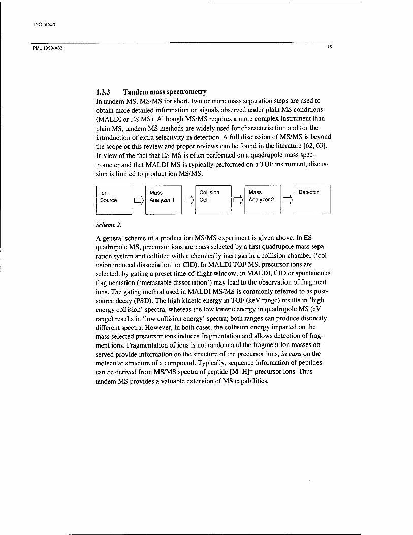
1.3.3 Tandem mass spectrometry In tandem MS, MS/MS for short, two or more mass separation steps are used to obtain more detailed information on signals observed under plain MS conditions (MALDI or ES MS). Although MS/MS requires a more complex instrument than plain MS, tandem MS methods are widely used for characterisation and for the introduction of extra selectivity in detection. A full discussion of MS/MS is beyond the scope of this review and proper reviews can be found in the literature [62, 63]. In view of the fact that ES MS is often performed on a quadrupole mass spec- trometer and that MALDI MS is typically performed on a TOF instrument, discus- sion is limited to product ion MS/MS.
Ion Source <3
Mass Analyzer 1 <=>
Collision Cell 3
Mass Analyzer 2 <3
Detector
Scheme 2.
A general scheme of a product ion MS/MS experiment is given above. In ES quadrupole MS, precursor ions are mass selected by a first quadrupole mass sepa- ration system and collided with a chemically inert gas in a collision chamber ('col- lision induced dissociation' or CID). In MALDI TOF MS, precursor ions are selected, by gating a preset time-of-flight window; in MALDI, CID or spontaneous fragmentation ('metastable dissociation') may lead to the observation of fragment ions. The gating method used in MALDI MS/MS is commonly referred to as post- source decay (PSD). The high kinetic energy in TOF (keV range) results in 'high energy collision' spectra, whereas the low kinetic energy in quadrupole MS (eV range) results in 'low collision energy' spectra; both ranges can produce distinctly different spectra. However, in both cases, the collision energy imparted on the mass selected precursor ions induces fragmentation and allows detection of frag- ment ions. Fragmentation of ions is not random and the fragment ion masses ob- served provide information on the structure of the precursor ions, in casu on the molecular structure of a compound. Typically, sequence information of peptides can be derived from MS/MS spectra of peptide [M+H]+ precursor ions. Thus tandem MS provides a valuable extension of MS capabilities.

TNO report
PML1999-A83 16
Mass spectrometry of bacteria
2.1 Analysis of lipids
Lipids are a class of compounds which has been used for typing of bacteria since the early 1960's [64, 65]. The characterisation of bacteria through FAB-MS of the lipid fraction isolated from lysate has proven a successful approach, already in 1987 [6, 7]. With the relatively new tools of ES and MALDI MS, this success should be continued. The glycerophospholipids from various bacteria were investigated, without prior separation, by negative ion ES MS [66]. Low energy CID MS/MS product ion spectra of [M-H]" ions generated from pure, isomeric compounds (i.e. isobaric) were studied for their specific fragmentation. The main MS/MS signals resulted from the fatty acid acyl groups, R-CO2", with intensity differences between acyl ions from the 1-, and the 2-substituent of the glycerol lipid backbone. Isobaric acyl groups, e.g. 17:1 and \lcyc fatty acids, could not be distinguished. However, with the partial identification of the two acyl substituents, the MW contribution of the polar head group could easily be derived, thus allowing classification of the phos- pholipids. If necessary, this classification was confirmed through the presence of low intensity, diagnostic fragment ions. The findings with pure compounds were applied to the analysis of chloroform/methanol extracts from Bacillus spp.,B. licheniformis, B. stearothermophilus, B. thuringiensis, and from Escherichia coli. Spectral patterns of selected precursor ions (from single MS) and product ions (from MS/MS) from these extracts were shown to differ significantly among the various species, thus allowing identification. The authors note that characterisation through fatty acid composition of the lipid fraction can only be used when bacteria have been cultured under standard conditions. Various phospholipids were identified in E. coli lipid extracts, by infusion negative ion ES MS [67]. The phospholipids were readily identified by their MW and, in addition, it was shown that in-source fragmentation, induced at elevated cone voltages, can be used to obtain fragments indicative of the fatty acid moieties. The authors indicate that full lipid identification can be achieved with less than 150 u.g (dry weight) of material, with the analysis accomplished in less than five minutes. It is concluded that all bacteria typing by lipid analysis, formerly done with FAB MS, can now be done less elaborately with ES MS. Lipid A obtained after acid hydrolysis of LPS from Enterobacter agglomerans and Salmonella minnesota was characterised by positive and negative ion ES MS, in the context of a more general investigation on bacterial endotoxin degradation [68, 69]. Positive ion detection of sodium adduct ions of Lipid A was only successful after dephosphorylation, whereas negative ion detection could be used with the intact Lipid A and with basic or acid hydrolysis products. This proved that basic hydrolysis is much more effective in removing fatty acyl chains from Lipid A and,

TNO report
PML1999-A83 17
therefore, in lowering the endotoxin level [69]. Lipid A of both species investi- gated, obtained from LPS by mild acid hydrolysis, shows some similarities in the (flow injection) ES MS spectrum, but species distinction can be made through diphosphoryl Lipid A (absent in & minnesota samples). This finding is corrobo- rated by a detailed study on Lipid A from Rhodobacter sphaeroides, which shows that MALDI MS and MS/MS can be used to characterise the Lipid A sugar moiety, degree of phosphorylation and substituent fatty acids [70]. The effect of various disinfection procedures on the lipid extracts from E. coli and B. cereus was investigated by flow injection ES MS [71], in a preliminary study of verification possibilities to strengthen the Biological and Toxin Weapons Conven- tion of 1972 [72]. Methanol/chloroform extracts from E. coli and B. cereus were obtained after treatment of the bacteria with ethanol, hypochlorite or heat and from untreated bacteria. The study does not allow conclusions on the real-world charac- terisation of bacteria from their disinfection debris, because the bacteria originated from one starter culture per species. Nevertheless, the spectra obtained from the various lipid fractions showed only minor differences, and even allowed identifi- cation of specific lipids. Although the study was limited to two species (one gram- positive and one gram-negative), this demonstrates that characterisation of bacteria from disinfected sites might generally be possible through MS of the lipid fraction. Thus, the use of on-line capillary electrophoresis (CE) or micro-column liquid chromatography (|X-LC) and ES MS allow detailed investigations of bacterial lipids. In contrast, no MALDI studies of bacterial lipids have appeared, as yet. Probably, the complexity of the lipid mixtures obtained from cells requires separa- tion prior to MS; therefore, on-line interfaces for CE-ES MS and U.-LC-ES MS give ES a decisive advantage over MALDI. Nevertheless, the separation step can be discarded, as was demonstrated in an investigation of the glycerophospholipids of selected strains of Bacillus spp., B. anthracis, B. cereus, B. thuringiensis and B. subtilis, by negative ion ES MS and ES MS/MS of crude lipid extracts [73]. The fatty acid substitution pattern of a variety of glycerophospholipids observed in ES MS spectra was determined by in- source fragmentation and by product ion MS/MS experiments on the [M-H]~ ions and used for bacteria typing. It was shown that the Bacillus spp. investigated could be distinguished by the fatty acid distribution in the glycerophospholipids; this is illustrated in Figure 6, which shows the fatty acid distribution in MS/MS spectra obtained from negative ions of glycerophospholipids with an MW of 694 Da. Lipids, particularly dimyceroserates, have also been detected in the whole bacteria positive ion mass spectra of Mycobacterium smegmatis ([74]; see below).

TNO report
PML1999-A83 18
1 Bacillus subtilis
CO c CD
CD >
J5 CD
GC
241 C15:0
J ■ I '
180
B
255 C16:0
269 C17:0 _A
x j_
200 220 240 260
m/z
280 300
Figure 6: Partial negative ES product ion MS/MS spectrum o/B. subtilis (top) and B. anthracis (bottom) glycerophospholipids with a molecular weight of 694 Da, showing the fatty acid distribution [73]; note that, despite the single selected mass (two fatty acids summing up to C30.0), the B. anthracis spectrum repre- sents at least three different glycerophospholipids, and that fragment ion in- tensity does not quantitatively reflect the exact fatty acid composition (e.g, C13.0 and C17:0 are not of equal intensity, although they must come from the same C30.0 type precursor molecules).
2.2 Analysis of lipopolysaccharides (LPS) and -oligosaccharides (LOS)
LPS and LOS are the subject of various studies, mainly because these outer cell membrane components from gram-negative bacteria carry important antigenic
TNO report
PML1999-A83 19
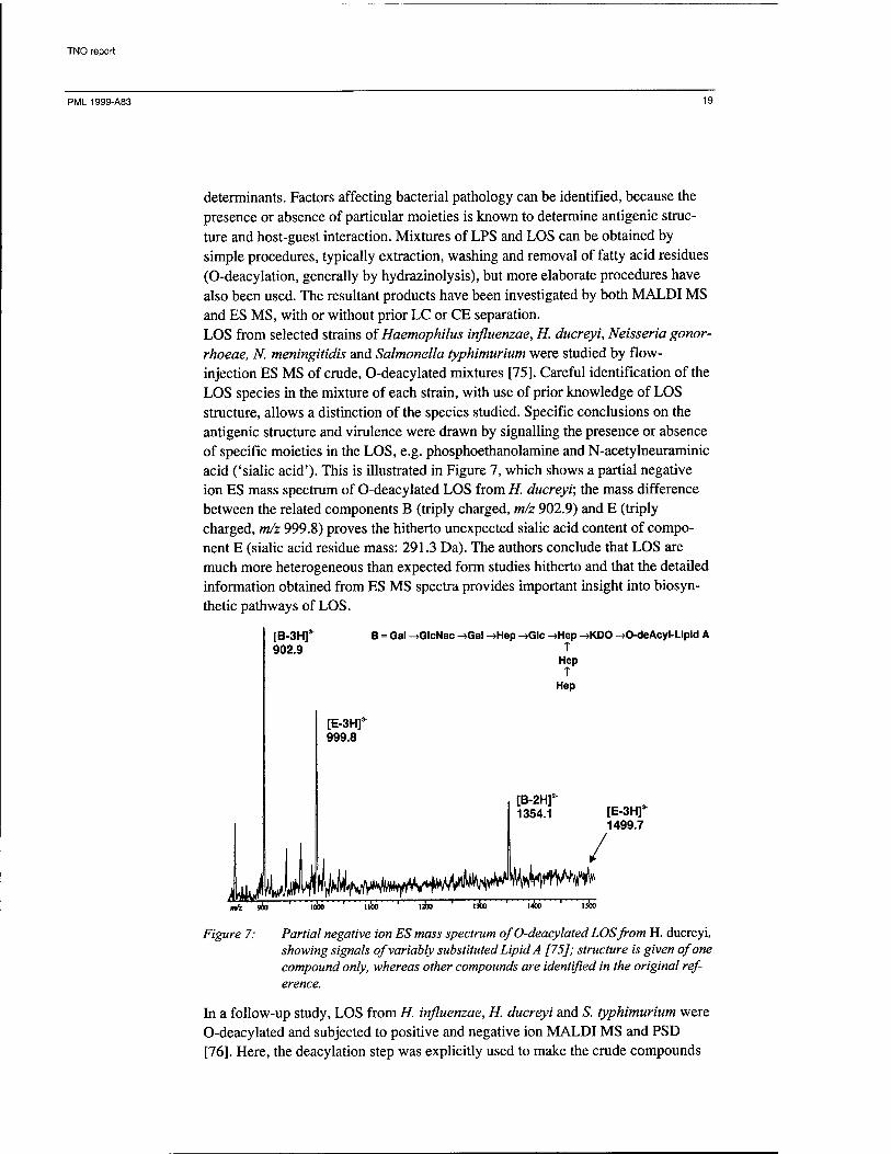
determinants. Factors affecting bacterial pathology can be identified, because the presence or absence of particular moieties is known to determine antigenic struc- ture and host-guest interaction. Mixtures of LPS and LOS can be obtained by simple procedures, typically extraction, washing and removal of fatty acid residues (O-deacylation, generally by hydrazinolysis), but more elaborate procedures have also been used. The resultant products have been investigated by both MALDI MS and ES MS, with or without prior LC or CE separation. LOS from selected strains of Haemophilus influenzae, H. ducreyi, Neisseria gonor- rhoeae, N. meningitidis and Salmonella typhimurium were studied by flow- injection ES MS of crude, O-deacylated mixtures [75]. Careful identification of the LOS species in the mixture of each strain, with use of prior knowledge of LOS structure, allows a distinction of the species studied. Specific conclusions on the antigenic structure and virulence were drawn by signalling the presence or absence of specific moieties in the LOS, e.g. phosphoethanolamine and N-acetylneuraminic acid ('sialic acid'). This is illustrated in Figure 7, which shows a partial negative ion ES mass spectrum of O-deacylated LOS from H. ducreyi; the mass difference between the related components B (triply charged, m/z 902.9) and E (triply charged, m/z 999.8) proves the hitherto unexpected sialic acid content of compo- nent E (sialic acid residue mass: 291.3 Da). The authors conclude that LOS are much more heterogeneous than expected form studies hitherto and that the detailed information obtained from ES MS spectra provides important insight into biosyn- thetic pathways of LOS.
[B-3Hf 902.9
B = Gal ->GlcNac ->Gal ->Hep ->Glc ->Hep ->KDO ->0-deAcyl-Lipid A T
Hep T
Hep
LkM
[E-3Hr 999.8
oö ' Tioö ' 13» ' 13» r
[B-2H]2
1354.1
%hw^ /
[E-3Hf 1499.7
»4 9O0 ' iooö ' Tioö ' !3» ' i3» ' I355 ' 13»
Figure 7: Partial negative ion ES mass spectrum of O-deacylated LOS from H. ducreyi, showing signals of variably substituted Lipid A [75]; structure is given of one compound only, whereas other compounds are identified in the original ref- erence.
In a follow-up study, LOS from H. influenzae, H. ducreyi and S. typhimurium were O-deacylated and subjected to positive and negative ion MALDI MS and PSD [76]. Here, the deacylation step was explicitly used to make the crude compounds

TNO report
PML1999-A83 20
amenable to MALDI, by improved miscibility with the matrix. The deacylated LOS appear as a variety of glycoforms, and typical spectra also show oligosaccha- ride and Lipid A fragment ion signals resulting from in source decay. It was dem- onstrated that PSD of the [M-H]- ions of selected glycoforms is extremely useful in further structure elucidation of LOS. In addition, it was shown that the methods developed could also be applied to a crude LOS extract (i.e. not deacylated). A more elaborate approach was applied in the analysis and characterisation of LPS from an H. influenzae (strain Eagan, serotype b) clinical isolate by CE-ES MS [77]. LPS were liberated from the cell wall of small quantities of the pathogen (0.2 mg dried cell mass) by proteinase K digest of whole cell solutions, subsequent treat- ment with DNase and RNase and O-deacylation. An on-line preconcentration step was used to achieve detection of the small quantities of LPS obtained, and com- pounds were detected as their negatively charged ions. The hydrophobic Lipid A part of the LPS provided the reversed phase affinity required for preconcentration from aqueous solution. Elution profiles, i.e. mass vs. retention time graphs, were used to achieve a broad characterisation of the specific LPS fractions by MW and charge.
Methylglucose LPS of M. smegmatis have been studied by negative ion CE-ES MS (and positive and negative ion LSIMS). These compounds are held responsible for alternative biosynthesis of mycolic acids which are known hydrophobic cell wall components involved in antibiotic resistance [78]. The methylglucose LPS was characterised and evidence for non-covalent complex formation between methyl- glucose LPS and palmitoyl-CoA, vital to the alternative biosynthesis of mycolic acids, was obtained by ES MS of mixtures of the two components. The appearance of the particular methylglucose LPS can be used for mycobacteria characterisation in general. It was noted [76, 77] that structure variation in LOS/LPS during bacterial growth, which is generally attributed to a response to external stimuli, would make these compounds less suitable for characterisation purposes. However, certain features of LOS/LPS are commonly used in serological typing and, therefore, chemical analy- sis of these compounds can provide a bridge between biological and chemical typing. In addition, MS investigation of LOS/LPS provides useful means for studying bacteria from clinical sources, thus providing valuable insight into rela- tions between structure alterations in bacterial LPS and pathology.
2.3 Analysis of proteins
The analysis of isolated whole proteins and their enzymatic digests by mass spec- trometry is now becoming a matter of routine. In principle, isolated single proteins from bacteria can be used for characterisation, provided that the protein is suffi- ciently specific for a genus or strain (see for example [79] concerning Clostridium thermosaccharolyticum). However, characterisation without isolation of one spe- cific protein is less elaborate, more general and, therefore, much more attractive.

TNO report
PML1999-A83 21
The protein fraction of bacteria has been used for MALDI MS characterisation purposes by various research groups. In all cases, a simple clean-up procedure was used to isolate the protein fraction from cell lysates, before MS analysis. The omission of separation methods in the MALDI MS analysis of complex protein mixtures is in marked contrast to the ES MS analysis of complex lipid mixtures. So far, the on-line separation capabilities with ES MS have only scarcely been applied in bacteria characterisation from bacterial protein fractions. It is noted that lysates of whole bacteria, which are generally assumed to consist of proteins, have been analysed by MALDI MS as well as by ES MS; however, lysate analysis is dis- cussed separately (vide infra). Nevertheless, MALDI MS of protein fractions already provides useful information for bacteria typing.
Cell lysis by sonification and subsequent solvent precipitation (or ultrafiltration) was used to obtain a crude water-soluble protein extract from several Bacillus and Pseudomonas spp. and other, gram-negative bacteria [80]. After addition of matrix, and with use of a polymer or a stainless steel target, MALDI mass spectra were obtained displaying protein profiles of cultured bacteria. A comparison with so- dium dodecyl sulphate - PAGE (SDS-PAGE) showed that the MALDI MS signals did not extend beyond 15 kDa, despite the presence of higher MW proteins. In contrast, the separation capabilities of PAGE were poor below 15 kDa. The dis- crepancy between the findings with PAGE and MALDI MS was attributed to the difference in sample preparation. Despite the apparently limited scope of protein profiles below 15 kDa, the information allowed distinction at the levels of genus and species. Interestingly, an artificial mixture of three bacteria produced a spec- trum which is almost a superposition of the three spectra of the single-component bacteria. The authors note that reproducibility, speed and sensitivity of analysis are major advantages of MALDI MS over SDS-PAGE. After this initial study, the characterisation method was extended with a capillary micro-LC separation between cell lysis and MALDI MS analysis [81]. The LC fractions obtained after gradient elution were mixed with matrix solution and deposited on a MALDI plate or on nitrocellulose substrate; the substrate reportedly improves ion yields [82]. The intermediate separation was used to prevent suppres- sion effects typically observed in desorption ionisation of mixtures. Indeed, the fractionated protein mixtures revealed more components than simple MALDI MS. In addition to species specific proteins, genus specific proteins were found in comparable LC fractions of Pseudomonas species. However, similar genus specific signals were not found for Bacillus spp., although species specific signals were observed; in this off-line LC MALDI MS procedure Bacillus spp. displayed a larger degree of heterogeneity. Despite the LC separation, high mass proteins observed with SDS-PAGE were not recovered, because the Ci8 micro-LC column used is not suited for the elution of large proteins. However, the additional dimen- sion of off-line separation greatly improves the distinction of the bacteria studied. MALDI MS analysis was performed with the protein fraction of cell lysates, ob- tained from whole bacteria in a simple 30-minute procedure, or with the tryptic digest of this fraction [83, 84]. Chemical lysis was found insufficient for gram-

TNO report
PML1999-A83 22
positive bacteria, because of more robust cell wall structures, and sonification was used for adequate lysis. The authors preferred external mass scale calibration, because it was noted that internal calibrants suppressed relevant sample signals. Although SDS-PAGE established that proteins with an MW of up to 160 kDa were present in the extracts, only compounds with an MW below 20 kDa were observed by MALDI MS (under the experimental conditions used). Although these findings are in line with the above-mentioned observations of Cain et al. [80], the discrep- ancy between PAGE and MALDI MS is attributed to suppression effects in ionisa- tion rather than to sample preparation. As in the aforementioned work [80], the mass range below 20 kDa yielded sufficiently distinct spectra for characterisation of various Bacillus spp. and distinction of these species from other pathogenic bacteria (Bruceila melitensis, Yersiniapestis, Francisella tularensis). It was even shown that particular marker signals could uniquely be found for B. anthracis grown on different media; however, the intensity of these marker signals varied considerably, as shown in Figure 8. Marker signals obtained from the tryptic di- gests of protein fractions were used for confirmation. This work showed that char- acterisation of bacteria can be based on the analysis of crude samples, obtained after simple sample preparation.
3075 4315
m/z
Figure 8: Positive ion MALDI mass spectrum of protein extracts from chemically lysed B. anthracis sterne cells, grown in casein acid digest medium (top) or in leighton doi medium (bottom); from reference [83].
As concerns the MALDI MS detection of high mass proteins (up to 500 kDa) in bacteria lysates, it was shown that sample preparation is a critical factor [85]. With a more elaborate sample preparation, involving repeated steps for extraction and solubilization of large proteins, high mass proteins could be observed. This was demonstrated with E. coli in a study of the proteins induced and suppressed by L- arabinose catabolism. Extraction and solubilization of the large proteins was en-

TNO report
PML1999-A83 23
hanced by using a combination of Triton buffer and guanidine-HCl, whereas the sensitivity of detection of MALDI MS was enhanced by the application of a nitro- cellulose film, prior to sample and matrix deposition. Proteins in the mass range of 20 to 120 kDa have a high abundance in the samples and they are readily observed; most MWs allow assignment of the MALDI spectral peaks to proteins known from the E. coli genome. Thus, the detection of high mass proteins allowed investigation of environmentally induced changes in gene expression of bacteria. A study on MALDI monitoring of recombinant protein expression in E. coli showed that small signals of the protein of interest could be obtained from whole bacteria, after on-target disruption of cell membranes with 1,1,1,3,3,3-hexafluoro- isopropanol [86]. In contrast, MALDI analysis of untreated bacteria, of methanol- water-treated bacteria, or of bacteria lysates did not produce significant signals for the desired recombinant protein. A positive MALDI MS screening result was followed by purification of the recombinant protein and confirmatory analysis by MALDI MS. The difficulties experienced in this screening of a single protein in a complex mixture demonstrate that suppression effects in the ionisation play an, as yet, unpredictable role and that such selectivity severely restricts conclusions with respect to (relative) quantities of the various components. The utility of MALDI MS for monitoring recombinant protein expression was further explored in a broader study with recombinant E. coli [87]. For larger pro- teins (>10 kDa), the broth solution was directly used for MALDI MS analysis, with minimum, on-target sample preparation. For smaller proteins, the precipitate from centrifuged culture was used for MALDI MS, instead. The developed procedure effectively limited total analysis time to ten minutes, thus allowing monitoring of the time dependency of expression. This time dependency was quantified by com- parison of the signal intensity of a recombinant protein (e.g. Mwaverage
-6000 Da) with that of a native E. coli protein (e.g. rpL29; MWav 7273.5). Nota- bly, the excess of recombinant proteins, present after induced expression, com- pletely suppresses MALDI MS signals of naturally occurring proteins oiE. coli. MALDI MS (with delayed extraction) shows the presence of target proteins and closely related compounds, e.g. methionine attached protein. All proteins studied (~5 - ~50 kDa) were successfully monitored by MALDI MS, with the considerable advantage of mass resolution and speed over SDS-PAGE. The influence of variations in some experimental parameters on the reproducibility of MALDI mass spectra was investigated systematically for crude protein extracts of E. coli and B. thuringiensis [88]. The 'sandwich method' of MALDI sample deposition was consistently used, while studying the extraction solvent composi- tion, extract salt content, sample solvent and protein extraction method. In addition, the analyses were performed in two laboratories and by different operators, thus giving some indication of interlaboratory variations. All parameters investigated were found to be important to the extent that spectra could be completely altered by using, for example, different extraction methods or pH. Nevertheless, persistent peaks could be observed under the various conditions (with the limited choice of bacteria) and these peaks should ideally be used as biomarkers for bacteria charac- terisation. The interlaboratory variations were in the order of the inter-operator
TNO report
PML1999-A83 24
variations, which finding suggests that the methods used are broadly applicable provided that extraction and sample preparation procedures are the same. Solvent and matrix combinations for the MALDI MS analysis of crude protein mixtures, extracted from E. coli, were used to establish optimum experimental conditions for bacteria typing through protein fractions [89]. From a cross- combination of five extraction solvents and four matrices it was found that extrac- tion is best performed with an isopropanohwatenformic acid mixture and that 2-(4- hydroxyphenylazo)benzoic acid performs best as a matrix. The authors note that spectrum quality, as judged from the width of the diagnostic mass range and from the number of diagnostic ions generated, is largely determined by the matrix- solvent system, i.e. by MALDI sample preparation, whereas reproducibility of spectra depends largely on bacteria sample handling techniques. A broad selection (2-100 kDa) of proteins from the whole E. coli proteome, ob- tained from dialysed lysates, was studied by CE, in the isoelectric focusing mode, and on-line ES MS with a high mass resolution Fourier transform ion cyclotron resonance (FT-ICR) instrument [90]. Between 400 and 1000 proteins were found in the electropherogram. Special automation software was used for the tentative identification of compounds from the dataset. The mass spectral patterns observed were enhanced by using lysates from bacteria grown on isotopically depleted media; this facilitated better pattern recognition in the protein charge state enve- lopes. The culturing in isotopically depleted media prohibits general use for bacte- ria typing, but it offers good means for the identification of specific proteins from a whole proteome; once the specific proteins have been identified, the CE-MS meas- urement of bacteria lysates from bacteria grown in 'isotopically natural' media, for typing purposes, becomes feasible. It is clear that this ES MS-based method has the possibility of very detailed fingerprinting.
2.4 Analysis of DNA
Analysis of DNA can be used for identification purposes and for investigating particular conditions of bacteria, e.g. phage infection or plasmid presence [65, 91]. Sensitive detection, even of DNA from a single cell, is generally achieved by using PCR amplification and appropriate detection methods. PCR with simple detection methods, e.g. colouring reactions, can be used routinely for the characterisation of bacteria (e.g. [92]). In the detection of PCR products, ES [31] and MALDI MS [93] provide more specific determination of oligonucleotide MW than other methods. With both MS methods, DNA is generally detected by its negative ions, i.e. in the singly or multiply deprotonated form. Various research groups have recently re- ported on the analysis of bacterial DNA and on PCR with subsequent MS analysis for the characterisation of bacteria. Plasmid DNA has been analysed by restriction enzyme digest and subsequent MALDI MS [56]. The 4658 base pair, double stranded B6BLEU5 plasmid, was digested by Alu I or Hae ///restriction enzymes (-15 pmol per digest). The per- formance of MALDI was enhanced by the application of a substrate coating
TNO report
PML1999-A83 25
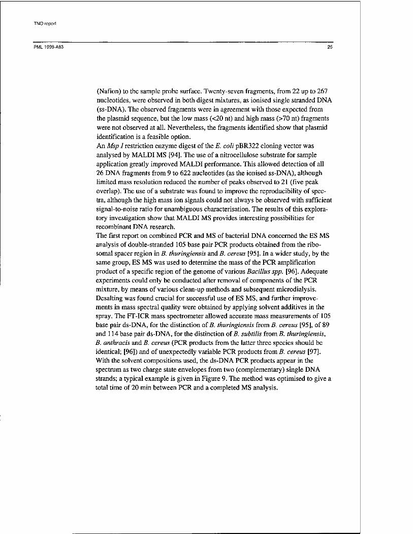
(Nafion) to the sample probe surface. Twenty-seven fragments, from 22 up to 267 nucleotides, were observed in both digest mixtures, as ionised single stranded DNA (ss-DNA). The observed fragments were in agreement with those expected from the plasmid sequence, but the low mass (<20 nt) and high mass (>70 nt) fragments were not observed at all. Nevertheless, the fragments identified show that plasmid identification is a feasible option. An Msp I restriction enzyme digest of the E. coli pBR322 cloning vector was analysed by MALDI MS [94]. The use of a nitrocellulose substrate for sample application greatly improved MALDI performance. This allowed detection of all 26 DNA fragments from 9 to 622 nucleotides (as the ionised ss-DNA), although limited mass resolution reduced the number of peaks observed to 21 (five peak overlap). The use of a substrate was found to improve the reproducibility of spec- tra, although the high mass ion signals could not always be observed with sufficient signal-to-noise ratio for unambiguous characterisation. The results of this explora- tory investigation show that MALDI MS provides interesting possibilities for recombinant DNA research. The first report on combined PCR and MS of bacterial DNA concerned the ES MS analysis of double-stranded 105 base pair PCR products obtained from the ribo- somal spacer region in B. thuringiensis and B. cereus [95]. In a wider study, by the same group, ES MS was used to determine the mass of the PCR amplification product of a specific region of the genome of various Bacillus spp. [96]. Adequate experiments could only be conducted after removal of components of the PCR mixture, by means of various clean-up methods and subsequent microdialysis. Desalting was found crucial for successful use of ES MS, and further improve- ments in mass spectral quality were obtained by applying solvent additives in the spray. The FT-ICR mass spectrometer allowed accurate mass measurements of 105 base pair ds-DNA, for the distinction of B. thuringiensis from B. cereus [95], of 89 and 114 base pair ds-DNA, for the distinction of B. subtilis from B. thuringiensis, B. anthracis and B. cereus (PCR products from the latter three species should be identical; [96]) and of unexpectedly variable PCR products from B. cereus [97]. With the solvent compositions used, the ds-DNA PCR products appear in the spectrum as two charge state envelopes from two (complementary) single DNA strands; a typical example is given in Figure 9. The method was optimised to give a total time of 20 min between PCR and a completed MS analysis.

TNO report
PML 1999-A83 26
88/89
2425
1275
Figure 9: ES-FTICR mass spectra ofPCR products produced from B. cereus strain 6464, showing charge states 18- to 27- ofds-DNA (top), with the inset dis- playing three ds-DNA products (1 nt difference in length, 88/88, 88/89 and 89/89), and charge states 18- to 23- ofss-DNA (bottom), with the inset dis- playing the presence of four strings (88 noncoding, 88N, and coding, 88C, 89 noncoding, 89N, and coding, 89C); from reference [97].
PCR with MALDI MS detection of reaction products was applied to identify Legionella species, in general, and L. pneumophila, in particular [98]. PCR was conducted using materials similar to those from a commercially available kit, but without the biotin labelling required for the colouring reaction used in dot-blot detection. PCR focuses on a 108 base pair region common to all species from genus Legionella and a 168 base pair region particular to L. pneumophila. Ampli- fied products from both sequences could be detected by MALDI MS, but only after thorough removal of the excess of unreacted primers and other low MW impurities, e.g. by the use of size exclusion microcolumn centrifugation. The same research group followed a similar but more extensive approach in the study of the effectivity of bioremediation, in this case: decontamination of soil by methanotrophic bacteria [99]. Specific, 56 and 99 base pair regions from the Meth- ylosinus trichosporum and Methylomicrobium albus genome were amplified by the PCR, using purpose-designed primers. The reaction product DNA was purified over column cartridges before MALDI MS determination of its MW. Only a small fraction (1-2 ul) from a single PCR mixture (100 jn.1) was required with the experi- mental conditions used, thus allowing other experiments with the same sample. In principle, the whole PCR sample could have been preconcentrated to gain sensitiv- ity in the overall analysis. The signal observed from the 56 nucleotide fragment, resolved under carefully selected conditions, shows that there is some heterogene- ity in the PCR products, attributed to primers with inosine bases. Even without this
TNO report
PML1999-A83 27
mass resolution, with the 99 bp gene region, the selectivity of the combination of PCR and MALDI MS allows clear establishment of the absence or presence of the bacteria investigated. The authors note that the ruggedness of PCR and MALDI MS should be tested for more general application in the characterisation of bacte- ria. Nevertheless, the observed sensitivity of MALDI MS approaches that of hy- bridisation or electrophoretic assay, thus making PCR and MALDI MS an attrac- tive, fast and automatable option for screening purposes.
In general, applications of combined PCR and MS are concerned with much smaller oligonucleotides (typically larger than 100 bp) than PCR with subsequent PAGE (typically larger than 300 bp). Although the compatibility of liquid-based PCR and PAGE is better than that of PCR and gas-phase orientated MS, and al- though commonly used 32P radiolabel detection is generally more sensitive than MS, MS does have several advantages over PAGE. MW determination by MS is absolute and the measured value has an error of less than 0.01% (1 Da in 10,000; 10,000 Da is the approximate MW of an ssDNA of 30 nucleotides); with specific MS methods, e.g. ES MS with an FT-ICR instrument, the error may be as low as 0.001% [96]. In contrast, typical MW errors with PAGE are in the order of 2-3%, not even considering artefacts of electrophoresis. Furthermore, PAGE is not appli- cable in the lower mass range (< ~10 kDa), whereas MALDI with TOF MS in principle covers the whole mass range. The MS accuracy implies that point muta- tions, i.e. single base substitutions, are easily distinguished directly (e.g. [100]), or after hybridisation with peptide nucleic acid probes [101]. A further advantage, particularly when using PCR and MALDI MS, lies in the possibility of applying additional conventional chemistry to the same sample. For example, ladder se- quencing by Edman degradation (e.g. [102]) and restriction enzyme digest (e.g. [94]) have been successfully applied for detailed investigations. Thus, as concerns the characterisation of bacteria, the major limitation lies in the non-generic nature of PCR: its application requires prior knowledge of a relevant and distinct DNA sequence.
2.5 Analysis of other specific compounds
Besides lipids, proteins and DNA, not many other compounds have been used for the characterisation of bacteria. This is mainly due to the fact that other compounds have a relatively low abundance in bacteria, their lysates or cultures. Characterisa- tion through secondary metabolites is only rarely applied, because useful, specific secondary metabolites are not often known. Various cyanobacteria have been identified by MALDI MS of a suspension of some micrograms of lyophilised bacteria in matrix solution [103]. From micro- scopic investigation of the MALDI target, the authors conclude that suspension in a matrix effectively extracts the secondary metabolites from the bacteria, through the apparently permeabilised cell membrane. In general, the cyclic peptide metabolites of the various subspecies, with MWs below 1500 Da, are very useful for typing,
TNO report
PML1999-A83 28
because they have been widely investigated by mass spectrometry (see for example [104, 105]) and produce a high response in MALDI. It was demonstrated [103, 106] that, despite the extensive studies of cyanobacterial peptide metabolites, hitherto unknown peptides could easily be identified and sequenced by MS/MS (PSD), without purification of the sample material. These findings facilitate dis- tinction of toxic and non-toxic algal blooms in a few minutes. Extracts of toxic phytoplankton and supernatant seawater were used for the LC-ES MS analysis of pectenotoxins (PTXs) from Dynophisis fortii and D. acuminata [107]. The bacteria were identified in seawater, before chemical analysis, by mi- croscopy. Solid phase extraction, with a C18 cartridge, was used as a means of concentration and sample clean-up. Positive ion spectra clearly show the PTX-2, a diarrhoeic shellfish toxin; the signals were used for quantification of the toxin per unit cell density (typically 180 pg/cell with D. fortii). It was also shown that the related PTX-6 was not directly excreted by D. fortii, but was formed in infected scallops {Patinopecten yessoensis). A similar approach was used for the analysis of ciguatoxins and brevetoxin, from Gambiediscus toxicus, in crude fish extracts [108]. These ES MS-based methods should be applicable in a more general ap- proach, but further studies are required to see if toxin distributions can be used for characterisation of bacteria. Secondary metabolites, particularly the lipopeptides surfactin, fengycin, iturin, mycosubtilin and bacillomycin, were used for typing of B. subtilis strains (b213, JH642/168, and ATCC 9943, 13952, 6633 and 21332) from whole bacteria [109]. Some low-mass markers are common to all investigated strains, whereas strain differentiation is possible from signals in the 950 to 1600 mass range. Slight differ- ences arise between cultures from petri dishes or from liquid fermentation, but the lipopeptide patterns allow a clear strain distinction. The structure of some of the lipopeptides was verified by MS/MS experiments (PSD) with sodium adduct precursor ions. In addition, some hitherto unidentified compounds were further investigated by MS/MS and partially identified as poly-glutamate derivatives. The authors note that signals of these derivatives have been reported for some Staphy- lococcus spp. [110] (see below) and may be specific to gram-positive bacteria. The bacteria were studied in more detail by culturing on three different media and subsequent measurements of isolated of subcellular fractions (intact cells, lysed protoplasts, periplasm and cytosol). The lipopeptides appeared in all cellular com- partments, whereas other, unidentified compounds (MWs up to 4000 Da) appeared in distinct locations. The poly-glutamate compounds were found exclusively in the membrane fraction, in support of earlier reports of J-glutamate compounds in Bacillus spp. membranes. Differences in compound profiles, observed for the three culture media, were used to test the consistency of specific marker compounds. This study pioneers the MALDI MS investigation of regulation of secondary metabolites and demonstrates the feasibility of localising compounds in subcellular fractions by MS. Flow injection ES MS and ES MS/MS was used to distinguish a wide variety of gram-negative and gram-positive bacteria (including B. anthracis) from fungi through the detection of muramic acid in crude cell lysates [111]. With the same

TNO report
PML1999-A83 29
objective, the compound has also recently been analysed in dust [112]. Muramic acid is an amino sugar that is exclusively found in eubacteria (but not in all eubac- teria) and that can be liberated from the cell wall peptidoglycan by simple acid hydrolysis. Neutralisation of the hydrolysate, addition of an internal standard and MS/MS detection allowed quantitation of the muramic acid content of bacteria and unequivocal distinction of bacteria and fungi. The authors suggest that, by analogy, other amino sugars may be used for chemotaxonomic characterisation of various bacteria, e.g. quinovosamine and fucosamine for Legionellaceae and galactosamine for the distinction of B. anthracis and B. cereus. With the results obtained so far, ES MS/MS detection of these compounds is less sensitive by several orders of magnitude than the more elaborate GC-MS and GC-MS/MS detection [111]. Peptidoglycan of Staphylococcus aureus was studied by MALDI MS and MS/MS (PSD) after degradation by muramidase; the resulting oligomeric glycopeptides (muropeptides) were used for typing of the peptidoglycan and investigation of structural changes after methicillin resistance [113]. A similar study, more detailed and with biological activity testing, was reported for the peptidoglycan of Strepto- coccus sanguis [114]. Amino acid substitution of the oligomeric backbone struc- ture could easily be detected by negative ion PSD of monomeric muropeptide [M- H]~ ions. An S. aureus specific protease, lysostaphin, was used to elucidate the branching structure of muropeptide mono- and oligomers [113]. It was possible to show that the peptidoglycan glycine content increased at the cost of the alanin content when the methicillin-resistant S. aureus strain was grown in a glycine-rich medium. Evidence that S. aureus peptidoglycan composition varies with the com- position of the growth medium shows that peptidoglycan would not be the first choice material for bacteria typing. However, only comparative studies can show if species typing, e.g. of S. aureus and of S. sanguis, can be achieved through their peptidoglycan.
2.6 Analysis of whole bacteria and bacteria lysates
Whole bacteria can generally be analysed by MALDI MS or ES MS. Obviously cell lysis is required for ES MS procedures, but MALDI MS may be performed on lysates or intact cells. In both cases, suitable preparations are commonly obtained after a minor sample preparation procedure or even by lysis upon addition of matrix, on the MALDI target. Most reported studies in this field are concerned with the characterisation through positive ion mass spectrum signals attributed to pro- teins and peptides. A single study identified low mass lipopeptides by MALDI MS of whole bacteria; because the compounds were well identified, this study was discussed in the previous section.
2.6.1 Electrospray MS Whole bacteria, i.e. their lysates, have not been widely investigated by ES MS. In fact, the feasibility of the approach has been demonstrated in flow-injection ES MS investigations of crude fungi extracts [115, 116]. The possibility of using ES MS

TNO report
PML1999-A83 30
with LC-MS and/or MS/MS, rather than with flow-injection, provides additional selectivity; however, these modes of detection have not been widely applied to bacteria typing, yet. Bacteria lysates can be analysed without much pre-treatment, by flow injection microdialysis with on-line ES MS [36]. A dual microdialysis set-up, with a low and a high MW cut-off membrane and countercurrent flow of the dialysis medium, was specially developed for this type of analysis. The two membranes were mounted before the conventional ES MS interface and served the removal of both high MW interferences and urea and salt from the treated lysates. The on-line dual dialysis improved spectrum quality and sensitivity to the extent that biomarker signals were clearly observed. In addition, selected biomarker ions were subjected to MS/MS to obtain a multidimensional 'fingerprint' of the biomarker signals and their MS/MS spectra. A typical result of the ES MS analysis of a lysate is given in Figure 10. Despite the fact that no particular compounds were identified from the product ion MS/MS spectra obtained from selected signals of the ES MS spectrum, spiking of the lysate with bradykinin showed that this target peptide could be identified among the variety of compounds.
800 1000 1200 1400 1600 1800 2000
m/z
Figure 10: Ion trap ES MS spectrum of an on-line dialysed crude E. coli extract, show- ing the full scan spectrum (A), and two expanded views (B and C);from ref- erence [36].
A different approach was used in the characterisation of bacteria from their centri- fuged and filtered lysates by LC with ES MS detection [117]. Biomarker proteins and peptides were separated in a 12-minute LC run and detected by their charge state envelope signals. A chromatogram of biomarker signals was used to distin-

TNO report
PML1999-A83 31
guish potential BW agents (gamma irradiated Bacillus spp., F. tularensis, B. meli- tensis, Y. pestis). Even strains of B. anthracis could easily be distinguished by the procedure developed. The authors noted that observed biomarkers were different from biomarkers observed in MALDI MS experiments with the same bacteria (see below). Some attention was devoted to repeatability, sensitivity and mixture analy- sis. The MW attribution of a selected biomarker protein, determined by deconvo- lution of ES mass spectra, was shown to vary over several days by ±2 Da. This variation is not likely to pose major problems in typing of bacteria, because the LC retention time provides additional distinction. Dilution of cell suspensions, prior to lysis and analysis, was used to establish that spectra obtained from the centrifuged and filtered material of 100-200 cells still provided sufficient biomarker informa- tion for typing. Mixtures of B. anthracis and B. melitensis were used to test the experimental procedure with respect to detection of the first species in a tenfold excess of the second. Selected biomarker signals of B. anthracis could readily be distinguished from among the B. melitensis signals. Although this study does not address the comparison of species of bacteria from different sources, the general potential of LC-ES MS typing of bacteria, even in mixtures, is clearly demon- strated.
2.6.2 Matrix-assisted laser desorption/ionisation MS Single visible cultures of various Staphylococcus spp., M. smegmatis and other bacteria were subjected to MALDI after direct mixing of the organisms with a matrix [110]. The mass spectra, taken in the range typical for peptides (500 to 2000 Da), allowed distinction of various strains of E. coli, with marked differences observed for a strain carrying an antibiotic resistance conferring plasmid. Low mass signals (1100-1400 Da) in the spectrum of M. smegmatis were attributed to specific lipids, dimyceroserates. Although the authors did not perform confirma- tory experiments, the spectra shown should have allowed straightforward MS/MS identification of these compounds. The demonstrated MALDI characterisation of a mycobacterium is of importance; due to the relatively long time required (weeks) for traditional characterisation of mycobacteria, among others, M. tuberculosis. Similarly, a method was devised for the rapid characterisation of target bacteria, directly from culture [74]. Bacteria were carefully removed from culture medium, mixed with a matrix solution, and subjected to MALDI. Five species, Enterobacter cloacae, Proteus mirabilis, Shigella flexneri, E. coli and Serratia marcescens, were used for the production of reference spectra. The same species from different cultures were then identified by comparison with the reference spectra. Despite poor mass resolution (300 at m/z around 8000) peak patterns from regrown bacteria sufficiently resembled reference peak patterns to allow identification. In addition, it was demonstrated that the method could also be used to distinguish between three species of the genus Pseudomonas (P. putida, P. mendocina and P. aeruginosa). Although it is not clear from these results that the characterisation of bacteria from any given environment can be performed with a library of spectra (or marker patterns), the characterisation capability is obvious.

TNO report
PML1999-A83 32
A similar but more elaborate study on direct analysis was conducted with lyophi- lised (and y-irradiated) pathogenic and non-pathogenic bacteria: again, bacteria samples were subjected to MALDI, immediately after redissolution and MALDI sample preparation [118]. The spectral marker patterns observed were attributed to proteins, although further characterisation of compounds was not performed. The experiments show that selective and reproducible marker patterns can be obtained from whole bacteria, even though the spectra were acquired at relatively low reso- lution (i.e. without delayed extraction of reflectron). Genus and species can be readily identified from the marker pattern, whereas strains can only sometimes be distinguished. Typical BW agents, like B. anthracis and Y. pestis, can readily be distinguished from less harmful species, e.g. B. cereus. In general, vegetative and sporulated forms of bacteria can successfully be detected. Moreover, preliminary results [118] indicate that bacteria can be detected by the presence of spectrum marker signals despite variations in growth conditions or sample preparation con- ditions. The notion that marker signals in MALDI MS spectra of whole bacteria might be used for characterisation was carried one step further in a study of computer com- parison of spectra from various Escherichia coli strains [119]. Computer compari- son should be preferred over visual assessment, to eliminate the subjectivity cre- ated by the margins of mass assignment and relative signal intensity. The spectral mass range of 3.5 to 10 kDa was used for comparison and it was found that repro- ducible spectra could only be obtained by following an established protocol; this unity in sample treatment is in agreement with the findings of Wang et al. [88], who limited their investigations to protein extracts. Before comparison, spectra had to be subjected to baseline correction and smoothing, to eliminate the effects of baseline drift and signal intensity variations. The inter- and intra-strain variations were investigated in a cross-correlation set-up and using developed procedures and various restraints of the comparison software. This cross-correlation approach allowed strain identification from among 25 strains. Whole bacteria MALDI MS of various strains of E. coli was used to identify the presence or absence of sex factor proteins [120]. Although the complex consists of more than 30 proteins, only one indicative signal for the presence of the sex factor plasmid, m/z 9743, could be identified in the complicated lysate spectra, albeit that the structure of this protein remains unknown. However, it was shown that this single signal could be used to reliably identify presence and conveyance of the sex factor plasmid. The observation of the single signal is indicative of the selectivity in ionisation; although this selectivity is desired for the study of a factor, it is less desirable from the point of view of characterisation. At least, this study shows that the single sex factor protein cannot be used as a marker of E. coli in general and introduces a variable in mass spectral characterisation.
Various pathogenic species of Haemophilus were investigated by MALDI MS of whole bacteria and their lysates, obtained from patients isolates [121]. Whole cells were subjected to MALDI, after washing and centrifugation, whereas lysates were obtained by additional SDS treatment and subsequent desalting. Spectra from

TNO report
PML 1999-A83 33
whole cells differ markedly from those of lysates, with the signals observed for whole cells attributed to excreted proteins. In the example spectra of H. ducreyi, the signals observed for whole cells are not reproduced in spectra of cell lysates; this may be due to suppression effects in ionisation, but also to proteolysis in the lysate. The authors prefer whole cells to lysates for characterisation purposes, and confirm the general notion that clear marker signals are produced. A notable ad- vantage of MALDI MS over established biochemical tests is the possibility of distinction between pathogenic Haemophilus species and other pathogens which often contaminate patients isolates. Specifically, spectra of whole cell Actinoba- cillus actinomycetemcomitans and N. gonorrhoeae differ widely from those of Haemophilus species. Another interesting aspect is the consistency of marker signals from various H. ducreyi strains, despite differentiation by plasmid analyses. This is illustrated in Figure 11, which shows a comparison of spectra from H. ducreyi isolates obtained from patients over a few days.
9356 10390
U 11540 35000
7326
X... L JL-.J^,..^ ( HD37
HD169
5526 ■ 6926
WWW HD138
5000 10000 Mass (m/z)
1 15000 20000
Figure 11: Partial MALDI mass spectra of whole bacteria: H. ducreyi strains 35000, HD37, HD169, andHD138 from different patients, isolated at different periods; species marker masses are 9356, 10390 and 11540, whereas other signals were strain specific; from reference [121].
The plasmid variability produces distinct signals, which allow strain differentia- tion. The analysis of isolates obtained from one patient, at subsequent time inter- vals, shows that MALDI MS even has potential for investigating dissemination and epidemiology. The elimination of subculturing from the overall diagnostic process,

TNO report
PML1999-A83 34
by direct MALDI MS analysis of the primary culture, reduces total analysis time for pathogenic Haemophilus spp. to less than 24 hours. In a similar study, Nilsson investigated pathogenic Helicobacter pylori strains. The various strain (13A, 32, 411, 412, 17874, and 17875) lysates were subjected to minimal clean-up and subsequent MALDI MS analysis [122]. Biomarker signals for H. pylori and for the various strains were established, given consistent, re- stricted conditions of culturing. From the three MALDI matrices used, ferulic acid (4-hydroxy-3-methoxycinnamic acid) gave the best results with TFA acidified lysates, whereas neutral lysates were best analysed with sinapinic acid (3,5- dimethoxy-4-hydroxycinnamic acid) as the matrix. The author concludes that, with the species- and strain-specific biomarkers generated from the H. pylori strains studied, it is possible to establish phenotypic drift of bacteria with unstable ge- nomes, cultured from the same host. Whole bacteria were investigated by MALDI MS, with special attention devoted to sample preparation and spectrum reproducibility [123]. Figure 12 presents some spectra obtained from various quantities of E. coli, subjected to the same sample pre-treatment procedure. Sample preparation and matrix choice were found to be critical factors in obtaining reproducible spectra. The authors established by elec- tron microscopy that repeated buffer washing of harvested bacteria (0.1% trifluoro- acetic acid in water), subsequent centrifugation, and mixing with the matrix (2,4- hydroxyphenylazabenzoic acid) did not lyse the cells. This supports the idea that cell wall proteins prevail in the mass spectra. The spectra obtained for the species of bacteria studied differ from other reported spectra, in that the mass range of biomarker signals was found to be wider (3 to 30 kDa) than commonly reported. This difference might be due to the matrix used and to the apparent lack in lysis, although the authors did not elaborate on this point. In principle, the larger mass range carries more information and the biomarker signals observed allowed clear distinction. With the well-defined sample preparation method developed, it was possible to obtain reproducible spectra from bacteria quantities of 0.5-4 mg.

TNO report
PML 1999-A83 35
100 E. coli (Strain B) 0.5mg
2000 100_
-i—|—i—r- 4000 6000
" J^*'*"—i|A>«x-«ft^v..i'>^ iJtKn*'
2000 100.,
->■»■ ■ ■■^w»»^—A»».
BO'OO ' ioAoÖ 12Ö0Ö 1460b I660Ö ' ' 18000 E. coli (Strain B) 1.0mg
»»^../yj.fcf >^W ^ ++<r**t —
1.9E3
-1.5E3
'_] .2E3
7.7E2
20000 .J.5E3
.1.2E3
.8.9E2
".5.9E2
40'00 ' 60'00 ' ' 80'00 ' I0A0Ö ' ' I2A0Ö ' ' I4A0Ö löAoÖ ' ' 18000 ' 201
E. coli (Strain B) 2.0mg
2000 100,
4000 6000 8000 lÖJ 10000
00 .2.3E3
.1.8E3
1.4E3
9.0E2
1 I E. coll (Strain B) 3.0m;
1260Ö I4A0Ö ' ' I660Ö ' ' isAoÖ ' ' 20Ö00
E. coli (Strain B) 3.0mg -1.4E3 1.1E3
8.2E2
E.5.5E2
'80'00 ' I0A0Ö ' ' 12(300 ' ' I460Ö ' ' I660Ö ' ' 186 00 20A00 E. coli (Strain B) 4.0mg
J<— 2000 4000 6000 8000 10 Aoo 12A00 ' 14A00 ' ' iöAoö ' ieAoö
3.1E3
2.5E3
1.9E3
1.2E3
20ÖOO
Figure 12: MALDITOF mass spectra of differing amounts of intact E. coli cells, after identical sample pre- treatment; from reference [123].
The first application of an IR laser in the MALDI MS and LDI MS analysis of whole bacteria was reported [124]. Although direct LDI analysis of whole bacteria produced useful lipid signals, predominantly from phosphatidylethanolamines, it was found that the addition of a matrix (glycerol) with IR-absorbing particles (cobalt) was much more efficient in generating lipid signals, also from phoshati- dylglycerols and dihexosyldiglycerides. The structure of the lipids was further probed by product ion MS/MS experiments with [M+K]+ and [M-H]" ions; this allowed distinction of the fatty acid moieties of the lipids. The lipid profiles, thus obtained from B. thuringiensis, B. sphaeroides, Erwinia herbicola and E. coli, allowed clear distinction of these species. It is remarkable that relatively small molecules (MW below 1 kDa) are obtained, whereas IR MALDI is renowned for its capability of generating ions from extremely large molecules (MW above 100 kDa, see for example [40, 85]). Nevertheless, this report demonstrates the huge potential of IR MALDI MS (and MS/MS), given the fact that lipid identification is a known valuable tool for bacteria typing [17, 64, 65]. Most recently, the exposure of bacteria (and viruses) to a corona plasma discharge, prior to MALDI analysis, was shown to produce biomarker signals distinct from those obtained from untreated cells [125]. It was shown that (vegetative) B. subtilis produced additional high mass biomarker signals after the electric treatment. Moreover, B. cereus spores yielded biomarker signals only after the electric treat-
TNO report
PML1999-A83 36
ment. Although the test set of bacteria is limited, the authors note that the corona plasma discharge pre-treatment has great potential, because electroporation must be the mechanism behind the extra yield of biomarkers. Interestingly, the extra bio- marker signals obtained from B. subtilis have tentatively been assigned to proteins from a protein database [126], known to be characteristic of the species.
2.6.3 Considerations Although studies reported so far are concerned with a limited number of species, all authors agree on the observation of marker signals in MALDI mass spectra. The single comparable study on LC-ES MS that has appeared so far [117], supports this notion of marker signals. The evidence shows that such marker signals are gener- ally genus and species specific, and sometimes even strain specific. The recent, tentative assignment of MALDI MS biomarker signals to B. subtilis proteins, with the help of a protein database, provides a first direct link of compound signals and species specific proteins [125]; this was recently followed by a similar tentative assignment of E. coli biomarker proteins [127]. However, it is generally clear from the number of marker signals that only a narrow selection of compounds is effec- tively observed. Therefore, the spectra are not representative for all bacterial com- pounds from a species under study. As a consequence, possible bias in marker signals may be caused by the measurement procedure, i.e. sample treatment and detection, or by the growth conditions of bacteria. Although the number of studies addressing the measurement procedure is still limited, it is clear that there is some tolerance in the specific conditions. With regard to sample treatment, high mass biomarker signals could be equally readily observed with lysates treated by various clean-up methods (see for example [85]); there is no indication that the clean-up conditions are critical. Major differences are generally observed between spectra obtained from whole bacteria and those from bacteria lysates. Notably, some reports stress that spectra from whole bacteria and from protein extracts display comparable information [88, 89]; these reports focus on MALDI MS of is. coli. On the basis of biomarker signals, it is often suggested that cell wall proteins prevail in MS of whole bacteria. This conjecture is corrobo- rated by the findings on electroporation before MALDI analysis [125]. As concerns the MS detection of biomarkers, the matter is more complicated. SDS PAGE shows the presence of compounds with an MW well above that observed with MALDI in a comparative analysis of the same bacteria lysates (see for example [81, 83]). Discrimination effects in MALDI are likely to result from variation in the ionisa- tion efficiency, which, in turn, depends on experimental conditions (e.g. laser wavelength, type of matrix). In general, MALDI of whole bacteria, bacteria lysates and protein fractions from lysates is performed with a-cyano-4-hydroxy-cinnamic acid as the matrix, although sinapinic or caffeic acid are used occasionally. With the present state of knowledge, the influence of experimental conditions on ionisa- tion efficiency in mixtures cannot be predicted to a sufficient extent as to allow conclusions regarding the presence or absence of specific proteins (biomarkers). Therefore, the use of marker signals for the distinction of genetically altered bacte- ria, demonstrated for example for E. coli [120], is not generally applicable. Some

TNO report
PML1999-A83 37
evidence has been presented that most E. coli biomarker signals, in whole cell MALDI MS up to 11,000 Da, do indeed result from proteins; a simple trypsin digest eliminated all biomarker signals from spectra [127]. Apart from such occa- sional distinction, the marker signals observed are only tentatively attributed to bacterial proteins, because detailed characterisation is only rarely pursued. The influence of growth conditions of bacteria on their mass spectra has not been widely investigated either. Some attention has been given to the growth medium: e.g. B. anthracis grown on different media produced sufficiently similar MALDI mass spectra to allow the observation of identical marker signals, albeit at a differ- ent intensity [83]. Most recently, the effect of growth conditions of E. coli on biomarkers in MALDI spectra of whole cells was studied [127]. It was found that differences in growth media, with cultures followed in time, result in considerable differences in the marker signals generated from E. coli, even to the extent of presence or absence of particular signals; obviously the use of such markers would result in highly unreliable typing. Another complicating factor is the fact that the expression of proteins can be altered by environmental stress, e.g. by hypochlorous acid [128]. Although environmental stress studies did not yet concern direct ES or MALDI MS of whole bacteria or their lysates, it demonstrates that the variation in protein expression can, in principle, combine to significantly alter a marker signal spectrum. Although data produced so far do not provide sound evidence for or against a relationship between growth conditions and biomarker signal, common sense suggests that growth conditions determine expression of proteins (and other compounds). In addition, growth medium components should be carefully removed from samples, in order to avoid contamination of marker spectra with growth medium signals. As long as the influence of growth conditions has not been inves- tigated in depth and marker signals have not been assigned to known compounds, characterisation methods have not been proven rugged; therefore, marker signals should be used with caution.
Given the present state of affairs, two lines of development will determine the practical use of ES and MALDI MS for bacteria characterisation. In one approach, marker signals are used for characterisation by comparison with a library of marker signals. A library must be compiled from spectra of known bacteria species, ob- tained under comparable experimental conditions (including the type of mass spectrometer). Such a library would allow fast spectrum comparison, without further knowledge of the compounds responsible for a given signal. A similar way of characterisation has proven quite successful with electron ionisation mass spec- tra of organic chemicals, where the control of experimental conditions is relatively easy. In the other approach, the compounds responsible for marker signals must be identified, in order to allow closer investigation of their representativeness. This approach will yield information for bacteria characterisation less dependent on experimental conditions, and it will also provide more general knowledge on bacteria. From the viewpoint of utility, library compilation provides a short-term starting point, and marker signal identification provides a long term foundation for characterisation.

TNO report
PML1999-A83 38
Outlook
Studies published on ES MS and MALDI MS of whole bacteria (and complex mixtures obtained from them) show that characterisation of bacteria by these MS methods is feasible and of practical value. Applications encompass monitoring in bioremediation, detection and monitoring of recombinant proteins, detection of BW agents and other pathogenic species, virulence assessment and the study of intracellular processes. In these applications, the use of MS characterisation pro- vides speed and a higher accuracy of MW determination as the major advantages over established methods like gel electrophoresis. The resolution of compounds, e.g. post-translationally modified proteins, which would remain unresolved by established methods follows as an additional benefit. The generic characterisation capability generally attributed to MS is somewhat flawed by the fact that experi- mental conditions strongly influence the outcome; particularly, desalting of sample material is of great importance in the development of reproducible experimental conditions. Although MS has proved useful in the field of micro-organism charac- terisation, much effort will still have to be devoted to the development of methods for the typing and detection of pathogens in real-world situations. From a research point of perspective, experience with ES and MALDI MS typing of bacteria, so far, does not allow a definite choice for the exclusive use of one of these methods. Analysis of whole cells can only be achieved by MALDI MS, but the analysis of cell lysates and of selected classes of compounds, both by MALDI and ES MS, provides equivalent information. Moreover, both methods allow the introduction of additional selectivity by combination with specific (but generally different) separation methods or with MS/MS capabilities. As concerns bacteria typing through analyses of proteins or DNA, either method can be used to produce equally useful information. As concerns the analysis of lipids and exotoxins, the on-line separation attainable with ES MS gives some advantages over MALDI MS; however, MALDI MS investigation of lipids and exotoxins has not been attempted sufficiently to discard the method altogether. Conversely, investigation of oligo- saccharides (LOS, LPS) is most often performed by MALDI MS, but the fact that ES MS of these compounds is rarely reported gives no sound reason for discarding ES MS. With the present state of technology and knowledge, ES MS and MALDI MS are complementary research tools. Further extension of capabilities is to be expected from the combination of solid phase (SP) trapping and mass spectrometry. This kind of trapping may be based on hydrophobic/hydrophilic interaction or on immunoaffinity. The potential of hydro- phobic SP trapping with MALDI MS analysis was recently demonstrated when a C18 self assembled monolayer was put on a MALDI target [129]; this allowed extraction and desalting of various biopolymer molecules. SP immunoaffinity (SPIA) has been demonstrated a feasible approach for peptide and protein trapping by antibodies on a MALDI target, and subsequent MS analysis (see for example [130, 131]). Interfacing of SP and SPIA trapping with LC and ES MS could be performed for compounds like lipids, peptides and proteins, in analogy to routine,

TNO report
PML1999-A83 39
on-line pesticides trapping and preconcentration column technology developed for water analysis [132]. The SP and SPIA approach seem amenable to the characteri- sation of bacteria or selected compounds from bacteria lysates or even to whole bacteria. Although the specificity of SPIA trapping seems somewhat contradictory to the generic detection provided by MS, the notorious cross-reactivity of trapping systems is adequately complemented by the specificity of MW determination. This complementarity is effectively demonstrated by a recent report on rather indis- criminate bacteria trapping, with the help of immobilised lectins, and subsequent washing and MALDI MS analysis [133]; this approach was used to isolate E. coli from spiked urine and detect bacteria at levels down to 5000 cells (on target), after a two-hour adsorption time. This initial work shows that solid phase trapping offers easy purification and preconcentration with a sensitivity of detection equal to or better than that for pure bacteria preparations. In the near future, SP and SPIA trapping will provide means for simple sample pre-treatment in the analysis of complex mixtures and for the characterisation of specific bacteria. Alternatively, immunoaffinity itself may be investigated through the application of MS. Recent work, concerning MS of non-covalently bound complexes, has re- vealed potential for epitope mapping [134, 135] and binding studies, e.g. in bacte- rial cell wall binding to antibiotics [136]. Thus, MS will become part of efforts directed towards the design of new vaccines, antibiotics and other medicine. As an alternative to solid phase adsorption methods, some improvement in MALDI MS of whole bacteria might also be attainable by the use of sample deposition methods developed for single mammalian cell lysates. The use of 'microspotting' in picolitre volumes, for example, enabled the detection of attomole quantities of haemoglobin from one lysed human erythrocyte [137]. It remains to be demon- strated, however, if similar analysis of single bacteria is possible. The further development of PCR technology will make the combined application of PCR and MS more attractive. Particularly, the advent of flow-through reactor systems opens up the possibility of on-line detection by nano-ES MS. Such PCR reactor systems have recently proven feasible, e.g. on a chip with differential resistive heating [138] or with IR heating of selected zones [139]. Although MS may be favourable for research purpose detection with PCR, the relatively easy application of DNA hybridisation with subsequent fluorescence detection is more broadly applicable, e.g. in screening, and less expensive. Hence, field applications of PCR will probably not use MS detection, but MS could play a role in cases where unequivocal identification, by multiple methods, is required. The development of rugged, easy-to-operate IR lasers is likely to enhance the capabilities of MALDI. Some studies have shown that IR MALDI performs better in ionisation of high MW molecules than UV MALDI (e.g. [140, 141, 142]), but the first application of IR lasers in MALDI MS of bacteria concerns low MW lipids [124]. Despite these promising developments, most applications so far use UV MALDI, because UV laser technology is more easily accessible. Future develop- ments in laser technology will open further possibilities for MS of micro- organisms.
TNO report
PML1999-A83 40
The development of on-line LC MALDI MS methods is still in an experimental stage and on-line CE MALDI MS has not even been demonstrated feasible yet [61]. Poor mass resolution and poor sensitivity, as compared to 'static' MALDI, are still major issues in interfacing LC and MALDI. Although the analysis of complex mixtures, e.g. from micro-organisms, by MALDI MS would be served by on-line coupling, the need for LC or CE separation can be often sufficiently cov- ered by off-line deposition or, alternatively, by PAGE (e.g. [53, 143]) or TLC (e.g. [144]). A further noteworthy development is the advent of real-time aerosol analysis by laser MS, which has the potential of detection of micro-organisms in the field. Several research groups have demonstrated that real-time laser MS analysis of organic aerosols is possible (e.g. [145, 146, 147, 148]), with one group using a mixture of Gramicidin S and a matrix for successful trials [149]. In addition, work on the miniaturisation of TOF mass spectrometers is in progress, e.g. in the group of Cotter [150]. In the near future, further efforts will show if micro-organisms are amenable to this method.
TNO report
PML1999-A83 41
References
[1] McFarlane, R.D. and Torgerson, D.F. (1976), Californium-252 Plasma Desorption Mass Spectrometry, Science 191, 920-925.
[2] Jonsson, G.P.; Hedin, A.G.; Hakansson, P.L.; Sundqvist, B.U.R.; Save, B.G.S.; Nielsen, P.F.; Roepstorff, P.; Johansson, K.-E.; Kemensky, I. and Lindberg, M.S.L. (1986), Plasma Desorption Mass Spectrometry of Peptides and Proteins Adsorbed on Nitrocellulose, Anal. Chem. 58,1084-1087.
[3] Chait, B.T. and Field, F.H. (1986), A Rapid, Sensitive Mass Spectrometric Method for Investigating Microscale Chemical Reactions of Surface Adsorbed Peptides and Proteins, Biochem. Biophys. Res. Commun. 134, 420-424.
[4] Barber, M.; Bordoli, R.S.; Sedgwick, R.D. and Tyler, A.N. (1981), Fast Atom Bombardment of Solids (F.A.B.): A New Ion Source for Mass Spectrometry, J. Chem. Soc, Chem. Commun. 325-327.
[5] Ross, M.M.; Neihof, R.A. and Campana, J.E. (1986), Direct Fatty Acid Profiling of Complex Lipids in Intact Algae by Fast Atom Bombardment Mass Spectrometry, Anal. Chim. Acta 181, 149-157.
[6] Heller, D.N.; Fenselau, C; Cotter, R.J.; Demirev, P.; Olthoff, J.K.; Honovich, J.; Uy, O.M.; Tanaka, T. and Kishimoto Y. (1987), Mass Spectral Analysis of Complex Lipids Desorbed Directly from Lyophilized Membranes and Cells, Biochem. Biophys. Res. Commun. 142, 2806-2809.
[7] Heller, D.N.; Cotter, R.J.; Fenselau, C. and Uy, O.M. (1987), Profiling of Bacteria by Fast Atom Bombardment Mass Spectrometry, Anal. Chem. 59, 2806-2809.
[8] Caroff, M.; Deprun, C. and Karibian, D. (1993), 252Cf Plasma Desorption Mass Spectrometry Applied to the Analysis of Underivatized Rough-type Endotoxin Preparations, J. Biol. Chem. 268, 12321-12324.
[9] Zarrouk, H.; Karibian, D.; Bodie, S.; Perry, M.B.; Richards, J.C. and Caroff, M. (1997), Structural Characterization of the Lipids A of Three Bordetella Bronchiseptica Strains: Variability of Fatty Acid Substitution, J. Bacteriol. 179, 3756-3760.
TNO report
PML1999-A83 42
[10] Andersen, J.S.; Svensson, B. and Roepstorff, P. (1996), Electrospray Ionization and Matrix Assisted Laser Desorption/Ionization Mass Spectrometry: Powerful Analytical Tools in Recombinant Protein Chemistry, Nature Biotechn. 14, 449-457.
[11] Mann, M. and Talbo, G. (1996), Developments in Matrix-assisted Laser Desorption/Ionization Peptide Mass Spectrometry, Curr. Opin. Biotechnol. 7, 11-19.
[12] Siuzdak, G. (1998), Probing Viruses with Mass Spectrometry, J. Mass Spectrom. 33, 203-211.
[13] Snyder, A.P.; Smith, P.B.W.; Dworzanski, J.P. and Meuzelaar, H.L.C. (1994), Pyrolysis - Gas Chromatography - Mass Spectrometry: Detection of Biological Warfare Agents, in Mass Spectrometry for the Characterization of Microorganisms, C. Fenselau (Ed.), ACS Symposium Series 541, American Chemical Society, Washington, DC, pp. 62-84.
[14] Voorhees, K.J.; Basile, F.; Beverly, M.B.; Abbashawks, C; Hendricker, A.; Cody, R.B. and Hadfield, T.L. (1997), The Use of Biomarker Compounds for the Identification of Bacteria by Pyrolysis Mass Spectrometry, J. Anal. Appl. Pyrol. 40, 111-134.
[15] Basile, F.; Beverly, M.B.; Voorhees, K.J. and Hadfield, T.L. (1998), Pathogenic Bacteria: Their Detection and Differentiation by Rapid Lipid Profiling with Pyrolysis Mass Spectrometry, Tr. Anal. Chem. 17, 95-109.
[16] Barshick, S.A.; Wolf, D.A. and Vass, A.A. (1999), Differentiation of Microorganisms Based on Pyrolysis - Ion Trap Mass Spectrometry Using Chemical Ionization, Anal. Chem. 71,633-641.
[17] Drucker D.B. (1994), Fast Atom Bombardment Mass Spectrometry of Phospholipids for Bacterial Taxonomy, in Mass Spectrometry for the Characterization of Microorganisms, C. Fenselau (Ed.), ACS Symposium Series 541, American Chemical Society, Washington, DC, pp. 18-35.
[18] Gharaibeh A.A. and Voorhees, K.J. (1996), Characterization of Lipid Fatty Acids in Whole-cell Microorganisms Using in Situ Supercritical Fluid Derivatization/Extraction and Gas Chromatogra- phy/Mass Spectrometry, Anal. Chem. 68, 2805-2810.

TNO report
PML1999-A83 43
[19] Drift, K.M.G.M. van der; Olsthoorn, M.M.A.; Brüll, L.P.; Blok-Tip, L. and Thomas-Oates, J.E. (1998), Mass Spectrometric Analysis of Lipo-chitin Oligosaccharides - Signal Mole- cules Mediating the Host-specific Legume - Rhizobium Symbiosis, Mass Spectrom. Rev. 17, 75-95.
[20] Yamashita, M. and Fenn, J.B. (1984), Electrospray Ion Source. Another Variation on the Free-jet Theme, J. Phys. Chem. 88, 4451-4459.
[21] Fenn, J.B.; Mann, M.; Meng, C.K.; Wong, S.F. and Whitehouse, CM. (1989), Electrospray Ionization for Mass Spectrometry of Large Biomolecules, Science 264, 64-71.
[22] Fenn, J.B.; Mann, M.; Meng, C.K.; Wong, S.F. and Whitehouse, CM. (1990), Electrospray Ionization - Principles and Practice, Mass Spectrom. Rev. 9, 37-70.
[23] Zeleny,J. (1917), Instability of Electrified Liquid Surfaces, Phys. Rev. 10, 1-6.
[24] Bruins, A.P.; Covey, T.R. and Henion, J.D. (1987), Ion Spray Interface for Combined Liquid Chromatography/Atmospheric Pressure Ionization Mass Spectrometry, Anal. Chem. 59, 2642-2646.
[25] Emmett, M.R. and Caprioli, R.M. (1994), Micro-electrospray Mass Spectrometry: Ultra-high-sensitivity Analysis of Peptides and Proteins, J. Am. Soc. Mass Spectrom. 5, 605-613.
[26] Wilm, M.S. and Mann, M. (1994), Electrospray and Taylor-Cone Theory, Dole's Beam of Macromolecules at Last?, Int. J. Mass Spectrom. Ion Proc. 136,167-180.
[27] Wilm, M. and Mann, M. (1996), Analytical Properties of the Nanoelectrospray Ion Source, Anal. Chem. 68, 1-8.
[28] Kebarle, P. and Tang, L. (1993), From Ions in Solution to Ions in the Gas Phase, Anal. Chem. 65, 972A-986A.
[29] Röllgen, F.W.; Bramer-Weger, E. and Bütfering, L. (1987), Field Ion Emission from Liquid Solutions: Ion Evaporation against Electrohydrodynamic Disintegration, J. Phys. (Paris), 48 C6, 253-256.

TNO report
PML1999-A83 44
[30] Loo, J.A. (1997), Studying Noncovalent Protein Complexes by Electrospray Ionization Mass Spectrometry, Mass Spectrom. Rev. 16, 1-23.
[31] Nordhoff, E.; Kirpekar, F. and Roepstorff, P. (1996), Mass Spectrometry of Nucleic Acids, Mass Spectrom. Rev. 15, 67-138.
[32] Esmans, E.L.; Broes, D.; Hoes, I.; Lemiere, F. and Vanhoutte, K. (1998), Liquid Chromatography - Mass Spectrometry in Nucleoside, Nucleotide and Modified Nucleotide Characterization, J. Chromatogr. A 794, 109-127.
[33] Dell, A.; Morris, H.R.; Panico, M.; Haslam, S.; Easton, R. and Khoo, K.-H. (1997), Trends in Mass Spectrometry of Carbohydrates and Glycoconjugates, Carbohydr. Eur., 10-17.
[34] Niessen, W.M.A. (1998), Advances in Instrumentation in Liquid Chromatography - Mass Spectrometry and Related Liquid-introduction Techniques, J. Chromatogr. A 794, 407-435.
[35] Severs, J.C. and Smith, R.D. (1997), Characterization of the Microdialysis Junction Interface for Capillary Electrophoresis / Microelectrospray Ionization Mass Spectrometry, Anal. Chem. 69, 2154-2158.
[36] Liu, C; Hofstadler, S.A.; Bresson, J.A.; Udseth, H.R.; Tsukuda, T.; Smith, R.D., and Snyder, A.P. (1998), On-line Dual Microdialysis with ESI-MS for Direct Analysis of Complex Biological Samples and Microorganism Lysates, Anal. Chem. 70, 1797-1801.
[37] Andren, P.E.; Emmett, M.R.; DaGue, B.B.; Steulet, A.F.; Waldmeier, P. and Caprioli, R.M. (1998), Blood-Brain Barrier Penetration of 3-Aminopropyl-n-butylphosphinic Acid (CGP 36742) in Rat Brain by Microdialysis Mass Spectrometry, J. Mass Spectrom. 33, 281-287.
[38] Karas, M. and Hillenkamp, F. (1988), Laser Desorption Ionization of Proteins with Molecular Masses Exceeding 10,000 Daltons, Anal.Chem. 60,2299-2301.
[39] Karas, M.; Bahr, U. and Giessman, U. (1991), Matrix-assisted Laser Desorption/Ionization Mass Spectrometry, Mass Spectrom. Rev. 10, 335-337.

TNO report
PML1999-A83 45
[40] Berkenkamp, S.; Menzel, C; Karas, M. and Hillenkamp, F. (1997), Performance of Infrared Matrix-Assisted Laser Desorption/Ionization Mass Spectrometry with Lasers Emitting in the 3 um Wavelength Range, Rapid Commun, Mass Spectrom. 11, 1399-1406.
[41] Kussman, M.; Nordhof, E.; Rahbek-Nielsen, H.; Haebel, S.; Rossel-Larsen, M.; Jakobsen, L.; Gobom, J.; Mirgorodskaya, E.; Kroll-Kristensen, A.; Palm, L. and Roepstorff, P. (1997), Matrix-assisted Laser Desorption/Ionization Mass Spectrometry Sample Preparation Techniques Designed for Various Peptide and Protein Analytes, J. Mass Spectrom. 32, 593-601.
[42] Westman, A.; Huth-Fehre, T.; Demirev, P. and Sundqvist, B.U.R. (1995), Sample Morphology Effects in Matrix-assisted Laser Desorption/Ionization Mass Spectrometry of Proteins, J. Mass Spectrom. 30, 206-211.
[43] Amado, F.M.L.; Domingues, P.; Santana-Marques, M.G.; Ferrer-Correia, A.J. and Tomer, K.B. (1997), Discrimination Effects and Sensitivity Variations in Matrix-Assisted Laser Desorption/Ionization, Rapid Commun. Mass Spectrom. 11, 1347-1352.
[44] Beavis, R.C.; Chaudhary, T. and Chait, B.T. (1992), Alfa-cyano-4-hydroxycinnamic Acid as a Matrix for Matrix-assisted Laser Desorption/Ionization Mass Spectrometry of Proteins, Org. Mass Spectrom. 27, 156-158.
[45] Beavis, R.C. and Chait, B.T. (1989), Cinnamic Acid Derivatives as Matrices for Ultraviolet Laser Desorption Mass Spectrometry of Proteins, Rapid Commun. Mass Spectrom. 3, 432-435.
[46] Wu, K.J.; Steding, A. and Becker, C.H. (1993), Matrix-assisted Laser Desorption Time-of-flight Mass Spectrometry of Oli- gonucleotides Using 3-Hydroxypicolinic acid as an Ultraviolet-sensitive Matrix, Rapid Commun. Mass Spectrom. 7, 142-146.
[47] Mamyrin, B.A.; Karatajev, V.J.; Shmikk, D.V. and Zagulin, V.A. (1973), Mass Reflectron. New Nonmagnetic Time-of-flight High-resolution Mass Spectrometer, Zh. Eksp. Teor. Fiz. 64, 82-89 (Sov. Phys. 37, 45-48).
[48] Wiley, W.C. and McLaren, I.H. (1955), Time-of-flight Mass Spectrometer with Improved Resolution, Rev. Sei. Instrum. 26, 1150-1157.
[49] Edmondson, R.D. and Russell, D.H. (1996), Evaluation of Matrix-assisted Laser Desorption Ionization - Time-of-flight Mass measurement Accuracy by Using Delayed Extraction, J. Am. Soc. Mass Spectrom. 7, 995-1001.
TNO report
PML1999-A83 46
[50] Juhasz, P.; Roskey, M.T.; Smirnov, I.P.; Haff, L.A.; Vestal, M.L. and Martin, S.A. (1996), Applications of Delayed Extraction Matrix-assisted Laser Desorption/Ionization Time-of-flight Mass Spectrometry to Oligonucleotide Analysis, Anal. Chem. 68,941-946.
[51] Nelson, R.W.; Krone, J.R. and Jansson, O. (1997), Surface Plasmon Resonance Biomolecular Interaction Analysis Mass Spectrometry. 1. Chip Based Analysis, Anal. Chem. 69, 4363-4368.
[52] Owega, S.; Lai, E.P.C. and Bawagan, A.D.O. (1998), Surface Plasmon Resonance - Laser Desorption/Ionization - Time-of-flight Mass Spectrometry, Anal. Chem. 70, 2360-2365.
[53] Vestling, M.M. and Fenselau, C. (1995), Surfaces for Interfacing Protein Gel Electrophoresis Directly with Mass Spectrometry, Mass Spectrom. Rev. 14, 169-178.
[54] Jensen, O.N.; Podtelejnikov, A.V. and Mann, M. (1997), Identification of the Components of Simple Protein Mixtures by High- accuracy Peptide Mass Mapping and Database Searching, Anal. Chem. 69, 4741-4750.
[55] Ogorzalek Loo, R.R.; Mitchell, C; Stevenson, T.I.; Loo, J.A. and Andrews, P.C. (1997), Diffusive Transfer to Membranes as an Effective Interface Between Gel Electrophoresis and Mass Spectrometry, Int. J. Mass Spectrom. Ion Proc. 169, 273-290.
[56] Bai, J.; Liu, Y.-H.; Lubman, D.M. and Siemieniak, D. (1994), Matrix-assisted Laser Desorption/Ionization Mass Spectrometry of Restriction Enzyme-digested Plasmid DNA Using an Active Nafion Substrate, Rapid Commun. Mass Spectrom. 8, 687-691.
[57] Worrall, T.A.; Cotter, R.J. and Woods, A.S. (1998), Purification of Contaminated Peptides and Proteins on Synthetic Membrane Surfaces for Matrix-Assisted Laser Desorption/Ionization Mass Spectrome- try, Anal. Chem. 70, 750-756.
[58] Cerpa-Poljak, A.; Jenkins, A. and Duncan, M.W. (1995), Recovery of Peptides and Proteins Following Matrix-assisted Laser Desorption/Ionization Mass Spectrometry, Rapid Commun. Mass Spectrom. 9, 233-239.
TNO report
PML1999-A83 47
[59] Veelen, P.A. van; Jimenez, C.R.; Li, K.W.; Wildering, W.C.; Geraerts, W.P.M.; Tjaden, U.R. and Van der Greef, J. (1993), Direct Peptide Profiling of Single Neurons by Matrix-assisted Laser Desorption-Ionization Mass Spectrometry, Org. Mass Spectrom. 28, 1542-1546.
[60] Caprioli, R.M.; Farmer, T.B. and Gile, J. (1997), Molecular Imaging of Biological Samples - Localization of Peptides and Proteins Using MALDI-TOF MS, Anal. Chem. 69, 4751-4760.
[61] Murray, K.K. (1997), Coupling Matrix-assisted Laser Desorption/Ionization to Liquid Separations, Mass Spectrom. Rev. 16, 283-299.
[62] Busch, K.L.; Glish, G.L. and McLuckey, S.A. (1989), Mass Spectrometry/Mass Spectrometry: Techniques and Applications of Tandem Mass Spectrometry, VCH Verlag, Weinheim.
[63] McLuckey, S.A. (1992), Principles of Collisional Activation in Analytical Mass Spectrometry, J. Am. Soc. Mass Spectrom. 3, 599-614.
[64] Shaw, N. (1974), Lipid Composition as a Guide to the Classification of Bacteria, in Advances in Applied Microbiology, Vol. 17, Perlman, D. (Ed.), Academic Press, New York, pp. 154-160.
[65] Towner, K.J. and Cockayne, A. (1993), Molecular Methods for Microbial Identification and Typing, Chapman and Hall, London (UK), pp. 93-122.
[66] Smith, P.B.W.; Snyder, A.P. and Harden, C.S. (1995), Characterization of Bacterial Phospholipids by Electrospray Ionization Tandem Mass Spectrometry, Anal. Chem. 67, 1824-1830.
[67] Sweetman, G.; Trinei, M.; Modha, J.; Kusel, J.; Freestone, P.; Fishov, I.; Joselau-Petit, D.; Redman, C; Farmer, P. and Norris, V. (1996), Electrospray Ionization Mass Spectrometric Analysis of Phospholipids of Escherichia coli, Mol. Microbiol. 20, 233-234.
[68] Harrata, A.K.; Domelsmith, L.N. and Cole, R.B. (1993), Electrospray Mass Spectrometry for Characterization of Lipid A from En- terobacter Agglomerans, Biol. Mass Spectrom. 22, 59-67.
[69] Cole, R.B. (1996), Electrospray Ionization Mass Spectrometry for Structural Characterization of the Lipid A Component in Bacterial Endotoxins, ACS Symp. Sen 619, 185-206.

TNO report
PML1999-A83 48
[70] Kaltashov, I.A.; Doroshenko, V.; Cotter, R.J.; Takayama, K. and Qureshi, N. (1997), Confirmation of the Structure of Lipid A Derived from the Lipopolysaccharide of Rhodobacter sphaeroides by a Combination of MALDI, LSIMS and Tandem Mass Spectrometry, Anal. Chem. 69, 2317-2322.
[71] Borrett, V.T.; Mathews, R.J.; Duncan, A. and Traeger, J.C. (1997), Negative Ion Electrospray Mass Spectra of Bacterial Phospholipids Following Inactivation Treatments, Eur. Mass Spectrom. 3, 251-255.
[72] Biological and Toxin Weapons Convention (1972), Convention on the Prohibition of the Development, Production and Stockpiling of Bacteriological (Biological) and Toxin Weapons and on Their Destruction, Signed at London, Moscow and Washington on 10 April 1972, Entered into force on 26 March 1975 (English text accessible at: http://www.sipri.se/cbw/docs/bw-btwc-text.html).
[73] Black, G.E.; Snyder, A.P. and Heroux, K.S. (1997), Chemotaxonomic Differentiation between the Bacillus cereus Group and Bacillus subtilis by Phospholipid Extracts Analyzed with Electrospray Ioni- zation Tandem Mass Spectrometry, J. Microbiol. Meth. 128, 187-199.
[74] Holland, R.D.; Wilkes, J.G.; Rafii, F.; Sutherland, J.B.; Persons, C.C.; Voorhees, K.J. and Lay Jr., J.O. (1996), Rapid Identification of Intact Whole Bacteria Based on Spectral Patterns using Matrix-assisted Laser Desorption/Ionization with Time-of-flight Mass Spectrometry, Rapid Commun. Mass Spectrom. 10, 1227-1232.
[75] Gibson, B.W.; Melaugh, W.; Philips, N.J.; Apicella, M.A.; Campagnari, A.A. and Griffiss, J.M. (1993), Investigation of the Structural Heterogeneity of Lipooligosaccharides from Pathogenic Haemophilus and Neisseria Species and of R-type Lipopolysaccharides from Salmonella typhimurium by Electrospray Mass Spectrometry, J. Bacteriol. 175, 2702-2712.
[76] Gibson, B.W.; Engstrom, J.J.; John, CM.; Hines, W. and Falick, A.M. (1997), Characterization of Bacterial Lipooligosaccharides by Delayed Extraction Matrix-assisted Laser Desorption Ionization Time-of-flight Mass Spectrometry, J. Am. Soc. Mass Spectrom. 8, 645-658.
TNO report
PML1999-A83 49
[77] Li, J.; Thibault, P.; Martin, A.; Richards, J.C.; Wakarchuk, W.W. and Wilp,W. van der (1998), Development of an On-line Preconcentration Method for the Analysis of Pathogenic Lipopolysaccharides Using Capillary Electrophoresis - Electrospray Mass Spectrometry. Application to Small Colony Isolates, J. Chromatogr. A 817, 325-336.
[78] Tuffal, G.; Tuong, A.; Dhers, C; Uzabiaga, F.; Riviere, M.; Picard, C. and Puzo, G. (1998), Direct Evidence of Methylglucose Lipopolysaccharides/Palmitoyl-CoA Noncovalent Complexes by Capillary Zone Electrophoresis- Electrospray/Mass Spectrometry, Anal. Chem. 70, 1853-1858.
[79] Allmaier, G.; Schaffer, C; Messner, P.; Rapp, U. and Mayer-Posner, FJ. (1995), Accurate Determination of the Molecular Weight of the Major Surface Layer Protein Isolated from Clostridium thermosaccharolyticum by Time-of-flight Mass Spectrometry, J. Bacteriol. 177, 1402-1404.
[80] Cain, T.C.; Lubman, D.M. and Weber Jr., WJ. (1994), Differentiation of Bacteria Using Protein Profiles from Matrix-assisted Laser Desorption/ionization Time-of-flight Mass Spectrometry, Rapid Commun. Mass Spectrom. 8, 1026-1030.
[81] Liang, X.; Zheng, K.F.; Qian, M.G. and Lubman, D.M. (1996), Determination of Bacterial Protein Profiles by Matrix-assisted Laser Desorption/ionization Mass Spectrometry with High-performance Liquid Chromatography, Rapid Commun. Mass Spectrom. 10, 1219-1226.
[82] Preston, L.M.; Murray, K.K. and Rüssel D.H. (1993), Reproducibility and Quantitation of Matrix-assisted Laser Desorption Ionization Mass Spectrometry: Effects of Nitrocellulose on Peptide Ion Yields, Biol. Mass Spectrom. 22, 544-550.
[83] Krishnamurthy, T.; Ross, P.L. and Rajamani, U. (1996), Detection of Pathogenic and Non-pathogenic Bacteria by Matrix-assisted Laser Desorption/ionization Time-of-flight Mass Spectrometry, Rapid Commun. Mass Spectrom. 10, 883-888.
[84] Krishnamurthy, T.; Ross, P.L.; Goode, M.T.; Menking, D.L.; Rajamani, U. and Heroux, K. (1997), Biomolecules and Mass Spectrometry, J. Nat. Toxins 6, 121-162.

TNO report
PML1999-A83 50
[85] Chong, B.E.; Wall, D.B.; Lubman, D.M. and Flynn, S.J. (1997), Rapid Profiling of E-Coli Proteins up to 500 KDa from Whole Cell Lysates Using Matrix-Assisted Laser Desorption/Ionization Time-of-flight Mass Spectrometry, Rapid Commun. Mass Spectrom. 11, 1900-1908.
[86] Parker, C.E.; Papac, D.I. and Tomer, K.B. (1996), Monitoring Cleavage of Fusion Proteins by Matrix-assisted Laser Desorption Ionization/Mass Spectrometry: Recombinant HIV-IJJJB p26, Anal. Biochem. 239, 25-34.
[87] Easterling, M.L.; Colangelo, CM.; Scott, R.A. and Amster, I.J. (1998), Monitoring Protein Expression in Whole Bacterial Cells with MALDI Time- of-flight Mass Spectrometry, Anal. Chem. 70, 2704-2709.
[88] Wang, Z.; Russon, L.; Li, L.; Roser, D.C. and Long, S.R. (1998), Investigation of Spectral Reproducibility in Direct Analysis of Bacteria Pro- teins by Matrix-Assisted Laser Desorption/Ionization Time-of-flight Mass Spectroscopy, Rapid Commun. Mass Spectrom. 12, 456-464.
[89] Domin, M.A.; Welham, K.J. and Ashton, D.S. (1999), The Effect of Solvent and Matrix Combinations on the Analysis of Bacteria by Matrix-assisted Laser Desorption/Ionization Time-of-flight Mass Spectrometry, Rapid Commun. Mass Spectrom. 13, 222-226.
[90] Jensen, P.K.; Pasa-Tolic, L.; Anderson, G.A.; Horner, J.A.; Lipton, M.S.; Bruce, J.E. and Smith, R.D. (1999), Probing Proteomes Using Capillary Isoelectric Focusing - Electrospray Ioni- zation Fourier Transform Ion Cyclotron Resonance Mass Spectrometry, Anal. Chem. 71, 2076-2084.
[91 ] Handfield, M. and Levesque, R.C. (1999), Strategies for Isolation of In Vivo Expressed Genes from Bacteria, FEMS Microbiol. Rev. 23, 69-91.
[92] Koch, W.H.; Payne, W.L. and Cebula, T.A. (1995), Detection of Enterotoxigenic Vibrio Cholerae in Foods by the Polymerase Chain Reaction, in: FDA Bacteriological Analytical Manual, 8th edition revision A, AOAC International, Gaithersburg MD, 1998, pp. 28.01-28.09.
[93] Fitzgerald, M.C.; Zhu, L. and Smith, L.M. (1993), The Analysis of Mock DNA Sequencing Reactions Using Matrix-assisted Laser Desorption/Ionization Mass Spectrometry, Rapid Commun. Mass Spectrom. 7, 895-897.
TNO report
PML1999-A83 51
[94] Liu, Y.-H.; Bai, J.; Zhu, Y.; Liang, X.; Siemieniak, D.; Venta, P.J. and Lubman, D.M. (1995), Rapid Screening of Genetic Polymorphisms Using Buccal Cell DNA with Detection by Matrix-assisted Laser Desorption/Ionization Mass Spectrometry, Rapid Commun. Mass Spectrom. 9, 735-743.
[95] Wunschel, D.S.; Fox, K.F.; Fox, A.; Bruce, J.E.; Muddiman, D.C. and Smith, R.D. (1996), Analysis of Double-stranded Polymerase Chain Reaction Products from the Bacillus cereus Group by Electrospray Ionization Fourier Transform Ion Cyclotron Resonance Mass Spectrometry, Rapid Commun. Mass Spectrom. 10, 29-35.
[96] Muddiman, D.C; Wunschel, D.S.; Liu, C; Pasa-Tolic, L.; Fox, K.F.; Fox, A.; Anderson, G.A. and Smith, R.D. (1996), Characterization of PCR Products from Bacilli Using Electrospray Ionization FTICR Mass Spectrometry, Anal. Chem. 68, 3705-3712.
[97] Wunschel, D.S.; Muddiman, D.C; Fox, K.F.; Fox, A. and Smith, R.D. (1998), Heterogeneity in Bacillus cereus PCR products Detected by ESI-FTICR Mass Spectrometry, Anal. Chem. 70, 1203-1207.
[98] Hurst, G.B.; Doktycz, M.J.; Vass, A.A. and Buchanan, M.V. (1996), Detection of Bacterial DNA Polymerase Chain Reaction Products by Matrix- assisted Laser Desorption/ionization Mass Spectrometry, Rapid Commun. Mass Spectrom. 10, 377-382.
[99] Hurst, G.B.; Weaver, K.; Doctycz, M.J.; Buchanan, M.V.; Costello, A.M., and Lidstrom, M.E. (1998), MALDI-TOF Analysis of Polymerase Chain Reaction Products from Methanotrophic Bacteria, Anal. Chem. 70, 2693-2698.
[100] Tsuneyoshi, T.; Ishikawa, K.; Koga, Y.; Naito, Y.; Baba, S.; Terunuma, H.; Arakawa, R. and Prockop, DJ. (1997), Mass Spectrometric Gene Diagnosis of One-base Substitution from Polymerase Chain Reaction Amplified Human DNA, Rapid Commun. Mass Spectrom. 11, 719-722.
[101] Ross, P.L.; Lee, K. and Belgrader, P. (1997), Discrimination of Single-Nucleotide Polymorphisms in Human DNA Using Peptide Nucleic Acid Probes Detected by MALDI-TOF Mass Spectrometry, Anal. Chem. 69, 4197-4202.
[102] Shaler, T.A.; Tan, Y.; Wickham, J.N.; Wu, K.J. and Becker, C.H. (1995), Analysis of Enzymatic DNA Sequencing Reactions by Matrix-assisted Laser Desorption/Ionization Time-of-flight Mass Spectrometry, Rapid Commun. Mass Spectrom. 9, 942-947.
TNO report
PML1999-A83 52
[103] Erhard, M.; Döhren, H. von and Jungblut, P.R. (1997), Rapid Typing and Elucidation of New Secondary Metabolites of Intact Cyanobacteria Using MALDI-TOF Mass Spectrometry, Nature Biotechn. 15, 906-909.
[104] Krishnamurthy, T.; Szafraniec, L.; Hunt, D.F.; Shabanowitz, J.; Yates, J.R.; Hauer, C.R.; Carmichael, W.W.; Skulberg, O.; Codd, G.A. and Missler, S. (1989), Structural Characterization of Toxic Cyclic Peptides from Bluegreen Algae by Tandem Mass Spectrometry, Proc. Nat. Acad. Sei. USA. 86, 770-774.
[105] Namikoshi, M.; Yuan, M.; Sivonen, K.; Carmicheal, W.W.; Rinehart, K.L.; Rouhiainen, L.; Sun, F.; Brittain, S. and Otsuki, A. (1998), Seven New Microcyctins Possessing Two L-Glutamic Acid Units, Isolated from Anabaena sp. Strain 186, Chem. Res. Toxicol. 11, 143-149.
[106] Erhard, M.; Döhren, H. von and Jungblut, P.R. (1999), Rapid Identification of the New Anabaenopeptin G from Planktothrix Agardii HUB 011 Using Matrix-assisted Laser Desorption/Ionization Time- of-flight Mass Spectrometry, Rapid Commun. Mass Spectrom. 13, 337-343.
[107] Suzuki, T.; Mitsuya, T.; Matsubara, H. and Yamasaki, M. (1998), Determination of Pectenotoxin-2 after Solid-Phase Extraction from Seawater and from the Dinoflagellate Dinophysis fortii by Liquid Chromatography with Electrospray Mass Spectrometry and Ultraviolet Detection. Evidence of Oxidation of Pectenotoxin-2 to Pectenotoxin-6 in Scallops, J. Chromatogr. A 815, 155-160.
[108] Lewis, R.J.; Jones, A. and Vernoux, J.-P. (1999), HPLC/Tandem Electrospray Mass Spectrometry for the Determination of Sub-ppb Levels of Pacific and Caribbean Ciguatoxins in Crude Extracts of Fish, Anal. Chem. 71, 247-250.
[109] Leenders, F.; Stein, T.H.; Kablitz, B.; Franke, P. and Vater, J. (1999), Rapid Typing of Bacillus Subtilis Strains by Their Secondary Metabolites Using Matrix-assisted Laser Desorption/Ionization Mass Spectrometry of Intact Cells, Rapid Commun. Mass Spectrom. 13, 943-949.
[110] Claydon, M.A.; Davey, S.N.; Edwards-Jones, V. and Gordon, D.B. (1996), The Rapid Identification of Intact Microorganisms Using Mass Spectrometry, Nature Biotechn. 14, 1584-1586.
[Ill] Black, G.E.; Fox, A.; Fox, K.; Snyder, A.P. and Smith, P.B.W. (1994), Electrospray Tandem Mass Spectrometry for Analysis of Native Muramic Acid in Whole Bacterial Cell Hydrolysates, Anal. Chem. 66, 4171-4176.

TNO report
PML1999-A83 53
[112] Shahgoli, M ; Ohorodnik, S.; Callahan, J.H. and Fox, A. (1997), Trace Detection of Underivatized Muramic Acid in Environmental Dust Samples by Microcolumn Liquid Chromatography - Electrospray Tandem Mass Spectrometry, Anal. Chem. 69, 1956-1960.
[113] Xu, N., Huang, Z.-H.; Jonge, B.L.M. de and Gage, D.A. (1997), Structural Characterization of Peptidoglycan Muropeptides by Matrix- Assisted Laser Desorption Ionization Mass Spectrometry and Postsource Decay Analysis, Anal. Biochem. 248, 7-14.
[114] Beranova-Georgianni, S.; Desiderio, D.M. and Pabst, M.J. (1998), Structures of Biologically Active Muramyl Peptides from Peptidoglycan of Streptococcus Sanguis, J. Mass Spectrom. 33, 1182-1191.
[115] Smedsgaard, J. and Frisvad, J.C. (1996), Using Direct Electrospray Mass Spectrometry in Taxonomy and Secondary Metabolite Profiling of Crude Fungal Extracts, J. Microbiol. Meth. 25, 5-17.
[116] Smedsgaard, J. and Frisvad, J.C. (1997), Terverticillate Penicillia Studied by Direct Electrospray Mass Spectrometric Profiling of Crude Extracts: 1. Chemosystematics, Biochem. Syst. Ecol. 25, 51-64.
[117] Krishnamurthy, T.; Davis, M.T.; Stahl, D.C. and Lee, T.D. (1999), Liquid Chromatography/Microspray Mass Spectrometry for Bacterial Investigations, Rapid Commun. Mass Spectrom. 13, 39-49.
[118] Krishnamurthy, T. and Ross, P.L. (1996), Rapid Identification of Bacteria by Direct Matrix-assisted Laser Desorption/Ionization Mass Spectrometric Analysis of Whole Cells, Rapid Commun. Mass Spectrom. 10,1992-1996.
[119] Arnold, RJ. and Reilly, J.P. (1998), Fingerprint Matching of E. coli Strains with Matrix-assisted Laser Desorption/Ionization Time-of-flight Mass Spectrometry of Whole Cells Using a Modified Correlation Approach, Rapid Commun. Mass Spectrom. 12, 630-636.
[120] Karty, J.A.; Lato, S. and Reilly, J.P. (1998), Detection of the Bacteriological Sex Factor in E. coli by Matrix-assisted Laser Desorption/Ionization Time-of-flight Mass Spectrometry, Rapid Commun. Mass Spectrom. 12, 625-629.
[121] Haag, A.M.; Taylor, S.N.; Johnston, K.H. and Cole, R.B. (1998), Rapid Identification and Speciation of Haemophilus Bacteria by Matrix- assisted Laser Desorption/Ionization Time-of-flight Mass Spectrometry, J. Mass Spectrom. 33, 750-756.

TNO report
PML1999-A83 54
[122] Nilsson, C.L. (1999), Fingerprinting of Helicobacter Pylori Strains by Matrix-assisted Laser Desorption/Ionization Mass Spectrometric Analysis, Rapid Commun. Mass Spectrom. 13, 1067-1071.
[123] Welham, K.J.; Domin, M.A.; Scannell, D.E.; Cohen, E. and Ashton, D.S. (1998), The Characterization of Micro-organisms by Matrix-assisted Laser Desorp- tion/Ionization Time-of-flight Mass Spectrometry, Rapid Commun. Mass Spectrom. 12,176-180.
[124] Ho, Y.-P. and Fenselau, C. (1998), Applications of 1.06-um IR Laser Desorption on a Fourier Transform Mass Spectrometer, Anal. Chem. 70, 4890-4895.
[125] Birmingham, J.; Demirev, P.; Ho, Y.-P.; Thomas, J.; Bryden, W. and Fenselau, C. (1999), Corona Plasma Discharge for Rapid Analysis of Microorganisms by Mass Spectrometry, Rapid Commun. Mass Spectrom. 13, 604-606.
[126] Swiss-prot protein sequence database, EMBL, European Bioinformatics Institute, Cambridge, UK.
[127] Arnold, R.J.; Karty, J.A.; Ellington, A.D. and Reilly, J.P. (1999), Monitoring the Growth of a Bacteria Culture by MALDI-MS of Whole Cells, Anal. Chem. 71, 1990-1996.
[128] Dukan, S.; Turlin, E.; Bolbach, G.; Touati, D.; Tabet, J.C. and Blais, J.C. (1998), Coupling 2D SDS-PAGE with CNBr Cleavage and MALDI-TOFMS: A Strategy Applied to the Identification of Proteins Induced by a Hypochlorous Acid Stress in Escherichia coli, Anal. Chem. 70, 4433-4440.
[129] Brockman, A.H.; Shah, N.N. and Orlando, R. (1998), Optimization of a Hydrophobie Solid-phase Extraction Interface for Matrix- assisted Laser Desorption/Ionization, J. Mass Spectrom. 33, 1141-1147.
[130] Brockman, A.H. and Orlando, R. (1995), Probe-immobilized Affinity Chromatography/Mass Spectrometry, Anal. Chem. 67, 4581-4585.
[131] Liang, X.; Lubman, D.M.; Rossi, D.T.; Nordblom, G.D. and Barksdale, CM. (1998), On-probe Immunoaffinity Extraction by Matrix-assisted Laser Desorption/Ionization Mass Spectrometry, Anal. Chem. 70, 498-503.

TNO report
PML1999-A83 55
[132] Slobodnik, J.; Baar, B.L.M. van and Brinkman, U.A.T. (1995), Column Liquid Chromatography - Mass Spectrometry: Selected Techniques in Environmental Applications for Polar Pesticides and Related Compounds, J. Chromatogr. A 703, 81-121.
[133] Bundy, J. and Fenselau, C. (1999), Lectin-based Affinity Capture for MALDI-MS Analysis of Bacteria, Anal. Chem. 71, 1460-1463.
[134] Water, J. van de.; Deininger, S.O.; Macht, M.; Przybylski, M. and Gershwin, M.E. (1997), Detection of Molecular Determinants and Epitope Mapping Using MALDI- TOF Mass Spectrometry, Clin. Immunol. Immunopathol. 85, 229-235.
[135] Yu, L.; Gaskell, S.J. and Brookman, J.L. (1998), Epitope Mapping of Monoclonal Antibodies by Mass Spectrometry: Identification of Protein Antigens in Complex Biological Sytems, J. Am. Soc. Mass Spectrom. 9, 208-215.
[136] J0rgenson, T.J.D.; Roepstorff, P. and Heck, A.J.R. (1998), Direct Determination of Solution Binding Constants for Noncovalent Com- plexes Between Bacterial Cell Wall Peptide Analogues and Vancomycin Group Antibiotics by Electrospray Ionization Mass Spectrometry, Anal. Chem. 70, 4427-4432.
[137] Li, L.; Golding, R.E. and Whittal, R.M. (1996), Analysis of Single Mammalian Cell Lysates by Mass Spectrometry, J. Am. Chem. Soc. 118, 11662-11663.
[138] Kopp, M.U.; De Mello, A.J. and Manz, A. (1998), Chemical Amplification: Continuous-Flow PCR on a Chip, Science 280, 1046-1048.
[139] Oda, R.P.; Strausbauch, M.A.; Huhmer, A.F.; Borson, N.; Jurrens, S.R.; Craighead, J.; Wettstein, P.J.; Eckloff, B.; Kline, B. and Landers, J.P. (1998), Infrared-mediated Thermocycling for Ultrafast Polymerase Chain Reaction Amplification of DNA, Anal. Chem. 70, 4361-4368.
[140] Berkenkamp, S.; Kirpekar, F. and Hillenkamp, F. (1998), Infrared MALDI Mass Spectrometry of Large Nucleic Acids, Science 281, 260-262.
[141] Niu, S.; Zhang, W.Z. and Chait, B.T. (1998), Direct Comparison of Infrared and Ultraviolet Wavelength Matrix-Assisted Laser Desorption/Ionization Mass Spectrometry of Proteins, J. Am. Soc. Mass Spectrom. 9, 1-7.

TNO report
PML1999-A83 56
[142] Kraft, P.; Alimpiev, S.; Dratz, E. and Sunner, J. (1998), Infrared, Surface-assisted Laser Desorption Ionization Mass Spectrometry on Frozen Aqueous Solutions of Proteins and Peptides (Cryo-IR-SALDI) Using Suspensions of Organic Solids, J. Am. Soc. Mass Spectrom. 9, 912-924.
[143] Li, G.; Waltham, M.; Anderson, N.; Unsworth, E.; Treston, A. and Weinstein, J. (1997), Rapid Mass Spectrometric Identification of Proteins from Two-dimensional Polyacrylamide Gels After In Gel Proteolytic Digestion, Electrophoresis 18, 391-402.
[144] Gusev, A.I.; Proctor, A.; Rabinovich, Y.I. and Hercules, D.M. (1995), Thin-layer Chromatography Combined with Matrix-assisted Laser Desorption/Ionization Mass Spectrometry, Anal.Chem. 67, 1805-1814.
[145] Marijnissen J.; Scarlett, B. and Verheijen, P. (1988), Proposed On-line Aerosol Analysis Combining Size Determination, Laser Induced Fragmentation and Time-of-flight Mass Spectrometry, J. Aerosol. Sei. 19, 1307-1310.
[146] Hinz, K.P.; Kaufman, R.L. and Spengler, B. (1994), Laser-induced Mass Analysis of Single Particles in the Airborne State, Anal. Chem. 66, 2071-2076.
[147] Gard, E.; Mayer, J.E.; Morrical, B.D.; Fergenson, D.P. and Prather, K.A. (1997), Real-time Analysis of Individual Atmospheric Particles: Design and Performance of a Portable ATOFMS, Anal. Chem. 69, 4083-4091.
[148] Weiss, M.; Verheijen, P.; Marijnissen, J. and Scarlett, B. (1997), On the Performance of an On-line Time of Flight Mass Spectrometer for Aerosols, J. Aerosol Sei. 28, 159-171.
[149] Weiss, M. (1997), An On-line Mass Spectrometer for Aerosols, Thesis, Technical University Delft, pp. 127-130.
[150] Henry, C. (1999), The Incredible Shrinking Mass Spectrometers, Anal. Chem., 71, 264A-268A.

TNO report
PML 1999-A83 57
Authentication
Dr. B.L.M. van Baar Author
Dr. C.E. Kientz Project Manager
Dr. M.S. Nieuwenhuizen Group Manager

TNO report
PML1999-A83 A.1
Annex A
Annex A List of abbreviations
API atmospheric pressure ionisation bp base pair(s) BW biological warfare CE capillary electrophoresis CID collision induced dissociation DNA deoxyribonucleic acid ds-DNA double stranded DNA ES electrospray FAB fast atom bombardment FT-ICR Fourier transform - ion cyclotron resonance GC gas chromatography IR infrared LC liquid chromatography LDI laser desorption/ionisation LOD limit of detection LOS lipooligosaccharides LPS lipopolysaccharides LSIMS liquid secondary ion mass spectrometry MALDI matrix-assisted LDI MS mass spectrometry MW molecular weight MWav average molecular weight nt nucleotide PAGE polyacrylamide gel electrophoresis PCR polymerase chain reaction PD plasma desorption PSD post-source decay PTX pectenotoxin ss-DNA single stranded DNA SDS sodium dodecyl sulfate SP solid phase SPIA solid phase immuno affinity SPR surface plasmon resonance TOF time-of-flight UV ultraviolet

TNO report
PML 1999-A83 B.1
Annex B
Annex B Overview of bacteria typing by ES MS and MALDI MS
Table 1: Overview of bacteria typing by ES MS and MALDI MS.
Organism Approach Reference
Bacillus anthracis protein MALDI MS [83] genome PCR, ES MS [95] whole cell MALDI MS [118]
Bacillus cereus lipid ES MS [71] whole cell MALDI MS [118, 125] genome PCR, ES MS [95, 97] protein MALDI MS [80,81,83]
Bacillus licheniformis lipid ES MS [66] Bacillus megaterium protein MALDI MS [80,81] Bacillus subtilis protein MALDI MS [80,81,83]
genome PCR, ES MS [95] whole cell MALDI MS [118,123,125] whole cell/lipopeptide MALDI MS [109]
Bacillus sphaeroides whole cell/lipid MALDI MS [124]
Bacillus stearothermophilus lipid ES MS [66] Bacillus thuringiensis lipid ES MS [66]
protein MALDI MS [83, 88] genome PCR, ES MS [95] whole cell MALDI MS [118] whole cell/lipid MALDI MS [124]
Bruceila melitensis protein MALDI MS [83] whole cell MALDI MS [118]
Dinophysis acuminata exotoxin ES MS [107] Dinophysis fortii exotoxin ES MS [107] Enterobacter aerogenes protein MALDI MS [80] Enterobacter agglomerans lipid ES MS [68,69] Enterobacter cloacae whole cell MALDI MS [74] Escherichiacoli protein MALDI MS [80,81,85,86,87,88,90]
lipid ES MS [66, 67, 71] plasmid PCR, MALDI MS [94] whole cell MALDI MS [74,110,119,120,123,
125] whole cell lysate ES MS [36] whole cell/lipid MALDI MS [124]
Francisella tularensis protein MALDI MS [83] Francisella tularensis whole cell MALDI MS [118] Haemophilus aphrophilus whole cell MALDI MS [121] Haemophilus ducreyi LOS ES MS [75,76]
whole cell MALDI MS [121] Haemophilus influenzae LOS ES MS [75, 76,77]
whole cell MALDI MS [121]
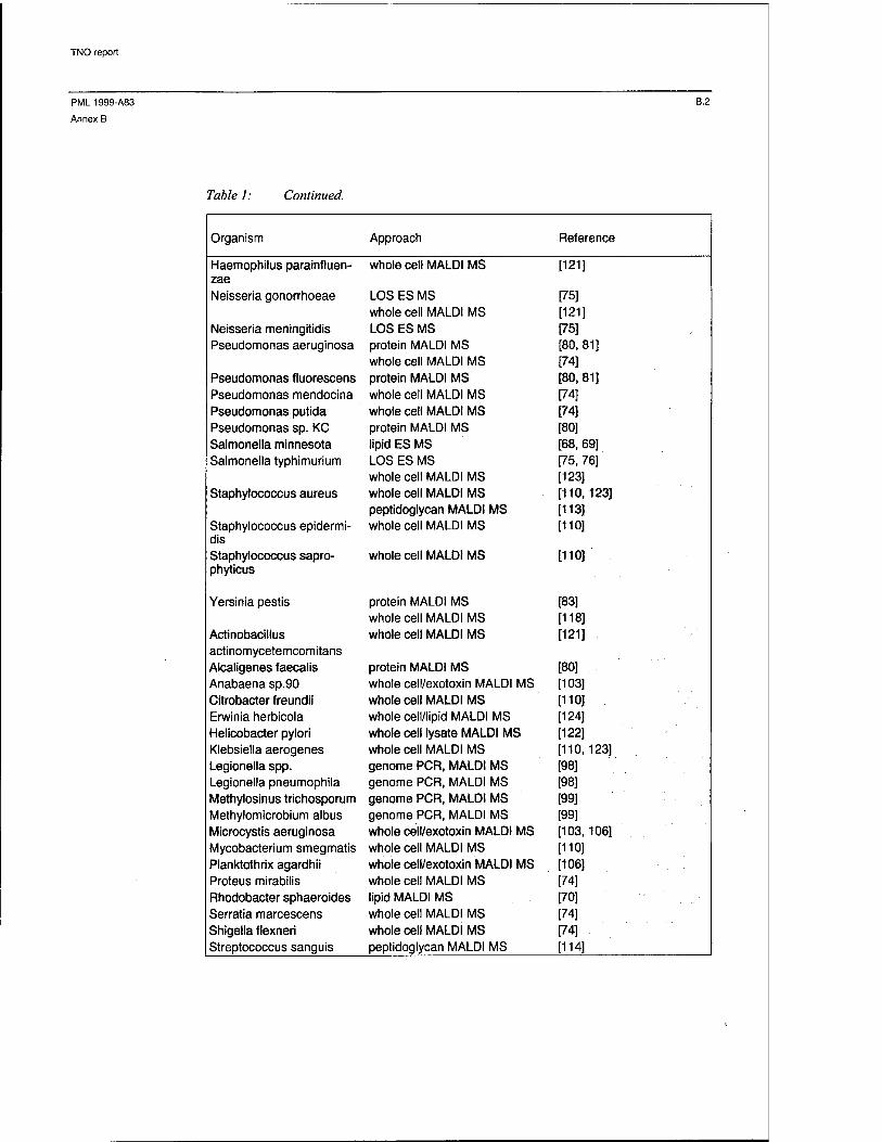
TNO report
PML1999-A83 B.2
Annex B
Table 1: Continued.
Organism Approach Reference
Haemophilus parainfluen- whole cell MALDI MS [121] zae Neisseria gonorrhoeae LOS ES MS
whole cell MALDI MS [75] [121]
Neisseria meningitidis LOS ES MS [75] Pseudomonas aeruginosa protein MALDI MS
whole cell MALDI MS [80, 81] [74]
Pseudomonas fluorescens protein MALDI MS [80, 81] Pseudomonas mendocina whole cell MALDI MS [74] Pseudomonas putida whole cell MALDI MS [74] Pseudomonas sp. KC protein MALDI MS [80] Salmonella minnesota lipid ES MS [68, 69] Salmonella typhimurium LOS ES MS
whole cell MALDI MS [75, 76] [123]
Staphylococcus aureus whole cell MALDI MS peptidoglycan MALDI MS
[110,123] [113]
Staphylococcus epidermi- whole cell MALDI MS [110] dis Staphylococcus sapro- whole cell MALDI MS [110] phyticus
Yersinia pestis protein MALDI MS whole cell MALDI MS
[83] [118]
Actinobacillus whole cell MALDI MS [121] actinomycetemcomitans Alcaligenes faecalis protein MALDI MS [80] Anabaena sp.90 whole cell/exotoxin MALDI MS [103] Citrobacter freundii whole cell MALDI MS [110] Erwinia herbicola whole cell/lipid MALDI MS [124] Helicobacter pylori whole cell lysate MALDI MS [122] Klebsiella aerogenes whole cell MALDI MS [110,123] Legionella spp. genome PCR, MALDI MS [98] Legionella pneumophila genome PCR, MALDI MS [98] Methylosinus trichosporum genome PCR, MALDI MS [99] Methylomicrobium albus genome PCR, MALDI MS [99] Microcystis aeruginosa whole cell/exotoxin MALDI MS [103,106] Mycobacterium smegmatis whole cell MALDI MS [110] Planktothrix agardhii whole cell/exotoxin MALDI MS [106] Proteus mirabilis whole cell MALDI MS [74] Rhodobacter sphaeroides lipid MALDI MS [70] Serratia marcescens whole cell MALDI MS [74] Shigella flexneri whole cell MALDI MS [74] Streptococcus sanguis peptidoglycan MALDI MS [114]
"

ONGERUBRICEERD
REPORT DOCUMENTATION PAGE (MOD-NL)
1. DEFENCE REPORT NO. (MOD-NL)
TD99-0176
2. RECIPIENTS ACCESSION NO. 3. PERFORMING ORGANIZATION REPORT NO.
PML 1999-A83
4. PROJECT/TASK/WORK UNIT NO.
014.11019
5. CONTRACT NO.
A98D420
6. REPORT DATE
December 1999
7. NUMBER OF PAGES
60 (incl. 2 annexes,
excl. RDP & distribution list)
8. NUMBER OF REFERENCES
150
9. TYPE OF REPORT AND DATES COVERED
Final
10. TITLE AND SUBTITLE
Characterisation of Bacteria by Matrix-assisted Laser Desorption/Ionisation and Electrospray Mass Spectrome- try
H.AUTHOR(S)
Dr. B.L.M. van Baar
12. PERFORMING ORGANIZATION NAME(S) AND ADDRESS(ES)
TNO Prins Maurits Laboratory, P.O. Box 45, 2280 AA Rijswijk, The Netherlands Lange Kleiweg 137, Rijswijk, The Netherlands
13. SPONSORING AGENCY NAME(S) AND ADDRESS(ES)
DICO/MGFB/GGB, P.O. Box 20701, 2500 ES The Hague, The Netherlands
14. SUPPLEMENTARY NOTES
The classification designation Ongerubriceerd is equivalent to Unclassified.
15. SUMMARY (MAXIMUM 200 WORDS (1044 BYTE))
Chemical analysis for the characterisation of micro-organisms is rapidly evolving, after the recent advent of new ionisation methods in mass spectrometry: electrospray (ES) and matrix-assisted laser desorption/ionisation (MALDI). These methods allow quick characterisation of micro-organisms, either directly or after minimum sample preparation. This report provides a brief introduction to ES and MALDI mass spectrometry and a dis- cussion of micro-organism characterisation capabilities. Some attention is devoted to the analysis of mixtures of proteins, lipids and other compounds, to the combination of polymerase chain reaction technology and mass spectrometry, and to the analysis of whole bacteria and their lysates. The review of results produced hitherto is concluded with an outlook on future developments.
16. DESCRIPTORS
Analysers Bacteria Biological agents Detection Desorption Electrospray Mass spectroscopy Mid-spectrum agents
17a.SECURITY CLASSIFICATION (OF REPORT)
Ongerubriceerd
17b.SECURITY CLASSIFICATION (OF PAGE)
Ongerubriceerd
17C.SECURITY CLASSIFICATION (OF SUMMARY)
Ongerubriceerd
18. DISTRIBUTION AVAILABILITY STATEMENT
Unlimited Distribution
17d.SECURITY CLASSIFICATION (OF TITLES)
Ongerubriceerd
ONGERUBRICEERD

Distributielijst*
1 DWOO
2 HWO-KL
3* HWO-KLu
4* HWO-KM
5 HWO-CO
6 Ministerie van Defensie, DICO/MGFB/GGB, drs. N.H.W. van Xanten, apotheker
7 DMKM/MARSTAF/OBS LTZT-1 ir. E.M. Pel
8 DMKIVAU/MT/VK/VINBC/MDW ir. A.A.M. Slagveer
9 DICO/MGFB/GGB L.kol. H.W. Poen
10 TNO-PG, Divisie Immunologische en Infectieziekten dr. N. Zegers
11 DOPKlu/AWGRO/SOB Maj. M. Kathmann
12 Norwegian ANNCP representative, Division for Environmental Toxicology, NDRE O. Busmundrud, M.Sc,
13 UK ANNCP representative DERA, CBD Porton Down dr. N.J. Underwood
14 TNO Voeding, Afdeling bio-farmaceutische analyse dr. S. Jespersen
15 DM&PTNO-DO
16* DM&P TNO-DO, accountcoördinator CO
17* TNO-FEL, Bibliotheek
18/20 Bibliotheek KMA
21 * Lid Instituuts Advies Raad PML BGen. prof. J.M.J. Bosch
22* Lid Instituuts Advies Raad PML prof. dr. U.A. Th. Brinkman
23 TNO-PML, Directie; daarna reserve
24 TNO-PML, Hoofd Divisie Toxische Stoffen dr. M.W. Leeuw
25 TNO-PML, Adjunct-hoofd Divisie Toxische Stoffen dr. ir. M.S. Nieuwenhuizen
26/28 TNO-PML Divisie Toxische Stoffen, groep Analyse Toxische en Explosieve Stoffen dr. B.L.M. van Baar, dr. ing. C.E. Kientz en ir. E.R.J. Wils
29 TNO-PML, Bibliotheek
30 TNO-PML, Archief
* De met een asterisk (*) gemerkte instanties/personen ontvangen uitsluitend de titelpagina, het managementuittreksel, de documentatiepagina en de distributielijst van het rapport.