targeted disruption of the mouse testis-enriched gene ... filetargeted disruption of the mouse...
TRANSCRIPT

Targeted disruption of the mouse testis-enriched gene Znf230 does not affectspermatogenesis or fertility
Yunqiang Liu, Dachang Tao, Yongjie Lu, Yuan Yang, Yongxin Ma and Sizhong Zhang
Department of Medical Genetics and Division of Human Morbid Genomics, State Key Laboratory of
Biotherapy, West China Hospital, West China Medical School, Sichuan Universtiy, Chengdu, Sichuan
Province, China.
Abstract
The mouse testis-enriched Znf230 gene, which encodes a type of RING finger protein, is present primarily in the nu-clei of spermatogonia, the acrosome and the tail of spermatozoa. To investigate the role of Znf230 in spermato-genesis, we generated Znf230-deficient mice by disrupting Znf230 exon-5 and exon-6 using homologousrecombination. The homozygous Znf230-knockout (KO) mice did not exhibit Znf230 mRNA expression and Znf230protein production. Znf230 KO mice exhibited no obvious impairment in body growth or fertility. Male Znf230 KO micehad integral reproductive systems and mature sperm that were regular in number and shape. The developmentalstages of male germ cells of Znf230 KO mice were also normal. We further examined variations in the transcriptomesof testicular tissue between Znf230 KO and wild-type mice through microarray analysis. The results showed that themRNA level of one unclassified transcript 4921513I08Rik was increased and that the mRNA levels of three othertranscripts, i.e., 4930448A20Rik, 4931431B13Rik and potassium channel tetramerisation domain containing14(Kctd14), were reduced more than two-fold in Znf230 KO mice compared with wild-type mice. Using our currentexamination techniques, these findings suggested that Znf230 deficiency in mice may not affect growth, fertility orspermatogenesis.
Keywords: Znf230, knockout mice, spermatogenesis, Kctd14.
Received: October 9, 2013; Accepted: June 24, 2014.
Introduction
Mammalian spermatozoa development is a complex
process that involves the renewal and differentiation of
spermatogonia, the meiosis of spermatocytes, and drastic
morphological changes accompanying the development
from round spermatids to mature spermatozoa (Russell et
al., 1990). Many environmental, behavioral and genetic
factors affect male fertility. An estimated 50% of human in-
fertility has been attributed to genetic abnormalities
(Hwang et al., 2011). Two previous microarray studies
(Schultz et al., 2003; Schlecht et al., 2004) showed that
haploid germ cells express a large number of germ cell-
specific genes (approximately 4% of mammalian genes).
Therefore, it is necessary to identify these unique genes and
characterize the precisely programmed cell- and stage-
specific gene expression that occurs during the regulation
of the developmental spermatogenesis process.
The human gene ZNF230 (also named RNF141),
which encodes a type of RING (Really Interesting New
Gene) finger protein, was first identified in our laboratory
to be restrictively expressed in the testicular tissue of fertile
men (Zhang et al., 2001). RING finger proteins, a sub-
family of zinc finger proteins (ZFP), often contain the
cysteine-rich CX2CX(9-39)CX(1-3)HX(2-3)CX2CX(4-48)CX2C
domain and are involved in a variety of biological pro-
cesses, including transcriptional regulation, signal trans-
duction, cell apoptosis and protein ubiquitination (Borden,
2000; Joazeiro and Weissman, 2000). The mouse homolog
of Znf230 was also identified in our laboratory (Qiu et al.,
2003). The expression of the mouse Znf230 gene is devel-
opmentally regulated, and the Znf230 protein functions as
an activator module in transcription. Additionally, the
mouse Znf230 protein is primarily expressed in the nuclei
of spermatogonia but has subsequent expression in the
acrosome system and the tails of developing spermatids
and spermatozoa (Song et al., 2008). Hence, we wondered
whether Znf230 may play a role in mammalian sperma-
togenesis.
Animal models have defined key signaling pathways
that are involved in reproductive physiology (Li et al.,
2001). To date, over 400 genes that are essential for male
Genetics and Molecular Biology, 37, 4, 708-715 (2014)
Copyright © 2014, Sociedade Brasileira de Genética. Printed in Brazil
www.sbg.org.br
Send correspondence to Yunqiang Liu. Department of Medical Ge-netics and Division of Human Morbid Genomics, State Key Labora-tory of Biotherapy, West China Hospital, West China MedicalSchool, Sichuan University, No. 1, Ke Yuan 4 Lu, Gao Peng DaDao, Chengdu, 610041, Sichuan, China. E-mail:[email protected].
Research Article

fertility have been identified using transgenic, chemically
induced, point mutants and KO/knock-in/gene-trap mouse
models (Yatsenko et al., 2010; Jamsai and O’Bryan, 2011).
To investigate the role of Znf230 in mouse sperma-
togenesis, we used a targeted gene KO strategy to generate
Znf230-deficient mice. We had previously constructed a
gene-targeting vector based on a modified pPNT vector and
generated mutant mice with exon-2 of the Znf230 gene dis-
rupted (Liu et al., 2013). However, a partial sequence from
the pPNT vector acted as an alternative exon-2, thus allow-
ing a new Znf230 transcript to be produced in the mutant
mice and a new protein product, possessing a C-terminal
amino acid sequence with a RING finger motif similar to
that of the wild-type Znf230 protein, to be generated. Thus,
Znf230 function in the mutant mice was not entirely inacti-
vated. In the current study, we changed the targeting strat-
egy such that the region of exon-5 and exon-6, which
encodes the essential RING finger domain of the Znf230
protein, was directly disrupted. This strategy successfully
generated Znf230-null mice for use in this study.
Materials and Methods
Construction of Znf230 KO targeting vector andgeneration of Znf230 KO mice
Using a highly efficient recombineering-based
method that has been previously described (Liu et al., 2003;
Chan et al., 2007), the Znf230 KO targeting vector was
constructed from a genomic DNA fragment derived from
the C57BL/6J bacterial artificial chromosome clone
bMQ-291L21. The targeting construct, which had a
4864 bp left arm containing introns 2 and 3 of the Znf230
gene and a 2902 bp right arm containing the partial untrans-
lated region of exon-6, was inserted into the
ABRLFn-pBR32 vector (Figure 1).
Thirty micrograms of the targeting vector was
linearized by Not I and transfected into CJ7 (derived from
129SV/J mice) embryonic stem cells (ESCs) by
electroporation. Ninety-six ESC clones were selected with
300 �g/mL G418 (Geneticin, Sigma-Aldrich Co., St. Louis,
MO, USA). Among these neomycin-resistant cells, 12
ESCs that had undergone homologous recombination were
identified by long polymerase chain reaction (L-PCR) anal-
ysis with two pairs of primers P1F:
5’-acctctggcctttacaaactcatg-3’, P1R:
5’-ggcctacccgcttccattgctc-3’ and P2F:
5’-ccgtgccttccttgaccctgg-3’, P2R:
5’-caagcagccttattacccagttg-3’.
Two correctly targeted ESC clones were micro-
injected into C57BL/6J blastocysts to generate chimeras
that were then crossed into a C57BL/6J genetic back-
ground. The offspring were screened by L-PCR analysis of
their genomic DNA using P1F/R and P2F/R primers.
The Znf230 KO mice were generated at the Shanghai
Research Center for Model Organisms, Shanghai, China.
Znf230 KO mice were crossed into a C57BL/6J back-
ground for at least eight generations before use. Germline
transmission of the targeted allele was monitored by PCR
with primers including Znf230 wild type (WT)-specific
primers: WTf: 5’-tgccccttgcccccataat-3’, WTr:
5’-gccacccaagaaaaagtcaaaata-3’ and Znf230 KO-specific
primers: Nf: 5’-ggcgcgagcccctgatgctc-3’, Nr:
5’-ttgggtggagaggctattcggctatgac-3’, respectively.
The locations of the aforementioned primers are
shown in Figure 1.
All animals used in this study were handled in com-
pliance with the National Cancer Center Research Insti-
tute’s guidelines for the use of animals (USA). All animal
experimental protocols were approved by the animal ethics
committee of West China Hospital, Sichuan University.
Reverse transcription (RT)-PCR analysis
Total RNA was extracted from the testes of at least
five 15-week-old mice per genotype using an RNApure kit
(Bioteke, Beijing, China). One microgram of total RNA
was reverse transcribed using a RevertAid First Strand
cDNA Synthesis Kit (Fermentas, Pittsburgh, PA, USA)
with oligo(dT) primers. The sequence of interest in the
Znf230 gene was amplified using two gene-specific prim-
ers: E4f: 5’-cccatcctcggtcacatctt-3’, located within the se-
quence of Exon-4, and E6r: 5’-cccccttctcctctacgacaac-3’,
the reverse complement primer of a sequence located
within Exon-6. A 982 bp fragment corresponding to the
mouse Gapdh gene was co-amplified as an internal control
using the following primers:
5’-tgaaggtcggtgtgaacggatttggc-3’ (Forward) and
5’-catgtaggccatgaggtccaccac-3’ (Reverse). Three inde-
pendent RT-PCR analyses were performed to validate the
results.
Western blot analysis
The testicular tissues of at least five 15-week-old
mice per genotype were removed and homogenized in a
Liu et al. 709
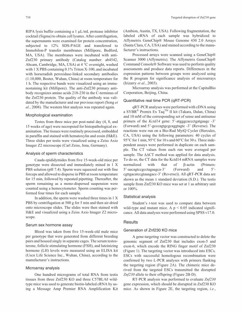
Figure 1 - The targeted knockout strategy for the Znf230 gene: wild-type
allele, targeting construct and targeted allele with the location of primers.
Primer pairs P1F/P1R and P2F/P2R, respectively, which flanked the tar-
geted region, were used to select positive ESC clones, and were used to
monitor the inheritance of the mutant allele. Primers WTf and WTr were
used to amplify the wild-type Znf230 allele. Primers Nf and Nr were used
to amplify the knockout allele.
RIPA lysis buffer containing a 1 �L/mL protease inhibitor
cocktail (Sigma) to obtain cell lysates. After centrifugation,
the supernatants were examined for protein concentration,
subjected to 12% SDS-PAGE and transferred to
Immobilon-P transfer membranes (Millipore, Bedford,
MA, USA). The membranes were incubated with anti-
Znf230 primary antibody (Catalog number: ab4542,
Abcam, Cambridge, MA, USA) at 4 °C overnight, washed
with 1 X PBS containing 0.1% Triton X-100, and incubated
with horseradish peroxidase-linked secondary antibodies
(1:10,000, Boster, Wuhan, China) at room temperature for
1 h. The respective bands were visualized using an immu-
nostaining kit (Millipore). The anti-Znf230 primary anti-
body recognizes amino acids 218-230 in the C-terminus of
the Znf230 protein. The quality of the antibody was vali-
dated by the manufacturer and our previous report (Song et
al., 2008). The western blot analysis was repeated again.
Morphological examination
Testes from three mice per post-natal day (4, 8, and
15 weeks of age) were necropsied for histopathological ex-
amination. The tissues were routinely processed, embedded
in paraffin and stained with hematoxylin and eosin (H&E).
Three slides per testis were visualized using a Zeiss Axio
Imager Z2 microscope (Carl Zeiss, Jena, Germany).
Analysis of sperm characteristics
Cauda epididymides from five 15-week-old mice per
genotype were dissected and immediately mined in 1 X
PBS solution (pH 7.4). Sperm were squeezed out with fine
forceps and allowed to disperse in PBS at room temperature
for 15 min, followed by repeated pipetting. Thereafter, the
sperm remaining as a mono-dispersed suspension were
counted using a hemocytometer. Sperm counting was per-
formed four times for each sample.
In addition, the sperm were washed three times in 1 X
PBS by centrifugation at 500 g for 5 min and then air-dried
onto microscope slides. The slides were then stained with
H&E and visualized using a Zeiss Axio Imager Z2 micro-
scope.
Serum sex hormone assay
Blood was taken from five 15-week-old male mice
per genotype that were generated from different breeding
pairs and housed singly in separate cages. The serum testos-
terone, follicle stimulating hormone (FSH), and luteinizing
hormone (LH) levels were measured using an ELISA kit
(Uscn Life Science Inc., Wuhan, China), according to the
manufacturer’s instructions.
Microarray analysis
One hundred micrograms of total RNA from testis
tissues from three Znf230 KO and three C57BL/6J wild-
type mice was used to generate biotin-labeled cRNA by us-
ing a Message Amp Premier RNA Amplification Kit
(Ambion, Austin, TX, USA). Following fragmentation, the
labeled cRNA of each sample was hybridized to
Affymetrix GeneChip® Mouse Genome 430 2.0 Arrays
(Santa Clara, CA, USA) and stained according to the manu-
facturer’s instructions.
Processed arrays were scanned using a GeneChip®
Scanner 3000 (Affymetrix). The Affymetrix GeneChip®
Command Console® Software was used to perform quality
assessments and produce data reports. Differences in the
expression patterns between groups were analyzed using
the R program for significance analysis of microarrays
(Irizarry et al., 2003).
Microarray analysis was performed at the CapitalBio
Corporation, Beijing, China.
Quantitative real time PCR (qRT-PCR)
qRT-PCR analyses were performed with cDNA using
a SYBR® Premix Ex TaqTM II kit (Takara, Dalan, China)
and 10 mM of the corresponding set of sense and antisense
primers of the Kctd14 gene: 5’-atgggcaccctgatgaagc -3’
(Forward) and 5’-gcccagtgcgcaggtagtc -3’ (Reverse). The
reactions were run on a Bio-Rad MyiQ Cycler (Hercules,
CA, USA) using the following parameters: 40 cycles of
95°C for 1 min, 95°C for 10 s and 60°C for 30 s. Three inde-
pendent assays were performed in duplicate on each sam-
ple. The CT values from each run were averaged per
sample. The ��CT method was applied for data analysis.
To do so, the CT data for the Kctd14 mRNA samples were
normalized with that of �-actin (Primers:
5’-aacagtccgcctagaagca-3’ (Forward) and 5’-
cgttgacatccgtaaagacc-3’ (Reverse)). All qRT-PCR data are
shown as the mean � standard deviation (S.D.). The testis
sample from Znf230 KO mice was set at 1 as arbitrary unit
or 100%.
Statistical analysis
Student’s t-test was used to compare data between
wild-type and mutant mice. A p < 0.05 indicated signifi-
cance. All data analyses were performed using SPSS v17.0.
Results
Generation of Znf230 KO mice
A gene-targeting vector was constructed to delete the
genomic segment of Znf230 that includes exon-5 and
exon-6, which encode the RING finger motif of Znf230
(Figure 1). The targeting vector was introduced into ESCs.
ESCs with successful homologous recombination were
confirmed by two L-PCR analyses with primers flanking
the targeting region (Figure 2A). The chimeric mice de-
rived from the targeted ESCs transmitted the disrupted
Znf230 allele to their offspring (Figure 2B-D).
RT-PCR analysis was performed to evaluate Znf230
gene expression, which should be disrupted in Znf230 KO
mice. As shown in Figure 2E, the targeting region, i.e.,
710 Targeted disruption of Znf230 gene

exon-5 and exon-6, of the Znf230 gene was not amplified
from total RNA of the testes in Znf230 KO mice using the
specific primers, in contrast to wild-type mice. Further-
more, western blot analysis with anti-Znf230 antibody
showed that the 26-kDa protein representing Znf230 was
present in the testes of wild-type mice, as expected, but it
was absent in the testes of Znf230 KO mice (Figure 2F).
Thus, the Znf230 gene was indeed disrupted in Znf230 KO
mice.
Znf230 KO mice appeared to be normal in growthand fertility
Znf230 KO mice exhibited no obvious impairment in
body growth and development because no significant dif-
ferences were observed in the weights of body or organs,
including brain, lung, heart, liver, spleen and kidney, or life
spans between Znf230 KO and wild-type mice (Table 1).
Because Znf230 was identified as a testis-enriched gene
that likely plays a role in male fertility, we focused our in-
vestigation on phenotypes related to male fertility. How-
ever, the fertility of Znf230 KO mice appeared to be normal
and the offspring of Znf230 KO intercrosses were born at
the expected Mendelian ratios. Compared with wild-type
mice, Znf230 KO mice displayed no detectable differences
in the male reproductive system, including the testis, semi-
nal vesicle, prostate and bladder. There were also no differ-
ences in serum testosterone, FSH or LH levels between
Znf230 KO and wild-type mice (Table 1, Figure 3A, B).
H& E staining demonstrated that the testicular tissue of
Znf230 KO mice was intact and that each developmental
stage of male germ cells was normal (Figure 3C-H). No sig-
nificant difference was detected in the shape and number of
sperm isolated from the epididymides of Znf230 KO and
C57BL/6J wild-type mice (Figure 3I, J and Table 1).
Changes in the mRNA expression profile of Znf230KO mice
Our previous report proposed that Znf230 was a
DNA-binding protein that may function as a transcriptional
activator (Qiu et al., 2003). We therefore investigated the
differences between the transcriptomes of testes from
Znf230 KO and wild-type mice using Affymetrix Mouse
Genome 430 2.0 Arrays. The expression levels of over
34,000 genes were assessed, and transcripts with fold
changes greater than 2 or less than 0.5 between the two
groups were analyzed (Table 2). Znf230 had the most
down-regulated expression level of the analyzed tran-
scripts, thereby confirming the validity of the experimental
system. The mRNA level of one unclassified transcript
4921513I08Rik (GenBank No. AK014883) was detected to
be more than two-fold higher, and the mRNA levels of
three transcripts including two protein coding genes
Kctd14 (GenBank No. NM_001136235) and
4930448A20Rik (GenBank No. Ak015411) and a non-
coding RNA 4931431B13Rik (GenBank No. NR_045183)
were detected to be more than two-fold lower in Znf230
KO mice compared with C57BL/6J wild-type mice. Be-
cause the Kctd14 gene may encode a functional protein, we
performed qRT-PCR analysis to verify the expression
changes of Kctd14 between Znf230 KO and wild-type
mice. The results showed that the mRNA level of Kctd14
Liu et al. 711
Figure 2 - The generation and identification of Znf230 KO mice. (A) L-PCR analysis using primer pairs P1F/P1R and P2F/P2R to amplify the targeted
Znf230 alleles from genomic DNA extracted from ESCs. ESCs from No. A7, E4 and H3 were the positive clones undergoing targeted homologous recom-
bination. (B) L-PCR analysis of the targeted Znf230 alleles amplified from genomic DNA derived from the offspring of chimeric mice backcrossed to
C57BL/6J. No. 1-10: the offspring members and N: negative control. (C) L-PCR analysis to monitor the inheritance of the targeted Znf230 allele in the
progeny of heterozygous Znf230 KO mice. (D) PCR analysis to monitor the inheritance of the targeted Znf230 allele in the progeny of heterozygous
Znf230 KO mice using primer pairs WTf/WTr and Nf/Nr. (E) RT-PCR analysis of the Znf230 gene in testes from Znf230 KO and C57BL/6J mice. M1,
M2: DNA ladders of 1 kb and 100 bp, respectively. Gapdh was used as an internal control. (F) Western blot analysis of the Znf230 protein in the testes
from Znf230 KO and C57BL/6J mice. �-actin was used as an internal control.
was reduced by more than 10-fold in Znf230 KO mice com-
pared with wild-type mice (Figure 4).
Discussion
The present work was undertaken in an effort to de-
fine the physiological role of the testis-enriched gene
Znf230 in mammalian spermatogenesis. We generated a
null mutation in the Znf230 gene by homologous recombi-
nation in mouse ESCs, which were used to produce homo-
zygous Znf230 KO mice. Mice that were homozygous for
the mutation lacked the intact mRNA and protein in germ
cells, but did not exhibit any detectable abnormality in body
growth or spermatogenesis. The absence of abnormality in
Znf230-null testes was unexpected because of the testis-
enriched expression pattern of Znf230. However, several
explanations may account for the lack of a clear phenotype
in Znf230 KO mice. First, the function of Znf230 may be
dispensable for male fertility. As an example, H1t is an H1
histone variant that is unique to late spermatocytes and
round spermatids, but H1t-null mice have no discernible
phenotype (Fantz et al., 2001). Another example is SPAG5,
which is an Odf1-interacting protein that is specifically ex-
pressed during meiosis. The disruption of SPAG5 does not
affect spermatogenesis or fertility (Xue et al., 2002). Other
examples include testicular orphan nuclear receptor 2
gene (Shyr et al., 2002), testicular haploid expressed gene
(Mannan et al., 2003), transition protein 2, proacrosin and
histone H1.1 genes (Nayernia et al., 2003), the tumor sup-
pressor LRP1b gene (Marschang et al., 2004), the UBC4-
testis gene (Bedard et al., 2005) and the testis-enriched
712 Targeted disruption of Znf230 gene
Table 1 - Comparison of phenotypes between Znf230 KO and C57BL/6J mice.
Znf230 KO C57BL/6J Pa
Total body weight (g)b30.18 � 1.45 30.31 � 2.07 NS
Organ weight (mg) b,c Brain 434.55 � 30.71 405.38 � 44.78 NS
Heart 151.8 � 16.08 139.54 � 8.48 NS
Lung 171.21 � 12.93 166.14 � 11.21 NS
Liver 1382.53 � 82.92 1386.72 � 192.7 NS
Kidney 212.32 � 25.95 204.1 � 21.18 NS
Spleen 60.37 � 7.17 63.64 � 8.29 NS
Seminal vesicle 268.2 � 14.91 236.1 � 40.69 NS
Prostate and Bladder 127 � 25.22 127.5 � 22.3 NS
Testis 108.5 � 15.46 109.6 � 23.67 NS
Total no. of sperm (x106)b24.4 � 3.6 23.7 � 2.8 NS
Serum sex hormone (ng/mL)b Testosterone 1.290.25 1.2 � 0.34 NS
Luteinizing hormone (LH) 8.02 � 1.37 8.21 � 1.72 NS
Follicle-stimulating hormone (FSH) 10.2 � 3.05 9.56 � 2.45 NS
Litter size d7.5 � 1.4 8.1 � 1.2 NS
Life Span (days) e713.2 � 147.6 722.5 � 168.5 NS
aStatistical analysis was carried out by Student’s t test, NS: Not significant.bFifteen-week-old mice were examine, n = 5 per group, Values are means � S.D.cWet weights of paired organs were averaged for each mouse, and the single value was used to calculate mean � S.D among same genotype.dData are mean values derived from six breeding pairs for each genotype.eData are mean values of 10 mice (5 male plus 5 female) per group.
Table 2 - Genes differentially expressed in the testes of Znf230 KO mice
Gene ID Gene symbol Gene name Fold changea ProbeSet IDb
70875 4921513108Rik RIKEN cDNA 492151308 gene 2.9705 1432299_at
67150 Znf230 ring finger protein 141 0.445/0.1841/0.158 1449086_at/ 1433655_at/ 1449087_at
233529 Kctd14 potassium channel tetramerisation domain containing 14 0.4276/0.4054 1426632_at/1426633_at
70971 4931431B13Rik RIKEN cDNA 4931431B13 gene 0.4054 1430416_at
73993 4930448A20Rik RIKEN cDNA 4930448A20 gene 0.2394 1454205_at
aThe ratio of signal values between Znf230 KO and Wild-type mice detected by every probe. Statistical p-values < 0.001.bAll probes hit one known transcript.

histone demethylase KDM4D gene (Iwamori et al., 2011).
One explanation for such phenomena is that an unidentified
protein may compensate for the loss of function of these
genes. It is reasonable that other RING finger proteins may
compensate for the loss of Znf230 function because RING
finger proteins belong to one of the largest zinc finger pro-
tein families. Second, the defects in Znf230 KO mice may
be too small to be detected using the techniques employed
here. A detailed ultrastructural examination of testicular
tissues and spermatozoa may be needed to confirm even-
tual minor changes in the mutant mice. Third, because
spermatogenesis is a very complex process that involves
many genes, the inactivation of one gene may not be suffi-
cient to produce a detectable phenotype. For example, Tyro
3, Axl, and Mer encode three structurally related receptors
that possess tyrosine kinase activity. Mice that lack any sin-
gle receptor or any combination of two receptors are viable
and fertile, but males that lack all three receptors produce
no mature sperm (Lu et al., 1999). Therefore, it will be of
interest to explore the interaction between Znf230 and its
related proteins by ablating their network and dissecting the
resulting phenotypes to help clarify the biological role of
the Znf230 gene in male fertility.
Indeed, we found that the mRNA levels of four tran-
scripts were changed more than two-fold in Znf230 KO
mice compared with C57BL/6J wild-type mice. Three of
the four transcripts were unclassified. However, Kctd14 en-
codes a putative member of the KCTD protein family that
contains the bric-a-brac/tramtrak/broad (BTB) complex
domain, which resembles the tetramerization domain of
voltage-gated potassium channels. The KCTD protein fam-
ily, which comprises 22 members, has been implicated in
many important biological processes (Schwenk et al.,
2010; Seddik et al., 2012; Cao-Ehlker et al., 2013; Skoblov
et al., 2013). However, because no report has yet described
the biological function of the Kctd14 protein, Znf230 KO
mice may provide a clue for investigating the pathways in
which these unclassified transcripts are involved.
Liu et al. 713
Figure 3 - Characteristics comparison of reproductive organs and sperm
shape between Znf230 KO and C57BL/6J mice. Morphology of reproduc-
tive organs from Znf230 KO (A) and C57BL/6J (B) mice. The seminal
vesicles (black arrows), bladder (black arrowheads), epididymis (white ar-
rows) and testes (red arrows) were highlighted. Scale bar = 1 cm.
Histological analysis of H&E stained testes from Znf230 KO (C,E,G) and
C57BL/6J (D,F,H) mice. SG: Spermatogonia, SC: Spermatocyte, RS:
Round spermatids, and LS: elongated spermatids. Scale bar = 100 �m.
Characteristics of H&E stained sperm from Znf230 KO (I) and C57BL/6J
(J) mice.. Ac: acrosome, T: sperm tail. Scale bar = 50 �m.
Figure 4 - Comparative qRT-PCR analysis of mRNA levels of the Kctd14
gene between Znf230 KO and C57BL/6J (WT) mice. Bars represent the
means ± S.D., Statistical p-values < 0.001.

Conversely, the Znf230 protein may act as a trans-
criptional factor, and the disruption of the Znf230 protein
may cause the related transcriptional complex to be de-
stroyed, this directly affecting the expression of the four
transcripts. It is interesting to note that Znf230 and the four
transcripts are all located on mouse chromosome 7 (Chr7).
As shown in Figure S1, the transcripts of 4921513I08Rik
and Kctd14 are located upstream of Znf230, while the other
two transcripts are located downstream of Znf230. It is thus
possible that cis-acting elements may have been destroyed
during the targeted disruption of the Znf230 gene. In addi-
tion, changes in the chromatin structure in the KO region of
Chr7 may be the cause for the changes in the expression
levels of the four transcripts.
In conclusion, we generated Znf230-deficient mice
that exhibited normal body growth and fertility based on
our current examination techniques. Using microarray
analysis to compare the transcriptomes of testicular tissue
from Znf230 KO and wild-type mice, we observed changes
in the expression levels of four transcripts in Znf230 KO
mice. In the future, the Znf230 KO mouse model may be
used to uncover the biological roles and explore the interac-
tion between Znf230 and its related transcripts.
Acknowledgments
This work was funded by a grant from the National
Basic Program of China (Program 973) from the Ministry
of Science and Technology of China (2012CB947600) and
the Fundamental Research Funds for the Central Univer-
sities (2010SCU11025).
ReferencesBedard N, Hingamp P, Pang Z, Karaplis A, Morales C, Trasler J,
Cyr D, Gagnon C and Wing SS (2005) Mice lacking the
UBC4-testis gene have a delay in postnatal testis develop-
ment but normal spermatogenesis and fertility. Mol Cell
Biol 25:6346-6354.
Borden KLB (2000) RING domains: master builders of molecular
scaffolds? J Mol Biol 295:1103-1112.
Chan W, Costantino N, Li R, Lee S, Su Q, Melvin D, Court DL
and Liu P (2007) A recombineering based approach for
high-throughput conditional knockout targeting vector con-
struction. Nucleic Acids Res 35:e64.
Cao-Ehlker X, Zong X, Hammelmann V, Gruner C, Fenske S,
Michalakis S, Wahl-Schott C and Biel M (2013) Up-
regulation of hyperpolarization-activated cyclic nucleo-
tide-gated channel 3 (HCN3) by specific interaction with K+
channel tetramerization domain-containing protein 3
(KCTD3). J Biol Chem 288:7580-7589.
Fantz DA, Hatfield WR, Horvath G, Kistler MK and Kistler WS
(2001) Mice with a targeted disruption of the H1t gene are
fertile and undergo normal changes in structural chromo-
somal proteins during spermiogenesis. Biol Reprod
64:425-431.
Hwang K, Yatsenko AN, Jorgez CJ, Mukherjee S, Nalam RL,
Matzuk MM and Lamb DJ (2011) Mendelian genetics of
male infertility. Ann N Y Acad Sci 1214:E1-E17.
Irizarry RA, Hobbs B, Collin F, Beazer-Barclay YD, Antonellis
KJ, Scherf U and Speed TP (2003) Exploration, normaliza-
tion, and summaries of high density oligonucleotide array
probe level data. Biostatistics 4:249-264.
Iwamori N, Zhao M, Meistrich ML and Matzuk MM (2011) The
testis-enriched histone demethylase, KDM4D, regulates
methylation of histone H3 lysine 9 during spermatogenesis
in the mouse but is dispensable for fertility. Biol Reprod
84:1225-1234.
Jamsai D and O’Bryan MK (2011) Mouse models in male fertility
research. Asian J Andro 13:139-151.
Joazeiro CAP and Weissman AM (2000) RING finger proteins:
mediators of ubiquitin ligase activity. Cell 102:549-552.
Li SW, Arita M, Fertala A, Bao Y, Kopen GC, Långsjö TK,
Hyttinen MM, Helminen HJ and Prockop DJ (2001) Trans-
genic mice with inactive alleles for procollagen N-
proteinase (ADAMTS-2) develop fragile skin and male ste-
rility. Biochem J 355:271-278.
Liu P, Jenkins NA and Copeland NG (2003) A highly efficient
recombineering-based method for generating conditional
knockout mutations. Genome Res 13:476-484.
Liu Y, Tao D, Ma S, Kuang Y, Su D, Zhang H, Yang Y, Ma Y and
Zhang S (2013) A new mutant transcript generated in
Znf230 exon 2 knockout mice reveals a potential exon struc-
ture in the targeting vector sequence. Acta Biochim Biophys
Sin 45:123-128.
Lu Q, Gore M, Zhang Q, Camenisch T, Boast S, Casagranda F,
Lai C, Skinner MK, Klein R, Matsushima GK, et al. (1999)
Tyro-3 family receptors are essential regulators of mamma-
lian spermatogenesis. Nature 398:723-728.
Mannan AU, Nayernia K, Mueller C, Burfeind P, Adham IM and
Engel W (2003) Male mice lacking the Theg (Testicular
Haploid Expressed Gene) protein undergo normal spermato-
genesis and are fertile. Biol Reprod 69:788-796.
Marschang P, Brich J, Weeber EJ, Sweatt JD, Shelton JM, Rich-
ardson JA, Hammer RE and Herz J (2004) Normal develop-
ment and fertility of knockout mice lacking the tumor sup-
pressor gene LRP1b suggest functional compensation by
LRP1. Mol Cell Biol 24:3782-3793.
Nayernia K, Drabent B, Adham IM, Möschner M, Wolf S,
Meinhardt A and Engel W (2003) Male mice lacking three
germ cell expressed genes are fertile. Biol Reprod
69:1973-1978.
Qiu W, Zhang S, Xiao C, Xu W, Ma Y, Liu Y and Wu Q (2003)
Molecular cloning and characterization of a mouse sper-
matogenesis-related ring finger gene znf230. Biochem
Biophys Res Commun 306:347-353.
Russell LD, Ettlin RA, Sinha HAP and Clegg ED (1990) Mamma-
lian spermatogenesis. In: Russell LD, Ettlin RA, Sinha HAP
and Clegg ED (eds) Histological and Histopathological
Evaluation of the Testis. Cache River Press, Saint Louis,
pp 1-40.
Schlecht U, Demougin P, Koch R, Hermida L, Wiederkehr C,
Descombes P, Pineau C, Jegou B and Primig M (2004) Ex-
pression profiling of mammalian male meiosis and gameto-
genesis identifies novel candidate genes for roles in the reg-
ulation of fertility. Mol Biol Cell 15:1031-1043.
Schultz N, Hamra FK and Garbers DL (2003) A multitude of
genes expressed solely in meiotic or postmeiotic
spermatogenic cells offers a myriad of contraceptive targets.
Proc Natl Acad Sci USA 100:12201-12206.
714 Targeted disruption of Znf230 gene

Schwenk J, Metz M, Zolles G, Turecek R, Fritzius T, Bildl W,
Tarusawa E, Kulik A, Unger A, Ivankova K, et al. (2010)
Native GABA(B) receptors are heteromultimers with a fam-
ily of auxiliary subunits. Nature 465:231-235.
Seddik R, Jungblut SP, Silander OK, Rajalu M, Fritzius T, Bes-
seyrias V, Jacquier V, Fakler B, Gassmann M and Bettler B
(2012) Opposite effects of KCTD subunit domains on
GABA(B) receptor-mediated desensitization. J Biol Chem
287:39869-39877.
Shyr CR, Collins LL, Mu XM, Platt KA and Chang C (2002)
Spermatogenesis and testis development are normal in mice
lacking testicular orphan nuclear receptor 2. Mol Cell Biol
22:4661-4666.
Skoblov M, Marakhonov A, Marakasova E, Guskova A, Chan-
dhoke V, Birerdinc A and Baranova A (2013) Protein part-
ners of KCTD proteins provide insights about their func-
tional roles in cell differentiation and vertebrate
development. Bioessays 35:586-596.
Song H, Su D, Lu P, Yang J, Zhang W, Yang Y, Liu Y and Zhang
S (2008) Expression and localization of the spermato-
genesis-related gene, Znf230, in mouse testis and spermato-
zoa during postnatal development. BMB Rep 41:664-669.
Xue J, Tarnasky HA, Rancourt DE and van Der Hoorn FA (2002)
Targeted disruption of the testicular SPAG5/deepest protein
does not affect spermatogenesis or fertility. Mol Cell Biol
22:1993-1997.
Yatsenko AN, Iwamori N, Iwamori T and Matzuk MM (2010)
The power of mouse genetics to study spermatogenesis. J
Andro 31:34-44.
Zhang S, Qiu W, Wu H, Zhang G, Xiao C and Yang J (2001) The
shorter zinc finger protein ZNF230 gene message is tran-
scribed in fertile male testes and may be related to human
spermatogenesis. Biochem J 359:721-727.
Supplementary Material
The following online material is available for this article:
Figure S1- The locations of the transcripts of Kctd14,
Znf230, 4921513I08Rik, 4930448A20Rik and
4931431B13Rik.
This material is available as part of the online article from
http://www.scielo.br/gmb.
Senior Editor: Emmanuel Dias Neto
License information: This is an open-access article distributed under the terms of theCreative Commons Attribution License, which permits unrestricted use, distribution, andreproduction in any medium, provided the original work is properly cited.
Liu et al. 715