targeted lung denervation for moderate to severe copd: a pilot study
TRANSCRIPT

ORIGINAL ARTICLE
Targeted lung denervation for moderate to severeCOPD: a pilot studyDirk-Jan Slebos,1 Karin Klooster,1 Coenraad F N Koegelenberg,2 Johan Theron,3
Dorothy Styen,2 Arschang Valipour,4 Martin Mayse,5 Chris T Bolliger2,3
1Department of Pulmonarydiseases, University ofGroningen, University MedicalCenter Groningen, Groningen,The Netherlands2Faculty of Medicine andHealth Sciences, Division ofPulmonology, Department ofMedicine, StellenboschUniversity, Cape Town,South Africa3Medi Clinic Panorama,Cape Town, South Africa4Department of Respiratoryand Critical Care Medicine,Ludwig-Boltzmann-Institute forCOPD and RespiratoryEpidemiology, Otto-Wagner-Spital, Vienna, Austria5Holaira, Inc., Minneapolis,Minnesota, USA
Correspondence toDr Dirk-Jan Slebos, UniversityMedical Center Groningen,Department of Pulmonarydiseases, AA11, PO Box30001, Groningen 9700 RB,The Netherlands;[email protected]
Received 7 August 2014Revised 9 February 2015Accepted 12 February 2015Published Online First4 March 2015
To cite: Slebos D-J,Klooster K,Koegelenberg CFN, et al.Thorax 2015;70:411–419.
ABSTRACTBackground Parasympathetic pulmonary nerves releaseacetylcholine that induces smooth muscle constriction.Disruption of parasympathetic pulmonary nervesimproves lung function and COPD symptoms.Aims To evaluate ‘targeted lung denervation’ (TLD),a novel bronchoscopic therapy based on ablation ofparasympathetic pulmonary nerves surrounding the mainbronchi, as a potential therapy for COPD.Methods This 1-year, prospective, multicentre studyevaluated TLD in patients with COPD forced expiratoryvolume in 1 s (FEV1)/forced vital capacity (FVC) (FEV1/FVC <0.70; FEV1 30%–60% predicted). Patientsunderwent staged TLD at 20 watts (W) or 15 Wfollowing baseline assessment off bronchodilators.Assessments were repeated on tiotropium beforetreatment and off bronchodilators at 30, 90, 180, 270and 365 days after TLD. The primary endpoint wasfreedom from documented and sustained worsening ofCOPD directly attributable to TLD to 1 year. Secondaryendpoints included technical feasibility, change inpulmonary function, exercise capacity, and quality of life.Results Twenty-two patients were included (n=12 at20 W, n=10 at 15 W). The procedures were technicallyfeasible 93% of the time. Primary safety endpoint wasachieved in 95%. Asymptomatic bronchial wall effectswere observed in 3 patients at 20 W. The clinical safetyprofiles were similar between the two energy doses. At1 year, changes from baseline in the 20 W dosecompared to the 15 W dose were: FEV1 (+11.6%±32.3vs +0.02%±15.1, p=0.324), submaximal cycleendurance (+6.8 min±12.8 vs 2.6 min±8.7, p=0.277),and St George’s Respiratory Questionnaire (−11.1 points±9.1 vs −0.9 points ±8.6, p=0.044).Conclusions Bronchoscopic TLD, based on the conceptof ablating parasympathetic pulmonary nerves, wasfeasible, safe, and well tolerated. Further investigation ofthis novel therapy is warranted.Trial registration number NCT01483534.
INTRODUCTIONCholinergic parasympathetic nerves innervate bothlarge and small airways and provide the dominantinnervation to human lungs.1 Acetylcholinereleased from these nerves regulates airway smoothmuscle tone, mucus secretion, and potentially localinflammation through interaction with muscarinicreceptors found throughout the bronchial tree.2 3
Furthermore, pulmonary parasympathetic activity isenhanced in COPD, and is the dominant reversiblecomponent of airway obstruction in this disease.2
As such, disruption of parasympathetic activity inthe lungs is a logical, well characterised, and aneffective approach to the treatment of COPD.Historically, parasympathetic nerve disruption via
surgical vagotomy has consistently demonstratedthe bronchodilator effect of vagotomy inanimals.4 5 Unfortunately, the majority of humanresearch of vagotomy as a treatment for COPD isanecdotal or highly subjective.6 In one notablestudy, surgical denervation of the lungs in 19patients with intractable bronchial asthma increasedvital capacity from 2.36 L to 2.79 L, and maximalvoluntary ventilation from 43 L to 50 L/min.7
Sputum production was essentially stopped in eightof the 11 patients with a previous history of heavysputum production.In more recent history, pharmacologic blockade of
acetylcholine binding to muscarinic receptors hasbeen shown to produce bronchodilation and decreasemucous production.8 One trial in COPD demon-strated tiotropium to have a 9.6% improvement intrough-forced expiratory volume in 1 s (FEV1) at1 year accompanied by a −4.5-point change in StGeorge’s Respiratory Questionnaire (SGRQ).9 Similarimprovements in lung function and health-relatedquality-of-life (HRQL) have been demonstrated by
Open AccessScan to access more
free content
Key messages
What is the key question?▸ Parasympathetic pulmonary nerves release
acetylcholine that causes smooth musclecontraction and increased mucus productioncontributing to airway obstruction andsymptoms in COPD. Is bronchoscopicradiofrequency (RF) ablation of thesepulmonary nerves safe and feasible in patientswith mild to moderate COPD?
What is the bottom line?▸ This first-in-human study evaluated
bronchoscopic RF ablation of parasympatheticpulmonary nerves running along the mainbronchi in patients with COPD. This approach isshown to be feasible and safe.
Why read on?▸ This new bronchoscopic therapy called
‘targeted lung denervation’, is a potentialfuture treatment option for patients withadvanced COPD.
Slebos D-J, et al. Thorax 2015;70:411–419. doi:10.1136/thoraxjnl-2014-206146 411
Chronic obstructive pulmonary disease
others.10 11 Tiotropium, when compared with placebo, alsoimproves cycle endurance time by 3.9 min, 2.5 h after inhalation,and 2.9 min, 8 h after inhalation.12 A significant focus on drugdevelopment has resulted in numerous pharmaceutical therapies tohelp manage COPD, and the majority of today’s treatment guide-lines recommend anticholinergics as first-line therapy for patientswith mild to advanced stages of COPD.13 14
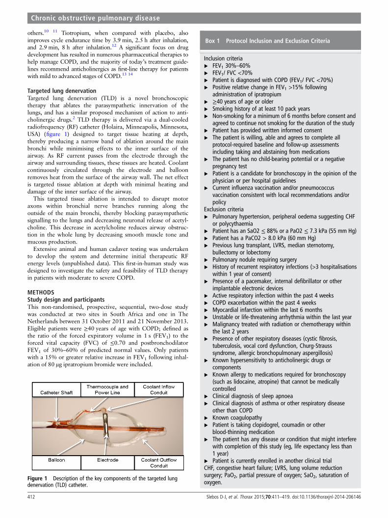
Targeted lung denervationTargeted lung denervation (TLD) is a novel bronchoscopictherapy that ablates the parasympathetic innervation of thelungs, and has a similar proposed mechanism of action to anti-cholinergic drugs.2 TLD therapy is delivered via a dual-cooledradiofrequency (RF) catheter (Holaira, Minneapolis, Minnesota,USA) (figure 1) designed to target tissue heating at depth,thereby producing a narrow band of ablation around the mainbronchi while minimising effects to the inner surface of theairway. As RF current passes from the electrode through theairway and surrounding tissues, these tissues are heated. Coolantcontinuously circulated through the electrode and balloonremoves heat from the surface of the airway wall. The net effectis targeted tissue ablation at depth with minimal heating anddamage of the inner surface of the airway.
This targeted tissue ablation is intended to disrupt motoraxons within bronchial nerve branches running along theoutside of the main bronchi, thereby blocking parasympatheticsignalling to the lungs and decreasing neuronal release of acetyl-choline. This decrease in acetylcholine reduces airway obstruc-tion in the whole lung by decreasing smooth muscle tone andmucous production.
Extensive animal and human cadaver testing was undertakento develop the system and determine initial therapeutic RFenergy levels (unpublished data). This first-in-human study wasdesigned to investigate the safety and feasibility of TLD therapyin patients with moderate to severe COPD.
METHODSStudy design and participantsThis non-randomised, prospective, sequential, two-dose studywas conducted at two sites in South Africa and one in TheNetherlands between 31 October 2011 and 21 November 2013.Eligible patients were ≥40 years of age with COPD; defined asthe ratio of the forced expiratory volume in 1 s (FEV1) to theforced vital capacity (FVC) of ≤0.70 and postbronchodilatorFEV1 of 30%–60% of predicted normal values. Only patientswith a 15% or greater relative increase in FEV1 following inhal-ation of 80 μg ipratropium bromide were included.
Figure 1 Description of the key components of the targeted lungdenervation (TLD) catheter.
Box 1 Protocol Inclusion and Exclusion Criteria
Inclusion criteria▸ FEV1 30%–60%▸ FEV1/ FVC <70%▸ Patient is diagnosed with COPD (FEV1/ FVC <70%)▸ Positive relative change in FEV1 >15% following
administration of ipratropium▸ ≥40 years of age or older▸ Smoking history of at least 10 pack years▸ Non-smoking for a minimum of 6 months before consent and
agreed to continue not smoking for the duration of the study▸ Patient has provided written informed consent▸ The patient is willing, able and agrees to complete all
protocol-required baseline and follow-up assessmentsincluding taking and abstaining from medications
▸ The patient has no child-bearing potential or a negativepregnancy test
▸ Patient is a candidate for bronchoscopy in the opinion of thephysician or per hospital guidelines
▸ Current influenza vaccination and/or pneumococcusvaccination consistent with local recommendations and/orpolicy
Exclusion criteria▸ Pulmonary hypertension, peripheral oedema suggesting CHF
or polycythaemia▸ Patient has an SaO2 ≤ 88% or a PaO2 ≤ 7.3 kPa (55 mm Hg)▸ Patient has a PaCO2 > 8.0 kPa (60 mm Hg)▸ Previous lung transplant, LVRS, median sternotomy,
bullectomy or lobectomy▸ Pulmonary nodule requiring surgery▸ History of recurrent respiratory infections (>3 hospitalisations
within 1 year of consent)▸ Presence of a pacemaker, internal defibrillator or other
implantable electronic devices▸ Active respiratory infection within the past 4 weeks▸ COPD exacerbation within the past 4 weeks▸ Myocardial infarction within the last 6 months▸ Unstable or life-threatening arrhythmia within the last year▸ Malignancy treated with radiation or chemotherapy within
the last 2 years▸ Presence of other respiratory diseases (cystic fibrosis,
tuberculosis, vocal cord dysfunction, Churg-Strausssyndrome, allergic bronchopulmonary aspergillosis)
▸ Known hypersensitivity to anticholinergic drugs orcomponents
▸ Known allergy to medications required for bronchoscopy(such as lidocaine, atropine) that cannot be medicallycontrolled
▸ Clinical diagnosis of sleep apnoea▸ Clinical diagnosis of asthma or other respiratory disease
other than COPD▸ Known coagulopathy▸ Patient is taking clopidogrel, coumadin or other
blood-thinning medication▸ The patient has any disease or condition that might interfere
with completion of this study (eg, life expectancy less than1 year)
▸ Patient is currently enrolled in another clinical trialCHF, congestive heart failure; LVRS, lung volume reductionsurgery; PaO2, partial pressure of oxygen; SaO2, saturation ofoxygen.
412 Slebos D-J, et al. Thorax 2015;70:411–419. doi:10.1136/thoraxjnl-2014-206146
Chronic obstructive pulmonary disease

Key exclusion criteria included: COPD exacerbation or activerespiratory infection within the past 4 weeks, more than 3respiratory-related hospitalisations within 1 year of enrolment,previous lung surgery, suspicious pulmonary nodule, pulmonaryhypertension, congestive heart failure, polycythaemia, SaO2
≤88% or a PaO2 ≤7.3 kPa, and PaCO2 >8.0 kPa. Formal pul-monary rehabilitation was not a requirement for inclusion. Referto the box 1 for a full list of inclusion and exclusion criteria.
This study was approved by local ethics committees and inaccordance with the Declaration of Helsinki (1996), GoodClinical Practice guidelines, and local requirements. An opera-tions committee, and a data monitoring committee, oversawprotocol management and safety for the study. An independentclinical event reviewer adjudicated all serious adverse events(SAE). This trial is registered with ClinicalTrials.gov, numberNCT01483534.
Study endpointsThe primary safety endpoint of freedom from documented andsustained worsening of COPD directly attributable to the investi-gational device or procedure to a 365-days post-TLD therapy,was defined as a decrease in the individual patient’s FEV1 by anyamount at all follow-up time points, along with a report of anadverse event that was reported to have a probable or definiterelation to the device. Secondary endpoints included technicalfeasibility of the device and change from baseline in pulmonaryfunction tests, exercise capacity assessments, and HRQL.Technical feasibility was defined as the ability to access the targettreatment area main bronchus and deliver RF energy to the entirecircumference of the bronchus at the target treatment site.
ProceduresAfter informed consent, patients underwent baseline testing aftera wash-out period of 8 days for long-acting muscarinic antago-nists (LAMA), 24 h for long-acting β agonists (LABA) and 12 hfor short-acting β agonists (SABA) and short-acting muscarinicantagonists (SAMA). Baseline and follow-up testing includedspirometry, body plethysmography, cycle ergometry, 6 min walktest (6MWT), COPD-specific SGRQ (score range 0–100 with aminimally clinically important difference (MCID) of ≥4 negativeunits15), the Clinical COPD Questionnaire (CCQ, 7-day version,score range 0–6, with an MCID of 0.4 units16), the commonBorg and modified Medical Research Council17 scales. Cycleergometry was first conducted as an incremental maximal test todetermine baseline maximum work rate (Wmax) off drugs, andsubsequently as an endurance test conducted at a constant workrate of 75% of the Wmax.
18 Current American Thoracic Society(ATS)/European Respiratory Society (ERS) guidelines were
followed for pulmonary function testing,19 20 and ATS/AmericanCollege of Chest Physicians (ACCP) guidelines for cycle ergome-try21 and the 6MWT.22 A baseline chest CT scan was required toconfirm appropriate bronchial anatomy and rule out other pul-monary abnormalities.
After completion of the wash-out baseline testing, patientsunderwent a minimum 8-day run-in period while on tiotropiumbromide, and similar testing was performed at drug trough 24 hafter the last dose of tiotropium to establish tiotropium troughbaseline values. LABAs were again held for 24 h and SABAs andSAMAs for 12 h before this testing. All patients underwent earlysafety evaluation by phone at 3 days and 10 days postprocedure.Testing was repeated at washout baseline, tiotropium troughbaseline, and 30, 90, 180, 270 and 365 days post-treatment,except for cycle ergometry which was performed at 90, 180 and365 days post-treatment. Patients were provided a daily memoryaid to track overall changes in health status. A total of three bron-choscopies were performed per patient; two in order to completethe two treatments and the third one to assess potential airwaysurface effects during the 90-day follow-up visit. Adverse eventswere tracked and recorded throughout the entire study period.
TreatmentPretreatment visual bronchoscopic inspection of the airways wasconducted to reconfirm that appropriate airway anatomy existed.Due to the construction of this first-generation device, procedureswere performed via rigid bronchoscopy under general anaesthesia.The dual-cooled catheter was placed through the rigid broncho-scope, and a flexible bronchoscope placed beside it for visualisa-tion. The electrode was placed and activated in up to eightrotational positions per bronchus to achieve complete circumfer-ential treatment. Total balloon inflation times were approximately3 min per activation. The initial subjects were treated with20 watts (W) in all positions except for the posterior-medialaspects of the left bronchus, where the power was reduced to15W due to the proximity of the oesophagus in those positions.Due to local airway effects, after a protocol amendment, add-itional patients underwent treatment with a more distal placementof the electrode along the medial wall to avoid the thin tissue ofthe carina, and a lower 15 W energy level in all position.Bronchoscopic and fluoroscopic visualisation was used to guidethe electrode positioning (figure 2) throughout treatment. Tomaximise safety, the protocol mandated staged treatment (2 thera-peutic procedures per patient) with the second bronchus beingtreated 30 days after the first. No special post procedure medica-tions were required, and the use of tiotropium was stopped.Investigators were allowed to treat respiratory symptoms per
Figure 2 (A and B) fluoroscopic view(A) and bronchoscopic view (B) of theelectrode during the procedure. Theelectrode is indicated by the arrow.
Slebos D-J, et al. Thorax 2015;70:411–419. doi:10.1136/thoraxjnl-2014-206146 413
Chronic obstructive pulmonary disease

standard-of-care and published guidelines, however, appropriatedrug wash-out was required before follow-up testing.
Statistical methodsAs this was a feasibility study, there was no primary studyhypothesis with statistical inference. All p values were presentedfor informational purposes. According to the prespecified ana-lysis plan, continuous data were summarised using means andSDs, or medians and quartiles, in the presence of non-normality.
Categorical data were tabulated, with counts and percentages.All available data was summarised, with no imputation formissing data. Final analyses were conducted using SAS V.9.3(SAS Institute, Cary, North Carolina, USA) by an independentstatistical group (NAMSA, Minneapolis, Minnesota, USA).
Role of the funding sourceThe sponsor, D-JS, MM and CB designed the trial. Sitesrecruited patients and collected data on standardised case report
Table 1 Baseline characteristics. Data are mean (SD) unless stated otherwise
20 W cohort (n=12) 15 W cohort (n=10) p Value
Age (years) 62.92 (11.37) 64.40 (8.87) 0.740Male (n, %) 7 (58) 4 (40) 0.669
Ethnic origin (n, %)White 12 (100) 8 (80) 0.195Black 0 (0) 2 (20) –
History of smoking (n, %) 12 (100) 10 (100) –
Pack-years 38.57 (19.97) 44.60 (25.86) 0.544Wash-out prebronchodilator FEV1 (L) 0.90 (0.28) 0.84 (0.15) 0.551Wash-out prebronchodilator FVC (L) 2.56 (0.59) 2.45 (0.60) 0.664Reversibility peak relative change in FEV1 (%) 25.95 (7.60) 19.89 (1.91) 0.019Run-in tiotropium trough FEV1 (L) 1.04 (0.39) 0.98 (0.23) 0.651Run-in tiotropium trough FVC (L) 2.89 (0.76) 2.75 (0.60) 0.661Cycle endurance time @ 75% of max (min) 6.71 (3.26) 4.57 (2.20) 0.1059SGRQ-C total score (points) 56.16 (13.71) 56.23 (20.85) 0.9931Average diameter: right main bronchus (mm)* 13.42 (1.57) 13.91 (1.62) 0.519Average diameter: left main bronchus (mm)* 12.02 (1.45) 12.33 (1.68) 0.672
*Measurements performed by the sponsor using Apollo Software (VIDA Diagnostics, Coralville, Iowa, USA).FEV1 (L), expiratory volume in 1 s; FVC (L), forced vital capacity (FVC)
Figure 3 Clinical trial profile and patient flowchart.
414 Slebos D-J, et al. Thorax 2015;70:411–419. doi:10.1136/thoraxjnl-2014-206146
Chronic obstructive pulmonary disease

Table 2 Summary of adverse events through 1 year
20 watt cohortn=12
15 watt cohortn=10
Alln=22
RF dosage, wattAdverse event
Adverse eventfrequencyn=60 (%)
Subject *frequency(%)
Adverse eventfrequencyn=39 (%)
Subject*frequency(%)
Adverse eventfrequencyn=99 (%)
Subject*frequency(%)
Device-related non-seriousBronchial perforation (carina) 2 (3) 2 (17) – – 2 (2) 2 (9)Bronchial stenosis 1 (2) 1 (8) – – 1 (1) 1 (5)Bronchial ulceration 1 (2) 1 (8) – – 1 (1) 1 (5)COPD Exacerbation – – 1 (3) 1 (10) 1 (1) 1 (5)Granulomas 1 (2) 1 (8) – – 1 (1) 1 (5)Worsening of FEV1† – – 1 (3) 1 (10) 1 (1) 1 (5)
Device related-seriousGastroparesis – – 1 (3) 1 (10) 1 (1) 1 (5)
Procedural related-non-serious (related or reported within 2 days of either procedure)Broken tooth 1 (2) 1 (8) 1 (1) 1 (5)Cough 2 (3) 2 (17) – – 2 (2) 2 (9)Dyspnoea 2 (3) 2 (17) 1 (3) 1 (10) 3 (3) 3 (14)Eczema – – 1 (3) 1 (10) 1 (1) 1 (5)Headache 2 (3) 2 (17) – – 2 (2) 2 (9)Mucus 3 (5) 3 (25) – – 3 (3) 3 (14)Sore throat 3 (5) 3 (25) 1 (3) 1 (10) 4 (4) 4 (18)Tracheal injury (due to rigidbronchoscope)
1 (2) 1 (8) – – 1 (1) 1 (5)
Procedural related-serious (related or reported within 2 days of either procedure)Anaphylactic reaction 1 (2) 1 (8) – – 1 (1) 1 (5)COPD exacerbation – – 1 (3) 1 (10) 1 (1) 1 (5)
Other non-seriousAbscess (skin) – – 1 (3) 1 (10) 1 (1) 1 (5)Back ache 1 (2) 1 (8) – – 1 (1) 1 (5)Bronchitis – – 5 (13) 2 (20) 5 (5) 2 (9)Candida – – 1 (3) 1 (10) 1 (1) 1 (5)Chest pain 1 (2) 1 (8) – – 1 (1) 1 (5)Common cold 2 (3) 2 (17) – – 2 (2) 2 (9)COPD exacerbation 12 (20) 7 (58) 13 (33) 6 (60) 25 (25) 13 (59)
Cough 1 (2) 1 (8) – – 1 (1) 1 (5)Diarrhoea 1 (2) 1 (8) – – 1 (1) 1 (5)Difficulty swallowing 1 (2) 1 (8) – – 1 (1) 1 (5)Dizziness 2 (3) 2 (17) 1 (3) 1 (10) 3 (3) 3 (14)Dyspnoea 3 (5) 2 (17) 1 (3) 1 (10) 4 (4) 3 (14)Flu 3 (5) 3 (25) 1 (3) 1 (10) 4 (4) 4 (18)Gastritis 2 (3) 2 (17) 1 (3) 1 (10) 3 (3) 3 (14)Gastroparesis 1 (2) 1 (8) – – 1 (1) 1 (5)Pneumonia 1 (1) 1 (8) – – 1 (1) 1 (5)Respiratory inflammation 4 (7) 4 (33) – – 4 (4) 4 (18)Sinusitis – – 1 (3) 1 (10) 1 (1) 1 (5)
Other seriousChest pain – – 1 (3) 1 (10) 1 (1) 1 (5)COPD exacerbation 2 (3) 2 (17) 5 (13) 3 (30) 7 (7) 5 (23)Coronary artery disease/CABG 1 (2) – – – 1 (1) 1 (5)Flu – – 1 (3) 1 (10) 1 (1) 1 (5)Lung infection 2 (3) 2 (17) – – 2 (2) 2 (9)Stomach cancer (stage 3) – – 1 (3) 1 (10) 1 (1) 1 (5)
A total of 99 adverse events were reported in 22 (100%) subjects.Serious/non-serious defined per ISO 14155.All adverse events were independently adjudicated.*Count reflects number of subjects with event reported one or more times.†Adverse event included based on primary endpoint definition.CABG, coronary artery bypass surgery; COPD, chronic obstructive pulmonary disease; RF, radiofrequency; SGRQ, St. George’s Respiratory Questionnaire.
Slebos D-J, et al. Thorax 2015;70:411–419. doi:10.1136/thoraxjnl-2014-206146 415
Chronic obstructive pulmonary disease

forms. The sponsor managed the overall study and safetycommittees.
RESULTSTwelve patients were included in the 20 W cohort, and an add-itional 10 patients were included as part of the 15 W cohort at thesame sites using the same study criteria (figure 3). Demographic
and baseline characteristics were similar between the two cohorts,with the 15 W cohort enrolling patients with slightly worse base-line FEV1 (table 1). One-year follow-up data were available in10 of 11 available patients in the 20 W cohort, and all 10 of the15 W cohort. One patient in the 20 W cohort underwent coronarybypass surgery for a calcified coronary lesion during follow-up, andwas withdrawn from the study by the site investigator before the
Figure 4 Bronchoscopic confirmation of airway healing after radiofrequency energy delivery: (A) Left main bronchus pretreatment. (B) Duringtreatment. (C) Immediately post-treatment. (D) 3-month follow-up.
Figure 5 CT and bronchoscopic findings seen before procedural enhancements: (A and B) A 4 mm granuloma indicated by white arrows on bothtransverse CT and bronchoscopic images. (C and D) Superficial airway effect indicated by white arrow on the bronchoscopic image with normaltransverse CT image. (E and F) A 1.5 mm perforation through carina indicated by white arrows on both coronal CT and bronchoscopic images.
416 Slebos D-J, et al. Thorax 2015;70:411–419. doi:10.1136/thoraxjnl-2014-206146
Chronic obstructive pulmonary disease

second procedure. One patient in the 15 W cohort experienced aCOPD exacerbation 10 days after the initial procedure and did notconsent to the 2nd treatment, but continued follow-up.
The primary safety endpoint was achieved in 100% (11/11)in the 20 W cohort and 90% (9/10) in the 15 W cohort. Theone subject that failed to meet the endpoint had a decreasefrom baseline in FEV1 at all follow-up time points, and a COPDexacerbation 1 day after the 2nd treatment that was reported asrelated to the lung denervation system and/or the bronchoscopyby the investigator.
No deaths occurred in this study. Seven postprocedure SAEsin the 30 days following treatment of either bronchus includedCOPD exacerbation (n=3), anaphylactic drug reaction, coron-ary artery bypass surgery, chest pain resulting in uneventful hos-pitalisation, and gastroparesis in a subject with a history ofgastric issues. Four of these seven events occurred in a singlepatient. Nine longer-term SAEs out to 1 year included COPDexacerbation (n=5), pneumonia (n=2), influenza (H1N1)(n=1), and stomach cancer (n=1). Events were evenly distribu-ted between both cohorts, with the majority of events reportedduring the periprocedural period (table 2).
The RF energy applied to the airway wall resulted in localasymptomatic airway blanching, which resolved at the 3-monthfollow-up bronchoscopy in all 10 of the 15 W patients, and ineight of the 11 W–20 W patients with long-term follow-up(figure 4). One patient had a superficial tissue defect at the firsttreatment site seen at 30 days, without any airway abnormalitieson CT. A second patient had a small 1.5 mm perforationthrough the thin tissue of the main carina, also discovered at30 days. A third patient had a superficial tissue defect at theinitial treatment site just distal to the main carina, as well as a4 mm granuloma at the second treatment site, with a 20% sten-osis observed during the follow-up bronchoscopy at 90 days
(figure 5). The granuloma was electively removed by cauterisa-tion. No other treatments or interventions were performed, andall showed complete or active healing during follow-up visualinspection.
Technical feasibility was 93%, with 39 of 42 procedures deli-vering full circumferential treatment (up to eight individual acti-vations) to the target treatment site of the main bronchus. Twopatients had only one bronchus treated as outlined above. Threepatients had incomplete (non-circumferential) treatment: twohad airway geometries that limited electrode/balloon contactwith the right mainstem bronchus wall, and one had an intra-procedural anaphylactic reaction to diclofenac that prohibitedfull left bronchus treatment.
No device-related adverse events occurred during the proced-ure. Mean (SD) procedure time (min) for the 20 W cohort was47.75±8.13 for the right and 56.10±12.79 for the left bronchi.Mean procedure time for the 15 W cohort was 42.10±8.23 forthe right and 51.33±25.76 for the left.
The results of FEV1 (% relative change), FVC, cycle ergometryendurance and SGRQ at each time point assessed are shown infigure 6. At the higher 20 W power level, significant differencesfrom baseline were seen at the following assessment time points:FVC at 90 days (p=0.016) and 270 days (p=0.036); cycle endur-ance at 180 days (p=0.03), and SGRQ at 90 days (p=0.042),180 days (p=0.019), 270 days (p=0.008) and 1 year (p=0.011).Variability across time is noted, and most likely due to the smallsample size. No statistically significant differences in any of thevariables measured were seen at the 15 W power level.
FEV1 per cent change from baseline at 1 year was 11.6±32.3%vs 0.02±15.1% (p=0.324) in the 20 W and 15 W cohorts,respectively (table 3). Similarly, FVC had a positive absolute posi-tive change in FVC at 1 year of 198±516 mL in the 20 W cohortvs 61±485 mL (p=0.256) in the 15 W cohort (figure 6).
Figure 6 Secondary efficacy endpoints. Data represented as mean. Error bars represent SEM. *p <0.05 compared with baseline; FEV1: forcedexpiratory volume in 1 s; Cycle Erg. Endurance, Cycle Ergometry Endurance; SGRQ, St. George’s Respiratory Questionnaire.
Slebos D-J, et al. Thorax 2015;70:411–419. doi:10.1136/thoraxjnl-2014-206146 417
Chronic obstructive pulmonary disease

Study results for exercise endurance and HRQL (figure 6)show improvement from baseline for endurance cycle ergometrywas 6.8±12.8 vs 2.6±8.7 min (p=0.277) at 1 year. A significantdifference was observed in HRQL as assessed by the SGRQ:
−11.1±9.1 vs −0.9±8.6 (p=0.044) in the 20 W and 15 Wcohorts, respectively.
At 90, 180 and 365 days, the effect on FEV1 of TLD therapyplus 80 μg ipratropium bromide at peak was assessed. As illustratedin figure 7, the combination therapy might result in an increase inFEV1 over those seen with an inhaled anticholinergic drug alone.Significant differences between TLD +drug vs TLD alone wereseen at each matching time points assessed for both energy cohorts.
DISCUSSIONThis first-in-human clinical trial evaluated the novel TLDtherapy, designed to ablate the parasympathetic pulmonarynerves surrounding the main bronchi, thereby decreasingbronchomotor tone in patients with COPD. As a feasibilityevaluation, this study included a drug ‘wash-out’ period toestablish baseline values in order to understand the effect of thisnew therapy alone. This study demonstrated TLD to be feasible,safe and well tolerated. The primary endpoint was met in 95%of patients, and technical feasibility was 93%. A tendencytoward improvements in lung function, exercise capacity, andHRQL were observed in the 20 W cohort with statistical signifi-cance achieved for FVC at 90 days (p=0.016) and 270 days(p=0.036); cycle endurance at 180 days (p=0.03) and SGRQat 90 days (p=0.042), 180 days (p=0.019), 270 days(p=0.008) and 1 year (p=0.011). These improvements tendedto be larger than those seen in the 15 W cohort with statisticallysignificant difference in SGRQ at 1 year (p=0.044).Additionally, early data suggest an additive effect when TLD iscombined with inhaled anticholinergic drugs.
Three procedural adjustments were made during the study inresponse to asymptomatic airway wall effects observed in threeof 12 patients in the 20 W group. These effects were postulatedto be attributable to a combination of energy delivery to the thin,thermally sensitive tissue of the main carina, imperfect ballooncontact that limited surface cooling and, potentially, the higherenergy level. Subsequent computer modelling and bench testingconfirmed heat accumulation in the thin tissue of the carina, andpost hoc analysis of procedural imaging indicated the balloonwas not able to accommodate itself to sharply tapering or suddengeometric changes of the airway. As a result, the procedure wasmodified to more distal electrode placement away from the maincarina, more detailed visual assessment of balloon contact beforeactivation, and decrease in overall power to 15 W to furtherreduce the potential for undesired airway wall effects.
Table 3 Comparison of outcomes at 1 year for patients withfollow-up testing
Outcome20 watt cohort(n=10)
15 watt cohort(n=10)
Overall(n=20) p Value
FEV1 (mL)Baseline 851.0 (277.3) 836.0 (148.0) 843.5 (216.5) 0.8821 year 900.0 (268.6) 835.0 (185.5) 867.5 (227.1) 0.537Per cent Δ 11.6 (32.3) 0.02 (151) 5.8 (25.3) 0.324
FVC (mL)Baseline 2439.0 (538.2) 2448.0 (602.9) 2443.5 (556.3) 0.9721 year 2637.0 (802.2) 2509.0 (665.6) 2573.0 (720.4) 0.702Per cent Δ 7.6 (21.9) 39 (19.1) 5.7 (20.1) 0.690
Total lung capacity (TLC)Baseline 7.2 (1.2) 6.8 (1.2) 7.0 (1.2) 0.5011 year 7.3 (1.8) 7.2 (1.2) 7.2 (1.5) 0.824Per cent Δ 1.0 (11.8) 5.2 (9.6) 3.2 (10.6) 0.405
Residual volume (RV)Baseline 4.6 (0.7) 4.1 (0.7) 44 (0.7) 0.1641 year 4.6 (1.2) 4.4 (0.9) 4.5 (1.0) 0.696Percent Δ 0.5 (21.2) 7.9 (20.6) 4.4 (20.6) 0.451
Inspiratory capacity (IC)Baseline 1.8 (0.5) 1.7 (0.6) 1.8 (0.5) 0.8851 year 1.7 (0.78) 1.8 (0.6) 1.8 (0.7) 0.743Percent Δ −2.0 (31.6) 7.3 (22.0) 2.9 (26.6) 0.462
Pulmonary resistance (kPa×s/L)Baseline 0.8 (0.3) 1.0 (0.2) 0.9 (0.2) 0.2331 year 0.8 (0.2) 0.8 (0.1) 0.8 (0.2) 0.695Percent Δ −4.6 (13.8) −19.1 (18.8) −12.2 (17.8) 0.074
Cycle ergometry endurance (min)Baseline 5.8 (2.3) 4.2 (2.1) 5.1 (2.3) 0.1391 year 12.7 (13.5) 6.9 (8.3) 10.1 (11.5) 0.301Absolute Δ 6.8 (12.8) 2.6 (8.7 5.0 (11.1) 0.441
Borg scale: post-testing (cycle ergometry): dyspnoeaBaseline 4.4 (1.6) 6.1 (1.3) 5.2 (1.7) 0.0231 year 4.1 (2.0) 5.3 (1.8) 4.6 (1.9) 0.223Absolute Δ −0.3 (2.0) −0.9 (2.0) −0.6 (2.0) 0.556
mMRC scaleBaseline 1.0 (1.3) 1.2 (1.1) 1.1 (1.1) 0.7881 year 0.3 (0.5) 1.8 (1.3) 1.0 (1.2) 0.031Absolute Δ −0.7 (0.8) 0.6 (1.3) −0.1 (1.2) 0.085
6 min walk test (metres)Baseline 388.1 (82.8) 425.4 (74.7) 406.8 (79.1) 0.3041 year 412.3 (51.7) 416.1 (120.0 414.2 (89.9) 0.928Absolute Δ 24.2 (45.6) −9.3 (70.6) 7.5 (60.4) 0.224
SGRQ-C: total scoreBaseline 53.2 (14.1) 57.9 (17.9) 55.4 (15.6) 0.5751 year 42.1 (12.) 57.1 (21.8) 49.1 (18.4) 0.120Absolute Δ −11.1 (9.1) −0.9 (8.6) −6.3 (10.1) 0.045
CCQ: total scoreBaseline 2.8 (1.0) 2.63 (1.0) 2.7 (1.0) 0.7321 year 2.2 (1.0) 2.77 (1.0) 2.4 (1.0) 0.230Absolute Δ −0.6 (0.8) 0.14 (0.7) −0.3 (0.8) 0.051
Data are mean (SD) unless stated otherwise.SGRQ, St. George’s Respiratory Questionnaire (COPD specific); CCQ, clinical COPDquestionnaire; mMRC, Modified Medical Research Council dyspnoea scale.p Values are for comparisons between cohorts.
Figure 7 Efficacy measures of targeted lung denervation (TLD)therapy +inhaled ipratropium bromide. Data represented as mean. Errorbars represent SEM. *p<0.05 compared with baseline. Follow-up datapoints were compared with results of TLD alone, and all werestatistically significant.
418 Slebos D-J, et al. Thorax 2015;70:411–419. doi:10.1136/thoraxjnl-2014-206146
Chronic obstructive pulmonary disease

In this study, the 12 patients treated with 20 W tended tohave greater changes from baseline in spirometry, exercise cap-acity and HRQL, when compared with the 10 patients treatedwith 15 W. The added effect in the 20 W group might be attrib-utable to more effective denervation due to a deeper tissueeffect at higher energy. On the other hand, the 20 W grouppatients were slightly more reversible to ipratropium at baseline,which theoretically, might mean that these patients are moresensitive to TLD therapy. However, we found no differencebetween the two groups in tiotropium through FEV1 levels.Apart from the airway effects observed, safety profiles for thetwo energy levels were similar. This might imply that futurecatheter designs that better accommodate human airway irregu-larities will ensure better surface cooling and, thus, allow higherenergy levels to be used.
In this paper, we introduced TLD, a novel bronchoscopictreatment concept for symptomatic patients suffering fromCOPD. Based on the concept of ablating parasympathetic pul-monary nerves, TLD was shown to be feasible, safe and well tol-erated. TLD has the potential to overcome many of thelimitations of inhaled drugs for the treatment of COPD. First,TLD may eliminate inhaler compliance issues for the 63% ofnew tiotropium users who discontinue treatment after 1 year.23
Second, TLD would not be subject to the peak and trough var-iations seen with drugs.24 Third, TLD may eliminate variableregional drug delivery and deposition in patients with obstruct-ive lung disease25 by ablating the nerves that travel throughoutthe bronchial tree independent of regional airflow obstruction.Fourth, by interfering with parasympathetic nerve-derivedacetylcholine by two different mechanisms, the combination ofTLD +inhaled anticholinergic drugs, as suggested in figure 7,may have a synergistic effect that results in a reduction in airwayobstruction and mucus production, as well as inhibition of localairway inflammation induced by non-neural muscarinicaction.8 26 Further investigation and progressive product devel-opment of this novel therapy is warranted, with focus onfurther refining energy delivery to ablate the nerves and opti-mise patient selection.
Acknowledgements We would like to thank the investigators, their staff andteams, and the patients who consented to be part of this investigation. Weacknowledge the work of the late Prof C Bolliger for his important contributions tothis work, and to the field of pulmonary medicine.
Collaborators Panorama Medi Clinic & University of Stellenbosch, Cape Town,South Africa (Ethics Committee reference # M11/05/013): Chris Bolliger CoenraadKoegelenberg, Johan Theron, Firdows Noor, Elvis Irusen, Dorothy Steyn; University ofGroningen, Groningen, The Netherlands (Ethics Committee reference #NL36608.042.11): Dirk-Jan Slebos, Nick Ten Hacken, Karin Klooster.
Contributors D-JS, KK, CFNK, JT, DS and CTB recruited and treated patients inthis trial. MM is the inventor of the lung denervation system. AV was involved indata analysis and data interpretation. All authors helped to write the report. Thecorresponding author had full access to all data in the study and had finalresponsibility for the decision to submit it for publication.
Funding Holaira, Inc., Minneapolis, Minnesota, USA.
Competing interests All clinical trial expenses were reimbursed by the studysponsor (Holaira, Inc.). D-JS is the overall study principal investigator. AV is theprinciple investigator of a second study conducted by the sponsor. MM is founderand chief technology officer of the study sponsor.
Ethics approval The medical ethics committees of the study sites involved, inSouth Africa and in The Netherlands.
Provenance and peer review Not commissioned; internally peer reviewed.
Open Access This is an Open Access article distributed in accordance with theCreative Commons Attribution Non Commercial (CC BY-NC 4.0) license, whichpermits others to distribute, remix, adapt, build upon this work non-commercially,and license their derivative works on different terms, provided the original work isproperly cited and the use is non-commercial. See: http://creativecommons.org/licenses/by-nc/4.0/
REFERENCES1 Canning B. Reflex regulation of airway smooth muscle tone. J Appl Physiol
2006;101:971–85.2 Belmonte K. Cholinergic pathways in the lungs and anticholinergic therapy for
chronic obstructive pulmonary disease. Proc Am Thor Soc 2005;2:297–305.3 Pera T, Zuidhof A, Valadas J, et al. Tiotropium inhibits pulmonary inflammation and
remodeling in a guinea pig model of COPD. Eur Respir J 2011;38:789–96.4 Klassen KD, Morton DR. The effect of vagus section on the cough reflex, bronchial
caliber, and clearance of bronchial secretions. Surgery 1951;29:483–90.5 Jammes Y, Mei N. Assessment of the pulmonary origin of bronchoconstrictor vagal
tone. J Physiol 1979;291:305–16.6 Abbott OA, Hopkins WA, Van Fleit WE, et al. A new approach to pulmonary
emphysema. Thorax 1953;8:116–31.7 Dimitrov-Szokodi D, Husveti A, Balogh G. Lung Denervation in the Therapy of
Intractable Bronchial Asthma. J Thoracic Surg 1957;33:166–84.8 Gosens R, Zaagsma J, Meurs H, et al. Muscarinic receptor signaling in the
pathophysiology of asthma and COPD. Respir Res 2006;7:73.9 Vincken W, van Noord JA, Greefhorst APM. Improved health outcomes in patients
with COPD during 1 yr’s treatment with tiotropium. Eur Respir J 2002;19:209–16.10 Tashkin DP, Celli B, Decramer M, et al. Bronchodilator responsiveness in patients
with COPD. Eur Respir J 2008;31:742–50.11 Tashkin DP, Celli B, Senn S, et al. A 4-year trial of tiotropium in chonic obstructive
pulmonary disease. NEJM 2008;359:1543–54.12 Maltais F, Hamilton A, Marciniuk D, et al. Improvements in symptom-limited
exercise performance over 8 h with once-daily tiotropium in patients with COPD.Chest 2005;128;1168–78.
13 Global Strategy for the Diagnosis, Management and Prevention of COPD. 2014.[http://www.goldcopd.org]
14 Qaseem A, Wilt T, Weinberger S, et al. Diagnosis and management of stablechronic obstructive pulmonary disease: a clinical practice Guideline update from theACP, ACCP, ATS and ERS. Ann intern Med 2011;155:179–91.
15 Meguro M, Barley E, Spencer S, et al. Development and Validation of an Improved,COPD-Specific Version of the St. George Respiratory Questionnaire. CHEST2007;132:456–63.
16 Van der Molen T, Willemse B, Schokker S, et al. Development, validity andresponsiveness of the Clinical COPD Questionnaire. Health Qual Life Outcomes2003;1:13.
17 Bestall JC, Paul EA, Garrod R, et al. Usefulness of the Medical Research Council(MRC) dyspnea scale as a measure of disability in patients with chronic obstructivepulmonary disease. Thorax 1999;54:581–6.
18 Maltais F, Hamilton A, Marciniuk D, et al. Improvement in Symptom-LimitedExercise Performance over 8 h With Once-Daily Tiotropium in Patients with COPD.Chest 2005;128:1168–78.
19 Miller M, Hankinson J, Brusasco V, et al. Series “ATS/ERS Task Force:Standardization of Lung Function Testing”. Eur Respir J 2005;26:319–38.
20 Wagner J, Clausen J, Coates A, et al. Series “ATS/ERS Task Force: Standardizationof Lung Function Testing”. Eur Respir J 2005;26:511–22.
21 Weisman IM, Beck KC, Casaburi R, et al. ATS/ACCP statement on cardiopulmonaryexercise testing. Am J Respir Crit Care Med 2003;167:211–77.
22 ATS Committee on Proficiency Standards for Clinical Pulmonary FunctionLaboratories. ATS statement: guidelines for the six-minute walk test. Am J RespirCrit Care Med 2002;166:111–7.
23 Breekveldt-Postma NS, Koerselmana J, Erkensa JA, et al. Enhanced persistencewith tiotropium compared with other respiratory drugs in COPD. Respir Med2007;101:1398–405.
24 Maesen FPV, Smeets JJ, Sledsens TJH, et al. Iprotropium bromide, a newlong-acting antimuscarinic bronchodilator: a pharmacodynamic study in patientswith chronic obstructive pulmonary disease (COPD). Eur Respir J 1995;8:1506–13.
25 Anderson P. Use of Respimat Soft Mist Inhaler in COPD patients. Int J ChronObstruct Pulmon Dis 2006;1:251–9.
26 Profita M, Albano GD, Riccobono L, et al. Increased levels of Th17 cells areassociated with non-neuronal acetylcholine in COPD patients. Immunobiology2014;219:392–401.
Slebos D-J, et al. Thorax 2015;70:411–419. doi:10.1136/thoraxjnl-2014-206146 419
Chronic obstructive pulmonary disease