targeting brain stem centers of cardiovascular control ... · targeting brain stem centers of...
TRANSCRIPT

doi:10.1152/physiolgenomics.00120.2004 20:165-172, 2005. First published Nov 23, 2004;Physiol. Genomics
Pickering and Sergey Kasparov Tina Lonergan, Anja G. Teschemacher, D. Y. Hwang, K.-S. Kim, Anthony E.transgene expression using adenoviral vectors: impact of promoters on Targeting brain stem centers of cardiovascular control
You might find this additional information useful...
43 articles, 21 of which you can access free at: This article cites http://physiolgenomics.physiology.org/cgi/content/full/20/2/165#BIBL
including high-resolution figures, can be found at: Updated information and services http://physiolgenomics.physiology.org/cgi/content/full/20/2/165
can be found at: Physiological Genomicsabout Additional material and information http://www.the-aps.org/publications/pg
This information is current as of June 14, 2005 .
http://www.the-aps.org/.the American Physiological Society. ISSN: 1094-8341, ESSN: 1531-2267. Visit our website at July, and October by the American Physiological Society, 9650 Rockville Pike, Bethesda MD 20814-3991. Copyright © 2005 bytechniques linking genes and pathways to physiology, from prokaryotes to eukaryotes. It is published quarterly in January, April,
publishes results of a wide variety of studies from human and from informative model systems withPhysiological Genomics
on June 14, 2005 physiolgenom
ics.physiology.orgD
ownloaded from

Targeting brain stem centers of cardiovascular control using adenoviralvectors: impact of promoters on transgene expression
Tina Lonergan,1 Anja G. Teschemacher,2 D. Y. Hwang,3
K.-S. Kim,3 Anthony E. Pickering,1 and Sergey Kasparov1
Departments of 1Physiology and 2Pharmacology, School of Medical Sciences, University of Bristol, Bristol, BS8 1TD, UnitedKingdom; and Molecular Neurobiology Laboratory, McLean Hospital, Harvard Medical School, Belmont, Massachusetts
Submitted 21 May 2004; accepted in final form 18 November 2004
Lonergan, Tina, Anja G. Teschemacher, D. Y. Hwang, K.-S.Kim, Anthony E. Pickering, and Sergey Kasparov. Targeting brainstem centers of cardiovascular control using adenoviral vectors: im-pact of promoters on transgene expression. Physiol Genomics 20:165–172, 2005. First published November 23, 2004; doi:10.1152/physiolgenomics.00120.2004.—Adenoviral vectors (AVV) arewidely used as tools for exploring gene function in studies of thecentral autonomic control, but the cellular specificity of the promoterscommonly used in these vectors has not been studied. We evaluatedAVV with four “wide-spectrum” promoters, human cytomegaloviruspromoter (HCMV), synapsin-1 promoter (Syn1), tubulin-�1 promoter(T�1), and neuron-specific enolase (NSE) for their ability to expressenhanced green fluorescent protein (EGFP) within the dorsal vagalcomplex and the adjacent brain stem. They were compared with thePRSx8 promoter, specifically designed to target catecholaminergicneurons. AdHCMVEGFP, AdSyn1EGFP-WHE (woodchuck hepatitisenhancer element), AdT�1EGFP, and AdNSEEGFP were unable todrive expression of EGFP in dopamine �-hydroxylase-immunoreac-tive neurons of the A2 cell group, although the adjacent dorsal vagalmotonucleus and especially hypoglossal motoneurons did expresshigh levels of EGFP. AdPRSx8EGFP efficiently drove EGFP expres-sion in the A2 cell group but also in choline acetyltransferase-positivevagal motoneurons. However, catecholaminergic neurons could beselectively and efficiently transduced via a retrograde route by inject-ing the vector into their target areas. Thus AVV with “wide-spectrum”promoters have strikingly different activity in the diverse cellularpopulations within brain stem cardiovascular control centers. ThePRSx8 promoter is a valuable tool for the study of the role ofcatecholaminergic neurons.
adenovirus; catecholamine; dorsal vagal complex; acetylcholine
ABNORMALITIES IN THE CARDIOVASCULAR system might result fromincreased or decreased expression of genes in autonomic con-trol centers in the brain stem. One rapidly emerging techniqueused to study the role of particular genes in cardiovasculardisease involves focal gene transfer, using adenoviral vectors(AVV), into various brain stem centers of autonomic control(11, 14–16, 25, 32, 36, 41, 43, 45). AVV allow relatively goodspatial and temporal control of gene expression. However,even when the injection is spatially localized, the viral vectorsare taken up by numerous cell types in the brain. Therefore,expression of a transgene in different cell types may severelycomplicate the interpretation of the physiological outcome.Conversely, because of the inevitable transductional tropism ofthe viral vectors and differences in the activity of the promoters
used to drive transgene expression, the particular cell typeresponsible for the physiological effect under study might notexpress the transgene (15), giving rise to a false-negativeoutcome of the experiment. To safeguard against this problem,to date, many studies have employed “constitutive” promoterssuch as human cytomegalovirus (HCMV), human synapsin-1(Syn1), tubulin-�1 (T�1), and neuron-specific enolase (NSE)for transgene expression in brain nuclei. It is known that suchpromoters may discriminate between neurons and glia; forexample, HCMV has been reported to be more active in gliathan neurons (17, 18), whereas Syn1, T�1, and NSE arereferred to as “pan-neuronal” and assumed to be active in allneurons (7, 8, 17, 18). However, the activity of these vectors indiverse neuronal populations essential for cardiovascular con-trol, such as the dorsal vagal complex, has not been documented.
Located in the caudal brain stem, the dorsal vagal complexcontains neurons of mixed phenotypes such as the A2 norad-renergic cell group, GABAergic and glutamatergic nucleustractus solitarius (NTS) neurons, and cholinergic neurons ofthe dorsal motonucleus of the vagus (DMNX). Catecholamin-ergic neurons play a key part in the autonomic control of bloodpressure, and altered catecholaminergic signaling has beenimplicated in rat models of hypertension (20, 29, 35, 44). It isbelieved that this excessive central catecholaminergic activityeventually translates to heightened sympathetic outflow lead-ing to hypertension and end organ damage. The DMNX com-prises preganglionic vagal motoneurons, which together withthe motoneurons of the nucleus ambiguus are critical for thegeneration of parasympathetic tone of the heart, a parametertypically impaired in hypertension (1, 4–6).
Given the growing interest in long-term genetic manipula-tion of signaling in autonomic control centers, we evaluated arange of promoters placed in an AVV backbone, for theirability to drive expression of enhanced green fluorescent pro-tein (EGFP) in the dorsal vagal complex. The promoterschosen were HCMV, Syn1, T�1, and NSE. We also evaluatedthe artificial PRSx8 promoter, which was specifically designedto target catecholaminergic neurons (12).
Here we report that AVV incorporating the above-men-tioned promoters show marked differences in their activity indifferent neuronal populations, with all of them, except forPRSx8 being inactive in catecholaminergic neurons. We alsodescribe a highly efficient and selective retrograde transductionof noradrenergic neurons using injections of AVV with thePRSx8 promoter into their projection areas.
EXPERIMENTAL METHODS
Viral constructs. AdHCMVEGFP and AdSyn1EGFP-WHE were akind gift from J. Uney, University of Bristol, UK (9). AdNSE-EGFP
Article published online before print. See web site for date of publication(http://physiolgenomics.physiology.org).
Address for reprint requests and other correspondence: S. Kasparov, Dept.of Physiology, School of Medical Sciences, Univ. of Bristol, Bristol, BS8 1TD,UK (E-mail: [email protected]).
Physiol Genomics 20: 165–172, 2005.First published November 23, 2004; doi:10.1152/physiolgenomics.00120.2004.
1094-8341/05 $8.00 Copyright © 2005 the American Physiological Society 165
on June 14, 2005 physiolgenom
ics.physiology.orgD
ownloaded from

and AdT�1EGFP were generously provided by S. Kugler, Universityof Gottingen, Germany (17).
AdPRSx8EGFP was constructed by homologous recombination ofthe shuttle vector pXCX-PRSx8-EGFP and pJM17 in HEK293 cells,followed by clone isolation (2, 10). pXCX-PRSx8-EGFP was clonedby inserting EGFP from pEGFP-N1 (Clontech) into pXCXCMV(obtained from Professor J. Uney, University of Bristol) using theHindIII and NotI restriction sites. The CMV promoter was thenremoved by MluI and BamHI digestion (MluI site blunted) andreplaced with the HindIII-BamHI fragment (HindIII site blunted) frompK18130, which contained the 240-bp PRSx8 promoter sequence(12). Except for the expression cassettes, all constructs containedessentially the same adenoviral serotype 5 backbone with deletions inthe E1/2 and E3 parts of the viral genome.
Delivery of viral vectors. Male Wistar rats (75–150 g) wereanesthetized with a mixture of ketamine (60 mg/kg) and medetomi-dine (250 �g/kg). They were placed in a stereotaxic head holder, andthe dorsal surface of the medulla was exposed through a midlineincision in the neck. Bilateral microinjections (1 �l over 2 min) ofAVV were made in the dorsal vagal complex, at the level of thecalamus scriptorius and 300 �m rostral and caudal to it, 300–400 �mfrom the midline and 350–400 �m ventral to the dorsal surface of themedulla. In the first series of experiments, the following AVV wereinjected at the same titer of 2.2 � 109 pfu/ml: AdPRSx8EGFP (n �5), AdHCMVEGFP (n � 2), AdSyn1EGFP-WHE (WHE � wood-chuck hepatitis enhancer element, n � 2), AdNSEEGFP (n � 2), andAdT�1EGFP (n � 2). In a second series of experiments, to testwhether this would affect their expression profile, the following AVVwere injected at �10-fold less titers: AdHCMVEGFP (2.7 � 108
pfu/ml, n � 3), AdSyn1EGFP-WHE (4.4 � 107 pfu/ml, n � 2),AdNSEEGFP (3 � 108 pfu/ml, n � 2), and AdT�1EGFP (3.3 � 108
pfu/ml, n � 2).Finally, since in our preliminary experiments we noticed possi-
ble retrograde transduction of catecholaminergic neurons withAdPRSx8EGFP (15), in the third series of experiments AdPRSx8EGFPwas injected into several regions in the spinal cord (a well-knownprojection area for several major catecholaminergic groups; 24). Intwo rats, we centered injections in the intermediolateral cell column(IML, 2 � bilateral injections; 0.8 mm lateral to midline and 0.5 mmventral, between thoracic segments 1 and 2, each injection: 1 �l over2 min). In two other animals, injections were centered on the dorsalhorn at the L3 level (500 �l bilateral injections, 0.5 mm lateral tomidline, 400 �m ventral at two rostrocaudal extents separated by �1mm, using 5 � 109 pfu/ml). These injections were aimed at thedescending catecholaminergic projections onto sympathetic pregan-glionic neurons and the dorsal horn, respectively.
In each case the wound was then sutured and treated with antisepticpowder, and the rat was allowed to recover for 6–7 days.
Immunohistochemistry. Rats were terminally anesthetized (pento-barbital sodium, 100 mg/kg ip) and perfused intracardially with 0.9%saline and 4% formaldehyde in 0.1 M phosphate buffer, at pH 7.4.Brain stems were removed and postfixed overnight and then placed in30% sucrose for 24 h. Serial 60-�m or 40-�m sections were cut on afreezing microtome. In the first series of experiments, alternate sectionsfrom the brain stems of rats injected with 2.2 � 109 pfu/ml AdHC-MVEGFP, AdSyn1EGFP-WHE, AdNSEEGFP, and AdT�1EGFPwere processed for dopamine �-hydroxylase (DBH), neuron-specificnuclear protein (NeuN, mouse anti-NeuN, 1:500, Chemicon), or glialfibrillary acidic protein (GFAP, mouse anti-GFAP, 1:500, Chemicon).In the second series of experiments using lower viral titers, sectionswere only processed for DBH. Of the five rats injected withAdPRSx8EGFP, three were processed for DBH (mouse anti-DBH,1:1,000, Chemicon) and two for choline acetyltransferase (ChAT,rabbit anti-ChAT, 1:500, Chemicon).
Details of the immunohistochemical procedures were describedpreviously (19). Briefly, sections were washed for 30 min with 50%ethanol, followed by three 30-min washes in Tris-phosphate buffered
saline (TPBS). Sections were then incubated in primary antibody and5% normal horse serum (NHS) in TPBS, for 72 h. This was followedby overnight incubations in either biotinylated donkey anti-mouseF(ab)2 fragments or Texas Red donkey anti-rabbit F(ab)2 fragments(1:500; Jackson Immunolabs, West Grove, PA) and 2% NHS inTPBS, then ExtrAvidin-Cy3 in TPBS (1:1,000, Sigma). Washes wereperformed between incubations (TPBS, three for 30 min), at roomtemperature. Sections were then mounted using Vectashield (VectorLabs).
Confocal images were obtained using a Leica SP spectral confocalmicroscope. Images were taken at 1- to 2-�m intervals through thethickness of the section. The two channels (EGFP, excitation 488 nmand emission 500–530 nm; Cy3, excitation 543 nm and emission590–650 nm) were scanned separately to avoid “bleed” of fluores-cence between channels and merged using Leica software.
To assess the activity of the AVV in catecholaminergic neurons,the presence of DBH immunoreactivity was determined in EGFP-expressing cells in the NTS. Cells were counted bilaterally over threecomparable sections at different rostrocaudal levels within the trans-fected area (approximately bregma �14.6, �14.2, �13.8 mm; 27). Tocompare the expression profile of AdHCMVEGFP, AdSyn1EGFP-WHE, AdT�1EGFP, and AdNSEEGFP within the NTS and DMNX,three comparable sections at different rostrocaudal levels (as above)were chosen from each experiment. The number of EGFP-expressingneurons in the NTS and DMNX were counted bilaterally, and thetotals for the three sections were summed. The numbers were thenexpressed as means � SE for each AVV. In the first series ofexperiments, NeuN was used as a marker of neuronal phenotype. Inthe second series, only morphological criteria were used to confirmthe neuronal nature of EGFP-fluorescent cells. Cells defined as neu-rons had the following features: soma 10 �m, detectable nucleus,few well defined asymmetric processes with limited branching prox-imal to cell body. As all of these constructs were highly active in thehypoglossal (XII) motonucleus, which lies directly ventral to theDMNX, neurons in the XII were also counted for comparison purposes.
RESULTS
Patterns of transduction following AdHCMVEGFP,AdSyn1EGFP-WHE, AdT�1EGFP, and AdNSEEGFP injec-tion in the dorsal vagal complex. When AdHCMVEGFP,AdSyn1EGFP-WHE, AdT�1EGFP, and AdNSEEGFP wereinjected into the dorsal vagal complex, we regularly observedtransduced cells in the NTS, DMNX, and the underlying XIImotonucleus, due to the spread of the injection. Neurons of theXII could be easily identified by their location and larger cellbodies whose axons traversed toward and exited the ventraledge of the medulla (Fig. 1A). DMNX neurons were founddorsal to the XII and ventral to DBH-immunoreactive A2neurons (Fig. 1B).
AdHCMVEGFP, AdSyn1EGFP-WHE, AdT�1EGFP, andAdNSEEGFP were inactive in DBH-immunoreactive A2 neu-rons (Fig. 1, C–E). For each vector, over 200 EGFP-expressingcells were counted, and none was found to be DBH im-munoreactive in rats injected with 2.2 � 109 pfu/ml. Despitetheir inactivity in noradrenergic cells, AdSyn1EGFP-WHE,AdT�1EGFP, and AdNSEEGFP were highly active in otherNTS neurons. The total number of EGFP-expressing NeuN-immunoreactive neurons in three different rostrocaudal sec-tions in the NTS (see EXPERIMENTAL METHODS) wereAdSyn1EGFP-WHE (60 � 10), AdT�1EGFP (74 � 8), andAdNSEEGFP (61 � 25). EGFP expression in the NTSfollowing AdSyn1EGFP-WHE and AdT�1EGFP, was re-stricted to neurons as was confirmed by colocalization withNeuN (Fig. 1G) and lack of colocalization with GFAP (Fig.
166 ADENOVIRAL TARGETING OF BRAIN STEM CARDIOVASCULAR NUCLEI
Physiol Genomics • VOL 20 • www.physiolgenomics.org
on June 14, 2005 physiolgenom
ics.physiology.orgD
ownloaded from
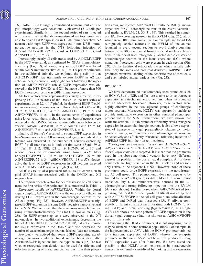
1H). AdNSEEGFP largely transduced neurons, but cells ofglial morphology were occasionally observed (2–3 cells perexperiment). Similarly, in the second series of rats injectedwith lower titers of the above-mentioned vectors, none wasable to drive EGFP expression in DBH-immunoreactive A2neurons, although EGFP was expressed in non-DBH-immu-noreactive neurons in the NTS following injection ofAdSyn1EGFP-WHE (22 � 7), AdT�1EGFP (21 � 11), andAdNSEEGFP (19 � 7).
Interestingly, nearly all cells transduced by AdHCMVEGFPin the NTS were glial, as confirmed by GFAP immunohisto-chemistry (Fig. 1I), although, very rarely, EGFP was foundwithin NeuN-immunoreactive cells (3 � 1.5 per experiment).In two additional animals, we explored the possibility thatAdHCMVEGFP may transiently express EGFP in A2 cat-echolaminergic neurons. Forty-eight hours following the injec-tion of AdHCMVEGFP, robust EGFP expression was ob-served in the NTS, DMNX, and XII, but none of more than 300EGFP-fluorescent cells was DBH immunoreactive.
All four vectors were approximately equally effective in ex-pressing EGFP in neurons of the DMNX. In the first series ofexperiments using 2.2 � 109 pfu/ml, the density of EGFP (NeuN-immunoreactive) neurons was as follows: AdSyn1EGFP-WHE,21 � 3; AdT�1EGFP, 24 � 6; AdNSEEGFP, 19 � 8; andAdHCMVEGFP, 11 � 1. In the second series of experimentsusing lower vector titers, slightly lower numbers of neurons wereobserved in the DMNX without striking differences between thevectors: AdSyn1EGFP-WHE, 8 � 7; AdT�1EGFP, 17 � 14;AdNSEEGFP, 7 � 4; and AdHCMVEGFP, 8 � 4.
Finally, all four AVV resulted in strong EGFP expression inNeuN-immunoreactive XII neurons, and again, no radical dif-ference was observed in the number of neurons expressingEGFP for all four vectors in both the first series (Syn1, 89 �14; T�1, 84 � 2; NSE, 121 � 19; HCMV, 80 � 16) andsecond series of experiments using lower AVV titers(AdSyn1EGFP-WHE, 107 � 30; AdT�1EGFP, 107 � 30;AdNSEEGFP, 72 � 34; AdHCMVEGFP, 118 � 57). Notice-ably, the level of EGFP expression in XII neurons targetedwith AdHCMVEGFP was very high (Fig. 1A).
AdHCMVEGFP also produced robust EGFP expression inglial (GFAP-immunoreactive) cells in the DMNX and XIImotonucleus.
The tropism of each vector for neurons (based on cell countsfrom the first series of experiments) is summarized in Table 1.
Expression profile of AdPRSx8EGFP. Within the dorsalvagal complex, AdPRSx8EGFP was the only vector efficient indriving EGFP expression in the noradrenergic neurons of theA2 cell group (Fig. 2A). However, AdPRSx8EGFP also trig-gered EGFP expression in some DBH-negative neurons ventralto the NTS. We confirmed that these neurons were cholinergicusing immunohistochemistry for ChAT in two animals (Fig.2B). No EGFP-expressing cells were observed in the XIImotonucleus. In two additional experiments, decreasing thetiter of AdPRSx8EGFP 10-fold to 2.2 � 108, did not eliminatethe EGFP expression in the DMNX and also decreased thenumber of catecholaminergic neurons labeled (data not shown).
Previously we noticed that some EGFP-expressing norad-renergic neurons could be found in the brain stem afterAdPRSx8EGFP injections into the hypothalamus (15). To testwhether retrograde transduction can be used for efficient andselective targeting of noradrenergic neurons from their projec-
tion areas, we injected AdPRSx8EGFP into the IML (a knowntarget area for C1 adrenergic neurons in the rostral ventrolat-eral medulla, RVLM; 24, 30, 31, 39). This resulted in numer-ous EGFP-expressing neurons in the RVLM (Fig. 2C), all ofwhich were DBH immunoreactive. For example, we found 156retrogradely labeled neurons in the RVLM of one animal(counted in every second section to avoid double countingbetween 0 to 800 �m caudal from the facial nucleus). Injec-tions in the dorsal horn retrogradely labeled dense clusters ofnoradrenergic neurons in the locus coeruleus (LC), wherenumerous fluorescent cells were present in each section (Fig.2D). Unlike traditional retrograde tracers which usually onlylabel only the soma and proximal dendrites, AdPRSx8EGFPproduced extensive labeling of the dendritic tree of most cellsand even labeled axonal varicosities (Fig. 2E).
DISCUSSION
We have demonstrated that commonly used promoters suchas HCMV, Syn1, NSE, and T�1 are unable to drive transgeneexpression in catecholaminergic neurons, when incorporatedinto an adenoviral backbone. However, these vectors werehighly effective in the two adjacent groups of cholinergicmotor neurons. Moreover, HCMV was apparently unable toprovide sustainable expression in other neuronal phenotypespresent within the NTS. Furthermore, we have shown thatwhile the artificial PRSx8 promoter efficiently drives transgeneexpression in catecholaminergic neurons, it also allows expres-sion of transgene in vagal preganglionic cholinergic motorneurons. Finally, we found that catecholaminergic neurons canbe selectively and efficiently transduced, following injection ofAdPRSx8EGFP into their target locations.
Transgene expression driven by AdHCMVEGFP,AdSyn1EGFP-WHE, AdT�1EGFP, and AdNSE-EGFP in thedorsal vagal complex is targeted. It is clear that the promotersused in the above-mentioned vectors have very differentexpression profiles in the dorsal vagal complex. All of theseconstructs are highly active in the XII nucleus and reason-ably active in the adjacent DMNX. However, none of thesepromoters could drive EGFP expression in the noradrener-gic A2 cell group. This phenomenon does not appear to belimited to the A2 cell group, as AdHCMVEGFP also did nottransduce any DBH-immunoreactive neurons in the C1adrenergic cell group following injection into the RVLM(data not shown). Furthermore, when AdHCMVDsRed (ex-pressing red coral fluorescent protein DsRed) was coinjectedwith AdPRSx8EGFP in the A6 cell group, no colocalizationof EGFP and DsRed was observed (15). Finally, a com-pletely different construct incorporating both HCMV (driv-ing EGFP) and PRSx8 (driving �-galactosidase) as a singleAVV (12) shows the same pattern of EGFP expression in thedorsal vagal complex (data not shown) as AdHCMVEGFPused in this study.
Concerning the HCMV promoter, it is not surprising that itmay be silenced in some neuronal populations. For example, inthe hippocampus, an AVV with the HCMV promoter only ledto a transient expression of EGFP, whereas the synapsinpromoter in the same AVV backbone still produced strongEGFP expression even after 9 mo (9). We have tested thepossibility that HCMV-driven expression in noradrenergicneurons is extremely short-lived by looking at the expression
167ADENOVIRAL TARGETING OF BRAIN STEM CARDIOVASCULAR NUCLEI
Physiol Genomics • VOL 20 • www.physiolgenomics.org
on June 14, 2005 physiolgenom
ics.physiology.orgD
ownloaded from
168 ADENOVIRAL TARGETING OF BRAIN STEM CARDIOVASCULAR NUCLEI
Physiol Genomics • VOL 20 • www.physiolgenomics.org
on June 14, 2005 physiolgenom
ics.physiology.orgD
ownloaded from

pattern after 48 h. Despite the very high overall level ofexpression, no DBH-positive neurons could be found, indicat-ing that the mechanisms which suppress HCMV activity inthese cells are already active at that stage. Moreover, not onlydo the catecholaminergic neurons appear to be resistant toHCMV-driven expression, but also so do other neuronal phe-notypes abundant in NTS, such as glutamatergic and GABAer-gic interneurons. Indeed, only a few EGFP-fluorescent NeuN-positive cells (e.g., neurons) were found in NTS of AdHCM-VEGFP-injected rats. Some authors have concluded thatHCMV preferentially expresses transgene in glia over neurons(9, 17, 18), but our study demonstrates that this is not neces-sarily the case. In fact HCMV was very active in XII motoneu-rons, whereas in the NTS, most of the transduced cells were ofglial morphology. Moreover, it appears that in several previ-ously published studies where HCMV was successfully used toexpress transgenes in neurons, the transduced areas containedcholinergic neurons (13, 22). The reasons for this selectiveexpression in glia and certain neural phenotypes are not knownbut might be related to the activity of the CREB pathway inthese phenotypes (for further discussion see Ref 15). Kuglerand colleagues (18) have also suggested that AVV-transducedglia may actively suppress the expression of transgene incertain neurons by secreting cytokines. In a more recent study,it was demonstrated that a very rapid shutdown of HCMV-driven transgene expression in skeletal muscle was due to theextensive methylation of this promoter (3), another possibilitythat has to be investigated.
Consistent with previously published data, Syn1- and T�1-driven EGFP expression appeared to be restricted to cells ofneuronal morphology (7, 18, 33, 42), whereas the NSE pro-moter also allowed expression in cells of glial morphology (17,18). It was more surprising to discover that all three neuronal-specific promoters were unable to drive EGFP expression incatecholaminergic neurons, at least not to levels detectable byspectral confocal microscopy (on the basis of our previousstudies, we estimate the threshold of detection in these studiesto be 100–300 nm of EGFP) (37). It is known, for example,that synapsin 1 is not expressed in all neuronal types in theretina (21) and in the thalamus (40). Thus it is possible that thisgene is inactive in catecholaminergic neurons, and thereforethe same pattern of transcriptional control could apply to theexogenously introduced proximal promoter sequence used inthe AVV. It is more difficult to explain why the T�1 and NSEpromoters are inactive in catecholaminergic neurons. Bothproteins are believed to ubiquitously expressed in neurons (15).However, no study has specifically addressed whether indeed
all neuronal phenotypes express these proteins nor whetherthey are colocalized with markers of catecholaminergic neu-rons. In addition, if the lack of expression in some phenotypesis due to the active suppression of viral transgenes, as men-tioned above in connection with HCMV, then it could be thatSyn1-, T�1-, NSE-containing expression cassettes can also besilenced in catecholaminergic neurons. Finally, it is also pos-sible that the sequences used as promoters in these vectors areinsufficient to drive expression in some neuronal populationssuch as catecholaminergic neurons. Whether the same is truefor other neuronal phenotypes such as GABAergic, serotoner-gic, dopaminergic neurons etc. remains to be established.
Targeting of catecholaminergic neurons usingAdPRSx8EGFP. The PRSx8 promoter is a synthetic constructbased on the binding motif for the transcription factors Phox2a/2b, found in the DBH promoter (12). These transcriptionfactors are critical for the expression of the catecholaminergicphenotype (23, 26, 34). The earlier published data consistentlydemonstrated that the PRSx8 promoter was able to efficientlydrive expression in noradrenergic neurons in the LC (12),where essentially all EGFP-expressing neurons were DBHimmunoreactive (15). However, when the same construct wasinjected into the dorsal vagal complex where catecholaminer-gic neurons are adjacent to cholinergic motoneurons, the latteralso expressed EGFP. This is not entirely surprising: whilethese neurons release a different neurotransmitter in the post-natal brain, they are believed to be ontogenetically related tocatecholaminergic neurons and are also known to expressPhox2a/2b (38). We attempted to avoid EGFP expression incholinergic neurons by altering the AVV titer, but this ap-
Table 1. Activity of AVV containing the promoters HCMV,Syn1, T�1, and NSE, in neurons (NeuN-immunoreactive) ofthe NTS, DMNX, and XII motonucleus
HCMV Syn1 T�1 NSE
NTS (excluding A2 neurons) DMNX XII
The cell counts are based on data obtained in animals injected with 2.2 �109 pfu/ml. Plus symbol () indicates that the promoter was able to induceEGFP expression in NeuN-immunoreactive cells: � 0–10 cells, �10–30 cells, � 30–50 cells, � 50–80 cells, and �80 cells per animal. AAV, adenoviral vectors; HCMV, human cytomegalo-virus; Syn1, synapsin-1; T�1, tubulin �1; NSE, neuron-specific enolase;EGFP, enhanced green fluorescent protein; NTS, nucleus tractus solitarius;DMNX, dorsal motonucleus of the vagus; XII, hypoglossal.
Fig. 1. Expression profile of AdHCMVEGFP, AdSyn1EGFP-WHE, AdT�1EGFP, and AdNSEEGFP, following injection into the dorsal vagal complex. A:low-power view of the dorsal vagal complex area injected with AdHCMVEGFP. Although no dopamine �-hydroxylase (DBH)-positive (red) A2 neuronsexpressed enhanced green fluorescent protein (EGFP) and no EGFP-expressing neurons of dorsal motonucleus of the vagus (DMNX) are visible at thismagnification, there is robust EGFP expression in the hypoglossal nucleus (XII). Axons of the XII nucleus can be seen traversing toward the ventral surface ofthe medulla. AdSyn1EGFP-WHE, AdT�1EGFP, and AdNSEEGFP were equally efficient in transducing XII motor neurons. B: EGFP expression in the dorsalmotonucleus of the vagus (DMNX) was evident following injection of AdHCMVEGFP, AdSyn1EGFP-WHE, AdT�1EGFP, and AdNSEEGFP into the dorsalvagal complex. In this example, AdNSEEGFP was used. EGFP-expressing neurons (green) of the DMNX can be clearly seen, dorsal to the XII nucleus (alsoexpressing EGFP) and ventral to the DBH-immunoreactive (red) A2 neurons. The red box on the cartoon in B (left) indicates the area and orientation of the imagein the right and in subsequent images. C–H: following injection into the dorsal vagal complex, AdHCMVEGFP (C), AdSyn1EGFP-WHE (D), AdT�1EGFP (E),and AdNSEEGFP (F) failed to induce EGFP expression (green) in DBH-immunoreactive (red) A2 neurons, as indicated by the lack of colocalization of EGFPand DBH. Despite the lack of expression of EGFP in catecholaminergic neurons, the neuronal promoters did express EGFP in other neuronal types as indicatedby the colocalization (yellow) of NeuN (red) and EGFP (green, G; AdT�1EGFP in this example). Adenoviral vectors (AVV) with neuron-specific promotersdid not express EGFP in glial cells, as indicated by the lack of colocalization of EGFP and glial fibrillary acidic protein (GFAP; H). AdHCMVEGFP largelyproduced EGFP expression in glia in the nucleus tractus solitarius (NTS), as indicated by the colocalization (yellow) of EGFP (green) with GFAP (red, I). Imagesare merged confocal projection stacks (40–50 �m) or single confocal planes (G and H). Scale bars are 20 �m.
169ADENOVIRAL TARGETING OF BRAIN STEM CARDIOVASCULAR NUCLEI
Physiol Genomics • VOL 20 • www.physiolgenomics.org
on June 14, 2005 physiolgenom
ics.physiology.orgD
ownloaded from

proach did not provide the desired degree of specificity, sug-gesting a fairly high level of Phox2 expression in the DMNX.This unsolicited expression in cholinergic neurons that can befound adjacent to most of the catecholaminergic cell groupsmay complicate experimentation with PRSx8-based constructsin vivo.
Previously we noticed that when AdPRSx8EGFP was in-jected in the hypothalamus, some retrogradely transduced neu-rons were found in the brain stem (15), suggesting that norad-
renergic neurons may be transduced via retrograde route, fromtheir projection areas. The present study proves the efficacy ofthis approach: indeed, numerous catecholaminergic cells werefound at the expected locations. The number of cells observedin this study following injection of AdPRSx8EGFP into theIML is comparable to that observed following retrogradetracing using cholera toxin �-chain (e.g., see Ref. 28). More-over, the present experiments show that retrograde targetingmay be used to alter the function of these cells as it allows high
Fig. 2. Expression profile of AdPRSx8EGFP, in the dorsal vagal complex. A: following injection of AdPRSx8EGFP into the dorsal vagal complex,EGFP-expressing neurons (green, Ai) were partially colocalized (Aii) with DBH-immunoreactive A2 neurons (red, Aiii). The remaining EGFP-expressing neuronsappeared to belong to the cholinergic population of the DMNX. B: the cholinergic phenotype of these cells was confirmed in separate studies where EGFPexpression (green, Bi) was colocalized (Bii) with choline acetyltransferase (ChAT) immunoreactivity (red, Biii). C–E: selective retrograde transduction ofcatecholaminergic neurons. Following injection of AdPRSx8EGFP into the intermediolateral cell column (IML) or locus coeruleus (LC), many EGFP-expressingadrenergic C1 neurons (green) are observed throughout the extent of the rostral ventrolateral medulla (RVLM, C) and LC (D, arrows), respectively. Many ofthe retrogradely labeled neurons were intensely fluorescent, and fine structural details became apparent including extensive dendritic arbor and axonal varicosities(E, also see inset, arrowheads indicate varicosities). A and B: merged confocal projection stacks (40–50 �m) or a single confocal plane (C) or a superimpositionof transmitted light and low-power confocal image (D). Scale bars are 20 �m (in D � 1 mm).
170 ADENOVIRAL TARGETING OF BRAIN STEM CARDIOVASCULAR NUCLEI
Physiol Genomics • VOL 20 • www.physiolgenomics.org
on June 14, 2005 physiolgenom
ics.physiology.orgD
ownloaded from
levels of transgene expression, as judged from the brightness ofEGFP fluorescence (Figs 2, C–E). Obviously, this approachwill work best after injections in the areas where there is a highconcentration of catecholaminergic terminals. For example,injection of AdPRSx8EGFP into the IML allowed selectivetargeting of C1 adrenergic neurons in the RVLM projecting tothat area. Injections of the same construct into the dorsal hornof the lumbar spinal cord selectively transduced another im-portant noradrenergic projection, which originates from theLC. Interestingly, when the PRSx8 promoter was incorporatedinto a lentiviral backbone, no retrograde labeling of cat-echolaminergic neurons was observed (T. Lonergan, A. G.Teschemacher, and S. Kasparov, unpublished observation). Webelieve the retrograde approach can be used to study variousaspects of function of different populations of central cat-echolaminergic neurons and their contribution to disorderssuch as pathological hypertension.
In conclusion, this study demonstrates the need for carefulchoice of promoter for gene manipulation in brain stem auto-nomic nuclei, and probably other brain regions. It illustrates thefact that the “constitutive” or “pan-neuronal” promoters may infact have a specific pattern of expression and have strikinglydifferent activity profiles in different subsets of neurons evenwithin the same nucleus. Importantly, within the brain regiontested, the most commonly used promoter, HCMV, was onlyactive in cholinergic neurons. In the context of genetic manip-ulation of catecholaminergic neurons, to date PRSx8 turns outto be the only promoter active in these neurons, at least withinan AVV backbone. Selective up- or downregulation of genes inthese neurons via a retrograde route opens a whole new avenuefor studies of their role in central autonomic control and inhypertension.
GRANTS
This work has been supported by the Wellcome Trust (AL/069061), BritishHeart Foundation (RG/02/011), and the Biotechnology and Biological Sci-ences Research Council (7/JE616459).
REFERENCES
1. Amerena J and Julius S. The role of the autonomic nervous system inhypertension. Hypertens Res 18: 99–110, 1995.
2. Bett AJ, Haddara W, Prevec L, and Graham FL. An efficient andflexible system for construction of adenovirus vectors with insertions ordeletions in early regions 1 and 3. Proc Natl Acad Sci USA 91: 8802–8806,1994.
3. Brooks AR, Harkins RN, Wang P, Qian HS, Liu P, and Rubanyi GM.Transcriptional silencing is associated with extensive methylation of theCMV promoter following adenoviral gene delivery to muscle. J Gene Med6: 395–404, 2004.
4. Davrath LR, Goren Y, Pinhas I, Toledo E, and Akselrod S. Earlyautonomic malfunction in normotensive individuals with a genetic predis-position to essential hypertension. Am J Physiol Heart Circ Physiol 285:H1697–H1704, 2003. First published June 12, 2003; doi:10.1152/ajpheart.00208.2003.
5. Ferrari AU, Daffonchio A, Franzelli C, and Mancia G. Cardiac para-sympathetic hyperresponsiveness in spontaneously hypertensive rats. Hy-pertension 19: 653–657, 1992.
6. Friberg P, Karlsson B, and Nordlander M. Sympathetic and parasym-pathetic influence on blood pressure and heart rate variability in Wistar-Kyoto and spontaneously hypertensive rats. J Hypertens Suppl 6: S58–S60, 1988.
7. Gloster A, Wu W, Speelman A, Weiss S, Causing C, Pozniak C,Reynolds B, Chang E, Toma JG, and Miller FD. The T�1 �-tubulinpromoter specifies gene expression as a function of neuronal growth andregeneration in transgenic mice. J Neurosci 14: 7319–7330, 1994.
8. Glover CP, Bienemann AS, Heywood DJ, Cosgrave AS, and Uney JB.Adenoviral-mediated, high-level, cell-specific transgene expression: aSYN1-WPRE cassette mediates increased transgene expression with noloss of neuron specificity. Mol Ther 5: 509–516, 2002.
9. Glover CP, Bienemann AS, Hopton M, Harding TC, Kew JN, andUney JB. Long-term transgene expression can be mediated in the brain byadenoviral vectors when powerful neuron-specific promoters are used.J Gene Med 5: 554–559, 2003.
10. Graham FL and Prevec L. Methods for construction of adenovirusvectors. Mol Biotechnol 3: 207–220, 1995.
11. Hirooka Y, Sakai K, Kishi T, Ito K, Shimokawa H, and Takeshita A.Enhanced depressor response to endothelial nitric oxide synthase genetransfer into the nucleus tractus solitarii of spontaneously hypertensiverats. Hypertens Res Clin Exp 26: 325–331, 2003.
12. Hwang DY, Carlezon WA Jr, Isacson O and Kim KS. A high-efficiency synthetic promoter that drives transgene expression selectivelyin noradrenergic neurons. Hum Gene Ther 12: 1731–1740, 2001.
13. Irnaten M, Walwyn WM, Wang J, Venkatesan P, Evans C, Chang KS,Andresen MC, Hales TG, and Mendelowitz D. Pentobarbital enhancesGABAergic neurotransmission to cardiac parasympathetic neurons, whichis prevented by expression of GABAA � subunit. Anesthesiology 97:717–724, 2002.
14. Ito K, Hirooka Y, Kishi T, Kimura Y, Kaibuchi K, Shimokawa H, andTakeshita A. Rho/Rho-kinase pathway in the brainstem contributes tohypertension caused by chronic nitric oxide synthase inhibition. Hyper-tension 43: 156–162, 2004.
15. Kasparov S, Teschemacher AG, Hwang DY, Kim KS, Lonergan T,and Paton JFR. Viral Vectors as Tools for Studies of Central Cardiovas-cular Control. Prog Biophys Mol Biol 84: 251–277, 2004.
16. Kishi T, Hirooka Y, Kimura Y, Sakai K, Ito K, Shimokawa H, andTakeshita A. Overexpression of eNOS in RVLM improves impairedbaroreflex control of heart rate in SHRSP. Rostral ventrolateral medullaStroke-prone spontaneously hypertensive rats. Hypertension 41: 255–260,2003.
17. Kugler S, Kilic E, and Bahr M. Human synapsin 1 gene promoterconfers highly neuron-specific long-term transgene expression from anadenoviral vector in the adult rat brain depending on the transduced area.Gene Ther 10: 337–347, 2003.
18. Kugler S, Meyn L, Holzmuller H, Gerhardt E, Isenmann S, Schulz JB,and Bahr M. Neuron-specific expression of therapeutic proteins: evalu-ation of different cellular promoters in recombinant adenoviral vectors.Mol Cell Neurosci 17: 78–96, 2001.
19. Lonergan T, Goodchild AK, Christie MJ, and Pilowsky PM. Presyn-aptic delta opioid receptors differentially modulate rhythm and patterngeneration in the ventral respiratory group of the rat. Neuroscience 121:959–973, 2003.
20. Lu D, Yu K, Paddy MR, Rowland NE, and Raizada MK. Regulation ofnorepinephrine transport system by angiotensin II in neuronal cultures ofnormotensive and spontaneously hypertensive rat brains. Endocrinology137: 763–772, 1996.
21. Mandell JW, Czernik AJ, De Camilli P, Greengard P, and Townes-Anderson E. Differential expression of synapsins I and II among ratretinal synapses. J Neurosci 12: 1736–1749, 1992.
22. Mohan RM, Heaton DA, Danson EJ, Krishnan SP, Cai S, ChannonKM, and Paterson DJ. Neuronal nitric oxide synthase gene transferpromotes cardiac vagal gain of function. Circ Res 91: 1089–1091, 2002.
23. Morin X, Cremer H, Hirsch MR, Kapur RP, Goridis C, and BrunetJF. Defects in sensory and autonomic ganglia and absence of locuscoeruleus in mice deficient for the homeobox gene Phox2a. Neuron 18:411–423, 1997.
24. Palkovits M and Brownstein MJ. Catecholamines in the dentral nervoussystem. In: Handbook of Experimental Pharmacology, edited by Tren-delenburg U and Weiner N. New York: Springer-Verlag, 1989, p. 1–26.
25. Paton JFR, Deuchars J, Ahmad Z, Wong LF, Murphy D, and Kas-parov S. Adenoviral vector demonstrates that angiotensin II-induceddepression of the cardiac baroreflex is mediated by endothelial nitric oxidesynthase in the nucleus tractus solitarii of the rat. J Physiol 531: 445–458,2001.
26. Pattyn A, Goridis C, and Brunet JF. Specification of the centralnoradrenergic phenotype by the homeobox gene Phox2b. Mol Cell Neu-rosci 15: 235–243, 2000.
171ADENOVIRAL TARGETING OF BRAIN STEM CARDIOVASCULAR NUCLEI
Physiol Genomics • VOL 20 • www.physiolgenomics.org
on June 14, 2005 physiolgenom
ics.physiology.orgD
ownloaded from

27. Paxinos G and Watson C. The Rat Brain in Stereotaxic Coordinates.London: Academic, 1986.
28. Phillips JK, Goodchild AK, Dubey R, Sesiashvili E, Takeda M,Chalmers J, Pilowsky PM, and Lipski J. Differential expression ofcatecholamine biosynthetic enzymes in the rat ventrolateral medulla.J Comp Neurol 432: 20–34, 2001.
29. Reja V, Goodchild AK, and Pilowsky PM. Catecholamine-related geneexpression correlates with blood pressures in SHR. Hypertension 40:342–347, 2002.
30. Ross CA, Armstrong DM, Ruggiero DA, Pickel VM, Joh TH, and ReisDJ. Adrenaline neurons in the rostral ventrolateral medulla innervatethoracic spinal cord: a combined immunocytochemical and retrogradetransport demonstration. Neurosci Lett 25: 257–262, 1981.
31. Ruggiero DA, Cravo SL, Golanov E, Gomez R, Anwar M, and ReisDJ. Adrenergic and non-adrenergic spinal projections of a cardiovascular-active pressor area of medulla oblongata: quantitative topographic analy-sis. Brain Res 663: 107–120, 1994.
32. Sakai K, Hirooka Y, Matsuo I, Eshima K, Shigematsu H, ShimokawaH, and Takeshita A. Overexpression of eNOS in NTS causes hypotensionand bradycardia in vivo. Hypertension 36: 1023–1028, 2000.
33. Schoch S, Cibelli G, and Thiel G. Neuron-specific gene expression ofsynapsin I. Major role of a negative regulatory mechanism. J Biol Chem271: 3317–3323, 1996.
34. Seo H, Hong SJ, Guo S, Kim HS, Kim CH, Hwang DY, Isacson O,Rosenthal A, and Kim KS. A direct role of the homeodomain proteinsPhox2a/2b in noradrenaline neurotransmitter identity determination.J Neurochem 80: 905–916, 2002.
35. Seyedabadi M, Goodchild AK, and Pilowsky PM. Differential role ofkinases in brain stem of hypertensive and normotensive rats. Hypertension38: 1087–1092, 2001.
36. Sinnayah P, Lindley TE, Staber PD, Davidson BL, Cassell MD, andDavisson RL. Targeted viral delivery of Cre recombinase induces condi-tional gene deletion in cardiovascular circuits of the mouse brain. Physiol
Genomics 18: 25–32, 2004. First published April 6, 2004; doi:10.1152/physiolgenomics.00048.2004.
37. Stokes CEL, Murphy D, Paton JFR, and Kasparov S. Dynamics of atransgene expression in acute rat brain slices transfected with adenoviralvectors. Exp Physiol 88: 459–466, 2003.
38. Tiveron MC, Hirsch MR, and Brunet JF. The expression pattern of thetranscription factor Phox2 delineates synaptic pathways of the autonomicnervous system. J Neurosci 16: 7649–7660, 1996.
39. Tucker DC, Saper CB, Ruggiero DA, and Reis DJ. Organization ofcentral adrenergic pathways: I. Relationships of ventrolateral medullaryprojections to the hypothalamus and spinal cord. J Comp Neurol 259:591–603, 1987.
40. Ullrich B and Sudhof TC. Differential distributions of novel synapto-tagmins: Comparison to synapsins. Neuropharmacology 34: 1371–1377,1995.
41. Waki H, Kasparov S, Wong LF, Murphy D, Shimizu T, and PatonJFR. Chronic inhibition of eNOS activity in NTS enhances baroreceptorreflex in conscious rats. J Physiol 546: 233–242, 2003.
42. Wang S, Wu H, Jiang J, Delohery TM, Isdell F, and Goldman SA.Isolation of neuronal precursors by sorting embryonic forebrain trans-fected with GFP regulated by the T alpha 1 tubulin promoter. NatBiotechnol 16: 196–201, 1998.
43. Wong LF, Polson JW, Murphy D, Paton JF, and Kasparov S. Geneticand pharmacological dissection of pathways involved in the angiotensinII-mediated depression of baroreflex function. FASEB J 16: 1595–1601,2002.
44. Yang H and Raizada MK. Role of phosphatidylinositol 3-kinase inangiotensin II regulation of norepinephrine neuromodulation in brainneurons of the spontaneously hypertensive rat. J Neurosci 19: 2413–2423,1999.
45. Zimmerman MC, Lazartigues E, Lang JA, Sinnayah P, Ahmad IM,Spitz DR, and Davisson RL. Superoxide mediates the actions of angio-tensin II in the central nervous system. Circ Res 91: 1038–1045, 2002.
172 ADENOVIRAL TARGETING OF BRAIN STEM CARDIOVASCULAR NUCLEI
Physiol Genomics • VOL 20 • www.physiolgenomics.org
on June 14, 2005 physiolgenom
ics.physiology.orgD
ownloaded from