targeting cxcl12 from fap-expressing carcinoma- … · targeting cxcl12 from fap-expressing...
TRANSCRIPT
Targeting CXCL12 from FAP-expressing carcinoma-associated fibroblasts synergizes with anti–PD-L1immunotherapy in pancreatic cancerChristine Feiga,1, James O. Jonesa,1, Matthew Kramana,1, Richard J. B. Wellsa,1, Andrew Deonarineb, Derek S. Chana,Claire M. Connella, Edward W. Robertsa,2, Qi Zhaoc, Otavia L. Caballeroc, Sarah A. Teichmannd, Tobias Janowitza,Duncan I. Jodrella, David A. Tuvesona,3, and Douglas T. Fearona,4
aCancer Research UK Cambridge Institute, Li Ka Shing Centre, University of Cambridge, Cambridge CB2 0RE, United Kingdom; bMedical Research CouncilLaboratory of Molecular Biology, Cambridge CB2 0QH, United Kingdom; cLudwig Collaborative Laboratory, Department of Neurosurgery, The Johns HopkinsUniversity School of Medicine, Baltimore, MD 21231; and dEuropean Molecular Biology Laboratory-European Bioinformatics Institute, Wellcome TrustGenome Campus, Hinxton, Cambridge, CB10 1SD, United Kingdom
Contributed by Douglas T. Fearon, October 31, 2013 (sent for review September 7, 2013)
An autochthonous model of pancreatic ductal adenocarcinoma(PDA) permitted the analysis of why immunotherapy is ineffectivein this human disease. Despite finding that PDA-bearing mice hadcancer cell-specific CD8+ T cells, the mice, like human patients withPDA, did not respond to two immunological checkpoint antago-nists that promote the function of T cells: anti-cytotoxic T-lympho-cyte-associated protein 4 (α-CTLA-4) and α-programmed cell death1 ligand 1 (α-PD-L1). Immune control of PDA growth was achieved,however, by depleting carcinoma-associated fibroblasts (CAFs)that express fibroblast activation protein (FAP). The depletion ofthe FAP+ stromal cell also uncovered the antitumor effects ofα-CTLA-4 and α-PD-L1, indicating that its immune suppressive activ-ity accounts for the failure of these T-cell checkpoint antagonists.Three findings suggested that chemokine (C-X-C motif) ligand 12(CXCL12) explained the overriding immunosuppression by the FAP+
cell: T cells were absent from regions of the tumor containing cancercells, cancer cells were coated with the chemokine, CXCL12, and theFAP+ CAF was the principal source of CXCL12 in the tumor. Admin-istering AMD3100, a CXCL12 receptor chemokine (C-X-C motif) re-ceptor 4 inhibitor, induced rapid T-cell accumulation amongcancer cells and acted synergistically with α-PD-L1 to greatly di-minish cancer cells, which were identified by their loss of hetero-zygosity of Trp53 gene. The residual tumor was composed only ofpremalignant epithelial cells and inflammatory cells. Thus, a singleprotein, CXCL12, from a single stromal cell type, the FAP+ CAF,may direct tumor immune evasion in a model of human PDA.
KPC mouse | tumor stroma | T cell exclusion | tumor immunogenicity
Immunotherapy of cancer has made recent progress by focusingon overcoming T-cell immunological checkpoints with block-
ing monoclonal antibodies to cytotoxic T-lymphocyte associ-ated protein-4 (CTLA-4) and the programmed cell death 1/programmed cell death 1 ligand 1 (PD-1/PD-L1) receptor/ligandpair (1–7). Many patients, however, did not respond to theseimmunological checkpoint antagonists for reasons that are notunderstood. In particular, patients with pancreatic ductal adeno-carcinoma (PDA), the fourth most common cause of cancer-relateddeaths in the United States, had no objective responses to anti(α)-CTLA-4 (7) or α-PD-L1 monoclonal antibodies (5).A mesenchymal tumoral stromal cell that is present in almost
all human adenocarcinomas (8) and is identified by its expressionof the membrane protein, fibroblast activation protein (FAP),was shown recently to mediate immunosuppression in a trans-planted murine tumor model (9). Because FAP+ stromal cellsare present in human PDA (8), we wished to investigate whetherthe immunosuppressive activity of the murine FAP+ stromal cellmight be involved in the resistance of this cancer to immuno-therapy. We were able to carry out this analysis because of theavailability of the autochthonous LSL-KrasG12D/+;LSL-Trp53R172H/+;
Pdx-1-Cre (KPC) model of PDA (10). We find that this PDAmodelreplicates the resistance of human PDA to checkpoint antagonists,despite the presence of systemic anti-PDA immunity. The failureof immune surveillance is attributable to local immunosuppressionmediated by the FAP+ stromal cell, which comprises essentiallyall carcinoma-associated fibroblasts (CAFs). Immunosuppressionmanifests as exclusion of T cells from regions of the tumor con-taining cancer cells and involves the production of chemokine(C-X-C motif) ligand 12 (CXCL12) by FAP+ CAFs. Inhibitingchemokine (C-X-C motif) receptor 4 (CXCR4), a CXCL12 re-ceptor, promotes T-cell accumulation and synergizes with thecheckpoint antagonist, α-PD-L1, to cause cancer regression.
Results and DiscussionIn the KPC model, Cre-mediated expression of Trp53R172H andKrasG12D is targeted to the pancreas, causing the development of
Significance
Cancer immune evasion is well described. In some cases, thismay be overcome by enhancing T-cell responses. We show thatdespite the presence of antitumor T cells, immunotherapeuticantibodies are ineffective in a murine pancreatic cancer modelrecapitulating the human disease. Removing the carcinoma-associated fibroblast (CAF) expressing fibroblast activationprotein (FAP) from tumors permitted immune control of tumorgrowth and uncovered the efficacy of these immunothera-peutic antibodies. FAP+ CAFs are the only tumoral source ofchemokine (C-X-C motif) ligand 12 (CXCL12), and administeringAMD3100, an inhibitor of chemokine (C-X-C motif) receptor 4,a CXCL12 receptor, also revealed the antitumor effects of animmunotherapeutic antibody and greatly diminished cancercells. These findings may have wide clinical relevance becauseFAP+ cells are found in almost all human adenocarcinomas.
Author contributions: C.F., J.O.J., M.K., R.J.B.W., and D.T.F. designed research; C.F., J.O.J.,M.K., R.J.B.W., D.S.C., C.M.C., and E.W.R. performed research; Q.Z., O.L.C., T.J., and D.I.J.contributed new reagents/analytic tools; C.F., J.O.J., M.K., R.J.B.W., A.D., S.A.T., D.A.T.,and D.T.F. analyzed data; and C.F., J.O.J., R.J.B.W., and D.T.F. wrote the paper.
The authors declare no conflict of interest.
Freely available online through the PNAS open access option.
Data deposition: The data reported in this paper have been deposited in the Gene Ex-pression Omnibus (GEO) database, www.ncbi.nlm.nih.gov/geo (accession no. GSE42605).1C.F., J.O.J., M.K., and R.J.B.W. contributed equally to this work.2Present address: Department of Pathology, University of California, San Francisco,CA 94143-0511.
3Present address: Cold Spring Harbor Laboratory, Cold Spring Harbor, NY 11724.4To whom correspondence should be addressed. E-mail: [email protected].
This article contains supporting information online at www.pnas.org/lookup/suppl/doi:10.1073/pnas.1320318110/-/DCSupplemental.
20212–20217 | PNAS | December 10, 2013 | vol. 110 | no. 50 www.pnas.org/cgi/doi/10.1073/pnas.1320318110
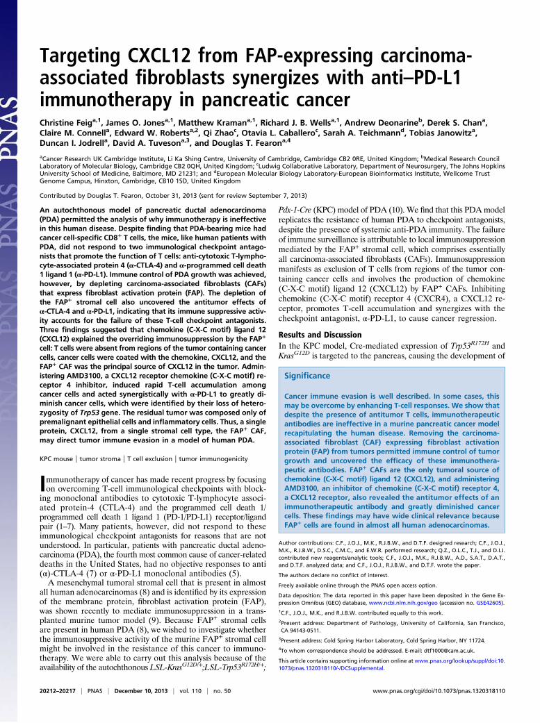
invasive and metastatic carcinoma that recapitulates many aspectsof human PDA, including the loss of heterozygosity (LOH) ofTrp53 in cancer cells but not in premalignant pancreatic intra-epithelial neoplasia (PanIN) (10). KPC mice with appropriatelysized tumors demonstrate consistent tumor growth, which per-mits robust analyses of experimental interventions (SI Appendix,Fig. S1). We examined whether blocking immunological check-points with α-CTLA-4 and α-PD-L1 would promote immunecontrol of the tumor. Administering these antibodies over 6 d tomice bearing PDA did not diminish the ∼80% increase in tumorvolume observed in mice receiving control IgG (Fig. 1A). Wedetermined whether this could be explained by the absence of animmune response to the PDA. Splenic CD8+ T cells from PDA-bearing and non–tumor-bearing mice were stimulated with tu-mor cells, and IFN-γ–secreting CD8+ T cells reporting antigenicstimulation were detected in an enzyme-linked immunosorbentspot (ELISpot) assay. Tumor cells induced significantly moreELISpots among CD8+ T cells from tumor-bearing mice than fromnon–tumor-bearing mice (Fig. 1B). The frequency of IFN-γ–secreting CD8+ T cells was the same when the stimulating tumorcells were from the T-cell donor or from another PDA-bearingmouse (Fig. 1C). An established PDA cell line also was stimula-tory, whereas dissociated cells from pancreata of LSL-KrasG12D/+;Pdx-1-Cre mice with premalignant PanIN lesions expressing onlyKrasG12D or from young KPC mice before the development ofcancer, were not (Fig. 1D and SI Appendix, Fig. S2). Therefore,PDA-bearing mice have a spontaneous adaptive immune responseto antigens that are shared by cancer cells from different PDAtumors, and the ineffectiveness of α-CTLA-4 and α-PD-L1 sug-gests an additional immunosuppressive mechanism.We considered the possibility that the FAP+ stromal cell
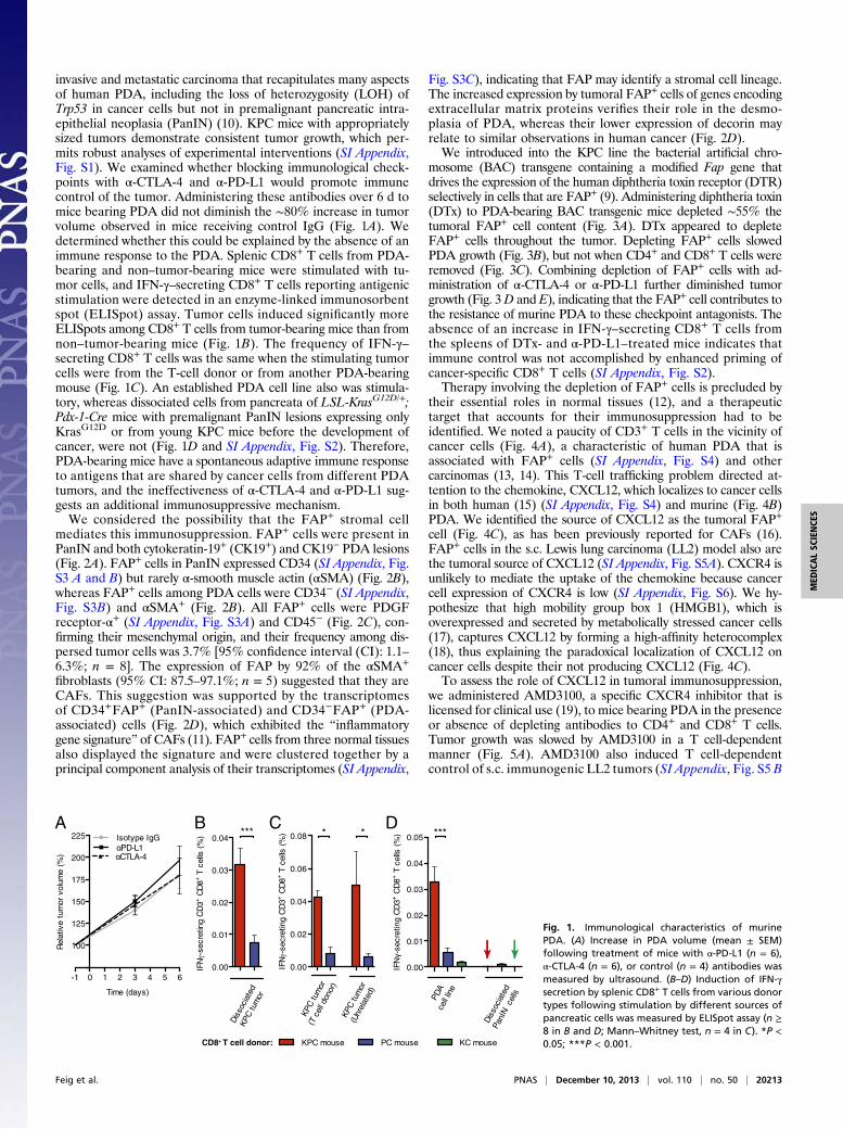
mediates this immunosuppression. FAP+ cells were present inPanIN and both cytokeratin-19+ (CK19+) and CK19− PDA lesions(Fig. 2A). FAP+ cells in PanIN expressed CD34 (SI Appendix, Fig.S3 A and B) but rarely α-smooth muscle actin (αSMA) (Fig. 2B),whereas FAP+ cells among PDA cells were CD34− (SI Appendix,Fig. S3B) and αSMA+ (Fig. 2B). All FAP+ cells were PDGFreceptor-α+ (SI Appendix, Fig. S3A) and CD45− (Fig. 2C), con-firming their mesenchymal origin, and their frequency among dis-persed tumor cells was 3.7% [95% confidence interval (CI): 1.1–6.3%; n = 8]. The expression of FAP by 92% of the αSMA+
fibroblasts (95% CI: 87.5–97.1%; n = 5) suggested that they areCAFs. This suggestion was supported by the transcriptomesof CD34+FAP+ (PanIN-associated) and CD34−FAP+ (PDA-associated) cells (Fig. 2D), which exhibited the “inflammatorygene signature” of CAFs (11). FAP+ cells from three normal tissuesalso displayed the signature and were clustered together by aprincipal component analysis of their transcriptomes (SI Appendix,
Fig. S3C), indicating that FAP may identify a stromal cell lineage.The increased expression by tumoral FAP+ cells of genes encodingextracellular matrix proteins verifies their role in the desmo-plasia of PDA, whereas their lower expression of decorin mayrelate to similar observations in human cancer (Fig. 2D).We introduced into the KPC line the bacterial artificial chro-
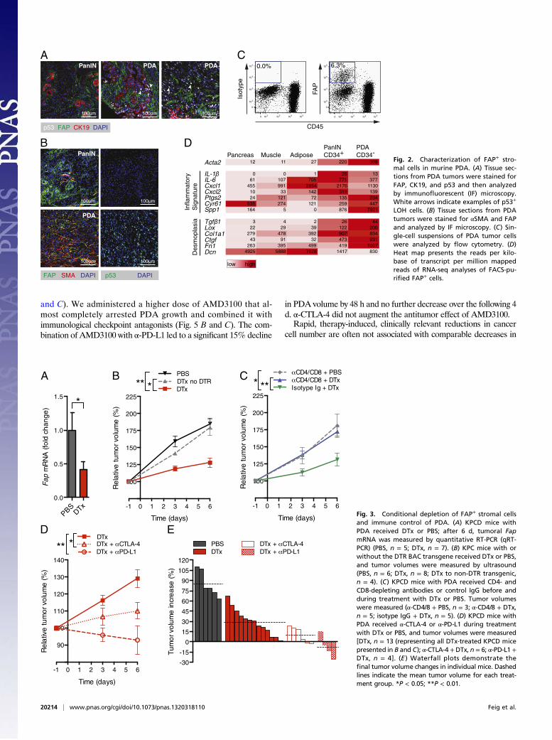
mosome (BAC) transgene containing a modified Fap gene thatdrives the expression of the human diphtheria toxin receptor (DTR)selectively in cells that are FAP+ (9). Administering diphtheria toxin(DTx) to PDA-bearing BAC transgenic mice depleted ∼55% thetumoral FAP+ cell content (Fig. 3A). DTx appeared to depleteFAP+ cells throughout the tumor. Depleting FAP+ cells slowedPDA growth (Fig. 3B), but not when CD4+ and CD8+ T cells wereremoved (Fig. 3C). Combining depletion of FAP+ cells with ad-ministration of α-CTLA-4 or α-PD-L1 further diminished tumorgrowth (Fig. 3D and E), indicating that the FAP+ cell contributes tothe resistance of murine PDA to these checkpoint antagonists. Theabsence of an increase in IFN-γ–secreting CD8+ T cells fromthe spleens of DTx- and α-PD-L1–treated mice indicates thatimmune control was not accomplished by enhanced priming ofcancer-specific CD8+ T cells (SI Appendix, Fig. S2).Therapy involving the depletion of FAP+ cells is precluded by
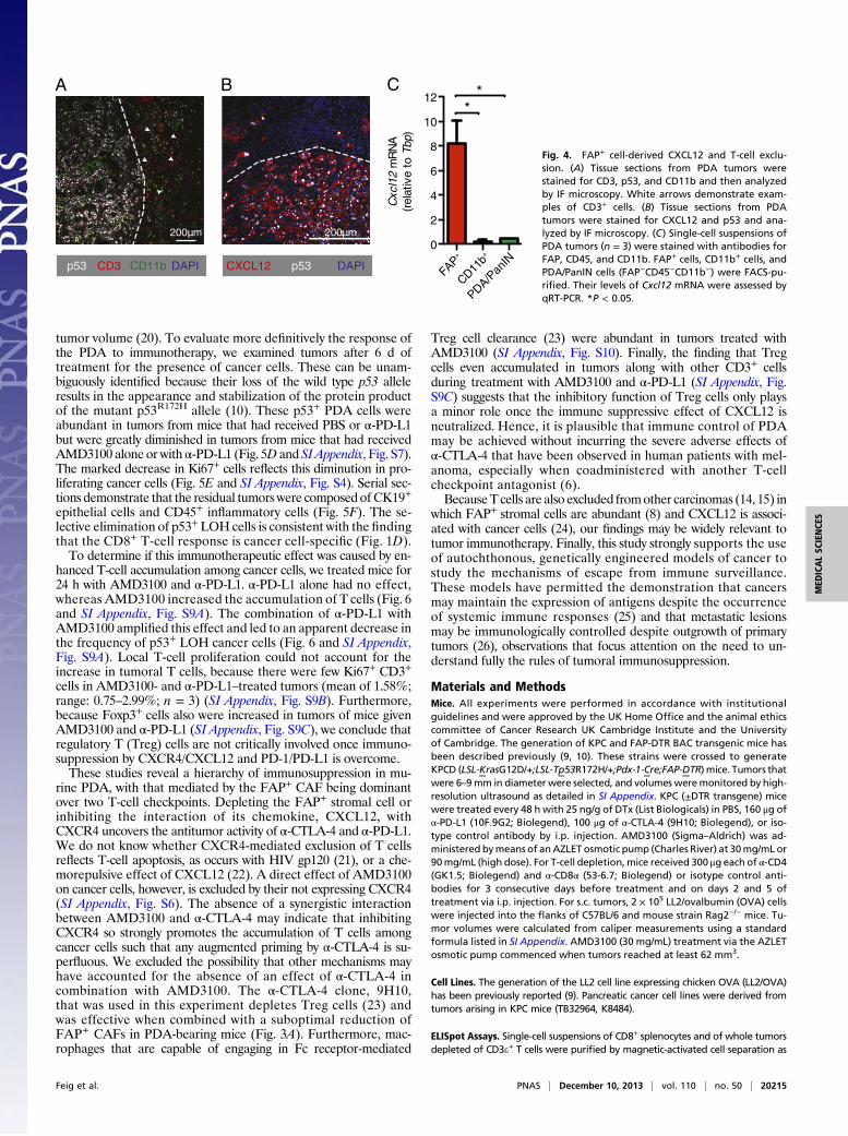
their essential roles in normal tissues (12), and a therapeutictarget that accounts for their immunosuppression had to beidentified. We noted a paucity of CD3+ T cells in the vicinity ofcancer cells (Fig. 4A), a characteristic of human PDA that isassociated with FAP+ cells (SI Appendix, Fig. S4) and othercarcinomas (13, 14). This T-cell trafficking problem directed at-tention to the chemokine, CXCL12, which localizes to cancer cellsin both human (15) (SI Appendix, Fig. S4) and murine (Fig. 4B)PDA. We identified the source of CXCL12 as the tumoral FAP+
cell (Fig. 4C), as has been previously reported for CAFs (16).FAP+ cells in the s.c. Lewis lung carcinoma (LL2) model also arethe tumoral source of CXCL12 (SI Appendix, Fig. S5A). CXCR4 isunlikely to mediate the uptake of the chemokine because cancercell expression of CXCR4 is low (SI Appendix, Fig. S6). We hy-pothesize that high mobility group box 1 (HMGB1), which isoverexpressed and secreted by metabolically stressed cancer cells(17), captures CXCL12 by forming a high-affinity heterocomplex(18), thus explaining the paradoxical localization of CXCL12 oncancer cells despite their not producing CXCL12 (Fig. 4C).To assess the role of CXCL12 in tumoral immunosuppression,
we administered AMD3100, a specific CXCR4 inhibitor that islicensed for clinical use (19), to mice bearing PDA in the presenceor absence of depleting antibodies to CD4+ and CD8+ T cells.Tumor growth was slowed by AMD3100 in a T cell-dependentmanner (Fig. 5A). AMD3100 also induced T cell-dependentcontrol of s.c. immunogenic LL2 tumors (SI Appendix, Fig. S5 B
-1 0 1 2 3 4 5 6
100
125
150
175
200
225
Time (days)
Rel
ativ
etu
mor
volu
me
(%)
Isotype IgGPD-L1CTLA-4
0.00
0.01
0.02
0.03
0.04
IFN
-sec
retin
gC
D3+
CD
8+T
cells
(%)
Diss
ocia
ted
KPC
tum
or
0.00
0.02
0.04
0.06
0.08
IFN
-sec
retin
gC
D3+
CD
8+T
cells
(%)
KPC
tum
or(T
cell
dono
r)KP
Ctu
mor
(Unr
elat
ed)
A C D
CD8+ T cell donor:
B
0.00
0.01
0.02
0.03
0.04
0.05
IFN
-sec
retin
gC
D3+
CD
8+T
cells
(%)
PDA
cell
line
Diss
ocia
ted
PanI
Nce
lls
*** * * ***
KPC mouse PC mouse KC mouse
Fig. 1. Immunological characteristics of murinePDA. (A) Increase in PDA volume (mean ± SEM)following treatment of mice with α-PD-L1 (n = 6),α-CTLA-4 (n = 6), or control (n = 4) antibodies wasmeasured by ultrasound. (B–D) Induction of IFN-γsecretion by splenic CD8+ T cells from various donortypes following stimulation by different sources ofpancreatic cells was measured by ELISpot assay (n ≥8 in B and D; Mann–Whitney test, n = 4 in C). *P <0.05; ***P < 0.001.
Feig et al. PNAS | December 10, 2013 | vol. 110 | no. 50 | 20213
MED
ICALSC
IENCE
S
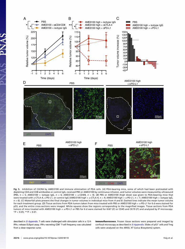
and C). We administered a higher dose of AMD3100 that al-most completely arrested PDA growth and combined it withimmunological checkpoint antagonists (Fig. 5 B and C). The com-bination of AMD3100 with α-PD-L1 led to a significant 15% decline
in PDA volume by 48 h and no further decrease over the following 4d. α-CTLA-4 did not augment the antitumor effect of AMD3100.Rapid, therapy-induced, clinically relevant reductions in cancer
cell number are often not associated with comparable decreases in
B
C
0 102 103 104 105
0
102
103
104
105 0.0%
0 102 103 104 105
0
102
103
104
105 6.3%
CD45
FAP
Isot
ype
FAP SMA DAPI p53 DAPI
PanIN
PDA
A
100 m 100 m
100 m 100 m
100 m 100 m 100 m
PanIN PDA PDA
p53 FAP CK19 DAPI
DPancreas
12 11 27 220 399
0 0 1 26 1361 107 705 771 377
455 991 2854 2176 113010 33 142 319 13924 121 72 135 234
535 274 121 259 447164 5 0 876 7621
3 4 2 26 4422 29 39 122 200
279 478 392 907 83443 91 32 473 931
263 395 499 419 10074925 5888 7828 1417 830
Muscle AdiposePanINCD34+
PDACD34-
Acta2
IL-6Cxcl1Cxcl2Ptgs2Cyr61Spp1
LoxCol1a1CtgfFn1Dcn
Infla
mm
ator
yS
igna
ture
Des
mop
lasi
a
low high
IL-1β
Tgf 1β
Fig. 2. Characterization of FAP+ stro-mal cells in murine PDA. (A) Tissue sec-tions from PDA tumors were stained forFAP, CK19, and p53 and then analyzedby immunofluorescent (IF) microscopy.White arrows indicate examples of p53+
LOH cells. (B) Tissue sections from PDAtumors were stained for αSMA and FAPand analyzed by IF microscopy. (C) Sin-gle-cell suspensions of PDA tumor cellswere analyzed by flow cytometry. (D)Heat map presents the reads per kilo-base of transcript per million mappedreads of RNA-seq analyses of FACS-pu-rified FAP+ cells.
-1 0 1 2 3 4 5 6
100
125
150
175
200
225
Time (days)
Re
lativ
e tu
mo
r vo
lum
e (%
)
DTx DTx no DTR **PBS
*B
-1 0 1 2 3 4 5 6
100
125
150
175
200
225
Time (days)
Re
lativ
e tu
mo
r vo
lum
e (%
)
CD4/CD8 + DTx Isotype Ig + DTx
CD4/CD8 + PBS
***C
-1 0 1 2 3 4 5 6
90
100
110
120
130
140
Time (days)
Re
lativ
e tu
mo
r vo
lum
e (%
)
DTx *** DTx + CTLA-4
DTx + PD-L1
D E
-30
-15
0
15
30
45
60
75
90
105
120
Tu
mo
r vo
lum
e in
cre
ase
(%)
DTxDTx + CTLA-4PBSDTx + PD-L1
A
PBSDTx
0.0
0.5
1.0
1.5
Fa
p m
RN
A (f
old
ch
an
ge
)
*
Fig. 3. Conditional depletion of FAP+ stromal cellsand immune control of PDA. (A) KPCD mice withPDA received DTx or PBS; after 6 d, tumoral FapmRNA was measured by quantitative RT-PCR (qRT-PCR) (PBS, n = 5; DTx, n = 7). (B) KPC mice with orwithout the DTR BAC transgene received DTx or PBS,and tumor volumes were measured by ultrasound(PBS, n = 6; DTx, n = 8; DTx to non-DTR transgenic,n = 4). (C) KPCD mice with PDA received CD4- andCD8-depleting antibodies or control IgG before andduring treatment with DTx or PBS. Tumor volumeswere measured (α-CD4/8 + PBS, n = 3; α-CD4/8 + DTx,n = 5; isotype IgG + DTx, n = 5). (D) KPCD mice withPDA received α-CTLA-4 or α-PD-L1 during treatmentwith DTx or PBS, and tumor volumes were measured[DTx, n = 13 (representing all DTx-treated KPCD micepresented in B and C); α-CTLA-4+DTx, n = 6; α-PD-L1 +DTx, n = 4]. (E ) Waterfall plots demonstrate thefinal tumor volume changes in individual mice. Dashedlines indicate the mean tumor volume for each treat-ment group. *P < 0.05; **P < 0.01.
20214 | www.pnas.org/cgi/doi/10.1073/pnas.1320318110 Feig et al.
tumor volume (20). To evaluate more definitively the response ofthe PDA to immunotherapy, we examined tumors after 6 d oftreatment for the presence of cancer cells. These can be unam-biguously identified because their loss of the wild type p53 alleleresults in the appearance and stabilization of the protein productof the mutant p53R172H allele (10). These p53+ PDA cells wereabundant in tumors from mice that had received PBS or α-PD-L1but were greatly diminished in tumors from mice that had receivedAMD3100 alone or with α-PD-L1 (Fig. 5D and SIAppendix, Fig. S7).The marked decrease in Ki67+ cells reflects this diminution in pro-liferating cancer cells (Fig. 5E and SI Appendix, Fig. S4). Serial sec-tions demonstrate that the residual tumorswere composed of CK19+
epithelial cells and CD45+ inflammatory cells (Fig. 5F). The se-lective elimination of p53+LOH cells is consistent with the findingthat the CD8+ T-cell response is cancer cell-specific (Fig. 1D).To determine if this immunotherapeutic effect was caused by en-
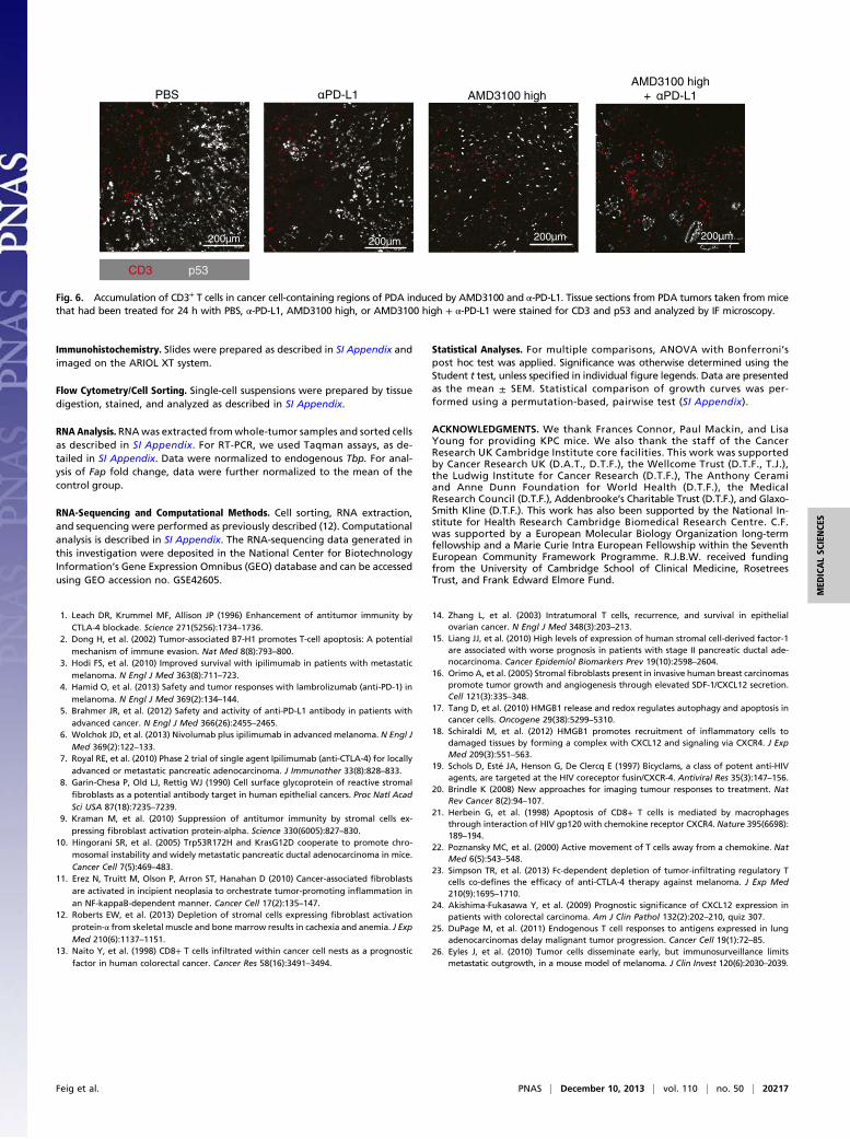
hanced T-cell accumulation among cancer cells, we treated mice for24 h with AMD3100 and α-PD-L1. α-PD-L1 alone had no effect,whereas AMD3100 increased the accumulation of T cells (Fig. 6and SI Appendix, Fig. S9A). The combination of α-PD-L1 withAMD3100 amplified this effect and led to an apparent decrease inthe frequency of p53+ LOH cancer cells (Fig. 6 and SI Appendix,Fig. S9A). Local T-cell proliferation could not account for theincrease in tumoral T cells, because there were few Ki67+ CD3+
cells in AMD3100- and α-PD-L1–treated tumors (mean of 1.58%;range: 0.75–2.99%; n = 3) (SI Appendix, Fig. S9B). Furthermore,because Foxp3+ cells also were increased in tumors of mice givenAMD3100 and α-PD-L1 (SI Appendix, Fig. S9C), we conclude thatregulatory T (Treg) cells are not critically involved once immuno-suppression by CXCR4/CXCL12 and PD-1/PD-L1 is overcome.These studies reveal a hierarchy of immunosuppression in mu-
rine PDA, with that mediated by the FAP+ CAF being dominantover two T-cell checkpoints. Depleting the FAP+ stromal cell orinhibiting the interaction of its chemokine, CXCL12, withCXCR4 uncovers the antitumor activity of α-CTLA-4 and α-PD-L1.We do not know whether CXCR4-mediated exclusion of T cellsreflects T-cell apoptosis, as occurs with HIV gp120 (21), or a che-morepulsive effect of CXCL12 (22). A direct effect of AMD3100on cancer cells, however, is excluded by their not expressing CXCR4(SI Appendix, Fig. S6). The absence of a synergistic interactionbetween AMD3100 and α-CTLA-4 may indicate that inhibitingCXCR4 so strongly promotes the accumulation of T cells amongcancer cells such that any augmented priming by α-CTLA-4 is su-perfluous. We excluded the possibility that other mechanisms mayhave accounted for the absence of an effect of α-CTLA-4 incombination with AMD3100. The α-CTLA-4 clone, 9H10,that was used in this experiment depletes Treg cells (23) andwas effective when combined with a suboptimal reduction ofFAP+ CAFs in PDA-bearing mice (Fig. 3A). Furthermore, mac-rophages that are capable of engaging in Fc receptor-mediated
Treg cell clearance (23) were abundant in tumors treated withAMD3100 (SI Appendix, Fig. S10). Finally, the finding that Tregcells even accumulated in tumors along with other CD3+ cellsduring treatment with AMD3100 and α-PD-L1 (SI Appendix, Fig.S9C) suggests that the inhibitory function of Treg cells only playsa minor role once the immune suppressive effect of CXCL12 isneutralized. Hence, it is plausible that immune control of PDAmay be achieved without incurring the severe adverse effects ofα-CTLA-4 that have been observed in human patients with mel-anoma, especially when coadministered with another T-cellcheckpoint antagonist (6).BecauseTcells are also excluded fromother carcinomas (14, 15) in
which FAP+ stromal cells are abundant (8) and CXCL12 is associ-ated with cancer cells (24), our findings may be widely relevant totumor immunotherapy. Finally, this study strongly supports the useof autochthonous, genetically engineered models of cancer tostudy the mechanisms of escape from immune surveillance.These models have permitted the demonstration that cancersmay maintain the expression of antigens despite the occurrenceof systemic immune responses (25) and that metastatic lesionsmay be immunologically controlled despite outgrowth of primarytumors (26), observations that focus attention on the need to un-derstand fully the rules of tumoral immunosuppression.
Materials and MethodsMice. All experiments were performed in accordance with institutionalguidelines and were approved by the UK Home Office and the animal ethicscommittee of Cancer Research UK Cambridge Institute and the Universityof Cambridge. The generation of KPC and FAP-DTR BAC transgenic mice hasbeen described previously (9, 10). These strains were crossed to generateKPCD (LSL-KrasG12D/+;LSL-Tp53R172H/+;Pdx-1-Cre;FAP-DTR) mice. Tumors thatwere 6–9mm in diameter were selected, and volumes weremonitored by high-resolution ultrasound as detailed in SI Appendix. KPC (±DTR transgene) micewere treated every 48 h with 25 ng/g of DTx (List Biologicals) in PBS, 160 μg ofα-PD-L1 (10F.9G2; Biolegend), 100 μg of α-CTLA-4 (9H10; Biolegend), or iso-type control antibody by i.p. injection. AMD3100 (Sigma–Aldrich) was ad-ministered bymeans of anAZLET osmotic pump (Charles River) at 30mg/mL or90mg/mL (high dose). For T-cell depletion,mice received 300 μg each of α-CD4(GK1.5; Biolegend) and α-CD8α (53-6.7; Biolegend) or isotype control anti-bodies for 3 consecutive days before treatment and on days 2 and 5 oftreatment via i.p. injection. For s.c. tumors, 2 × 10⁵ LL2/ovalbumin (OVA) cellswere injected into the flanks of C57BL/6 and mouse strain Rag2−/− mice. Tu-mor volumes were calculated from caliper measurements using a standardformula listed in SI Appendix. AMD3100 (30 mg/mL) treatment via the AZLETosmotic pump commenced when tumors reached at least 62 mm3.
Cell Lines. The generation of the LL2 cell line expressing chicken OVA (LL2/OVA)has been previously reported (9). Pancreatic cancer cell lines were derived fromtumors arising in KPC mice (TB32964, K8484).
ELISpot Assays. Single-cell suspensions of CD8+ splenocytes and of whole tumorsdepleted of CD3e+ T cells were purified by magnetic-activated cell separation as
FAP+
CD11b+
PDA/Pan
IN0
2
4
6
8
10
12
Cxc
l12
mR
NA
(rel
ativ
eto
Tbp)
BA
DAPICD3p53 CD11b DAPICXCL12 p53
200 m200 m
C*
*
Fig. 4. FAP+ cell-derived CXCL12 and T-cell exclu-sion. (A) Tissue sections from PDA tumors werestained for CD3, p53, and CD11b and then analyzedby IF microscopy. White arrows demonstrate exam-ples of CD3+ cells. (B) Tissue sections from PDAtumors were stained for CXCL12 and p53 and ana-lyzed by IF microscopy. (C) Single-cell suspensions ofPDA tumors (n = 3) were stained with antibodies forFAP, CD45, and CD11b. FAP+ cells, CD11b+ cells, andPDA/PanIN cells (FAP−CD45−CD11b−) were FACS-pu-rified. Their levels of Cxcl12 mRNA were assessed byqRT-PCR. *P < 0.05.
Feig et al. PNAS | December 10, 2013 | vol. 110 | no. 50 | 20215
MED
ICALSC
IENCE
S
described in SI Appendix. T cells were challenged with stimulator cells in a 12-hIFN-γ release ELISpot assay. IFN-γ–secreting CD8+ T-cell frequency was calculatedfrom a dose–response curve.
Immunofluorescence. Frozen tissue sections were prepared and imaged byconfocal microscopy as described in SI Appendix. Slides of p53+ cells and Tregcells were analyzed on the ARIOL XT (Leica Biosystems) system.
-45-30-15
0153045607590
105120135150
Tu
mo
rvo
lum
ein
cre
ase
(%)
PBSAMD3100 high + isotype IgGAMD3100 high + PD-L1
-1 0 1 2 3 4 5 6
100
125
150
175
200
225
Time (days)
Re
lativ
etu
mo
rvo
lum
e(%
)AMD3100 + isotype IgG
PBSAMD3100 + CD4/CD8
**
-1 0 1 2 3 4 5 6
80
85
90
95
100
105
110
115
Time (days)
Re
lativ
etu
mo
rvo
lum
e(%
)
AMD3100 high + PD-L1
AMD3100 high + isotype IgGAMD3100 high + CTLA-4
***
A B
PBSAMD3100 high
200µm
C
D
200µm
AMD3100 high AMD3100 high
200µm
Ki67 DAPI
PBSPBS
DAPIp53
1mm1mm
200µm
E F
200µm 200µm
DAPICK19 CD45
Fig. 5. Inhibition of CXCR4 by AMD3100 and immune elimination of PDA cells. (A) PDA-bearing mice, some of which had been pretreated withdepleting CD4 and CD8 antibodies or control IgG, received PBS or AMD3100 by continuous infusion, and tumor volumes were measured by ultrasound(PBS, n = 5; AMD3100 + isotype IgG, n = 6; AMD3100 + α-CD4/8, n = 4). (B) PBS or AMD3100 (high dose) was given to PDA-bearing mice thatwere treated with α-CTLA-4, α-PD-L1, or control IgG (AMD3100 high + α-CTLA-4, n = 4; AMD3100 high + α-PD-L1, n = 7; AMD3100 high + isotype IgG,n = 6). (C ) Waterfall plots present the final changes in tumor volumes in individual mice from A and B. Dashed lines indicate the mean tumor volumefor each treatment group. (D) Tissue sections from PDA tumors taken from mice treated with PBS or AMD3100 high + α-PD-L1 for 6 d were stained forp53, and the entire cross-sections were imaged. White squares show the regions corresponding to the magnified images. Tissue sections from PDAtumors of mice treated with AMD3100 high + α-PD-L1 or PBS for 6 d were stained for Ki67 (E ) or CD45 and CK19 (F ) and analyzed by IF microscopy.*P < 0.05; **P < 0.01.
20216 | www.pnas.org/cgi/doi/10.1073/pnas.1320318110 Feig et al.
Immunohistochemistry. Slides were prepared as described in SI Appendix andimaged on the ARIOL XT system.
Flow Cytometry/Cell Sorting. Single-cell suspensions were prepared by tissuedigestion, stained, and analyzed as described in SI Appendix.
RNA Analysis. RNAwas extracted fromwhole-tumor samples and sorted cellsas described in SI Appendix. For RT-PCR, we used Taqman assays, as de-tailed in SI Appendix. Data were normalized to endogenous Tbp. For anal-ysis of Fap fold change, data were further normalized to the mean of thecontrol group.
RNA-Sequencing and Computational Methods. Cell sorting, RNA extraction,and sequencing were performed as previously described (12). Computationalanalysis is described in SI Appendix. The RNA-sequencing data generated inthis investigation were deposited in the National Center for BiotechnologyInformation’s Gene Expression Omnibus (GEO) database and can be accessedusing GEO accession no. GSE42605.
Statistical Analyses. For multiple comparisons, ANOVA with Bonferroni’spost hoc test was applied. Significance was otherwise determined using theStudent t test, unless specified in individual figure legends. Data are presentedas the mean ± SEM. Statistical comparison of growth curves was per-formed using a permutation-based, pairwise test (SI Appendix).
ACKNOWLEDGMENTS. We thank Frances Connor, Paul Mackin, and LisaYoung for providing KPC mice. We also thank the staff of the CancerResearch UK Cambridge Institute core facilities. This work was supportedby Cancer Research UK (D.A.T., D.T.F.), the Wellcome Trust (D.T.F., T.J.),the Ludwig Institute for Cancer Research (D.T.F.), The Anthony Ceramiand Anne Dunn Foundation for World Health (D.T.F.), the MedicalResearch Council (D.T.F.), Addenbrooke’s Charitable Trust (D.T.F.), and Glaxo-Smith Kline (D.T.F.). This work has also been supported by the National In-stitute for Health Research Cambridge Biomedical Research Centre. C.F.was supported by a European Molecular Biology Organization long-termfellowship and a Marie Curie Intra European Fellowship within the SeventhEuropean Community Framework Programme. R.J.B.W. received fundingfrom the University of Cambridge School of Clinical Medicine, RosetreesTrust, and Frank Edward Elmore Fund.
1. Leach DR, Krummel MF, Allison JP (1996) Enhancement of antitumor immunity byCTLA-4 blockade. Science 271(5256):1734–1736.
2. Dong H, et al. (2002) Tumor-associated B7-H1 promotes T-cell apoptosis: A potentialmechanism of immune evasion. Nat Med 8(8):793–800.
3. Hodi FS, et al. (2010) Improved survival with ipilimumab in patients with metastaticmelanoma. N Engl J Med 363(8):711–723.
4. Hamid O, et al. (2013) Safety and tumor responses with lambrolizumab (anti-PD-1) inmelanoma. N Engl J Med 369(2):134–144.
5. Brahmer JR, et al. (2012) Safety and activity of anti-PD-L1 antibody in patients withadvanced cancer. N Engl J Med 366(26):2455–2465.
6. Wolchok JD, et al. (2013) Nivolumab plus ipilimumab in advanced melanoma. N Engl JMed 369(2):122–133.
7. Royal RE, et al. (2010) Phase 2 trial of single agent Ipilimumab (anti-CTLA-4) for locallyadvanced or metastatic pancreatic adenocarcinoma. J Immunother 33(8):828–833.
8. Garin-Chesa P, Old LJ, Rettig WJ (1990) Cell surface glycoprotein of reactive stromalfibroblasts as a potential antibody target in human epithelial cancers. Proc Natl AcadSci USA 87(18):7235–7239.
9. Kraman M, et al. (2010) Suppression of antitumor immunity by stromal cells ex-pressing fibroblast activation protein-alpha. Science 330(6005):827–830.
10. Hingorani SR, et al. (2005) Trp53R172H and KrasG12D cooperate to promote chro-mosomal instability and widely metastatic pancreatic ductal adenocarcinoma in mice.Cancer Cell 7(5):469–483.
11. Erez N, Truitt M, Olson P, Arron ST, Hanahan D (2010) Cancer-associated fibroblastsare activated in incipient neoplasia to orchestrate tumor-promoting inflammation inan NF-kappaB-dependent manner. Cancer Cell 17(2):135–147.
12. Roberts EW, et al. (2013) Depletion of stromal cells expressing fibroblast activationprotein-α from skeletal muscle and bone marrow results in cachexia and anemia. J ExpMed 210(6):1137–1151.
13. Naito Y, et al. (1998) CD8+ T cells infiltrated within cancer cell nests as a prognosticfactor in human colorectal cancer. Cancer Res 58(16):3491–3494.
14. Zhang L, et al. (2003) Intratumoral T cells, recurrence, and survival in epithelialovarian cancer. N Engl J Med 348(3):203–213.
15. Liang JJ, et al. (2010) High levels of expression of human stromal cell-derived factor-1are associated with worse prognosis in patients with stage II pancreatic ductal ade-nocarcinoma. Cancer Epidemiol Biomarkers Prev 19(10):2598–2604.
16. Orimo A, et al. (2005) Stromal fibroblasts present in invasive human breast carcinomaspromote tumor growth and angiogenesis through elevated SDF-1/CXCL12 secretion.Cell 121(3):335–348.
17. Tang D, et al. (2010) HMGB1 release and redox regulates autophagy and apoptosis incancer cells. Oncogene 29(38):5299–5310.
18. Schiraldi M, et al. (2012) HMGB1 promotes recruitment of inflammatory cells todamaged tissues by forming a complex with CXCL12 and signaling via CXCR4. J ExpMed 209(3):551–563.
19. Schols D, Esté JA, Henson G, De Clercq E (1997) Bicyclams, a class of potent anti-HIVagents, are targeted at the HIV coreceptor fusin/CXCR-4. Antiviral Res 35(3):147–156.
20. Brindle K (2008) New approaches for imaging tumour responses to treatment. NatRev Cancer 8(2):94–107.
21. Herbein G, et al. (1998) Apoptosis of CD8+ T cells is mediated by macrophagesthrough interaction of HIV gp120 with chemokine receptor CXCR4. Nature 395(6698):189–194.
22. Poznansky MC, et al. (2000) Active movement of T cells away from a chemokine. NatMed 6(5):543–548.
23. Simpson TR, et al. (2013) Fc-dependent depletion of tumor-infiltrating regulatory Tcells co-defines the efficacy of anti-CTLA-4 therapy against melanoma. J Exp Med210(9):1695–1710.
24. Akishima-Fukasawa Y, et al. (2009) Prognostic significance of CXCL12 expression inpatients with colorectal carcinoma. Am J Clin Pathol 132(2):202–210, quiz 307.
25. DuPage M, et al. (2011) Endogenous T cell responses to antigens expressed in lungadenocarcinomas delay malignant tumor progression. Cancer Cell 19(1):72–85.
26. Eyles J, et al. (2010) Tumor cells disseminate early, but immunosurveillance limitsmetastatic outgrowth, in a mouse model of melanoma. J Clin Invest 120(6):2030–2039.
PD-L1
CD3 p53
PBS AMD3100 highAMD3100 high
+
200 m200 m 200 m200 m
Fig. 6. Accumulation of CD3+ T cells in cancer cell-containing regions of PDA induced by AMD3100 and α-PD-L1. Tissue sections from PDA tumors taken from micethat had been treated for 24 h with PBS, α-PD-L1, AMD3100 high, or AMD3100 high + α-PD-L1 were stained for CD3 and p53 and analyzed by IF microscopy.
Feig et al. PNAS | December 10, 2013 | vol. 110 | no. 50 | 20217
MED
ICALSC
IENCE
S