targeting the expression of a putative rna ... · targeting the expression of a putative rna...
TRANSCRIPT

1
Thesis for the Master`s degree in Molecular Bioscience Main
field of study in Molecular Biology
Targeting the expression of a putative
RNA pyrophosphohydrolase (rppH) gene
by artificial microRNAs in
Chlamydomonas reinhardtii
Pedro Yahyavi
60 study points
Department of Molecular Bioscience Faculty of mathematics and Natural Sciences
University of OSLO 06/2013

2
Acknowledgement
Acknowledgement The project was performed at the Department of Molecular Biosciences at the University of Oslo
under the supervision of Professor Uwe Klein.
I am grateful to Professor Uwe Klein for his guidance and support. I would like to thank Biruk,
Anders, and Ragnhild for their support and good times in the lab.
Finally I wish to thank all my friends and family member for their unending support and love.
Pedro Yahyavi
Oslo, May 2013

3
Abbreviations
Abbreviations Nudix Nucleoside diphosphate linked to X
RppH RNA pyrophosphohydrolase
miRNA MicroRNA
RNAi RNA interference
mRNA Messenger RNA
UTR Untranslated regions
amiRNA Artificial microRNA

4
Summary
Summary RNA pyrophosphohydrolase (RppH) catalyzes the removal of pyrophosphate from the 5'
terminus of bacterial RNAs leaving a 5' monophosphate that is thought to be a prerequisite for
RNA degradation. A homolog of the bacterial rppH gene has been found in the nuclear genome
of the unicellular green alga Chlamydomonas reinhardtii by a BLAST homology search. In order
to identify the function of the putative rppH gene in Chlamydomonas artificial microRNAs
(amiRNAs) were used to silence expression of the rppH gene.
Four amiRNAs corresponding to four different sequences of the putative Chlamydomonas rppH
transcripts were designed by web-based tools. The four amiRNAs were cloned into a
transformation vector in between the promoter and 3' transcription terminator sequences of the
Chlamydomonas psaD gene and stably inserted into the nuclear genome of the alga.
Selected transformants were screened for the presence of the chimeric psaD-amiRNA genes by
PCR. Positive transformants were further analyzed by northern analysis for an effect of the
amiRNAs on expression of the rppH gene. Preliminary results indicate that one of the
microRNAs successfully silences expression of the rppH gene.

5
1. Introduction
1.1. RNA pyrophosphohydrolase…………………………………………………………………7
1.1.1. The Nudix motif…………………………………………………………………………….7
1.2. RNA Silencing ………………………………………………………………...……………..8
1.2.1 mRNA degradation………………………………………………………………………….9
1.2.2 MicroRNAs in Chlamydomonas…………………………………………………………...10
1.3 Chlamydomonas as a model system………………………………………………………….11
1.3.1. The cell wall of Chlamydomonas………………………………………………………..12
1.3.2 The chloroplast of Chlamydomonas……………………………………………………….12
1.3.3 Vegetative cell growth……………………………………………………………………...12
1.3.4 The sexual cycle…………………………………………………………………………..12
Aim of project …………………….……………………………………………………………..14
2. Materials and Methods
2.1. Micro RNA.………………………………………………………………………..…..……15
2.2. Cloning methods 2.2.1 Oligonucleotide cloning.………………………………………………………...………....16
2.2.2 Dephosphorylation ……………….…………………………………….………….……....17
2.2.3 Ligation mix ……………………………………………………………………………….17
2.2.4 Transformation of E.coli…………………………………………………………………..18
2.2.5 Small Scale isolation of plasmids from E.coli TB1cells.…………………………..………18
2.2.6 Maxi prerp……………………………………………………………………………….....18
2.2.7. Cloning of microRNA……………………………………………………………………..19
2.3. Nuclear transformation ………………………………………………..……………………20
2.4 Work with Chlamydomonas
2.4.1 Isolation of genomic DNA from Chlamydomonas………………………………………...21
2.4.2 RNA isolation from Chlamydomonas…………………………………………………..…22
2.5 Analytic methods 2.5.1 PCR ……………………………………………………………………………………..…22
2.5.2 Southern blotting.…………………………………………………………………………..23
2.5.3 Southern blotting.…………………………………………………………………………..23
3. Result 3.1 Cloning of the microRNAs into the transformation vector …………………………………25
3.2 Screening of transformants by PCR …………………………………………..…………….29
3.3 Detecting the paromomycin resistance gene by Southern analysis ……….……..………….31
3.4 Effect of microRNA1 on expression of the rppH gene ……………………….…………….32

6
4. Discussion 4.1 MicroRNA….………………………………………........………………………..…………34
4.2 Nuclear transformation….……………………………………..…………………………….34
4.3 Southern blotting ………………………………………………...…………………………..34
4.4 Northern blotting…….………………………...………………………………………….….35
Conclusion………………………………………………………………………..36
Future perspectives…….………………………………………………………...37
References.…………………………………...……………………………..………38
Appendix 1………………………………………………………………………..………….…..40
Appendix 2.…………………………………………………………..…………….…………….41
Appendix 3.…………………………………………………………….…………..…………….42
Appendix 4.…………………………………...……………………………………..………...…43
Appendix5.…………………………………………………………………………….…………46
Appendix 6…………………………………….……...………………………………………….48
Appendix 7.……………………………………...……………………………………………….50

7
Introduction
1. Introduction
1.1. RNA pyrophosphohydrolase
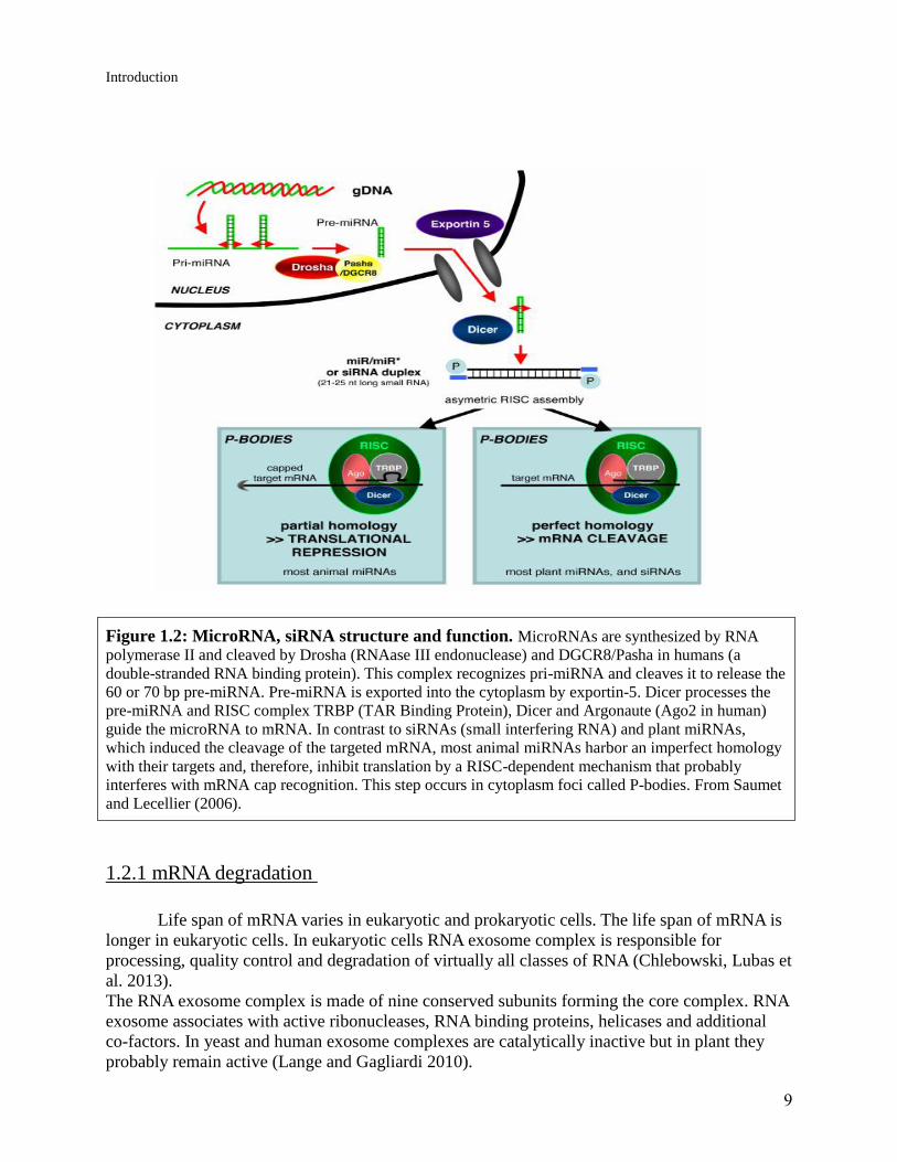
In E. coli RNA pyrophosphohydrolase is thought to initiate RNA degradation by
removing pyrophosphate from the 5' end of mRNAs. Removal of pyrophosphate enables RNase
E to digest the mRNAs (Figure 1.1).
Figure 1.1: mRNA degradation pathway in bacteria. a) Bacteria that contain the
endoribonuclease RNaseE or a homolog use RppH to remove 2 phosphates from the 5`terminus of
mRNAs. This allows RNase E to bind to the 5' end of the mRNAs and start degradation. b) In bacteria
containing RNase J an internal cleavage by an endonuclease generates a monophosphorylated
intermediate that is susceptible to 5′-to-3′ digestion by RNase J. A pathway analogous to the RppH
pathway may also exist in these bacteria. From Belasco (2010).
Plastids in plants and algae harbor a prokaryotic molecular machinery including mechanisms of
RNA degradation similar to what is found in bacteria. It is therefore thought that RppH homologs
in these organisms are located in plastids where they catalyze the removal of pyrophosphate from
plastid RNAs in the course of RNA degradation (Richards, Luciano et al. 2012).
In this project artificial microRNAs suited for silencing RppH expression were introduced into
Chlamydomonas in an effort to identify the function of a putative Chlamydomonas rppH gene.
1.1.1. The Nudix motif
RNA pyrophosphohydrolase contains a Nudix motif. Nudix stands for nucleoside
diphosphate linked to X. Nudix proteins contain two parts, a Nudix fold beta sheet with alpha
helices on either side and Nudix sequence motif or box which contains catalytic and metal
binding amino acids (Mildvan, Xia et al. 2005).
Nudix is a motif that uses water to hydrolyze phosphate groups and removes the phosphate from
organic compounds. This process is called phosphohydrolysis which is a means to activate or
deactivate nucleotides and proteins. Dcp2 of the decapping complex, ADP-ribose diphosphatase,
MutT ADPRase, Ap4A, RppH are examples of enzymes that contain a Nudix motif.

8
Introduction
Members of this family are recognized by a highly conserved 23-residue sequence, GX (5) EX
(7) REUXEEXGU, where U is a bulky hydrophobic residue and X is any residue. The Nudix box
contains Mg2
+ at both the binding and the catalytic site. The enzyme RppH contains a Nudix
motif and mediates degradation of mRNA within bacteria (Mildvan, Xia et al. 2005).
RppH is not the only protein containing a Nudix motif within the cell and thus when PCR or
Southern blotting is done with Nudix detecting primers or probes, the primer will bind to other
Nudix genes as well.
1.2. RNA silencing
In RNA silencing or post-transcriptional gene silencing (PTGS) expression of a gene is
downgraded or inhibited.
RNA interference is a well-studied method of mRNA silencing. In RNA interference a small 22-
24 nucleotide RNA is produced. This small RNA is complementary to a specific region of
mRNA and can bind to that region. This prevents ribosomes from attaching themselves to mRNA
and preventing the protein synthesis. Tagged mRNA can either be destroyed or remain in an
arrested state and to be reversed at a later date. MicroRNA can be separated from the mRNA and
mRNA can be activated again in order to produce the protein. When microRNA is removed,
ribosomes attach themselves to the mRNA and start the translation (Susi, P., et al. 2004).
RNA interference can occur with microRNA (miRNA) and a small interfering RNA (siRNA). It
is believed that the RNA silencing is a method used to combat viral infections (Ding 2000).
What distinguishes the miRNA from siRNA is the mode by which it attaches itself to the mRNA
and ultimately leads to destruction of mRNA. With siRNAs, a perfect match with a target mRNA
marks the duplex for destruction by endonucleases. However; miRNA does not match the target
sequence exactly and is unable to distinguish small variations in the recognition sites. As a
consequence of that a bulge in the duplex is formed between the miRNA and its mRNA. This
leads to two events. First It blocks the target mRNA from translation. Second it protects the
target mRNA from destruction by the endonucleases.
Usually miRNA genes are transcribed by polymerase II (Pol II) within the nucleus (Lee, Kim et
al. 2004).
Polymerase II produces pri-miRNA. The pri-miRNA has a stem loop, a 5`cap, and a polytail A.
This pri-miRNA can contain between 1 to 6 miRNA precursors. Protein Drosha pasha removes
the 5`cap and ploy tail A and makes pre-miRNA. The pre-miRNA exported out of the nucleus
with Exportin 5. Exportin 5 is a transporter channel that uses GTP bound to RAN protein. Once
inside cytoplasm the enzyme Dicer attaches itself to pre-miRNA and unwinds the double helix
(Lund and Dahlberg 2006). One of the strands is kept and the other strand is digested (Cenik and
Zamore 2011). What determines the selection of the strand is its thermodynamic stability and
strong base pairing. The remaining strand stays attached to the Dicer and another protein called
Argonaute nuclease. This complex is called RISC (RNA-induced silencing complex) and its role
is to identify the targeted mRNA and to bind itself to it and ultimately prevent the translation of
the mRNA to a protein by preventing binding of ribosomal RNA (Vermeulen, Behlen et al.2005).
9
Introduction
Figure 1.2: MicroRNA, siRNA structure and function. MicroRNAs are synthesized by RNA
polymerase II and cleaved by Drosha (RNAase III endonuclease) and DGCR8/Pasha in humans (a
double-stranded RNA binding protein). This complex recognizes pri-miRNA and cleaves it to release the
60 or 70 bp pre-miRNA. Pre-miRNA is exported into the cytoplasm by exportin-5. Dicer processes the
pre-miRNA and RISC complex TRBP (TAR Binding Protein), Dicer and Argonaute (Ago2 in human)
guide the microRNA to mRNA. In contrast to siRNAs (small interfering RNA) and plant miRNAs,
which induced the cleavage of the targeted mRNA, most animal miRNAs harbor an imperfect homology
with their targets and, therefore, inhibit translation by a RISC-dependent mechanism that probably
interferes with mRNA cap recognition. This step occurs in cytoplasm foci called P-bodies. From Saumet
and Lecellier (2006).
1.2.1 mRNA degradation
Life span of mRNA varies in eukaryotic and prokaryotic cells. The life span of mRNA is
longer in eukaryotic cells. In eukaryotic cells RNA exosome complex is responsible for
processing, quality control and degradation of virtually all classes of RNA (Chlebowski, Lubas et
al. 2013).
The RNA exosome complex is made of nine conserved subunits forming the core complex. RNA
exosome associates with active ribonucleases, RNA binding proteins, helicases and additional
co-factors. In yeast and human exosome complexes are catalytically inactive but in plant they
probably remain active (Lange and Gagliardi 2010).
10
Introduction
In prokaryotic cells degradosome is responsible for processing rRNA and degradation of mRNA.
Degradosome have DEAD box RNA helicaseB, RNAse and polynucleotide phosphorylase.
The DEAD box helices unwind parts of the RNA that interferes with degradation.
Polyadenylation can promote degradation by creating a toehold for the degradation machinery.
PNPase and RNase E are important in RNA processing and degradation of mRNA in bacteria.
Chloroplasts contain homologous gene for both of these two proteins, suggesting that the
prokaryotic degradosome is the ancestral origin of the chloroplast (Carpousis et al., 1999).
Major portions of mRNAs in the chloroplast are flanked with stem and loop structure 3`s of their
coding region that take part in the mature 3`end processing.
Stem and loop structures are necessary for the correct 3`-end processing and mRNA
accumulation (Barkan and Stern, 1998).
Chloroplast mRNAs contain poly (A) tails like bacteria and yeast. Poly (A) tails in chloroplast is
very long and is about 270 nucleotides; however, poly (A) tails in bacteria and yeast are only 40
to 60 nucleotides long. poly(A) tails in eukaryotic cell are made up of only adenosine versus
poly(A) tails in chloroplast are made up of 70% adenosine, 25% guanosines and 5% cytidine and
uridine (Lisitsky et al., 1996).
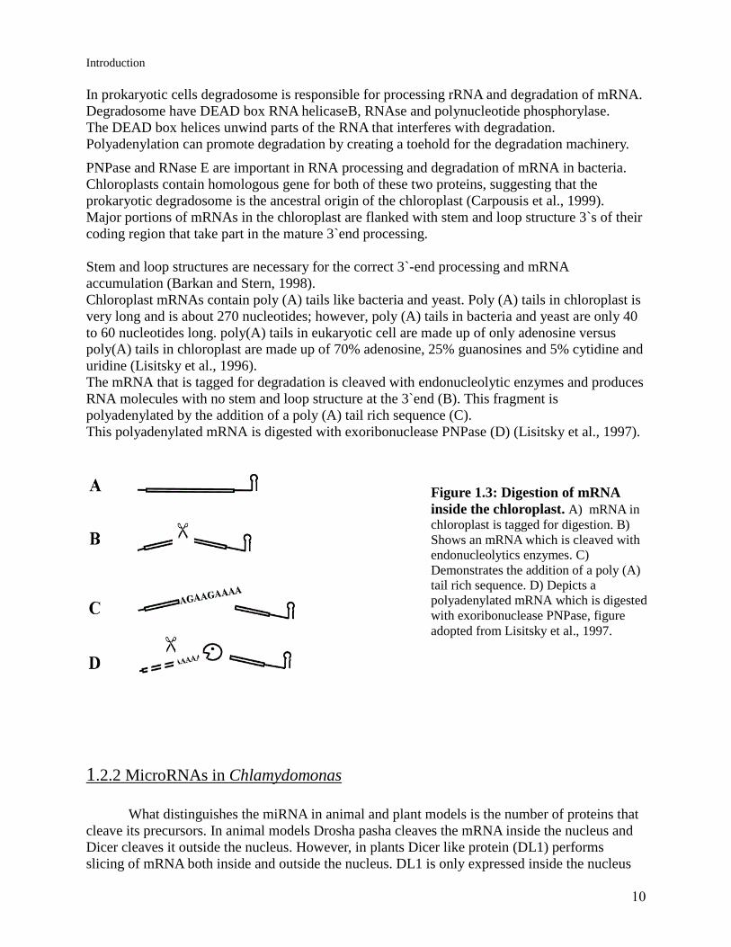
The mRNA that is tagged for degradation is cleaved with endonucleolytic enzymes and produces
RNA molecules with no stem and loop structure at the 3`end (B). This fragment is
polyadenylated by the addition of a poly (A) tail rich sequence (C).
This polyadenylated mRNA is digested with exoribonuclease PNPase (D) (Lisitsky et al., 1997).
1.2.2 MicroRNAs in Chlamydomonas
What distinguishes the miRNA in animal and plant models is the number of proteins that
cleave its precursors. In animal models Drosha pasha cleaves the mRNA inside the nucleus and
Dicer cleaves it outside the nucleus. However, in plants Dicer like protein (DL1) performs
slicing of mRNA both inside and outside the nucleus. DL1 is only expressed inside the nucleus
Figure 1.3: Digestion of mRNA
inside the chloroplast. A) mRNA in
chloroplast is tagged for digestion. B)
Shows an mRNA which is cleaved with
endonucleolytics enzymes. C)
Demonstrates the addition of a poly (A)
tail rich sequence. D) Depicts a
polyadenylated mRNA which is digested
with exoribonuclease PNPase, figure
adopted from Lisitsky et al., 1997.

11
Introduction
of plant cells and is a Dicer homolog. After that the 3`overhang is ethylated by an enzyme called
Hua-Enhancer1 (HEN). Another homolog to the exportin 5 called hasty (HST) transports the
microRNA into the cytoplasm (Lelandais-Briere, Sorin et al. 2010).
Land plants are thought to be the descendants of green algae-like ancestors (Lewis and McCourt
2004; Palmer et al 2004). The Chlamydomonas genome encodes both the dicer and the
Agronaute nuclease (AGO) indicating that it is fully competent to perform RNA silencing
(Molnar et al. 2007).
Considerable proportions of Chlamydomonas miRNA genes are in the introgenic regions; while
the remaining mRNA genes reside within the intergenic regions. It is found that multiple
Chlamydomonas miRNAs can be derived from a single stem and a loop.
The existence of miRNAs in the green alga Chlamydomonas raised the possibility that plants and
Chlamydomonas might be sharing some common miRNAs; however, comparisons between
Chlamydomonas miRNAs with all known plant and animal miRNAs in the entire data base has
found no homologues to miRNAs in Arabidopsis. The lack of universally conserved miRNA
genes among all living organisms suggests miRNA genes may have evolved independently in the
lineage leading to animal, plant, and green algae (Zhao, Li et al. 2007).
1.3 Chlamydomonas as a model system
Chlamydomonas is a unicellular organism of the green algae family that has two flagella.
It is a photosynthetic organism of the family Chlamydomonadaceae, genus Chlamydomonas.
Chlamydomonas contains different organelles such as a chloroplast, mitochondria, vacuoles and
flagella. The alga is a model organism which is used in studies like motility, chloroplast studies
and genetics. Chlamydomonas performs photosynthesis through utilizing sunlight and making
organic products from inorganic materials. Most of the Chlamydomonas species are obligate
prototrophs with the exception of C.reinhardtii which is a facultative heterotroph. This means C.
reinhardtii is capable of producing organic substances in the presence of both light and dark. In
dark it can use acetate, when present, whereas in the presence of light it can do photosynthesis
(Harris 2001).
C. reinhardtii is a haploid organism with 17 chromosomes. It has two types of organelles,
mitochondria and a single chloroplast. The alga divides by mitosis in favorable times. Under
nitrogen deprivation gametes are formed. Two gametes of opposite mating fuse to make a diploid
zygote. The zygote remains dormant in the soil until such time when nitrogen becomes plentiful.
Then the zygote becomes active and undergoes meiosis and it forms four haploid
Chlamydomonas.
One of the reasons that Chlamydomonas is used as a model organism is its short reproductive
cycle and its rapid multiplication. As a model organism Chlamydomonas has been modified in
many different ways. The strain that was used in this project is the cell wall-less mutant CW-15.
Because the mutant lacks a cell wall it can easily be transformed.
Paromomycin sulfate is used as a selection marker. Paromomycin binds to 16S ribosomal RNA
and stops translation of proteins (Vicens and Westhof 2001). Only Chlamydomonas cells that
have incorporated into their genome the vector that harbors the paromomycin-resistance gene are
able to grow on paromomycin plates

12
Introduction
1.3.1. The cell wall of Chlamydomonas
The wild type C .reinhardtii averages about 10μm in diameter and is enclosed within a
cell wall consisting primarily of several layers of hydroxyproline-rich glycoproteins that
resemble plant extensions. The cell wall does not contain cellulose to make it hard and inflexible
like other plants cells. The CW-15 type of Chlamydomonas produces the precursor protein
required to form the cell wall in normal quantity but prevents it from assembling into a complete
cell wall. Lack of the cell wall makes this mutant susceptible to transformation with foreign
DNA and further genetic manipulation (Harris 2001).
1.3.2 The chloroplast of Chlamydomonas
The chloroplast occupies two thirds of the cell in Chlamydomonas. This is where
photosynthesis occurs. Photosynthesis leads to production of nutrients; the chloroplast of
Chlamydomonas contains a 195 kb genome (Harris 2001).
1.3.3 Vegetative cell growth
C. reinhardtii is capable of both sexual and asexual reproduction. It can grow on agar or
liquid medium without co-factors and vitamins. In the absence of light when the strain has access
to acetate as a carbon source, energy can be produced.
Wild-type cells grow faster in light with or without acetate in comparison to dark. Optimal
growth conditions recommended to grow the algae are a temperature between 20° to 25° in a
medium of high salt with adequate lighting (200-400μEinstein’s/m2 sec photosynthetically active
radiation ). This will support an average doubling time of 6 to 8 hours (Harris 2001).
1.3.4 The sexual cycle
Under normal conditions Chlamydomonas undergoes mitosis. The cell divides in the
dark phase with 2 to 3 rounds of mitosis and makes 4 to 8 daughter cells. Daughter cells are
retained inside the cell wall of the mother cell and are released when the cell wall has been
digested by a lytic enzyme (Harris 2001).
Under starving conditions such as lack of nitrogen, cells develop into gametes and reproduce
sexually. C .reinhardtii are haploids (n) and genetically fixed to plus (mt+) and minus (mt-).
Under starving conditions the plus and minus gametes make contact with each other, the cell
walls are removed and the mating structure is activated. The flagella of the plus and minus cells
meet and adhere together and form a complete cell. Subsequently the fused cell loses it flagella
and makes a spore. The spore is a diploid zygote (2n) and is ready to germinate under favorable
conditions. In the presence of light zygote undergoes the meiosis and releases 4 cells.

13
Introduction
Figure 1.4: Sexual reproduction of the Chlamydomonas. Under starving condition plus and
minus gametes make a zygote and reproduce sexually. From Zhao, Lu et al. 2001.
14
Aim of the project
Aim of the project
The project is based on the hypothesis that a homolog of the bacterial RppH is
present in the chloroplast of Chlamydomonas where it participates in initiation of RNA
degradation. In order to test this assumption we wanted to inhibit expression of the putative rppH
gene in Chlamydomonas using artificial micro RNAs. If successful less RppH should be
produced and RNA degradation in the chloroplast should be affected, e.g. levels of mRNAs in
the chloroplast could be higher in transformants than in wild-type cells.

15
Materials and Methods
2. Materials and Methods
2.1 MicroRNA WMD3 (http://wmd3.weigelworld.org) is a site that specifically designs microRNA for
different organisms.WMD3 suggests different 21bp DNA strands since DNA is more stable than
RNA. Suggested DNA can have different colors; green is the best choice for the microRNA.
A flanking sequence specific to the Chlamydomonas reinhardii is added to the 21 bp. Flanking
sequence is necessary to make the stem and to give the stem and loop form to the artificial
microRNA.
Artificial miRNA resembles the natural RNA. Both start with U, and display 5' instability
relative to their miRNA. Their 10th nucleotide is either an A or a U (Schwab, Ossowski et al.
2006).
Certain characteristics associated with siRNA's functionality have been identified and they are:
low G/C content, a bias towards low internal stability at the sense strand 3'-terminus, lack of
inverted repeats, and sense strand base preferences (positions 3, 10, 13 and 19), (Figure 2.1).
Further analysis has revealed that the application of an algorithm incorporating all of the criteria
significantly improves potent siRNA selection. This highlights the utility of rational design for
selecting potent siRNAs and facilitating functional gene knockdown studies (Reynolds, Leake et
al. 2004)
Figure 2.1: Artificial microRNA mismatches and structure. In an artificial microRNA the
5`region is sensitive to mismatch but the 3`part can have up to 4 mismatches. Base pairs located at 13, 14
and 15 position must be mismatched too.
To construct the MicroRNA used in this experiment following steps were undertaken:
1- Designer inn (WMD3.com) was accessed
2- The rppH gene from the Chlamydomonas was inserted in the target gene part.
3- Chlamydomonas Reinhardtii from genomic section was chosen.
4- 1 as a Minimum number of included targets was chosen.
5- My E-mail was provided and sends inn was pressed
6- A list containing the best suited MicroRNA was received in a couple of days. The four best
suited sequences were selected and were placed in the oligo part of the MWD3 page.
7-for vector choice Chlamydomonas was chosen and send inn was pressed.

16
Materials and Methods
Shortly after I received a sequence containing MicroRNA and the flanking sequence needed to
transform the Chlamydomonas reinhardtii with the MicroRNA.
With flanking sequence, oligonucleotides were about 90 base pair and later were sent to the
Eurofins Company in order to design the microRNA used in this experiment.
All the suggested microRNA were specific for 3`and 5`UTR
Figure 2.2: Structure of mRNA contains cap, 5`UTR, coding sequence, 3`UTR and polytail
A. MicroRNA1 and 3 binds to 5`UTR and microRNA4, 6 attaches to 3`UTR. MicroRNA1 attaches to
position 506 microRNA3 to 565, microRNA4 to 3593 and microRNA 6 to 3646 in rppH
2.2 Cloning methods
2.2.1 Oligonucleotide cloning -Mix 5 μl of each of the single stranded oligonucleotides (100pmol/μl) with 30μl of sterile water
(total volume 40μl).
-heat for 2 minutes at 100°C.
-collect liquid on the bottom of the tube by centrifuge (room temperature) and let it cool down
slowly (15 min) on bench.
-add 5μl of poly nucleotide kinas buffer (10X) and 5μl ATP (10mM, pH 7) and mix
- add 1 μl of T4 polynucleotide kinas, mix again and spin briefly and incubate at 37ºC for 60 min
-Run the Oligonucleotides on the 1.3% agrose gel.
-Weigh a DNAase free 1.5 ml eppendorf
-use a scalpel and a large wavelength to cut the band from the gel
-weight the eppendorf tube with gel.
-add 10μl of capture buffer type 3 for each 10 mg of gel.
-mix and incubate at 60°C for 15 min to dissolve, shake the mixture every 3 min.
-once the gel is dissolved check the color of the capture buffer 3 to ensure it is still yellow.
-place one GFX micro spin column into one of the collection tubes
-Transfer DNA mix in GFX column within the collection tube.
-Incubate at RT for 1min.
-Spin the assembled column and collect tube at 1600 rpm for 30 sec
-Discard the flow through.
-add 500 μl of wash buffer type 1 to column.
-spin the column for 30 min in 1600g.

17
Materials and Methods
-Transfer GFX to an eppendorf tube.
-Add 20μl of elution buffer type 6 to GFX column
-wait 1 min in RT and spin in 1 min in 1600g.
2.2.2 Dephosphorylation -Linearized Sk+ vector is ethanol precipitated.
-Add 90μl of water to dry pellet.
-Add 10μl buffer 10X dephosphorylation buffer
-Add 1μl CIP (calf intestine phosphate), mix and incubate at 37°C. Add the same amount 100μl
Phenol/chloroform /isoamyl alcohol [25:24:1], vortex and centrifuge transfer the upper phase to
a new tube.
-Add the same amount of Chloroform /isoamyl alcohol [24:1], vortex and centrifuge transfer the
upper phase to a new tube.
-Precipitate DNA by adding 1/10 of the volume Na-acetate (3M) and 2 volumes ethanol. Leaves
tube at freezer or dry ice for 30 min.
-Collect the Precipitate by centrifuge (10 min, 4 min), wash the pellet with ice cold 70 % ethanol
and dry.
-Dissolve DNA at 20μl water.
-Add the same amount of 100μl Phenol/chloroform /isoamyl alcohol [25:24:1],
vortex and centrifuge then transfer the upper phase to a new tube.
-Add the same amount of Chloroform /isoamyl alcohol [24:1], vortex and centrifuge and transfer
the upper phase to a new tube.
-Precipitate DNA by adding 1/10 of the volume of Na-acetate (3M) and 2 volumes of ethanol.
Leave tube at freezer or dry ice for 30 min.
-Collect the Precipitate by centrifuge (10 min, 4 min), wash the pellet with ice cold 70 % ethanol
and dry.
-Dissolve DNA at 20μl of water.
2.2.3 Ligation mix -Add the insert to eppendorf tube
-Add appropriate amount of the vector
-Add water to final concentration of the 6.5μl
-Incubated for 45°C for 5 min put on ice.
-Add 1μl of ligase buffer, 2μl of PEG (remove water), 0.5μl of ligase T4 to the mixture
-Incubated at 19°C for 3 hours.
2.2.8 Transformation of E.coli
-Add 3μl of ligation mixes to the competent cell and keep on ice for 30 min . -Heat shock the
cell for 1 min at 42 °C
-Add 0.8ml LB to mixture and keep at 37 °C for 1 hour. Plate 75 μl of the mixture on LB +
ampicillin plate and keep over night at 37 °C.

18
Materials and Methods
2.2.4 Transformation of E.coli
-Add 3μl of ligation mixes to the competent cell and keep on ice for 30 min . . Heat shock the
cell for 1 min at 42 °C
-Add 0.8ml LB to mixture and keep at 37 °C for 1 hour. Plate 75 μl of the mixture on LB +
ampicillin plate and keep over night at 37 °C.
2.2.5 Small Scale isolation of plasmids from E.coli TB1 cells (Mini
prep) -Pipette 1.5 ml of each culture into the microfuge tube. Spin for 20 sec at full speed
-Remove the medium with a pipette leaving the bacterial pellet as dry as possible.
-Add 100μl of ice -cold TEG buffer and resuspend the pellet by vigorous mixing.
-Leave the tubes 5 min at RT. During this time , prepare a solution of 0.2 N NaOH /1% SDS by
gently mixing in a eppendorf tube 800μl of destilled water with 100μl of NaOH[2N] and 100μl
of SDS [10%]. Keep at RT.
-Add 200μl of the 0.2N NaOH /1%SDS solution to each tube. Mix by inversion. Do not vortex.
Incubate 5 min on the ice.
-Add 150μl of ice cold potassium acetate [5M, pH 4.8]. Mix by inversion for about10 sec. Do
not vortex. Incubate on the ice for 5 min.
-Centrifuge in a cold (4°C) microfuge 5 min at max speed.
-With pipette transfer the supernatant to a new tube avoiding the white precipitate on the side of
the tubes.
-Add 410μl of phenol/chloroform /isoamyl alcohol [25:24:1] into the supernatant .Mix by
vortaxing (mixture will turn milky white) and centrifuge for 2 min at RT at max speed to separate
the phases.
-Transfer the upper phase (containing the plasmids) to the new tube and add 410μl of
Chloroform /isoamyl alcohol [24:1]. Mix by overtaxing and centrifuge for 2 min as before.
-Transfer 310μl of the upper phase to a new tube. Add 750μl of ice cold 96% ethanol. Mix and
precipitate nucleic acids for 10 min on ice.
-Centrifuge 10 min in the cold at max speed.
-Remove and discard the liquid with a pipette and add 1 ml of ice-cold 70% ethanol to the pellet.
Mix by inversion and centrifuge in the cold for 5 min as before.
-Remove the supernatant leaving the pellet as dry as possible. Leave the pellet at RT to let t air
dry or vacuum dry.
-Dissolve the dry pellet with pipette in 15μl of sterile distilled water.
2.2.6 Maxi prep Maxi prep or E.coli plasmid preparation by CsCl gradient centrifugation
-Grow 100ml of LB with 100μl ampicillin (1μg/μl) culture over night at 37°C shaking.
-Collect the cells by centrifugation for 5 min at 4°C at 5000 rpm.
-Resuspends cells in 3.6 ml of cold TEG (25mM Tris (pH 8), 10mM EDTA, and 50mM glucose)
and remove cells to 50 ml tubes.
-Add 0.4 ml of 10 mg/ml lysozyme (0.01 g lysozyme/1ml TEG) in TEG (prepared fresh) to the
cells and leave at RT for 5 min and move to an ice bath for an additional 5 min.
- Add 8 ml of 0.2 N NaOH/1% SDS (prepared fresh), invert to mix and leave on ice for 5 min.

19
Materials and Methods
-Add 6 ml of cold 5 M potassium acetate (pH 4.8), invert to mix, and leave on ice bath for 5 min.
-Centrifuge in SS-34 rotor at 6K for 10min at 4°C to pellet debris.
-Pour supernatant through cheesecloth into a 50 ml tube and add 12.5ml of isopropanol.
-Leave at RT for 15 min and centrifuge in SS-34 rotor at 10K for 10 min at RT.
-Aspirate supernatant thoroughly and add 3 ml of 50 Tris (pH 8)/1mM EDTA to the pellet.
-Resuspends the pellet and TE to 4.2 grams in 30 ml tube.
-Add 4.5g CsCl and mix until the pellet is dissolved and allow solution to warm to RT.
-Add 0.5 ml of 10 mg/ml ethidium bromide, mix and centrifuge in SS-34 rotor at 4K for 5 min at
RT.
-Remove supernatant to a Vti80 tube and heat seal.
-Load rotor and centrifuge at >50K for more than 15 hours at 15°C.
-Decelerate the mixture without brake, extract band with 3 ml syringe and 16 gauge needles.
-Remove EtBr by repeated (4-5X) extraction with isopropanol /water (7:1).
-Dialyze against 1 liter of sterile TE at 4°C for >3 hours, replace buffer with fresh 1 liter sterile
TE and leave overnight.
-Find the concentration with nano drop.
2.2.7 Cloning of microRNA Oligonucleotides were ordered from Eurofins MWG operon. Water was added to
the oligonucleotides to make the concentration of the DNA 100 pmol/μl. Oligonucleotides are
single strands and have to be double stranded for it to be cloned within the vector.
Oligonucleotide cloning method was used to purify the DNA and to make it complementary.
Linearized SK+ vector was precipitated with ethanol precipitation method and dephosphorylated.
To ligate the microRNA inside the vector, appropriate amount of the vector and insert were
added together. Mixture incubated at 45°C for 5 minutes and then placed on ice.
2μl of PEG (removes water), 1μl of ligase buffer and 0.5μl of ligase T4 were added to the
mixture of the insert and SK+ vector. Ligase mix was incubated at 19°C for 3 hours.
Competent E.coli was transformed with 3μl of ligation mix, plated and kept overnight.
Four colonies were picked up from the plate, and mini preps were performed on them to isolate
the plasmids.
Plasmids from mini prep were checked to make sure they contain the insert and specifically to
ensure that insert sites were in the right direction (5` to 3` direction).
3μl of plasmids were cut with AfeI restriction enzyme. Plasmids without the insert were not cut
and remained supercoiled in the gel. The plasmids containing the insert were cut and linearized.
MicroRNA was inserted in 5` to 3` or 3` to 5 ` direction. Plasmids with insert (linearized with
AfeI digestion) were further cut with SpeI and XbaI. The plasmids containing the insert in the
right direction is about 160bp versus the one in the wrong direction is about 230 bp.
Plasmids containing the microRNA were used to inoculate 100 ml of LB with
ampicillin. Maxi prep was used to isolate plasmids in a much larger quantity.
Appropriate amount of the SK+ (10000ng) was digested with XbaI and XhoI, Insert was placed
between XbaI and XhoI site. SK+ digestion resulted in a fragment which is about 1200 bp. This
fragment was then ligated within the pChlamiRNA3int vector (plasmids). Plasmid
pChlamiRNA3int is 6481bp which contains Paromomycin sulfate resistance gene is used to Materials and Methods

20
select the with the insert.
Plasmid pChlamiRNA3int was digested with the XbaI and XhoI. Digested plasmids ran on the
gel. Electrophoresis of the digested material formed two bands. The smaller band was discarded
while the larger band was isolated from the gel.
Using transformation method SK+ plasmids (~1200bp) was ligated within the
pChlamiRNA3int (~4600bp).The transformed Product was the pChlamiRNA3int with
micoRNA. Plasmids were cloned within the E.coli genome and then ran through the mini prep.
Plasmids were cut sequentially with the AfeI, and then with XbaI and SpeI enzymes to ensure
that plasmids contain the insert and that the insert is inserted in the right direction. Sample with
the right insert was selected and inoculated in 100ml of LB+amp. Plasmids were isolated with
the maxi prep and dialyzed and later ethanol precipitated for purification. The Purified plasmids
were dissolved in 20μl of water. To find the concentration of plasmids nano drop was used.
Plasmids (10000ng) were digested with KpnI enzyme in order to make them linearized for
transformation of Chlamydomonas.
Figure 2.3: Cloning step for microRNA. A) MicroRNA is inserted within SK+ vector after it has
been digested with SpeI. B) SK+ vector is further digested with XhoI and XbaI and fragments
containing microRNA is separated. C) Fragments are inserted within the pChlamiRNA3int vector.
2.3. Nuclear Transformation MicroRNA is inserted inside the pChlamiRNA3int and transformed into the C .reinhardtii
cell. Since Chlamydomonas cell does not have a cell wall it can be transformed easily. Plasmids
are cut with KpnI in order to make them linearized and to transform the cell. Plasmids are
inserted within the nuclear genome.

21
Materials and Methods
The shotgun method can be used to do this, but our experiment method is cheaper,
easier and more effective. If plasmids are inserted inside the genome, the transformation is
permanent and the cell will never loses the insert.
Linearized pChlamiRNA3int with microRNA was transformed within Chlamydomonas with no
cell wall (CW-). 40 ml of Chlamydomonas was centrifuged and the supernatant was removed.
The remaining pellet was dissolved in 0.5 ml of HS solution. To make the transformation mix,
337μl of Chlamydomonas, 300μg of beads, 66μl of TEG and 7.5μl of DNA were added and
vortexed for 15 seconds. The solution was removed and plated on the plate with paromomycin.
Then the plate was left undisturbed for over 2 weeks.
If the Chlamydomonas is not transformed with the vector it cannot survive. However; if it is
transformed with the pChlamiRNA3int vector it can grow on the plate. The pChlamiRNA3int
with the insert was transformed within the Chlamydomonas and was integrated inside the
genomic DNA of Chlamydomonas and began to produce the paromomycin and microRNA.
Colonies from the plate were later transferred to a medium of HS and placed under the light.
After 30 days, colonies started to grow in the HS solution. To speed the growth of
Chlamydomonas the solution was transferred to the CO2 chamber. DNA isolation methods were
used to isolate the DNA from Chlamydomonas.
2.4 Work with Chlamydomonas
2.4.1 Isolation of genomic DNA from Chlamydomonas -Spin down (5 min 4000 rpm, SS-34 rotor) 40-50 ml of Chlamydomonas culture.
-Resuspend the pellet in 0.75ml of DNA extraction buffer (100mM Tris pH 8.0; 50mM Na2-
EDTA; 0.5M NaCl; 10mM β-mercapto ethanol) and transfer suspension to a 2ml microfuge tube.
-Add 60μl of SDS (21%), mix, and incubate the mixture for 15 min at 65°C.
-Let the mixture cool down on RT and add 0.7 ml of phenol (equilibrated with 0.1 M Tris, pH
8.0). Mix carefully by inverting the tube and spin for 5 min in microfuge at RT.
-Transfer supernatant to a new microfuge and add 0.750ml Phenol/chloroform /isoamyl alcohol
[25:24:1], and mix and spin.
-Transfer 0.650 ml of supernatant to a new 1.5 microfuge tube and add equal volume of
isopropanol. Mix by inverting the tube and letting it stand at RT for 5 min.
-Collect the precipitated nucleic acids at the bottom of the tube at low speed centrifugation
(2000rpm) at RT if DNA is visible you can remove it with glass or pipette tips.
-Discard as much supernatant as possible and wash pellet in a 1ml ice cold 70% ethanol. To dry
the pellet spin for 2 min in full speed.
-Resuspend pellet in 90μl of Te buffer (10mM Tris, pH 8.0; mM EDTA), add 10μl of RNase a
(1mg/ml), and incubate at 37°C for 1 hour.
-Extract the mixture once with 100μl Phenol/chloroform /isoamyl alcohol 25:24:1] and once with
100μl Chloroform /isoamyl alcohol [24:1].
-Precipitate DNA in freezer with 0.3 M Na-acetate /2 volumes ethanol.
-Collect DNA precipitate in microfuge in cold, wash with ice-cold 70% ethanol and dry the
pellet.
-Resuspend the DNA in 20μl of sterile water. DNA concentration will be ~200 ng/μl, and yield of
~4μg.

22
Materials and Methods
2.4.2 RNA isolation from Chlamydomonas reinhardtii -Centrifuge at 5000rpm at 4°C for 5 minutes 40 ml of Chlamydomonas culture containing 2X10⁶
cells /ml.
-Re suspends the pellet in 1.5 ml of ice-cold lyses buffer (0.6mNaCl, 200mMTris
pH8.0, and 10mMNa2-EDTA); add 150μg of RNAase inhibitor (200mM vandal rib nucleoside
(NEB)).
-Pipet cells into a 15ml sarstedt tube containing 2 ml of phenol and 1.5 ml of SDS (4%)
preheated to 65°C. Cap and mix.
-Incubate at 65°C for 15 min (mix occasionally [3-4 times] by shaking).
-Cool down on ice and add 1ml of ice cold Chloroform /isoamyl alcohol (24:1) and Mix.
-Centrifuge at 7000rpm for 5 min (SS34, 4 °C).
-Take 3 ml of the supernatant and transfer to another 15ml sarstedt tube containing 3ml of ice
cold Phenol/chloroform /isoamyl alcohol [ 25:24:1]. Mix and spin as before.
-Pipet 2.5ml of the supernatant into the tube containing 2.5ml of ice cold isopropranolol and
250μl of 3M Na-acetat (pH5.2) .Mix and incubate at -20°C for at last 1 hours ( over night is
better).
-Spin for 15 min at 12000rpm. Decant supernatant; watch pellet it as it may get loose. Pellet
should be almost dry; Re suspends the pellet in 300 μl of DEPC -treated water and transfer to a
sterile 1.5ml Eppendorf tube containing 100μl of 1ice- cold 8m LiCl. Mix and incubate on ice for
2 hours.
Spin in microfuge at 4°C for 30 min. Remove supernatant with a pipet and resuspend the pellet
in 100μl of DEPC treated water. Keep on ice.
Dilute 10μl of RNA solution in 1 ml of DEPC treated water and measure OD260nm
(OD1.0 =40μg RNA/ml). To the rest of the RNA solution add 10μl of 3M
Sodium acetate (pH5.2) and 200μl of ice cold ethanol. Mix and incubate at -20°C for 1 hour.
Spin in microfuge in a cold room for 10min.
-Wash pellet with 1 ml of ice cold 70%ethanol (10 min spin). Store the RNA in 70 % ethanol if
it is not used immediately .Dry pellet briefly in a vacuum centrifuge. Resuspend RNA at a
concentration of 2μg/μl in DEPC -treated water.
2.5 Analytic methods
2.5.1 PCR
Genomic DNA was used as the template. In the experiment 100ng of genomic DNA was
added to 50μl of PCR solution. Primer 5`paro4331 and 3`paro4743 was used at the annealing
temperature of 59°C .The Product was about 412 bp which proves paromomycin is present
within the genome as the Primers used were specific for paromomycin.
A new PCR on the samples containing paromomycin gene was performed using new primer
3`psaD495 and 5`psaD6401 at annealing temperature of 63°C. The Primers used at this step were
specific for promoter region in the vector and the product was around 713bp.

23
Materials and Methods
Primer5`interon and 3`psaD495 at annealing temperature of 62°C was used on the sample
containing paromomycin and promoter region. Primers are specific to intron in the vector and
promoter region in the vector and can give different bonds. Our targeted Product must be around
543bp.
2.5.2 Southern blotting
Southern blotting utilizes DNA interaction between probe and DNA to detect a
specific sequence within the genomic DNA. In this experiment a probe for detecting the
paromomycin sequence was used to detect the paromomycin gene in the genomic DNA.
PCR with paromomycin primer proved paromomycin was already present within the genomic
DNA and southern blotting used to confirm the PCR result. Southern blotting is another
common method used in laboratories. It was performed to reconfirm the PCR result.
Plasmid pChlamiRNA3int was digested by BamHI and XhoI enzymes. After digestion was
completed, the product was fractionated on the gel and an 1800bp fragment containing
paromomycin gene was isolated from the gel. The probe was prepared by Professor Uwe Klein
using the 1800bp fragment.
Isolated genomic DNA was digested by Bathe and XhoI and ethanol precipitate.
-Resuspend the DNA in 20μl of water, including DNA loading gel buffer.
- Run the reaction on the agarose gel.
-Check the gel under UV light and take a photo.
-Transfer DNA to nylon membrane according to protocol
-Put the membrane in SSC [2X] .Check gel under UV – light for complete transfer.
-Wrap the membrane into plastic Wrap and cross link DNA to membrane with CL-1000
ultraviolet cross linker, UVP (USA), set to 1500 energy.
-Hybridize the membrane with a radioactive probe and develop by auto radiography.
2.5.3 Northern blotting
Northern blotting is utilized to detect RNA using complementary DNA strands.
Chlamydomonas RNA was isolated using RNA isolation method and was subsequently added to
paromomycin probe to detect the mRNA produced by the Chlamydomonas containing
paromomycin resistance gene. Probes specific for Nudix was used to detect Nudix mRNA within
the pool of mRNA.
-Wash the tray and comb in the 3% H2O2 overnight.
-Dissolve 0.78 g agarose in 37ml DEPC treated water (1.3% agarose gel). Cool down in water
bath at 65°C.
-Add 12ml of MOPS buffer [5X], and 11ml of formaldehyde [37%].
-Mix gently by swirling and pouring into a gel trey: Put in the comb.
-Prepare the samples by mixing every sample in a microfuge tube:
2.5μl of DEPC treated water
2.0μl of MOPS buffer [5x]
3.5μl of formaldehyde

24
Materials and Methods
3.5μl of ethidium bromide [100μg/ml]
10μl of formamide
2μl of RNA sample [2μg/μl]
-Incubate samples at 65°C for 15 min.
-Put the gel into the electrophoresis chamber and add running buffer to cover the gel. (315ml of
DEPC treated water, MOPS [5X] and 45 ml of formaldehyde).
- Pre run for 5 minutes at 60mA.
-Cool down samples. Spin in centrifuge at maximum speed for 1 min and add 2μl of RNA gel
loading buffer.
-Mix with pipette and add 20μl of sample for every well.
-Run the sample at 60mA for 10 min in reverse direction, then in normal direction until the
bromphenol blue bands is at the bottom of the gel.
-Take a photograph from the gel.
-Wash briefly in DEPC treated water and transfer RNA to a nylon membrane following the
protocol.
-Transfer for 5.5 hours. Put membrane into SSC [2X].Check gel under UV light for complete
transfer.
-Wrap the membrane into plastic wrap, and cross link DNA to the membrane with CL-1000
Ultraviolet Cross linker, UVP, set to 1500 energy.
-Hybridize the membrane with a radioactive probe and develop by auto-radiography.

25
Results
3. Results Four different artificial microRNAs were used to silence the rppH gene (see Appendix
5). These microRNAs were supposed to bind to transcripts of the putative rppH gene in
Chlamydomonas in the 5' UTR region (microRNAs 1 and 4) and in the 3' UTR region
(microRNAs 3 and 6). The four microRNAs were chosen from a list of possible microRNAs that
was provided by the WMD3 - Web MicroRNA Designer tool (http://wmd3.weigelworld.org)
using the putative rppH gene from Chlamydomonas as the target sequence. Besides suggesting
sequences for microRNAs the tool provides also the sequence of an oligonucleotide (≈ 90 nt
including the microRNA sequences) that can be directly cloned into the SpeI site of the
pChlamiRNA3int transformation vector (Molnar, Bassett et al. 2009) .
3.1 Cloning of the microRNAs into the transformation vector
Complementary ≈ 90 nt oligonucleotides, obtained from Eurofins MWG (Ebersberg, Germany),
were annealed (Materials and Methods) and purified on an agarose gel (Figure 3.1).
The purified oligonucleotides were cloned into the SpeI site of plasmid psaD/SK+, a pBluescript
SK+ vector into which the 1200 bp XhoI-XbaI fragment from the pChlamiRNA3int vector
(Appendix 2) has been cloned. The XhoI-XbaI fragment contains the Chlamydomonas psaD
Figure 3.1: Purification of annealed
oligonucleotides. Lane 2, 3, 4, 5 show the
four microRNAs purified on a 1% gel. The
bands were cut from the gel and purified with
GFX PCR DNA gel band purification kit (GE
Healthcare). Lane 1 contains the DNA ladder,
lane 2 contains microRNA1, lane 3 contains
microRNA3, lane 4 microRNA4, and lane 5
contain microRNA6.

26
Results
promoter and 3' terminator sequences that flank the SpeI site into which the oligonucleotides
were inserted. The total size of the psaD/SK+ vector is about 4200 bp.
Prior to cloning the linearized psaD/SK+ vector was dephosphorylated to avoid religation.
Competent E.coli TB1 cells were transformed and minipreps of colonies screened for the
presence of the oligonucleotides by digestion with SpeI and XbaI (Figure 3.3). Positive
transformants released a fragment of ≈150 bp.
Figure 3.2:
psaD/Sk+ vector
digested with SpeI
(lane 2).

27
Results
In addition to digesting the miniprep DNA with SpeI and XbaI positive samples were also
digested with AfeI (not shown) because the oligonucleotides contained a unique AfeI site.
Plasmids that did not contain microRNA were not digested by AfeI and remained supercoiled.
Plasmids were amplified and isolated by maxi preps, digested with XhoI and XbaI (Figure 3.4)
and cloned into the XhoI/XbaI sites of the final transformation vector pChlamiRNA3int
(Appendix 2).
Figure 3.3: Example for
screening of transformant DNA
for the presence and orientation
of microRNA-containing
oligonucleotides. Miniprep DNA was digested with
SpeI and XbaI releasing a fragment
of 150 bp or 250 bp depending on
the orientation of the
oligonucleotide in the vector.
Figure 3.4: Plasmid psaD/SK+
and the final transformation
vector pChlamiRNA3int
digested with XbaI and XhoI. The smaller fragment from
psaD/SK+ (≈ 1300 bp) containing
the microRNA was isolated and
cloned into the pChlamiRNA3int
vector (large fragment of about
5200 bp).
28
Results
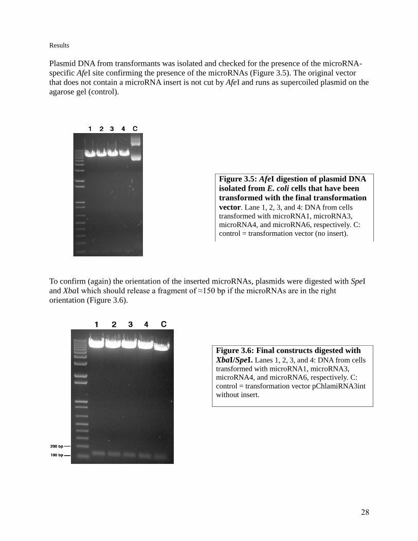
Plasmid DNA from transformants was isolated and checked for the presence of the microRNA-
specific AfeI site confirming the presence of the microRNAs (Figure 3.5). The original vector
that does not contain a microRNA insert is not cut by AfeI and runs as supercoiled plasmid on the
agarose gel (control).
To confirm (again) the orientation of the inserted microRNAs, plasmids were digested with SpeI
and XbaI which should release a fragment of ≈150 bp if the microRNAs are in the right
orientation (Figure 3.6).
Figure 3.5: AfeI digestion of plasmid DNA
isolated from E. coli cells that have been
transformed with the final transformation
vector. Lane 1, 2, 3, and 4: DNA from cells
transformed with microRNA1, microRNA3,
microRNA4, and microRNA6, respectively. C:
control = transformation vector (no insert).
Figure 3.6: Final constructs digested with
XbaI/SpeI. Lanes 1, 2, 3, and 4: DNA from cells
transformed with microRNA1, microRNA3,
microRNA4, and microRNA6, respectively. C:
control = transformation vector pChlamiRNA3int
without insert.

29
Results
Maxipreps of the plasmids harboring the four microRNAs were made and linearized by digestion
with KpnI. Chlamydomonas cells were transformed by the glass beads method (Materials and
Methods) and plated on agar containing 60 µg/ml paromomycin. Individual colonies could be
seen on the plates about 4 weeks after transformation.
3.2 Screening of transformants by PCR Individual colonies of Chlamydomonas were picked and grown on liquid HS medium to a cell
density of about 1-5 million cells per ml. Total DNA was isolated and screened by PCR for the
presence of the paromomycin gene and the microRNAs. PCR primers used, binding sites, and
expected sizes of PCR products are indicated in Figure 3.7.
The presence of the paromomycin resistance gene in the nuclear genome of Chlamydomonas was
checked with primers 5'paro4331 and 3'paro4743 yielding a PCR product of 412 bp (Figure 3.7).
All transformants checked contained the paromomycin resistance gene (Figure 3.8) which was
not surprising because they have been selected by growing on paromomycin plates. For one of
the cell lines, the presence of the paromomycin gene was also confirmed by Southern analysis
(see 3.3 and Figure 3.11).
Figure 3.7: Map of the linearized final transformation vector in which the primer binding
sites are marked. Sizes of the expected PCR products are as follows: 5'psaD6401/3'psaD495,
691 bp. 5'intron/3'psaD495, 543 bp. 5'paro4331/3'paro4743, 412 bp. apHVIII, paromomycin
resistance gene. The map is not drawn to scale.

30
Results
Two PCR reactions were done to verify the presence of the microRNA sequence in the nuclear
genome of the transformants. A PCR with primer pair 5' intron/3'psaD495, with an expected
fragment size of 543 bp (Figure 3.7), and a second PCR with primer pair 5'psaD6401/3'psaD495,
with expected fragment sizes of 691 bp and 778 bp, the latter being a PCR product from the
endogenous psaD gene in the genome. The results of these PCRs are shown in Figures 3.9 and
3.10.
Besides the expected products of 691bp and 778 bp, PCR reactions with primer pair
5'psaD6401/3'psaD495 also gave a product of around 600 bp (lowest band in Figure 3.10). This
is assumed to be an unspecific amplification product from unspecific binding of the primers to
the genomic DNA.
Figure 3.8: PCR products from primers
5`paro4331 and 3`paro 4743. The primers
amplify a 412bp fragment.
Lanes 2, 3, 4, and 5: template DNA from cells
transformed with microRNA1, microRNA3,
microRNA4, and microRNA6, respectively.
Lane 1: 1 kb plus DNA ladder.
Figure 3.9: PCR products from
primers 5` intron and 3`psaD495. Expected fragment size is 543 bp.
Lanes 1, 2, 3, and 4: template DNA
from cells transformed with
microRNA1, microRNA3,
microRNA4, and microRNA6,
respectively.

31
Results
Of the four transformant cell lines, the microRNA4 transformant did not give the expected
fragment of 691 bp in PCRs with primer pair 5'psaD6401/3'psaD495. Because of this result and
because of time constraints the following analyses were only done with the microRNA1
transformant.
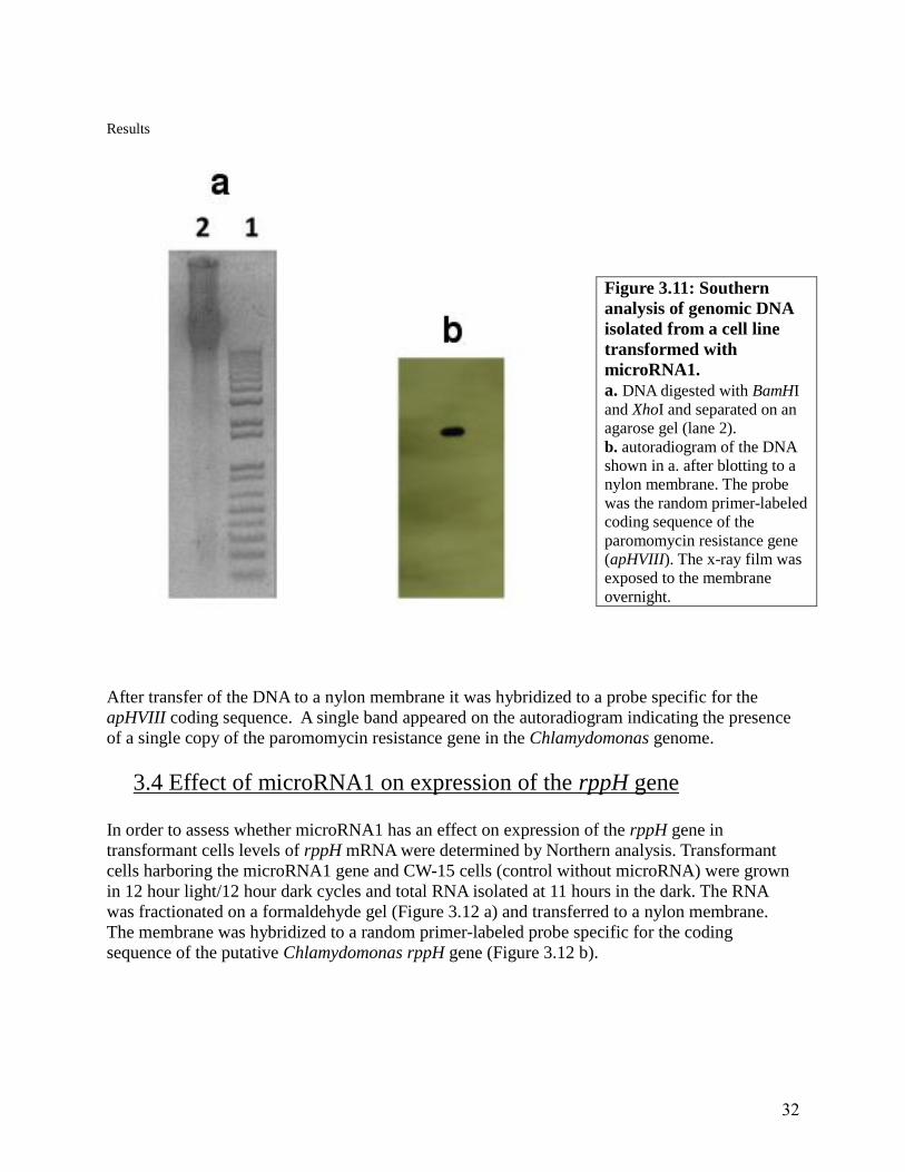
3.3 Detecting the paromomycin resistance gene by Southern analysis
In addition to PCR, the presence of the paromomycin resistance gene in Chlamydomonas
genomic DNA was confirmed by Southern analysis. Genomic DNA from transformant
microRNA1 was digested with BamHI and XhoI and separated on an agarose gel (Figure 3.11 a).
Figure 3.10: PCR products from
primers 5` psaD6401 and 3`psaD495. Expected fragment sizes are 691 bp and 778
bp (from the endogenous psaD gene.
Lanes 1, 2, 3, and 4: template DNA from cells
transformed with microRNA1, microRNA3,
microRNA4, and microRNA6, respectively.
32
Results
After transfer of the DNA to a nylon membrane it was hybridized to a probe specific for the
apHVIII coding sequence. A single band appeared on the autoradiogram indicating the presence
of a single copy of the paromomycin resistance gene in the Chlamydomonas genome.
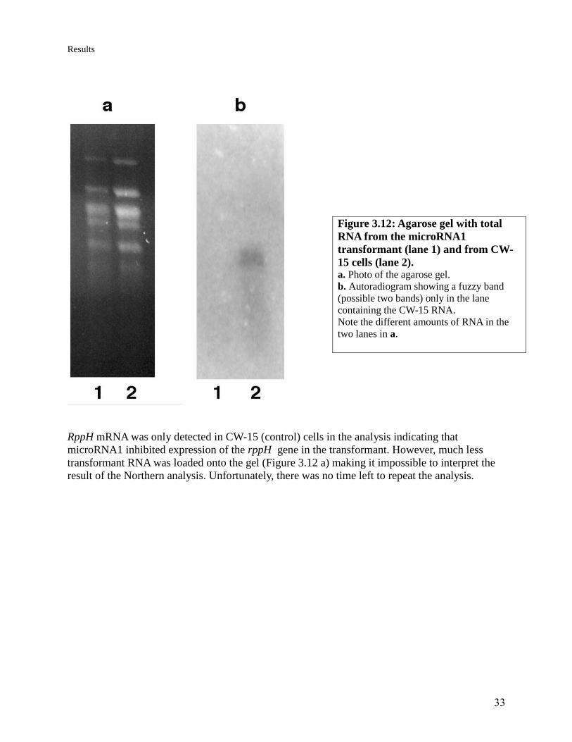
3.4 Effect of microRNA1 on expression of the rppH gene
In order to assess whether microRNA1 has an effect on expression of the rppH gene in
transformant cells levels of rppH mRNA were determined by Northern analysis. Transformant
cells harboring the microRNA1 gene and CW-15 cells (control without microRNA) were grown
in 12 hour light/12 hour dark cycles and total RNA isolated at 11 hours in the dark. The RNA
was fractionated on a formaldehyde gel (Figure 3.12 a) and transferred to a nylon membrane.
The membrane was hybridized to a random primer-labeled probe specific for the coding
sequence of the putative Chlamydomonas rppH gene (Figure 3.12 b).
Figure 3.11: Southern
analysis of genomic DNA
isolated from a cell line
transformed with
microRNA1.
a. DNA digested with BamHI
and XhoI and separated on an
agarose gel (lane 2).
b. autoradiogram of the DNA
shown in a. after blotting to a
nylon membrane. The probe
was the random primer-labeled
coding sequence of the
paromomycin resistance gene
(apHVIII). The x-ray film was
exposed to the membrane
overnight.
33
Results
RppH mRNA was only detected in CW-15 (control) cells in the analysis indicating that
microRNA1 inhibited expression of the rppH gene in the transformant. However, much less
transformant RNA was loaded onto the gel (Figure 3.12 a) making it impossible to interpret the
result of the Northern analysis. Unfortunately, there was no time left to repeat the analysis.
Figure 3.12: Agarose gel with total
RNA from the microRNA1
transformant (lane 1) and from CW-
15 cells (lane 2). a. Photo of the agarose gel.
b. Autoradiogram showing a fuzzy band
(possible two bands) only in the lane
containing the CW-15 RNA.
Note the different amounts of RNA in the
two lanes in a.

34
Discussion
4. Discussion In this project four artificial microRNAs, targeting sequences in the untranslated regions
of transcripts of a putative Chlamydomonas rppH gene, were inserted into the nuclear genome of
CW-15, a cell wall-less Chlamydomonas mutant. The presence of the artificial microRNAs in
nuclear transformants was checked by PCR. Due to time constraints, only one of the
transformant cell lines was analyzed for an effect of the artificial microRNA on expression of the
rppH gene.
4.1 MicroRNAs
Four different microRNAs were used in this project. All four microRNAs bind to the 5` and 3`
UTR regions of the mRNA. None of them is specific for the exon regions of the rppH gene. The
logic behind using these four miRNA is as follows: there are at least eight other genes with a
Nudix motif in the Chlamydomonas genome. If selected microRNAs were specific for exon
sequences, they could silence other genes containing the Nudix motif as well increasing the
chance to generate non-viable transformants.
4.2 Nuclear transformation
The glass beads method of nuclear transformation method was used to transform
Chlamydomonas with microRNA 1, 3, 4, and 6. MiRNAs are inserted in random locations into
the Chlamydomonas genome. Upon successful transformation Chlamydomonas cells can grow
on paromomycin plates. Chlamydomonas transformants from plates were selected for DNA
isolation and their DNA was screened by PCR for the presence of the paromomycin resistance
gene and the presence of the microRNA sequences. However, expression of the artificial
microRNAs in transformants was not shown directly, e.g. by using a probe specific for the
microRNAs or by RT-PCR. Therefore it is not sure that the microRNA genes are actually
transcribed and processed correctly such that they are functionally active. Such an analysis
would require more resources than have been available in the project.
4.3 Southern analysis
Southern blotting was used to check if the vector is inserted into the Chlamydomonas genome.
DNA from Chlamydomonas with microRNA1 was isolated and digested with BamHI and XhoI.
Digested DNA then was fractionated on an agarose gel. The gel was inspected under UV light
and the DNA was transferred to a membrane. A probe specific to the sequence of the
aminoglycoside 3`phosphotransferase gene (apHVIII) responsible for paromomycin resistance
was hybridized to the membrane. After exposure to an x-ray film the presence of a strong band
on the film proved that the paromomycin resistance gene was inserted. Most likely the other
sequences in the transformation vector, including the microRNA genes, have been inserted into
the genome as well.

35
Discussion
4.4 Northern analysis
Total RNA was isolated from one of the transformants and from the wild type. RNA from the
microRNA1 transformant was fractionated on a gel and transferred to a membrane. Later an
rppH probe was added to the membrane and the film was developed.
RppH mRNA could only be detected in CW-15 (control) cells that did not harbor an artificial
microRNA gene. This seems to indicate that the microRNA in the transformant does indeed
silence expression of the rppH gene. However, as less mRNA from the transformant than for the
wild-type was used in the analysis, no such conclusion can be drawn. The analysis needs to be
repeated with equal amounts of total RNA.
In addition to Northern analysis, qRT-PCR could be used to estimate the levels of rppH
transcripts in transformants and wild-type cells but there was no time to do this.
Another possible approach to assess whether expression of the rppH gene is silenced by the
artificial microRNAs would be to analyze the level of RppH protein by immunoblots.
Unfortunately, no antibodies against the Chlamydomonas RppH protein are available and
production of antibodies would take a few months.

36
Conclusion
Conclusion
The goal of the project was to identify the function of a putative RNA pyrophosphohydrolase
gene in Chlamydomonas reinhardtii by silencing the expression of the gene and analyzing the
effect of silencing on mRNA degradation. In this work artificial microRNA genes, targeting
transcripts of the putative rppH gene, were inserted into the nuclear genome of Chlamydomonas.
Preliminary analysis by northern blotting suggests an effect of one of the microRNAs on rppH
transcript levels indicating functioning of the artificial microRNAs in silencing expression of the
target gene. However, the analysis needs to be repeated and additional analyses are needed to
come to a clear conclusion. If silencing is confirmed by additional analyses, the effect of rppH
silencing on mRNA levels has to be determined in order to verify that the putative rppH gene is
indeed an RNA pyrophosphohydrolase.

37
Future perspectives
Future perspectives
Only one cell line transformed with an artificial microRNA has been analyzed in the
project. As the results of this analysis are not conclusive, cell lines harboring the other three
artificial microRNAs should be analyzed too.
Transformants in which expression of the putative rppH gene is silenced should be analyzed for
an effect of silencing on mRNA degradation. RppH is thought to initiate mRNA degradation in
bacteria by removing pyrophosphate from the 5' terminus of mRNAs. Mutant bacterial cell lines
that do not produce a functional RppH protein have been found to have significantly higher
levels of mRNAs. If the putative RppH in Chlamydomonas functions in the same way, levels of
mRNAs should also be higher in the alga. As Chlamydomonas RppH is supposed to be located in
the chloroplast, only chloroplast mRNA levels should be affected (nuclear mRNAs do not have
triphosphates at their 5' terminus but a 5' cap).
Isolated RNA can be checked with probes specific to different chloroplast mRNAs. RbcL and
psaB gene sequences are candidates for this analysis because of high abundance of their
transcripts in the Chlamydomonas chloroplast making it possible to use traditional quantitative
northern analysis.
38
References
References
Barkan A, Stern D (1998) Chloroplast mRNA processing: intron splicing and 3'-end metabolism.
In J Bailey-Serres, DR Gallie, eds, A Look Beyond Transcription: Mechanisms Determining
mRNA stability and Translation in Plants. American Society of Plant Physiologists, Rockville,
MD, pp 162–173
Belasco, J. G. (2010). "All things must pass: contrasts and commonalities in eukaryotic and
bacterial mRNA decay." Nat Rev Mol Cell Biol 11(7): 467-478.
Carpousis AJ, Vanzo NF, Raynal LC(1999) mRNA degradation, a tale of poly (A) and
multiprotein machines. Trends Genet 15:24–28.
Cenik, E. S. and P. D. Zamore (2011). "Argonaute proteins." Curr Biol 21(12): R446-449.
Chlebowski, A., Lubas M, Jensen TH, Dziembowski A.(2013). "RNA decay machines: The
exosome." Biochim Biophys Acta.
Ding, S. W. (2000). "RNA silencing." Curr Opin Biotechnol 11(2): 152-156.
Harris, E. H. (2001). "CHLAMYDOMONAS AS A MODEL ORGANISM." Annu Rev Plant
Physiol Plant MolBiol 52: 363-406.
Lange, H. and D. Gagliardi (2010). "The exosome and 3'-5' RNA degradation in plants." Adv
Exp Med Biol 702: 50-62.
Lee, Y., Kim M, Han J, Yeom KH, Lee S, Baek SH, Kim VN.(2004). "MicroRNA genes are
transcribed by RNA polymerase II." EMBO J 23(20): 4051-4060.
Lelandais-Briere, C., et al. (2010). "Small RNA diversity in plants and its impact in
development." Curr Genomics 11(1): 14-23.
Lewis, L. A. and R. M. McCourt (2004). "Green algae and the origin of land plants." Am J Bot
91(10): 1535-1556.
Lisitsky I, Klaff P, Schuster G(1996) Addition of poly(A)-rich sequences to endonucleolytic
cleavage sites in the degradation of spinach chloroplast mRNA. Proc Natl Acad Sci USA
93:13398–13403.
Lisitsky I, Klaff P, Schuster G(1997) Blocking polyadenylation of mRNA in the chloroplast
inhibits its degradation. Plant J 12:1173–1178.
Lund, E. and J. E. Dahlberg (2006). "Substrate selectivity of exportin 5 and Dicer in the
biogenesis of microRNAs." Cold Spring Harb Symp Quant Biol 71: 59-66.
Mildvan, A. S., Xia Z, Azurmendi HF, Saraswat V, Legler PM, Massiah MA, Gabelli SB,
Bianchet MA, Kang LW, Amzel LM. (2005). "Structures and mechanisms of Nudix hydrolases."

39
References
Archives of Biochemistry and Biophysics 433(1): 129-143.
Molnar, A., Schwach F, Studholme DJ, Thuenemann EC, Baulcombe DC. (2007). "miRNAs
control gene expression in the single-cell alga Chlamydomonas reinhardtii." Nature 447(7148):
1126-1129.
Attila Molnar,Andrew Bassett, Eva Thuenemann, Frank Schwach, Shantanu Karkare, Stephan
Ossowski,Detlef Weigel and David Baulcombe, (2009). "Highly specific gene silencing by
artificial microRNAs in the unicellular alga Chlamydomonas reinhardtii." Plant J 58(1): 165-174.
Reynolds, A., Leake D, Boese Q, Scaringe S, Marshall WS, Khvorova A . (2004). "Rational
siRNA design for RNA interference." Nat Biotechnol 22(3): 326-330.
Richards, J., Luciano DJ, Belasco JG. (2012). "Influence of translation on RppH-dependent
mRNA degradation in Escherichia coli." Mol Microbiol 86(5): 1063-1072.
Saumet, A. and C. H. Lecellier (2006). "Anti-viral RNA silencing: do we look like plants?"
Retrovirology 3: 3.
Schwab, R., Ossowski S, Riester M, Warthmann N, Weigel D.(2006). "Highly specific gene
silencing by artificial microRNAs in Arabidopsis." Plant Cell 18(5): 1121-1133.
Susi, P., Hohkuri M, Wahlroos T, Kilby NJ. (2004). "Characteristics of RNA silencing in plants:
similarities and differences across kingdoms." Plant Mol Biol 54(2): 157-174.
Vermeulen, A. Behlen L, Reynolds A, Wolfson A, Marshall WS, Karpilow J, Khvorova A.
(2005). "The contributions of dsRNA structure to Dicer specificity and efficiency." RNA 11(5):
674-682.
Vicens, Q. and E. Westhof (2001). "Crystal Structure of Paromomycin Docked into the
Eubacterial Ribosomal Decoding A Site." Structure 9(8): 647-658.
Zhao, T. Li G, Mi S, Li S, Hannon GJ, Wang XJ, Qi Y.(2007). "A complex system of small RNAs
in the unicellular green alga Chlamydomonas reinhardtii." Genes Dev 21(10): 1190-1203.

40
Appendix
Appendix 1 Details about pBlueScript II SK (+) cloning vector for cloning of the microRNA, map and
method the digestion of the SK+ with SpeI.
pBlueScript II SK(+)
Vector Type: Bacterial
Viral/Non-viral: Nonviral
Promoter: lac
Backbone size: 3000
Sequencing Primer: T7 Fwd
Bacteria Resistance: Ampicillin
GenBank Accession Number:X52328
Comments:MCS oriented as SacI-KpnI; f1 ori can be in either orientation; contains lacZ reporter
(Figure 1).
SK+ vector. SK+ vector used
for cloning in the experiment. Ampicillin resistance gene and
promoter (pBR322- origin)
41
Appendix
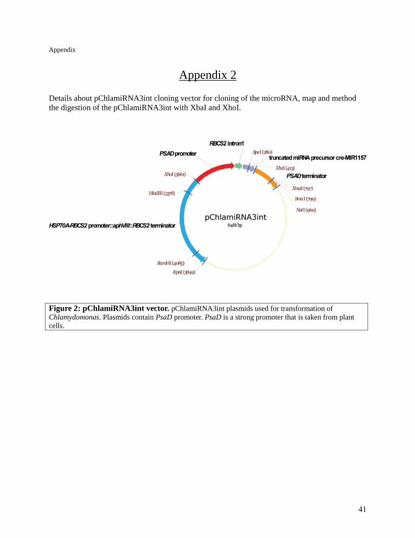
Appendix 2 Details about pChlamiRNA3int cloning vector for cloning of the microRNA, map and method
the digestion of the pChlamiRNA3int with XbaI and XhoI.
Figure 2: pChlamiRNA3int vector. pChlamiRNA3int plasmids used for transformation of
Chlamydomonas. Plasmids contain PsaD promoter. PsaD is a strong promoter that is taken from plant
cells.

42
Appendix
Appendix 3
Sequence of the rppH gene with 3`and 5`UTR sequence , exon and intron.
ATATCGTAGGCTTGGGGGCCGAGTCAGGGACGGGCTCGGGGAAAACACGGCGTGTCTGCGAGATTCCGCGTGGCGCA
TTATCCCTTGCTTGCTGGGTGCCACTACAGAAACAGTCACCAGAAAACCTATCCCTGCCATCGCAAGCTTATAACCG
CGTTTGTTGGTTCTTTCTTGACAACTAGGGTCATCATCCCCTCCTGACCTGTTTGCGCTGCGCGGCCCGTCGCGAGG
CTCTCACGTCAACATATACTTGCTTTCATGCTGGCTACAAGGTGTGCGATTTTCAGGCCGTGACTCTTTCCCGCCCT
CGGTCTCGCGGAACTGCTTCATGCTCTGCTTTCTGCACAGTGACCATGTAATTTATTAAGAACGTGACACATAGTAT
CCAGTGCAAGTTGAGTGCTGCAGCTCGCGCGGGCTCTTGAGCGACTTCAGTTTGCCCTCCGAAAACCCCCGCACTGT
CGCAGGCCGCCCGTCGGTGCGCCCTCGCGGGGAGCCCTGTGCCGCGCGCCTCGCGCTCTGTCCCGCCGCTCGCTCTT
GGCCGTGCCGCCTGCTGCGGCGCCAGCCTCGGGCCCCGGACCGGCATCTTCGGGACCGCCCGGCCCGTCGCCATCCT
CGAGCGACTACATAAACCCAGCGTACTTCTACGCCAACCCCTTCGCGCCAGCTCAGGGCGCAACTTCCTCCAGCTCA
TCGCCCGGATCCAGCGGCCCCAGCTCCCACGCCACTGTCCTCCGCACCGTTGACTCGGTCGTTGCGGCCGCCAGCGC
AGCACGCGCCGCAGCCACCGCCCGCGCGGCCGTAGCGGCACGCTCTGCTTCCCCTTCCCCCTCCTCCTCGAGCGCTT
CCAGCTCTCCCGGCAGCCAGACGCACGCCCGTGCGCCCGCGGTATCCACAGCTGCTGCTGCCCCGTCTTCTTCCCCC
GCGGCCCCCGCCGCGCAGGCCGCCTCCACCGCTCCGGTGCAGCTGTCGCCTGCAGCCGCTCCTGCGCTAGCCACCAA
CACCTTTGCCAGCATGGACGAGGGAGACGCGCCCGCTGCCGCCCCTGGTAGGTTGCTGGGCAGTCGCTGAGCACCAG
TCAGCCACTACCGGGCTCAGGCCCAACTGCTTCCGGGCAACGCTTGTGCAGGCCCCTCCTGCGCGCGGCCCTGTCTT
GTGCGCACTGCATGCGGCTTGCCAGTGGAACTGGCTTGGCACAGGCACCACCGGCAGGCCCTGCCCCTTCAATACGA
GCGCTTTCAGGAGCGCTTCTGTGTTTCACGGTGCTAACCTCGCTCTGTTCTTGTACTAGCAACAGGTGGCGTTCGGC
TGCTTGTGGTGGTGGGTGTGGTCCTGCTGGACGACCCACTGTGGGACCACGAGACGGGCGAGCCCAGCAAGGGTAAG
CCGGCACAGGGGGTAGTGTTTTCCCAACGCCGGGCCGGGGGTACTGTGCCGAGCTCAACTAACGATGCTGGGGGAGG
GGCAAGGGAAAAGGCGCGGGCTCACAGGGGTCAGAGTCTATAGCAGTAACGTTACTGCGGCCGTGGAGCAGTGACAG
CGGTGAGGGGAGAGGATCGCCGGTGTAGGAAGCGTGTGGGCGATGGGGGCATAAGGTGATCAGGGGATGTGGGGTGG
GGCATGGCATGTAGGGCAGTGACGGCACCTGCGCCTGGTTTAGTGTACGGCGTGTTGGCAGCGTATTGCACTGCTTG
CTATGGGTGGAAATGTAGCTTGTTGGTCATGCCCTGTCACGCGCAGGCGCCGCCGCCGCGGATCGGCCTGTGCGGGT
GCTGCTGGCTCAGCGGCCTGTGGGCAAGAGCAACGCGGGGCTGTGGGAGTTCCCGGGCGGCAAGGTGGACCCAGGGG
AGACGCCCGAGGTGAGGACAGCGCTGCGTGGGATGGGGGGTAGGCGTCCATGAAAGACGGAATGGAAAGAAGTATGA
AGTGCCTGCGGGGGCAATGATAGCTCAGGGAAAGCAATGACAGCACACTGGGGAGCAATGATGCAATGAGTCAAGTA
GCCAAACGGGTGTTGGGGTGGGGGGACAGGAGAGGGAAAAGGGCGGGCGGGCAACACAGAAGAGTAAGGGTGCCGAG
CATGTGTGTATGGACTTGCATGTATGCCTGCGTTGGGATCACTGACTATTGATGGGCCGGTGGTGGGTTGCCAGTCC
AATTGTAGCGGGGACGGCGCCAGCCATCGGCTTTCTGCGACACATCAGAAGGATATCGTACTTGCAGTACCGCTGCG
TCTTTGGACGCAGCTATTCCTAAGCACGGCATGCAACACATGACAACAATGGAAACCAACACAGGCGGCGCTGGTTC
GCGAGCTGTATGAGGAGCTGGGCATCTCGGTGGACCCGGCGGACCTGGCGCCGCTCACATTTGCCTCCCACACCTAC
CCCACCTTCCACCTGCTCATGCCGCTGTATGGTGAGTGGGCTGCGACCTGCGGGGGCGTTTGGCCGTGGTCGTGGTG
GTGGTGGTGGTGGTCGTGTCGTGGTTGTGGAGGGGCAAGGATGTGGCCATAGAGGAGGTAGGACATGGCCGCAGGAC
GAGAGAGAGCGGGATTAAAAGACACGGCAATGAAGTACCGGTAACTTGGGTTGCGCTCCGCGTATAGTTCATGCAAA
ACTGTGGACCTTGGCACCACAACCCCTTCCCTCCACCTTAAATCCCTCCTGTCTGTCCCTGTGTGCTTCATTACAGC
CTGCCGGCGCTGGTCGGGCGTGCCTGTGGGCGCGGAGGGCCAGGCGGTGGCGTGGGCGGCTGCGGGCGAGGTGACGT
CTTTCAACCTGACGCCTGCAGACATACCGCTGGTACCGGCTGTGCTGGCGGCTATGCGGCACTACCCCAGCCAGTAG
GCAGGGCAGCAGAGGCGCAGCAGAGGCGCTGAGGGCTGGCCACACAGGCGGCGACGACCGCCACGCGCCATGCGCGC
AGGCGCACGTGTAGGTCAGCGGTTGGGCTGTGATGCCGGCTGGGGACCAGCTGCGCGGTAGCTGTGGGGGCGGCTAG
CGCTGCCGACTAACGCGAGCTGCAAGTTGCACAGGCAGCTCGTCGTGCTGACAGCCAGAAGCCAGGGGCAAGGCCCG
GCGACGATGGGTAGCATAAACACGCCTGGATGGTCCTGTGGCGGCTCGGAGGCGGATGCACGGTTGCTTCAGTCATG
CGCACAGCACGTGCCCGAGTGACCTACGTCATAGGTGCTGCTTACAGCTATGCGCTAGTTTGGAGTTTGGAACTCAC
AGAGAAGAGAAAACACGGTTTTGCCCATTATGCTTCATTTGTGGTGTGATTCTTGTTTCGGTGTGATTCTTGTTTCG
GGCTTGCAGACTTGCTACGATACAGCTGCGCCAGTCGGCGGCCTGGCGCCGTCACGGCTGTTGTCTTGATTTCGCGG
TACAGGGCTGTACTTATATCCGGCTGTACATTGATTGGGATTGCATTTGGGATGGTTGGAGTTAGAGAATTGGTCCG
TAACTCTGAACGTCGCAGATTCCGATGGCGTGTGCCAAAGCGACTTACATATGATTGGCTGTAGGCTCTGAAGGCTG
TGCCTGCGTGCGGTAACGTTGCGAGGCTCTTGGCATCTAGGTGCCGACCCGTAGATGGGGGATCGTGCCAGATGGTA
GGGTAGGACGCGCAGTCGGAAAGGACATGGCGGCGCTTCGTTGCAGCGGGAACACCTAGCATGTGTGGCGGCGGTCT
GACAGACAGGCATGAGATCGGACGAGCTAGACGTGTGGGTCAGCAGAGATTGGTCGAGGTTCTCCCGATGTAATACG
CAGTCGGA
The green colour is 5`UTR, blue is 3` UTR, black is exon and red is intron in rppH. The brown
color is the site of the microRNA attachment.

43
Appendix
Appendix 4 PCR
Tabel 1: Primers used to amplify the paromomycin resistance gene and annealing temperature.
Primers Annealing temperature °C
5`paro 4331 69.6
3`paro 4743 59.8
Tabel 2: Primers used to amplify the psaD vector and annealing temperature.
Primer Annealing temperature °C
3`psaD 495 65.8
5`psaD 6401 63.5
Tabel 3: Primers used to amplify the psaD region and intron part of the vector and annealing
temperature.
Primer Annealing temperature °C
5`intron 63.7
3`psaD 495 65.8
Tabel 4: Primers used in PCR and sequences.
Primer Sequence
5`paro 4331 ACGGCCGACCCGCCCCACGT
3`paro 4743 GATTCCCGTACCTCGTGTTGT
3`psaD495 GCGAAAGCCTCCGAGCTCCGAT
5`psaD6401 CGCCGAGCAAGCCAGGGTTA
5`intron GCAACGCCCGCATTGTGTCGA
PCR condition
1. 94°C 1 minute
2. 94 °C 30 second
3. Annealing temperature 30 sec
4. 72°C 45 second

44
Go to step 2 45 times
5. 72°C 10 minutes
6. 4 ∞
Appendix
Sequence of pChlamiRNA3int.
tatgaacaagtgagtcgacgagcaagcccggcggatcaggcagcgtgcttgcagatttgacttgcaacgcccgcattgtg
tcgacgaaggcttttggctcctctgtcgctgtctcaagcagcatctaaccctgcgtcgccgtttccatttgcaggatgtcatatgggtgt
tgggtcggtgtttttggtcttggttggggtgttggtggtgctggtggaacatgtcaacatgcccaggaaaccaaggcgcgctagcttcct
gggcgcagtgttccagctactagtagccggaacactgccaggaaggagggggaggctgggtgggagaagcggtgtggggc
ggattagccttggagaccgattgctttgggttagtttgggctggcatagtttgggctggcttagttacacctctagatggcagcagctgg
accgcctgtaccatggagaagagctttacttgccgggatggccgatttcgctgattgatacgggatcggagctcggaggctttcgcgcta
ggggctaggcgaagggcagtggtgaccagggtcggtgtgg ggtcggcccacggtcaattagccacaggaggatcaggggg
aggtaggcacgtcgacttggtttgcgaccccgcagttttggcggacgtgctgttgtagatgttagcgtgtgcgtgagccagtggccaacg
tgccacacccattgagaagaccaaccaacttactggcaatatctgccaatgccatactgcatgtaatggccaggccatgtgagagtttgc
cgtgcctgcgcgcgccccgggggcgcagtttagctgaccagccgtgggatgatgcacgca tttgcaaggacagggtaatc
acagcagcaacatggtgggcttaggacagctgtgggtcagtggacggacggcaggggagggacggcgcagctcgggagac
agggggagacagcgtgactgtgcaatgcggccgccaccgcggtggagctccaattcgccctatagtgagtcgtattacgc
gcgctcactggccgtcgttttacaacgtcgtgactgggaaaaccctggcgttacccaacttaatcgccttgcagcacatccccctttcgc
cagctggcgtaatagcgaagaggcccgcaccgatcgcccttcccaacagttgcgcagcctgaatggcgaatgggacgcgc
cctgtagcggcgcattaagcgcggcgggtgtggtggttacgcgcagcgtgaccgctacacttgccagcgccctagcgcccgctcctttcg
ctttcttcccttcctttctcgccacgttcgccggctttccccgtcaagctctaaatcgggggctccctttagggttccgatttagtgctt
tacggcacctcgaccccaaaaaacttgattagggtgatggttcacgtagtgggccatcgccctgatagacggtttttcgccctttgacgt
tggagtccacgttctttaatagtggactcttgttccaaactggaacaacactcaaccctatctcggtctattcttttgatttataaggga
ttttgccgatttcggcctattggttaaaaaatgagctgatttaacaaaaatttaacgcgaattttaacaaaatattaacgcttacaattt
aggtggcacttttcggggaaatgtgcgcggaacccctatttgtttatttttctaaatacattcaaatatgtatccgctcatgagacaata
accctgataaatgcttcaataatattgaaaaaggaagagtatgagtattcaacatttccgtgtcgcccttattcccttttttgcggcatt
ttgccttcctgtttttgctcacccagaaacgctggtgaaagtaaaagatgctgaagatcagttgggtgcacgagtgggttacatcgaact
ggatctcaacagcggtaagatccttgagagttttcgccccgaagaacgttttccaatgatgagcacttttaaagttctgctatgtggcgc
ggtattatcccgtattgacgccgggcaagagcaactcggtcgccgcatacactattctcagaatgacttggttgagtactcaccagtcac
agaaaagcatcttacggatggcatgacagtaagagaattatgcagtgctgccataaccatgagtgataacactgcggccaacttacttct
gacaacgatcggaggaccgaaggagctaaccgcttttttgcacaacatgg gggatcatgtaactcgccttgatcgttgggaaccggagct
gaatgaagccataccaaacgacgagcgtgacaccacgatgcctgtagcaatggcaacaacgttgcgcaaactattaactggcgaactact
tactctagcttcccggcaacaattaatagactggatggaggcggataaagttgcaggaccacttctgcgctcggcccttccggctggctg
gtttattgctgataaatctggagccggtgagcgtgggtctcgcggtatcattgcagcactggggccagatggtaagccctcccgtatcgt
agttatctacacgacggggagtcaggcaactatggatgaacgaaatagacagatcgctgagataggtgcctcactgattaagcattggta
actgtcagaccaagtttactcatatatactttagattgatttaaaacttcatttttaatttaaaaggatctaggtgaagatcctttttgataatctcatg
accaaaatcccttaacgtgagttttcgttccactgagcgtcagaccccgtagaaaagatcaaaggatcttcttgagatcctttttttctg
cgcgtaatctgctgcttgcaaacaaaaaaaccaccgctaccagcggtggtttgtttgccggatcaagagctaccaactctttttccgaag
gtaactggct tcagcagagcgcagataccaaatactgtccttctagtgtagccgtagttaggccaccacttcaagaactctgtagcaccg
cctacatacc tcgctctgct aatcctgttaccagtggctgctgccagtggcgataagtcgtgtcttaccgggttggactcaagacgatag
ttaccggataaggcgcagcggtcgggctgaacggggggttcgtgcacacagcccagcttggagcgaacgacctacaccga
actgagatacctacagcgtgagctatgagaaagcgccacgcttcccgaagggagaaaggcggacaggtatccggtaagcg
gcagggtcggaacaggagagcgcacgagggagcttccagggggaaacgcctggtatctttatagtcctgtcgggtttcgccacctctgac
ttgagcgtcgatttttgtgatgctcgtcaggggggcggagcctatggaaaaacgccagcaacgcggcctttttacggttcctggcctttt
gctggccttttgctcacatgttctttcctgcgttatcccctgattctgtggataaccgtattaccgcctttgagtgagctgataccgctc
gccgcagccgaacgaccgagcgcagcgagtcagtgagcgaggaagcggaagagcgcccaatacgcaaaccgcctctcccc

45
gcgcgttggccgattcattaatgcagctggcacgacaggtttcccgactggaaagcgggcagtgagcgcaacgcaattaatgtgagttag
ctcactcattaggcaccccaggctttacactttatgcttccggctcgtatgttgtgtggaattgtgagcggataacaatttcacacagga
aacagctatgaccatgattacgccaagcgcgcaattaaccctcactaaagggaacaaaagctgggtacccgcttcaaatacgcccagccc
gcccatggagaaagaggccaaaatcaacggaggatcgttacaaccaacaaaattgcaaaactcctccgctttttacgtgttgaaaaagac
Appendix
tgatcagcacgaaacggggagctaagctaccgcttcagcacttgagagcagtatcttccatccaccgccgttcgtcagggggcaaggctc
agatcaacgagcgcctccatttacacggagcggggatcccaacgtccacactgtgctgtcacccacgcgacgcaacccta
cccagccaccaacaccatcaggtccctcagaagaactcgtccaacagccggtaaaacgccagcttttcctccgataccgccccatcccac
ccgcgcccgtactcccgcaggaacgccgcggaacactccggcccgaaccacgggtcctcctcgtgggccagctcgcgcag
caccagcgcgagatcggagtgccggtccgcacggccgacccgccccacgtcgatcagcccggtcacctcgcaggtacgag
ggtcgagcagcacgttgtccgggcacaggtgaccgtggcaaaccgccagatcctcgtccgcaggccgagtccgctccagc
tcggcgagaagccgctcccccgaccaccccttccgctcctcgtccagatcctccaagtcgacgctcccttcagcgacagcacgggccgcc
tgcggcaccgtcaccgcgagactgcgatcgaacggacaccgctcccagtccagcgcgtgcagcgaacgagcgagccccgc
gagcgccaccgccacgtccagccgctgctcccgcggccaccgcgcactggccggacgccccggaaccgcttcggtgacca
accaggcgaccctctcgtccccaccaccctccacaacacgaggtacgggaatccccacctccgccaaccacaccagccgc
tcagcctcacccaacaagcccaccccggcccccagagctgccaccttgacaaacaactcccgcccaccaccccgaagccg
ataaacaccagcccccgaggccccatcctccacaacaacccactcacaaccgggataccgaccccgcagtgcacgcaacg
catcgtccatgcttcgaaattcttcagcaccggggagggcggagtggccatcctgcaaatggaaacggcgacgcagggttagatgctgct
tgagacagcgacagaggagccaaaagccttcgtcgacacaatgcgggcgttgcaagtcaaatctgcaagcacgctgcctg
atccgccgggcttgctcgtcgactcacctggccattttaagatgttgagtgacttctcttgtaaaaaagtaaagaacataggccccctgg
ccggtttatcaggagggcaccgctccaggggctgcatgcgaactgcttgcattggcgcctagcctttgtgggccagggggcttccggata
agggttgcaagtgctcaaataccccatcaaacatcatcctggtttggctgcgctccttctggcgcgcccggcatgcaagcttgatgggat
cttaagctagctgagtggttatgtatagcggcagaatagtcgcgtatgtataagtgctcgtttgtcgctgaaagtggaggtcaccgttcg
gggtcgcgggcttttataccggatgggtgccgccagcgggccgtatggcgccttctggacgccgcgcgccccatcgcggc
ccttccagagcttcccgcgccctcatagcccgccaaatcagtcctgtagcttcatacaaacatacgcaccaatcatgtcaagcctcagcg
agctccccgctcgagcacacacctgcccgtctgcctgacaggaagtgaacgcatgtcgagggaggcctcaccaatcgtca
cacgagccctcgtcagaaacacgtctccgccacgctctccctctcacggccgaccccgcagcccttttgccctttcctaggccaccgaca
ggacccaggcgctctcagcatgcctcaacaacccgtactcgtgccagcggtgcccttgtgctggtgatcgcttggaagcgcatgcgaaga
cgaaggggcggagcaggcggcctggctgttcgaagggctcgccgccagttcgggtgcctttctccacgcgcgcctccaca
cctaccgatgcgtgaaggcaggcaaatgctcatgtttgcccgaactcggagtccttaaaaagccgcttcttgtcgtcgttccgagacatg
ttagcagatcgcagtgccacctttcctgacgcgctcggccccatattcggacgcaattgtcatttgtagcacaattggagcaaatctggc
gaggcagtaggcttttaagttgcaaggcgagagagcaaagtgggacgcggcgtgattattggtatttacgcgacggcccggcgcgttagc
ggcccttcccccaggccagggacgattatgtatcaatattgttgcgttcgggcactcgtgcgagggctcctgcgggctggggagggggat
ctgggaattggaggtacgaccgagatggcttgctcggggggaggtttcctcgccgagcaagccagggttaggtgttgcgctcttgactcg
ttgtgcattctaggaccccactgctactcacaacaagccca
Tabel 5: Different primers attached itself to the pChlamiRNA3int.
Primer Color
5`paro 4331 Blue
3`paro 4743 Blue
3`psaD 495 Orange
5`psaD6401 Orange

46
5`intron Green
Appendix
Appendix 5 MicroRNA
Wmd3.weigelworld.org (http://wmd3.weigelworld.org) was used to suggest MicroRNA. We
choose 4 different microRNA for the experiment.
Tabel 6: Sequences for microRNA1, 5` and 3`
Micro 1 Sequence
5`microRNA1 TAAATGAAGCATAATGGGCAA
3`microRNA1 TTGCCCATTATGCTTTGTTTA
Tabel 7: Sequences for microRNA 3, 5` and 3`
Micro3 Sequence
5`microRNA3 TACATACGCGGTTATAAGCTT
3`microRNA3 AAGCTTATAACCGCGTTTGTA
Tabel 8: Sequences for microRNA 4, 5` and 3`
Micro 4 Sequence
5`microRNA4 TATGATGACCCTAGTTGTCAA
3`microRNA4 AAGCTTATAACCGCGTTTGTA
Tabel 9: Sequences for microRNA 6, 5` and 3`
Micro 6 Sequence
5`microRNA6 TCAATCTCCAAACTAGCGCAT
3`microRNA6 ATGCCGCTAGTTTGGAGATTGA
To transform the Chlamydomonas flanking sequences were added to the MicroRNA (suggested
by the site)

47
Appendix
Tabel 10: MicroRNA 1, 5` and 3` with flanking sequence
microRNA 1with flanking
sequence
Sequence
5`micro1 ctatTTGCCCATTATGCTTTGTTTAtctcgctgatcggcaccatggggg
tggtggtgatcagcgctaTAAATGAAGCATAATGGGCAAg
3`micro1 ctatTTGCCCATTATGCTTTGTTTAtagcgctgatcaccaccaccccca
tggtgccgatcagcggagaTAAATGAAGCATAATGGGCAAg
Tabel 11: MicroRNA 3, 5` and 3` with flanking sequence
microRNA 3with flanking
sequence
Sequence
5`micro3 ctagcAAGCTTATAACCGCGTTTGTAtagcgctgatcaccaccaccc
catggtgccgatcagcgagaTACATACGCGGTTATAAGCTTa
3`micro3 ctagcAAGCTTATAACCGCGTTTGTAtagcgctgatcaccaccaccc
ccatggtgccgatcagcgagaTACATACGCGGTTATAAGCTTa
Tabel 12: MicroRNA 4 , 5` and 3` with flanking sequence
microRNA 4 with flanking
sequence
Sequence
5`micro4 ctagtTTGACAACTAGGGTCAACATAtctcgctgatcggcaccatgg
gggtggtggtgatcagcagcgtaTATGATGACCCTAGTTGTCAAg
3`micro4 ctagcTTGACAACTAGGGTCATCATAtagcgctgatcaccaccaccc
ccatggtgccgatcagcgagaTATGTTGACCCTAGTTGTCAAa
Tabel 13: MicroRNA6, 5` and 3` with flanking sequence
microRNA 6 with flanking
sequence
Sequence
5`micro6 ctagtATGCCGCTAGTTTGGAGATTGAtctcgctgatcggcaccatg
ggggtggtggtgatcagcgtaTCAAACTCCAAACTAGCGCATg
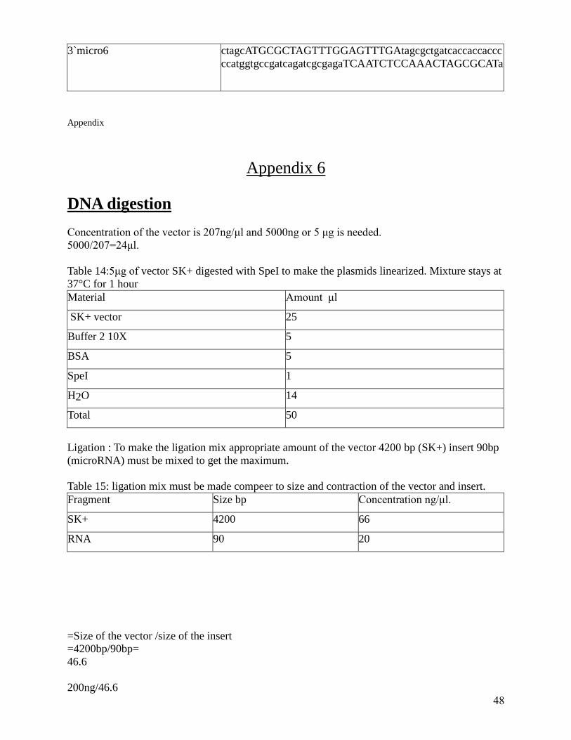
48
3`micro6 ctagcATGCGCTAGTTTGGAGTTTGAtagcgctgatcaccaccaccc
ccatggtgccgatcagatcgcgagaTCAATCTCCAAACTAGCGCATa
Appendix
Appendix 6
DNA digestion
Concentration of the vector is 207ng/μl and 5000ng or 5 μg is needed.
5000/207=24μl.
Table 14:5μg of vector SK+ digested with SpeI to make the plasmids linearized. Mixture stays at
37°C for 1 hour
Material Amount μl
SK+ vector 25
Buffer 2 10X 5
BSA 5
SpeI 1
H2O 14
Total 50
Ligation : To make the ligation mix appropriate amount of the vector 4200 bp (SK+) insert 90bp
(microRNA) must be mixed to get the maximum.
Table 15: ligation mix must be made compeer to size and contraction of the vector and insert.
Fragment Size bp Concentration ng/μl.
SK+ 4200 66
RNA 90 20
=Size of the vector /size of the insert
=4200bp/90bp=
46.6
200ng/46.6

49
=4.34 ng of insert.
To make ligation mix add 200ng vector to 4.4ng insert and add water to final concentration of
6.5μl.
Appendix
Table 16: Appropriate amount of material for digestion of the SK+ with XbaI , SpeI
Material Amount (μl)
DNA (SK+) 3
Buffer 2 10X 1
Enzyme(XbaI , SpeI) 0.25 , 0,25
RNAase 1
BSA 1
Water 3,5
Total 10
Table 17: Appropriate amount of material for digestion of the SK+ with AfeI
Material Amount ( μl)
Buffer 4 10X 1
RNAase 1
DNA 5
Water 2,7
Enzyme AfeI 0.3
Total 10
Table 18: Digestion of the pChlamiRNA3int with XbaI and XhoI.
Material Amount ( μl)
Vector 15 (12000ng )
Buffer 2 10X 5
BSA 5
XbaI 1
XhoI 1
Water 23
Total 50

50
Appendix
Appendix 7
To Remove the DNA from the gel GFX PCR DNA gel band purification kit (GE healthcare)
were used.
LB medium/L
10g tryptone
5 g yeast extract
10g NaCl
LB amp/L plates
10g tryptone
5 g yeast extract
10g NaCl
15 g agar
100μg Amp
TAE (50x)/L
242g Tris base
57.1ml glacial acetic acid
100ml 0,5 M EDTA (pH 8.0)
Loading buffer
0.25% Bromphenol blue
0.25% xylene cyanol FF
30% glycerol
HS media (high salt)
For 1 liter:
20 ml salt stock
20ml phosphate stock
1ml trace element (Hunter)
Salt stock(50x):
For 500ml :
12.5g NH4Cl
0.50g MgSO4. 7 H2O
0.25 g CaCl2. 2 H2O
Phosphate stock (50X)

51
For 500ml:
47g K2HPO4. 3H2O
18g KH2 PO4
Appendix
Materials for min prep:
1,5 ml eppendorf tubes, sterile
Pipettes and tips
box with ice
tube rack for microfuge tube
TEG buffer ( 25 mM Tris-HCl, pH 8,0)
10mM Na2 -EDTA, 50 mM glucose, ice cold(1ml)
NaOH [2n] (1m)
SDS (sodium dodecyl sulfate) [10%] (1m)
Potassium acetate [5 M, pH4.8], ice -cold 5M acetate and 3 M potassium(1ml)
Phenol/chloroform /isoamyl alcohol [ratio 25:24:1] (2ml)
Chloroform /isoamyl alcohol [ratio 24:1] (2ml)
Sterile distilled water (1ml)
Absolute ethanol [ice- cold] (500ml)
70% ethanol [ice- cold] (500ml).
Figure 3: 1kb ladder used in the lab as DNA size marker.

52