task-specific codes for face recognition: how they shape...
TRANSCRIPT
Task-Specific Codes for Face Recognition: How theyShape the Neural Representation of Features forDetection and IndividuationAdrian Nestor., Jean M. Vettel., Michael J. Tarr.*
Department of Cognitive and Linguistic Sciences, Brown University, Providence, Rhode Island, United States of America
Abstract
Background: The variety of ways in which faces are categorized makes face recognition challenging for both synthetic andbiological vision systems. Here we focus on two face processing tasks, detection and individuation, and explore whetherdifferences in task demands lead to differences both in the features most effective for automatic recognition and in thefeatural codes recruited by neural processing.
Methodology/Principal Findings: Our study appeals to a computational framework characterizing the featuresrepresenting object categories as sets of overlapping image fragments. Within this framework, we assess the extent towhich task-relevant information differs across image fragments. Based on objective differences we find among task-specificrepresentations, we test the sensitivity of the human visual system to these different face descriptions independently of oneanother. Both behavior and functional magnetic resonance imaging reveal effects elicited by objective task-specific levels ofinformation. Behaviorally, recognition performance with image fragments improves with increasing task-specificinformation carried by different face fragments. Neurally, this sensitivity to the two tasks manifests as differentiallocalization of neural responses across the ventral visual pathway. Fragments diagnostic for detection evoke larger neuralresponses than non-diagnostic ones in the right posterior fusiform gyrus and bilaterally in the inferior occipital gyrus. Incontrast, fragments diagnostic for individuation evoke larger responses than non-diagnostic ones in the anterior inferiortemporal gyrus. Finally, for individuation only, pattern analysis reveals sensitivity to task-specific information within the right‘‘fusiform face area’’.
Conclusions/Significance: Our results demonstrate: 1) information diagnostic for face detection and individuation isroughly separable; 2) the human visual system is independently sensitive to both types of information; 3) neural responsesdiffer according to the type of task-relevant information considered. More generally, these findings provide evidence for thecomputational utility and the neural validity of fragment-based visual representation and recognition.
Citation: Nestor A, Vettel JM, Tarr MJ (2008) Task-Specific Codes for Face Recognition: How they Shape the Neural Representation of Features for Detection andIndividuation. PLoS ONE 3(12): e3978. doi:10.1371/journal.pone.0003978
Editor: Ernest Greene, University of Southern California, United States of America
Received September 12, 2008; Accepted November 18, 2008; Published December 29, 2008
Copyright: � 2008 Nestor et al. This is an open-access article distributed under the terms of the Creative Commons Attribution License, which permitsunrestricted use, distribution, and reproduction in any medium, provided the original author and source are credited.
Funding: This research was supported by NSF Award #0339122, by the Temporal Dynamics of Learning Center (NSF Science of Learning Center SBE-0542013),and through the generosity of The Ittleson Foundation. The MRI system used in the study was purchased in part by an MRI grant from the NSF. JMV wassupported by a DOD SMART Graduate Fellowship. The funders had no role in study design, data collection and analysis, decision to publish, or preparation of themanuscript.
Competing Interests: The authors have declared that no competing interests exist.
* E-mail: [email protected]
. These authors contributed equally to this work.
Introduction
One of the hallmarks of human face processing is our ability to
recognize faces across a multitude of levels. At the most general
level, we are able to locate and distinguish faces from non-faces in
a visual scene. We can also categorize faces by any number of
traits including gender, ethnicity, expression, and age. Finally, we
routinely discriminate faces from one another at the individual
level (which we refer to as ‘‘individuation’’). This range of abilities
leads us to ask whether the visual system carries out such
categorization tasks using a single general type of category
representation or, alternatively, translates task-specific constraints
into different representations of the overall category. Our study
focuses on the two ends of this categorization spectrum by
comparing the sensitivity of the human visual system to face
detection and face individuation.
Artificial systems for automatic face recognition typically treat
detection and individuation as two separate problems with two
different goals [1]. However it is less clear to what extent biological
systems such as the human visual system adopt a similar dual
approach. Models of human face processing acknowledge the
difference between multiple face recognition tasks. For instance,
two classical models of face processing [2] and its neural basis [3]
are centered around task differences. However, the main
dichotomy these models emphasize is that between expression
recognition and individuation. While an early stage of facial
feature processing is separated from these tasks, the locus of face
detection as well as its relationship with this early stage and the
PLoS ONE | www.plosone.org 1 December 2008 | Volume 3 | Issue 12 | e3978

other tasks are less spelled out. Neuroimaging results also provide
mixed evidence for the neural separability of detection and
individuation. Some studies found that a set of common areas in
the fusiform gyrus [4–7] and the inferior occipital gyrus [5,7] are
sensitive to both face detection and individuation. However, a
recent study [8] uncovered an area sensitive to face individuation
in the right anterior inferior temporal cortex, outside the typical
face-selective regions recruited by detection. Consistent with this
result, studies of white matter connectivity linked behavioral
individuation performance with the structural integrity of fibers
connecting the fusiform gyrus with more anterior areas in the right
hemisphere, including the inferior temporal cortex [9,10]. Finally,
the neural markers of the time course for these two processes also
seem to be different: detection and individuation were associated
with separate M100 and M170 components in a magnetoencelo-
graphy study [11].
If face detection and individuation do recruit different brain
areas and exhibit different time courses, this may point to
processing and representational differences that characterize and
motivate their separation. One possibility is that the two tasks we
consider here pose objectively different constraints on the featural
codes underlying these two types of face recognition. The present
study investigates the neural separability of detection and
individuation precisely by exploring this issue. We hypothesize
that detection and individuation require separate sets of facial
features to optimally achieve their goals and that the visual system
adopts this separation to perform different aspects of face
recognition more effectively. Moreover, we surmise this difference
in the representational bases of the two tasks leads in turn to
differences in neural processing sufficiently robust to be tested by
functional magnetic resonance imaging (fMRI).
The present study investigates this hypothesis by appealing to a
framework for synthetic vision initially developed in the context of
automatic object detection [12]. More recently this framework has
been extended to a model of human vision and has been evaluated
with respect to its original use as a method for category detection
[13,14] and category learning [15]. Within this framework,
categories of objects are represented as sets of overlapping
rectangular image fragments of different aspect ratios, sizes and
resolutions. Many other candidates for the role of object and facial
features have been proposed in the literature: edge structures
[16,17], principal components of images [18,19] or image
segments [20,21], to name just a few. However, for the goals of
our present study, we find fragment features appealing for a
number of reasons. First, they are cue-agnostic in that they do not
commit themselves from the start to a single type of cue, for
example, edges. Second, they naturally account for configural
information, an important aspect of face processing [22], in that
allowing features to overlap constrains the spatial relationship of
otherwise disjoint image fragments. Finally, and most relevant for
the objectives of our present study, Ullman et al’s framework
provides a principled means for establishing optimal task-specific
sets of fragment features for a given category. In the case of faces,
it has been shown, for instance, that such features lend themselves
naturally to deal not only with detection [12] but also with other
categorization tasks, for example, individuation or expression
recognition [23].
We note the problem of mapping diagnostic areas for specific
object categories and particular tasks has been investigated in
human observers using other approaches. For instance, reverse
correlation methods [24–26] and ‘bubbles’ [27,28] in particular,
can effectively produce maps of task-diagnostic regions of images
with respect to a visually-homogeneous category such as faces.
Critically, the task-diagnostic maps produced by these methods are
compatible with different models of how one might divide an
object into features. The fragment-based approach we adopt here
produces concrete ways to decompose a stimulus category into
feature components. We take advantage of this property to gain a
finer-grained view of the representational codes used in task-
specific face processing.
Our investigation proceeds in three stages. First, we evaluate
and compare systematically the task-specific information of face
fragments for detection and individuation. This evaluation enables
the selection of sets of fragments whose task-specific information
varies independently in the two tasks. Second, using these
fragments, we assess and confirm the sensitivity of human subjects
to task-diagnostic fragment features. Third and finally, an fMRI
study tests and reveals that different cortical areas exhibit different
patterns of sensitivity to task-specific information with respect to
our two tasks. These results jointly confirm the separability of
detection and individuation in the human visual system and
provide evidence for different representational codes underlying
the two tasks and driving the noted separation.
Materials and Methods
Evaluation of Task-Specific InformationStimuli. Image face fragments were extracted from a set of 60
face images (12 individuals65 expressions)–Figure S1–selected
from the Tarrlab face database (available online at www.face-
place.org). This set contains near-front-view grayscale Caucasian
faces, half of which were male and half female, wearing no glasses
or other facial accessories and displaying variable affective
expressions. The faces were cropped, down-sampled (60640
pixels) and normalized with the position of the main features, the
eyes and the nose, to permit the mapping of corresponding face
fragments across faces. A similar set containing 12 different
individuals was used for cross-validation of the computational
results. In addition, two sets of 605 natural scene images were
randomly selected from the McGill Calibrated Color Image
Database (tabby.vision.mcgill.ca). These images, mapped to
grayscale, were used in the computation of the amount of
detection-specific information and also provided non-face stimuli
for behavioral testing.
The image fragments were rectangular image patches of
different sizes and aspect ratios [12]. More precisely, we
systematically varied the size, aspect ratio and position of a
rectangular window across each face in steps of 4 pixels. Thus, the
smallest fragments were 464 pixel image patches while the largest
ones contained an entire face. For each face this procedure yielded
6600 image fragments. Consequently, application of the same
procedure to all face images yielded 6600 fragment types, where by
fragment type we mean a class of fragments corresponding to the
same area of the face across different images of the same or
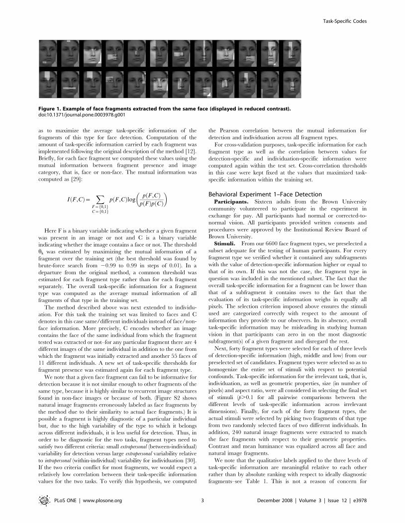
different individuals. Examples of face fragments extracted from
the same image are shown in Figure 1.
Methods. The amount of face-detection information was
computed for each of 396000 fragments generated by extracting
6600 rectangular fragment types from 60 face images. We refer to
these fragments as well as the face images from which they were
extracted as the training set. A separate test set was composed of
an equal number of fragments extracted from a different set of face
images.
To estimate the task-specific information of a fragment of a
given type k, we cross-correlated the given fragment with all face
and non-face images. If the maximum correlation value surpassed
a certain threshold hk, the fragment was considered present in the
image. The threshold hk was computed for each fragment type k so
Task-Specific Codes
PLoS ONE | www.plosone.org 2 December 2008 | Volume 3 | Issue 12 | e3978
as to maximize the average task-specific information of the
fragments of this type for face detection. Computation of the
amount of task-specific information carried by each fragment was
implemented following the original description of the method [12].
Briefly, for each face fragment we computed these values using the
mutual information between fragment presence and image
category, that is, face or non-face. The mutual information was
computed as [29]:
I F ,Cð Þ~X
F~ 0,1f gC~ 0,1f g
p F ,Cð Þlogp F ,Cð Þ
p Fð Þp Cð Þ
� �
Here F is a binary variable indicating whether a given fragment
was present in an image or not and C is a binary variable
indicating whether the image contains a face or not. The threshold
hk was estimated by maximizing the mutual information of a
fragment over the training set (the best threshold was found by
brute-force search from 20.99 to 0.99 in steps of 0.01). In a
departure from the original method, a common threshold was
estimated for each fragment type rather than for each fragment
separately. The overall task-specific information for a fragment
type was computed as the average mutual information of all
fragments of that type in the training set.
The method described above was next extended to individu-
ation. For this task the training set was limited to faces and C
denotes in this case same/different individuals instead of face/non-
face information. More precisely, C encodes whether an image
contains the face of the same individual from which the fragment
tested was extracted or not–for any particular fragment there are 4
different images of the same individual in addition to the one from
which the fragment was initially extracted and another 55 faces of
11 different individuals. A new set of task-specific thresholds for
fragment presence was estimated again for each fragment type.
We note that a given face fragment can fail to be informative for
detection because it is not similar enough to other fragments of the
same type, because it is highly similar to recurrent image structures
found in non-face images or because of both. (Figure S2 shows
natural image fragments erroneously labeled as face fragments by
the method due to their similarity to actual face fragments.) It is
possible a fragment is highly diagnostic of a particular individual
but, due to the high variability of the type to which it belongs
across different individuals, it is less useful for detection. Thus, in
order to be diagnostic for the two tasks, fragment types need to
satisfy two different criteria: small extrapersonal (between-individual)
variability for detection versus large extrapersonal variability relative
to intrapersonal (within-individual) variability for individuation [30].
If the two criteria conflict for most fragments, we would expect a
relatively low correlation between their task-specific information
values for the two tasks. To verify this hypothesis, we computed
the Pearson correlation between the mutual information for
detection and individuation across all fragment types.
For cross-validation purposes, task-specific information for each
fragment type as well as the correlation between values for
detection-specific and individuation-specific information were
computed again within the test set. Cross-correlation thresholds
in this case were kept fixed at the values that maximized task-
specific information within the training set.
Behavioral Experiment 1–Face DetectionParticipants. Sixteen adults from the Brown University
community volunteered to participate in the experiment in
exchange for pay. All participants had normal or corrected-to-
normal vision. All participants provided written consents and
procedures were approved by the Institutional Review Board of
Brown University.
Stimuli. From our 6600 face fragment types, we preselected a
subset adequate for the testing of human participants. For every
fragment type we verified whether it contained any subfragments
with the value of detection-specific information higher or equal to
that of its own. If this was not the case, the fragment type in
question was included in the mentioned subset. The fact that the
overall task-specific information for a fragment can be lower than
that of a subfragment it contains owes to the fact that the
evaluation of its task-specific information weighs in equally all
pixels. The selection criterion imposed above ensures the stimuli
used are categorized correctly with respect to the amount of
information they provide to our observers. In its absence, overall
task-specific information may be misleading in studying human
vision in that participants can zero in on the most diagnostic
subfragment(s) of a given fragment and disregard the rest.
Next, forty fragment types were selected for each of three levels
of detection-specific information (high, middle and low) from our
preselected set of candidates. Fragment types were selected so as to
homogenize the entire set of stimuli with respect to potential
confounds. Task-specific information for the irrelevant task, that is,
individuation, as well as geometric properties, size (in number of
pixels) and aspect ratio, were all considered in selecting the final set
of stimuli (p.0.1 for all pairwise comparisons between the
different levels of task-specific information across irrelevant
dimensions). Finally, for each of the forty fragment types, the
actual stimuli were selected by picking two fragments of that type
from two randomly selected faces of two different individuals. In
addition, 240 natural image fragments were extracted to match
the face fragments with respect to their geometric properties.
Contrast and mean luminance was equalized across all face and
natural image fragments.
We note that the qualitative labels applied to the three levels of
task-specific information are meaningful relative to each other
rather than by absolute ranking with respect to ideally diagnostic
fragments–see Table 1. This is not a reason of concern for
Figure 1. Example of face fragments extracted from the same face (displayed in reduced contrast).doi:10.1371/journal.pone.0003978.g001
Task-Specific Codes
PLoS ONE | www.plosone.org 3 December 2008 | Volume 3 | Issue 12 | e3978
recognition performance in that recognition, in this case detection,
does not rely on single ideal features but on multiple features that
jointly contribute to the process possibly based on their
independent amounts of information they provide [12].
Task. Participants were asked to perform a face detection task
by pressing one of two buttons on a buttonbox. More precisely,
participants were asked to judge whether the single image
fragment displayed at a time was a face fragment or not. The
response was made by pushing one of two buttons with the index
fingers of the left and right hands randomly assigned to signal a
face/non-face response across participants.
On each trial, a cross was presented in the center of the screen
for 400 ms followed by an image fragment for 250 ms. A black
screen replaced the stimulus until the participant made a response
signaling the end of a trial. A stimulus subtended on the average a
visual angle of 2.162.3 from a distance of 70 cm after doubling the
size of the image by pixel replication. Each participant completed
480 trials over the course of two blocks in a 30-minute session.
Each stimulus was shown only once in the entire experimental
session. Trial order was randomized for each participant.
Experimental trials were preceded by a short practice session
allowing the participants to familiarize with the task and the
stimuli. At the end of the experiment participants were asked to
report whether they were familiar with and able to recognize any
of the individuals whose faces were presented in the experiment.
Stimulus design and presentation relied on Matlab 7.5 (Math-
works, Natick, MA, USA) and the Psychophysics Toolbox 3
[31,32] running on an Apple Macintosh using OS X.
Behavioral Experiment 2–Face IndividuationParticipants. Another sixteen adults from the Brown
University community with normal or corrected-to-normal vision
participated in the experiment. All participants provided written
consents and procedures were approved by the Institutional
Review Board of Brown University.
Stimuli. From all our face fragment types, we preselected a
subset in a manner analogous to the one described for the first
experiment. However, this time the relevant task-specific
information was computed for individuation instead of detection.
Next, the procedure described above was followed to select 40
fragment types for each of three levels of individuation-specific
information. In addition to controlling for low-level properties of
the stimuli we also attempted to homogenize the overall set with
respect to detection-specific information–see Table 1. Finally, for
each of the forty fragment types, the actual stimuli were selected by
picking four fragments of that type from two individuals where
each of these individuals supplied two distinct fragments of that
type showing two different expressions.
Interestingly, we note that, as a result of our selection
procedure, the average size of a fragment in this experiment was
significantly larger than the one in the first experiment (two-tailed
t-test p,0.01). This difference is mainly due to the fact that it is
more difficult to find intermediate-sized fragments with small
detection-specific information than it is to find small fragments
with high task-specific information. Conversely, it is more difficult
to find small fragments with high individuation-specific informa-
tion than to find intermediate-sized fragments with low amounts of
relevant information.
Task. Participants were asked to judge whether two
fragments of the same type shown in succession belonged to the
same individual or not. Each trial had the following structure: a
cross appeared for 400 ms in the center of the screen followed by
the first image fragment for 250 ms, a white noise mask with the
same size as the previously presented image for 200 ms, the second
face fragment for another 250 ms and a black screen until the
subject made a button press. Trials were equally divided between
same-individual versus different–individual trials for each
condition. A stimulus subtended on the average a visual angle of
3.462.3 from a distance of 70 cm after doubling the size of the
image. Each participant completed 240 trials over the course of
two blocks in a single a 45-minute session. Each stimulus was
shown only once in the entire experimental session and trial order
was randomized for each participant. In all other respects we
followed the procedure described for Experiment 1.
Functional Magnetic Resonance Imaging (fMRI)Experiment
Participants. Eleven healthy adult members (7 female, age
range: 18–30) of the Brown University community, including one
of the authors JMV, volunteered to participate in the experiment
for pay. None of them took part in the behavioral experiments
described above. Participants were right-handed, with normal or
corrected-to-normal vision and reported no contraindications for
MRI scanning. All participants provided written consents and
procedures were approved by the Institutional Review Board of
Brown University.
Stimuli and behavioral task. Participants were presented
with the same face fragment stimuli as the ones from the
behavioral experiments but using only two levels of task-specific
information: high and low. Participants lay supine and viewed the
rear-projection display through an angled mirror in the bore of the
magnet. Stimuli were presented in 30-second blocks of face
fragments with high/low levels of information for detection or
individuation. The order of the blocks was counterbalanced across
participants. Half of the stimuli in each condition were presented
twice during the experiment but at most once within a block.
Stimulus duration was 800 ms with 700 ms inter-stimulus interval
(20 stimuli per block). The stimuli in the detection and
individuation conditions subtended different viewing angles
similar to those in the behavioral experiment. In each time-
Table 1. Task-specific information* carried by fragments used in the two behavioral tasks.
Level of Information Detection task Individuation task
detection MI individuation MI detection MI individuation MI
high 0.79 (0.02) 0.28 (0.03) 0.70 (0.09) 0.75 (0.03)
middle 0.51 (0.05) 0.25 (0.06) 0.65 (0.11) 0.55 (0.03)
low 0.21 (0.02) 0.3 (0.11) 0.64 (0.9) 0.25 (0.03)
*task-specific information is presented here as the average mutual information (MI) and the standard deviation of each fragment set relative to the mutual informationof a task-ideal fragment (one that is detected in all and only those instances in which the class of interest is present).
doi:10.1371/journal.pone.0003978.t001
Task-Specific Codes
PLoS ONE | www.plosone.org 4 December 2008 | Volume 3 | Issue 12 | e3978
series there were four stimulus blocks separated by 30-second
fixation blocks during which participants were instructed to look at
a fixation cross displayed in the center of the screen. In total, we
acquired 6 time series with image fragments and 2 additional time
series with blocks of faces and objects serving as a standard face-
localizer.
On every trial the participants performed an unrelated task.
The stimuli were randomly jittered 1u to the left/right of the
center fixation cross and the participants performed a one-back
location task by pushing one of two buttons randomly assigned
with the index fingers of the two hands.
Scanning parameters. Scanning was carried out at the
Brown University MRI Research Facility with a Siemens 3T TIM
Trio magnet with an 8-channel phased-array head coil. Functional
images were acquired with an ascending interleaved echo-planar
imaging (EPI) pulse sequence (90 time points per time series;
TR = 3 s; TE = 30 ms; flip angle 90u; 3 mm isotropic voxels; field
of view 19261926144 mm3; 48 slices covering the entire cerebral
cortex). At the beginning of each session, we also acquired a T1-
weighted anatomical image (1 mm isotropic voxels; 160 slices of
size 2566256 mm).
Analysis of imaging data. Analysis was carried out using
AFNI [33] and custom in-house Matlab (Mathworks, Natick MA)
code. The first 5 images of each fMRI time series, during which
subjects maintained fixation, were removed to allow the
hemodynamics to achieve a steady state and to minimize
transient effects of magnetic saturation. Further preprocessing
involved slice scan time correction, 3-D motion correction,
smoothing with a Gaussian kernel of 6 mm FWHM,
normalization (each voxels’s time series was divided by its mean
intensity to convert the data from arbitrary image intensity units to
percent signal modulation) and linear trend removal. Group
analyses were performed after converting functional images into
Talairach space [34].
Conventional univariate mapping analysis was performed on
each participant’s data. For each experimental condition we
constructed a box-car predictor and convolved it with a gamma
function. The general linear model [35] was applied to compute
the coefficient of each predictor independently for each voxel.
Significance maps of the brain were computed by t-tests of
pairwise comparisons between relevant conditions. Significance
levels were corrected by taking into account cluster size and its
false detection probability[36] (p = 0.05 corrected). This type of
analysis was used to contrast the high and low information
conditions for detection and individuation as well as faces versus
objects in a standard face localizer test [7,37].
In addition, we performed multivariate pattern analysis [38] to
distinguish between the two levels of task-specific information for
each task in face selective areas. Principal component analysis
(PCA) was first applied to the coefficients of all voxels in a given
area across all blocks to reduce the dimensionality of the patterns.
A ‘leave-one-out’ (‘jackknife’) classification procedure was then
carried out on the resultant patterns. More precisely, we trained a
linear classifier on the patterns corresponding to all blocks except
one and tested it on this remaining pattern. This procedure was
repeated in turn for all blocks, every time leaving out a different
pattern. For the purposes of pattern classification we used a linear
support vector machine (SVM)–other classifiers we tested, such as
a single-layer perceptron, yielded similar results. Importantly,
multivariate analysis was carried out on a version of the data that
had not been spatially smoothed, thus preserving high-frequency
spatial information.
Results
Task-Specific Fragment Information for Detection andIndividuation
Task-specific information values for fragment types reliably
transferred from the training face dataset to a new dataset. The
correlation between the scores for the training and the test dataset
were significant for both detection (r = 0.93, p,0.001) and
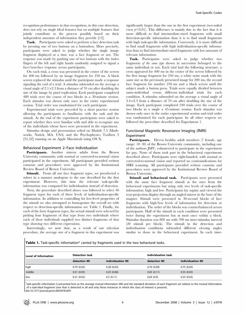
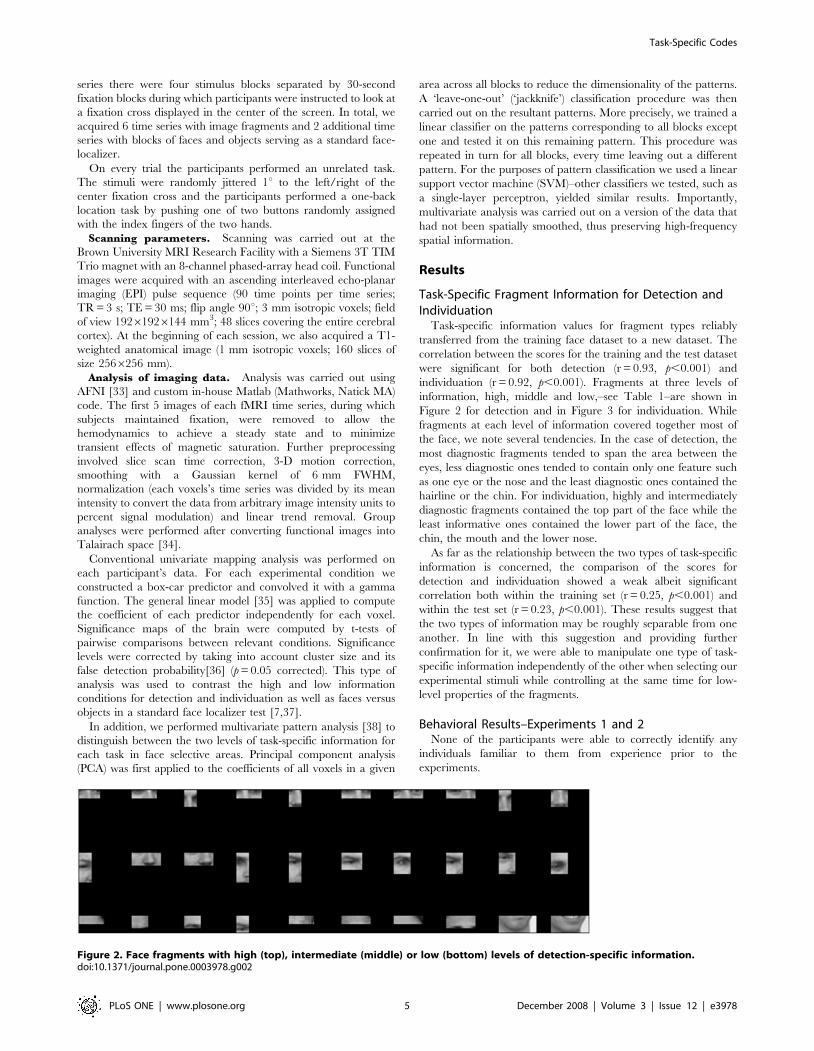
individuation (r = 0.92, p,0.001). Fragments at three levels of
information, high, middle and low,–see Table 1–are shown in
Figure 2 for detection and in Figure 3 for individuation. While
fragments at each level of information covered together most of
the face, we note several tendencies. In the case of detection, the
most diagnostic fragments tended to span the area between the
eyes, less diagnostic ones tended to contain only one feature such
as one eye or the nose and the least diagnostic ones contained the
hairline or the chin. For individuation, highly and intermediately
diagnostic fragments contained the top part of the face while the
least informative ones contained the lower part of the face, the
chin, the mouth and the lower nose.
As far as the relationship between the two types of task-specific
information is concerned, the comparison of the scores for
detection and individuation showed a weak albeit significant
correlation both within the training set (r = 0.25, p,0.001) and
within the test set (r = 0.23, p,0.001). These results suggest that
the two types of information may be roughly separable from one
another. In line with this suggestion and providing further
confirmation for it, we were able to manipulate one type of task-
specific information independently of the other when selecting our
experimental stimuli while controlling at the same time for low-
level properties of the fragments.
Behavioral Results–Experiments 1 and 2None of the participants were able to correctly identify any
individuals familiar to them from experience prior to the
experiments.
Figure 2. Face fragments with high (top), intermediate (middle) or low (bottom) levels of detection-specific information.doi:10.1371/journal.pone.0003978.g002
Task-Specific Codes
PLoS ONE | www.plosone.org 5 December 2008 | Volume 3 | Issue 12 | e3978

Accuracy scores for each participant were computed using d’, a
signal detection measure of discrimination performance between
two classes combining the relative contribution of hits and false
alarms [39]. In the case of detection, hits and false alarms were
provided by correct and incorrect ‘face’ responses, while for
individuation they were provided by correct and incorrect ‘same-
individual’ responses.
Repeated measures analysis of variance was conducted across
the discrimination performance of the participants in each
experiment. We found a main effect of information level for both
detection (F(15, 30) = 3.92, p,0.001) and individuation (F(15,
30) = 4.88, p,0.001) indicating that participants are less accurate
at recognizing faces with decreasing amounts of relevant
information–Figure 4. In addition, we performed a two-way
analysis of variance across the level of information and task
combining the results of the two experiments. The analysis
revealed significant effects for both the task factor (F(1,
90) = 231.71, p,0.001) and the interaction of the two factors
(F(2, 90) = 7.33, p,0.01). We also note that performance was
above chance for all information levels in both experiments
(significantly above d’ = 0).
Similar analyses computed for reaction times revealed no
significant effect of task-specific information for either task
(p.0.1)–Figure 5. In the two-way analysis of variance, we found
a main effect of task (F(1, 90) = 23.63, p,0.001) but no significant
interaction (p.0.5). The main effect of the task is a good indicator
of task difficulty: face individuation was more difficult than face
detection despite the larger size of the stimuli as reflected by both
discrimination performance and reaction times.
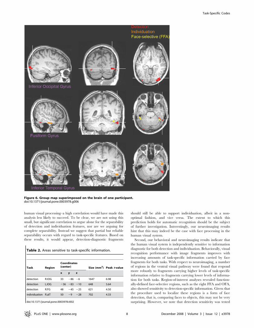
fMRI ResultsGroup maps of task-specific information effects were obtained
by averaging the statistical parametric maps of individual
participants–Figure 6. The comparison of the two detection
conditions revealed two areas more active for diagnostic fragments
than non-diagnostic fragments across participants (p,0.05,
Figure 3. Face fragments with high (top), intermediate (middle) or low (bottom) individuation-specific information.doi:10.1371/journal.pone.0003978.g003
Figure 4. Discrimination accuracy for detection and individuation across three levels of task-specific information (mean6SEM).doi:10.1371/journal.pone.0003978.g004
Task-Specific Codes
PLoS ONE | www.plosone.org 6 December 2008 | Volume 3 | Issue 12 | e3978

corrected): a region in the right posterior fusiform gyrus (pFG) and
a bilateral region in the inferior occipital gyrus (IOG)–see Table 2.
The first of these regions borders the functionally-localized face-
selective region we find in the right fusiform gyrus, also known as
the right ‘fusiform face area’ (FFA) [27], while the second
surrounds and completely contains the functionally-localized
face-selective region we found in the right IOG, the right ‘occipital
face area’ (OFA) [26]. (Consistent with other studies, the left OFA
was not reliably found across a number of participants and
therefore excluded from analyses). In contrast, the comparison of
the two individuation conditions revealed one area more active in
the right anterior inferior temporal gyrus (aIT).
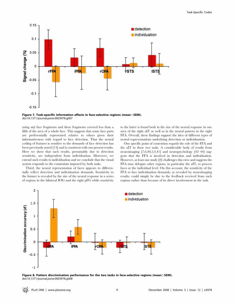
Specific region-of-interest analyses were performed in the
functionally-localized face-selective areas nominally forming the
core system for face processing [3]: the FFA, the OFA and another
region located bilaterally in the superior temporal sulcus (STS).
The areas were individually localized across participants using the
data from the standard face-localizer scans, and the average
percentage signal change was computed for each area and each
task. Detection effects were reliably found in the right FFA
(t(7) = 4.14, p,0.05) and the right OFA (t(7) = 3.22, p,0.05)–
Figure 7. In contrast, no individuation-specific information effects
were found in any of these regions (p.0.1).
In addition to the analysis of face-selective regions of interest, we
examined the response of regions localized for one task, with the
other task as well; for example, we examined whether there is
sensitivity to detection-specific information in the aIT region
already identified as sensitive to individuation-specific information.
No significant effects were found for any of these comparisons.
Next, pattern analysis was applied across blocks within each
face-selective region for each subject. The discriminability of the
two levels of information for each task was encoded using again the
d’ measure. Neural responses elicited by higher-information
fragments were encoded as hits or false alarms when recognized
correctly and incorrectly, respectively. The only region that
showed significant sensitivity across participants was the right FFA
when viewing fragments varying across levels of individuation-
specific information (t(7) = 3.67, p,0.01)–Figure 8. No other
region was significantly different from chance for either task
(p.0.05).
Discussion
Our study examines how different tasks impact the featural code
used in human face processing. From the many tasks that
constitute face recognition, we focused on detection and
individuation in that they represent two ends of the face
recognition spectrum. This comparison is made tractable by
adopting a general computational framework–fragment-based
category representations. The concrete question we ask within
this framework is twofold: how does the task-specific information
carried by face fragments objectively vary within and across tasks
and how sensitive is the visual system to these types of variation?
First, from a computational perspective, we find that the two
types of task-specific information, for detection and individuation,
are roughly separable when considering the mutual information
between fragment presence and the category of interest. This
result is not entirely unexpected given that in order to be
diagnostic for the two tasks a fragment type would need to satisfy
two criteria presumably at tension with each other. For detection,
the area of the face captured by a fragment would need to exhibit
small extrapersonal variability and be visually dissimilar from
recurrent non-face image structures. For individuation, on the
other hand, the same area would need to exhibit large extrapersonal
variability relative to intrapersonal variability [30]. Consistent with
this tension, the correlation we found between the two types of
task-specific information is relatively small although still signifi-
cant. The small size of this correlation is what justifies and explains
our ability to select (with relative ease) two subsets of fragments
that vary independently with respect to their task-specific
information for the two tasks. We then use these separate subsets
to examine the relationship between detection and identification in
Figure 5. Reaction times for detection and individuation across three levels of task-specific information (mean6SEM).doi:10.1371/journal.pone.0003978.g005
Task-Specific Codes
PLoS ONE | www.plosone.org 7 December 2008 | Volume 3 | Issue 12 | e3978
human visual processing–a high correlation would have made this
analysis less likely to succeed. To be clear, we are not using this
small, but significant correlation to argue alone for the separability
of detection and individuation features, nor are we arguing for
complete separability. Instead we suggest that partial but reliable
separability occurs with regard to task-specific features. Based on
these results, it would appear, detection-diagnostic fragments
should still be able to support individuation, albeit in a non-
optimal fashion, and vice versa. The extent to which this
prediction holds for automatic recognition should be the subject
of further investigation. Interestingly, our neuroimaging results
hint that this may indeed be the case with face processing in the
human visual system.
Second, our behavioral and neuroimaging results indicate that
the human visual system is independently sensitive to information
diagnostic for both detection and individuation. Behaviorally, visual
recognition performance with image fragments improves with
increasing amounts of task-specific information carried by face
fragments for both tasks. With respect to neuroimaging, a number
of regions in the ventral visual pathway were found that respond
more robustly to fragments carrying higher levels of task-specific
information relative to fragments carrying lower levels of informa-
tion for both tasks. Region-of-interest analyses revealed function-
ally-defined face-selective regions, such as the right FFA and OFA,
also showed sensitivity to detection-specific information. Given that
the procedure used to localize these regions is a form of face
detection, that is, comparing faces to objects, this may not be very
surprising. However, we note that detection sensitivity was tested
Figure 6. Group map superimposed on the brain of one participant.doi:10.1371/journal.pone.0003978.g006
Table 2. Areas sensitive to task-specific information.
Task Region
Coordinates(center) Size (mm3) Peak t-value
x y z
detection R.IOG 33 286 26 1647 6.98
detection L.IOG 236 283 210 648 5.64
detection R.FG 48 245 225 621 4.50
individuation R.aIT 50 29 228 702 4.33
doi:10.1371/journal.pone.0003978.t002
Task-Specific Codes
PLoS ONE | www.plosone.org 8 December 2008 | Volume 3 | Issue 12 | e3978
using only face fragments and these fragments covered less than a
fifth of the area of a whole face. This suggests that some face parts
are preferentially represented relative to others given their
informativeness with regard to face detection. That the neural
coding of features is sensitive to the demands of face detection has
been previously noted [14] and is consistent with our present results.
Here we show that such results, presumably due to detection
sensitivity, are independent from individuation. Moreover, we
extend such results to individuation and we conclude that the visual
system responds to the constraints imposed by both tasks.
Third, the neural representation of faces appears to differen-
tially reflect detection and individuation demands. Sensitivity to
the former is revealed by the size of the neural response in a series
of regions in the bilateral IOG and the right pFG while sensitivity
to the latter is found both in the size of the neural response in one
area of the right aIT as well as in the neural pattern in the right
FFA. Overall, these findings support the idea of different types of
neural representations underlying detection or individuation.
One specific point of contention regards the role of the FFA and
the aIT in these two tasks. A considerable body of results from
neuroimaging [7,6,40,5,4,41] and neuropsychology [42–44] sug-
gests that the FFA is involved in detection and individuation.
However, at least one study [8] challenges this view and suggests the
FFA may delegate other regions, in particular the aIT, to process
faces at the individual level. On this account, the sensitivity of the
FFA to face individuation demands, as revealed by neuroimaging
results, could simply be due to the feedback received from such
regions rather than because of its direct involvement in the task.
Figure 7. Task-specific information effects in face-selective regions (mean6SEM).doi:10.1371/journal.pone.0003978.g007
Figure 8. Pattern discrimination performance for the two tasks in face-selective regions (mean6SEM).doi:10.1371/journal.pone.0003978.g008
Task-Specific Codes
PLoS ONE | www.plosone.org 9 December 2008 | Volume 3 | Issue 12 | e3978

An interesting perspective on this issue comes from the study of
congenital prosopagnosia, a condition characterized by profound
impairment in face recognition, particularly at the individual level,
in the absence of an obvious insult to the brain. A recent diffusion
tensor imaging study [10] associated recognition performance in
prosopagnosics with the degree of structural integrity of the right
Inferior Longitudinal Fasciculus (ILF). ILF is one of the two major
fiber tracts passing through the fusiform gyrus and connects the
lingual and fusiform gyri with the superior, inferior and middle
temporal gyri as well as the hippocampus and the parahippo-
campus. While, given the length of the tract, this result by itself
may fail to directly involve the aIT, it could explain cortical
volume alterations in the inferotemporal cortex observed in this
population [45]. Interestingly, activation in face-selective areas in
prosopagnosics and normal humans appears to be comparable
[46,47]–but see [44]. This pattern of results seems to suggest the
aIT is important for face recognition and a partial breakdown in
the communication between the fusiform gyrus and the aIT may
be a plausible source of face individuation deficits.
Our results can help bridge two potentially divergent lines of
evidence. In agreement with most neuroimaging studies, we find
evidence for the direct involvement of the FFA in face individuation
and against the hypothesis of an indirect feedback-conditioned role.
At the same time, we also find evidence for the role of the aIT in
individuation [8], a role also suggested by the neuropsychological
literature. However, the FFA and the aIT turned out to exhibit two
different types of sensitivity to individuation, one revealed by
multivariate pattern analysis and the other by univariate analysis.
This difference by itself does not explain, of course, why the same
studies fail to involve both areas in individuation–they typically
implicate only the FFA or aIT, but not both. In addition to the
difference in sensitivity revealed by our two analysis methods, this
discrepancy can be accounted for in several other ways. First, face-
localizer tests are optimized primarily for detection, that is, they
compare faces to other categories of objects, and thus can fail to
involve neural structures that serve primarily face individuation.
Second, given the special status conferred to a group of regions
including the FFA as the ‘core system’ for face processing [3],
neuroimaging research has particularly focused on these restricted
brain areas and, thus, may fail to observe relevant activation in
other areas, for example, the aIT. Third, and most importantly, our
stimuli were face fragments selected based on their task-specific
information instead of whole faces. Thus, our study is aimed at
dealing in a more direct manner with individuation sensitivity than
previous studies using whole face images.
Overall, we interpret our current results as supporting the
involvement of the FFA primarily in detection and of the right aIT
in individuation. However, individual face differences are already
represented in the FFA. These differences seem to be further
amplified and recoded in the right aIT insofar as they lead to
different types of sensitivity to individuation-specific information.
If this is the case, we expect the FFA to support individuation
without the help of the aIT at least to some extent. However, if the
features encoded in the FFA serve primarily face detection,
individuation processing in this area is likely to be suboptimal.
This motivates and explains the recruitment of a different area, the
aIT, dedicated to a task critical in our everyday life, individuation.
Our current results are also interesting from the perspective of
the hierarchy of visual processing along the ventral visual pathway
[48]. The idea that visual features of increasing complexity build
successively upon each other at different levels of visual processing
has been incorporated in many neurally-inspired models of object
[16,49] and face recognition [17]. More recently, this approach
has also been extended to fragment-based processing [50,51].
Composing larger, more specialized fragments successively out of
smaller and more generic fragments across a series of represen-
tational levels is a computationally attractive means for instanti-
ating hierarchical processing. Relevant to our study, this hints that
larger individuation-dedicated fragments may be separately
represented and built upon smaller detection-dedicated fragments.
One concrete possibility suggested by the present results is that
features optimal for individuation are represented as patterns over
face detection features within the FFA and then recoded in a more
localist fashion within the right aIT. The neural plausibility of this
hypothesis is the goal of further research.
Finally, our results reinforce the assumption that overlapping
image fragments provide a neurally-plausible representational
schema for object features. The argument the present study makes
for their plausibility and utility is that they help clarify how
different tasks shape the neural code underlying face recognition.
However, we should also acknowledge the limits of this approach
as an actual model. One question regards the effectiveness of using
rectangular fragments to represent what are, most likely, non-
rectangular features. In response to this, we take rectangular-
shaped features to be a rough but reasonable approximation of the
actual features encoded by the visual system. More plausible
features with smoother edges without sharp corners should be
furthered examined. However, we argue, our investigation of task-
specific information as carried by fragments is systematic and
sufficiently detailed that the precise shape of the features should
not alter significantly the conclusions we reached above.
Consistent with this, replacement of square-like features with
circular ones did not significantly alter measurements of detection-
specific information in a related study [14]. Another more critical
issue concerns the manner in which fragments are actually
represented by the neural code. For instance, every fragment
contains a multitude of cues with different contributions to various
recognition tasks. How such cues are separately considered,
encoded and integrated into a unified representation is an
important problem that should be addressed further. For present
purposes, we treat rectangular image patches as reasonable stand-
ins for the actual biological representations of different face parts
and subparts.
To conclude, we examined and found that face detection and
individuation place different task constraints on the representa-
tional code required for automatic and human face recognition.
More generally, we interpret these results as further evidence for
the soundness of fragment-based models of human object
processing.
Supporting Information
Figure S1 Training set of face images
Found at: doi:10.1371/journal.pone.0003978.s001 (0.48 MB TIF)
Figure S2 Natural image fragments erroneously labeled as face
fragments by the method
Found at: doi:10.1371/journal.pone.0003978.s002 (0.08 MB TIF)
Acknowledgments
We thank the members of the Perceptual Expertise Network (PEN) for
many helpful and insightful comments as well as the staff of the Brown
University Magnetic Resonance Facility for assistance with data collection.
Author Contributions
Conceived and designed the experiments: AN JMV MJT. Performed the
experiments: AN JMV. Analyzed the data: AN JMV. Wrote the paper: AN
JMV MJT.
Task-Specific Codes
PLoS ONE | www.plosone.org 10 December 2008 | Volume 3 | Issue 12 | e3978

References
1. Zhao W, Chellappa R, Phillips PJ, Rosenfeld A (2003) Face recognition: A
literature survey. ACM Computing Surveys 35: 399–458.2. Bruce V, Young A (1986) Understanding Face Recognition. British Journal of
Psychology 77: 305–327.3. Haxby JV, Hoffman EA, Gobbini MI (2000) The distributed human neural
system for face perception. Trends Cogn Sci 4: 223–233.
4. Rotshtein P, Henson RN, Treves A, Driver J, Dolan RJ (2005) MorphingMarilyn into Maggie dissociates physical and identity face representations in the
brain. Nat Neurosci 8: 107–113.5. Pourtois G, Schwartz S, Seghier ML, Lazeyras F, Vuilleumier P (2005) Portraits
or people? Distinct representations of face identity in the human visual cortex.
J Cogn Neurosci 17: 1043–1057.6. Grill-Spector K, Knouf N, Kanwisher N (2004) The fusiform face area subserves
face perception, not generic within-category identification. Nat Neurosci 7:555–562.
7. Gauthier I, Tarr MJ, Moylan J, Skudlarski P, Gore JC, Anderson AW (2000)The fusiform ‘‘face area’’ is part of a network that processes faces at the
individual level. J Cogn Neurosci 12: 495–504.
8. Kriegeskorte N, Formisano E, Sorger B, Goebel R (2007) Individual faces elicitdistinct response patterns in human anterior temporal cortex. Proc Natl Acad
Sci U S A 104: 20600–20605.9. Thomas C, Moya L, Avidan G, Humphreys K, Jung KJ, et al. (2008) Reduction
in white matter connectivity, revealed by diffusion tensor imaging, may account
for age-related changes in face perception. J Cogn Neurosci 20: 268–284.10. Thomas C, Avidan G, Humphreys K, Jung K, Gao F, Behrmann M (in press)
Reduced structural connectivity in ventral visual cortex in congenitalprosopagnosia. Nat Neurosci.
11. Liu J, Harris A, Kanwisher N (2002) Stages of processing in face perception: anMEG study. Nat Neurosci 5: 910–916.
12. Ullman S, Vidal-Naquet M, Sali E (2002) Visual features of intermediate
complexity and their use in classification. Nat Neurosci 5: 682–687.13. Harel A, Ullman S, Epshtein B, Bentin S (2007) Mutual information of image
fragments predicts categorization in humans: Electrophysiological and behav-ioral evidence. Vision Res 47: 2010–2020.
14. Lerner Y, Epshtein B, Ullman S, Malach R (2008) Class information predicts
activation by object fragments in human object areas. J Cogn Neurosci 20:1189–1206.
15. Hegde J, Bart E, Kersten D (2008) Fragment-based learning of visual objectcategories. Curr Biol 18: 597–601.
16. Riesenhuber M, Poggio T (1999) Hierarchical models of object recognition incortex. Nat Neurosci 2: 1019–1025.
17. Jiang X, Rosen E, Zeffiro T, VanMeter J, Blanz V, Riesenhuber M (2006)
Evaluation of a shape-based model of human face discrimination using fMRIand behavioral techniques. Neuron 50: 159–172.
18. Turk M, Pentland A (1991) Eigenfaces for recognition. J Cogn Neurosci 3:71–86.
19. Hancock PJB, Burton AM, Bruce V (1996) Face processing: Human perception
and principal components analysis. Memory and Cognition 24: 26–40.20. Nestor A, Tarr MJ (2008) The segmental structure of faces and its use in gender
recognition. J Vis 8: 1–12.21. Balas BJ, Sinha P (2006) Region-based representations for face recognition.
ACM Transactions on Applied Perception 3: 354–375.22. Maurer D, Grand RL, Mondloch CJ (2002) The many faces of configural
processing. Trends Cogn Sci 6: 255–260.
23. Zhang L, Cottrell GW (2005) Holistic processing develops because it is good.Proceedings of the Cognitive Science Society. pp 2428–2433.
24. Neri P, Levi DM (2006) Receptive versus perceptive fields from the reverse-correlation viewpoint. Vision Res 46: 2465–2474.
25. Murray RF, Bennett PJ, Sekuler AB (2002) Optimal methods for calculating
classification images: Weighted sums. J Vis 2: 79–104.
26. Eckstein MP, Ahumada AJ (2002) Classification images: a tool to analyze visual
strategies. J Vis 2: 1x.27. Gosselin F, Schyns PG (2001) Bubbles: a technique to reveal the use of
information in recognition tasks. Vision Res 41: 2261–2271.28. Gosselin F, Schyns PG (2004) No troubles with bubbles: a reply to Murray and
Gold. Vision Res 44: 471–477.
29. Cover T, Thomas J (1991) Elements of information theory. New York: Wiley.30. Moghaddam B, Jebara T, Pentland A (2000) Bayesian face recognition. Pattern
Recognition 33: 1771–1782.31. Brainard DH (1997) The psychophysics toolbox. Spat Vis 10: 433–436.
32. Pelli DG (1997) The VideoToolbox software for visual psychophysics:
Transforming numbers into movies. Spat Vis 10: 437–442.33. Cox RW (1996) AFNI: software for analysis and visualization of functional
magnetic resonance neuroimages. Comput Biomed Res 29: 162–173.34. Talairach J, Tournoux P (1988) Co-planar stereotaxic atlas of the human brain:
3-dimensional proportional system - an approach to cerebral imaging. NewYork: Thieme Medical Publishers.
35. Friston KJ, Holmes AP, Worsley KJ, Poline JP, Frith CD, Frackowiak RSJ
(1995) Statistical parametric maps in functional imaging: a general linearapproach. Hum Brain Mapp 2: 189–210.
36. Forman SD, Cohen JD, Fitzgerald M, Eddy WF, Mintun MA, Noll DC (1995)Improved assessment of significant activation in functional magnetic resonance
imaging (fMRI): use of a cluster-size threshold. Magn Reson Med 33: 636–647.
37. Kanwisher N, McDermott J, Chun MM (1997) The fusiform face area: amodule in human extrastriate cortex specialized for face perception. J Neurosci
17: 4302–4311.38. O’Toole AJ, Jiang F, Abdi H, Penard N, Dunlop JP, Parent MA (2007)
Theoretical, statistical, and practical perspectives on pattern-based classificationapproaches to the analysis of functional neuroimaging data. J Cogn Neurosci 19:
1735–1752.
39. Snodgrass JG, Corwin J (1988) Pragmatics of measuring recognition memory:applications to dementia and amnesia. J Exp Psychol Gen 117: 34–50.
40. Loffler G, Yourganov G, Wilkinson F, Wilson HR (2005) fMRI evidence for theneural representation of faces. Nat Neurosci 8: 1386–1390.
41. Winston JS, Henson RN, Fine-Goulden MR, Dolan RJ (2004) fMRI-adaptation
reveals dissociable neural representations of identity and expression in faceperception. J Neurophysiol 92: 1830–1839.
42. Barton JJ, Press DZ, Keenan JP, O’Connor M (2002) Lesions of the fusiformface area impair perception of facial configuration in prosopagnosia. Neurology
58: 71–78.43. Damasio AR, Damasio H, Van Hoesen GW (1982) Prosopagnosia: anatomic
basis and behavioral mechanisms. Neurology 32: 331–341.
44. Hadjikhani N, de Gelder B (2002) Neural basis of prosopagnosia: an fMRIstudy. Hum Brain Mapp 16: 176–182.
45. Behrmann M, Avidan G, Gao F, Black S (2007) Structural imaging revealsanatomical alterations in inferotemporal cortex in congenital prosopagnosia.
Cereb Cortex 17: 2354–2363.
46. Hasson U, Avidan G, Deouell LY, Bentin S, Malach R (2003) Face-selectiveactivation in a congenital prosopagnosic subject. J Cogn Neurosci 15: 419–431.
47. Avidan G, Hasson U, Malach R, Behrmann M (2005) Detailed exploration offace-related processing in congenital prosopagnosia: 2. Functional neuroimaging
findings. J Cogn Neurosci 17: 1150–1167.48. Felleman DJ, Van Essen DC (1991) Distributed hierarchical processing in the
primate cerebral cortex. Cerebral Cortex 1: 1–47.
49. Serre T, Oliva A, Poggio T (2007) A feedforward architecture accounts for rapidcategorization. Proc Natl Acad Sci U S A 104: 6424–6429.
50. Ullman S (2007) Object recognition and segmentation by a fragment-basedhierarchy. Trends Cogn Sci 11: 58–64.
51. Epshtein B, Lifshitz I, Ullman S (2008) Image interpretation by a single bottom-
up top-down cycle. Proc Natl Acad Sci U S A 105: 14298–14303.
Task-Specific Codes
PLoS ONE | www.plosone.org 11 December 2008 | Volume 3 | Issue 12 | e3978