tauopathy in transgenic (shr72) rats impairs function … · modulates energy metabolism, the...
TRANSCRIPT

RESEARCH Open Access
Tauopathy in transgenic (SHR72) ratsimpairs function of central noradrenergicsystem and promotes neuroinflammationBoris Mravec1,2*, Katarina Lejavova1,2, Peter Vargovic1, Katarina Ondicova1,2, Lubica Horvathova1, Petr Novak3,4,Georg Manz5, Peter Filipcik3,4, Michal Novak3,4 and Richard Kvetnansky1
Abstract
Background: Brain norepinephrine (NE) plays an important role in the modulation of stress response andneuroinflammation. Recent studies indicate that in Alzheimer’s disease (AD), the tau neuropathology begins in thelocus coeruleus (LC) which is the main source of brain NE. Therefore, we investigated the changes in brain NE systemand also the immune status under basal and stress conditions in transgenic rats over-expressing the human truncatedtau protein.
Methods: Brainstem catecholaminergic cell groups (LC, A1, and A2) and forebrain subcortical (nucleus basalisof Meynert), hippocampal (cornu ammonis, dentate gyrus), and neocortical areas (frontal and temporal associationcortices) were analyzed for NE and interleukin 6 (IL-6) mRNA levels in unstressed rats and also in rats exposed to singleor repeated immobilization. Moreover, gene expression of NE-biosynthetic enzyme, tyrosine hydroxylase (TH), andseveral pro- and anti-inflammatory mediators were determined in the LC.
Results: It was found that tauopathy reduced basal NE levels in forebrain areas, while the gene expression of IL-6 wasincreased in all selected areas at the same time. The differences between wild-type and transgenic rats in brain NE andIL-6 mRNA levels were observed in stressed animals as well. Tauopathy increased also the gene expression of TH in theLC. In addition, the LC exhibited exaggerated expression of pro- and anti-inflammatory mediators (IL-6, TNFα, induciblenitric oxide synthases 2 (iNOS2), and interleukin 10 (IL-10)) in transgenic rats suggesting that tauopathy affectsalso the immune background in LC. Positive correlation between NE and IL-6 mRNA levels in cornu ammonis in stressedtransgenic animals indicated the reduction of anti-inflammatory effect of NE.
Conclusions: Our data thus showed that tauopathy alters the functions of LC further leading to the reduction of NElevels and exaggeration of neuroinflammation in forebrain. These findings support the assumption that tau-relateddysfunction of LC activates the vicious circle perpetuating neurodegeneration leading to the development of AD.
Keywords: Alzheimer’s disease, Locus coeruleus, Neuroinflammation, Stress, Tauopathy, Transgenic rats
BackgroundSporadic Alzheimer’s disease (AD) represents the mostprevalent form of dementia in the elderly, with severalhypotheses being proposed to explain its etiopathogenesis.These hypotheses have focused either on the role ofamyloid β, hyperphosphorylated and truncated tau protein,
neuroinflammation, altered insulin signalization, impairedblood-brain barrier permeability, or other related factorsand mechanisms [1, 2]. However, despite such enormousscientific effort, the primary factors responsible forthe development of AD still remain only vaguely defined.Recently accumulated evidence indicates that alter-
ation of the brain’s noradrenergic system plays animportant role in the development of AD-relatedneuropathology during the early stages of AD. Centralnorepinephrine (NE)-synthesizing neurons are locatedpredominantly in the brainstem, particularly in the locus
* Correspondence: [email protected] of Experimental Endocrinology, Slovak Academy of Sciences,Vlarska 3, 833 06 Bratislava, Slovakia2Institute of Physiology, Faculty of Medicine, Comenius University inBratislava, Bratislava, SlovakiaFull list of author information is available at the end of the article
© 2016 Mravec et al. Open Access This article is distributed under the terms of the Creative Commons Attribution 4.0International License (http://creativecommons.org/licenses/by/4.0/), which permits unrestricted use, distribution, andreproduction in any medium, provided you give appropriate credit to the original author(s) and the source, provide a link tothe Creative Commons license, and indicate if changes were made. The Creative Commons Public Domain Dedication waiver(http://creativecommons.org/publicdomain/zero/1.0/) applies to the data made available in this article, unless otherwise stated.
Mravec et al. Journal of Neuroinflammation (2016) 13:15 DOI 10.1186/s12974-016-0482-1

coeruleus (LC). These neurons richly ramify and innervatestructures throughout the neuraxis, providing NE to allbrain and spinal cord structures [3]. NE released fromthese neurons reduces neuroinflammation and potentiatessynaptic plasticity and neurogenesis. In addition, NEmodulates energy metabolism, the activity of astrocytesand microglia, cortical perfusion, and permeability of theblood-brain barrier [4–7]; all functions impaired in ADpatients. Moreover, recent findings indicate that taupathology initially occurs in specific brainstem nuclei,particularly the LC, and spreads from these nuclei tothe forebrain [8–10]. Therefore, it has been suggestedthat the LC may represent the center of AD-relatedneuropathology [11].Based on the abovementioned facts, we investigated
the interrelations between brain NE and markers of neu-roinflammation in discrete brain areas of both wild-typeas well as in transgenic (SHR72) rats expressing a patho-logical form of tau protein at basal conditions. However,since brain NE-synthesizing neurons are activated duringstress, which is implicated in the development of AD[12–15], we also investigated the effect of both singleand repeated immobilization (IMO) stress on NE andinterleukin 6 (IL-6) mRNA levels in brain structuresknown to be involved in AD neuropathology. In ourstudy, we focused on brainstem catecholaminergic cellgroups (LC, A1, A2) as well as the forebrain structuresinnervated by the LC, including cornu ammonis (CA),dentate gyrus (DG), nucleus basalis of the Meynert (NBM),and both frontal (FCx) and temporal (TCx) associationcortices. In addition, in the LC, we analyzed gene ex-pression of the NE-biosynthetic enzyme, and tyrosinehydroxylase (TH), as well as several pro- and anti-inflammatory mediators.
MethodsAnimalsExperiments were performed on 6-month-old male trans-genic (SHR72) and age-matched wild-type rats of thesame SHR genetic background. The transgenic (SHR72)animals expressed a fragment of tau protein containingfour C-terminal repeats designed to correspond with thepathological tau protein found in human AD brains. Thetransgenic (SHR72) animals were prepared by microinjec-tion of human cDNA encoding N- and C-terminal trun-cated tau protein encompassing the region of amino acids151–391 into the male pronucleus of 1-day-old rat em-bryos. The transgenic construct (human truncated MAPTcDNA under Thy-1 promoter) was devoid of all prokary-otic sequences before injection. The injected embryos wereincubated in vitro and implanted to the oviducts of pseudo-pregnant foster mothers about 12 h after the injection. Thisanimal model recapitulates basic features of human neuro-fibrillary pathology typical of AD sufferers and is the unique
transgenic model that unequivocally demonstrates thatexpression of pathologically truncated tau protein is suffi-cient to drive neuropathology typical of AD [16–18].The homozygous transgenic (SHR72) animals with
eight copies of the transgene per genome only survivedto the age of 4 months. Therefore, we bred hemizygoustransgenic (SHR72) animals with wild-type animals ofthe same genetic background, which expanded the life-span of transgenic (SHR72) animals to an average of7 months. The transgene was transmitted to subsequentgenerations according to Mendelian law, and the colonywas phenotypically stable at the time the animals used inthis study were taken (12th generation).The animals, bred at the Institute of Neuroimmunology,
were kept under controlled conditions in the animalfacility (12-h light/12-h dark cycle, lights on/off at0600 hours; temperature, 22 ± 1 °C) with free accessto tap water and standard pelleted rat chow. All experi-ments were performed between 0800 and 1200 hours,with all external noises or any other stressful stimuli ofthe animals being strictly avoided. The experiments werecarried out in accordance with the Council Directive2010/63EU of the European Parliament and the Councilof 22 September 2010 on the protection of animals usedfor scientific purposes.
Immobilization stressIn the experiments, we used unstressed (control) andstressed groups of wild-type and transgenic (SHR72) rats(Fig. 1):
(a)Control groups (C): unstressed animals (n = 9 ofboth wild-type and transgenic rats) that weredecapitated immediately after being taken out fromtheir home cages.
(b)Single IMO group (1×IMO): the animals (n = 9 ofboth wild-type and transgenic rats) that weresubjected once to 2 h IMO and were decapitatedimmediately after the end of stressor exposure.
(c)Repeated IMO group (7×IMO): the animals (n = 10of both wild-type and transgenic rats) were subjectedrepeatedly to IMO for 2 h daily for seven successivedays and decapitated immediately after the last IMOwas finished.
(d)Adapted IMO group (6×IMO + 24 h): the animals(n = 10 of both wild-type and transgenic rats) weresubjected to IMO for 2 h daily for six successivedays and decapitated 24 h after the last IMO, at thetime when the seventh IMO would have beenfinished. This group enables to determine the effectof seventh exposure to IMO in repeatedly stressedrats and therefore to investigate the mechanisms ofadaptation to chronic stress. The IMO stress wasperformed as previously described [19].
Mravec et al. Journal of Neuroinflammation (2016) 13:15 Page 2 of 17

Microdissection of brain areasThe tissue of brainstem catecholaminergic cell groups aswell as that of the forebrain subcortical and corticalareas (Fig. 2) was dissected bilaterally by the micropunchtechnique [20] according to the original punching guideatlas [21]. Briefly, after the quick removal, the brains wereimmediately frozen on dry ice and cut into 300-μm-thickserial coronal sections using a cryostat (Reichert-Jung)at −10 °C. The tissue was isolated under a dissectionmicroscope by specialized metal punching needles with adiameter ranging from 600 to 1200 μm (for details, seeFig. 2) and then stored at −70 °C for the later analyses.
Determination of catecholamine in brain areasTissues of isolated brain areas were sonicated in 100 μLof 0.01 M HCl. Two microliters of homogenate wereused for the determination of proteins using a bicinchoni-nic acid (BCA) protein assay (Thermo Scientific, Rockford,IL.). The appropriate amount of supernatant (containing10–400 μg of protein, depending on brain area) was usedfor norepinephrine measurement using 3-CAT ResearchRIA kits (Labor Diagnostica Nord, Nordhorn, Germany)according to the manufacturer’s protocol. We have testedthis method for different brain areas, and we obtaineda good linearity of measurements between 10 and800 pg of NE or 10 and 400 μg protein in homogenates.The final loading amounts of homogenates were LC andNBM, 10–20 μg of protein; A1 and A2 noradrenergic cellgroups, 20–30 μg of protein; temporal cortex, 100–200 μgof protein; frontal cortex, 300–400 μg of protein; and CA,300–400 μg of protein. The sensitivity of the RIA assay
for NE in the brain tissue was 7.2 pg/sample making itsensitive enough for the performed experiments. Thevalues were normalized to proteins determined in thehomogenate and expressed as nanograms of catecholamineper milligram of protein.
Isolation of RNA and measurement of mRNA levels byRT-PCRTotal RNA was isolated from the frozen tissue samplesof the brain areas using TRI REAGENT (MRC, Inc.).Reverse transcription was performed from total RNA usingIlustra™ Ready-To-Go™ RT-PCR beads (GE Healthcare,Buckinghamshire, UK) with pd(N)6 primer according tomanufacturer’s protocol.Gene expression was determined by RT-PCR analyses.
For all PCR amplifications, cDNA aliquots containing20 ng of total RNA were used. Transcripts were amplifiedwith specific primers (shown in Table 1) using a SensiFastHi-ROX qPCR Kit (Bioline, Taunton, MA) according tothe manufacturer’s instructions in 20 μL of total reactionvolume using an ABI Prism 7900HT Sequence DetectionSystem thermocycler (Applied Biosystems, Inc, FosterCity, CA, USA). Data were analyzed with sodium dodecylsulfate (SDS) software version 2.3 (Applied Biosystems)and inspected to determine artifacts (loading errors,threshold errors, etc.). Count numbers (Ct values) wereexported to an Excel spreadsheet and analyzed accordingto the ΔΔCT method described by Livak and Schmittgen[22]. For internal controls, several genes were tested in-cluding 18S rRNA, beta tubulin, GAPDH, TATA-bindingprotein, RPS21, and beta actin. We normalized the results
decapitationof rats
Groups
Control group (C)
1
Days of experiment
IMO - immobilization stress (2 hours)
Single immobilizatio group (1xIMO)
Repeated immobilization group (7xIMO)
Adapted IMO group (6xIMO+24h)
2 3 4 5 6 7
A
B
C
D
Fig. 1 Schematic illustration of experimental design. a Control group. b Single immobilization group (1×IMO). c Repeated immobilization group(7×IMO). d Adapted IMO group (6×IMO+24 h)
Mravec et al. Journal of Neuroinflammation (2016) 13:15 Page 3 of 17

of the investigated genes to these internal controls andfound similar results regarding both gene expressionchanges and patterns. Subsequently, we examined thevariability of values between samples within experimentalgroups and concluded that 18S rRNA would be the mostappropriate internal control.
Western blot analysisFor detection of TH protein, freshly frozen brain sam-ples were homogenized in ice-cold extraction buffer(20 mM Tris-HCl, pH 8, 100 mM NaCl, 1 mM EDTA,1 mM dithiothreitol, 0.5 % Triton X-100, 20 mM NaF,and 1 mM activated Na3VO4) supplemented with protease
C
D
E
F
B
A
Fig. 2 Localization of brainstem catecholaminergic cell groups (LC, A1, and A2) and forebrain areas in the rat brain (a-f) and diameters ofpunching needles used for their microdissections. Axons of A1 and A2 noradrenergic cell groups are a part of the ascending ventralnoradrenergic bundle (VNB) innervating hypothalamus. Axons of the locus coeruleus (LC/A6) cell group constitute the ascending dorsalnoradrenergic bundle (DNB) providing noradrenergic innervation of hippocampal cornu ammonis (CA) and dentate gyrus (DG), nucleus basalis ofthe Meynert (NBM), as well as frontal (FCx) and temporal cortex (TCx). Red circles norepinephrine synthesizing cell groups. Blue circles forebrainstructures innervated by the locus coeruleus. Modified according to [23, 48, 49]
Mravec et al. Journal of Neuroinflammation (2016) 13:15 Page 4 of 17
inhibitor cocktail (Complete without EDTA; Roche,Manheim, Germany) using the disposable polypropylenehomogenizers fitted to the tip of 1.5 mL eppendorf tube(E&K Scientific Products, CA, USA). The samples werecentrifuged for 20 min at 16,000×g, and supernatants weretransferred into new tubes. Concentrations of total pro-teins were measured by PierceTM BCA protein assay kit(Thermo Scientific, USA). Samples were mixed withstandard 2× sample loading buffer (10 mM Tris-HCl, 4 %SDS, 4 % 2-mercaptoethanol, 20 % glycerol, and 0.1 %Coomassie Brilliant Blue G-250) and heated at 95 °C for5 min. Twenty micrograms of total protein was then sepa-rated by electrophoresis in 12 % SDS-polyacrylamide gels.Separated proteins were transferred onto nitrocellulosemembranes (Whatman, Maidstone, UK), which wereblocked in Odyssey blocking buffer (LI-COR, Lincoln,NE, USA) containing 0.1 % Tween 20 (Sigma, St. Louis,MO, USA). Membranes were incubated overnight with pri-mary monoclonal antibody (anti-TH, MAB5280, Millipore,dilution 1:3000) in Odyssey blocking buffer (LI-COR,Lincoln, NE, USA) containing 0.1 % Tween 20 (Sigma, St.Louis, MO, USA). Secondary infrared 800 CW goatanti-mouse IgG (LI-COR, Lincoln, NE, USA) was diluted1:10,000 in Odyssey blocking buffer (LI-COR, Lincoln,NE, USA) containing 0.1 % Tween 20 (Sigma, St. Louis,MO, USA) and incubated with membrane for 2 h at RT.The blots were scanned with Odyssey infrared imagingsystem (LI-COR, Lincoln, NE, USA). Detected bandswere quantified using Image Studio software ver. 2.0.38(LI-COR, Lincoln, NE, USA).
Statistical analysisAll statistical analyses were performed using SigmaPlot11.0 (Systat Software, Inc., Germany). The normal distri-bution of the quantitative data was verified for all ana-lyzed data using the Kolmogorov-Smirnov test allowingfor parametric tests to be applied. Statistical analysis ofthe data was performed by t test (used for the compari-son of changes in NE and IL-6 mRNA levels betweenunstressed wild-type and transgenic rats; Fig. 3) and byone-way ANOVA measurement followed by a Dunnett’stest (used for the comparison of changes in NE levels aswell as in mRNA levels of TH and cytokines in ratsexposed to a single or repeated immobilization; Figs. 4,5, 6, 7, and 8). The correlation between NE levels andIL-6 mRNA levels was determined by linear regression test(Fig. 9). The results are expressed as means ± SEMand represent an average of four to ten animals. A P valueof <0.05 was taken as indicative of statistical significancefor the tests.
ResultsEffect of tauopathy on basal norepinephrine and IL-6mRNA levels in brainstem and forebrain areasTauopathy significantly affected basal levels of NE aswell as gene expression of IL-6, one of the major pro-inflammatory cytokines in the brain (Fig. 3). AlthoughNE levels were not considerably changed in the brain-stem catecholaminergic cell groups, they were clearlydecreased in all forebrain structures innervated by theLC, particularly in hippocampal areas such as the CAand DG, as well as NBM, FCx, and TCx in transgenic(SHR72) animals compared to wild-type rats (P < 0.05;Fig. 3a). On the other hand, basal levels of IL-6 mRNAwere elevated in all abovementioned investigated areas,the most prominent elevation being observed in the LCand DG of transgenic rats (P < 0.01; Fig. 3b).These data led us to further study the question of
whether the tauopathy itself affects NE and IL6 mRNAlevels in the investigated brain areas of transgenic ratsunder the conditions of single and repeated stress byimmobilization.
Effect of tauopathy on stress-induced changes ofnorepinephrine levels in brainstem and forebrain areasWe found that in stressed rats, there were differences inNE levels within selected brain structures between thewild-type and transgenic groups. In general, in stressedtransgenic rats, the concentrations of NE remained un-changed compared to their unstressed, transgenic controls.In the LC, a significant reduction of NE concentration
was found in wild-type animals exposed to either singleor repeated IMO (P < 0.05; Fig. 4a). However, transgenicanimals did not show this stress-induced reduction inNE levels (Fig. 4a). Furthermore, we did not find any
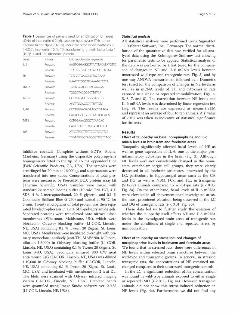
Table 1 Sequences of primers used for amplification of targetcDNA of interleukin 6 (IL-6), tyrosine hydroxylase (TH), tumornecrosis factor alpha (TNF-α), inducible nitric oxide synthases 2(iNOS2), interleukin 10 (IL-10), transforming growth factor beta 1(TGFβ1), and 18S ribosomal protein
Gene Primer Oligonucleotide sequence
IL-6 Forward AAGTCGGAGGCTTAATTACATATGTTC
Reverse TCATCGCTGTTCATACAATCAGAA
TH Forward TCTCCCTGAGGGGTACAAAA
Reverse GAATTTTGGCTTCAAATGTCTCA
TNF-α Forward TGATCGGTCCCAACAAGGA
Reverse TGGGCTACGGGCTTGTCA
iNOS2 Forward GCTTCAGAATGGGGAGCTG
Reverse AGGTTGGAGGCCTTGTGTC
IL-10 Forward CCCTGGGAGAGAAGCTGAAGA
Reverse CACTGCCTTGCTTTTATTCTCACA
TGFβ1 Forward CCTGGAAAGGGCTCAACAC
Reverse CAGTTCTTCTCTGTGGAGCTGA
18S Forward ATGGTTCCTTTGTCGCTCGCTCC
Reverse TGGATGTGGTAGCCGTTTCTCAGG
Mravec et al. Journal of Neuroinflammation (2016) 13:15 Page 5 of 17

significant changes in NE concentrations within the A1noradrenergic cell group when examining singly or re-peatedly stressed wild-type and transgenic rats (Fig. 4c).But, the A2 noradrenergic cells group showed a non-significant reduction in NE levels in both wild-type andtransgenic animals exposed to single IMO (Fig. 4e).We found that NE levels were reduced in both hippo-
campal regions (CA and DG) compared to wild-type ani-mals in both the single (P < 0.01) and repeatedly stressed(P < 0.01) transgenic rats as well as in transgenic ratsfrom the adapted IMO group (P < 0.05). Whereas repeatedIMO non-significantly increased NE levels in the CA andDG of wild-type rats the NE levels remained unchangedor were even non-significantly reduced in transgenic ratswhen compared to controls (Fig. 5a, c).Although we have seen significantly reduced NE
levels in the FCx of transgenic compared to wild-type rats(P < 0.01, single and repeatedly stressed rats; P < 0.05,adapted IMO rats; Fig. 6a), we found no difference in the
levels of NE levels in the TCx between stressed transgenicand wild-type rats (Fig. 6c). Whereas repeated stressnon-significantly increased NE levels in the FCx ofwild-type rats, NE levels did not change or were evennon-significantly reduced in transgenic rats when com-pared to controls. Additionally, NE levels in the TCx wereunchanged, while reduced NE levels were found inthe entorhinal cortex (data not shown).Repeated stress non-significantly increased NE levels
in the NBM of wild-type rats. In transgenic rats, NElevels remained unchanged when compared to those ofunstressed transgenic animals, but they were signifi-cantly lower than these in repeatedly stressed wild-typerats (P < 0.01; Fig. 6e).
Effect of tauopathy on stress-induced changes of IL-6mRNA levels in brainstem and forebrain areasBoth, single as well as repeated exposure to IMO stressinduced a significant elevation of IL-6 mRNA level in
Norepinephrine
LC A1 A2 CA DG NBM FCx TCx0
1
2
3
45
10152025303540
AWild-type rats
Transgenic rats
*
*
*
*
*
Brainstem catecholaminergiccell groups
Forebrain subcortical and cortical areas
NE
ng
/mg
pro
tein
IL-6 mRNA
LC A1 A2 CA DG NBM FCx TCx0
1
2
3
4
5
6
7
8B
*
*
*
*
*
Brainstem catecholaminergiccell groups
Forebrain subcortical and cortical areas
**
*
**
*
*
IL-6
/18S
mR
NA
Fig. 3 Basal levels of norepinephrine (NE) and interleukine-6 mRNA (IL-6 mRNA) in brainstem catecholaminergic cell groups containing NE cellbodies (LC, A1, A2) and forebrain subcortical and cortical areas in wild-type (white square) and transgenic (SHR72) rats (black square). Each value isthe mean ± SEM (n = 4–10). Statistical significance vs. corresponding wild-type group: *P < 0.05; **P < 0.01
Mravec et al. Journal of Neuroinflammation (2016) 13:15 Page 6 of 17

Norepinephrine
C 1xIMO 6xIMO+24h 7xIMO0
5
10
15
20
25
30
35
40
45A
Wild-type ratsTransgenic rats
#
#
*
NE
ng
/mg
pro
tein
IL-6 mRNA
C 1xIMO 6xIMO+24h 7xIMO0123456789
101112
B# #
** #
#
IL-6
/18S
mR
NA
Norepinephrine
C 1xIMO 6xIMO+24h 7xIMO0
5
10
15
20
25C
NE
ng
/mg
pro
tein
IL-6 mRNA
C 1xIMO 6xIMO+24h 7xIMO0
1
2
3
4
5
6
7
8D
#
**
*
#
IL-6
/18S
mR
NA
Norepinephrine
C 1xIMO 6xIMO+24h 7xIMO0
5
10
15
20
25
30
35E
NE
ng
/mg
pro
tein
IL-6 mRNA
C 1xIMO 6xIMO+24h 7xIMO0.0
0.5
1.0
1.5
2.0
2.5
3.0
3.5F
*
# #
*
IL-6
/18S
mR
NA
Locus coeruleus
A1 cell group
A2 cell group
Fig. 4 Levels of norepinephrine (NE) and interleukine-6 mRNA (IL-6 mRNA) in the LC, A1, and A2 brainstem noradrenergic areas in wild-type(white square) and transgenic (SHR72) rats (black square) exposed to a single (1 × 2 h) or repeated (7 × 2 h) immobilization stress (IMO). C controlrats; 6×IMO + 24 h-adapted IMO rats exposed to 6×IMO for 2 h and decapitated 24 h after the last IMO. Each value is the mean ± SEM (n = 5–8).Statistical significance vs. corresponding wild-type group: *P < 0.05; **P < 0.01; statistical significance vs. wild-type control group: #P < 0.05; ##P < 0.01
Mravec et al. Journal of Neuroinflammation (2016) 13:15 Page 7 of 17

the LC (P < 0.01, single stress; P < 0.05; adapted IMOand repeated stress; Fig. 4b) and A1 noradrenergic cellgroup (P < 0.05 single and repeated stress; Fig. 4d) ofwild-type animals, However, IL-6 mRNA levels wereonly elevated in wild-type animals by repeated stress inthe A2 noradrenergic cell group (P < 0.01; Fig. 4f ), CA(P < 0.01; Fig. 5b), and FCx (P < 0.05; Fig. 6b).Similar to the abovementioned findings of NE levels in
stressed transgenic rats, exposure of rats to IMO did notinduce further increases in IL-6 gene expression in se-lected brain areas of transgenic animals when comparedto values found in unstressed transgenic animals (Figs. 4,5, and 6). However, in some brainstem and in hippocam-pal areas, IL-6 mRNA levels were significantly elevatedin stressed transgenic rats compared to correspondingwild-type groups (Figs. 4d, e, and 5b, d).
Effect of tauopathy on basal and stress-induced changesof TH gene expression and TH protein levels in brainstemnoradrenergic cell groupsBrainstem noradrenergic neurons, particularly thesein the LC, provide NE to all levels of the neuraxis.Reduced NE levels and increased levels of IL-6mRNA in investigated forebrain structures (Fig. 3a, b)indicate altered function of brainstem noradrenergicneurons. Therefore, when attempting to assess thefunctionality of the LC, we selected TH mRNA andprotein levels as markers of NE synthesis. We alsoinvestigated the effect of stress on gene expressionand protein levels of TH.Repeated IMO significantly increased TH mRNA
levels in LC of wild-type rats (P < 0.05; Fig. 7a). This isnot surprising as TH gene expression in the LC of
Norepinephrine
C 1xIMO 6xIMO+24h 7xIMO0.0
0.2
0.4
0.6
0.8
1.0
1.2
1.4
1.6
1.8
2.0A
Wild-type ratsTransgenic rats
**
** **
NE
ng
/mg
pro
tein
IL-6 mRNA
C 1xIMO 6xIMO+24h 7xIMO0.0
0.5
1.0
1.5
2.0
2.5
3.0
3.54.0
4.5
5.0
5.5
6.0B
*
# #
*
**
IL-6
/18S
mR
NA
Norepinephrine
C 1xIMO 6xIMO+24h 7xIMO0.0
0.5
1.0
1.5
2.0
2.5
3.0
3.5
4.0
4.5
5.0C
*
** ***
NE
ng
/mg
pro
tein
IL-6 mRNA
C 1xIMO 6xIMO+24h 7xIMO0.0
0.5
1.0
1.5
2.0
2.5
3.0
3.54.0
4.5
5.0
5.5
6.0D
** **
+IL-6
/18S
mR
NA
Cornu ammonis
Dentate gyrus
Fig. 5 Levels of norepinephrine (NE) and interleukine-6 mRNA (IL-6 mRNA) in the cornu ammonis (CA) and dentate gyrus (DG) in wild-type (white square)and transgenic (SHR72) rats (black square) exposed to a single (1 × 2 h) or repeated (7 × 2 h) immobilization stress (IMO). C control; 6×IMO+ 24 h-adaptedIMO rats exposed to 6×IMO for 2 h and decapitated 24 h after the last IMO. Each value is the mean ± SEM (n = 5–8). Statistical significance vs.corresponding wild-type group: *P < 0.05; **P < 0.01; statistical significance vs. wild-type control group: #P < 0.05; ##P < 0.01; statistical significance vs.transgenic control group: +P < 0.05
Mravec et al. Journal of Neuroinflammation (2016) 13:15 Page 8 of 17

Norepinephrine
C 1xIMO 6xIMO+24h 7xIMO0.0
0.2
0.4
0.6
0.8
1.0
1.2
1.4
1.6
1.8
2.0 Wild-type ratsTransgenic rats
A
*
** * **
NE
ng
/mg
pro
tein
IL-6 mRNA
C 1xIMO0.0
0.5
1.0
1.5
2.0
2.5
3.0
3.5
4.0
4.5
5.0B
*
#
IL-6
/18S
mR
NA
Norepinephrine
C 1xIMO 6xIMO+24h 7xIMO0.0
0.5
1.0
1.5
2.0
2.5
3.0
3.5C
*
NE
ng
/mg
pro
tein
IL-6 mRNA
C 1xIMO0.0
0.5
1.0
1.5
2.0
2.5
3.0
3.5D
IL-6
/18S
mR
NA
Norepinephrine
C 1xIMO 6xIMO+24h 7xIMO0
2
4
6
8
10
12
14
16
18
20E
***
NE
ng
/mg
pro
tein
IL-6 mRNA
C 1xIMO0.0
0.2
0.4
0.6
0.8
1.0
1.2
1.4
1.6
1.8
2.0
2.2
2.4F
IL-6
/18S
mR
NA
Frontal cortex
Temporal cortex
Nucleus basalis of Meynert
Fig. 6 Levels of norepinephrine (NE) and interleukine-6 mRNA (IL-6 mRNA) in frontal cortex, temporal cortex, and nucleus basalis of Meynert inwild-type (white square) and transgenic (SHR72) rats (black square) exposed to a single (1 × 2 h) or repeated (7 × 2 h) immobilization stress (IMO).C control; 6×IMO + 24 h-adapted IMO rats exposed to 6×IMO for 2 h and decapitated 24 h after the last IMO. Each value is the mean ± SEM(n = 4–10). Statistical significance vs. corresponding wild-type group: *P < 0.05; **P < 0.01; statistical significance vs. wild-type control group: #P < 0.05
Mravec et al. Journal of Neuroinflammation (2016) 13:15 Page 9 of 17

transgenic rats was already significantly increased in un-stressed (P < 0.05) as well as in singly stressed transgenicrats when compared to corresponding wild-type animals(P < 0.05; Fig. 7a). Also, non-significantly increased THmRNA levels were observed in repeatedly stressed trans-genic rats (Fig. 7a). These stress-induced increases of THin transgenic rats were confirmed by similar results foundin TH protein levels (Fig. 7c).We also investigated TH gene expression in the A1
and A2 noradrenergic cell groups. We found that THmRNA levels in the A1 noradrenergic cell group of wild-type animals were significantly elevated only by repeatedIMO stress (P < 0.05; Fig. 7b). When compared to wild-
type animals, transgenic rats showed elevated basallevels (P < 0.05), but no further elevation was detectedafter either single or repeated stress (Fig. 7b). Similarlyto the A1 noradrenergic cell group, gene expression ofTH in the A2 noradrenergic cell group of wild-type ani-mals was significantly elevated only by repeated IMOstress (P < 0.05; Fig. 7d).In transgenic rats, stress did not induce any sig-
nificant changes in TH mRNA levels. Similarly, nochanges were seen when comparing the values ofTH mRNA levels in the A2 noradrenergic cell groupsbetween transgenic rats and corresponding wild-typeanimals (Fig. 7d).
TH
GAPDH
+24h
TH mRNA
C 1xIMO 6xIMO+24h 7xIMO0.0
0.5
1.0
1.5
2.0
2.5
3.0
3.5
4.0A
Wild-type ratsTransgenic rats
**
# #
TH
/18S
mR
NA
TH mRNA
C 1xIMO 6xIMO+24h 7xIMO0
1
2
3
4
5
6B
* #*
TH
/18S
mR
NA
TH mRNA
C 1xIMO 6xIMO+24h 7xIMO0.0
0.5
1.0
1.5
2.0
2.5
3.0D
#
TH
/18S
mR
NA
Locus coeruleus A1 cell group
A2 cell group
C
Locus coeruleus
TH protein
WT TG WT TG WT TG WT TG
6xIMO C 1xIMO 7xIMO
1.96 1.93 1.00 1.38 1.38 1.42 1.56 1.80A.U.
Fig. 7 Gene expression (a, b, and d) and protein (c) levels of tyrosine hydroxylase (TH) in the LC, A1, and A2 brainstem noradrenergic cell groupsof wild-type (white square) and transgenic (SHR72) rats (black square) exposed to a single (1 × 2 h) or repeated (7 × 2 h) immobilization stress(IMO). Each sample of TH protein represents a pool of two to three animals (c), values are expressed as arbitrary units (A.U.) calculated as a ratiobetween TH and GAPDH in a given sample. C control; 6×IMO + 24 h-adapted IMO rats exposed to 6×IMO for 2 h and decapitated 24 h after thelast IMO. Each value is the mean ± SEM (n = 5–8; a, b, and d). Statistical significance vs. corresponding wild-type group: *P < 0.05; statisticalsignificance vs. wild-type control group: #P < 0.05
Mravec et al. Journal of Neuroinflammation (2016) 13:15 Page 10 of 17

Effect of tauopathy on basal and stress-induced changesof levels of pro- and anti-inflammatory factors in thelocus coeruleusIn transgenic rats, tauopathy is localized mainly to thebrainstem [16]. Because tauopathy also induces neuroin-flammation, we also investigated levels of selected pro-and anti-inflammatory factors as markers of the immunemilieu of the LC.Besides IL-6 mRNA, we determined in the LC expres-
sion of genes of other inflammatory factors, particularlythe tumor necrosis factor alpha (TNF-α), inducible nitricoxide synthases 2 (iNOS2), interleukin 10 (IL-10), andtransforming growth factor beta 1 (TGFβ1). WhereasmRNA levels of the pro-inflammatory factor TNF-αwere only non-significantly elevated in unstressed trans-genic rats (P = 0.078; Fig. 8a), the expression of theiNOS2 gene showed significant increases in the LC oftransgenic animals during basal conditions (P < 0.01;
Fig. 8c). However, after the exposure to repeated stress,TNF-α mRNA levels were reduced compared to corre-sponding wild-type rats (P < 0.05; Fig. 8a), while gene ex-pression of iNOS2 was significantly reduced in repeatedlystressed transgenic rats when compared to unstressedtransgenic rats (P < 0.05; Fig. 8c).Gene expression of the anti-inflammatory cytokine
IL-10 was significantly elevated during basal conditions inthe LC of transgenic rats (P < 0.01; Fig. 8b). Even if stresshad no effect on IL-10 mRNA levels in transgenic rats,gene expression of IL-10 was significantly elevated in bothsingly stressed and adapted IMO transgenic rats whencompared to values found in corresponding wild-typeanimals (P < 0.05; Fig. 8b).Gene expression of neuroprotective factor TGFβ1 in
wild-type animals was not affected by stress. However,TGFβ1 mRNA levels were significantly lower in repeat-edly stressed and adapted IMO transgenic rats compared
TNF-αα mRNA
C 1xIMO 6xIMO+24h 7xIMO0
1
2
3
4
5
6A
Wild-type ratsTransgenic rats
#
#
* *
TN
F-α
/18S
mR
NA
IL-10 mRNA
C 1xIMO 6xIMO+24h 7xIMO0.0
0.5
1.0
1.5
2.0
2.5
3.0
3.5
4.0
4.5
5.0B
**
# #
*
*
# # #
IL-1
0/18
S m
RN
AiNOS2 mRNA
C 1xIMO 6xIMO+24h 7xIMO0.0
0.5
1.0
1.5
2.0
2.5
3.0
3.5
4.0C
**#
#
+
iNO
S2/
18S
mR
NA
TGFβ1 mRNA
C 1xIMO 6xIMO+24h 7xIMO0.0
0.2
0.4
0.6
0.8
1.0
1.2
1.4
1.6
1.8
2.0D
*
*
TG
Fβ
1/18
S m
RN
A
Locus coeruleus
Fig. 8 Gene expression of pro-inflammatory factors TNF-α (a), iNOS2 (c), and anti-inflammatory factors IL-10 (b) and TGFβ1 (d) in the LC of wild-type(white square) and transgenic (SHR72) rats (black square) exposed to a single (1 × 2 h) or repeated (7 × 2 h) immobilization stress (IMO). C control;6×IMO+ 24 h-adapted IMO rats exposed to 6×IMO for 2 h and decapitated 24 h after the last IMO. Each value is the mean ± SEM (n = 5–8). Statisticalsignificance vs. corresponding wild-type group: *P < 0.05; **P < 0.01; statistical significance vs. wild-type control group: #P < 0.05, ##P < 0.01
Mravec et al. Journal of Neuroinflammation (2016) 13:15 Page 11 of 17

to levels found in the corresponding wild-type animals(P < 0.05; Fig. 8d).
Effect of tauopathy on correlation betweennorepinephrine and IL-6 mRNA levels in cornu ammonisunder basal and stress conditionsThe reduced NE and elevated IL-6 mRNA levels foundin unstressed transgenic rats (Fig. 3), differences in NEand IL-6 mRNA levels in stressed transgenic animals(Figs. 4, 5, and 6), and altered immune background inthe LC (Fig. 8) indicate the effect of deregulated immu-nomodulation on the brain’s noradrenergic system.Therefore, we investigated the effect of stress in bothwild-type and transgenic rats on the relationship be-tween NE and IL-6 mRNA levels in the CA, a brainstructure innervated almost exclusively by the LC.
We did not find any correlation between NE and IL-6mRNA levels in the CA of either unstressed or stressedwild-type (Fig. 9a, c) or transgenic rats (Fig. 9b, d). How-ever, whereas NE levels are similar between unstressedwild-type and transgenic rats (ranging from 0.7 to1.5 ng/mg of protein), the mean gene expression of IL-6 isalmost three times higher in transgenic rats (Fig. 9a, b).
DiscussionThe brain’s noradrenergic system regulates the centralstress response [23] and maintains several cognitive,affective, and behavioral functions [3, 24], while contrib-uting to the consolidation of learning and memory [25].However, Gibbs and Summers showed that NE releasedby brainstem noradrenergic neurons, besides its neuro-transmitter role, is also involved in maintaining the
Fig. 9 Correlation plots illustrating the relationship of norepinephrine (NE) concentration and interleukine-6 mRNA (IL-6 mRNA) in cornu ammonis(CA) of wild-type controls (a, white circle), transgenic controls (b, black circle), wild-type rats exposed to a single 2-h immobilization stress (c, white circle),and transgenic rats exposed to a single 2-h immobilization stress (d, black circle)
Mravec et al. Journal of Neuroinflammation (2016) 13:15 Page 12 of 17

brain’s tissue milieu [4]. Furthermore, centrally releasedNE modulates synaptic plasticity, neurogenesis, energymetabolism, activity of astrocytes and microglia, corticalperfusion, and the permeability of the blood-brain barrier[4–7]. Moreover, it also exerts several potent anti-in-flammatory and anti-oxidative effects on the brain tissue[26]. All of the abovementioned “homeostatic” processesmodulated by NE are impaired in the brains of AD pa-tients. Importantly, recent findings of Braak et al. [9] indi-cate that in humans, pathological tau species are originallyformed in the LC (the brainstem structure representingthe main accumulation of noradrenergic neurons in thebrain) and then spread by axonal transport and interneur-onal communications to the transentorhinal region, thento the entorhinal region, the hippocampal formation, andthen later to the association cortices [9]. Therefore, it issuggested that the LC plays a crucial role in the develop-ment of AD-related neuropathology [11]. The importanceof the LC in AD development is further supported by thefinding that LC destruction results in the worsening ofneuroinflammation and amyloid β deposition in brains oftransgenic Aβ animal models, as well as in elevated IL-1βand Ccl2 cytokines in laboratory animals [27, 28]. Interest-ingly, monoaminergic deficiency and severe depletion ofNE has been found in most LC projection areas in ADpatients [29]. Morphological studies of brains of AD pa-tients have shown that the LC undergoes significantdegeneration that precedes pathological changes in theforebrain [30]. It is suggested that this degeneration startsearly in the course of AD pathogenesis and that theLC represents the centre from which altered tau proteinsspread throughout the forebrain structures [9]. Therefore,we investigated the effect of tauopathy on the LC by usingtransgenic (SHR72) rats that over-express human trun-cated tau protein [17].Preclinical investigation of the LC role in AD pathogen-
esis is based mainly on the study of the effects resultingfrom the destruction of this noradrenergic cell group onthe development of AD-related neuropathology. However,in the available literature, information on tauopathy’seffect on LC functions and on the immune background ofbrain tissue during basal and stress conditions is lacking.Based on the abovementioned facts, we investigated
the mutual relationship between the brain’s noradrener-gic system and immune activity in unstressed and singlyor repeatedly stressed wild-type and transgenic rats thatover-express human truncated tau protein [17]. Here, wedemonstrate for the first time that expression of humantruncated tau protein, whose primary sequence wasderived from paired helical filaments obtained from thebrains of AD patients, is sufficient to induce dysfunctionof LC neurons in vivo and this dysfunction is most likelyfollowed by exaggerated synthesis of pro-inflammatorycytokines in brain structures innervated by the LC
that are known to be affected by neuropathology inAD patients.We have found significant reduction of basal NE levels
in hippocampus (CA and DG) and NBM as well as incortical areas (frontal and temporal association cortex)of transgenic rats, structures innervated almost entirelyby noradrenergic LC neurons [3]. Our findings indicatethat the function of the noradrenergic system in thebrains of transgenic rats is significantly impaired, at leastat the level of NE synthesis. This is in agreement withobserved significant reductions of NE levels on the cere-brospinal fluid of AD patients compared to matchedcontrols [31].Recent studies have revealed that NE exerts a potent
anti-inflammatory effect on the brain [32]. In support ofthis, decreased cerebral NE levels and exacerbated neu-roinflammatory processes have been demonstrated inmurine AD models [27, 33]. Moreover, it is suggestedthat the reduction of LC noradrenergic neurons in thebrains of AD patients could be one factor responsiblefor the exaggerated neuroinflammation in the brains ofthese patients [34]. Based on these facts and assump-tions, the question has been raised as to whether the re-duction of brain’s tissue NE levels in transgenic (SHR72)rats is accompanied by altered immune status in thebrain of these animals. Therefore, we analyzed the geneexpression of IL-6, one of the main immune system fac-tors in the brain [35]. In transgenic rats, we found a sig-nificant elevation of IL-6 mRNA in CA, DG, and FCx.Moreover, significantly elevated IL-6 mRNA levels werealso found in the brainstem, particularly in the LC, A1,and A2 noradrenergic cell groups. Increased gene ex-pression of IL-6 in forebrain structures may reflect thereduced anti-inflammatory effect of NE due to reducedNE concentrations in these forebrain areas, while theincreased IL-6 mRNA levels in brainstem noradrenergicareas may result from the burden of tau pathology, asit is localized mainly in the brainstem of transgenic(SHR72) rats [16].It is suggested that stress represents important factor
participating in the development of AD (for review, see[12]). Moreover, AD-related neuropathology may affectthe stress response as well [36]. Because the LC repre-sents a key structure modulating stress responses in thebrain [23] and that we found that tauopathy impairs LCfunction, we investigated the effect of single and repeatedstress on tissue levels of NE against the background oftauopathy. Interestingly, the exposure of transgenic rats tostress did not significantly affect NE levels when comparedto unstressed transgenic animals. However, in the CA,DG, FCx, and NBM, NE levels in transgenic animals weresignificantly lower when compared to the NE levels ofcorresponding wild-type groups. These findings indicatethat tauopathy reduces noradrenergic neurotransmission
Mravec et al. Journal of Neuroinflammation (2016) 13:15 Page 13 of 17

not only at basal conditions but also in animals exposed tostressors. Furthermore, these reduced NE levels in theforebrain areas of stressed, transgenic rats were accom-panied by elevated gene expression of IL-6 in the CA andDG. Moreover, IL-6 mRNA levels were also elevated inbrainstem A1 noradrenergic cells of stressed, transgenicanimals. These findings indicate that the immune milieuremains altered in investigated brain areas in stressed,transgenic rats, but stress is not able to significantly affectgene expression of IL-6. It can be speculated that exposureto immobilization stress daily for 7 days is not sufficient toalter the immune status of transgenic rats and thereforelonger period of chronic stress exposure will be necessaryto determine the effect of stress on neuroinflammation atthe background of tauopathy.In transgenic (SHR72) rats, tau pathology is predomin-
antly found in the brainstem [16]. However, even if re-duced NE levels in forebrain areas of transgenic ratsindicate functional impairment of the LC, we did notfind any neurodegenerative changes at the level of theLC morphology (unpublished data). Using immunohisto-chemistry, we found accumulation of tau pathology inthe form of AT8-immunopositive tau, pre-tangles, andneurofibrillary tangles in brainstem areas that innervatethe LC, particularly in the paragigantocellular nucleusand hypoglossal preposite nucleus (unpublished data).Neurons of the paragigantocellular nucleus project tothe LC and increase noradrenergic neurotransmission inmany forebrain areas, including the hippocampus, viamostly glutamatergic excitatory fibers [37]. Therefore,we speculate that in transgenic (SHR72) rats, the tauopa-thy found in the paragigantocellular nucleus attenuatesglutamatergic neurotransmission from the LC, which con-sequently reduces levels of NE in forebrain structures.However, alternative mechanisms may participate in theimpairment of the LC as well, including reduced axonaltransport of trophic factors to the nuclei of LC neuronsfrom structures innervated by the LC.To further investigate the functional impairment of
the LC, we determined gene expression and proteinlevels of TH, the rate-limiting enzyme of NE biosyn-thesis as TH mRNA levels reflect changes in activity ofthe noradrenergic cells in this brain area [23]. We de-tected a significant increase of TH mRNA in the LC ofrepeatedly stressed wild-type animals that is in accord-ance with our previous reports [23, 38–41]. Importantly,TH protein concentration in the LC is in good agree-ment with TH expression. When compared to wild-typerats, transgenic (SHR72) animals started from the higherbasal levels of TH mRNA, but did not show any signifi-cant changes after stress exposure. Increased TH levelsin transgenic rats indicate exaggerated NE biosynthesis.However, as discussed above, NE levels in forebrainareas innervated by the LC were reduced. There is
published data showing that even if the number of LCneurons in the brain of AD patients is significantly re-duced, the remaining LC neurons undergo significantcompensatory changes [42]. Our contradictory findingsindicate that even if compensatory mechanisms in nor-adrenergic neurotransmission machinery are activated atthe level of the LC, these responses are not able to com-pensate for the functional deficits manifested by reducedNE concentrations in brain areas innervated by the LC.Besides the LC, we also analyzed TH mRNA in other
noradrenergic cell groups that participate in noradrener-gic innervation of forebrain structures, particularly inthe A1 and A2 noradrenergic cell groups. However, incontrast to the LC, the A1 and A2 noradrenergic cellgroups predominantly innervate the hypothalamus [23].Therefore, sparse information is available as to howneurofibrillary pathology affects these cell groups. Wefound that TH mRNA levels in wild-type animals weresignificantly elevated in A1 and A2 noradrenergic cellgroups after repeated stress. Also, while TH mRNA inA1 noradrenergic cells group of transgenic rats startedfrom elevated basal levels, no further changes were de-tected after either single or repeated stress. We suggestthat similarly to LC, increased TH expression in the A1noradrenergic cell group might represent a compensa-tory response to impairment of TH-positive A1 neuronsin this transgenic (SHR72) animal model of tauopathy.However, AD-related neuropathology of A1 noradrener-gic neurons has not yet been described in available lit-erature. However, we found that the A2 noradrenergiccell group shows an increase of TH mRNA levels inrepeatedly stressed wild-type animals not seen in trans-genic rats. Because the A1 and A2 noradrenergic cellgroups innervate mainly the hypothalamic nuclei, wealso investigated changes in NE level in the hypothal-amic paraventricular nucleus. Importantly, in contrast tosignificantly reduced NE levels in forebrain structuresinnervated by the LC, we found that NE levels were onlynon-significantly reduced in the hypothalamic paraven-tricular nucleus in transgenic rats (9.93 ± 0.94 ng/mg pro-tein) compared to wild-type animals (16.38 ± 2.57 ng/mgprotein). These findings indicate that tauopathy in trans-genic (SHR72) rats affects predominantly noradrenergicneurotransmission in the hippocampus, nucleus basalis ofMeynert, and association cortices, all of which are areasinnervated by the LC and affected in AD patients.To further to determine the mechanisms responsible
for tau-related alteration of the LC in transgenic(SHR72) rats, we measured tissue mRNA levels ofboth pro- and anti-inflammatory factors, including TNF-α,iNOS2, IL-10, and TGFβ1. We found that gene expressionof these factors differs between wild-type and transgenicrats at both basal and stressed conditions, although TNF-αmRNA levels in unstressed, transgenic rats were only
Mravec et al. Journal of Neuroinflammation (2016) 13:15 Page 14 of 17

non-significantly increased. However, even if gene ex-pression of TNF-α declined in repeatedly stressed trans-genic rats compared to unstressed transgenic rats, TNF-αmRNA in rats exposed to repeated stress was significantlyelevated compared to the matched, wild-type group. Geneexpression of iNOS2, another pro-inflammatory factor,was significantly increased at basal conditions and findingsrelated to gene expression of TNF-α and iNOS2 indicate apro-inflammatory milieu in the LC tissue of transgenicrats. Moreover, gene expression of the anti-inflammatorycytokine IL-10 was increased in unstressed transgenic ratsas well as in transgenic animals exposed to a single stres-sor, indicating activation of compensatory immune mech-anisms at the level of the LC against a background oftauopathy. Comparison of NE, IL-6 mRNA, TNF-αmRNA, and IL-10 mRNA in the LC of transgenic rats in-dicate that the anti-inflammatory effect of NE in the LC isimpaired. Because tauopathy is localized predominantly tothe brainstem of transgenic (SHR72) rats, several local fac-tors may promote an inflammatory milieu in the LC aswell. Reduced gene expression of the neuroprotective fac-tor TGFβ1 in repeatedly stressed transgenic rats highlightsimpairment of neuroprotective mechanisms in the LC.Based on the abovementioned findings, we suggest thattauopathy affects function of brainstem nuclei innervatingthe LC and alters the brainstem immune milieu. Thesealterations might participate in neuroinflammation andthe reduced neuroprotection in the LC accompanied byreduced release of NE by LC neurons into the forebrainareas of transgenic (SHR72) rats. Reduced NE releasemight then participate in the development of neuroinflam-mation in forebrain structures innervated by the LC.To investigate the role of the LC dysfunction in ob-
served alteration of immune milieu in forebrain areas oftransgenic rats, we assessed the correlation between NEand IL-6 mRNA levels in both wild-type and transgenicrats at basal and stress conditions. Importantly, we foundthat even if there was no correlation between NE and IL-6mRNA levels at basal conditions, transgenic rats exhibitapproximately threefold higher IL-6 mRNA levels. Thesefindings indicate that the inhibitory effect of brain NEon neuroinflammation is severally impaired against abackground of tauopathy. However, the mechanisms andpathways responsible for this phenomenon need furtherinvestigation.It is necessary to note that all abovementioned param-
eters (e.g., interleukins) were investigated in brain tissuesobtained by microdissection (punching) technique. How-ever, this method does not allow to determine whetheralterations are present in neurons, glia, or both types ofcells. Therefore, for more complex view of neuropatho-logical consequences of tau pathology on the function ofthe LC and on neuroinflammation, other approaches(e.g., immunohistochemistry) must be used.
ConclusionsOur data have shown that tau pathology in the brains oftransgenic (SHR72) rats alters central noradrenergicneurotransmission, particularly in brain’s structures inner-vated by noradrenergic neurons, under basal conditions aswell as after an exposure to single or chronic stress. Weobserved increases in IL-6 gene expression in brain struc-tures that may reflect a reduced anti-inflammatory effect-iveness of the LC noradrenergic system. Importantly, somein vitro and in vivo studies have provided evidence thatincreases in NE levels can attenuate AD-related neuro-pathology and neuroinflammation, while enhancing cogni-tion [43, 44]. In support of this, some clinical studies havealready confirmed that pharmacotherapy potentiatingnoradrenergic neurotransmission is able to improve cogni-tive symptoms, while reducing depression and aggressionin AD patients [45–47].However, it is necessary to note that it remains unclear
whether tauopathy in transgenic (SHR72) rats affect LCneurons directly or indirectly. Indirect mechanisms ofaltered LC function in transgenic rats may include (i)impaired transport of trophic factors from brainstemstructures affected by tauopathy to the LC and (ii) re-duced synthesis of norepinephrine in varicosities of theLC neurons as a result of either the pro-inflammatorymilieu or other mechanisms. Moreover, tauopathy intransgenic (SHR72) rats may also potentially affect othercatecholaminergic cell groups, exaggerating the norad-renergic deficit followed by impairment of the brain mi-lieu and development of neuropathology. However, it isunclear whether other monoaminergic cell groups areaffected in this transgenic model, and therefore, thisneeds further investigation.Based on our data, we suggest that transgenic rats of this
study can be efficiently employed in detailed investigationof molecular mechanisms of neuronal degeneration relatedto tauopathy, neuroinflammation, and, more importantly,in the development of disease modifying strategy fortreatment of Alzheimer’s disease and other tauopathies.
AbbreviationsAD: Alzheimer’s disease; CA: cornu ammonis; DG: dentate gyrus; FCx: frontalcortex; IL-10: interleukin 10; IL-6: interleukin 6; IMO: immobilization;iNOS2: inducible nitric oxide synthases 2; LC: locus coeruleus; NBM: nucleusbasalis of the Meynert; NE: norepinephrine; TCx: temporal cortex;TGFβ1: transforming growth factor beta 1; TH: tyrosine hydroxylase;TNF-α: tumor necrosis factor alpha.
Competing interestThe authors declare that they have no competing interests.
Authors’ contributionsBM, PF, and RK designed the experiments and supervised the project.KL, PV, KO, LH, and PN carried out the in vivo experiments. KL, PV, and GMcarried out the molecular analyses. BM, PF, MN, and RK analyzed data andwrote the manuscript. All authors have read and approved the finalversion of the manuscript.
Mravec et al. Journal of Neuroinflammation (2016) 13:15 Page 15 of 17
AcknowledgementsThis work was supported by the Slovak Research and Development Agencyunder the contracts no. APVV-0088-10 and APVV-0677-12, BrainCentrum2013–2014, and by grant VEGA 2/0067/14.
Author details1Institute of Experimental Endocrinology, Slovak Academy of Sciences,Vlarska 3, 833 06 Bratislava, Slovakia. 2Institute of Physiology, Faculty ofMedicine, Comenius University in Bratislava, Bratislava, Slovakia. 3Institute ofNeuroimmunology, Slovak Academy of Sciences, Bratislava, Slovakia. 4AxonNeuroscience SE, Bratislava, Slovakia. 5LDN, Labor Diagnostika Nord,Nordhorn, Germany.
Received: 29 June 2015 Accepted: 13 January 2016
References1. Ramanan VK, Saykin AJ. Pathways to neurodegeneration: mechanistic
insights from GWAS in Alzheimer’s disease, Parkinson’s disease, and relateddisorders. Am J Neurodegener Dis. 2013;2(3):145–75.
2. Armstrong RA. What causes Alzheimer’s disease? Folia Neuropathol.2013;51(3):169–88. doi:21432 [pii].
3. Benarroch EE. The locus ceruleus norepinephrine system: functionalorganization and potential clinical significance. Neurology. 2009;73(20):1699–704.doi:10.1212/WNL.0b013e3181c2937c.
4. O’Donnell J, Zeppenfeld D, McConnell E, Pena S, Nedergaard M.Norepinephrine: a neuromodulator that boosts the function of multiple celltypes to optimize CNS performance. Neurochem Res. 2012;37(11):2496–512.doi:10.1007/s11064-012-0818-x.
5. Hertz L, Lovatt D, Goldman SA, Nedergaard M. Adrenoceptors in brain:cellular gene expression and effects on astrocytic metabolism and [Ca(2+)]i.Neurochem Int. 2010;57(4):411–20. doi:10.1016/j.neuint.2010.03.019.
6. Marien MR, Colpaert FC, Rosenquist AC. Noradrenergic mechanisms inneurodegenerative diseases: a theory. Brain Res Brain Res Rev. 2004;45(1):38–78.doi:10.1016/j.brainresrev.2004.02.002. S0165017304000165 [pii].
7. Toussay X, Basu K, Lacoste B, Hamel E. Locus coeruleus stimulation recruits abroad cortical neuronal network and increases cortical perfusion. J Neurosci.2013;33(8):3390–401. doi:10.1523/JNEUROSCI.3346-12.2013.
8. Clavaguera F, Hench J, Goedert M, Tolnay M. Invited review: Prion-liketransmission and spreading of tau pathology. Neuropathol Appl Neurobiol.2015;41(1):47–58. doi:10.1111/nan.12197.
9. Braak H, Thal DR, Ghebremedhin E, Del Tredici K. Stages of the pathologicprocess in Alzheimer disease: age categories from 1 to 100 years. J NeuropatholExp Neurol. 2011;70(11):960–9. doi:10.1097/NEN.0b013e318232a379.
10. Pamphlett R, Kum JS. Different populations of human locus ceruleusneurons contain heavy metals or hyperphosphorylated tau: implications foramyloid-beta and tau pathology in Alzheimer’s disease. J Alzheimers Dis.2015;45(2):437–47. doi:10.3233/JAD-142445. 130X574785821T62 [pii].
11. Mravec B, Lejavova K, Cubinkova V. Locus (coeruleus) minoris resistentiae inpathogenesis of Alzheimer’s disease. Curr Alzheimer Res. 2014;11(10):992–1001.doi:CAR-EPUB-63259 [pii].
12. Machado A, Herrera AJ, de Pablos RM, Espinosa-Oliva AM, Sarmiento M,Ayala A, et al. Chronic stress as a risk factor for Alzheimer’s disease. RevNeurosci. 2014;25(6):785–804. doi:10.1515/revneuro-2014-0035/j/revneuro.ahead-of-print/revneuro-2014-0035/revneuro-2014-0035.xml [pii].
13. Ricci S, Fuso A, Ippoliti F, Businaro R. Stress-induced cytokines and neuronaldysfunction in Alzheimer’s disease. J Alzheimers Dis. 2012;28(1):11–24.doi:10.3233/JAD-2011-110821.
14. Carroll JC, Iba M, Bangasser DA, Valentino RJ, James MJ, Brunden KR, et al.Chronic stress exacerbates tau pathology, neurodegeneration, and cognitiveperformance through a corticotropin-releasing factor receptor-dependentmechanism in a transgenic mouse model of tauopathy. J Neurosci.2011;31(40):14436–49. doi:10.1523/JNEUROSCI.3836-11.2011.
15. Marcello E, Gardoni F, Di Luca M. Alzheimer’s disease and modern lifestyle: whatis the role of stress? J Neurochem. 2015;134(5):795–8. doi:10.1111/jnc.13210.
16. Zilka N, Filipcik P, Koson P, Fialova L, Skrabana R, Zilkova M, et al. Truncated taufrom sporadic Alzheimer’s disease suffices to drive neurofibrillary degenerationin vivo. FEBS Lett. 2006;580(15):3582–8. doi:10.1016/j.febslet.2006.05.029.
17. Koson P, Zilka N, Kovac A, Kovacech B, Korenova M, Filipcik P, et al.Truncated tau expression levels determine life span of a rat model oftauopathy without causing neuronal loss or correlating with terminal
neurofibrillary tangle load. Eur J Neurosci. 2008;28(2):239–46.doi:10.1111/j.1460-9568.2008.06329.x.
18. Stozicka Z, Zilka N, Novak P, Kovacech B, Bugos O, Novak M. Geneticbackground modifies neurodegeneration and neuroinflammation driven bymisfolded human tau protein in rat model of tauopathy: implication forimmunomodulatory approach to Alzheimer’s disease. J Neuroinflammation.2010;7:64. doi:10.1186/1742-2094-7-64.
19. Kvetnansky R, Mikulaj L. Adrenal and urinary catecholamines in rats duringadaptation to repeated immobilization stress. Endocrinology. 1970;87(4):738–43.doi:10.1210/endo-87-4-738.
20. Palkovits M. Isolated removal of hypothalamic or other brain nuclei of therat. Brain Res. 1973;59:449–50. doi:0006-8993(73)90290-4 [pii].
21. Palkovits M, Brownstein MJ. Maps and guide to microdissection of the ratbrain. New York: Elsevier Science Publishing Co; 1988.
22. Livak KJ, Schmittgen TD. Analysis of relative gene expression data usingreal-time quantitative PCR and the 2(−Delta Delta C(T)) Method. Methods.2001;25(4):402–8. doi:10.1006/meth.2001.1262S1046-2023(01)91262-9 [pii].
23. Kvetnansky R, Sabban EL, Palkovits M. Catecholaminergic systems in stress:structural and molecular genetic approaches. Physiol Rev. 2009;89(2):535–606.doi:10.1152/physrev.00042.2006.
24. Herrmann N, Lanctot KL, Khan LR. The role of norepinephrine in thebehavioral and psychological symptoms of dementia. J Neuropsychiatry ClinNeurosci. 2004;16(3):261–76. doi:10.1176/appi.neuropsych.16.3.26116/3/261 [pii].
25. Gibbs ME, Summers RJ. Role of adrenoceptor subtypes in memoryconsolidation. Prog Neurobiol. 2002;67(5):345–91. doi:S0301008202000230 [pii].
26. Feinstein DL, Heneka MT, Gavrilyuk V, Dello Russo C, Weinberg G, Galea E.Noradrenergic regulation of inflammatory gene expression in brain.Neurochem Int. 2002;41(5):357–65. doi:S0197018602000499 [pii].
27. Heneka MT, Nadrigny F, Regen T, Martinez-Hernandez A, Dumitrescu-Ozimek L,Terwel D, et al. Locus ceruleus controls Alzheimer’s disease pathology bymodulating microglial functions through norepinephrine. Proc NatlAcad Sci U S A. 2010;107(13):6058–63. doi:10.1073/pnas.0909586107.
28. Jardanhazi-Kurutz D, Kummer MP, Terwel D, Vogel K, Thiele A, Heneka MT.Distinct adrenergic system changes and neuroinflammation in response toinduced locus ceruleus degeneration in APP/PS1 transgenic mice.Neuroscience. 2011;176:396–407. doi:10.1016/j.neuroscience.2010.11.052.
29. Matthews KL, Chen CP, Esiri MM, Keene J, Minger SL, Francis PT.Noradrenergic changes, aggressive behavior, and cognition in patients withdementia. Biol Psychiatry. 2002;51(5):407–16.
30. Trillo L, Das D, Hsieh W, Medina B, Moghadam S, Lin B, et al. Ascendingmonoaminergic systems alterations in Alzheimer’s disease. Translating basicscience into clinical care. Neurosci Biobehav Rev. 2013;37(8):1363–79.doi:10.1016/j.neubiorev.2013.05.008.
31. Kaddurah-Daouk R, Rozen S, Matson W, Han X, Hulette CM, Burke JR, et al.Metabolomic changes in autopsy-confirmed Alzheimer’s disease. AlzheimersDement. 2011;7(3):309–17. doi:10.1016/j.jalz.2010.06.001.
32. Chavarria A, Cardenas G. Neuronal influence behind the central nervoussystem regulation of the immune cells. Front Integr Neurosci. 2013;7:64.doi:10.3389/fnint.2013.00064.
33. Heneka MT, Galea E, Gavriluyk V, Dumitrescu-Ozimek L, Daeschner J,O’Banion MK, et al. Noradrenergic depletion potentiates beta -amyloid-inducedcortical inflammation: implications for Alzheimer’s disease. J Neurosci.2002;22(7):2434–42.
34. Eikelenboom P, van Exel E, Hoozemans JJ, Veerhuis R, Rozemuller AJ, vanGool WA. Neuroinflammation—an early event in both the history andpathogenesis of Alzheimer’s disease. Neurodegener Dis. 2010;7(1–3):38–41.doi:10.1159/000283480.
35. Erta M, Quintana A, Hidalgo J. Interleukin-6, a major cytokine in thecentral nervous system. Int J Biol Sci. 2012;8(9):1254–66. doi:10.7150/ijbs.4679ijbsv08p1254 [pii].
36. Lejavova K, Ondicova K, Horvathova L, Hegedusova N, Cubinkova V,Vargovic P, et al. Stress-induced activation of the sympathoadrenal system isdetermined by genetic background in rat models of tauopathy. JAlzheimers Dis. 2015;43(4):1157–61. doi:10.3233/JAD-141329.
37. Mello-Carpes PB, Izquierdo I. The nucleus of the solitary tract → nucleusparagigantocellularis → locus coeruleus → CA1 region of dorsalhippocampus pathway is important for consolidation of object recognitionmemory. Neurobiol Learn Mem. 2013;100:56–63. doi:10.1016/j.nlm.2012.12.002.
38. Sabban EL, Kvetnansky R. Stress-triggered activation of gene expression incatecholaminergic systems: dynamics of transcriptional events. TrendsNeurosci. 2001;24(2):91–8. doi:S0166-2236(00)01687-8 [pii].
Mravec et al. Journal of Neuroinflammation (2016) 13:15 Page 16 of 17

39. Sabban EL. Catecholamines in stress: molecular mechanisms of geneexpression. Endocr Regul. 2007;41(2–3):61–73.
40. Kvetnansky R, Sabban EL. Stress and molecular biology of neurotransmitter-related enzymes. Ann N Y Acad Sci. 1998;851:342–56.
41. Mravec B, Vargovic P, Filipcik P, Novak M, Kvetnansky R. Effect of a singleand repeated stress exposure on gene expression of catecholaminebiosynthetic enzymes in brainstem catecholaminergic cell groups in rats.Eur J Neurosci. 2015.
42. Szot P, White SS, Greenup JL, Leverenz JB, Peskind ER, Raskind MA.Compensatory changes in the noradrenergic nervous system in the locusceruleus and hippocampus of postmortem subjects with Alzheimer’sdisease and dementia with Lewy bodies. J Neurosci. 2006;26(2):467–78.doi:10.1523/JNEUROSCI.4265-05.2006.
43. Yang JH, Lee EO, Kim SE, Suh YH, Chong YH. Norepinephrine differentiallymodulates the innate inflammatory response provoked by amyloid-betapeptide via action at beta-adrenoceptors and activation of cAMP/PKApathway in human THP-1 macrophages. Exp Neurol. 2012;236(2):199–206.doi:10.1016/j.expneurol.2012.05.008S0014-4886(12)00206-3 [pii].
44. Counts SE, Mufson EJ. Noradrenaline activation of neurotrophic pathwaysprotects against neuronal amyloid toxicity. J Neurochem. 2010;113(3):649–60.doi:10.1111/j.1471-4159.2010.06622.xJNC6622 [pii].
45. Shankle WR, Nielson KA, Cotman CW. Low-dose propranolol reducesaggression and agitation resembling that associated with orbitofrontaldysfunction in elderly demented patients. Alzheimer Dis Assoc Disord.1995;9(4):233–7.
46. Reifler BV, Teri L, Raskind M, Veith R, Barnes R, White E, et al. Double-blindtrial of imipramine in Alzheimer’s disease patients with and withoutdepression. Am J Psychiatry. 1989;146(1):45–9.
47. Riekkinen Jr P, Riekkinen M. THA improves word priming and clonidine enhancesfluency and working memory in Alzheimer’s disease. Neuropsychopharmacology.1999;20(4):357–64. doi:10.1016/S0893-133X(98)00093-1.
48. Max MB, Stewart WF. The molecular epidemiology of pain: a new discipline fordrug discovery. Nat Rev Drug Discov. 2008;7(8):647–58. doi:10.1038/nrd2595.
49. Paxinos G, Watson C. The rat brain in stereotaxic coordinates. New York:Academic; 1997.
• We accept pre-submission inquiries
• Our selector tool helps you to find the most relevant journal
• We provide round the clock customer support
• Convenient online submission
• Thorough peer review
• Inclusion in PubMed and all major indexing services
• Maximum visibility for your research
Submit your manuscript atwww.biomedcentral.com/submit
Submit your next manuscript to BioMed Central and we will help you at every step:
Mravec et al. Journal of Neuroinflammation (2016) 13:15 Page 17 of 17