taxane resistance in breast cancer: mechanisms, predictive biomarkers and circumvention strategies
TRANSCRIPT
Cancer Treatment Reviews 38 (2012) 890–903
Contents lists available at SciVerse ScienceDirect
Cancer Treatment Reviews
journal homepage: www.elsevierheal th.com/ journals /c t rv
Laboratory-Clinic Interface
Taxane resistance in breast cancer: Mechanisms, predictive biomarkers andcircumvention strategies
S. Murray a,⇑, E. Briasoulis b, H. Linardou c, D. Bafaloukos b, C. Papadimitriou d
a Department of Molecular Oncology, GeneKOR, Athens, Greeceb Cancer Biobank Center, University of Ioannina Medical School, Ioannina, Greecec 1st Department of Medical Oncology, Metropolitan Hospital, Athens, Greeced Department of Clinical Therapeutics, University of Athens School of Medicine, Alexandra Hospital, Athens, Greece
a r t i c l e i n f o
Article history:Received 17 September 2011Received in revised form 15 February 2012Accepted 24 February 2012
Keywords:StathminP-glycoproteinResistanceTaxanep53HER2BRCA1/2MRP-1
0305-7372/$ - see front matter � 2012 Elsevier Ltd. Ahttp://dx.doi.org/10.1016/j.ctrv.2012.02.011
⇑ Corresponding author. Address: GeneKor A.E., 52Athens, Greece. Tel.: +30 210 603 2138, mobile: +30 62148.
E-mail address: [email protected] (S. Murray)
a b s t r a c t
Background: Taxanes are established in the treatment of metastatic breast cancer (MBC) and early breastcancer (EBC) as potent chemotherapy agents. However, their therapeutic usefulness is limited by de-novorefractoriness or acquired resistance, which are common drawbacks to most anti-cancer cytotoxics. Con-sidering that the taxanes will remain principle chemotherapeutic agents for the treatment of breast can-cer, we reviewed known mechanisms of resistance in with an outlook of optimizing their clinical use.Methods: We searched the PubMed and MEDLINE databases for articles (from inception through to 9thJanuary 2012; last search 10/01/2012) and journals known to publish information relevant to taxane che-motherapy. We imposed no language restrictions. Search terms included: cancer, breast cancer, response,resistance, taxane, paclitaxel, docetaxel, taxol. Due to the possibility of alternative mechanisms of resis-tance all combination chemotherapy treated data sets were removed from our overview.Results: Over-expression of the MDR-1 gene product Pgp was extensively studied in vitro in associationwith taxane resistance, but data are conflicting. Similarly, the target components microtubules, whichare thought to mediate refractoriness through alterations of the expression pattern of tubulins or micro-tubule associated proteins and the expression of alternative tubulin isoforms, failed to confirm such asso-ciations. Little consensus has been generated for reported associations between taxane-sensitivity andmutated p53, or taxane-resistance and overexpression of Bcl-2, Bcl-xL or NFkB. In contrary sufficientin vitro data support an association of spindle assembly checkpoint (SAC) defects with resistance. Clinicaldata have been limited and inconsistent, which relate to the variety of methods used, lack of standard-ization of cut-offs for quantitation, differences in clinical endpoints measured and in methods of tissuecollection preparation and storage, and study/patient heterogeneity. The most prominent finding is thatpharmaceutical down-regulation of HER-2 appears to reverse the taxane resistance.Conclusions: Currently no valid practical biomarkers exist that can predict resistance to the taxanes inbreast cancer supporting the principle of individualized cancer therapy. The incorporation of several bio-marker analyses into prospectively designed studies in this setting are needed.
� 2012 Elsevier Ltd. All rights reserved.
Introduction
Breast cancer remains the most common type of cancer in wo-men, with more than one million reported new cases diagnosedper year.1 Of those, 20–30% present with metastatic or locally ad-vanced disease, and other 30% will develop recurrent or metastaticdisease.2 Treatment options include surgery, radiotherapy and sys-temic treatment. Among the most commonly used cytotoxic drugsfor breast cancer are the taxanes; paclitaxel and docetaxel.3
ll rights reserved.
Spaton Ave, Gerakas, 15433944644281; fax: +30 210 603
.
Taxanes were first introduced into clinical use during the1990’s. Both, paclitaxel and docetaxel compared favorable byterms of efficacy in metastatic breast cancer (MBC) and early stagebreast cancer (EBC) when tested against older drugs.4–7 Today,both taxanes have been established as a viable option in the treat-ment of MBC and have been incorporated into the management ofEBC in association with anthracyclins and trastuzumab where andwhen appropriate.3,8–10
Although improvements have been made, for virtually all ther-apeutic strategies, many patients have and eventually almost allpatients will develop tumors that are non-responsive to our cur-rent treatment strategies whether they are of the so called ‘tar-geted’ or ‘non-targeted’ class.11 Efforts to move more and morepatients into adjuvant based therapeutic strategies has highlighted
S. Murray et al. / Cancer Treatment Reviews 38 (2012) 890–903 891
the need to either identify those that are less likely to be resistantand/or to develop strategies to circumvent such resistancemechanisms.12,13
In a more simplified view resistance can be de-novo (inherentinsensitivity) or acquired (due to the emergence of resistantpopulations). The development of tumor resistance (acquired) ispotentially a result of several alterations in the tumor includingbut not limited to protein isoform switching/dysregulation/mutations; alterations in drug efflux mechanisms, apoptotic mod-ulation, and a number of other candidate mechanisms have beensuggested.14,15
One of the most often studied mechanisms with regard totaxane resistance has centered on that of de-novo and acquiredresistance with respect to drug efflux proteins. These are anever-enlarging family of proteins that are known to limit drugefficacy by removal at their site of action. These proteins clearexcessive extra- and/or intra-cellular concentrations of a varietyof substrates and toxins. It is now well known that various cancercell types express proteins of the adenosine triphosphate (ATP)-binding cassette (ABC) transporter family. The most well knownmember of the family is the P-glycoprotein (P-gp) membrane pro-tein encoded by the MDR1 gene, and other similarly functionaltransporters that have been correlated with reduced efficacy of avariety of different chemotherapeutics, including the taxanes.16,17
Circumvention or blocking resistance mediated by these mech-anisms has been both a therapeutic target, but also a clinicalchallenge.18 The synthesis of low susceptibility to resistancemechanism analogs of several chemotherapeutic agents has beena continual process.17 Furthermore, several small molecule inhibi-tors of Pgp and MRP1 have entered clinical development, unfortu-nately with limited success.19–21 Other strategies have included thedevelopment of alternative forms of taxanes that are poor sub-strates to Pgp, the most clinically advanced of which are theepithiliones.22,23
Whatever agents or strategies we develop will ultimately de-pend upon our understanding of the mechanism of action of eachof the therapeutic agents we develop and administer to our pa-tients. Classification of all patients tumors based upon severalmeasures will hopefully achieve this goal.24–30 Considering thatthe taxanes will remain a principle chemotherapeutic agent forthe treatment of breast cancer, a rational understanding not onlyof predictors of response but also potential predictors of resistance(de-novo or acquired) may assist in this personalized approach.This review aims to document our current best evidence regardingmechanisms of taxane resistance and propose potential avenuesfor circumvention.
Table 1Formulations of taxanes for cancer therapy.
Cationic PEGylated liposomal paclitaxel (EndoTAG-1)Docetaxel (Taxotere�)Liposomal docetaxel (ATI-1123 PSN™)Liposomal docetaxel (ThermoDox�)Liposomal paclitaxel (LipoTaxen™)Nanoparticle albumin-bound (NAB) paclitaxel (Abraxane�)OncoGel (a biocompatible, biodegradable, controlled release depot
formulation of paclitaxel in ReGel)Paclitaxel (Taxol�)Paclitaxel poliglumex (CT-2103), paclitaxel linked to a biodegradable
polyglutamate polymer (OPAXIO™)Vitamin E based paclitaxel emulsion (Tocosol�)
Incomplete list of several of the most widely known taxane formulations.
Research methodology
The information for this review was obtained by searching thePubMed and MEDLINE databases for articles published until 9th
January 2012 (last search 10/01/2011). Electronic early-releasepublications were also included. We searched journals known topublish information relevant to our topic and cross-referencedthe reference lists of recovered articles. We did not impose lan-guage restrictions. Search terms included: cancer, breast cancer,response, resistance, taxane, paclitaxel, docetaxel, taxol. Cell lineand other in-vitro data have been used for mechanistic descrip-tions; however, precedence has been given to clinical evidence.Data was limited by treatment or pre-treatment strategies, where-in data included in the tables are derived solely from studies withtaxane resistant populations or single agent taxane treated popula-tions, i.e. studies with polychemotherapy inclusive of a taxanehave not been included due to unknown characterization of ‘other’agent(s) effect on resistance. Due to the possibility of alternative
mechanisms of resistance all combination chemotherapy treateddata sets were removed from our overview. Observations conveyedto the authors by personal communication and unpublished obser-vations were also included. We also contacted experts in the fieldto broaden our yield of potentially eligible articles. Studies pub-lished exclusively in abstract form were not considered (they wereconsidered open to subsequent modification).
The taxanes
Paclitaxel is a plant derivative of the Pacific Yew (Taxus brevifo-lia) and a potent cytotoxic microtubule-stabilizing agent.31 It hasbeen found to be efficacious in the treatment of a number of hu-man cancers including ovarian cancer, breast cancer, NSCLC, andother malignancies.32–38 However, it has become obvious thatmany patients treated with paclitaxel present de-novo or will ac-quire resistance to this agent.
Docetaxel is regarded as a second-generation taxane. It is semi-synthetically derived from the esterification of a side chain to 10-deacetyl-beccatin III.39 The chemical status of the two taxanes isalmost identical. Docetaxel is typically administered in a vehiclewith low hypersensitivity. The both share similar, but not identical,pharmacokinetics and related side effects.40 Reported mechanismsof resistance are typically if not identical for both.
A list of some of the taxane formulations available in clinicalpractice or under investigation is shown in Table 1.
Mechanisms of taxane action
Classically taxanes exert their action through binding to b-tubu-lin, components of microtubules resulting in the formation ofstable microtubules.39,41–43 Subsequent arrest at the mitotic check-point results in apoptosis presumably through G2/M arrest andsubsequent apoptosis through the mitochondrial pathway.44–46
Paclitaxel can also cause disruption of microtubules during inter-phase, thereby disrupting growth and metabolism.
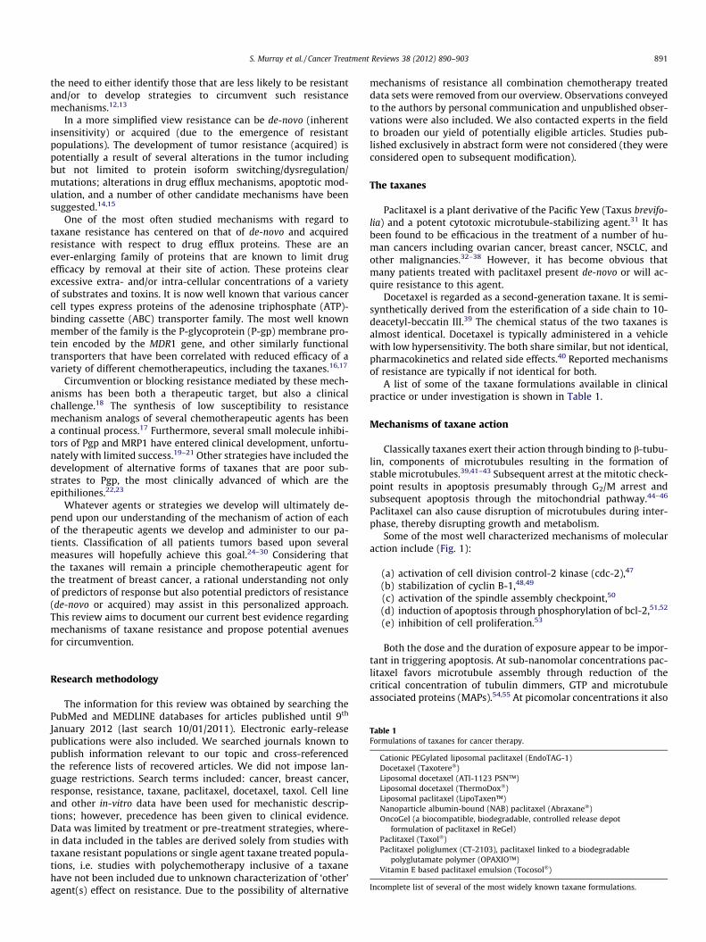
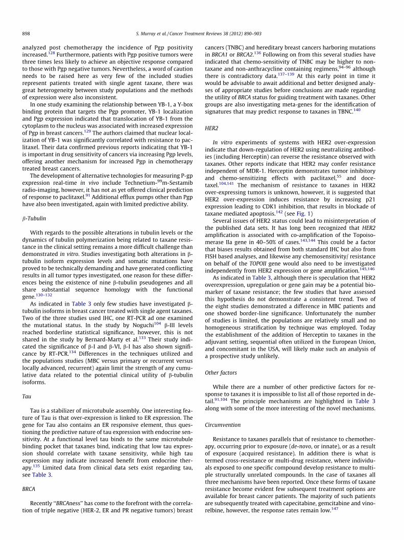
Some of the most well characterized mechanisms of molecularaction include (Fig. 1):
(a) activation of cell division control-2 kinase (cdc-2),47
(b) stabilization of cyclin B-1,48,49
(c) activation of the spindle assembly checkpoint,50
(d) induction of apoptosis through phosphorylation of bcl-2,51,52
(e) inhibition of cell proliferation.53
Both the dose and the duration of exposure appear to be impor-tant in triggering apoptosis. At sub-nanomolar concentrations pac-litaxel favors microtubule assembly through reduction of thecritical concentration of tubulin dimmers, GTP and microtubuleassociated proteins (MAPs).54,55 At picomolar concentrations it also
G0
M
S
G2
G1
Committed
Cyclin D1,2,3
CDK4/6
INK4
p16INK4a
p15INK4b
p18INK4c
p19INK4d
Cyclin E1,2
CDK2
Cip/Kip
p21Cip1
p27Kip1
p57Kip2
HDAC
E2F1-5HDAC
pRbP107p130
E2F1-5
pRbP107p130
P P
P
Cyclin A
CDK2
CDC2
Cyclin ACyclin B
CDC2
Cyclin A
CDK2
CDC25C
CDC2
WEE1
Myt1pRbP107p130
P P
P
PP-I
14-3-3σ
14-3-3σ
* *
*
*
Mitogens:Growth factors,
hormones
Quinescence
Anti-mitogenic signals
AktP HER2P13k
p53P
ATR
CHKP
Grb2MAPK
KAP
SAC
BubR1
KAP
Mad2Inactive
APCActiveAPC
Spindle Inhibitors BRCA1
ActiveStathminInactive
StathminP P
PP
Fig. 1. Regulation of cell cycle in relation to taxane resistance. Cell cycle and phase commitment. Mitogenic stimuli converge to activate cyclin D complexes; these allow E2Fto activate the expression of genes required for S-phase entry in a pRb dependant fashion. Antiproliferative signals, by means of as yet unclear mechanisms, affect p27Kip1 thatin turn antagonizes cyclin E-CDK2 activity. The cyclin dependent kinases (cdk1 (cdc2), cdk2, cdk4/6) bind to and regulate cyclin function. Phosphorylation of the complexesregulates various transcriptional events in cell cycle progression. The spindle assembly checkpoint (SAC) appears to be the principal site for taxane induced cell death signalsto take effect. As indicated in the text, taxanes have been correlated with differential regulation of a number of key genes (proteins) associated with the cell cycle. Many ofthese are considered to specifically regulate the S/G2 transition preceding M. Resistance to taxanes is thought to occur through a variety of mechanisms, many of which havebeen linked to defects in the SAC. Several chemotherapeutic agents act in distinct phases of the cell cycle: G1 (alkylating agents, platinums, cytotoxic antibiotics), S (anti-metabolites, topoisomerase inhibitors, docetaxel), and M (paclitaxel, docetaxel, epithiliones, vinca alkaloids). CDK, cyclin-dependent kinase; Cip, CDK interacting protein;HDAC, histone deacetylase; Ink4, Inhibitor of CDK4; Kip, Kinase inhibitory protein, PP-I, protein phosphatase 1; pRb, product of the retinoblastoma susceptibility gene. ⁄Cellcycle checkpoint.
892 S. Murray et al. / Cancer Treatment Reviews 38 (2012) 890–903
exerts its effects on interphase microtubules and genes controllingapoptosis.56 Increasing concentrations result in shifting the equi-librium of dimers to polymers thereby preventing disassociationeven under conditions of extreme stress.41,57 As a result of theincreasing number of patients being treated with taxanes thedevelopment of taxane resistance is becoming a clinically impor-tant issue. Therefore, the elucidations of resistance are not onlyimportant for the development of strategies to overcome it, butalso in possibly predicting response of patients to taxane basedregimens.
Molecular mechanisms of taxane resistance
Although numerous mechanisms of drug resistance have beenrecognized we focus on those specifically reporting on taxaneresistance. Several mechanisms have been identified in breastcancer cell lines, while characterization of resistance has provenmore difficult in clinical specimens. We primarily report onin vivo data and supplement with in vitro data for a number ofthe best-characterized mechanisms.
P-glycoprotein (Pgp)
A feature common to most cancer types is multi-drug resis-tance, i.e. cross resistance of cancer cells to structurally unrelatedcytotoxic agents.58,59 Several mechanisms of variable drug and
cancer specificity have been associated with the study of cancercells to cytotoxic xenobiotics.60–64
One of the most well known mechanisms relates to drug resis-tance associated with the over-expression of the MRD-1 geneproduct Pgp (permeability-glycoprotein) in cancer.65–67 Increasedexpression (as assessed by immunohistochemistry) of this proteinhas been extensively studied in taxane resistant breast carcino-mas.68 However, it remains an elusive marker for clinical imple-mentation due to conflicting data and a lack of standardization inlight of consensus recommendations that date back to the mid1990’s.69 Pgp is a member of a growing family of at least 49 aden-osine triphosphate (ATP) binding cassette (ABC) transporters.70,71
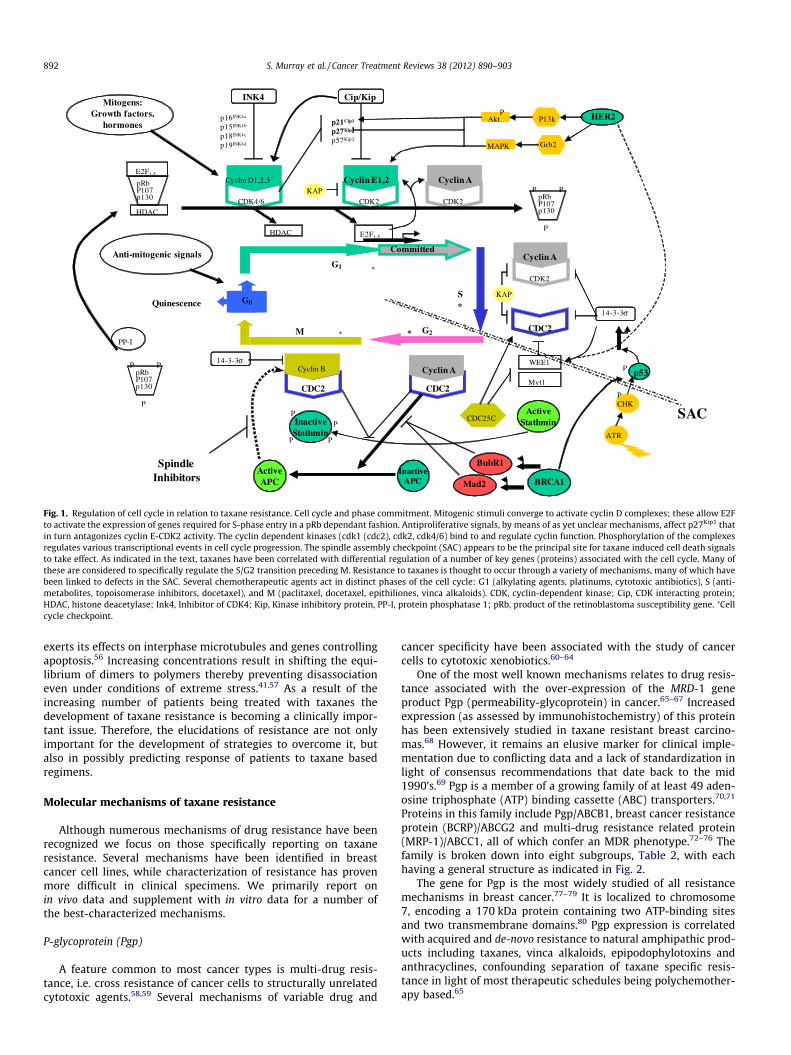
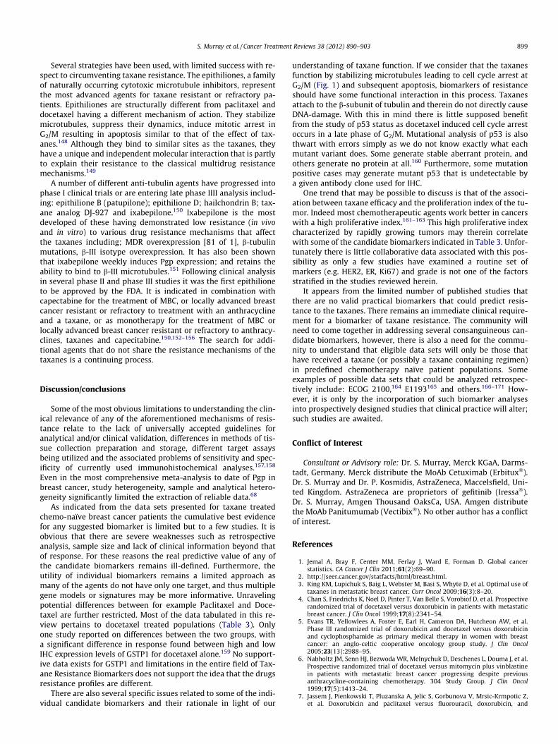
Proteins in this family include Pgp/ABCB1, breast cancer resistanceprotein (BCRP)/ABCG2 and multi-drug resistance related protein(MRP-1)/ABCC1, all of which confer an MDR phenotype.72–76 Thefamily is broken down into eight subgroups, Table 2, with eachhaving a general structure as indicated in Fig. 2.
The gene for Pgp is the most widely studied of all resistancemechanisms in breast cancer.77–79 It is localized to chromosome7, encoding a 170 kDa protein containing two ATP-binding sitesand two transmembrane domains.80 Pgp expression is correlatedwith acquired and de-novo resistance to natural amphipathic prod-ucts including taxanes, vinca alkaloids, epipodophylotoxins andanthracyclines, confounding separation of taxane specific resis-tance in light of most therapeutic schedules being polychemother-apy based.65
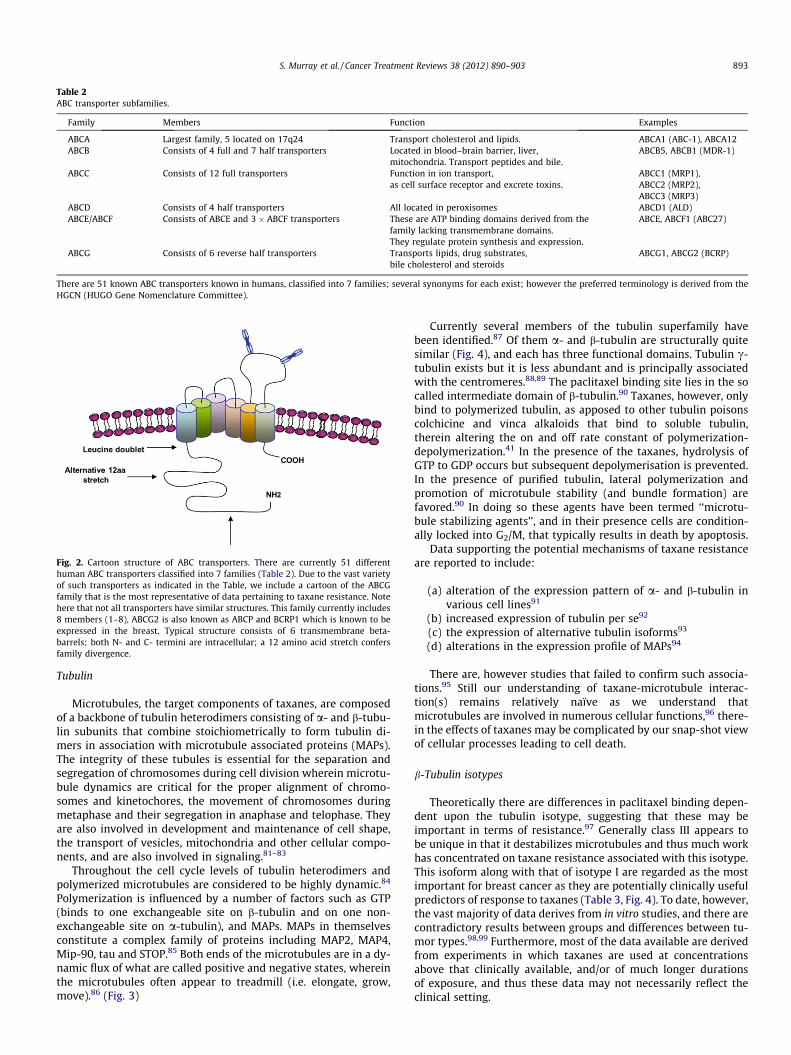
Table 2ABC transporter subfamilies.
Family Members Function Examples
ABCA Largest family, 5 located on 17q24 Transport cholesterol and lipids. ABCA1 (ABC-1), ABCA12ABCB Consists of 4 full and 7 half transporters Located in blood–brain barrier, liver,
mitochondria. Transport peptides and bile.ABCB5, ABCB1 (MDR-1)
ABCC Consists of 12 full transporters Function in ion transport,as cell surface receptor and excrete toxins.
ABCC1 (MRP1),ABCC2 (MRP2),ABCC3 (MRP3)
ABCD Consists of 4 half transporters All located in peroxisomes ABCD1 (ALD)ABCE/ABCF Consists of ABCE and 3 � ABCF transporters These are ATP binding domains derived from the
family lacking transmembrane domains.They regulate protein synthesis and expression.
ABCE, ABCF1 (ABC27)
ABCG Consists of 6 reverse half transporters Transports lipids, drug substrates,bile cholesterol and steroids
ABCG1, ABCG2 (BCRP)
There are 51 known ABC transporters known in humans, classified into 7 families; several synonyms for each exist; however the preferred terminology is derived from theHGCN (HUGO Gene Nomenclature Committee).
COOH
NH2
Alternative 12aastretch
Leucine doublet
Fig. 2. Cartoon structure of ABC transporters. There are currently 51 differenthuman ABC transporters classified into 7 families (Table 2). Due to the vast varietyof such transporters as indicated in the Table, we include a cartoon of the ABCGfamily that is the most representative of data pertaining to taxane resistance. Notehere that not all transporters have similar structures. This family currently includes8 members (1–8), ABCG2 is also known as ABCP and BCRP1 which is known to beexpressed in the breast. Typical structure consists of 6 transmembrane beta-barrels; both N- and C- termini are intracellular; a 12 amino acid stretch confersfamily divergence.
S. Murray et al. / Cancer Treatment Reviews 38 (2012) 890–903 893
Tubulin
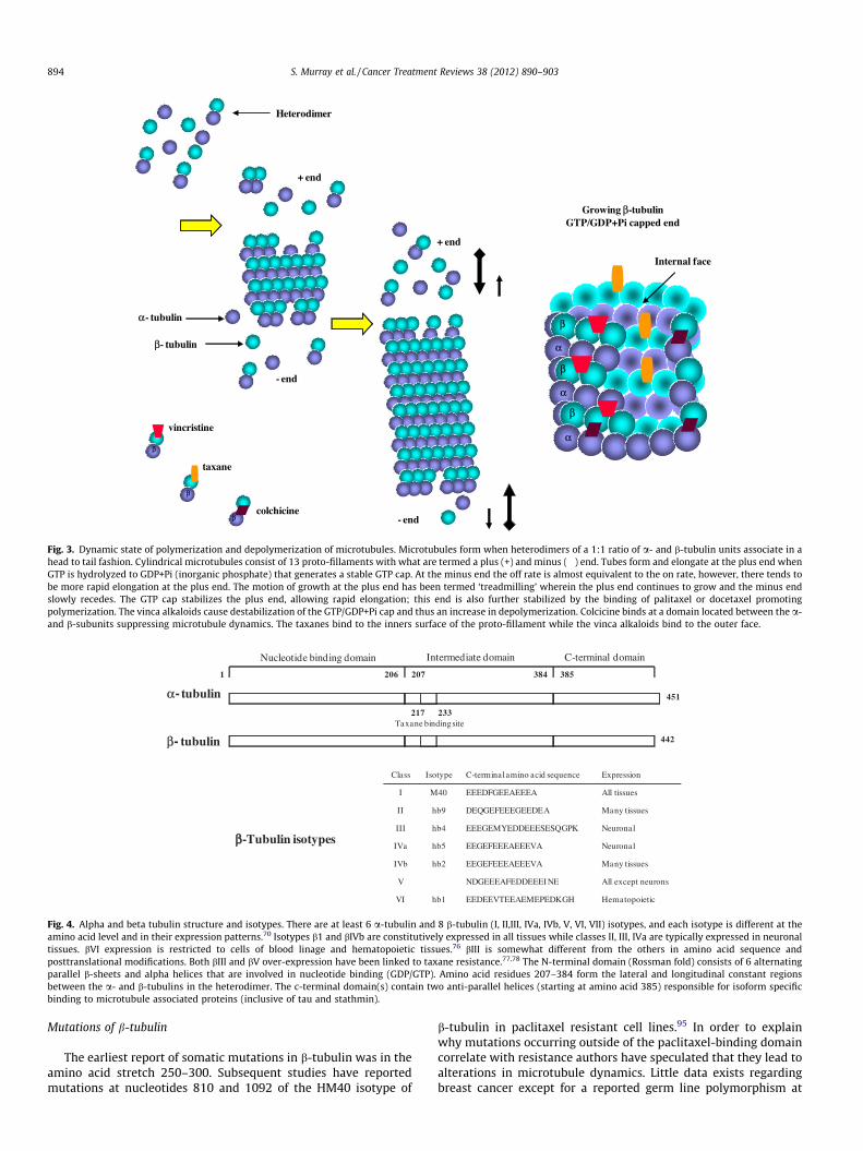
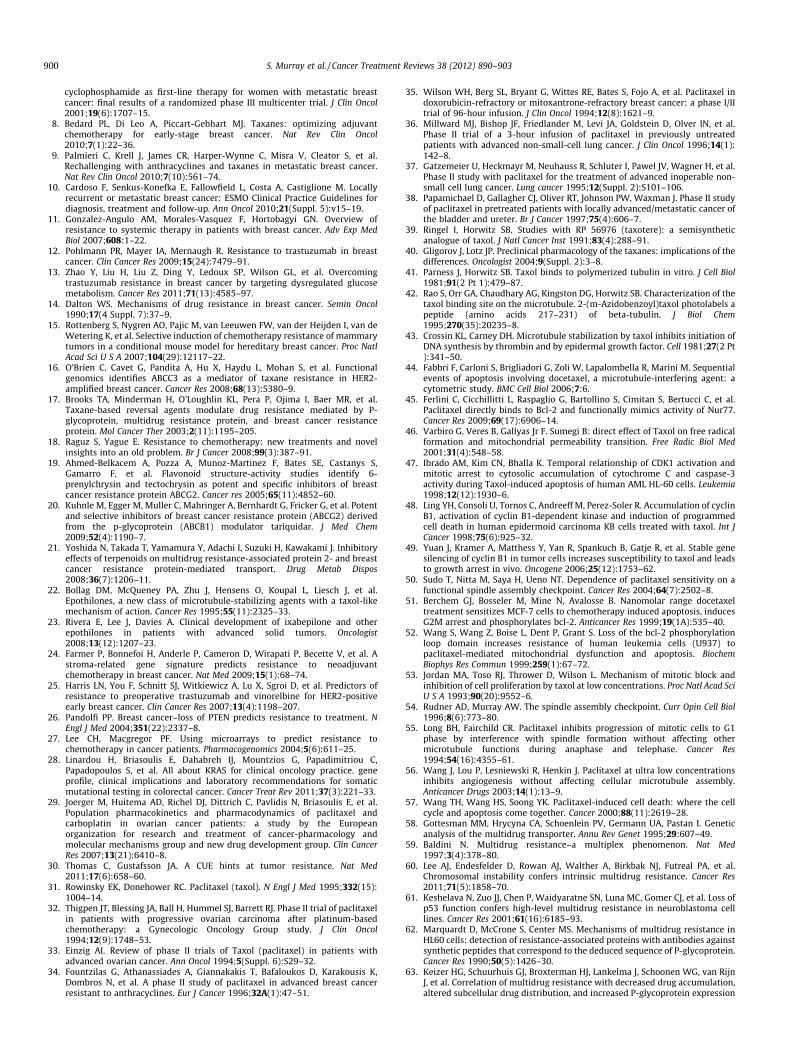
Microtubules, the target components of taxanes, are composedof a backbone of tubulin heterodimers consisting of a- and b-tubu-lin subunits that combine stoichiometrically to form tubulin di-mers in association with microtubule associated proteins (MAPs).The integrity of these tubules is essential for the separation andsegregation of chromosomes during cell division wherein microtu-bule dynamics are critical for the proper alignment of chromo-somes and kinetochores, the movement of chromosomes duringmetaphase and their segregation in anaphase and telophase. Theyare also involved in development and maintenance of cell shape,the transport of vesicles, mitochondria and other cellular compo-nents, and are also involved in signaling.81–83
Throughout the cell cycle levels of tubulin heterodimers andpolymerized microtubules are considered to be highly dynamic.84
Polymerization is influenced by a number of factors such as GTP(binds to one exchangeable site on b-tubulin and on one non-exchangeable site on a-tubulin), and MAPs. MAPs in themselvesconstitute a complex family of proteins including MAP2, MAP4,Mip-90, tau and STOP.85 Both ends of the microtubules are in a dy-namic flux of what are called positive and negative states, whereinthe microtubules often appear to treadmill (i.e. elongate, grow,move).86 (Fig. 3)
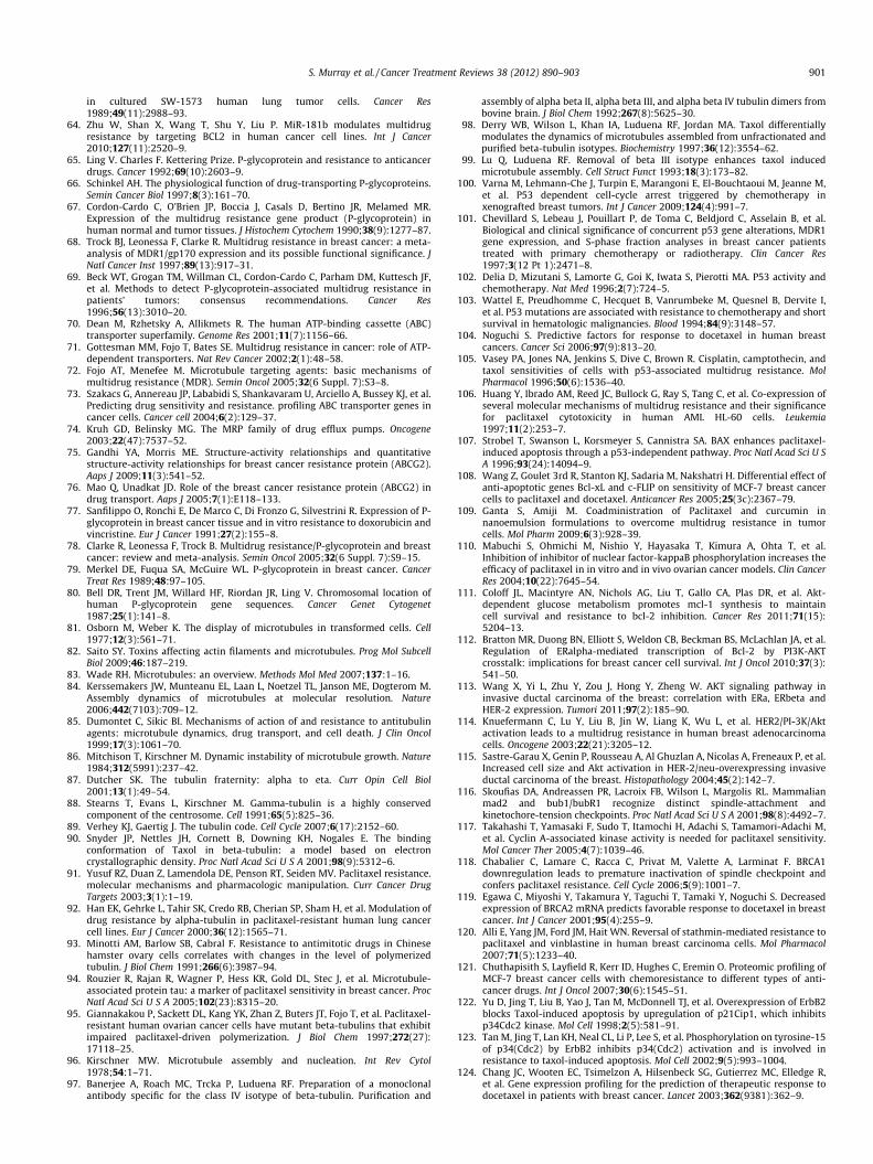
Currently several members of the tubulin superfamily havebeen identified.87 Of them a- and b-tubulin are structurally quitesimilar (Fig. 4), and each has three functional domains. Tubulin c-tubulin exists but it is less abundant and is principally associatedwith the centromeres.88,89 The paclitaxel binding site lies in the socalled intermediate domain of b-tubulin.90 Taxanes, however, onlybind to polymerized tubulin, as apposed to other tubulin poisonscolchicine and vinca alkaloids that bind to soluble tubulin,therein altering the on and off rate constant of polymerization-depolymerization.41 In the presence of the taxanes, hydrolysis ofGTP to GDP occurs but subsequent depolymerisation is prevented.In the presence of purified tubulin, lateral polymerization andpromotion of microtubule stability (and bundle formation) arefavored.90 In doing so these agents have been termed ‘‘microtu-bule stabilizing agents’’, and in their presence cells are condition-ally locked into G2/M, that typically results in death by apoptosis.
Data supporting the potential mechanisms of taxane resistanceare reported to include:
(a) alteration of the expression pattern of a- and b-tubulin invarious cell lines91
(b) increased expression of tubulin per se92
(c) the expression of alternative tubulin isoforms93
(d) alterations in the expression profile of MAPs94
There are, however studies that failed to confirm such associa-tions.95 Still our understanding of taxane-microtubule interac-tion(s) remains relatively naïve as we understand thatmicrotubules are involved in numerous cellular functions,96 there-in the effects of taxanes may be complicated by our snap-shot viewof cellular processes leading to cell death.
b-Tubulin isotypes
Theoretically there are differences in paclitaxel binding depen-dent upon the tubulin isotype, suggesting that these may beimportant in terms of resistance.97 Generally class III appears tobe unique in that it destabilizes microtubules and thus much workhas concentrated on taxane resistance associated with this isotype.This isoform along with that of isotype I are regarded as the mostimportant for breast cancer as they are potentially clinically usefulpredictors of response to taxanes (Table 3, Fig. 4). To date, however,the vast majority of data derives from in vitro studies, and there arecontradictory results between groups and differences between tu-mor types.98,99 Furthermore, most of the data available are derivedfrom experiments in which taxanes are used at concentrationsabove that clinically available, and/or of much longer durationsof exposure, and thus these data may not necessarily reflect theclinical setting.
- end
- end
+ end
+ end
Heterodimer
α- tubulin
Growing β-tubulinGTP/GDP+Pi capped end
β- tubulin
vincristineα
α
α
β
β
β
Internal face
taxane
colchicine
β
β
β
Fig. 3. Dynamic state of polymerization and depolymerization of microtubules. Microtubules form when heterodimers of a 1:1 ratio of a- and b-tubulin units associate in ahead to tail fashion. Cylindrical microtubules consist of 13 proto-fillaments with what are termed a plus (+) and minus (�) end. Tubes form and elongate at the plus end whenGTP is hydrolyzed to GDP+Pi (inorganic phosphate) that generates a stable GTP cap. At the minus end the off rate is almost equivalent to the on rate, however, there tends tobe more rapid elongation at the plus end. The motion of growth at the plus end has been termed ‘treadmilling’ wherein the plus end continues to grow and the minus endslowly recedes. The GTP cap stabilizes the plus end, allowing rapid elongation; this end is also further stabilized by the binding of palitaxel or docetaxel promotingpolymerization. The vinca alkaloids cause destabilization of the GTP/GDP+Pi cap and thus an increase in depolymerization. Colcicine binds at a domain located between the a-and b-subunits suppressing microtubule dynamics. The taxanes bind to the inners surface of the proto-fillament while the vinca alkaloids bind to the outer face.
ββ-Tubulin isotypes
Class Isotype C-terminal amino acid sequence Expression
I M40 EEEDFGEEAEEEA All tissues
II hb9 DEQGEFEEEGEEDEA Many tissues
III hb4 EEEGEMYEDDEEESESQGPK Neuronal
IVa hb5 EEGEFEEEAEEEVA Neuronal
IVb hb2 EEGEFEEEAEEEVA Many tissues
V NDGEEEAFEDDEEEI NE All except neurons
VI hb1 EEDEEVTEEAEMEPEDKGH Hematopoietic
α- tubulin
β- tubulin
Nucleotide binding domain
206 2071 384 385
451
442
217 233Taxane binding site
Intermediate domain C-terminal domain
Fig. 4. Alpha and beta tubulin structure and isotypes. There are at least 6 a-tubulin and 8 b-tubulin (I, II,III, IVa, IVb, V, VI, VII) isotypes, and each isotype is different at theamino acid level and in their expression patterns.70 Isotypes b1 and bIVb are constitutively expressed in all tissues while classes II, III, IVa are typically expressed in neuronaltissues. bVI expression is restricted to cells of blood linage and hematopoietic tissues.76 bIII is somewhat different from the others in amino acid sequence andposttranslational modifications. Both bIII and bV over-expression have been linked to taxane resistance.77,78 The N-terminal domain (Rossman fold) consists of 6 alternatingparallel b-sheets and alpha helices that are involved in nucleotide binding (GDP/GTP). Amino acid residues 207–384 form the lateral and longitudinal constant regionsbetween the a- and b-tubulins in the heterodimer. The c-terminal domain(s) contain two anti-parallel helices (starting at amino acid 385) responsible for isoform specificbinding to microtubule associated proteins (inclusive of tau and stathmin).
894 S. Murray et al. / Cancer Treatment Reviews 38 (2012) 890–903
Mutations of b-tubulin
The earliest report of somatic mutations in b-tubulin was in theamino acid stretch 250–300. Subsequent studies have reportedmutations at nucleotides 810 and 1092 of the HM40 isotype of
b-tubulin in paclitaxel resistant cell lines.95 In order to explainwhy mutations occurring outside of the paclitaxel-binding domaincorrelate with resistance authors have speculated that they lead toalterations in microtubule dynamics. Little data exists regardingbreast cancer except for a reported germ line polymorphism at
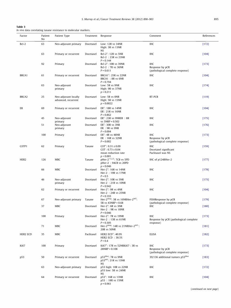
Table 3In vivo data correlating taxane resistance to molecular markers.
Factor PatientNo
Patient Type Treatment Response Comment References
Bcl-2 63 Neo-adjuvant primary Docetaxel Low: 12R vs 34NRHigh: 3R vs 13NRNS
IHC [172]
63 Primary or recurrent Docetaxel Bcl-2+: 12R vs 5NRBcl-2�: 23R vs 23NRP = 0.144
IHC [104]
92 Primary Docetaxel Bcl-2+: 10R vs 39NRBcl-2�: 7R vs 36NRP = 0.611
IHCResponse by pCR(pathological complete response)
[173]
BRCA1 61 Primary or recurrent Docetaxel BRCA1+: 25R vs 22NRBRCA1�: 8R vs 6NRP = 0.794
IHC [104]
63 Neo-adjuvantprimary
Docetaxel Low: 5R vs 9NRHigh: 9R vs 37NRp = 0.211
IHC [174]
BRCA2 25 Neo-adjuvant locallyadvanced, recurrent
Docetaxel Low: 5R vs 0NRHigh: 5R vs 15NRp = 0.0022
RT-PCR [119]
ER 69 Primary or recurrent Docetaxel ER+: 18R vs 14NRER-: 21R vs 16NRP = 0.862
IHC [104]
45 Neo-adjuvantprimary
Docetaxel ER+: 23R vs 9NRER�: 8Rvs 5NRP = 0.502
IHC [175]
51 Neo-adjuvantprimary
Docetaxel ER+: 30R vs 3NRER�: 9R vs 9NRP = 0.004
IHC [176]
100 Primary Docetaxel ER+: 4R vs 48NRER�: 16R vs 32NRP = 0.002
IHCResponse by pCR(pathological complete response)
[173]
GSTP1 62 Primary Taxane GST+: 0.31 ± 0.09GST-: 0.73 ± 0.04mean reduction ratep < 0.001
IHCDocetaxel significantPaclitaxel was NS
[159]
HER2 126 MBC Taxane pHer-2+1,2,3: 7CB vs 5PDpHer-2�: 94CB vs 20PDp = 0.046
IHC of p1248Her-2 [177]
66 MBC Docetaxel Her-2+: 16R vs 14NRHer-2�: 19R vs 17NRP = 0.5
IHC [178]
46 Neo-adjuvantprimary
Docetaxel Her-2+: 10R vs 5NRHer-2�: 21R vs 10NRP = 0.942
IHC [175]
62 Primary or recurrent Docetaxel Her-2+: 9R vs 4NRHer-2�: 24R vs 25NRP = 0.193
IHC [104]
67 Neo-adjuvant primary Taxane Her-2Amp: 3R vs 16NRHer-2WT:5R vs 43NRP = 0.68
FISHResponse by pCR(pathological complete response)
[179]
37 MBC Docetaxel Her-2+: 6R vs 3NRHer-2�: 9R vs 18NRP = 0.046
IHC [180]
100 Primary Docetaxel Her-2+: 7R vs 19NRHer-2�: 13R vs 61NRP = 0.305
IHCResponse by pCR (pathological completeresponse)
[173]
71 MBC Docetaxel Her-2Amp: 14R vs 21NRHer-2WT-:20R vs 50NR
[181]
HER2 ECD 35 MBC Paclitaxel HER2 ECD+: 40.9%HER2 ECD�: 38.5%P = 0.4
ELISA [182]
Ki67 100 Primary Docetaxel Ki67+: 17R vs 52NRKi67-: 3R vs28NRP = 0.108
IHCResponse by pCR(pathological complete response)
[173]
p53 50 Primary or recurrent Docetaxel p53Mut: 7R vs 9NRp53WT: 21R vs 13NRNS
39/136 additional tumors p53Mut [183]
63 Neo-adjuvant primary Docetaxel p53 high: 10R vs 22NRp53 low: 5R vs 24NRNS
IHC [172]
64 Primary or recurrent Docetaxel p53+: 16R vs 13NRp53�: 18R vs 15NRp = 0.961
IHC [104]
(continued on next page)
S. Murray et al. / Cancer Treatment Reviews 38 (2012) 890–903 895
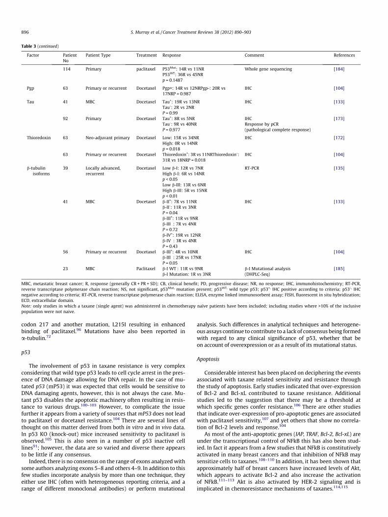
Table 3 (continued)
Factor PatientNo
Patient Type Treatment Response Comment References
114 Primary paclitaxel P53Mut: 14R vs 11NRP53WT: 36R vs 45NRp = 0.1487
Whole gene sequencing [184]
Pgp 63 Primary or recurrent Docetaxel Pgp+: 14R vs 12NRPgp-: 20R vs17NRP = 0.987
IHC [104]
Tau 41 MBC Docetaxel Tau+: 19R vs 13NRTau-: 2R vs 2NRP = 0.99
IHC [133]
92 Primary Docetaxel Tau+: 8R vs 5NRTau-: 9R vs 40NRP = 0.977
IHCResponse by pCR(pathological complete response)
[173]
Thioredoxin 63 Neo-adjuvant primary Docetaxel Low: 15R vs 34NRHigh: 0R vs 14NRp = 0.018
IHC [172]
63 Primary or recurrent Docetaxel Thioredoxin+: 3R vs 11NRThioredoxin-:31R vs 18NRP = 0.018
IHC [104]
b-tubulinisoforms
39 Locally advanced,recurrent
Docetaxel Low b-I: 12R vs 7NRHigh b-I: 6R vs 14NRp < 0.05
RT-PCR [135]
Low b-III: 13R vs 6NRHigh b-III: 5R vs 15NRp < 0.01
41 MBC Docetaxel b-II+: 7R vs 11NRb-II-: 11R vs 3NRP = 0.04
IHC [133]
b-III+: 11R vs 9NRb-III�: 7R vs 4NRP = 0.72b-IV+: 19R vs 12NRb-IV�: 3R vs 4NRP = 0.43
56 Primary or recurrent Docetaxel b-III+: 4R vs 10NRb-III�: 25R vs 17NRP = 0.05
IHC [104]
23 MBC Paclitaxel b-I WT : 11R vs 9NRb-I Mutation: 1R vs 3NR
b-I Mutational analysis(DHPLC-Seq)
[185]
MBC, metastatic breast cancer; R, response (generally CR + PR + SD); CB, clinical benefit; PD, progressive disease; NR, no response; IHC, immunohistochemistry; RT-PCR,reverse transcriptase polymerase chain reaction; NS, not significant, p53Mut, mutation present; p53WT, wild type p53; p53+, IHC positive according to criteria; p53-, IHCnegative according to criteria; RT-PCR, reverse transcriptase polymerase chain reaction; ELISA, enzyme linked immunosorbent assay; FISH, fluorescent in situ hybridization;ECD, extracellular domain.Note: only studies in which a taxane (single agent) was administered in chemotherapy naïve patients have been included; including studies where >10% of the inclusivepopulation were not naive.
896 S. Murray et al. / Cancer Treatment Reviews 38 (2012) 890–903
codon 217 and another mutation, L215I resulting in enhancedbinding of paclitaxel.96 Mutations have also been reported ina-tubulin.72
p53
The involvement of p53 in taxane resistance is very complexconsidering that wild type p53 leads to cell cycle arrest in the pres-ence of DNA damage allowing for DNA repair. In the case of mu-tated p53 (mP53) it was expected that cells would be sensitive toDNA damaging agents, however, this is not always the case. Mu-tant p53 disables the apoptotic machinery often resulting in resis-tance to various drugs.100–103 However, to complicate the issuefurther it appears from a variety of sources that mP53 does not leadto paclitaxel or docetaxel resistance.104 There are several lines ofthought on this matter derived from both in vitro and in vivo data.In p53 KO (knock-out) mice increased sensitivity to paclitaxel isobserved.105 This is also seen in a number of p53 inactive celllines91; however, the data are so varied and diverse there appearsto be little if any consensus.
Indeed, there is no consensus on the range of exons analyzed withsome authors analyzing exons 5–8 and others 4–9. In addition to thisfew studies incorporate analysis by more than one technique, theyeither use IHC (often with heterogeneous reporting criteria, and arange of different monoclonal antibodies) or perform mutational
analysis. Such differences in analytical techniques and heterogene-ous assays continue to contribute to a lack of consensus being formedwith regard to any clinical significance of p53, whether that beon account of overexpression or as a result of its mutational status.
Apoptosis
Considerable interest has been placed on deciphering the eventsassociated with taxane related sensitivity and resistance throughthe study of apoptosis. Early studies indicated that over-expressionof Bcl-2 and Bcl-xL contributed to taxane resistance. Additionalstudies led to the suggestion that there may be a threshold atwhich specific genes confer resistance.106 There are other studiesthat indicate over-expression of pro-apoptotic genes are associatedwith paclitaxel sensitivity,107 and yet others that show no correla-tion of Bcl-2 levels and response.104
As most of the anti-apoptotic genes (IAP, TRAF, Bcl-2, Bcl-xL) areunder the transcriptional control of NFkB this has also been stud-ied. In fact it appears from a few studies that NFkB is constitutivelyactivated in many breast cancers and that inhibition of NFkB maysensitize cells to taxanes.108–110 In addition, it has been shown thatapproximately half of breast cancers have increased levels of Akt,which appears to activate Bcl-2 and also increase the activationof NFkB.111–113 Akt is also activated by HER-2 signaling and isimplicated in chemoresistance mechanisms of taxanes.114,115
S. Murray et al. / Cancer Treatment Reviews 38 (2012) 890–903 897
Cell cycle
The spindle assembly checkpoint (SAC) appears critical for tax-ane mediated cell death. Various mechanisms involved in thischeckpoint appear to influence and be influenced by taxanes, anddefects in the SAC correlate with resistance. (Fig. 1)
Various data sets seem to support this strong interaction:
(1) Upon activation of the SAC both Mad2 and BubR1 interactwith Cdc2 inhibiting its ability to activate APC.50 Destructionof cyclin B and other regulators of mitosis by APC are respon-sible for proper metaphase-anaphase transition and mitoticexit. MAPs including Mad2 and BubR1 are thought to regu-late SAC preventing anaphase until chromosomes areattached to bipolar spindles.116 In the presence of spindleinhibitors cyclin B degradation is inhibited, cells arrest atpro-metaphase and maintain constitutive Cdk1 activity(destruction of cyclin B inactivates Cdk1).50
(2) When Mad2 levels are low SAC is non-functional. Sensitivityis restored with re-establishment of Mad2 levels.50
(3) Over-expression of cyclins E and A have been associatedwith adverse outcomes. These cyclins are important media-tors of G1–S phase transition and subsequent S–G2 phasetransition. Cyclin A appears to be the more important as itis directly involved in regulating Cdk1 (cdc2) activity as acti-vated Cdk1 is required for cells to enter mitosis and for SACfunctionality, both key requirements of taxane sensitivity.117
(4) BRCA1 is also implicated in SAC control. BubR1 transcriptionis regulated by BRCA1, and also to some extent by p53.BRCA1 is a co-activator of p53 and positively regulatesMad2, thus inhibiting APC activity. Therefore, in BRCA1 defi-cient cells there is a premature onset of anaphase activatedby APC by ubiquitination and degradation of cyclin B andsubsequent activation of Cdk1,118 this being linked to paclit-axel resistance. Similarly there is data showing thatdecreased levels of BCRA2 correlate with better responsesto docetaxel.119 (Table 3)
(5) Stathmin, a microtubule regulator, destabilizes microtubulesby two mechanisms, catastrophic promotion and tubulinsequestration. It is active in G2/M transition where it is inac-tivated by Cdk1 allowing for M phase entry. Stathmin over-expression has been correlated with resistance to taxanes.120
More recently in a two-dimensional gel electrophoresis andMALDI-TOF peptide mass fingerprinting study stathmin wasprofiled as one of 9 proteins differentially expressed in pac-litaxel resistant MCF-7 cells.121
(6) HER2 over-expression inhibits taxol induced apoptosis bytranscriptionally up-regulating p21cip1 which associateswith p34Cdc2 inhibiting taxol mediated p34 activation delay-ing cells from entering G2/M and thereby inhibiting apopto-sis.122 HER2 may also directly phosphorylate Cdc2 leading toresistance. There is also evidence that HER2 positive tumorshave low levels of Cdk1 resulting in delayed mitosis and pac-litaxel resistance.123 HER2 has also been shown to promoteG1/S progression and tumor cell proliferation by reducingp27Kip1 stability and reducing p27-cdc2 complexes. Stimula-tion through HER2 and other RTKs has also been shown toresult in increased levels of Pgp, without affecting transcrip-tion, through the MEK–ERK–RSK pathway as inhibitors tothis pathway decrease Pgp mediated resistance topaclitaxel.123
Thus it appears that the microtubule composition of the mitoticspindle, the dynamics of microtubule assembly and the associatedanaphase–metaphase block induced by anti-microtubule agentshighlight the importance of the transition into M phase in deter-
mining their sensitivity and that deficits in any of the multitudeof proteins that regulate the SAC would be sufficient for conferringresistance to taxanes.
Gene signatures/metagenes
Several groups have investigated high throughput screening ofthousands of genes as a method to identify patterns of expressionof single genes or gene combinations (gene signatures) that corre-late with outcome to given therapies. A first study to investigatetaxane related outcomes of response in breast cancer was reportedby Chang, et al.124 They identified 92 genes that correlated withdocetaxel response of primary breast cancer in the neo-adjuvantsetting. Their RNA profile from 24 patients included higher expres-sion of genes involved in cell cycle, cell adhesion, protein modifica-tion, transcription and apoptosis; while resistant tumors showedincreased expression of some transcriptional and signal transduc-tion genes. The 92 gene predictor had positive and negative predic-tive values of 92% and 83%, respectively.124
Utilizing another technique, adaptor-tagged competitive(ATAC)-polymerase chain reaction (PCR), Iwao-Koizumi et al., mea-sured the expression of 2453 genes in a series of 70 (44 learningset, 26 validation set) primary or locally recurrent breast cancersreceiving docetaxel.125 They identified an algorithm consisting of85 genes that predicted clinical response to docetaxel with positiveand negative predictive values of 73.3% and 90.9%, respectively.Non-responders were characterized as having elevated expressionof genes controlling cellular redox, thioredoxin, glutathione-S-transferase and peroxiredoxin.125
The utility of gene expression signatures based predictive algo-rithms will advance as they allow, if standardized, potentially im-proved positive and negative predictive ability over single genepredictors. Similar algorithms of metagenes may also enter intoclinical development as our knowledge base increases per predic-tive marker/signature.
Clinically relevant prediction of taxane resistance
While the majority of data presented relates to in vitro experi-mentation, limited hypothesis of taxane resistance have beeninvestigated in vivo. Therefore, insufficient in vivo data exist to gaina clear picture of numerous hypotheses that have been generatedfor molecules of predictive/prognostic significance in breast can-cer. To date studies have failed to indicate any particular mecha-nism or marker as immediately clinically relevant with respect tooffering insights into patient stratification.
P-glycoprotein (Pgp)
Increased expression in breast cancers and in other cancer typeshas generally been correlated with MDR.68 However, much of thedata is conflicting. Some of the problems in assessing the predictivenature of Pgp may relate to the variety of methods used and lack ofstandardization of cut offs for quantitation, clinical endpoints mea-sured and study/patient heterogeneity that exists across all studies.Furthermore, one of the main antibodies used for its detection hasbeen reported to cross react with HER2 and also the heavy chain ofmyosin leading to distinct difficulties in interpretation.126
Putting this aside Pgp expression (depending on the method ofanalysis and thresholds used) shows a broad range of expression.From 0–30% in newly diagnosed breast cancer rising to over 70%in many cases of relapsed breast cancer.127 Indeed in a meta-analysis of MDR1/Pgp expression in breast cancers Trock et al.,showed that approximately 40% of breast tumors expressed Pgpat RNA level or protein assessed by IHC, and that in tumors
898 S. Murray et al. / Cancer Treatment Reviews 38 (2012) 890–903
analyzed post chemotherapy the incidence of Pgp positivityincreased.128 Furthermore, patients with Pgp positive tumors werethree times less likely to achieve an objective response comparedto those with Pgp negative tumors. Nevertheless, a word of cautionneeds to be raised here as very few of the included studiesrepresent patients treated with single agent taxane, there wasgreat heterogeneity between study populations and the methodsof expression were also inconsistent.
In one study examining the relationship between YB-1, a Y-boxbinding protein that targets the Pgp promoter, YB-1 localizationand Pgp expression indicated that translocation of YB-1 from thecytoplasm to the nucleus was associated with increased expressionof Pgp in breast cancers.129 The authors claimed that nuclear local-ization of YB-1 was significantly correlated with resistance to pac-litaxel. Their data confirmed previous reports indicating that YB-1is important in drug sensitivity of cancers via increasing Pgp levels,offering another mechanism for increased Pgp in chemotherapytreated breast cancers.
The development of alternative technologies for measuring P-gpexpression real-time in vivo include Technetium-99m-Sestamibradio-imaging, however, it has not as yet offered clinical predictionof response to paclitaxel.91 Additional efflux pumps other than Pgphave also been investigated, again with limited predictive ability.
b-Tubulin
With regards to the possible alterations in tubulin levels or thedynamics of tubulin polymerization being related to taxane resis-tance in the clinical setting remains a more difficult challenge thandemonstrated in vitro. Studies investigating both alterations in b-tubulin isoform expression levels and somatic mutations haveproved to be technically demanding and have generated conflictingresults in all tumor types investigated, one reason for these differ-ences being the existence of nine b-tubulin pseudogenes and allshare substantial sequence homology with the functionalgene.130–132
As indicated in Table 3 only few studies have investigated b-tubulin isoforms in breast cancer treated with single agent taxanes.Two of the three studies used IHC, one RT-PCR ad one examinedthe mutational status. In the study by Noguchi104 b-III levelsreached borderline statistical significance, however, this is notshared in the study by Bernard-Marty et al.133 Their study indi-cated the significance of b-I and b-VI, b-I has also shown signifi-cance by RT-PCR.134 Differences in the techniques utilized andthe populations studies (MBC versus primary or recurrent versuslocally advanced, recurrent) again limit the strength of any cumu-lative data related to the potential clinical utility of b-tubulinisoforms.
Tau
Tau is a stabilizer of microtubule assembly. One interesting fea-ture of Tau is that over-expression is linked to ER expression. Thegene for Tau also contains an ER responsive element, thus ques-tioning the predictive nature of tau expression with endocrine sen-sitivity. At a functional level tau binds to the same microtubulebinding pocket that taxanes bind, indicating that low tau expres-sion should correlate with taxane sensitivity, while high tauexpression may indicate increased benefit from endocrine ther-apy.135 Limited data from clinical data sets exist regarding tau,see Table 3.
BRCA
Recently ‘‘BRCAness’’ has come to the forefront with the correla-tion of triple negative (HER-2, ER and PR negative tumors) breast
cancers (TNBC) and hereditary breast cancers harboring mutationsin BRCA1 or BRCA2.136 Following on from this several studies haveindicated that chemo-sensitivity of TNBC may be higher to non-taxane and non-anthracycline containing regimens,94–96 althoughthere is contradictory data.137–139 At this early point in time itwould be advisable to await additional and better designed analy-ses of appropriate studies before conclusions are made regardingthe utility of BRCA status for guiding treatment with taxanes. Othergroups are also investigating meta-genes for the identification ofsignatures that may predict response to taxanes in TBNC.140
HER2
In vitro experiments of systems with HER2 over-expressionindicate that down-regulation of HER2 using neutralizing antibod-ies (including Herceptin) can reverse the resistance observed withtaxanes. Other reports indicate that HER2 may confer resistanceindependent of MDR-1. Herceptin demonstrates tumor inhibitoryand chemo-sensitizing effects with paclitaxel,55 and doce-taxel.104,141 The mechanism of resistance to taxanes in HER2over-expressing tumors is unknown, however, it is suggested thatHER2 over-expression induces resistance by increasing p21expression leading to CDK1 inhibition, that results in blockade oftaxane mediated apoptosis.142 (see Fig. 1)
Several issues of HER2 status could lead to misinterpretation ofthe published data sets. It has long been recognized that HER2amplification is associated with co-amplification of the Topoiso-merase IIa gene in 40–50% of cases.143,144 This could be a factorthat biases results obtained from both standard IHC but also fromFISH based analyses, and likewise any chemosensitivity/ resistanceon behalf of the TOPOII gene would also need to be investigatedindependently from HER2 expression or gene amplification.145,146
As indicated in Table 3, although there is speculation that HER2overexpression, upregulation or gene gain may be a potential bio-marker of taxane resistance; the few studies that have assessedthis hypothesis do not demonstrate a consistent trend. Two ofthe eight studies demonstrated a difference in MBC patients andone showed border-line significance. Unfortunately the numberof studies is limited, the populations are relatively small and nohomogeneous stratification by technique was employed. Todaythe establishment of the addition of Herceptin to taxanes in theadjuvant setting, sequential often utilized in the European Union,and concomitant in the USA, will likely make such an analysis ofa prospective study unlikely.
Other factors
While there are a number of other predictive factors for re-sponse to taxanes it is impossible to list all of those reported in de-tail.91,104 The principle mechanisms are highlighted in Table 3along with some of the more interesting of the novel mechanisms.
Circumvention
Resistance to taxanes parallels that of resistance to chemother-apy, occurring prior to exposure (de-novo, or innate), or as a resultof exposure (acquired resistance). In addition there is what istermed cross-resistance or multi-drug resistance, where individu-als exposed to one specific compound develop resistance to multi-ple structurally unrelated compounds. In the case of taxanes allthree mechanisms have been reported. Once these forms of taxaneresistance become evident few subsequent treatment options areavailable for breast cancer patients. The majority of such patientsare subsequently treated with capecitabine, gemcitabine and vino-relbine, however, the response rates remain low.147
S. Murray et al. / Cancer Treatment Reviews 38 (2012) 890–903 899
Several strategies have been used, with limited success with re-spect to circumventing taxane resistance. The epithiliones, a familyof naturally occurring cytotoxic microtubule inhibitors, representthe most advanced agents for taxane resistant or refractory pa-tients. Epithiliones are structurally different from paclitaxel anddocetaxel having a different mechanism of action. They stabilizemicrotubules, suppress their dynamics, induce mitotic arrest inG2/M resulting in apoptosis similar to that of the effect of tax-anes.148 Although they bind to similar sites as the taxanes, theyhave a unique and independent molecular interaction that is partlyto explain their resistance to the classical multidrug resistancemechanisms.149
A number of different anti-tubulin agents have progressed intophase I clinical trials or are entering late phase IIII analysis includ-ing: epithilione B (patupilone); epithilione D; hailchondrin B; tax-ane analog DJ-927 and ixabepilone.150 Ixabepilone is the mostdeveloped of these having demonstrated low resistance (in vivoand in vitro) to various drug resistance mechanisms that affectthe taxanes including; MDR overexpression [81 of 1], b-tubulinmutations, b-III isotype overexpression. It has also been shownthat ixabepilone weekly induces Pgp expression; and retains theability to bind to b-III microtubules.151 Following clinical analysisin several phase II and phase III studies it was the first epithilioneto be approved by the FDA. It is indicated in combination withcapectabine for the treatment of MBC, or locally advanced breastcancer resistant or refractory to treatment with an anthracyclineand a taxane, or as monotherapy for the treatment of MBC orlocally advanced breast cancer resistant or refractory to anthracy-clines, taxanes and capecitabine.150,152–156 The search for addi-tional agents that do not share the resistance mechanisms of thetaxanes is a continuing process.
Discussion/conclusions
Some of the most obvious limitations to understanding the clin-ical relevance of any of the aforementioned mechanisms of resis-tance relate to the lack of universally accepted guidelines foranalytical and/or clinical validation, differences in methods of tis-sue collection preparation and storage, different target assaysbeing utilized and the associated problems of sensitivity and spec-ificity of currently used immunohistochemical analyses.157,158
Even in the most comprehensive meta-analysis to date of Pgp inbreast cancer, study heterogeneity, sample and analytical hetero-geneity significantly limited the extraction of reliable data.68
As indicated from the data sets presented for taxane treatedchemo-naïve breast cancer patients the cumulative best evidencefor any suggested biomarker is limited but to a few studies. It isobvious that there are severe weaknesses such as retrospectiveanalysis, sample size and lack of clinical information beyond thatof response. For these reasons the real predictive value of any ofthe candidate biomarkers remains ill-defined. Furthermore, theutility of individual biomarkers remains a limited approach asmany of the agents do not have only one target, and thus multiplegene models or signatures may be more informative. Unravelingpotential differences between for example Paclitaxel and Doce-taxel are further restricted. Most of the data tabulated in this re-view pertains to docetaxel treated populations (Table 3). Onlyone study reported on differences between the two groups, witha significant difference in response found between high and lowIHC expression levels of GSTP1 for docetaxel alone.159 No support-ive data exists for GSTP1 and limitations in the entire field of Tax-ane Resistance Biomarkers does not support the idea that the drugsresistance profiles are different.
There are also several specific issues related to some of the indi-vidual candidate biomarkers and their rationale in light of our
understanding of taxane function. If we consider that the taxanesfunction by stabilizing microtubules leading to cell cycle arrest atG2/M (Fig. 1) and subsequent apoptosis, biomarkers of resistanceshould have some functional interaction in this process. Taxanesattach to the b-subunit of tubulin and therein do not directly causeDNA-damage. With this in mind there is little supposed benefitfrom the study of p53 status as docetaxel induced cell cycle arrestoccurs in a late phase of G2/M. Mutational analysis of p53 is alsothwart with errors simply as we do not know exactly what eachmutant variant does. Some generate stable aberrant protein, andothers generate no protein at all.160 Furthermore, some mutationpositive cases may generate mutant p53 that is undetectable bya given antibody clone used for IHC.
One trend that may be possible to discuss is that of the associ-ation between taxane efficacy and the proliferation index of the tu-mor. Indeed most chemotherapeutic agents work better in cancerswith a high proliferative index.161–163 This high proliferative indexcharacterized by rapidly growing tumors may therein correlatewith some of the candidate biomarkers indicated in Table 3. Unfor-tunately there is little collaborative data associated with this pos-sibility as only a few studies have examined a routine set ofmarkers (e.g. HER2, ER, Ki67) and grade is not one of the factorsstratified in the studies reviewed herein.
It appears from the limited number of published studies thatthere are no valid practical biomarkers that could predict resis-tance to the taxanes. There remains an immediate clinical require-ment for a biomarker of taxane resistance. The community willneed to come together in addressing several consanguineous can-didate biomarkers, however, there is also a need for the commu-nity to understand that eligible data sets will only be those thathave received a taxane (or possibly a taxane containing regimen)in predefined chemotherapy naïve patient populations. Someexamples of possible data sets that could be analyzed retrospec-tively include: ECOG 2100,164 E1193165 and others.166–171 How-ever, it is only by the incorporation of such biomarker analysesinto prospectively designed studies that clinical practice will alter;such studies are awaited.
Conflict of Interest
Consultant or Advisory role: Dr. S. Murray, Merck KGaA, Darms-tadt, Germany. Merck distribute the MoAb Cetuximab (Erbitux�).Dr. S. Murray and Dr. P. Kosmidis, AstraZeneca, Maccelsfield, Uni-ted Kingdom. AstraZeneca are proprietors of gefitinib (Iressa�).Dr. S. Murray, Amgen Thousand OaksCa, USA. Amgen distributethe MoAb Panitumumab (Vectibix�). No other author has a conflictof interest.
References
1. Jemal A, Bray F, Center MM, Ferlay J, Ward E, Forman D. Global cancerstatistics. CA Cancer J Clin 2011;61(2):69–90.
2. http://seer.cancer.gov/statfacts/html/breast.html.3. King KM, Lupichuk S, Baig L, Webster M, Basi S, Whyte D, et al. Optimal use of
taxanes in metastatic breast cancer. Curr Oncol 2009;16(3):8–20.4. Chan S, Friedrichs K, Noel D, Pinter T, Van Belle S, Vorobiof D, et al. Prospective
randomized trial of docetaxel versus doxorubicin in patients with metastaticbreast cancer. J Clin Oncol 1999;17(8):2341–54.
5. Evans TR, Yellowlees A, Foster E, Earl H, Cameron DA, Hutcheon AW, et al.Phase III randomized trial of doxorubicin and docetaxel versus doxorubicinand cyclophosphamide as primary medical therapy in women with breastcancer: an anglo-celtic cooperative oncology group study. J Clin Oncol2005;23(13):2988–95.
6. Nabholtz JM, Senn HJ, Bezwoda WR, Melnychuk D, Deschenes L, Douma J, et al.Prospective randomized trial of docetaxel versus mitomycin plus vinblastinein patients with metastatic breast cancer progressing despite previousanthracycline-containing chemotherapy. 304 Study Group. J Clin Oncol1999;17(5):1413–24.
7. Jassem J, Pienkowski T, Pluzanska A, Jelic S, Gorbunova V, Mrsic-Krmpotic Z,et al. Doxorubicin and paclitaxel versus fluorouracil, doxorubicin, and
900 S. Murray et al. / Cancer Treatment Reviews 38 (2012) 890–903
cyclophosphamide as first-line therapy for women with metastatic breastcancer: final results of a randomized phase III multicenter trial. J Clin Oncol2001;19(6):1707–15.
8. Bedard PL, Di Leo A, Piccart-Gebhart MJ. Taxanes: optimizing adjuvantchemotherapy for early-stage breast cancer. Nat Rev Clin Oncol2010;7(1):22–36.
9. Palmieri C, Krell J, James CR, Harper-Wynne C, Misra V, Cleator S, et al.Rechallenging with anthracyclines and taxanes in metastatic breast cancer.Nat Rev Clin Oncol 2010;7(10):561–74.
10. Cardoso F, Senkus-Konefka E, Fallowfield L, Costa A, Castiglione M. Locallyrecurrent or metastatic breast cancer: ESMO Clinical Practice Guidelines fordiagnosis, treatment and follow-up. Ann Oncol 2010;21(Suppl. 5):v15–19.
11. Gonzalez-Angulo AM, Morales-Vasquez F, Hortobagyi GN. Overview ofresistance to systemic therapy in patients with breast cancer. Adv Exp MedBiol 2007;608:1–22.
12. Pohlmann PR, Mayer IA, Mernaugh R. Resistance to trastuzumab in breastcancer. Clin Cancer Res 2009;15(24):7479–91.
13. Zhao Y, Liu H, Liu Z, Ding Y, Ledoux SP, Wilson GL, et al. Overcomingtrastuzumab resistance in breast cancer by targeting dysregulated glucosemetabolism. Cancer Res 2011;71(13):4585–97.
14. Dalton WS. Mechanisms of drug resistance in breast cancer. Semin Oncol1990;17(4 Suppl. 7):37–9.
15. Rottenberg S, Nygren AO, Pajic M, van Leeuwen FW, van der Heijden I, van deWetering K, et al. Selective induction of chemotherapy resistance of mammarytumors in a conditional mouse model for hereditary breast cancer. Proc NatlAcad Sci U S A 2007;104(29):12117–22.
16. O’Brien C, Cavet G, Pandita A, Hu X, Haydu L, Mohan S, et al. Functionalgenomics identifies ABCC3 as a mediator of taxane resistance in HER2-amplified breast cancer. Cancer Res 2008;68(13):5380–9.
17. Brooks TA, Minderman H, O’Loughlin KL, Pera P, Ojima I, Baer MR, et al.Taxane-based reversal agents modulate drug resistance mediated by P-glycoprotein, multidrug resistance protein, and breast cancer resistanceprotein. Mol Cancer Ther 2003;2(11):1195–205.
18. Raguz S, Yague E. Resistance to chemotherapy: new treatments and novelinsights into an old problem. Br J Cancer 2008;99(3):387–91.
19. Ahmed-Belkacem A, Pozza A, Munoz-Martinez F, Bates SE, Castanys S,Gamarro F, et al. Flavonoid structure-activity studies identify 6-prenylchrysin and tectochrysin as potent and specific inhibitors of breastcancer resistance protein ABCG2. Cancer res 2005;65(11):4852–60.
20. Kuhnle M, Egger M, Muller C, Mahringer A, Bernhardt G, Fricker G, et al. Potentand selective inhibitors of breast cancer resistance protein (ABCG2) derivedfrom the p-glycoprotein (ABCB1) modulator tariquidar. J Med Chem2009;52(4):1190–7.
21. Yoshida N, Takada T, Yamamura Y, Adachi I, Suzuki H, Kawakami J. Inhibitoryeffects of terpenoids on multidrug resistance-associated protein 2- and breastcancer resistance protein-mediated transport. Drug Metab Dispos2008;36(7):1206–11.
22. Bollag DM, McQueney PA, Zhu J, Hensens O, Koupal L, Liesch J, et al.Epothilones, a new class of microtubule-stabilizing agents with a taxol-likemechanism of action. Cancer Res 1995;55(11):2325–33.
23. Rivera E, Lee J, Davies A. Clinical development of ixabepilone and otherepothilones in patients with advanced solid tumors. Oncologist2008;13(12):1207–23.
24. Farmer P, Bonnefoi H, Anderle P, Cameron D, Wirapati P, Becette V, et al. Astroma-related gene signature predicts resistance to neoadjuvantchemotherapy in breast cancer. Nat Med 2009;15(1):68–74.
25. Harris LN, You F, Schnitt SJ, Witkiewicz A, Lu X, Sgroi D, et al. Predictors ofresistance to preoperative trastuzumab and vinorelbine for HER2-positiveearly breast cancer. Clin Cancer Res 2007;13(4):1198–207.
26. Pandolfi PP. Breast cancer–loss of PTEN predicts resistance to treatment. NEngl J Med 2004;351(22):2337–8.
27. Lee CH, Macgregor PF. Using microarrays to predict resistance tochemotherapy in cancer patients. Pharmacogenomics 2004;5(6):611–25.
28. Linardou H, Briasoulis E, Dahabreh IJ, Mountzios G, Papadimitriou C,Papadopoulos S, et al. All about KRAS for clinical oncology practice. geneprofile, clinical implications and laboratory recommendations for somaticmutational testing in colorectal cancer. Cancer Treat Rev 2011;37(3):221–33.
29. Joerger M, Huitema AD, Richel DJ, Dittrich C, Pavlidis N, Briasoulis E, et al.Population pharmacokinetics and pharmacodynamics of paclitaxel andcarboplatin in ovarian cancer patients: a study by the Europeanorganization for research and treatment of cancer-pharmacology andmolecular mechanisms group and new drug development group. Clin CancerRes 2007;13(21):6410–8.
30. Thomas C, Gustafsson JA. A CUE hints at tumor resistance. Nat Med2011;17(6):658–60.
31. Rowinsky EK, Donehower RC. Paclitaxel (taxol). N Engl J Med 1995;332(15):1004–14.
32. Thigpen JT, Blessing JA, Ball H, Hummel SJ, Barrett RJ. Phase II trial of paclitaxelin patients with progressive ovarian carcinoma after platinum-basedchemotherapy: a Gynecologic Oncology Group study. J Clin Oncol1994;12(9):1748–53.
33. Einzig AI. Review of phase II trials of Taxol (paclitaxel) in patients withadvanced ovarian cancer. Ann Oncol 1994;5(Suppl. 6):S29–32.
34. Fountzilas G, Athanassiades A, Giannakakis T, Bafaloukos D, Karakousis K,Dombros N, et al. A phase II study of paclitaxel in advanced breast cancerresistant to anthracyclines. Eur J Cancer 1996;32A(1):47–51.
35. Wilson WH, Berg SL, Bryant G, Wittes RE, Bates S, Fojo A, et al. Paclitaxel indoxorubicin-refractory or mitoxantrone-refractory breast cancer: a phase I/IItrial of 96-hour infusion. J Clin Oncol 1994;12(8):1621–9.
36. Millward MJ, Bishop JF, Friedlander M, Levi JA, Goldstein D, Olver IN, et al.Phase II trial of a 3-hour infusion of paclitaxel in previously untreatedpatients with advanced non-small-cell lung cancer. J Clin Oncol 1996;14(1):142–8.
37. Gatzemeier U, Heckmayr M, Neuhauss R, Schluter I, Pawel JV, Wagner H, et al.Phase II study with paclitaxel for the treatment of advanced inoperable non-small cell lung cancer. Lung cancer 1995;12(Suppl. 2):S101–106.
38. Papamichael D, Gallagher CJ, Oliver RT, Johnson PW, Waxman J. Phase II studyof paclitaxel in pretreated patients with locally advanced/metastatic cancer ofthe bladder and ureter. Br J Cancer 1997;75(4):606–7.
39. Ringel I, Horwitz SB. Studies with RP 56976 (taxotere): a semisyntheticanalogue of taxol. J Natl Cancer Inst 1991;83(4):288–91.
40. Gligorov J, Lotz JP. Preclinical pharmacology of the taxanes: implications of thedifferences. Oncologist 2004;9(Suppl. 2):3–8.
41. Parness J, Horwitz SB. Taxol binds to polymerized tubulin in vitro. J Cell Biol1981;91(2 Pt 1):479–87.
42. Rao S, Orr GA, Chaudhary AG, Kingston DG, Horwitz SB. Characterization of thetaxol binding site on the microtubule. 2-(m-Azidobenzoyl)taxol photolabels apeptide (amino acids 217–231) of beta-tubulin. J Biol Chem1995;270(35):20235–8.
43. Crossin KL, Carney DH. Microtubule stabilization by taxol inhibits initiation ofDNA synthesis by thrombin and by epidermal growth factor. Cell 1981;27(2 Pt):341–50.
44. Fabbri F, Carloni S, Brigliadori G, Zoli W, Lapalombella R, Marini M. Sequentialevents of apoptosis involving docetaxel, a microtubule-interfering agent: acytometric study. BMC Cell Biol 2006;7:6.
45. Ferlini C, Cicchillitti L, Raspaglio G, Bartollino S, Cimitan S, Bertucci C, et al.Paclitaxel directly binds to Bcl-2 and functionally mimics activity of Nur77.Cancer Res 2009;69(17):6906–14.
46. Varbiro G, Veres B, Gallyas Jr F. Sumegi B: direct effect of Taxol on free radicalformation and mitochondrial permeability transition. Free Radic Biol Med2001;31(4):548–58.
47. Ibrado AM, Kim CN, Bhalla K. Temporal relationship of CDK1 activation andmitotic arrest to cytosolic accumulation of cytochrome C and caspase-3activity during Taxol-induced apoptosis of human AML HL-60 cells. Leukemia1998;12(12):1930–6.
48. Ling YH, Consoli U, Tornos C, Andreeff M, Perez-Soler R. Accumulation of cyclinB1, activation of cyclin B1-dependent kinase and induction of programmedcell death in human epidermoid carcinoma KB cells treated with taxol. Int JCancer 1998;75(6):925–32.
49. Yuan J, Kramer A, Matthess Y, Yan R, Spankuch B, Gatje R, et al. Stable genesilencing of cyclin B1 in tumor cells increases susceptibility to taxol and leadsto growth arrest in vivo. Oncogene 2006;25(12):1753–62.
50. Sudo T, Nitta M, Saya H, Ueno NT. Dependence of paclitaxel sensitivity on afunctional spindle assembly checkpoint. Cancer Res 2004;64(7):2502–8.
51. Berchem GJ, Bosseler M, Mine N, Avalosse B. Nanomolar range docetaxeltreatment sensitizes MCF-7 cells to chemotherapy induced apoptosis, inducesG2M arrest and phosphorylates bcl-2. Anticancer Res 1999;19(1A):535–40.
52. Wang S, Wang Z, Boise L, Dent P, Grant S. Loss of the bcl-2 phosphorylationloop domain increases resistance of human leukemia cells (U937) topaclitaxel-mediated mitochondrial dysfunction and apoptosis. BiochemBiophys Res Commun 1999;259(1):67–72.
53. Jordan MA, Toso RJ, Thrower D, Wilson L. Mechanism of mitotic block andinhibition of cell proliferation by taxol at low concentrations. Proc Natl Acad SciU S A 1993;90(20):9552–6.
54. Rudner AD, Murray AW. The spindle assembly checkpoint. Curr Opin Cell Biol1996;8(6):773–80.
55. Long BH, Fairchild CR. Paclitaxel inhibits progression of mitotic cells to G1phase by interference with spindle formation without affecting othermicrotubule functions during anaphase and telephase. Cancer Res1994;54(16):4355–61.
56. Wang J, Lou P, Lesniewski R, Henkin J. Paclitaxel at ultra low concentrationsinhibits angiogenesis without affecting cellular microtubule assembly.Anticancer Drugs 2003;14(1):13–9.
57. Wang TH, Wang HS, Soong YK. Paclitaxel-induced cell death: where the cellcycle and apoptosis come together. Cancer 2000;88(11):2619–28.
58. Gottesman MM, Hrycyna CA, Schoenlein PV, Germann UA, Pastan I. Geneticanalysis of the multidrug transporter. Annu Rev Genet 1995;29:607–49.
59. Baldini N. Multidrug resistance–a multiplex phenomenon. Nat Med1997;3(4):378–80.
60. Lee AJ, Endesfelder D, Rowan AJ, Walther A, Birkbak NJ, Futreal PA, et al.Chromosomal instability confers intrinsic multidrug resistance. Cancer Res2011;71(5):1858–70.
61. Keshelava N, Zuo JJ, Chen P, Waidyaratne SN, Luna MC, Gomer CJ, et al. Loss ofp53 function confers high-level multidrug resistance in neuroblastoma celllines. Cancer Res 2001;61(16):6185–93.
62. Marquardt D, McCrone S, Center MS. Mechanisms of multidrug resistance inHL60 cells: detection of resistance-associated proteins with antibodies againstsynthetic peptides that correspond to the deduced sequence of P-glycoprotein.Cancer Res 1990;50(5):1426–30.
63. Keizer HG, Schuurhuis GJ, Broxterman HJ, Lankelma J, Schoonen WG, van RijnJ, et al. Correlation of multidrug resistance with decreased drug accumulation,altered subcellular drug distribution, and increased P-glycoprotein expression
S. Murray et al. / Cancer Treatment Reviews 38 (2012) 890–903 901
in cultured SW-1573 human lung tumor cells. Cancer Res1989;49(11):2988–93.
64. Zhu W, Shan X, Wang T, Shu Y, Liu P. MiR-181b modulates multidrugresistance by targeting BCL2 in human cancer cell lines. Int J Cancer2010;127(11):2520–9.
65. Ling V. Charles F. Kettering Prize. P-glycoprotein and resistance to anticancerdrugs. Cancer 1992;69(10):2603–9.
66. Schinkel AH. The physiological function of drug-transporting P-glycoproteins.Semin Cancer Biol 1997;8(3):161–70.
67. Cordon-Cardo C, O’Brien JP, Boccia J, Casals D, Bertino JR, Melamed MR.Expression of the multidrug resistance gene product (P-glycoprotein) inhuman normal and tumor tissues. J Histochem Cytochem 1990;38(9):1277–87.
68. Trock BJ, Leonessa F, Clarke R. Multidrug resistance in breast cancer: a meta-analysis of MDR1/gp170 expression and its possible functional significance. JNatl Cancer Inst 1997;89(13):917–31.
69. Beck WT, Grogan TM, Willman CL, Cordon-Cardo C, Parham DM, Kuttesch JF,et al. Methods to detect P-glycoprotein-associated multidrug resistance inpatients’ tumors: consensus recommendations. Cancer Res1996;56(13):3010–20.
70. Dean M, Rzhetsky A, Allikmets R. The human ATP-binding cassette (ABC)transporter superfamily. Genome Res 2001;11(7):1156–66.
71. Gottesman MM, Fojo T, Bates SE. Multidrug resistance in cancer: role of ATP-dependent transporters. Nat Rev Cancer 2002;2(1):48–58.
72. Fojo AT, Menefee M. Microtubule targeting agents: basic mechanisms ofmultidrug resistance (MDR). Semin Oncol 2005;32(6 Suppl. 7):S3–8.
73. Szakacs G, Annereau JP, Lababidi S, Shankavaram U, Arciello A, Bussey KJ, et al.Predicting drug sensitivity and resistance. profiling ABC transporter genes incancer cells. Cancer cell 2004;6(2):129–37.
74. Kruh GD, Belinsky MG. The MRP family of drug efflux pumps. Oncogene2003;22(47):7537–52.
75. Gandhi YA, Morris ME. Structure-activity relationships and quantitativestructure-activity relationships for breast cancer resistance protein (ABCG2).Aaps J 2009;11(3):541–52.
76. Mao Q, Unadkat JD. Role of the breast cancer resistance protein (ABCG2) indrug transport. Aaps J 2005;7(1):E118–133.
77. Sanfilippo O, Ronchi E, De Marco C, Di Fronzo G, Silvestrini R. Expression of P-glycoprotein in breast cancer tissue and in vitro resistance to doxorubicin andvincristine. Eur J Cancer 1991;27(2):155–8.
78. Clarke R, Leonessa F, Trock B. Multidrug resistance/P-glycoprotein and breastcancer: review and meta-analysis. Semin Oncol 2005;32(6 Suppl. 7):S9–15.
79. Merkel DE, Fuqua SA, McGuire WL. P-glycoprotein in breast cancer. CancerTreat Res 1989;48:97–105.
80. Bell DR, Trent JM, Willard HF, Riordan JR, Ling V. Chromosomal location ofhuman P-glycoprotein gene sequences. Cancer Genet Cytogenet1987;25(1):141–8.
81. Osborn M, Weber K. The display of microtubules in transformed cells. Cell1977;12(3):561–71.
82. Saito SY. Toxins affecting actin filaments and microtubules. Prog Mol SubcellBiol 2009;46:187–219.
83. Wade RH. Microtubules: an overview. Methods Mol Med 2007;137:1–16.84. Kerssemakers JW, Munteanu EL, Laan L, Noetzel TL, Janson ME, Dogterom M.
Assembly dynamics of microtubules at molecular resolution. Nature2006;442(7103):709–12.
85. Dumontet C, Sikic BI. Mechanisms of action of and resistance to antitubulinagents: microtubule dynamics, drug transport, and cell death. J Clin Oncol1999;17(3):1061–70.
86. Mitchison T, Kirschner M. Dynamic instability of microtubule growth. Nature1984;312(5991):237–42.
87. Dutcher SK. The tubulin fraternity: alpha to eta. Curr Opin Cell Biol2001;13(1):49–54.
88. Stearns T, Evans L, Kirschner M. Gamma-tubulin is a highly conservedcomponent of the centrosome. Cell 1991;65(5):825–36.
89. Verhey KJ, Gaertig J. The tubulin code. Cell Cycle 2007;6(17):2152–60.90. Snyder JP, Nettles JH, Cornett B, Downing KH, Nogales E. The binding
conformation of Taxol in beta-tubulin: a model based on electroncrystallographic density. Proc Natl Acad Sci U S A 2001;98(9):5312–6.
91. Yusuf RZ, Duan Z, Lamendola DE, Penson RT, Seiden MV. Paclitaxel resistance.molecular mechanisms and pharmacologic manipulation. Curr Cancer DrugTargets 2003;3(1):1–19.
92. Han EK, Gehrke L, Tahir SK, Credo RB, Cherian SP, Sham H, et al. Modulation ofdrug resistance by alpha-tubulin in paclitaxel-resistant human lung cancercell lines. Eur J Cancer 2000;36(12):1565–71.
93. Minotti AM, Barlow SB, Cabral F. Resistance to antimitotic drugs in Chinesehamster ovary cells correlates with changes in the level of polymerizedtubulin. J Biol Chem 1991;266(6):3987–94.
94. Rouzier R, Rajan R, Wagner P, Hess KR, Gold DL, Stec J, et al. Microtubule-associated protein tau: a marker of paclitaxel sensitivity in breast cancer. ProcNatl Acad Sci U S A 2005;102(23):8315–20.
95. Giannakakou P, Sackett DL, Kang YK, Zhan Z, Buters JT, Fojo T, et al. Paclitaxel-resistant human ovarian cancer cells have mutant beta-tubulins that exhibitimpaired paclitaxel-driven polymerization. J Biol Chem 1997;272(27):17118–25.
96. Kirschner MW. Microtubule assembly and nucleation. Int Rev Cytol1978;54:1–71.
97. Banerjee A, Roach MC, Trcka P, Luduena RF. Preparation of a monoclonalantibody specific for the class IV isotype of beta-tubulin. Purification and
assembly of alpha beta II, alpha beta III, and alpha beta IV tubulin dimers frombovine brain. J Biol Chem 1992;267(8):5625–30.
98. Derry WB, Wilson L, Khan IA, Luduena RF, Jordan MA. Taxol differentiallymodulates the dynamics of microtubules assembled from unfractionated andpurified beta-tubulin isotypes. Biochemistry 1997;36(12):3554–62.
99. Lu Q, Luduena RF. Removal of beta III isotype enhances taxol inducedmicrotubule assembly. Cell Struct Funct 1993;18(3):173–82.
100. Varna M, Lehmann-Che J, Turpin E, Marangoni E, El-Bouchtaoui M, Jeanne M,et al. P53 dependent cell-cycle arrest triggered by chemotherapy inxenografted breast tumors. Int J Cancer 2009;124(4):991–7.
101. Chevillard S, Lebeau J, Pouillart P, de Toma C, Beldjord C, Asselain B, et al.Biological and clinical significance of concurrent p53 gene alterations, MDR1gene expression, and S-phase fraction analyses in breast cancer patientstreated with primary chemotherapy or radiotherapy. Clin Cancer Res1997;3(12 Pt 1):2471–8.
102. Delia D, Mizutani S, Lamorte G, Goi K, Iwata S, Pierotti MA. P53 activity andchemotherapy. Nat Med 1996;2(7):724–5.
103. Wattel E, Preudhomme C, Hecquet B, Vanrumbeke M, Quesnel B, Dervite I,et al. P53 mutations are associated with resistance to chemotherapy and shortsurvival in hematologic malignancies. Blood 1994;84(9):3148–57.
104. Noguchi S. Predictive factors for response to docetaxel in human breastcancers. Cancer Sci 2006;97(9):813–20.
105. Vasey PA, Jones NA, Jenkins S, Dive C, Brown R. Cisplatin, camptothecin, andtaxol sensitivities of cells with p53-associated multidrug resistance. MolPharmacol 1996;50(6):1536–40.
106. Huang Y, Ibrado AM, Reed JC, Bullock G, Ray S, Tang C, et al. Co-expression ofseveral molecular mechanisms of multidrug resistance and their significancefor paclitaxel cytotoxicity in human AML HL-60 cells. Leukemia1997;11(2):253–7.
107. Strobel T, Swanson L, Korsmeyer S, Cannistra SA. BAX enhances paclitaxel-induced apoptosis through a p53-independent pathway. Proc Natl Acad Sci U SA 1996;93(24):14094–9.
108. Wang Z, Goulet 3rd R, Stanton KJ, Sadaria M, Nakshatri H. Differential effect ofanti-apoptotic genes Bcl-xL and c-FLIP on sensitivity of MCF-7 breast cancercells to paclitaxel and docetaxel. Anticancer Res 2005;25(3c):2367–79.
109. Ganta S, Amiji M. Coadministration of Paclitaxel and curcumin innanoemulsion formulations to overcome multidrug resistance in tumorcells. Mol Pharm 2009;6(3):928–39.
110. Mabuchi S, Ohmichi M, Nishio Y, Hayasaka T, Kimura A, Ohta T, et al.Inhibition of inhibitor of nuclear factor-kappaB phosphorylation increases theefficacy of paclitaxel in in vitro and in vivo ovarian cancer models. Clin CancerRes 2004;10(22):7645–54.
111. Coloff JL, Macintyre AN, Nichols AG, Liu T, Gallo CA, Plas DR, et al. Akt-dependent glucose metabolism promotes mcl-1 synthesis to maintaincell survival and resistance to bcl-2 inhibition. Cancer Res 2011;71(15):5204–13.
112. Bratton MR, Duong BN, Elliott S, Weldon CB, Beckman BS, McLachlan JA, et al.Regulation of ERalpha-mediated transcription of Bcl-2 by PI3K-AKTcrosstalk: implications for breast cancer cell survival. Int J Oncol 2010;37(3):541–50.
113. Wang X, Yi L, Zhu Y, Zou J, Hong Y, Zheng W. AKT signaling pathway ininvasive ductal carcinoma of the breast: correlation with ERa, ERbeta andHER-2 expression. Tumori 2011;97(2):185–90.
114. Knuefermann C, Lu Y, Liu B, Jin W, Liang K, Wu L, et al. HER2/PI-3K/Aktactivation leads to a multidrug resistance in human breast adenocarcinomacells. Oncogene 2003;22(21):3205–12.
115. Sastre-Garau X, Genin P, Rousseau A, Al Ghuzlan A, Nicolas A, Freneaux P, et al.Increased cell size and Akt activation in HER-2/neu-overexpressing invasiveductal carcinoma of the breast. Histopathology 2004;45(2):142–7.
116. Skoufias DA, Andreassen PR, Lacroix FB, Wilson L, Margolis RL. Mammalianmad2 and bub1/bubR1 recognize distinct spindle-attachment andkinetochore-tension checkpoints. Proc Natl Acad Sci U S A 2001;98(8):4492–7.
117. Takahashi T, Yamasaki F, Sudo T, Itamochi H, Adachi S, Tamamori-Adachi M,et al. Cyclin A-associated kinase activity is needed for paclitaxel sensitivity.Mol Cancer Ther 2005;4(7):1039–46.
118. Chabalier C, Lamare C, Racca C, Privat M, Valette A, Larminat F. BRCA1downregulation leads to premature inactivation of spindle checkpoint andconfers paclitaxel resistance. Cell Cycle 2006;5(9):1001–7.
119. Egawa C, Miyoshi Y, Takamura Y, Taguchi T, Tamaki Y, Noguchi S. Decreasedexpression of BRCA2 mRNA predicts favorable response to docetaxel in breastcancer. Int J Cancer 2001;95(4):255–9.
120. Alli E, Yang JM, Ford JM, Hait WN. Reversal of stathmin-mediated resistance topaclitaxel and vinblastine in human breast carcinoma cells. Mol Pharmacol2007;71(5):1233–40.
121. Chuthapisith S, Layfield R, Kerr ID, Hughes C, Eremin O. Proteomic profiling ofMCF-7 breast cancer cells with chemoresistance to different types of anti-cancer drugs. Int J Oncol 2007;30(6):1545–51.
122. Yu D, Jing T, Liu B, Yao J, Tan M, McDonnell TJ, et al. Overexpression of ErbB2blocks Taxol-induced apoptosis by upregulation of p21Cip1, which inhibitsp34Cdc2 kinase. Mol Cell 1998;2(5):581–91.
123. Tan M, Jing T, Lan KH, Neal CL, Li P, Lee S, et al. Phosphorylation on tyrosine-15of p34(Cdc2) by ErbB2 inhibits p34(Cdc2) activation and is involved inresistance to taxol-induced apoptosis. Mol Cell 2002;9(5):993–1004.
124. Chang JC, Wooten EC, Tsimelzon A, Hilsenbeck SG, Gutierrez MC, Elledge R,et al. Gene expression profiling for the prediction of therapeutic response todocetaxel in patients with breast cancer. Lancet 2003;362(9381):362–9.
902 S. Murray et al. / Cancer Treatment Reviews 38 (2012) 890–903
125. Iwao-Koizumi K, Matoba R, Ueno N, Kim SJ, Ando A, Miyoshi Y, et al. Predictionof docetaxel response in human breast cancer by gene expression profiling. JClin Oncol 2005;23(3):422–31.
126. Liu B, Sun D, Xia W, Hung MC, Yu D. Cross-reactivity of C219 anti-p170(mdr-1) antibody with p185(c-erbB2) in breast cancer cells: cautions on evaluatingp170(mdr-1). J Natl Cancer Inst 1997;89(20):1524–9.
127. Mechetner E, Kyshtoobayeva A, Zonis S, Kim H, Stroup R, Garcia R, et al. Levelsof multidrug resistance (MDR1) P-glycoprotein expression by human breastcancer correlate with in vitro resistance to taxol and doxorubicin. Clin CancerRes 1998;4(2):389–98.
128. Trock BJ, Hilakivi-Clarke L, Clarke R. Meta-analysis of soy intake and breastcancer risk. J Natl Cancer Inst 2006;98(7):459–71.
129. Fujita T, Ito K, Izumi H, Kimura M, Sano M, Nakagomi H, et al. Increasednuclear localization of transcription factor Y-box binding protein 1accompanied by up-regulation of P-glycoprotein in breast cancer pretreatedwith paclitaxel. Clin Cancer Res 2005;11(24 Pt 1):8837–44.
130. Orr GA, Verdier-Pinard P, McDaid H, Horwitz SB. Mechanisms of Taxolresistance related to microtubules. Oncogene 2003;22(47):7280–95.
131. Kavallaris M. Microtubules and resistance to tubulin-binding agents. Nat RevCancer 2010;10(3):194–204.
132. Berrieman HK, Lind MJ, Cawkwell L. Do beta-tubulin mutations have a role inresistance to chemotherapy? Lancet Oncol 2004;5(3):158–64.
133. Bernard-Marty C, Treilleux I, Dumontet C, Cardoso F, Fellous A, Gancberg D,et al. Microtubule-associated parameters as predictive markers of docetaxelactivity in advanced breast cancer patients: results of a pilot study. Clin BreastCancer 2002;3(5):341–5.
134. Hasegawa S, Miyoshi Y, Egawa C, Ishitobi M, Taguchi T, Tamaki Y, et al.Prediction of response to docetaxel by quantitative analysis of class I and IIIbeta-tubulin isotype mRNA expression in human breast cancers. Clin CancerRes 2003;9(8):2992–7.
135. Smoter M, Bodnar L, Duchnowska R, Stec R, Grala B, Szczylik C. The role of Tauprotein in resistance to paclitaxel. Cancer Chemother Pharmacol2011;68(3):553–7.
136. Comen E, Davids M, Kirchhoff T, Hudis C, Offit K, Robson M. Relativecontributions of BRCA1 and BRCA2 mutations to ‘‘triple-negative’’ breastcancer in Ashkenazi Women. Breast Cancer Res Treat 2011;129(1):185–90.
137. Chang HR, Glaspy J, Allison MA, Kass FC, Elashoff R, Chung DU, et al.Differential response of triple-negative breast cancer to a docetaxel andcarboplatin-based neoadjuvant treatment. Cancer 2010;116(18):4227–37.
138. Wang S, Shi Y, Yuan Z, Wang X, Liu D, Peng R, et al. Classical CMF regimen asadjuvant chemotherapy for triple-negative breast cancer may be moreeffective compared with anthracycline or taxane-based regimens. Med Oncol2011.
139. Wysocki PJ, Korski K, Lamperska K, Zaluski J, Mackiewicz A. Primary resistanceto docetaxel-based chemotherapy in metastatic breast cancer patientscorrelates with a high frequency of BRCA1 mutations. Med Sci Monit2008;14(7):SC7–10.
140. Juul N, Szallasi Z, Eklund AC, Li Q, Burrell RA, Gerlinger M, et al. Assessment ofan RNA interference screen-derived mitotic and ceramide pathway metageneas a predictor of response to neoadjuvant paclitaxel for primary triple-negative breast cancer: a retrospective analysis of five clinical trials. LancetOncol 2010;11(4):358–65.
141. Yu D. Mechanisms of ErbB2-mediated paclitaxel resistance and trastuzumab-mediated paclitaxel sensitization in ErbB2-overexpressing breast cancers.Semin Oncol 2001;28(5 Suppl. 16):12–7.
142. Pegram MD, Finn RS, Arzoo K, Beryt M, Pietras RJ, Slamon DJ. The effect ofHER-2/neu overexpression on chemotherapeutic drug sensitivity in humanbreast and ovarian cancer cells. Oncogene 1997;15(5):537–47.
143. Jarvinen TA, Liu ET. Simultaneous amplification of HER-2 (ERBB2) andtopoisomerase IIalpha (TOP2A) genes–molecular basis for combinationchemotherapy in cancer. Curr Cancer Drug Targets 2006;6(7):579–602.
144. Arriola E, Marchio C, Tan DS, Drury SC, Lambros MB, Natrajan R, et al. Genomicanalysis of the HER2/TOP2A amplicon in breast cancer and breast cancer celllines. Lab Invest 2008;88(5):491–503.
145. Jarvinen TA, Tanner M, Barlund M, Borg A, Isola J. Characterization oftopoisomerase II alpha gene amplification and deletion in breast cancer. GenesChromosomes Cancer 1999;26(2):142–50.
146. Andre F, Mazouni C, Liedtke C, Kau SW, Frye D, Green M, et al. HER2expression and efficacy of preoperative paclitaxel/FAC chemotherapy inbreast cancer. Breast Cancer Res Treat 2008;108(2):183–90.
147. Lee JJ, Swain SM. Development of novel chemotherapeutic agents to evade themechanisms of multidrug resistance (MDR). Semin Oncol 2005;32:S22–6.
148. Schneider J, Lucas R, Sanchez J, Ruibal A, Tejerina A, Martin M. Modulation ofmolecular marker expression by induction chemotherapy in locally advancedbreast cancer: correlation with the response to therapy and the expression ofMDR1 and LRP. Anticancer Res 2000;20:4373–7.
149. Filipits M, Pohl G, Rudas M, Dietze O, Las X, Grill R, et al. Clinical role ofmultidrug resistance protein 1 expression in chemotherapy resistance inearly-stage breast cancer: the Austrian Breast and Colorectal Cancer StudyGroup. JClin Oncol 2005;23:1161–8.
150. Perez EA. Microtubule inhibitors: differentiating tubulin-inhibiting agentsbased on mechanisms of action, clinical activity, and resistance. Mol CancerTher 2008;8:2086–95.
151. Rivera E, Fomez H. Chemotherapy resistance in metastatic breast cancer: theevolving role of ixabepilone. Breast Cancer Res 2010;12(Suppl. 2):S2.
152. Thomas E, Taberon J, Fornier M, Conte P, Furnoleau P, Lluch A, et al. Phase IIclinical trial of ixabepilone (BMS-247550), an epothilone B analog, in patientswith taxane-resistant metastatic breast cancer. J Clin Oncol 2007;25:3399–406.
153. Perez EA, Lerzo G, Pivot X, Thomas E, Vahdat L, Bossserman L, et al. Efficacyand safety of ixabepilone (BMS-247550) in a phase II study of patients withadvanced breast cancer resistant to an anthracycline, a taxane, andcapecitabine. J Clin Oncol 2007;25:3407–14.
154. Bunnel C, Vahdat L, Schwatzberg L, Gralow J, Klimovsky J, Poulart V, et al.Phase I/II study of ixabepilone plus capecitabine in anthracycline-pretreated/resistant and taxane-resistant metastatic breast cancer. Clin Breast Cancer2008;8:234–41.
155. Cleator S, Heller W, Coombes RC. Triple-negative breast cancer: therapeuticoptions. Lancet Oncol 2007;8:235–44.
156. Thomas E. Ixabepilone plus capecitabine for metastatic breast cancerprogressing after anthracycline and taxane treatment. J Clin Oncol2007;25:5210–7.
157. Valero V, Jones SE, Von Hoff DD, Booser DJ, Mennel RG, Ravdin PM, et al. Aphase II study of docetaxel in patients with paclitaxel-resistant metastaticbreast cancer. J Clin Oncol 1998;16(10):3362–8.
158. Szakacs G, Paterson JK, Ludwig JA, Booth-Genthe C, Gottesman MM. Targetingmultidrug resistance in cancer. Nat Rev Drug Discov 2006;5(3):219–34.
159. Arai T, Miyoshi Y, Kim SJ, Akazawa K, Maruyama N, Taguchi T, et al.Association of GSTP1 expression with resistance to docetaxel and paclitaxel inhuman breast cancers. Eur J Surg Oncol 2008;34(7):734–8.
160. Soussi T, Ishioka C, Claustres M, Beroud C. Locus-specific mutation databases:pitfalls and good practice based on the p53 experience. Nat Rev Cancer2006;6(1):83–90.
161. Vincent-Salomon A, Rousseau A, Jouve M, Beuzeboc P, Sigal-Zafrani B,Freneaux P, et al. Proliferation markers predictive of the pathologicalresponse and disease outcome of patients with breast carcinomas treatedby anthracycline-based preoperative chemotherapy. Eur J Cancer2004;40(10):1502–8.
162. Daidone MG, Silvestrini R. Prognostic and predictive role of proliferationindices in adjuvant therapy of breast cancer. J Natl Cancer Inst Monogr2001;30:27–35.
163. Amadori D, Volpi A, Maltoni R, Nanni O, Amaducci L, Amadori A, et al. Cellproliferation as a predictor of response to chemotherapy in metastatic breastcancer: a prospective study. Breast Cancer Res Treat 1997;43(1):7–14.
164. Miller K, Wang M, Gralow J, Dickler M, Cobleigh M, Perez EA, et al. Paclitaxelplus bevacizumab versus paclitaxel alone for metastatic breast cancer. N Engl JMed 2007;357(26):2666–76.
165. Sledge GW, Neuberg D, Bernardo P, Ingle JN, Martino S, Rowinsky EK, et al.Phase III trial of doxorubicin, paclitaxel, and the combination of doxorubicinand paclitaxel as front-line chemotherapy for metastatic breast cancer: anintergroup trial (E1193). J Clin Oncol 2003;21(4):588–92.
166. Bishop JF, Dewar J, Toner G, Tattersall MH, Olver I, Ackland S, et al. Arandomized study of paclitaxel versus cyclophosphamide/methotrexate/5-fluorouracil/prednisone in previously untreated patients with advancedbreast cancer: preliminary results. Taxol Investigational Trials Group,Australia/New Zealand. Semin Oncol 1997; 24(5 Suppl 17): S17-15–S17-19.
167. Stemmler HJ, Harbeck N, Groll de Rivera I, Vehling Kaiser U, Rauthe G,Abenhardt W, et al. Prospective multicenter randomized phase III study ofweekly versus standard docetaxel plus doxorubicin (D4) for first-linetreatment of metastatic breast cancer. Oncology 2010;79(3–4):204–10.
168. Joensuu H, Sailas L, Alanko T, Sunela K, Huuhtanen R, Utriainen M, et al.Docetaxel versus docetaxel alternating with gemcitabine as treatments ofadvanced breast cancer: final analysis of a randomised trial. Ann Oncol2010;21(5):968–73.
169. Robert N, Leyland-Jones B, Asmar L, et al. Randomized phase III study oftrastuzumab, paclitaxel, and carboplatin compared with trastuzumab andpaclitaxel in women with HER-2-overexpressing metastatic breast cancer. JClin Oncol 2006;24:2786–92.
170. Pegram M, Forbes J, Pienkowski T, et al. BCIRG 007: first overall survivalanalysis of randomized phase III trial of trastuzumab plus docetaxel with orwithout carboplatin as first line therapy in HER2 amplified metastatic breastcancer (MBC). J Clin Oncol 2007;25:968S.
171. Marty M, Cognetti F, Maraninchi D, et al. Superior long-term survival benefitsof trastuzumab plus docetaxel compared to docetaxel alone in patients withHER2-positive metastatic breast cancer: patients surviving more than 4 yearsin the M77001 study. Breast Cancer Res Treat 2006;100(Suppl. 1):S103.
172. Kim SJ, Miyoshi Y, Taguchi T, Tamaki Y, Nakamura H, Yodoi J, et al. Highthioredoxin expression is associated with resistance to docetaxel in primarybreast cancer. Clin Cancer Res 2005;11(23):8425–30.
173. Martin M, Romero A, Cheang MC, Lopez Garcia-Asenjo JA, Garcia-Saenz JA,Oliva B, et al. Genomic predictors of response to doxorubicin versusdocetaxel in primary breast cancer. Breast Cancer Res Treat 2011;128(1):127–36.
174. Sankaran S, Starita LM, Groen AC, Ko MJ, Parvin JD. Centrosomal microtubulenucleation activity is inhibited by BRCA1-dependent ubiquitination. Mol CellBiol 2005;25(19):8656–68.
175. Estevez LG, Cuevas JM, Anton A, Florian J, Lopez-Vega JM, Velasco A, et al.Weekly docetaxel as neoadjuvant chemotherapy for stage II and III breastcancer: efficacy and correlation with biological markers in a phase II,multicenter study. Clin Cancer Res 2003;9(2):686–92.
S. Murray et al. / Cancer Treatment Reviews 38 (2012) 890–903 903
176. Miyoshi Y, Kim SJ, Akazawa K, Kamigaki S, Ueda S, Yanagisawa T, et al. Down-regulation of intratumoral aromatase messenger RNA levels by docetaxel inhuman breast cancers. Clin Cancer Res 2004;10(24):8163–9.
177. Modi S, DiGiovanna MP, Lu Z, Moskowitz C, Panageas KS, Van Poznak C, et al.Phosphorylated/activated HER2 as a marker of clinical resistance to singleagent taxane chemotherapy for metastatic breast cancer. Cancer Invest2005;23(6):483–7.
178. Sjostrom J, Collan J, von Boguslawski K, Franssila K, Bengtsson NO, Mjaaland I,et al. C-erbB-2 expression does not predict response to docetaxel or sequentialmethotrexate and 5-fluorouracil in advanced breast cancer. Eur J Cancer2002;38(4):535–42.
179. Gonzalez-Angulo AM, Krishnamurthy S, Yamamura Y, Broglio KR, Pusztai L,Buzdar AU, et al. Lack of association between amplification of her-2 andresponse to preoperative taxanes in patients with breast carcinoma. Cancer2004;101(2):258–63.
180. Camerini A, Donati S, Viacava P, Siclari O, Puccetti C, Tartarelli G, et al.Evaluation of HER2 and p53 expression in predicting response to docetaxel-based first-line chemotherapy in advanced breast cancer. J Exp Clin Cancer Res2011;30:38.
181. Di Leo A, Chan S, Paesmans M, Friedrichs K, Pinter T, Cocquyt V, et al. HER-2/neu as a predictive marker in a population of advanced breast cancer patientsrandomly treated either with single-agent doxorubicin or single-agentdocetaxel. Breast Cancer Res Treat 2004;86(3):197–206.
182. Luftner D, Henschke P, Flath B, Akrivakis C, Schnabel S, Prinz B, et al. SerumHER-2/neu as a prediction and monitoring parameter in a phase II study withweekly paclitaxel in metastatic breast cancer. Anticancer Res 2004;24(2B):895–906.
183. Ooe A, Kato K, Noguchi S. Possible involvement of CCT5, RGS3, and YKT6 genesup-regulated in p53-mutated tumors in resistance to docetaxel in humanbreast cancers. Breast Cancer Res Treat 2007;101(3):305–15.
184. Chrisanthar R, Knappskog S, Lokkevik E, Anker G, Ostenstad B, Lundgren S,et al. Predictive and prognostic impact of TP53 mutations and MDM2promoter genotype in primary breast cancer patients treated withepirubicin or paclitaxel. PLoS One 2011;6(4):e19249.
185. Maeno K, Ito K, Hama Y, Shingu K, Kimura M, Sano M, et al. Mutation of theclass I beta-tubulin gene does not predict response to paclitaxel for breastcancer. Cancer Lett 2003;198(1):89–97.