teaching course 11 differential diagnoses - ectrims congress · teaching course 11 differential...
TRANSCRIPT
Teaching Course 11
Differential diagnoses
Chairs: S. Fredrikson (Stockholm, SE) A. Siva (Istanbul, TK)
49 Diagnostic and differential diagnostic aspects in MS S. Fredrikson (Stockholm, SE)
32 Multiple Sclerosis Differential diagnoses MRI - diagnostic possibilities and pitfalls A. Siva (Istanbul, TK)
33 Blood and CSF Biomarkers in the Diagnostic Process F. Sellebjerg (Copenhagen, DK)

2016-08-15
1
15/08/2016Name Surname 1
Diagnostic and differential diagnostic aspects of multiple sclerosis
Teaching course 11, ECTRIMS 2016, London
Sten FredriksonProfessor of NeurologyDivision of Neurology
Department of Clinical NeuroscienceKarolinska Institutet
Stockholm, Sweden
”Multiple sclerosis is what a good clinician would call multiple sclerosis”
John Kurtzke, 1970
”Nothing shuts off criticalneurological thought processesfaster than a diagnosis of multiple sclerosis”
Semin Neurol 1985:5:94-98

2016-08-15
2
Some possible pitfalls and problems
� There is no definitive diagnostic test for MS
� Misdiagnosis estimated to be 5-10% (or higher?, up to 35% reported)
� Diagnosis is based on (changing) criteria – what is the interrater reliability of the criteria for atypical cases?
� The occurence of ”preclinical” diagnosis based on MRI findings
� A broad spectrum of (treatable) differential diagnoses
� Coincidence of MS and psychiatric/psychological problems (and the latter symptoms constitute the major part of the functionaldisability)
15/08/2016Name Surname 4
Key Steps in the Diagnostic Process
� History:� Previous episodes� Other diseases� Family history
� Comprehensive physical examination:� ‘Objective evidence’� Other lesions
� Additional tests:� MRI� CSF
Megacystic MS
Balo-like MS
InfiltrativeMS
Benign MS
RIS
TumefactiveMS
PPMSSPMS
RRMS
Devic
Schilder
Marburg
NMOAHL
ADEM
CIS
Idiopathic inflammatory demyelinating lesion
2016-08-15
3
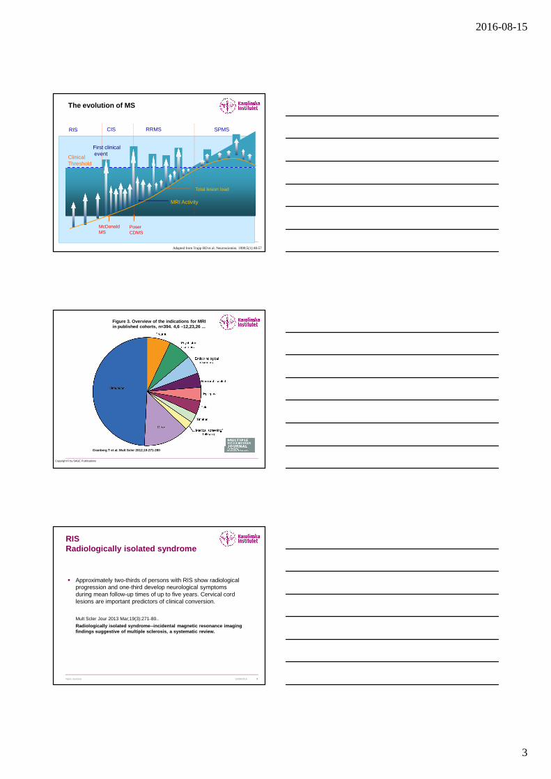
The evolution of MS
Clinical Threshold
MRI Activity
Total lesion load
CIS RRMS SPMSRIS
McDonaldMS
PoserCDMS
First clinicalevent
Adapted from Trapp BD et al. Neuroscientist. 1999;5(1):48-57
Figure 3. Overview of the indications for MRI in published cohorts, n=394. 4,6 –12,23,26 ...
Granberg T et al. Mult Scler 2012;19:271-280
Copyright © by SAGE Publications
RISRadiologically isolated syndrome
� Approximately two-thirds of persons with RIS show radiological progression and one-third develop neurological symptoms during mean follow-up times of up to five years. Cervical cord lesions are important predictors of clinical conversion.
Mult Scler Jour 2013 Mar;19(3):271-80..
Radiologically isolated syndrome--incidental magneti c resonance imaging findings suggestive of multiple sclerosis, a system atic review.
15/08/2016Name Surname 9

2016-08-15
4
What is a Clinically Isolated Syndrome?
� A clinically isolated syndrome (CIS) is a first acute or subacuteepisode of neurological dysfunction with a high suspicion of development of multiple sclerosis
� A CIS is usually the first clinical event in an MS patient
� Magnetic resonance imaging (MRI) findings compatible with: � No or minimal oedema/mass effect� T2-hyperintense lesions� Contrast enhancing lesions� Location of lesion
� Spontaneous or steroid responsive remissions
To exclude other pathologies that may underly the same syndrome(Always look for RED FLAGS, Miller DH, Mult Scler 2008:14:1157-74)
11Tobias Granberg
Multiple sclerosis and diagnosis
� The main principle: dissemination in time (DIT) and space (DIS)
� Schumacher 19655
� Poser 19836
� McDonald 20017
� Reviderade McDonald 2005 (Polman)8
� Reviderade McDonald 2010 (Polman)9
� Demonstration of DIS and DIT with MRI:
� Paty 198810
� Barkhof 199711
� Reviderade Barkhof (Tintoré) 200012
� Swanton 200613
5. Schumacher GA, et al. Problems of Experimental Trials of Therapy in Multiple Sclerosis: Report by the Panel on the Evaluation of Experimental Trials of Therapy in Multiple Sclerosis. Ann N Y Acad Sci. 1965;122(1):552–68.
6. Poser CM, et al. New diagnostic criteria for multiple sclerosis: Guidelines for research protocols. Ann Neurol. 1983;13(3):227–31.
7. McDonald WI, et al. Recommended diagnostic criteria for multiple sclerosis: guidelines from the International Panel on the diagnosis of multiple sclerosis. Ann Neurol. 2001;50(1):121–7.
8. PolmanCH, et al. Diagnostic criteria for multiple sclerosis: 2005 revisions to the “McDonald Criteria.”AnnNeurol. 2005;58(6):840–6.
9. PolmanCH, et al. Diagnostic criteria for multiple sclerosis: 2010 Revisions to the McDonald criteria. Ann Neurol. 2011;69(2):292–302.
10. Paty DW, et al. MRI in the diagnosis of MS: a prospective study with comparison of clinical evaluation, evoked potentials, oligoclonal banding, and CT. Neurology. 1988;38(2):180–5.
11. Barkhof F, et al. Comparison of MRI criteria at first presentation to predict conversion to clinically definite multiple sclerosis. Brain. 1997;120(11):2059–69.
12. Tintoré M, et al. Isolated demyelinating syndromes: comparison of different MR imaging criteria to predict conversion to clinically definite multiple sclerosis. Am J Neuroradiol. 2000;21(4):702–6.
13. Swanton JK, et al. Modification of MRI criteria for multiple sclerosis in patients with clinically isolated syndromes. J Neurol Neurosurg Psychiatry. 2006;77(7):830–3.
20 mars 2014
Ann Neurol 2011:69:292-

2016-08-15
5
Recent radiological classificationsfor MS lesions
15/08/2016Name Surname 13
Tintoré M, et al. Isolated demyelinating syndromes: comparison of different MR imaging criteria to predict conversion to clinically definite multiple sclerosis. Am J Neuroradiol. 2000;21(4):702–6.
Swanton JK, et al. Modification of MRI criteria for multiple sclerosis in patients with clinically isolated syndromes. J Neurol Neurosurg Psychiatry. 2006;77(7):830–3.
Diagnostic criteria for Multiple Sclerosis:2010 Revisions to the McDonald Criteria
Ann Neurol 2011:69:292-302
© 2014 American Academy of Neurology. Published by American Academy of Neurology. 3
Defining the clinical course of multiple sclerosis: The
2013 revisions.
Lublin, Fred et al
Neurology. 83(3):278-286, July 15, 2014.
DOI: 10.1212/WNL.0000000000000560
The 1996 vs 2013 multiple sclerosis phenotype
descriptions for progressive disease Figure 2.
*Activity determined by clinical relapses assessed at
least annually and/or MRI activity (contrast-
enhancing lesions; new and unequivocally enlarging
T2 lesions).
**Progression measured by clinical evaluation,
assessed at least annually. If assessments are not
available, activity and progression are
"indeterminate."
MS = multiple sclerosis; PP = primary progressive; PR
= progressive relapsing; SP = secondary progressive.

2016-08-15
6
Oligoclonal bands
Nature Rev Neurol 2013:9:267-276
Frequencies of abnormal CSF variables in clinically definite MS
Oligoclonal IgG in CSF >95%Increased IgG index 70-80%Increased cell count 50%Abnormal albumin ratio 12%
Cerebrospinal fluid in the diagnosis of multiple sclerosis: a consensus reportJ Neurol Neurosurg Psych 1994:57:897-902
Andersson M, Alvarez-Cermeno J, Bernardi G, Cogato I, Fredman P, Frederiksen J, Fredrikson S, Gallo P, Grimaldi LM, G ronning M, Keir G, Lamers K, Link H, Magalhaes A, Massaro AR, Ohman S, Rei ber H, Rönnbäck L, Schluep M, Schuller E, Sindic CJM, Thompso n EJ, Trojano M, Wurster W.
CSF is of particular value in patients:-older than 50 years-with vascular risk factors-with migraine-with non-specific neurologic symptoms
Differential diagnosis based on presenting symptoms from the brainstem, spinal cord, optic nerves or cerebrum.
Mult Scler 2008:14:1157-74

2016-08-15
7
MS Less common Atypical
Internuclearophthalmoplegia
Facial palsy, facial myokymia
Ataxia and multidirectional nystagmus
Deafness Vascular territory syndrome, e.g., lateral medullary
Sixth nerve palsy One-and-a-half syndrome
Third nerve palsy
Facial numbness Trigeminal neuralgia
Progressive trigeminal sensory neuropathy
Paroxysmal tonic spasms
Focal dystonia, torticollis
Brain stem presentation
MS Less common Atypical
Unilateral optic neuritis
Bilateral simultaneous optic neuritis
Progressive optic neuropathy
Pain on eye movement
No pain Severe, continuous orbital pain
Partial and mainly central visual blurring
No light perception
Persistent complete loss of vision
Normal disc or mild disc swelling
Severe disc swelling
Neuroretinitis (optic disc swelling with macular star)
Uveitis (mild, posterior)
Uveitis (severe, anterior)
Optic nerve presentation
MS Less common Atypical
Partial myelopathy Complete transverse myelitis
Anterior spinal artery territory lesion (sparing posterior columns only)
Lhermitte’s symptom Radiculopathy, areflexia
Cauda equina syndrome
Deafferented hand Segmental loss of pain and temperature sensation
Sharp sensory level to all modalities & localised spinal pain
Numbness Partial Brown-Sequard syndrome (sparing posterior columns)
Complete Brown-Sequard syndrome
Urinary urgency, incontinence, erectile dysfunction
Faecal incontinence Acute urinary retention
Progressive spastic paraplegia (asymmetrical)
Progressive spastic paraplegia (symmetrical)
Progressive sensory ataxia (posterior columns)
Spinal cord presentation

2016-08-15
8
Cerebral presentation
MS Less common Atypical
Mild subcorticalcognitive impairment
Epilepsy Encephalopathy (obtundation, confusion, drowsiness)
Hemianopia Cortical blindness
Hemiparesis Chorea, myoclonus
Generalized movement disorder or Parkinsoniansyndrome
Some differential diagnosis to MS
� ADEM, NMO, AHL, PML, Balo� Systemic : Sarcoidosis, SLE, Behcet, Sjögren, Wegener� Vascular : Stroke, Vasculitis, CADASIL, anti-phospholipid
syndrome, AV-malformations, hemangioma� Metabolic : Leukodystrophies (metachromatic/adreno-),
mitochondrial disorders (MERFF, MELAS, Leber), B12-deficiency
� Genetic : SCAs, Friedreich, HSP� Neoplastic : Lymphomas, paraneoplastic syndromes� Infection : HIV, syphilis, Borrelia, herpes, Whipple� Psychiatric� Others (toxic, compression, neuromuscular (MG) etc)
RED FLAGS� Lung involvement� Multiple cranial neuropathies or
polyradiculopathy� Peripheral neuropathy� Tendon xanthomas� Cerebral venous sinus thrombosis� Cardiac disease� Myopathy� Renal involvement� Cortical infarcts� Haemorrhages/microhaemorrhages� Extrapyramidal features/ Movement
disorders� Livedo reticularis� Retinopathy� Calcifications on CT scans � Bone lesions� Diabetes insipidus� Increase serum lactate level� Selective involvement of the anterior
temporal and inferior frontal lobe� Hematological manifestations� Lacunar infarcts� Mucosal ulcers� Myorhythmia� Hypothalamic disturbance
� Recurrent spontaneous abortion or thrombotic events
� Simultaneous enhancement of all lesions� Rash� Arthritis, polyarthalgias, myalgias� Amyotrophy� Headache or meningismus
REMEMBER:� Age?� Abrupt onset?� Short duration or poor
recovery?� Lack of typical symptoms?� Nonspecific symptoms?� Family history?� Normal examination?� Normal MRI/CSF?

2016-08-15
9
What differ age related changes on MRI from MS?
15/08/2016Name Surname 25
Red flags on MRI
� Persistent Gd-enhancement and continued enlargement of lesions
� Persistently unifocal manifestations � Large and infiltrating brainstem lesions� Predominance of lesions at the cortical/subcortical junction� Meningeal enhancement� T2-hyperintensity in the dentate nuclei� No "black holes“� Large lesions� Marked asymmetry of WM lesions� No enhancement
Blood tests in MS diagnosis???
15/08/2016Name Surname 27

2016-08-15
10
In conclusion…
MS remains a diagnosis requiring an expert neurologist
Differential diagnosis are many, but they can usually be excluded by considering “ red flags ”
Although a disease specific marker does not exist, a robust diagnosis can usually be established early
after onset in most cases based on compatible clinical-, CSF- and MRI-data
Atypical MRI changes should be interpreted cautiously
References
� Miller DH, et al: Differential diagnosis of suspecte d multiple sclerosis: a consensus approach. Mult Scler 2008:14:1 157-1174
� Rolak LA, Fleming JO: The differential diagnosis of multiple sclerosis. The neurologist. 2007:13:57-72
� Stangel M et al: The utility of cerebrospinal fluid analysis in patients with multiple sclerosis. Nature Rev Neurol 2013:9:267-276
� Hahn JS, et al: Differential diagnosis and evaluati on in pediatricmultiple sclerosis. Neurology 2007:68 (suppl 2):S13- S22
� Polman CH et al: Diagnostic criteria for Multiple Sc lerosis:2010 revisions to the McDonald Criteria. Ann Neurol 2011: 69:292-302
� Lublin F et al: Defining the clinical course of multiple sclerosis. The 2013 revisions. Neurology 20 14:83:278-286
1
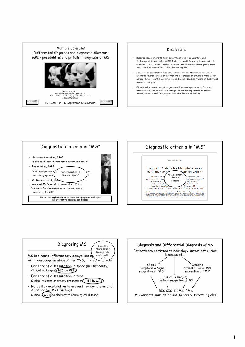
Multiple SclerosisDifferential diagnoses and diagnostic dilemmas
MRI - possibilities and pitfalls in diagnosis of MS
Aksel Siva, M.D.MS Clinic & Department Of Neurology
Istanbul University Cerrahpaşa School Of [email protected]
ECTRIMS – 14 – 17 September 2016, London
Disclosure
• Received research grants to my department from The Scientific and Technological Research Council Of Turkey - Health Sciences Research Grants numbers : 109S070 and 112S052.; and also unrestricted research grants from Merck-Serono to our Clinical Neuroimmunology Unit
• Honoraria or consultation fees and/or travel and registration coverage for attending several national or international congresses or symposia, from Merck Serono, Teva, Novartis, Genzyme, Roche, Biogen Idec/Gen Pharma of Turkey and Bayer-Schering AG
• Educational presentations at programmes & symposia prepared by Excemedinternationally and at national meetings and symposia sponsored by Merck-Serono; Novartis and Teva; Biogen Idec/Gen Pharma of Turkey
Diagnostic criteria in “MS”
• Schumacher et al, 1965"a clinical disease disseminated in time and space"
• Poser et al, 1983 "additional paraclinical and / or laboratory evidence: neuroimaging, neurophysiology & CSF (IgG/OCB)"
• McDonald et al, 2001 & • revised McDonald; Polman et al, 2005
“evidence for dissemination in time and spacesupported by MRI”
No better explanation to account for symptoms and signs(no alternative neurological disease)
"dissemination in time and space"
Diagnostic criteria in “MS”
MRI dominant disease criteria
Diagnosing MS
MS is a neuro-inflammatory demyelinating diseasewith neurodegeneration of the CNS, in which there is• Evidence of dissemination in space (multifocality)
Clinical sx & signs + DIS by MRI
• Evidence of dissemination in timeClinical relapses or steady progression + DIT by MRI
• No better explanation to account for symptoms and signs and/or MRI findingsClinical & MRI: no alternative neurological disease
Clinical HxNeuro-exam + findings to be confirmed by
MRI!
ClinicalSymptoms & Signssuggestive of ”MS”
ImagingCranial & Spinal MRIsuggestive of “MS”
Clinical & Imagingfindings suggestive of MS
Diagnosis and Differential Diagnosis of MSPatients are admitted to neurology outpatient clinics
because of...
RIS CIS RRMS PMSMS variants, mimics or not so rarely something else!
2
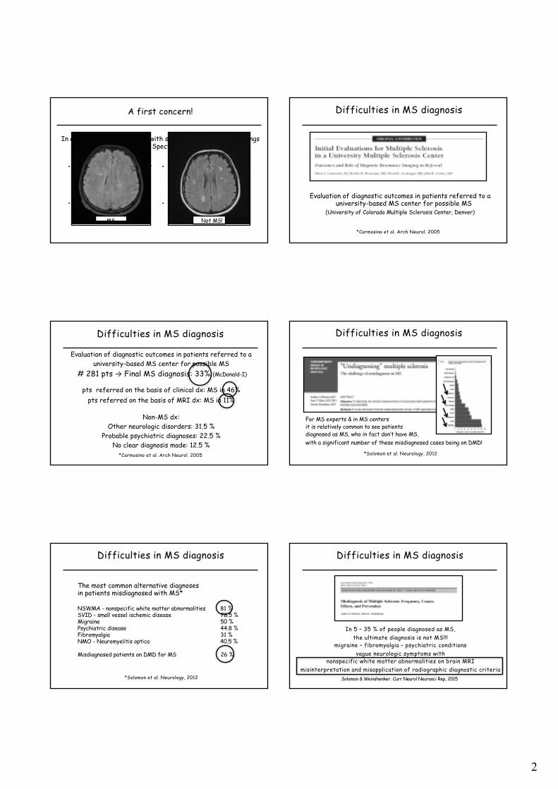
A first concern!
• What should be our firstconcern in the general neurology out-patientclinic?
• Could it be MS?
• What should be our firstconcern in the MS out-patient clinic?
• Could it not be MS?
In a patient who is admitted with symptoms and/or MRI findingssuggestive of MS or MS Spectrum / related disorders
MS Not MS!
Difficulties in MS diagnosis
Evaluation of diagnostic outcomes in patients referred to a university-based MS center for possible MS
(University of Colorado Multiple Sclerosis Center, Denver)
*Carmosino et al. Arch Neurol. 2005
Difficulties in MS diagnosis
Evaluation of diagnostic outcomes in patients referred to auniversity-based MS center for possible MS
# 281 pts → Final MS diagnosis: 33% (McDonald-I)
pts referred on the basis of clinical dx: MS in 46% pts referred on the basis of MRI dx: MS in 11%
Non-MS dx: Other neurologic disorders: 31.5 %
Probable psychiatric diagnoses: 22.5 %No clear diagnosis made: 12.5 %
*Carmosino et al. Arch Neurol. 2005
Difficulties in MS diagnosis
*Solomon et al. Neurology, 2012
For MS experts & in MS centers it is relatively common to see patients diagnosed as MS, who in fact don’t have MS,with a significant number of these misdiagnosed cases being on DMD!
Difficulties in MS diagnosis
*Solomon et al. Neurology, 2012
The most common alternative diagnoses in patients misdiagnosed with MS*
NSWMA - nonspecific white matter abnormalities 81 %SVID - small vessel ischemic disease 78.5 %Migraine 50 %Psychiatric disease 44.8 %Fibromyalgia 31 %NMO - Neuromyelitis optica 40.5 %
Misdiagnosed patients on DMD for MS 26 %
Difficulties in MS diagnosis
In 5 – 35 % of people diagnosed as MS,the ultimate diagnosis is not MS!!!
migraine – fibromyalgia – psychiatric conditionsvague neurologic symptoms with
nonspecific white matter abnormalities on brain MRImisinterpretation and misapplication of radiographic diagnostic criteria
Solomon & Weinshenker. Curr Neurol Neurosci Rep, 2015
3
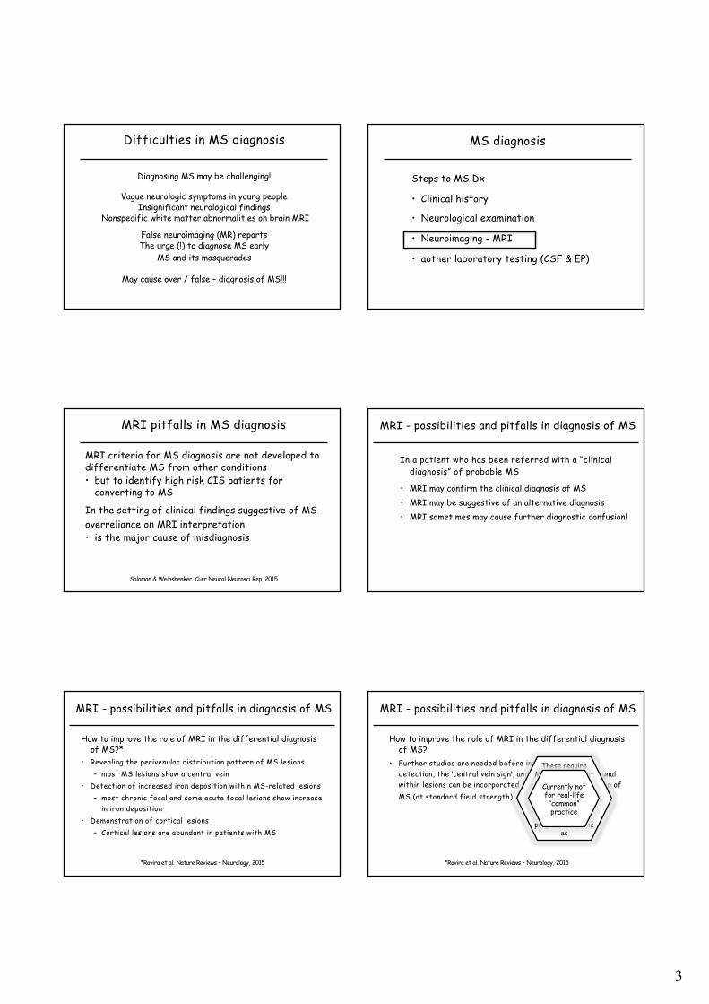
Difficulties in MS diagnosis
Diagnosing MS may be challenging!
Vague neurologic symptoms in young peopleInsignificant neurological findings
Nonspecific white matter abnormalities on brain MRI
False neuroimaging (MR) reportsThe urge (!) to diagnose MS early
MS and its masquerades
May cause over / false – diagnosis of MS!!!
MS diagnosis
Steps to MS Dx
• Clinical history
• Neurological examination
• Neuroimaging - MRI
• aother laboratory testing (CSF & EP)
MRI pitfalls in MS diagnosis
MRI criteria for MS diagnosis are not developed to differentiate MS from other conditions• but to identify high risk CIS patients for
converting to MS
In the setting of clinical findings suggestive of MS overreliance on MRI interpretation • is the major cause of misdiagnosis
Solomon & Weinshenker. Curr Neurol Neurosci Rep, 2015
MRI - possibilities and pitfalls in diagnosis of MS
In a patient who has been referred with a “clinicaldiagnosis” of probable MS
• MRI may confirm the clinical diagnosis of MS• MRI may be suggestive of an alternative diagnosis• MRI sometimes may cause further diagnostic confusion!
MRI - possibilities and pitfalls in diagnosis of MS
How to improve the role of MRI in the differential diagnosisof MS?*
• Revealing the perivenular distribution pattern of MS lesions– most MS lesions show a central vein
• Detection of increased iron deposition within MS-related lesions– most chronic focal and some acute focal lesions show increase
in iron deposition • Demonstration of cortical lesions
– Cortical lesions are abundant in patients with MS
*Rovira et al. Nature Reviews – Neurology, 2015
MRI - possibilities and pitfalls in diagnosis of MS
How to improve the role of MRI in the differential diagnosisof MS?
• Further studies are needed before intracortical lesiondetection, the ‘central vein sign’, and the susceptibility signalwithin lesions can be incorporated in the diagnostic work-up of MS (at standard field strength)
These require MRI systems that
operate at highmagnetic field
strengths (≥3.0 T) & sophisticated
software programs/sequenc
es
Currently not for real-life
“common”practice
*Rovira et al. Nature Reviews – Neurology, 2015
4
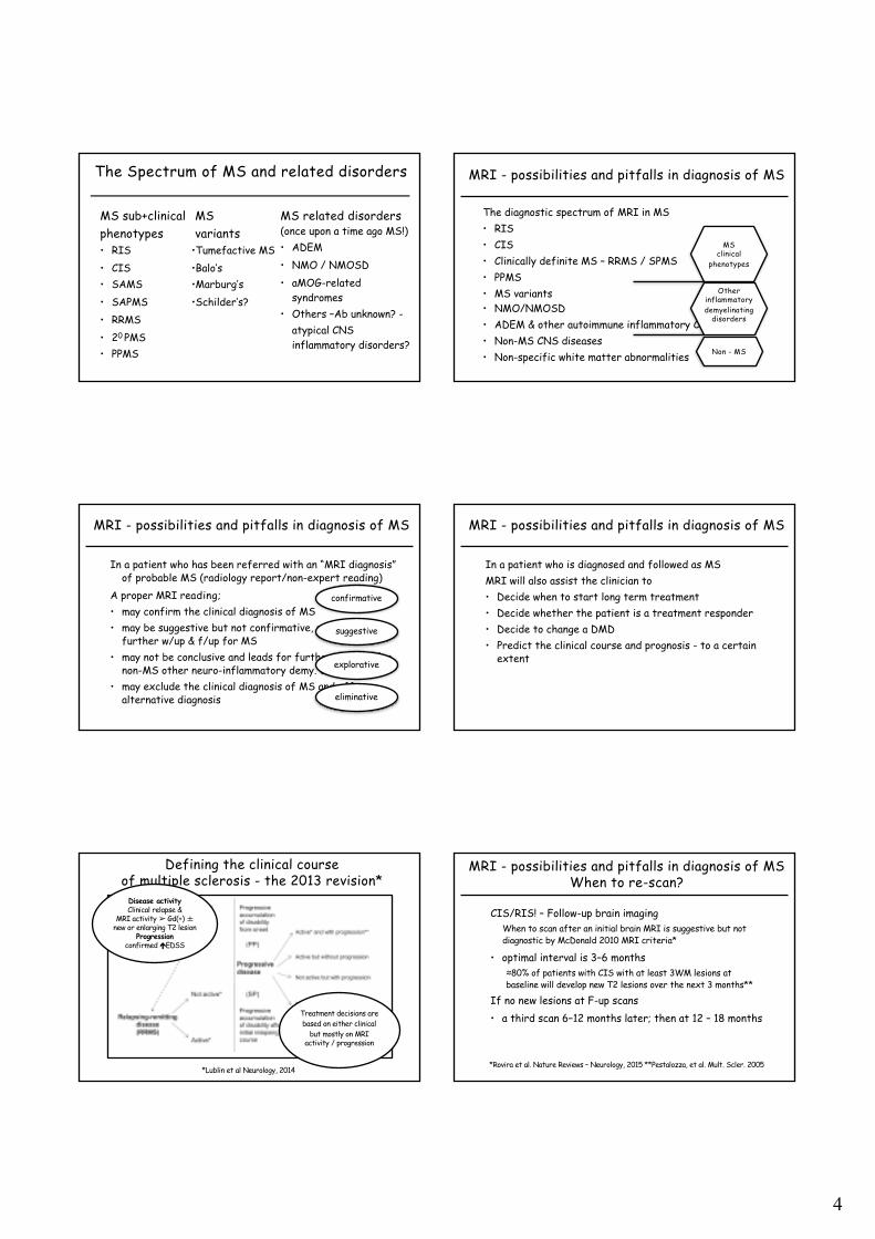
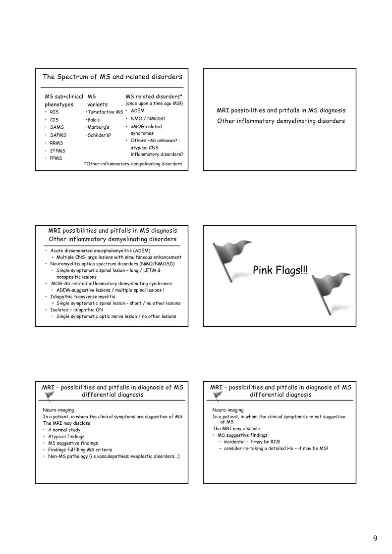
The Spectrum of MS and related disorders
MS sub+clinicalphenotypes• RIS• CIS• SAMS• SAPMS• RRMS• 20 PMS• PPMS
MS variants
•Tumefactive MS•Balo’s•Marburg’s•Schilder’s?
MS related disorders(once upon a time ago MS!)• ADEM• NMO / NMOSD• aMOG-related
syndromes• Others –Ab unknown? -
atypical CNS inflammatory disorders?
MRI - possibilities and pitfalls in diagnosis of MS
The diagnostic spectrum of MRI in MS • RIS• CIS• Clinically definite MS – RRMS / SPMS• PPMS• MS variants• NMO/NMOSD• ADEM & other autoimmune inflammatory CNS diseases• Non-MS CNS diseases• Non-specific white matter abnormalities
MSclinical
phenotypes
Other inflammatory demyelinating
disorders
Non - MS
MRI - possibilities and pitfalls in diagnosis of MS
In a patient who has been referred with an “MRI diagnosis” of probable MS (radiology report/non-expert reading)
A proper MRI reading; • may confirm the clinical diagnosis of MS• may be suggestive but not confirmative, necessitating
further w/up & f/up for MS• may not be conclusive and leads for further work-up for
non-MS other neuro-inflammatory demy. diseases• may exclude the clinical diagnosis of MS and offer an
alternative diagnosis
confirmative
suggestive
explorative
eliminative
MRI - possibilities and pitfalls in diagnosis of MS
In a patient who is diagnosed and followed as MS MRI will also assist the clinician to• Decide when to start long term treatment• Decide whether the patient is a treatment responder• Decide to change a DMD• Predict the clinical course and prognosis - to a certain
extent
*Lublin et al Neurology, 2014
Treatment decisions are based on either clinical
but mostly on MRI activity / progression
Defining the clinical courseof multiple sclerosis - the 2013 revision*
Disease activityClinical relapse &
MRI activity ➢ Gd(+) ±new or enlarging T2 lesion
Progressionconfirmed éEDSS
MRI - possibilities and pitfalls in diagnosis of MSWhen to re-scan?
CIS/RIS! – Follow-up brain imaging When to scan after an initial brain MRI is suggestive but not diagnostic by McDonald 2010 MRI criteria*
• optimal interval is 3–6 months≈80% of patients with CIS with at least 3WM lesions at baseline will develop new T2 lesions over the next 3 months**
If no new lesions at F-up scans• a third scan 6–12 months later; then at 12 – 18 months
*Rovira et al. Nature Reviews – Neurology, 2015 **Pestalozza, et al. Mult. Scler. 2005
5
MRI - possibilities and pitfalls in diagnosis of MSWhen to re-scan?
RRMS on DMD • Follow-up brain imaging depends on availability and reimbursement
Suggested practice• Within the first month of the onset of the given drug efficacy!
(i.e. 3-4th months after initiating INF-beta drugs & orals; 6-7th
months of GA ) – this may serve as the baseline/reference MRI• in the absence of any clinical episode first f/up MRI at month 12• then in case of “not active disease” at month 24 and if patient
continues to be NEDA(+) then every other year x 2; afterwards longer intervals
MRI - possibilities and pitfalls in diagnosis of MSWhen to re-scan?
RRMS on DMD • Follow-up brain imaging depends on availability and reimbursement
Suggested practice• in case of “minimally active disease by MRI” and no switch of DMD
then at 6 months x 2; and if patient converts back to be NEDA(+) MRI f/up accordingly – as above
MRI - possibilities and pitfalls in diagnosis of MSWhen to re-scan?
RRMS not on DMD (is it possible? It may…) not active –Follow-up brain imaging
Depends on the duration of the disease and for how long thepatient is clinically & by imaging inactive and the MRI load!
• The longer duration & lower MRI load safer to scan at longerintervals – first at 6 – 12, then at 18 – 24 months; and thenevery 3 years or until a new clinical episode occurs…
*Rovira et al. Nature Reviews – Neurology, 2015 **Pestalozza, et al. Mult. Scler. 2005
Neuroimaging in MS diagnosis
McDonald 2010 diagnostic criteria for MS (CIS!) MRI criteria
What’s new?
McDonald 2010 - MRI diagnostic criteria
DIS – Dissemination in space≥ 1 T2 lesions in ≥ 2 regions of the following CNS areas• juxtacortical • periventricular• infratentorial• spinal cord
DIT – Dissemination in time• ≥ 1 asymptomatic Gd enhancing lesion/s in the initial MRI• New T2 lesion/s (Gd+Ø) on follow-up MRI
*Polman et al, Ann. Neurol 2011

6
McD 2010 MS diagnostic MR findings – DISjuxtacortical & periventricular & post fossa & spinal cord
Sub-cortical periventricularJuxta-cortical
Spinal cord lesionsPosterior fossalesions
Corpus callosumlesions
Sub-cortical
1
3
4
2
McD 2010 MS diagnostic MR findings – DIT*
DIT – Dissemination in time• ≥ 1 asymptomatic Gd enhancing lesion/s in the initial MRI• New T2 lesion/s (Gd+Ø) on follow-up MRI
*Polman et al, Ann. Neurol 2011
MS suggestive MR findingsGadolinium enhancing lesions
Ringpattern Nodular
enhencement
Open ring pattern
MS suggestive MR findingsGadolinium enhancing lesions
McD 2010 MS diagnostic MR findingsspinal cord lesions
Spinal cord lesions
multiple Gd +
lesions
MR findings in MS"black holes!"
(T1) black holes

7
MR findings in MS”cerebral atrophy"
2011 / OT, 35, M; SPMS On DMD since 03/02
at 10 yrs: EDSS: 62015 EDSS: 6.5
2001 / OT, 25, M; New DxSx: Visual blurring
EDSS: 1
Enlarged sulci
Severe atrophyof the corpus callosum
*Fillipi et al. Lancet Neurol, 2016
We propose an increase in the number of lesions necessaryto confirm involvement of the periventricular area from one to three,
and to add an additional cardinal CNS location, the optic nerve
An update on MRI criteria for the diagnosis of MS the 2016 MAGNIMS consensus guidelines
An update on MRI criteria for the diagnosis of MS the 2016 MAGNIMS consensus guidelines
*Fillipi et al. Lancet Neurol, 2016
Clinically MS & MR consistent with MS
Not all white spotsseen on MRI are MS!!!
32, M admitted because of dizziness & numbness in handshe receives an MRI diagnosis (radiologic report) of MS
The clinician agrees with this diagnosis and starts a DMD !A year later continues to describe the same symptoms – no change on MRI
But accepted as a non-responder (!) and his DMD is changedAbout another year later he comes for another opinion to our center...
Neuroexam normal; MRI unchanged! CSF: normal, no OCBsFurther work up including vasculitis/collagen disease panel & serology all normal
A young man with dizziness, numbness in handsand T2 lesions on MRI
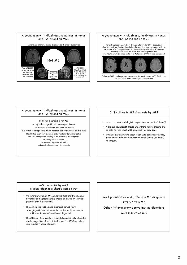
8
Lesions are bilateral & semi-symmetrical & largely subcorticalthere was no gadolinium enhancement in any study!
Follow up MRIs: no change in any MRI – no enhancement in any scanno atrophy – no T1 Black holes
Not MS
A young man with dizziness, numbness in handsand T2 lesions on MRI
First MRI 2004Second MRI 2005
DMD initiatedThird MRI 2006DMD switched!
MRI 2008Pt self-admittedto our MS center
Dx of MS orother CNS disease
is ruled out!No
spinal cordlesions
Nocorpus callosum
Nopost fossa
Not MS
A young man with dizziness, numbness in handsand T2 lesions on MRI
Patient was seen again about 3 years later in Apr 2014 because of dizziness and tension type headache - he was fine over the years with the
exception of a few episodes of dizziness closely related to life events!He was given duloxetine in 05.2014 and responded well.
His neuro-exam is normal and a f/up MRI done on 02/15 was unchanged
Follow up MRI: no change – no enhancement – no atrophy – no T1 Black holesNo posterior fossa and no spinal cord lesions
A young man with dizziness, numbness in handsand T2 lesions on MRI
His final diagnosis is not MSor any other significant neurologic disease
This individual is someone who turns out to have“NSWMA - nonspecific white matter abnormalities” on his MRI
He also has an anxiety disorder and a tendency for somatizationHis MRI changes are unlikely to be related to his symptoms
or to any other disorderHe was overdiagnosed with MS
and received unnecessary treatments
Difficulties in MS diagnosis by MRI
• Never rely on a radiologist’s report (whom you don’t know)!
• A clinical neurologist should understand neuro-imaging and be able to read what MRI abnormalities may say…
• When you are not sure about what MRI abnormalities may mean, then find a good neuroradiologist (whom you trust) to consult…
MS diagnosis by MRIclinical diagnosis should come first!
• the interpretation of MRI abnormalities and the imagingdifferential diagnosis always should be based on “clinicalgrounds” (Hx & Sx & signs)
• The clinical impression and diagnosis comes first!Ø imaging/MRI and all other lab tools should be used to
confirm or to exclude a clinical diagnosis!
• The MRI may lead you to a clinical diagnosis, only when it’shighly suggestive of a certain disease (i.e. MS!) and whenyour mind isn’t clear clinically!
MRI possibilities and pitfalls in MS diagnosis
RIS & CIS & MS
Other inflammatory demyelinating disorders
MRI mimics of MS
9
The Spectrum of MS and related disorders
MS sub+clinicalphenotypes• RIS• CIS• SAMS• SAPMS• RRMS• 20 PMS• PPMS
MS variants
•Tumefactive MS•Balo’s•Marburg’s•Schilder’s?
MS related disorders*(once upon a time ago MS!)• ADEM• NMO / NMOSD• aMOG-related
syndromes• Others –Ab unknown? -
atypical CNS inflammatory disorders?
*Other inflammatory demyelinating disorders
MRI possibilities and pitfalls in MS diagnosis
Other inflammatory demyelinating disorders
MRI possibilities and pitfalls in MS diagnosisOther inflammatory demyelinating disorders
• Acute disseminated encephalomyelitis (ADEM)• Multiple CNS large lesions with simultaneous enhancement
• Neuromyelitis optica spectrum disorders (NMO/NMOSD)• Single symptomatic spinal lesion – long / LETM &
nonspesific lesions• MOG-Ab related inflammatory demyelinating syndromes
• ADEM-suggestive lesions / multiple spinal lesions !• Idiopathic transverse myelitis• Single symptomatic spinal lesion – short / no other lesions
• Isolated – idiopathic ON• Single symptomatic optic nerve lesion / no other lesions
Pink Flags!!!
MRI - possibilities and pitfalls in diagnosis of MS differential diagnosis
Neuro-imagingIn a patient, in whom the clinical symptoms are suggestive of MSThe MRI may disclose• A normal study• Atypical findings• MS suggestive findings• Findings fulfilling MS criteria• Non-MS pathology (i.e.vasculopathies; neoplastic disorders...)
MRI - possibilities and pitfalls in diagnosis of MS differential diagnosis
Neuro-imagingIn a patient, in whom the clinical symptoms are not suggestive
of MSThe MRI may disclose• MS suggestive findings
– incidental – it may be RIS!– consider re-taking a detailed Hx – it may be MS!
10
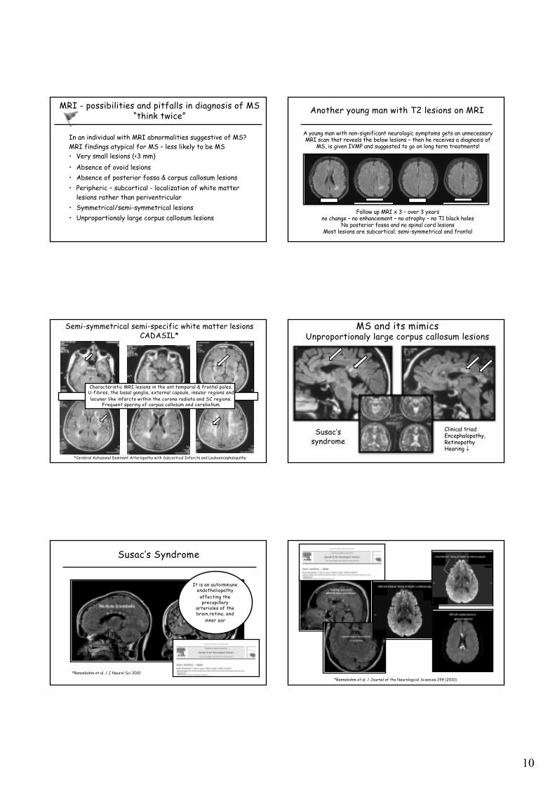
MRI - possibilities and pitfalls in diagnosis of MS “think twice”
In an individual with MRI abnormalities suggestive of MS?MRI findings atypical for MS – less likely to be MS • Very small lesions (<3 mm)• Absence of ovoid lesions• Absence of posterior fossa & corpus callosum lesions• Peripheric – subcortical - localization of white matter
lesions rather than periventricular• Symmetrical/semi-symmetrical lesions• Unproportionaly large corpus callosum lesions
Another young man with T2 lesions on MRI
A young man with non-significant neurologic symptoms gets an unnecessaryMRI scan that reveals the below lesions – then he receives a diagnosis of
MS, is given IVMP and suggested to go on long term treatments!
Follow up MRI x 3 – over 3 yearsno change – no enhancement – no atrophy – no T1 black holes
No posterior fossa and no spinal cord lesionsMost lesions are subcortical; semi-symmetrical and frontal
AA 64 F, (28.01.2004)
Semi-symmetrical semi-specific white matter lesionsCADASIL*
Characteristic MRI lesions in the ant.temporal & frontal poles,U-fibres, the basal ganglia, external capsule, insular regions andlacunar like infarcts within the corona radiata and SC regions.
Frequent sparing of corpus callosum and cerebellum
(
*Cerebral Autosomal Dominant Arteriopathy with Subcortical Infarcts and Leukoencephalopathy
MS and its mimicsUnproportionaly large corpus callosum lesions
Clinical triadEncephalopathy,RetinopathyHearing ¯
Susac’ssyndrome
Susac’s Syndrome
*Rennebohm et al. / J Neurol Sci 2010
It is an autoimmune endotheliopathy
affecting the precapillary
arterioles of the brain,retina, and
inner ear
*Rennebohm et al. / Journal of the Neurological Sciences 299 (2010)
11
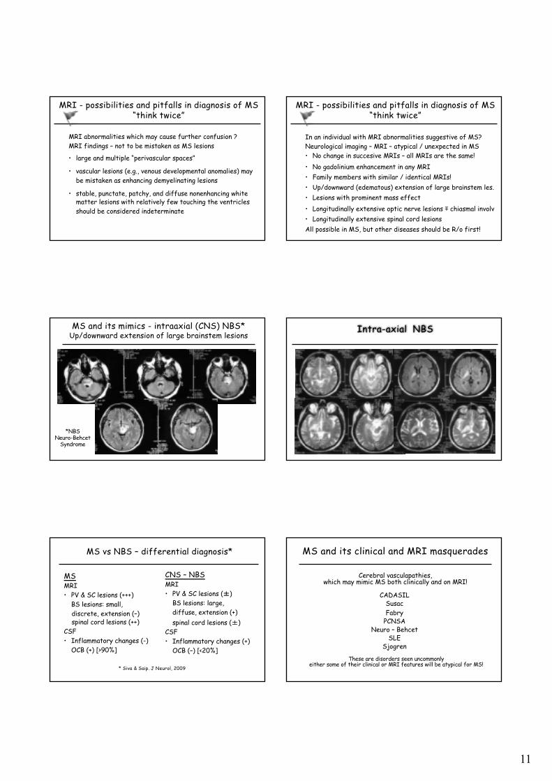
MRI - possibilities and pitfalls in diagnosis of MS “think twice”
MRI abnormalities which may cause further confusion ?MRI findings – not to be mistaken as MS lesions
• large and multiple “perivascular spaces”
• vascular lesions (e.g., venous developmental anomalies) maybe mistaken as enhancing demyelinating lesions
• stable, punctate, patchy, and diffuse nonenhancing whitematter lesions with relatively few touching the ventriclesshould be considered indeterminate
MRI - possibilities and pitfalls in diagnosis of MS “think twice”
In an individual with MRI abnormalities suggestive of MS?Neurological imaging – MRI – atypical / unexpected in MS• No change in succesive MRIs – all MRIs are the same!• No gadolinium enhancement in any MRI• Family members with similar / identical MRIs!• Up/downward (edematous) extension of large brainstem les.• Lesions with prominent mass effect• Longitudinally extensive optic nerve lesions ∓ chiasmal involv• Longitudinally extensive spinal cord lesionsAll possible in MS, but other diseases should be R/o first!
MS and its mimics - intraaxial (CNS) NBS*Up/downward extension of large brainstem lesions
*NBS Neuro-Behcet
Syndrome
Intra-axial NBS
MS vs NBS – differential diagnosis*
MSMRI• PV & SC lesions (+++)
BS lesions: small, discrete, extension (–)spinal cord lesions (++)
CSF• Inflammatory changes (-)
OCB (+) [>90%]
CNS – NBSMRI• PV & SC lesions (±)
BS lesions: large, diffuse, extension (+)spinal cord lesions (±)
CSF• Inflammatory changes (+)
OCB (–) [<20%]
* Siva & Saip. J Neurol, 2009
MS and its clinical and MRI masquerades
CADASILSusacFabry
PCNSANeuro – Behcet
SLESjogren
Cerebral vasculapathies, which may mimic MS both clinically and on MRI!
These are disorders seen uncommonlyeither some of their clinical or MRI features will be atypical for MS!

12
MRI possibilities and pitfallsin MS diagnosis
“Tumefactive lesions”when they may be MS and when not?
Tumefactive lesions
What could they be?• Brain neoplasms• CNS lymphoma • Abscesses• PML!• Vasculitic disorders & NBS!• Tumefactive demyelinative
lesions (TDL)
Clues to the diagnosis of TDL include* • Less mass effect than
expected for their size• open ring enhancement• no increased perfusion• visualisation of veins
coursing through the lesion
*Kaschka et al 2014
Tumefactive lesions – MS or not?
Tumefactive demyelinating lesions are well-demarcated, hyperintense on T2, hypointense on T1-wMRI. Ring enhanc with Gd is characteristic, >open ring, the open portion abuts the GM of the cortex (or BG). Size of the lesion (>20mm), the relative lack of mass effect, and edema are helpful radiological findings
Biopsy provenacute demyl
Tumefactive lesions – MS
“Tumefactive MS” lesions may be seen with other MS suggestive lesionswhen the diagnosis becomes easier,
However, it should be kept in mind that MS and brain tumorsalthough highly unlikely may be seen together in an unfortunate individual!
MS and its MRI mimics – multifocal glial tumors
18 02 09
MS and its mimics – multifocal glial tumors
06 03 09
Brain biopsy:multifocal
GBM!
13
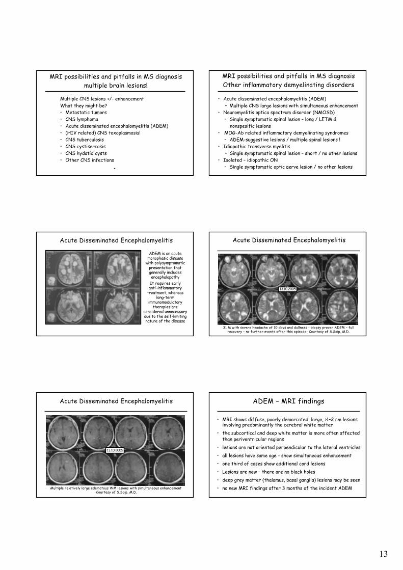
MRI possibilities and pitfalls in MS diagnosismultiple brain lesions!
*
Multiple CNS lesions +/- enhancement What they might be?• Metastatic tumors• CNS lymphoma• Acute disseminated encephalomyelitis (ADEM)• (HIV related) CNS toxoplasmosis!• CNS tuberculosis• CNS cystisercosis• CNS hydatid cysts• Other CNS infections
MRI possibilities and pitfalls in MS diagnosisOther inflammatory demyelinating disorders
*
• Acute disseminated encephalomyelitis (ADEM)• Multiple CNS large lesions with simultaneous enhancement
• Neuromyelitis optica spectrum disorder (NMOSD)• Single symptomatic spinal lesion – long / LETM &
nonspesific lesions• MOG-Ab related inflammatory demyelinating syndromes
• ADEM-suggestive lesions / multiple spinal lesions !• Idiopathic transverse myelitis• Single symptomatic spinal lesion – short / no other lesions
• Isolated – idiopathic ON• Single symptomatic optic nerve lesion / no other lesions
Acute Disseminated Encephalomyelitis
ADEM is an acute monophasic disease
with polysymptomaticpresentation that generally includes encephalopathy
It requires early anti-inflammatory
treatment, whereaslong-term
immunomodulatorytherapies are
considered unnecessarydue to the self-limiting nature of the disease
Acute Disseminated Encephalomyelitis
31 M with severe headache of 10 days and dullness - biopsy proven ADEM – fullrecovery – no further events after this episode- Courtesy of S.Saip, M.D.
13.10.2005
Acute Disseminated Encephalomyelitis
Multiple relatively large edematous WM lesions with simultaneous enhancementCourtesy of S.Saip, M.D.
13.10.2005
ADEM – MRI findings
• MRI shows diffuse, poorly demarcated, large, >1–2 cm lesions involving predominantly the cerebral white matter
• the subcortical and deep white matter is more often affected than periventricular regions
• lesions are not oriented perpendicular to the lateral ventricles• all lesions have same age - show simultaneous enhancement • one third of cases show additional cord lesions• Lesions are new – there are no black holes•
• deep grey matter (thalamus, basal ganglia) lesions may be seen• no new MRI findings after 3 months of the incident ADEM

14
MRI possibilities and pitfalls in MS diagnosisOther inflammatory demyelinating disorders - NMOSD
• much of the optic nerve• a trend to more posterior
involvement of the optic nerve including chiasm
• simultaneous bilateral disease
*
Neuromyelitis optica spectrum disorder (NMOSD)pattern of optic nerve involvement*
*Kim et al. Neurology 2015;
Neuromiyelitis Optica – optic nerve imaging
41 F; with recurrent - ON05/2012 painful L-ON limited improvement with IVMP05/2013 painful R-ONMRI – enhancing longitudinal optic nerve lesion extending to chiasmaCSF-OCB (-); AQP+-Ab (+)
MRI - possibilities and pitfalls in diagnosis of MS “think twice”
In an individual with parenchymal spinal MRI abnormalitiessuggestive of inflammatory pathology...possibilities are;
When it is a “small spinal cord lesion” [<3 segments]• MS • Transverse myelitis• Short segment or recovering NMO / NMOSD – myelitis• Myelitis associated with systemic vasculitic or collagen
tissue disorders• Tumors (i.e.astrocytoma; ependymoma)• Infectious disorders
MRI - possibilities and pitfalls in diagnosis of MS “think twice”
MSBrain MR+
Spinal lesionSmall
LateralGd+
RecoveringNMO-myelitis
Transversemyelitis
MRI - possibilities and pitfalls in diagnosis of MS “think twice”
In an individual with parenchymal (intra-axial) spinal MRI abnormalitiessuggestive of inflammatory pathology...possibilities;
When it is a “longitudinally extensive spinal cord lesion” [>3 segments]• NMO / NMOSD – myelitis• Transverse myelitis• MS – multiple small lesions in contiguity suggestive of a single LETM-lesion• Myelitis assoiated with systemic vasculitides & collagen tissue disorders• Spinal venous dural fistula• Tumors (i.e.astrocytoma; ependymoma)• Infectious disorders (i.e. viral, tbc, lyme)• Granulomatous disorders (i.e. Sarcoidosis)
Longitudinally extensive spinal cord lesionssuggestive of NMO
Longitudinally extensivespinal cord lesionsLETM / NMO / NMOSD
widespread Gd enhancement
15
Brighter T2-hyperintense spotty lesions (T1W-hypo) within the ordinary spinal cord lesion may differentiate NMO from MS!
MRI possibilities and pitfalls in MS diagnosisOther inflammatory demyelinating disorders - NMOSD
Yonezu et al. MS J 2014 & Hyun et al JNNP 2015
From Hyun et al JNNP 2015
MRI possibilities and pitfalls in MS diagnosisOther inflammatory demyelinating disorders - NMOSD
*Wingerchuk et al. Neurology 2015
MRI possibilities and pitfalls in MS diagnosisOther inflammatory demyelinating disorders - NMOSD
MS - Spinal cord lesions
• do not extend over more than three vertebral segments
• are eccentric /laterally located (on axial images)
• may have focal (nodular or peripheral) gad-enhancement
NMOSD - Spinal cord lesions
• extend over more than three vertebral segments
• are centrally located (on axial images)
• may have patchy or long-extensive gad-enhancement
• bright spotty cord lesions • short segmental lesions, may
be seen at onset and during periods of recovery
• if present other CNS lesions are nonspesific
MOG-Ab+ related inflammatory demyelinating syndromes
NS, 49 F - 29 12 2006 Dx. APS & SLE & MS? NMOSD? AQP4-Ab all times negativeRx with oral AC + mycophenolate mofetil (MMF) + GA - stable
MOG-Ab+ related inflammatory demyelinating syndromes
NS, 49 F - 29 01 2007 Dx. APS & SLE & MS? NMOSD? AQP4-Ab all times negativeRx with oral AC + mycophenolate mofetil (MMF) + GA - MOG-Ab recently was detected (+)
No spesific patternTo be suspected in ptswith ADEM-like lesions
orTumefactive lesions
orin pts with no or atypical
cerebral lesions and multiple spinal cord lesions ± dorsocaudal LETM
Longitudinally extensive spinal cord lesions
spinal venous dural fistula
Gd +enchancing
vascularstructures
2o to dural fistules
peri-cordial vascular abnormalities

16
Longitudinally extensive spinal cord lesions
Longitudinally extensive spinal cord lesionneurosarcoidosis
widespread Gd enhancement
large spinalcord lesions
52F recent onset of disturbing paresthesiasand mild weakness of lower extremities
neurosarcoidosisMRI possibilities and pitfalls in diagnosis
Inflammatory granulomatous disorders- Neurosarcoidosis
*
Neurosarcoidosis• Basal meningitis / Leptomeningeal enhancement in about 40% • Hydrocephalus • Hypothalamic involvement and/or pituitary fossa involvement• Parenchymal – nonspecific lesions - areas of increased T2 signal with or
without enhancement at SC and/or PV regions• intraparenchymal lesions exhibit Gad enhancement that persists without
treatment!• Sarcoid myelitis can be longitudinally extensive. Root involvement as well as
linear and/or nodular enhancement along the surface of the spinal cord ±intramedullary extension suggests sarcoidosis
MRI possibilities and pitfalls in MS diagnosisInflammatory granulomatous disorders- Neurosarcoidosis
55 yrs university prof admitted with dysarthria, imbalance, L-sided weaknesswith known past Hx of sarcoidosis
Neuromyelitis Optica – Area postrema involvement
A.G. 18y F • 03/2015 develops nausea and
vomiting with hiccup: • Hosp in gastro clinic! No Dx!
Discharged withoutimprovement,
• Develops numbness, itching, burning sensation and allodyniain extremities and part of body
• Lhermitte & tonic spasms!!!• Neuro – first time; MR+• AQP4-Ab (+++)
MR lesions in NMO/NMOSDare closely correlated with aquaporin-4 expression
Wingerchuk et al. Lancet Neurology, 2007
Brain lesions typicalof NMO localiseat the sites whereaquaporin 4 expressionare normally highest
Strange looking brain abnormalities!Neuromiyelitis Optica
Pittock et al. Arch Neurol. 2006
Brain MRI lesions were detected in 36 patients (60%). Most were nonspecific, but 6 patients (10%) had
MS–like lesions, usually asymptomatic.. Another 5 patients (8%), mostly children,
had diencephalic, brainstem or cerebral lesions,atypical for MS
17
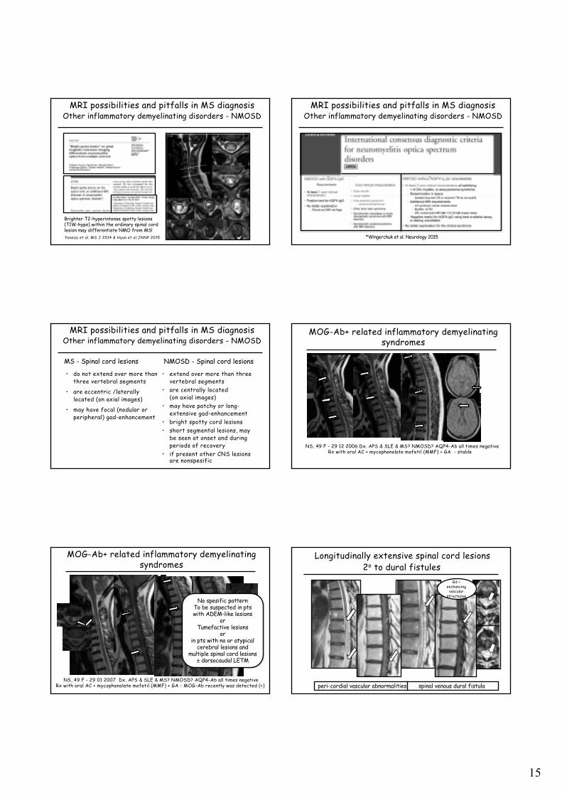
Strange looking brain abnormalities!NMO & NMOSD
Cortical and leptomeningeal involvement in NMO – rare but possible“Cloud-like enhancement” on postcontrast T1W images – as multiplepatchy enhancement with blurred margin in adjacent regionsTahara et al Eur J Neurol 2012 & Ito et al Ann Neurol 2009
Strange looking brain abnormalities!NMO & NMOSD
O.C.E. 34 F• 12/2007 first admission with itching and pain in
the neck, followed by weakness more on the leftMRI C2-3 (short) spinal lesion
• Dx w/up: APS & SLE & AQP4-Ab+ (NMOSD)• Later develop enhancing corpus callosum lesion
MRI possibilities and pitfalls in MS diagnosisOther inflammatory demyelinating disorders - NMOSD
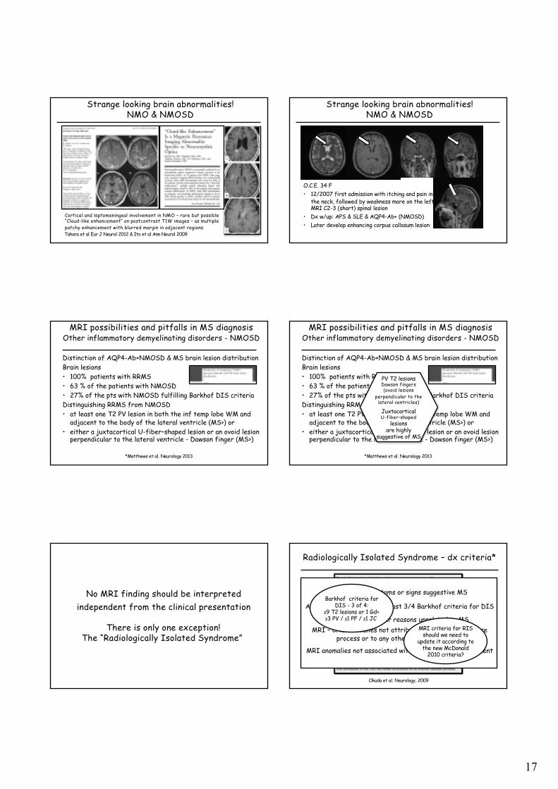
*Matthews et al. Neurology 2013
Distinction of AQP4-Ab+NMOSD & MS brain lesion distributionBrain lesions• 100% patients with RRMS• 63 % of the patients with NMOSD • 27% of the pts with NMOSD fulfilling Barkhof DIS criteriaDistinguishing RRMS from NMOSD • at least one T2 PV lesion in both the inf temp lobe WM and
adjacent to the body of the lateral ventricle (MS>) or • either a juxtacortical U-fiber–shaped lesion or an ovoid lesion
perpendicular to the lateral ventricle - Dawson finger (MS>)
MRI possibilities and pitfalls in MS diagnosisOther inflammatory demyelinating disorders - NMOSD
*Matthews et al. Neurology 2013
Distinction of AQP4-Ab+NMOSD & MS brain lesion distributionBrain lesions• 100% patients with RRMS• 63 % of the patients with NMOSD • 27% of the pts with NMOSD fulfilling Barkhof DIS criteriaDistinguishing RRMS from NMOSD • at least one T2 PV lesion in both the inf temp lobe WM and
adjacent to the body of the lateral ventricle (MS>) or • either a juxtacortical U-fiber–shaped lesion or an ovoid lesion
perpendicular to the lateral ventricle - Dawson finger (MS>)
PV T2 lesionsDawson fingers(ovoid lesions
perpendicular to the lateral ventricles)
JuxtacorticalU-fiber–shaped
lesionsare highly
suggestive of MS
No MRI finding should be interpretedindependent from the clinical presentation
There is only one exception!The “Radiologically Isolated Syndrome”
Radiologically Isolated Syndrome – dx criteria*
Okuda et al. Neurology, 2009
No clinical symptoms or signs suggestive MS
An initial MRI fulfilling at least 3/4 Barkhof criteria for DIS
MRI done for other reasons unrelated to MSMRI – CNS anomalies not attributable to another disease
process or to any other medical condition
MRI anomalies not associated with any functional impairment
MRI criteria for RISshould we need to
update it according tothe new McDonald
2010 criteria?
Barkhof criteria forDIS - 3 of 4:
≥9 T2 lesions or 1 Gd+ ≥3 PV / ≥1 PF / ≥1 JC
18
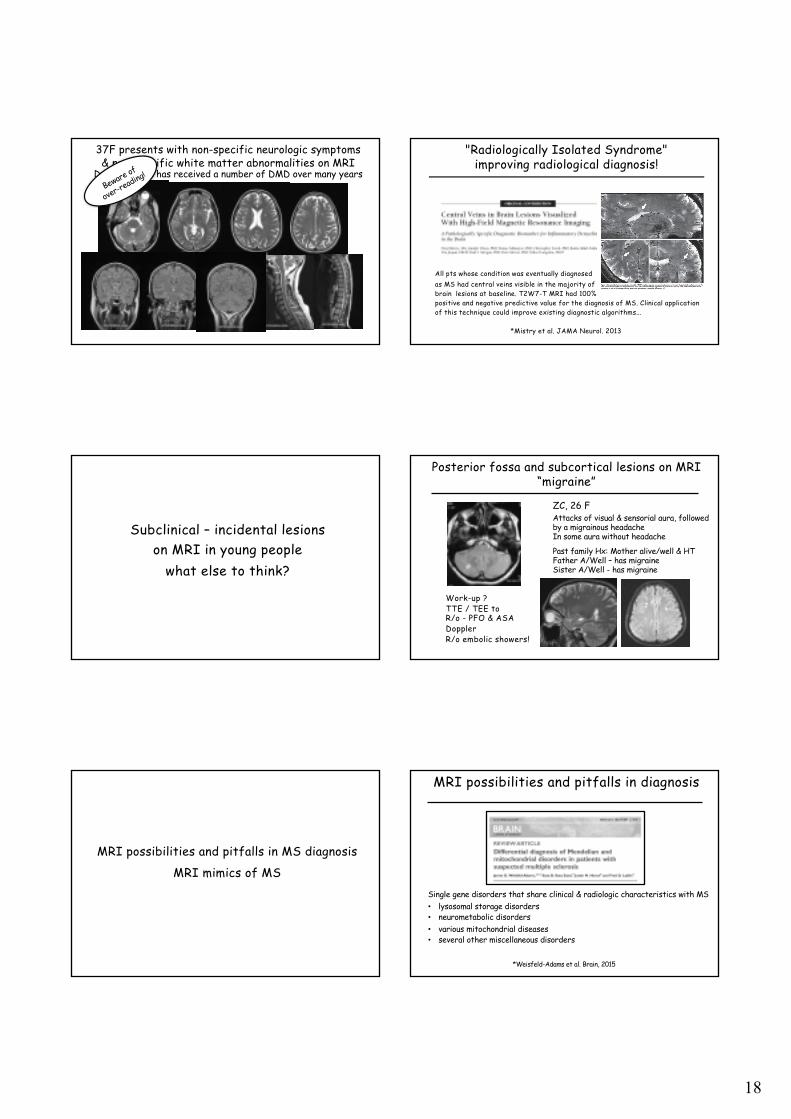
37F presents with non-specific neurologic symptoms& non-specific white matter abnormalities on MRI
Dx as MS and has received a number of DMD over many years
"Radiologically Isolated Syndrome"improving radiological diagnosis!
All pts whose condition was eventually diagnosedas MS had central veins visible in the majority of brain lesions at baseline. T2W7-T MRI had 100% positive and negative predictive value for the diagnosis of MS. Clinical application of this technique could improve existing diagnostic algorithms...
*Mistry et al. JAMA Neurol. 2013
Subclinical – incidental lesionson MRI in young people
what else to think?
Posterior fossa and subcortical lesions on MRI “migraine”
ZC, 26 FAttacks of visual & sensorial aura, followedby a migrainous headacheIn some aura without headache•
Past family Hx: Mother alive/well & HT Father A/Well – has migraineSister A/Well - has migraine
Work-up ?TTE / TEE toR/o - PFO & ASADopplerR/o embolic showers!
MRI possibilities and pitfalls in MS diagnosis
MRI mimics of MS
MRI possibilities and pitfalls in diagnosis
*Weisfeld-Adams et al. Brain, 2015
Single gene disorders that share clinical & radiologic characteristics with MS• lysosomal storage disorders • neurometabolic disorders• various mitochondrial diseases• several other miscellaneous disorders

19
MRI possibilities and pitfalls in diagnosis
Single gene disorders sharing clinical & radiologic characteristics with MSPresence of the following findings - not suggestive of MS… • Symmetrical WM involvement of the cerebral hemispheres• Cerebral involvement limited to long tracts (>post int cap & brain stem)• Spinal cord involvement limited to long tracts – longitudinal lesions• T1 hyperintensities of thalamic pulvinar• T2 (symmetric) hyperintensities of dentate nucleus • Multiple cystic cavitationsAbsence of the following findings - not likely in MS… • Lack of ovoid lesions• Lack of spinal cord involvement • Lack of Gad-enhancement (exception – adrenoleucodystrophies)
*Weisfeld-Adams et al. Brain, 2015
MS and Its Masquerades
32 F; Admitted with a diagnosis of MS!Hx: subacute onset of weakness in both legs at age 30Paraparesis progressing to significant gait difficultyover a month; then shows a fluctuating course withlimited progression + affected with major depression.
MS and Its Masquerades MS and Its Masquerades
MS and Its Masquerades MS and Its Masquerades
20
MRI possibilities and pitfalls in MS diagnosismitochondrial disorders
This is a group of rare multisystem disorders caused by a variety of genetic defects affecting the mitochondrial metabolism
• Multisystem involvement • Clinical presentation • A positive family history • Cortical & deep gray matter involvement in a non-vascular pattern• Asymmetrical or symmetrical white matter non-specific involvement• Calcified cerebral lesions might be the clue
Leber’s hereditary optic neuropathy, chronic progressive ophthalmoplegiaand mitochondrial encephalomyelopathy, lactic acidosis and stroke-like episodes (MELAS) might be difficult to distinguish radiologically from MS
MRI possibilities and pitfallsin diagnosis of MSWRAP-UP
MRI possibilities and pitfalls in diagnosis
*Rovira et al. Nature Reviews – Neurology, 2015
MRI possibilities and pitfalls in diagnosis
Shortcomings that may lead to erroneous diagnoses in patients “who are suspected to have MS”* • MRI examinations –that- are nonstandardized and often of
inadequate quality• scans –that- might be read by radiologists lacking expertise
in this field and without consideration of relevant clinical and laboratory data
• the simplified and less-restrictive McDonald’s 2010 MRI criteria –that- might compromise diagnostic specificity leading to overdiagnosis
*Rovira et al. Nature Reviews – Neurology, 2015
MRI possibilities and pitfalls in diagnosis
Shortcomings that may lead to erroneous diagnoses in patients “who are suspected to have MS” • MRI examinations –that- are nonstandardized and often of
inadequate quality• scans –that- might be read by radiologists lacking expertise
in this field and without consideration of relevant clinical and laboratory data
• the simplified and less-restrictive McDonald’s 2010 MRI criteria –that- might compromise diagnostic specificity leading to overdiagnosis
*Rovira et al. Nature Reviews – Neurology, 2015
and –that-is real-
life“common”practice
Incorrect MRI reports and its consequences
Incorrect radiological-MRI reports• False T1 Gd+ lesions• Incorrectly reported additional T2 lesions• Overreadings...
Such incorrect reports may result in unnecessary IVMP treatments and/or
inappropriate DMT switches!!!

21
Problem areas in MRI diagnosis of MS
Technical• Having f/up MRI studies at different centers / with dif MRs• Lack of standardization of MRI studies• Lack of knowledge of MS MRI protocols at the study centers• Different slice thicknesses / different sequences• Application of Gd-contrast at low dose• Scanning without waiting after giving the Gd-contrast media
Problem areas in MRI diagnosis of MS
Incorrect MRI evaluations – radiologist related causes• Lack of knowledge – lack of proper training• Lack of experience• Overload of work – too many MRI reports / too little time
• The non-professional understanding of “I will reporteverything I see - or I think I see - and will leave it to the clinicianto decide what they are!”
Problem areas in MRI diagnosis of MS
Incorrect MRI evaluations – neurologist related causes• Not looking to the images – just reading the report!• Not interpreting the MRI her/him-self, lack of
neuroradiology training!• The unfortunate changes in the healthcare and
educational systems, in which the “patient centric evaluation and care” understanding is losing grounds or completely forgotten!
Not likely to be MS!
Normal CSF & (-) OCBs ⇒ think twice!Normal MRI* ⇒ unlikely to be MS
Normal MRI & CSF ➢ can’t be MS!!!
*MRI of brain and spinal cord
The other side of the coin!
Abnormal CSF ➣ (+) OCBs not always MS!Abnormal MRI ➣ not always MSor not clinically significant “MS!”

Differential diagnoses: Blood and CSF biomarkers in the diagnostic
process
Finn Sellebjerg
Danish Multiple Sclerosis Center, Department of Neurology, Rigshospitalet, University of
Copenhagen, Copenhagen, Denmark
Introduction
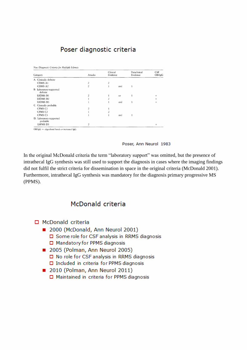
Although widely used in the diagnostic process, blood biomarkers are not included in current
diagnostic criteria for multiple sclerosis (MS). In the Poser criteria for the diagnosis of MS, which
preceded the currently used McDonald criteria, laboratory support for the diagnosis could be
provided by the finding of intrathecal IgG synthesis by cerebrospinal fluid (CSF) analysis. Today
CSF analysis is still used to support the MS diagnosis, especially in patients with a progressive
onset of disease, although the use of CSF analysis has clearly decreased with the development of
modern imaging criteria for the diagnosis.
Concensus criteria for CSF analysis
The most consistent CSF feature of MS is the presence of intrathecal synthesis of IgG, which can be
detected in at least 90% of patients. The detection of IgG oligoclonal bands by isoelectric focusing
and immununodetection is the most sensitive method, and is preferred over quantitative methods,
i.e., IgG-index and intrathecal IgG synthesis rate calculations. IgG oligoclonal bands are also a

common finding in patients with neuroinfections or inflammatory diseases, and may be an
unexpected finding in some 5-10% of controls without neurological diseases. Thus, the isolated
detection of IgG oligoclonal bands should not prompt extensive diagnostic investigations. Lack of
oligoclonal bands are considered a “red flag” which should lead to reconsideration of the diagnosis.
However, the 5-10% of MS patients without oligoclonal bands do not show major differences in
clinical disease course compared to oligoclonal bands negative patients. Furthermore, some patients
who are initially oligoclonal bands negative may develop oligoclonal bands on follow-up.
Minor increases in CSF mononuclear cell counts are often observed, especially in younger patients,
whereas the CSF protein content and the CSF-serum glucose ratio is usually normal. Thus, CSF cell
counts > 50 cells/µl, the presence of granulocytes in the absence of blood contamination, an
increase in CSF protein concentration above 1 g/l or a markedly increased CSF-serum albumin
concentration quotient, and a low CSF-serum glucose ratio are also considered “red flags” which
may indicate an alternative diagnosis such as infection, sarcoidosis or lymphoma.
CSF analysis in diagnostic criteria
As mentioned above the Poser criteria allowed for “laboratory support” for diagnosing MS.
Laboratory support allowed for a diagnosis of “laboratory-supported definite MS” in patients where
evidence of either dissemination in time or dissemination in space was lacking (Poser 1983).
In the original McDonald criteria the term “laboratory support” was omitted, but the presence of
intrathecal IgG synthesis was still used to support the diagnosis in cases where the imaging findings
did not fulfil the strict criteria for dissemination in space in the original criteria (McDonald 2001).
Furthermore, intrathecal IgG synthesis was mandatory for the diagnosis primary progressive MS
(PPMS).
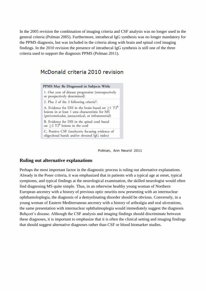
In the 2005 revision the combination of imaging criteria and CSF analysis was no longer used in the
general criteria (Polman 2005). Furthermore, intrathecal IgG synthesis was no longer mandatory for
the PPMS diagnosis, but was included in the criteria along with brain and spinal cord imaging
findings. In the 2010 revision the presence of intrathecal IgG synthesis is still one of the three
criteria used to support the diagnosis PPMS (Polman 2011).
Ruling out alternative explanations
Perhaps the most important factor in the diagnostic process is ruling out alternative explanations.
Already in the Poser criteria, it was emphasized that in patients with a typical age at onset, typical
symptoms, and typical findings at the neurological examination, the skilled neurologist would often
find diagnosing MS quite simple. Thus, in an otherwise healthy young woman of Northern
European ancestry with a history of previous optic neuritis now presenting with an internuclear
ophthamoloplegia, the diagnosis of a demyelinating disorder should be obvious. Conversely, in a
young woman of Eastern Mediterranean ancestry with a history of arthralgia and oral ulcerations,
the same presentation with internuclear ophthalmoplegia would immediately suggest the diagnosis
Behçcet’s disease. Although the CSF analysis and imaging findings should discriminate between
these diagnoses, it is important to emphasize that it is often the clinical setting and imaging findings
that should suggest alternative diagnoses rather than CSF or blood biomarker studies.
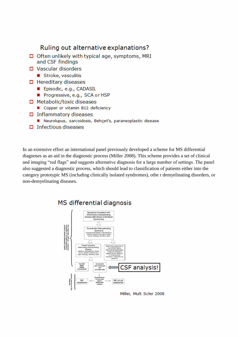
In an extensive effort an international panel previously developed a scheme for MS differential
diagnoses as an aid in the diagnostic process (Miller 2008). This scheme provides a set of clinical
and imaging “red flags” and suggests alternative diagnosis for a large number of settings. The panel
also suggested a diagnostic process, which should lead to classification of patients either into the
category prototypic MS (including clinically isolated syndromes), othe r demyelinating disorders, or
non-demyelinating diseases.
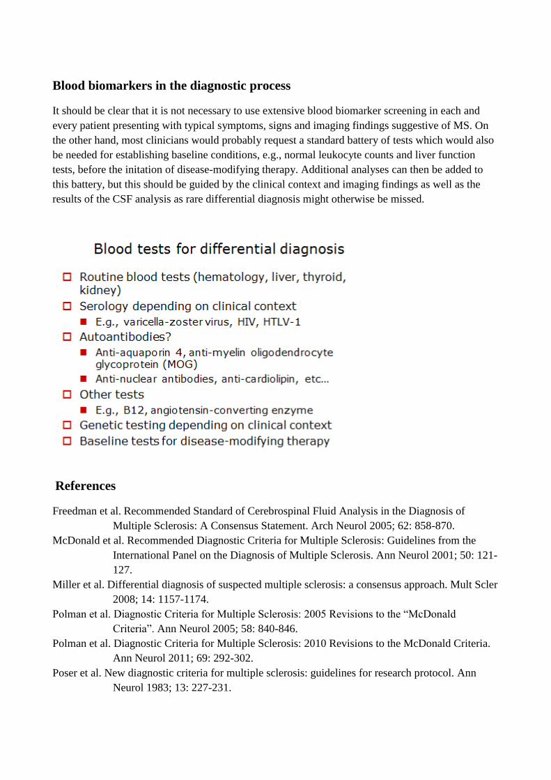
Blood biomarkers in the diagnostic process
It should be clear that it is not necessary to use extensive blood biomarker screening in each and
every patient presenting with typical symptoms, signs and imaging findings suggestive of MS. On
the other hand, most clinicians would probably request a standard battery of tests which would also
be needed for establishing baseline conditions, e.g., normal leukocyte counts and liver function
tests, before the initation of disease-modifying therapy. Additional analyses can then be added to
this battery, but this should be guided by the clinical context and imaging findings as well as the
results of the CSF analysis as rare differential diagnosis might otherwise be missed.
References
Freedman et al. Recommended Standard of Cerebrospinal Fluid Analysis in the Diagnosis of
Multiple Sclerosis: A Consensus Statement. Arch Neurol 2005; 62: 858-870.
McDonald et al. Recommended Diagnostic Criteria for Multiple Sclerosis: Guidelines from the
International Panel on the Diagnosis of Multiple Sclerosis. Ann Neurol 2001; 50: 121-
127.
Miller et al. Differential diagnosis of suspected multiple sclerosis: a consensus approach. Mult Scler
2008; 14: 1157-1174.
Polman et al. Diagnostic Criteria for Multiple Sclerosis: 2005 Revisions to the “McDonald
Criteria”. Ann Neurol 2005; 58: 840-846.
Polman et al. Diagnostic Criteria for Multiple Sclerosis: 2010 Revisions to the McDonald Criteria.
Ann Neurol 2011; 69: 292-302.
Poser et al. New diagnostic criteria for multiple sclerosis: guidelines for research protocol. Ann
Neurol 1983; 13: 227-231.