teaching digital histology - formatex · 2008, the system ( is fully operational for the...
TRANSCRIPT

Teaching Digital Histology
Carlos R. Morales
Department of Anatomy and Cell Biology, McGill University, Montreal, Quebec, Canada
The light microscope is one of the most widely used scientific instruments in teaching, diagnosis and research. For the teaching of histology the microscope is used in conjunction with tissue sections and cell specimens mounted on glass slides that must be stained for their visualization. This requires the maintenance of the instrument and the histological collection. Presently, many pathology laboratories generate sections that are digitized and viewed in computers via internal computer networks and/or the internet. This new approach permits the visualization of tissues from a remote location and allows the rapid consultation and discussion of difficult cases with experts around the world. The use of digital sections is now expanding to teaching as companies and commercial websites start offering services and virtual histology material for purchase. The advantages of teaching digital histology are many, such as the availability of high quality histological sections, the use of synchronized teaching, the possibility of self-study, the use of digital slides in lectures, reviews and demonstrations, etc. Using a special software that simulate the use of a microscope, the whole digital section can be viewed in a computer, moved in all directions and magnified up to x1000. Here we report our experience with this innovative approach and its benefits for the teaching of histology.
Keywords Digital Histology, Digital Scanners, Digital Slides
1. Teaching Histology with Microscopes
One of the major obstacles in teaching histology to College and University students is the availability, preservation and maintenance of a large collection of tissue sections. Over time, glass slides tend to break and entire collections are decimated. As a result, professors of histology and pathology are constantly replacing the damaged material. These activities are not only expensive but also time consuming. Another problem arises when viewing the slides, which entails the use of microscopes in good condition, equipped with acceptable optics. Good quality instruments are expensive and require regular maintenance. To decrease these expenses, some universities have transferred the cost of the instruments to the students, who are obligated to purchase and maintain them. The use of microscopes also involves the utilization of oil immersion which frequently results in the contamination of the objectives, particularly the high dry objective. Additionally, the teaching of histology requires the use of both an atlas and a text book, adding to the expenses of students. Finally and most importantly, the use of microscopes impedes the teaching dynamics of laboratories, as the demonstrator must change sections, objectives, and/or clean the equipment after the use of oil immersion. This situation is not ideal for small group teaching, as learning is more effective when it is free of distractions.
2. Taking Advantage of the Digital Age
The advent of the internet and the availability of digital images have revolutionized the traditional delivery of knowledge and information. Anatomy, histology and pathology are ideal disciplines for the use of digital images since analysis and interpretation of structures is based on visual specimens. In fact, the majority of professors in the field have taken advantage of the use of digital micrographs in conjunction with the use of internet facilities for teaching purposes [1-3]. Presently, there are many digital atlases available for teaching and several have been adopted for use by McGill University. By the end of the 1990’s, our medical curriculum incorporated a laboratory guide with digital images. The undergraduate program of our department also developed an atlas of digital histology which has been improved by combining light and electron microscope digital images with schematic drawings (http://people.mcgill.ca/carlos.morales/inthist). To encourage attendance and participation, we presented these images with open questions allowing students to find the answers during the laboratory. More recently, medical and undergraduate students of histology at McGill University benefited from the installation of new microscopes, equipped with digital cameras that are connected to computers. However, the increase in the student population, coupled with the lack of space and sufficient microscope/computer stations, has obliged us to develop in 2003 a small group teaching strategy whereby five or more students are grouped around one station under the guidance of a teaching assistant. This approach not only proved to be effective, but also reduced the difficulties created by the expanding student population. Nevertheless, the basic challenges of teaching histology did not disappear as this approach depended on the use of microscopes and histological sections.
Current Microscopy Contributions to Advances in Science and Technology (A. Méndez-Vilas, Ed.)
© 2012 FORMATEX 994

3. The Use of Digital Slides
A few years ago, while attending the annual conference of the American Society of Cell Biology (ASCB), we visited a couple of firms promoting the use of a new technology for pathologists, which consisted of creating crisp and clear digitized histological sections. In other words, entire sections were digitized by precision scanners that created flawless digital slide images of an entire section in minutes. By using state of the art software that simulated the use of a microscope, the whole section could be examined via computer in situ or in a remote location via the internet in real time. The section could be moved in all directions and magnified up to x1000. When we asked the representatives of these firms if the devices were used for the teaching of histology, they responded that they were unaware of their use other than in pathology laboratories. The scanner alone had a cost that ranged between $90,000 and $110,000 U.S. making it impossible for some institutions to even consider the possibility of acquiring such an instrument for teaching purposes. Three years ago, we learned of a U.S. based company that offered a scanning service at an accessible cost. We contacted the firm and informed them of our desire to digitize slides for our undergraduate course “Introduction to Dynamic Histology” as well as slides used in the “Medical Curriculum”. We selected the best available sections to digitize, which are now available for the teaching of histology at our university. We started implementing the use of digital slides as an experimental tool for the teaching of histology in the undergraduate course at the end of 2006. Since 2008, the system (http://www.medicine.mcgill.ca/histology) is fully operational for the laboratories of histology of the medical curriculum, with an impressive collection of 105 slides. By adding digital slides to our curriculum, our medical school furthered its ambition of being incorporated amongst the most modern academic medical teaching centers worldwide. One small caveat to this approach is that digital sections consume a large amount of memory. To circumvent this inconvenience at a minimal cost, we purchased and developed our own server allocating this equipment to the exclusive storage of digital sections. The system works flawlessly and the students like it (Table 1). The advantages of this new approach for the teaching of histology are enormous, due largely to the availability of high quality histological sections. The digitized sections permit the application of various techniques, including classroom digital teaching, synchronized examination of slides, self-study in situ and from a remote place via the internet in real time. Digital slides can also be used in lectures, reviews and demonstrations. This system is also ideal for the rapid design of lab, mid-term and final exams since it permits the capture of histological fields at different magnifications in a short period of time. This approach also dissipated the criticisms of “traditionalists” who argued, with good reason, that the use of digital images or pictures only offers a limited field at a single magnification, removing the sense of reality and limiting the student to a collection of selected images that are not representative of the entire section. Another extraordinary advantage of this new approach is that a digital collection of histological sections can be improved and enriched over the years with little effort as histologists often share the “perfect” fixed, cut and/or stained material with their colleagues.
Table 1. Undergraduate Course of Histology
Comparison of lab evaluations and lab exam success rates before (2005) and after (2006-2009) the implementation of Digital Histology
2005 2006 2007 2008 2009 Laboratory 3.6 (± 0.2) 3.7 (±0.2) 3.7 (±0.1) 4.2 (±0.2)* 4.2 (±0.1)* Evaluation Audio Visual 3.7 (± 0.1) 4.1 (±0.2)* 4.1 (±0.1)* 4.2 (+0.1)* 4.3 (±0.1)* Lab Exams 35% 75/100 (n=250) 73% 75/100 (n=360)
* Statistical analysis was performed by t-test. Asterisks indicate that when compared to 2005 the difference is significant (p<0.05).
4. Requirements, Pros, Cons, and Tips
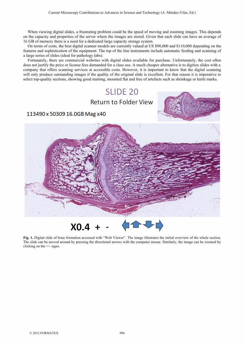
To produce digital slides in situ, one must have a digital microslide scanner. This is a “closed box” system in which a slide is progressively scanned at high resolution. Aperio’s ScanScope (www.aperio.com) and Vassalo’s Claro (www.claro-inc.jp) have probably the top instruments in the market. Such instruments use a series of accurate nanomotors to move and scan slides in a matter of minutes, maintaining the focus throughout the scanning process. The acquired digital images are then amalgamated and compressed with digital software. We have experience with Aperio. The images can be accessed with “Web Viewer” or “Image Scope”. In the case of the former, the initial overview of the whole area of the section can be moved around by pressing on directional arrows with the computer mouse (Fig. 1). The image can be also grabbed and displaced. Specific areas can be selected for enlargement, which digitally come into focus in a similar manner to zooming an area with Google EarthTM. Image Scope also provides an initial overview of the digital slide, but the zooming is achieved with a magnifier located on top of the image (Fig. 2). Image Scope permits the capture of sharp histological fields at different magnifications in short periods of time (Fig. 3).
Current Microscopy Contributions to Advances in Science and Technology (A. Méndez-Vilas, Ed.)
© 2012 FORMATEX 995
When viewing digital slides, a frustrating problem could be the speed of moving and zooming images. This depends on the capacity and properties of the server where the images are stored. Given that each slide can have an average of 16 GB of memory there is a need for a dedicated large capacity storage system. On terms of costs, the best digital scanner models are currently valued at US $90,000 and $110,000 depending on the features and sophistication of the equipment. The top of the line instruments include automatic feeding and scanning of a large series of slides (ideal for pathology labs). Fortunately, there are commercial websites with digital slides available for purchase. Unfortunately, the cost often does not justify the price or license fees demanded for a class use. A much cheaper alternative is to digitize slides with a company that offers scanning services at accessible costs. However, it is important to know that the digital scanning will only produce outstanding images if the quality of the original slide is excellent. For that reason it is imperative to select top-quality sections, showing good staining, mounted flat and free of artefacts such as shrinkage or knife marks.
Fig. 1. Digital slide of bone formation accessed with “Web Viewer”. The image illustrates the initial overview of the whole section. The slide can be moved around by pressing the directional arrows with the computer mouse. Similarly, the image can be zoomed by clicking on the +/- signs.
Current Microscopy Contributions to Advances in Science and Technology (A. Méndez-Vilas, Ed.)
© 2012 FORMATEX 996
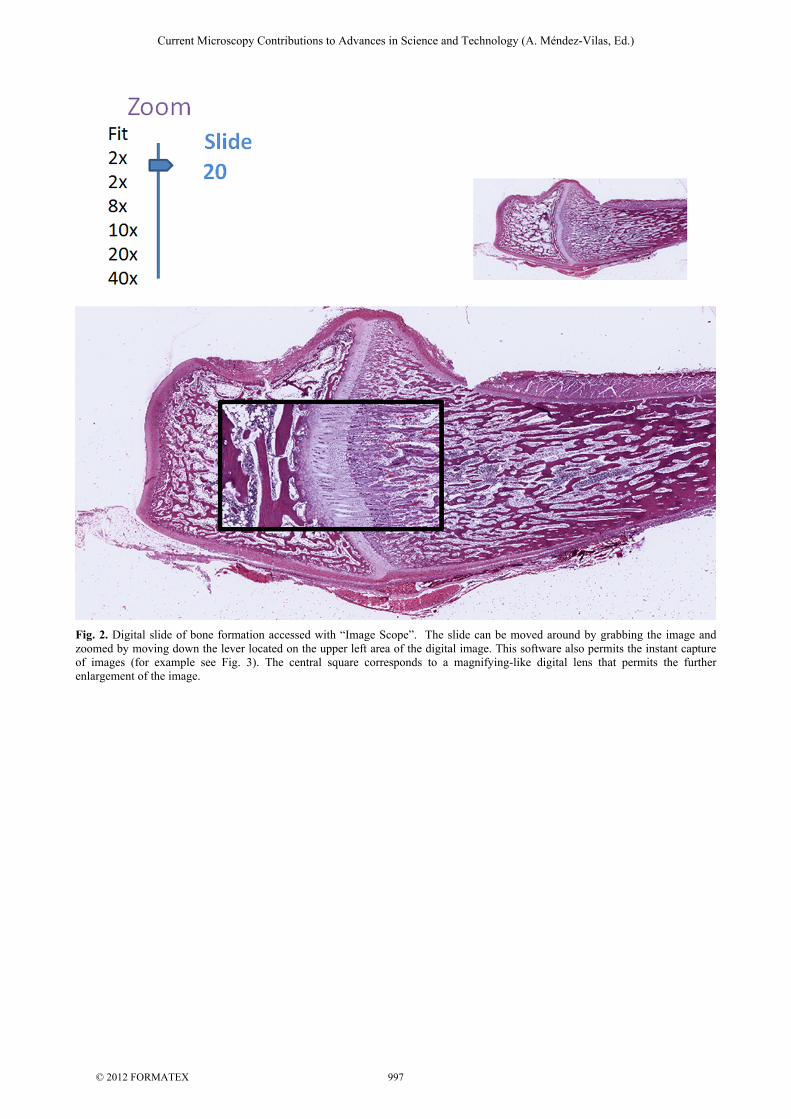
Fig. 2. Digital slide of bone formation accessed with “Image Scope”. The slide can be moved around by grabbing the image and zoomed by moving down the lever located on the upper left area of the digital image. This software also permits the instant capture of images (for example see Fig. 3). The central square corresponds to a magnifying-like digital lens that permits the further enlargement of the image.
Current Microscopy Contributions to Advances in Science and Technology (A. Méndez-Vilas, Ed.)
© 2012 FORMATEX 997

Fig. 3. Digital slide of bone formation accessed with “Image Scope”. The field is a captured image taken from the epiphyseal cartilage with Image Scope (x400). In conclusion, the adoption of this new approach will help to introduce our medical students to the new way of viewing and analyzing pathological sections, as is currently done in some highly rated U.S. hospitals and medical centers worldwide. In the near future this system will be extended to scan fluorescent, phase-contrast, polarized and confocal images and will become the preferred way to judge images during the peer review process of manuscripts and scientific grant applications. Note of the author: Since http://www.medicine.mcgill.ca/histology is a McGill University restricted website, we have set up a link to allow the readers to play with a digital slide in order to have full appreciation of the potentials of the described approach. Please contact: http://www.medicine.mcgill.ca/morales/Medical%20Histology/SLIDES%2020%20to%2039/20.html
Acknowledgements: We appreciate the technical assistance of George Rodas and Boyko Kouchev, Medical Information Technology, McGill University for their invaluable assistance and to Andrew Fox and María Luisa Garicoche for proofreading this article.
References
[1] P.M. Heidger, Jr., F. Dee, D. Consoer, T. Leaven, J. Duncan, C. Kreiter, Integrated approach to teaching and testing in histology with real and virtual imaging, Anat Rec 269 (2002) 107-112.
[2] J. Gu, R.W. Ogilvie, Virtual microscopy and virtual slides in teaching, diagnosis, and research, Advances in pathology, microscopy & molecular morphology, Taylor & Francis, Boca Raton, 2005, p. 356 p.
[3] R. Coleman, Can histology and pathology be taught without microscopes? The advantages and disadvantages of virtual histology, Acta Histochem 111 (2009) 1-4.
Current Microscopy Contributions to Advances in Science and Technology (A. Méndez-Vilas, Ed.)
© 2012 FORMATEX 998