tel. +34 930 116 062 comercial@iffservice
TRANSCRIPT


1
Technical data
IMAGE RESOLUTION 6 mp
ACQUISITION MODE Multi shot, movie
FOCUS Autofocus, manual focus
ISO MANAGEMENT Variable
GRIDS Placido disc, NIBUT grid
CAMERA Coloured, sensitive to infrared (NIR)
LIGHT SOURCE Infrared LED – Blue and white Led
I.C.P. OSAA full assessment of the ocular surface through a combination of tests for dry eye diagnosis, from tear break up time to the tear volume production test.
Integrated system for the analysis of the ocular surface
The instrument is fit in the slit lamp tonometer’s hall, it is designed to make all the related tear film tests, from the quality of the same to the analysis of Meibomian glands, as well as various measurements and classifications according to international grading scales.
Invented and developed 100% in Italy Medical instrument in CLASS I registered to the Ministry of Health Medical electrical equipment CLASS I complies with the norm En. 60601-1. The technical features of the instrument and its accessories can be improved in any time and without notice. To obtain an updated description we suggest to visit the website www.sbmsistemi.com
MINIMUM HARDWARE REQUIREMENTS:
• Intel® Pentium® Dual Core 2.00 Ghz
• SSD Hard Drive
• 4 GB RAM
• Screen resolution: 1600x900
• 1 available usb 3.0 port
• 1 other available usb port
• Microsoft® Windows® 7, 8, 10 Professional (Pro)
CONSIDERING THE HIGH QUALITY OF THE VIDEOS, FOR OPTIMAL VIDEO RECORDING AND PLAYBACK WE SUGGEST:
• Intel® Core™ i7
• 8GB RAM

2
Ocular Surface Workup with Automated Non-invasive
Measurements for the Diagnosis of Meibomian Gland Dysfunction
INTRODUCTIONDry eye disease (DED) was recently redefined as a “multifactorial disease of the ocular surface characterized by a loss of homeostasis of the tear film, and accompanied by ocular symptoms, in which tear film instability and hyperosmolarity, ocular surface inflammation and damage, and neurosensory abnormalities play etiological roles”. Meibomian gland dysfunction (MGD) represents the leading cause of evaporative dry eye, the most common subtype of DED. MGD is characterized by hyperkeratinization of the meibomian gland ductal epithelium, leading to obstruction and plugging of the gland orifice. Moreover, quantitative and qualitative changes in the meibum lipid composition lead to increased viscosity and reduced gland outflow onto the tear film. The stasis of meibum inside the gland promotes the proliferation of bacteria producing lipases and esterases that increase the viscosity and melting temperature of the meibum, thus setting up a vicious circle. The hyposecretion of meibomian lipids causes a thinning of the tear film lipid layer, with consequent tear film instability, increased evaporation rate and DED onset...
DISCUSSIONThe accurate diagnosis and classification of dry eye are complicated by the heterogeneous nature of the disease and the variability of signs and symptoms. Various diagnostic assessments have been proposed to qualitatively and quantitatively characterize the entire ocular surface system. However, to date no universally accepted diagnostic workup for the diagnosis of MGD has been established. Several tests used routinely in daily practice require the direct contact with the eye and/or the use of eye drops. The resulting alteration of the tear film volume and composition may not only influence the measured variable itself, but also have disruptive effects on the results of subsequent tests. In addition, some tests require the clinician’s judgment to reach a score, and, therefore, are open to significant observer bias. Furthermore, measurements obtained with the use of traditional tests are often affected by low values of repeatability and reproducibility.Recently, new automated non-invasive quantitative tests have been developed in order to overcome these drawbacks. They include, among others, tear film interferometry, non-contact meibography and tear osmolarity. In particular, interferometry is a technique that studies the surface reflection pattern and dynamics of the lipid layer of the tear film, thus allowing the measurement of the tear film stability and the thickness of the lipid layer. The measurement of break-up time with a non-invasive technique eliminates the disturbance on tear film caused by the instillation of fluorescein dye. Meibography allows the in vivo observation of the meibomian glands morphology; the gland structural changes may be graded with different scoring systems. In addition, new digital software allows the automated calculation of the total meibomian gland area in the lower and upper eyelids. Tear film osmolarity has been reported as the single best metric to diagnose and grade severity of DED. However, some authors questioned its clinical utility due to the high variability of measurements and the lack of correlation with dry eye signs and symptoms.In the present study, we performed the diagnostic workup by utilizing automated non-invasive measurements of various ocular surface parameters in both MGD patients and healthy controls. A significant difference in OSDI, NIBUT and MGL was found between the two groups; on the contrary, no differences were found in LLT and tear osmolarity. However, it was not unexpected that OSDI score was significantly higher in our MGD patients compared to controls, since the presence of ocular discomfort symptoms was one of the inclusion criteria in the MGD group, Non-invasive break-up time was significantly shorter in the MGD group compared to the control group. This result is consistent with previous studies, and confirms that MGD reduces the stability of the tear film. The ROC analysis showed that the NIBUT was the parameter with the highest AUC, indicating that it has the highest power to differentiate between MGD and control patients. No association between NIBUT and OSDI was found, in disagreement with other Authors. However this finding is not surprising, since it is well known that ocular surface symptoms have low and inconsistent correlations with clinical signs, including also NIBUT or TBUT.
IN CONCLUSION the automated non-invasive ocular surface diagnostic workup used in the present study may represent a promising diagnostic tool for MGD diagnosis. Although no single test has proved able to reach the diagnosis with sufficient accuracy, MGD may be strongly suspected when one between NIBUT and meibography combined in parallel is abnormal. Therefore, in case of positivity of either NIBUT or MGL, subsequent qualitative clinical tests should be performed to achieve a reliable diagnosis and a more precise characterization of MGD.
Giuseppe Giannaccare, MD, PhD,1* Luca Vigo, MD,2* Marco Pellegrini, MD,1 Stefano Sebastiani, MD,1 Francesco Carones, MD2
1 Ophthalmology Unit, DIMES, S.Orsola-Malpighi University Hospital, University of Bologna, Bologna, Italy
2 Carones Ophthalmology Center, Milan, Italy
* The Authors contributed equally and should be considered co-first authors

3
Functions
The instrument has adopted in an exclusive way the diagnostic protocol developed by Dr. Vigo Luca “By Carones ophtalmic surgical center lacrimation study center”.
I.C.P. Osa the new instrument of individual analysis of lacrimal layers that allow with a quick detailed structural research of the tear composition.
Possibility of researsch on the single layers: Lipid, Aqueous, Mucin
Thanks to ICP Osa is possible to identify the type of DED (Dry Eye Desease) and determine which deficient layers can be treated with a specific treatment.
THERAPEUTIC DIAGNOSTIC PROTOCOL
Tear meniscus-heightEvaluation of the tear film quantity.With a various magnification tools, you can measure the tear meniscus height and evaluate its characteristics along the lower lid margin.Palpebral angle measurementMeasurement of the nasal lower palpebral angle useful in the management of the contact lens.
N.I.B.U.T.Evaluation of tear film break-up time non invasive and fully automati. In the B U T test the presence of fluorescein in the tears may stimulate reflex tearing and may also result in changes to the tear film properties. To overcome these potential limitations, using a non-invasive procedure because the eye is not touched.
White to white measurementEvaluation of corneal diameter from limbus to limbus (white-to-white distance, WTW).
PupillometryMeasurement of the pupil reaction to light with and without glare.Measurement mode: SCOTOPIC, MESOPIC, PHOTOPIC
Evaluation of the lipid layer thicknessThe color and structure of the lipid layer is visible and can be recorded. This shows the lipid layer thickness, which correlates with tear film evaporation and dry eye symptoms.
Blepharitis and cylindrical dandrufThis test helps in detection of blepharitis, which can be performed on the outer surface of the eyeball and eyelids.
Bulbar redness classificationDetected the fluidity of the blood vessels of the conjunctiva, evaluating the degree of redness, it will be possible to compare the classification sheets of the degree of redness of bulbar and limbal.
Contact lenses application simulation without fluoresceinAbility to digitally test the CL application from database with simulation with fluorescein.

4
Analysis meniscus tear with calculation of automated heights
and parameters
Analysis stability and calculation of the lipid layer thickness
Practical display of the exam list with graph of trend Comparation on the international grading scale of the interferometry value
Comprehensive and easy to use report off all the examinations
related to DED
Interferometry test
Through a quick and easy acquisition of a series of 3 blinks, ICP Osa allows to obtain the thickness of the single Lipid Layer of the tear film classifying it in 7 different categories in a quick and precise way the secretion of the lipids by the Meibomian Glands.
Presence of grading scale and comparison in the time for detailed and precise follow up.
For a detailed analysis of the mucin layer, ICP osa evaluates in both modes the break up time of the lipid layer and, so, the stability through the classic TBUT with possibilities of use of fluorescein in blue light the non-invasive and quick N.I.B.U.T.

5
Tear meniscus height measurement
The tear film is the thin layer of liquid (about 8 μ, its thickness is variable on basis the considered portion and it results at maximum at cornea level) composed 98% of water and for the remaining 2% by protein and lipids, that is continuously and uniformly distributed on the ocular surface of the closing of the eyelids and that performs irreplaceable functions for our sight.
In fact it is able to improve the optic quality of the image regularizing the corneal surface (it has an index of refraction of 1,33, very close to that of the cornea); it allows an adequate lubrication reducing the friction of the eyelids, it allows the transport and the diffusion of molecules (oxygen, carbon dioxide, ions, mucins, lipids with a slightly alkaline pH 7,3/7,8), vital elements for the survival of the epithelia and of the cornea, it has strong antibacterial activity thanks to the presence of some enzymes and it guarantees the parts and keeps the ocular surface clean removing impurities from the environment, the waste of metabolism and exfoliated cells.
In the photos (on the left) is possible to recognize the diffraction of light on the lipid layer, on the right is possible to see the meniscus composed by tear film between the edge of the eyelid and the cornea (normal if its height is included between 0.2-0.5 mm).
Evaluation of the tear film quantity. With a various magnification tools, you can measure the tear meniscus height and evaluate its characteristics along the lower lid margin. The tear film is the thin layer of liquid (about 8 μ, its thickness is variable on basis the considered portion and it results at maximum at cornea level) composed 98% of water and for the remaining 2% by protein and lipids, that is continuously and uniformly distributed on the ocular surface of the closing of the eyelids and that performs irreplaceable functions for our sight.
Specific reports per exam

6
Supplied accessories
The system is provided with a kit of useful grids to perform various screening, all filters are already present in the system software and includes tests to evaluate and diagnose dry eye problems and can recommend artificial tears.
• Measurement of BLACK LINE (MLMI)
• Evaluation of the integrity of cornea and ascertaining the presence of corneal scars and bruises
The product is already ready for the connection to Digital Imaging and Communications in Medicine (DICOM)
• Blue and white Led
• A thick grid to observe the quality of the tear film and measure the N.I.B.U.T.
• A fine grid to evaluate the quality and the structure of tear
• A Placido’s disc to highlight possible distortions or corneal irregularities
• A yellow and cobalt blue filter via software for applicative evaluation of rigid contact lenses.
N.I.B.U.T.
Evaluation of tear film break-up time non invasive and fully automati. In the B U T test the presence of fluorescein in the tears may stimulate reflex tearing and may also result in changes to the tear film properties. To overcome these potential limitations, using a non-invasive procedure because the eye is not touched.

7
ICP can, in case of a good quality of image, in a guided way detect the length and width of meibomian glands imaged by infrared meibography without requiring any input from the user. The images are then automatically classified.
It serves to build the morphology, diagnosis and drop out of the Meibomian Glands and for the diagnosis of the vital dysfunctions.
Meiboscopy is the visualization of the glands through trans-illumination of the eyelid with infrared light. The software allows to analyze the working and not working areas, and to compare the glands of the patient with the diagnostic evaluation scales.
Meibomian gland dysfunction (MGD) is a chronic, diffuse abnormality of the meibomian glands, commonly characterized by terminal duct obstruction and/or qualitative/quantitative changes in the glandular secretion. It may result in alteration of the tear film, symptoms of eye irritation, clinically apparent inflammation, and ocular surface disease.
MEIBOGRAPHY

8
Extensive press reports containing all the exam information
MGD Analysis meibomian gland desease
Easily and efficiently integrates complex examination, such as meibography into the ophthalmological and optometric practices. Dry Eye is most commonly caused by the Meibomian Gland Dysfunction (MGD). The Meibo-Scan shows the morphological changes in the glandular tissue.
System analysis of the images obtained through a sensitive infrared camera (NIR) in order to locate in a guided way:
• The position detected from the image, valid both for the superior both for the inferior part of the eye
• Calculating percentage of the extension in area of the present glands, taken by the operator
• Calculating percentage of the area of the missing glands
• Absent and present coloring area
• Classification in 4 different degrees
• Loss between 0 and 25%
• Loss between 25 and 50% in yellow
• Loss between 50 and 75% in orange
• Loss between 75 and 100% in red
• Through the editor system is possible to modify the brightness of the picture for a better evaluation.
8 megapixel infrared image captured automatic - semi automatic - manual procedure of the gland area present and absent
Follow up with graph

9
White to white measurement
Evaluation of corneal diameter from limbus to limbus (white-to-white distance, WTW).
Blepharitis and cylindrical dandruf
This test helps in detection of blepharitis, which can be performed on the outer surface of the eyeball and eyelids.
PupillometryMeasurement of the pupil reaction to light with and without glare.
Measurement mode:
• SCOTOPIC
• MESOPIC
• PHOTOPIC
Bulbar redness classification
Detected the fluidity of the blood vessels of the conjunctiva, evaluating the degree of redness, it will be possible to compare the classification sheets of the degree of redness of bulbar and limbal.
An outstanding diagnostic evaluation is needed to demonstrate to the patient the effectiveness of the IPL treatment.
INTENSE PULSED LIGHT

10
Contact lenses application simulation without fluorescein
Ability to digitally test the CL application from database with simulation with fluorescein.
An assessment of grading scales for meibography images
The evaluation of the meibomian gland dysfunction appears to be of increasing interests in research and clinical practice. Consequently the evaluation of meibomian gland morphology using meibography is of high interests for both, researchers and clinicians.
The measurement of pupil diameter
The measurement of pupil diameter has become increasingly important in the fi eld of refractive surgery. Larger scotopic pupil sizes may be partially responsible for the occurrence of postoperative symptoms such as halos, glare, and monocular diplopia.1,2 Refractive surgeons also need an accurate scotopic pupil measurement to determine appropriate treatment zones for excimer laser, corneal, and intraocular surgery.
Comparison with all international scalescales such as
EFRON - CCLRU - JENVIS - GLAUCOMA - FERNING TEST - MEIBOGRAPHY
11
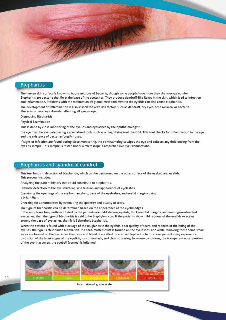
Blepharitis
The human skin surface is known to house millions of bacteria, though some people have more than the average number. Blepharitis are bacteria that lie at the base of the eyelashes. They produce dandruff-like flakes in the skin, which lead to infection and inflammation. Problems with the meibomian oil gland (meibomianitis) in the eyelids can also cause blepharitis.
The development of inflammation is also associated with risk factors such as dandruff, dry eyes, acne rosacea, or bacteria. This is a common eye disorder affecting all age groups.
Diagnosing Blepharitis
Physical Examination
This is done by close monitoring of the eyelids and eyelashes by the ophthalmologist.
the eye must be evaluated using a specialized tools such as a magnifying tool like OSA. This tool checks for inflammation in the eye and the existence of bacteria/fungi/viruses.
If signs of infection are found during close monitoring, the ophthalmologist wipes the eye and collects any fluid oozing from the eyes as sample. This sample is tested under a microscope. Comprehensive Eye Examinations.
Blepharitis and cylindrical dandruf This test helps in detection of blepharitis, which can be performed on the outer surface of the eyeball and eyelids. This process includes:
Analyzing the patient history that could contribute to blepharitis.
Extrinsic detection of the eye structure, skin texture, and appearance of eyelashes.
Examining the openings of the meibomian gland, base of the eyelashes, and eyelid margins using a bright light.
Checking for abnormalities by evaluating the quantity and quality of tears.
The type of blepharitis can be determined based on the appearance of the eyelid edges. If the symptoms frequently exhibited by the patients are mild sticking eyelids, thickened lid margins, and missing/misdirected eyelashes, then the type of blepharitis is said to be Staphylococcal. If the patients show mild redness of the eyelids or scales around the base of eyelashes, then it is Seborrheic blepharitis.
When the patient is found with blockage of the oil glands in the eyelids, poor quality of tears, and redness of the lining of the eyelids, the type is Meibomian blepharitis. If a hard, matted crust is formed on the eyelashes, and while removing these some small sores are formed on the eyelashes that ooze and bleed, it is called Ulcerative blepharitis. In this case, patients may experience distortion of the front edges of the eyelids, loss of eyelash, and chronic tearing. In severe conditions, the transparent outer portion of the eye that covers the eyeball (cornea) is inflamed.
International grade scale

12
What is demodex brevis?
Demodex brevis is a kind of mite found on humans. Like its counterpart Demodex folliculorum, brevis is naturally occurring. D. brevis is so small that you can’t see the mites with a naked eye. In fact, the average mite is only www.dermnetnz.org/topics/demodex/long. They only cause noticeable reactions and problems in people if the mites exist in large quantities.
Symptoms
Symptoms of D. brevis usually only surface in cases of large infestations. Signs might include:
Red skin
Rough or tough skin
Scaly or patchy skin
The symptoms of D. brevis are similar to those of D. folliculorum. The key difference is location. While folliculorum tend to stay on the face, D. brevis can distribute all over the body. The chest and neck are common areas of D. brevis infestation, so you might notice more symptoms there if you have it.
Causes
Once in the skin, D. brevis feed off sebum in the oil glands. These glands are attached to hair follicles underneath the skin’s surface.
Infestations of D. brevis aren’t common in young children, but their numbers naturally grow with age. The mites may also be spread between humans.

13
Comparative table
LIPIDIC LAYER
INTERFEROMETRY
N.I.B.U.T
TEAR MENISCUS
MEIBOGRAPHY
PUPILLOMETRY
W. TO W.
BLEPHARITIS
DEMODEX
BULBAR REDNESS
INTERNATIONAL GRADING SCALE
RESULTS EXAMS
LIPIDIC LAYER
INTERFEROMETRY
N.I.B.U.T
TEAR MENISCUS
BULBAR REDNESS
INTERNATIONAL GRADING SCALE
RESULTS EXAMS
MEIBOGRAPHY
RESULTS EXAMS
AUTO MANUAL
AUTO MANUAL
AUTO MANUAL AUTO MANUAL
AUTO MANUAL
AUTO MANUAL
PORTABLE DEVICE

14
Exam Report
14/09/2016 09:38 219.00 mOsm/L 289.00 mOsm/L 70.00 mOsm/L
14/09/2016 09:26 312.00 mOsm/L 320.00 mOsm/L 8.00 mOsm/L
13/09/2016 16:27 333.00 mOsm/L 289.00 mOsm/L 44.00 mOsm/L
13/09/2016 16:22 297.00 mOsm/L 266.00 mOsm/L 31.00 mOsm/L
13/09/2016 16:22 288.00 mOsm/L 288.00 mOsm/L 0.00 mOsm/L
13/09/2016 16:21 299.00 mOsm/L 299.00 mOsm/L 0.00 mOsm/L
13/09/2016 15:31 299.00 mOsm/L 299.00 mOsm/L 0.00 mOsm/L
13/09/2016 15:31 333.00 mOsm/L 333.00 mOsm/L 0.00 mOsm/L
13/09/2016 15:30 289.00 mOsm/L 310.00 mOsm/L 21.00 mOsm/L
SBM Sistemi s.r.l.Strada Torino, 43Orbassano, 10043+390117791800
SBM Sistemi srlStrada Torino 43,Orbassano (TO)01119923378www.sbmsistemi.com
PATIENTDoe, John
TELEPHONE01119923378
REPORT DESCRIPTIONOsmolarity
BIRTH DATE02/03/1990 (26)
SEXM
IDDOEJHN90C02I480P
ADDRESSStrada Torino , 43 Orbassano 10043 TO
Date Value Right Eye Value Left Eye Difference Value
SBM Sistemi ICP Medical System - http://www.sbmsistemi.com/ - Page 1/2
With all exams values of a given day.
Report with values and graph of a specific exam value
Daily reportMGD
Values and graph of lost area
MGD
Exam Reporta
SBM Sistemi s.r.l.Strada Torino, 43Orbassano, 10043+390117791800
SBM Sistemi srlStrada Torino 43,Orbassano (TO)01119923378www.sbmsistemi.com
PATIENTDoe, John
TELEPHONE01119923378
REPORT DESCRIPTION8/9/2016
BIRTH DATE02/03/1990 (26)
SEXM
IDDOEJHN90C02I480P
ADDRESSStrada Torino , 43 Orbassano 10043 TO
Right eye (O.D.) Left eye (O.S.)
10.0 sNIBUT
11.0 s
7.0 sBUT
9.0 s
~30 nm - close meshworkLipid Layer Type
~80 nm - amorphous
83 %Meibomian Glands - Loss area
24 %
3Ferning test type
2
78O.S.D.I. result
98
319 mOsm/LOsmolarity
328 mOsm/L
1 - TRACERedness
3 - MODERATE
2LiPCoF (Lid Parallel Conjunctival
Folds) grade 0
1 - TRACELissamin
4 - SEVERE
SBM Sistemi ICP Medical System - http://www.sbmsistemi.com/ - Page 1/1
Exam Report
Occhio sinistro 13/09/2016 15:46 8.0 s
Occhio destro 13/09/2016 15:35 8.0 s
Occhio sinistro 13/09/2016 15:14 9.0 s
Occhio sinistro 08/09/2016 15:15 11.0 s
Occhio sinistro 08/09/2016 15:15 8.0 s
Occhio destro 08/09/2016 15:00 10.0 s
Occhio destro 08/09/2016 15:00 9.0 s
Occhio destro 08/09/2016 14:00 9.0 s
Occhio destro 08/09/2016 10:16 6.0 s
SBM Sistemi s.r.l.Strada Torino, 43Orbassano, 10043+390117791800
SBM Sistemi srlStrada Torino 43,Orbassano (TO)01119923378www.sbmsistemi.com
PATIENTDoe, John
TELEPHONE01119923378
REPORT DESCRIPTIONNIBUT
BIRTH DATE02/03/1990 (26)
SEXM
IDDOEJHN90C02I480P
ADDRESSStrada Torino , 43 Orbassano 10043 TO
Eye Date Value
SBM Sistemi ICP Medical System - http://www.sbmsistemi.com/ - Page 1/2
These values have been grouped in a new section in the exam results screen with all these new values.All values from “Grading scales” were deliberately put together in a single section later providing the ability to filter the values seeing only those of interest (Redness, Staining, ...)
Daily reportwith all exams values of a given day
Report with values and graph of a specific exam value (nibut, osdi, osmolarity)
N.I.B.U.T.Values and graph of nibut values
NEW RESULTS EXAMS
OsmolarityValues and graph of osmolarity
values
Exam Report
14/09/2016 09:38 219.00 mOsm/L 289.00 mOsm/L 70.00 mOsm/L
14/09/2016 09:26 312.00 mOsm/L 320.00 mOsm/L 8.00 mOsm/L
13/09/2016 16:27 333.00 mOsm/L 289.00 mOsm/L 44.00 mOsm/L
13/09/2016 16:22 297.00 mOsm/L 266.00 mOsm/L 31.00 mOsm/L
13/09/2016 16:22 288.00 mOsm/L 288.00 mOsm/L 0.00 mOsm/L
13/09/2016 16:21 299.00 mOsm/L 299.00 mOsm/L 0.00 mOsm/L
13/09/2016 15:31 299.00 mOsm/L 299.00 mOsm/L 0.00 mOsm/L
13/09/2016 15:31 333.00 mOsm/L 333.00 mOsm/L 0.00 mOsm/L
13/09/2016 15:30 289.00 mOsm/L 310.00 mOsm/L 21.00 mOsm/L
SBM Sistemi s.r.l.Strada Torino, 43Orbassano, 10043+390117791800
SBM Sistemi srlStrada Torino 43,Orbassano (TO)01119923378www.sbmsistemi.com
PATIENTDoe, John
TELEPHONE01119923378
REPORT DESCRIPTIONOsmolarity
BIRTH DATE02/03/1990 (26)
SEXM
IDDOEJHN90C02I480P
ADDRESSStrada Torino , 43 Orbassano 10043 TO
Date Value Right Eye Value Left Eye Difference Value
SBM Sistemi ICP Medical System - http://www.sbmsistemi.com/ - Page 1/2

15
• Comparison of database’s images
• Saving of images and movies comparing the situation before and post application
• Direct comparison with the taken images and the tables of Efron
• Direct comparison with the taken images and the tables of CCLRU
• Direct comparison with the taken images and the tables of Jenvis
• Possibility to point out and to show to your patient the pathology
• Evaluation of the visual acuity from far
• Evaluation of the visual acuity from near
• Show visually with the help of the iPad the difference between the use of the lac and the use of the ophthalmic lens
• Database dedicated and structured for the saving of the sensible data and of the made exams during the time
• Technical follow up targeted to the LAC application
• The electronic medical record with the essential registry of the Patient
• The medical history of the patient
• Optometric data
• Results of examinations
• Privacy management
• The archive of images and movies (photographs)
• Test and results
• Follow up visits allowing the simultaneous visualization of images related the next visits. With this function is possible to evaluate the upgradability of the pathology through direct comparison
• Reporting and printing up to 10 printable reports.
The software functions present in all versions of the program
The system allows you to securely access all your data from iPad to PC and from PC to iPad.
• You will always have the most up-to-date version of your patients’ data at the right time.
• From the iPad device you will have the ability to send all patient records and related exams, or parts of them to the Windows system.
• All patient examinations, or part of them, can be sent to the iOS device
• The system allows you to save space on the iPad, having the ability to only preserve patients who are going to be visited in portability
• Data storage
• share all information inside the clinic
• Possibility to receive exams from assistants allover the world for referral or sharing
I Cloud
“Unlike words, and their interpretation, the image is a neutral constant a benchmark reference frozen in time.
It becomes the quintessentially unbiased witness to the condition at the moment of capture”.

16
Windows system
A practical personal archive to bring always with you and with the possibility to share the archive by Wi-Fi with the various ICP medical instrumentation
The electronical medical cartel that includes:• Essential patient registry• Medical history• Formula of the current therapeutic cycle• Archive of images• Report
With this module is possible to manage the database of patient, his medical history, the medical examinations and their Follow Up.It follows that, through a unique screen, will be possible to visualize the whole clinical history of the patient and will be made all related prints.The doctor has then, at the end of the visit, the possibility to print and deliver to the patient a report that presents only the data of the clinical cartel that he considers appropriate, inserting also a textual discursive section. He can also choose to send these documents via email.Multisite/multi iPad with synchronization between them to visualize all medical reports from a computerExternal memory to download exams when the memory of the various supports is finishedBackup and recovery of the achieveWorking also in backgroundSaving photo and video on FTP instead that on the device to safeguard the space.
We are working for an innovative web tele-diagnostic application dedicated to the world of Vision. The mission is to make easily available rapid reporting of the main Ophthalmological diagnostic tests, according to the European laws about sensitive data protection.
This service will offers expertise and professional skills of a huge number of Ophthalmologists. Physicians will report only exams acquired by Sbm and other system that are selected diagnostic centers really easily accessible by patients.
The reporting will be granted within 72 working hours after acquiring data, although statistically, thanks to Ophthalmologists professionalism and cooperation, the report will be performed within 24 hours. The project’s goal is to even reduce this timing in order to provide to the patients a fast, safe and effective eye care service. The spread out of this diagnostic network will allow patient to get the nearest doctor in order to access immediately to a diagnostic service and a skilled Specialist reporting without any waiting list and at convenient price.
The web-application is written in dynamic multilingual, translated into the main foreign languages, in order to allow an international use, anticipating the recent European Community provisions about health care. Finally the patient will have the opportunity to save all his exams on his personal account without any loss or damage risk. By the use of a free mobile app (compatible with iOS, Android and Windows Mobile), the patient will can also share his exams with his Family Doctor or ask for a second opinion to his Ophthalmologist.
The website has been developed with the latest technology IT, ensuring technical reliability and operational safety in the management and storage of data and guarantee of privacy in compliance with current legislation..
OUR PROJECT IN TELEMEDICINE

17
History
The SBM was founded in 1984. The core activity of the new company is to produce software for optical centers.
The territorial development begins with the opening of new distribution facilities on the Italian territory, especially to offer services and assistance. In the ‘90s, the company began its real growth path that is still the main goal for the future.
Thanks to large investments and sacrifices in those years began working for production. Later in time also starts working as a provider of information technology services and optics and ophthalmology instrumentation.
After more than twenty years these services are still carried out at the office of our customers or our operating offices on the Italian territory and abroad.
The experience achieved over the years with important and prestigious brands allows us to be present on the market as a serious and reliable partners for Strategic Outsourcing services or part of them.
Production
A good product must be made from a precise projecting including a careful materials choice. The final difference consists in availability of personalized solutions for every need.
SBM Sistemi customers have a complete post-sale support. Any request related to the software is solved with remote assistance from local partners or directly by the SBM company. Any need related to the hardware can be managed by our local partners all-over the world that, in constant contact with SBM company, have the opportunity to ship the instrument for complete check-up.
All SBM Sistemi products are realized with hi-precision by factory technicians. Pre-sale checks and verifications consist in 24 hours non-stop test session to ensure the quality of the components. Only after this task an instrument can be packaged and considered ready for delivery.

18
Registration made in the following extra cee countries
Medical certificate
All SBM Sistemi medical products are marked EC under the Medical Device 93/42 / EEC Directive and also conform to a set of international standards as applicable.
However, the interior of the medical commitment SBM Sistemi for product quality goes beyond the adherence of internationally recognized standards and extends in the attitude of our highly skilled production staff and the quality dedicated team, who are always aware the fact that the products they manufacture are used to save lives in critical care applications both locally and worldwide.
ID registration BD/RDN
I.C.P. IOL registration number at the Ministry: 1340867/R
I.C.P. SLIT LAMP registration number at the Ministry: 1340861/R
I.C.P. SLIT LAMP PLUS registration number at the Ministry: 1340862/R
I.C.P. TEARSCOPE registration number at the Ministry: 1340864/R
I.C.P. MEASUREMENT registration number at the Ministry: 1340865/R
I.C.P. OSA registration number at the Ministry: 1556084/R
Furthermore all devices are certified CE and are accordant to the European regulations in force.
Electro medical test – photobiological test – etc
ISO 13485 Quality Management System for Medical Devices
ISO 13485ISO 13485 is a quality management system standard specifically for the medical devices industry, which encompasses aspects of the ISO 9001 standard, plus additional industry-specific medical device requirements.
ISO 13485: 2003 has been harmonized against the three EU Medical Devices Directives (Medical Devices, In-vitro Diagnostic Devices and Active Implantable Devices), so certification to this standard by an accredited certification body such as ECM automatically demonstrates compliance with specific clauses in these directives.
The certification of a quality management system, specifically for medical devices, to ISO 13485 is essential, for medical companies which export their products to the global market. In the European Union, the fulfilment of EU Directives (e.g., Active Implantable Medical Devices Directive, Medical Devices Directive and In Vitro Diagnostic Directive) allows the free trade of medical devices. A significant portion of demonstrating compliance with the EU directive requirements is the establishment and independent assessment of the quality system.
uni en iso 9001:2015 Nr. 8631/0uni cei en iso 13485:2012 Nr. 8632/0

1
Complete holder for OSA Foot pedal USBUSB connection
Briefcase
Bags, resistant material to large stresses, have the particularity to be watertight with IP 67 impermeability and the perfect seal for liquids and dust, this is ensured by a rubber seal along the entire closure profile; is also present in all models a balancing valve of the internal and external pressure. Customizations are possible such as bespoke interior, screen prints or stickers.
I.C.P. OSA film DEALER
Cylindrical dandrufOSA table holder
Table
Accessory