telepathology: clinical utility and methodology · 4 telepathology became a newsworthy item on...
TRANSCRIPT

Telepathology: Clinical utility and methodology
F Joel W-M Leong1, Andrew K Graham1, Thomas Gahm2, James O'D McGee1
1999
1Nuffield Department of Pathology & Bacteriology University of Oxford John Radcliffe Hospital Headington, Oxford OX3 9DU UK 2Autocyte Inc. Elon College North Carolina, USA This is a modified version of a review article by the same name and authors published in Recent Advances in Histopathology 18 (Edited by DG Lowe, JCE Underwood 1999, Churchill Livingstone, London ISBN 0-443-06036-3) Similar copyright restrictions apply. This article may not be copied, reproduced or distributed in whole or in part in any way, shape or form, electronic or otherwise. Please direct all enquiries to Dr Joel Leong Affiliations were correct at the time of paper publication. Please note that the Nuffield Department of Pathology and Bacteriology is now part of the Nuffield Department of Clinical Laboratory Sciences (same address) and Autocyte Inc has become TriPath Imaging. Click here to return to Telepathology City

2
INTRODUCTION
Most histopathologists are aware of the concept of transmitting textual, numerical, macroscopic and microscopic images via telecommunication technologies though the majority have yet to see it work in practise. Telepathology and medical telematics however, have progressed beyond the experimental stage and advances in computer image processing, the development of the internet and telelcommunications technology have evolved to the stage where telepathology is now in use in many institutions.
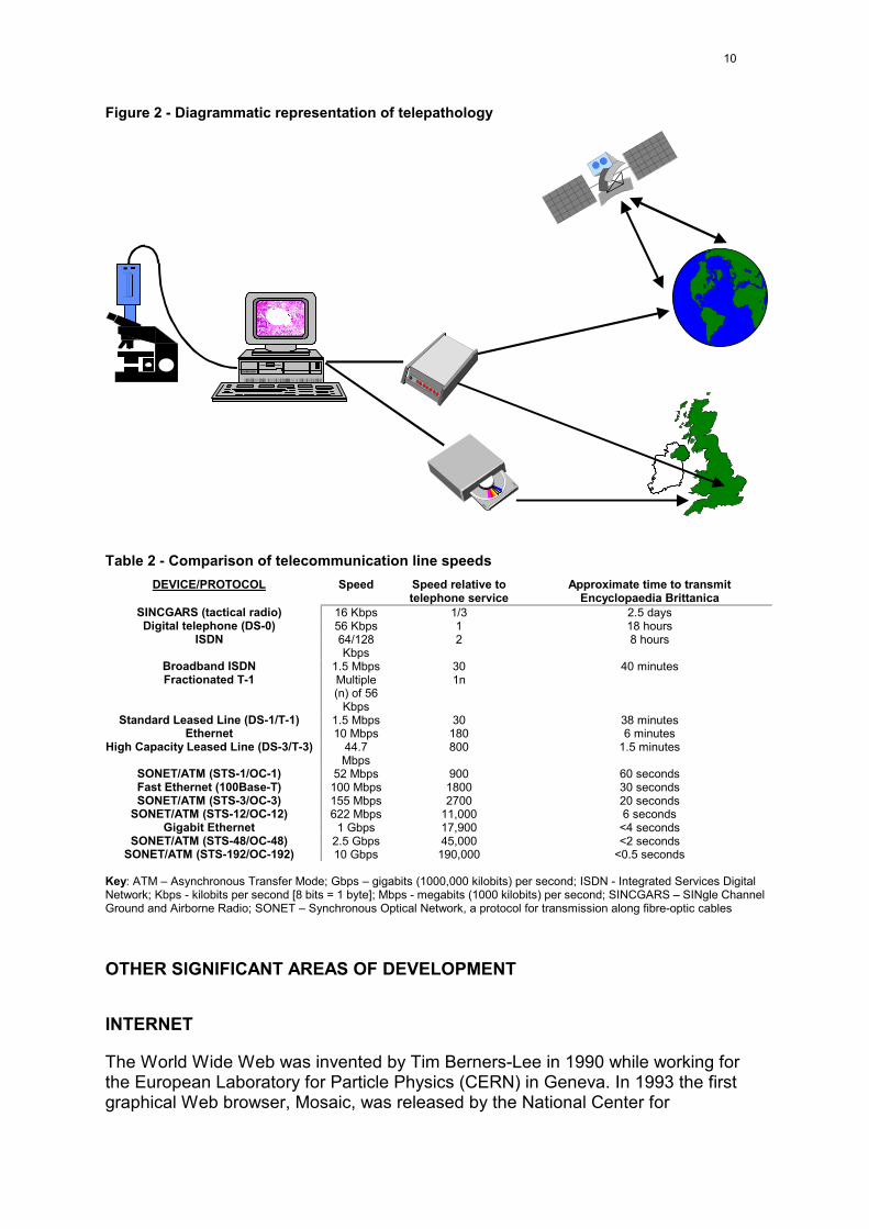
Telepathology is the acquisition of histological and or macroscopic images for transmission along telecommunication pathways for diagnosis, consultation or continuing medical education. In brief, a telepathology system comprises a conventional microscope; a method of image capture, commonly a camera mounted on a light microscope; telecommunications link between sending and receiving sites; and a workstation at the receiving site with a high-quality monitor to view the images (Figure 2). There may also be mechanical hardware to allow the receiving pathologist to control the microscope from a distance and view the entire slide in 'real-time'. The images are viewed on a computer screen, rather than through microscope oculars.
No one will challenge the fact that telemicroscopy in its current form, falls short of what pathologists are used to seeing through their conventional microscopes. Despite this, emerging literature suggests that in most cases, an accurate diagnosis can still be made.1-8 The authors experience in this area is that image quality is such that diagnosis is unduly difficult and cannot be made with the same level of confidence as with conventional microscopy.
We describe the recent advances in this field, with particular focus on our experience with this technology in Oxford. In addition, we discuss the exponential growth and popularity of the internet and how various spin-off technologies in image delivery may be utilised. This chapter is presented in two parts, the first describes and delineates the clinical utility of telepathology, and to a lesser extent telecytopathology. The second describes the technological background on which future developments in telepathology will evolve. Both sections can be perused independently (and are cross-referenced) but a comprehensive understanding of the state of the art and evolution is dependent on both sections of the presentation.
3
SECTION I
HISTORY
The initial impetus in the development of telepathology was the provision of the best possible diagnostic opinion to all patients irrespective of geographical locale and social/economic circumstances. Telepathology has its origins in telemedicine, a generic term encompassing the use of visual telecommunications in health care. Radiology was one of the earliest medical specialties to utilise this as early as 1959 when Albert Jutra used coaxial cable to transmit videotaped telefluoroscopy examinations between two hospitals in Montreal, some five miles apart.9 Pathologists came somewhat later to the technology probably because it was not of a sufficient standard. A histological image is exponentially more complex that a black-and-white radiological one and colour television was yet to be mass-produced. One of the earliest instances of telepathology took place in Boston in 1968 when a microwave-based telecommunications system was established, linking Massachusetts General Hospital with a medical station at Logan Airport, outside of downtown Boston.10 Live black and white images of histological sections and stained blood smears were transmitted from a clinic at the airport to the hospital. The video quality was equivalent to the then current United States commercial television (300-330 lines). A technician at the airport was directed by the pathologist at the hospital and also provided information on stain colours as requested.
The aim of this experiment, organised by Dr KT Bird, a Harvard physician, was to demonstrate the feasibility of remote physical diagnosis, telestethoscopy, teleauscultation, teledermatology, speech pathology and even telepsychiatry.11 Interestingly, even at this embryonic stage of development, Professor Scully made the correct diagnosis on all microscopic images (personal communication).12
In 1974, satellite communication was used to transmit histological images and clinical data from a 17-year-old male on board a hospital ship moored off the coast of Brazil to a hospital in Washington DC.13 The system provided the equivalent of two telephone lines, one of which was used for voice communication, the other to transmit images of tissue sections, blood and bone marrow smears from a microscope video camera. In addition, teletype provided textual data communications while facsimiles of drawing and diagrams were sent at a rate of one page every six minutes. An electrocardiogram transmission unit and electronic stethoscope relayed ECGs and heart sounds. While the patient was not actually visualised, the data provided allowed a team of specialist consultants to diagnose 'mediastinal lymphosarcoma with leukemic transformation', initiate a regimen of chemotherapy and irradiation to the mediastinum, and even transmit a relevant journal article to the ship. During follow-up under similar circumstances thirty-five days later, a chest x-ray showed a reduction in tumour size.
4
Telepathology became a newsworthy item on August 20, 1986 with a public demonstration of a satellite-linked colour-video dynamic telepathology system between Fort William Beaumont Army Medical Center in El Paso, Texas and Washington DC.14 In Texas, a frozen section slide of breast tissue was placed on the stage of a custom-designed, Olympus Vanox motorised microscope equipped with a video camera. The full-colour image was transmitted via the SBS-3 COMSAT satellite (Communications Satellite Corp, Washington, DC) to Washington DC where a pathologist seated at a prototype workstation (Corabi International Telemetrics, Rockville, Md) was able to control stage movements, magnification, focus and illumination, while viewing a real-time image at 525 lines of resolution. A second monitor displayed other parameters such as location of the image in relation to the whole slide, coordinates, and stage speed. Two-way audio communication was also available. The exercise was a success, although the Washington Post only afforded it page seven newsworthiness.
Since then, work by independent groups in Europe,4,15-24 the United States8,25,26 and Japan3,27 have each made significant contributions to the development and assessment of the clinical utility of telepathology equipment.
TYPES OF TELEPATHOLOGY
Telepathology or telemicroscopy has developed along two lines designated ‘static telepathology’ and ‘Robotic Interactive Pathology’ (RITPath). The former is also referred to as ‘store-and-forward’ or passive telepathology.
STATIC IMAGING
The static or 'store-and-forward' approach to telepathology is cheap, simple and needs only a standard telephone line or internet connection.1 By definition, the images are static - there is no facility for recipient control. In practise, only a limited number of images are captured for each case, varying between one and forty1,26 with the average number varying between 4.926 and 6.3.1 To the pathologist trained in the doctrine that one must view the entire slide in order to make a confident diagnosis, this method would appear suboptimal, and many find it initially quite disconcerting. And yet, international static telepathology consulting services do exist.1,26
The major disadvantage is of course, sampling error, although interpretation and video image quality have also been cited as problems.5 Sampling error has been well-demonstrated in various studies.2,3,6,26 Attempts to overcome this have included use of a trained pathologist to select the images6,26 however few pathologists are completely happy with relinquishing control to another person. Halliday et al26 pointed out that referring pathologists would often selected images that supported their own diagnosis and that ambiguous fields were often ignored. The limited number of images could underrepresent the complexity of the case and lead to a false sense of security as to the ease of the diagnosis. Sampling error or bias remains a serious flaw as it precludes the pathologist being consulted from applying his skills in identifying significant regions of the slide while scanning at low-power.

5
Assessing the accuracy of telepathology is difficult. Involving multiple pathologists is the best option as the results from a single pathologist is influenced by his degree of skill and experience in viewing static images. Eusebi et al1 used the Pathmaker image acquisition software28 to package thirty-six diagnostically difficult cases from a quality assurance program over the course of a year. Images included immunohistochemistry and were digitised at 640 x 480 x 24 bit colour resolution before being sent as email attachment to Dr Juan Rosai, in the Memorial Sloan-Kettering Center, New York. Of the thirty-six cases, thirty-five were also sent for final diagnosis based upon actual histological slides and/or further stains. Dr Rosai deferred a telepathology diagnosis in eight cases. In three of these cases, his final diagnosis concurred with the referring pathologists. While a telepathology diagnosis was provided in only 77% of the cases (27/35) the concordance with the final diagnosis in those 77% was 100%. It is difficult to ascertain whether this is due to the accuracy of the technique or the skill of the histopathologist. Anecdotal evidence would suggest the latter.
Shimosato et al27 reported a diagnostic accuracy of 88.1% also based upon performance of one pathologist. Weinberg et al5 used four pathologists at the Brigham And Women's Hospital in Boston, showing them fifty static images selected from cases on CD-ROM, comparing their diagnoses with the conventional light microscopy diagnoses they gave at a later date. The overall diagnostic accuracy was 88.5% however marked interobserver variability was noted.
Lisa Weinstein et al6 examined the diagnostic accuracy of static imaging telepathology in assessing archival frozen sections of 46 skin excisions showing a spectrum of benign (n=9) and malignant (n=37) lesions. Using a single pathologist, they found 100% concordance in discriminating benign from malignant lesions, with minor discrepancies relating to precise characterisation of the lesion (actinic keratosis vs. irritated seborrhoeic keratosis, basal cell carcinoma vs. squamous cell carcinoma). Where errors occurred in determining clearance at the margin, they were attributed to field selection error. The images were stored at 1024 x 768 x 24-bit colour with no complaints regarding image quality.
Work published from the Arizona-International Telemedicine Network (AITN)26 showed an 88.2% concordance (127/144) between telepathology and glass slide diagnosis on a variety of referred cases, with 96.5% concordance (139/144) for clinically important diagnoses. They divided the reasons for discordance into three categories - field selection, diagnostic interpretation, and video image quality. Diagnostic interpretation was the most commonly cited reason, with field selection second. Poor image quality was not a problem using a resolution of 1024 x 768 x 24-bit colour and 6.5:1 JPEG compression. Comments were made on a possible reluctance on the part of the telepathologist to overturn the provisional diagnosis provided by the referring pathologist based upon a small set of images.
Michael Weinstein & Epstein7 conducted an analysis of the diagnostic accuracy of a static telepathology system in the interpretation of prostatic needle biopsies. They examined 200 needle biopsies of prostate divided into two groups and cited accuracy of up to 99%. However, no mention was made of image resolution, colour depth or the colour depth of the computer monitor used for viewing. Average image
6
size was 300 Kb using JPEG compression (incorrectly referred to as a lossless algorithm) which would correspond to a 1024 x 768 image using moderate compression.
These studies have provided us with an indication of the skill of several individual pathologists in interpreting ‘bitmapped’ histological images. It is difficult to make comparisons between studies however due to the lack of standardisation in hardware, image resolution, storage format and colour-depth. This, combined with the narrow range of pathologists and wide range of cases, is a limitation preventing the extrapolation of results. The future of this technique may lie in teaching and external quality assessment rather than as a diagnostic tool.
ROBOTIC INTERACTIVE TELEPATHOLOGY (RITPATH)
RITPath systems allow the receiving pathologist to control the movement of the slide on the stage, and to see the image in 'real-time' on a high-resolution monitor. Synonyms used for this modality include dynamic telepathology, active telepathology and real-time telepathology.
The limiting factor in RITPath is transmission time and understanding of the various type of telecommunications protocols is essential knowledge in assessing the viability of a dynamic system in your particular area.
Norwegian geography necessitated development by the Norwegian health care system in 1991 of one of the earliest working European frozen section telepathology services. This was established between University Hospital of Tromsø, Tromsø and an outlying hospital, Kirkenes Hospital, some 420km to the north-east.15 Supported by a Norwegian Telecom network with a 2 Mbps capacity, both dynamic or static resolution images can be transmitted. In 1991, the average time taken for assessment of a frozen section was fifteen minutes, but it ranged from five to thirty minutes. An image resolution of 256 x 286 pixels appears quite small by present standards (see Figure 1, Table 1). Magnification shifts are performed manually by the local technician, although there are plans to automate this. Recently, two additional diagnostic centres have been established in Oslo and Trondheim.

7
Figure 1 - A graphical representation of image resolutions and resultant file sizes (refer to Table 1 for details)
Table 1 - Image resolutions, resultant file sizes and current popular usage (refer to Figure 1 for graphical comparison) KEY Image
dimensions (pixels)
Size using 24-bit colour
Size in greyscale or 8-bit colour (256 shades/colours)
Current usage
F 160 x 120 57 KB 19 KB • Video conferencing E 320 x 240 225 KB 75 KB • High-quality video conferencing
• Approximate resolution using Apollo/Corabi telepathology system in dynamic mode (actual resolution 352 x 288)
• Early digital snapshot cameras • Personal digital assistants, high-end
electronic diaries D 640 x 480 900 KB 300 KB • High-end personal digital assistants
• Outdated computer systems with 14-inch monitors
• Entry-level digital snapshot cameras • Home digital video (DV) cameras • Most microscope cameras used by
pathology departments for clinical presentations
C 800 x 600 1.37 MB 469 KB • Budget-priced computer systems • Mid-range digital snapshot cameras • Approximate resolution of
HISTKOM dynamic telepathology system resolution (actual resolution 768 x 576)
B 1024 x 768 2.25 MB 768 KB • Entry-level office workstations (requires 2 MB graphics RAM and 15 inch flatscreen monitor for best

8
viewing) • Upper-range digital snapshot
cameras A 1536 x 1152 5.06 MB 1.69 MB • Minimum resolution for desktop
publishing, computer graphics editing, computer-aided design (requires >19 inch monitor)
• Static image resolution using Apollo/Corabi telepathology system
2048 x 1536 9.00 MB 3.00 MB 3072 x 2304 20.3 MB 6.75 MB • Maximum resolution of Kontron
ProgRes 3012 ultra high resolution digital camera
Key: KB - kilobytes; MB - megabytes [1024 KB = 1 MB]
The HISTKOM RITPath system29 is a joint project of the Institut für Physikalische Elektronik of the University of Stuttgart and Deutsche Telekom. The prototype has been tested in our department under simulated line speeds. It consists essentially of a robotic Zeiss microscope with a triple charge-couple device sensor (CCD) Sony video camera (refer to part 2 for an explanation of CCD technology) transmitting images to a remote station. At this station the user is able to control all the functions of the microscope, including the scanning stage, magnification and light intensity. Manual focussing may also be exercised in preference to the autofocus. Designed to be used with low-bandwidth networks, eg. Integrated Services Digital Network (ISDN), compression algorithms are employed to minimise the data transmitted (refer to Section II for terminology). Transmission along multiple ISDN channels is also possible. The system also conforms to Transmission Control Protocol/Internet Protocol (TCP/IP), the de facto standard for data transmission over networks to ensure compatibility with current network systems. The remote station is designed for ease of use with two monitors, one displaying current position relative to the whole slide, the other providing the current view at 768 x 576 x 24-bit colour (see Table 1, Figure 1 for a comparison with other systems). Stage movement is controlled via a joystick, with optional mouse control. The images are transmitted in compressed JPEG format (refer to part 2 for an explanation of image compression formats), encoded 'on-the-fly' via a hardware JPEG board. The software at the receiving station 'stitches' the images together, thus simulating a moving image. The degree of JPEG compression employed is proportional to the speed of movement, with the image quality improving when the slide is at rest.
Images and position on the slide can be marked and saved. Response time is sufficient to review a slide in under ten minutes, although in our preliminary studies, experienced users have been able to reduce this to less than three.
Two field trials have been conducted in 1997 using this system (personal communication).30 A prototype was tested in Stuttgart in a double blind study involving three pathologists, 119 lung frozen sections and a single ISDN connection providing a 64 Kbps transmission rate. Overall accuracy of telepathology diagnosis was 63.4% compared with 81.5% using conventional light microscopy. Transmission time was unsatisfactory.
A second field trial was conducted in the University of Tübingen, using two pathologists, 139 frozen sections of lung and eight times the bandwidth (8 x 64 Kbps). The two pathologists averaged 105 and 154 seconds per case respectively,

9
compared with 24 seconds using conventional light microscopy. Concordance with conventional light microscopy was 94.2% and 96.4% respectively. There were no complaints regarding image quality.
The Apollo Image Management System/Corabi® Dynamic Module (IMS/CDM™) is based around the Corabi DX 1000, the result of a corporately-financed joint United States-Japanese venture. It was first used in 1986 and since then has been tested in several American universities.31 Apollo software, a corporation in Maryland, has licensed a telepathology system patented by Professor RS Weinstein and Corabi International Telemetric, Inc. and coupled it with its own image management software and teleconferencing facilities. The system provides remote stage movement, focus, light intensity and magnification, as well as the ability to view a gross image of the slide with the relative position of the current objective over the specimen. It also has the capability of functioning as a fully bi-directional telepathology system. This system differs from the Histkom system in that it is a hybrid system capable of both static telepathology, at resolutions up to 1520 x 1144 x 24-bit colour, and dynamic images at 352 X 288 x 24-bit colour via video codec over a T-1 line. One disadvantage of using video conferencing technology is susceptibility of the system to blurring or interruptions in image display. The use of a T-1 line greatly improves transmission speeds compared with ISDN (see Table 2)
This system is in use in the Veterans Affairs Hospitals in Milwaukee, Wisconsin and Iron Mountain, Michigan. Dunn et al32 reported that in using this system they achieved an overall concordance with consensus ‘truth’ diagnoses of 97.5% based upon a test set of 100 consecutive routine surgical pathology cases. Quality of the video images was cited as the reason for diagnostic error in two cases. The rest were attributed to diagnostic difficulty. The time spent on each case varied between 2.8 and 4.7 minutes.
10
Figure 2 - Diagrammatic representation of telepathology
Table 2 - Comparison of telecommunication line speeds DEVICE/PROTOCOL Speed Speed relative to
telephone service Approximate time to transmit
Encyclopaedia Brittanica SINCGARS (tactical radio) 16 Kbps 1/3 2.5 days Digital telephone (DS-0) 56 Kbps 1 18 hours
ISDN 64/128 Kbps
2 8 hours
Broadband ISDN 1.5 Mbps 30 40 minutes Fractionated T-1 Multiple
(n) of 56 Kbps
1n
Standard Leased Line (DS-1/T-1) 1.5 Mbps 30 38 minutes Ethernet 10 Mbps 180 6 minutes
High Capacity Leased Line (DS-3/T-3) 44.7 Mbps
800 1.5 minutes
SONET/ATM (STS-1/OC-1) 52 Mbps 900 60 seconds Fast Ethernet (100Base-T) 100 Mbps 1800 30 seconds SONET/ATM (STS-3/OC-3) 155 Mbps 2700 20 seconds
SONET/ATM (STS-12/OC-12) 622 Mbps 11,000 6 seconds Gigabit Ethernet 1 Gbps 17,900 <4 seconds
SONET/ATM (STS-48/OC-48) 2.5 Gbps 45,000 <2 seconds SONET/ATM (STS-192/OC-192) 10 Gbps 190,000 <0.5 seconds
Key: ATM – Asynchronous Transfer Mode; Gbps – gigabits (1000,000 kilobits) per second; ISDN - Integrated Services Digital Network; Kbps - kilobits per second [8 bits = 1 byte]; Mbps - megabits (1000 kilobits) per second; SINCGARS – SINgle Channel Ground and Airborne Radio; SONET – Synchronous Optical Network, a protocol for transmission along fibre-optic cables
OTHER SIGNIFICANT AREAS OF DEVELOPMENT
INTERNET
The World Wide Web was invented by Tim Berners-Lee in 1990 while working for the European Laboratory for Particle Physics (CERN) in Geneva. In 1993 the first graphical Web browser, Mosaic, was released by the National Center for

11
Supercomputing Applications (NCSA). The exponential growth of the internet can be attributed to the popularity of this graphical, animated, interactive interface. In the long-term, developers would like the internet to become as ubiquitous and as simple to use as the television set and such enticing terms as ‘WebTV’ and ‘Digital TV’ have already appeared in popular press. In the short-term however, full-motion internet video and video-conferencing is erratic and possible only with software technologies that attempt to circumvent the immediate problems of narrow bandwidth and network congestion.
The advantage of using the internet for telepathology is cost and availability. Sending static images as email attachments is not only easy for most people, it costs nothing to send. The internet however is too slow and unreliable for satisfactory dynamic telepathology to take place at this point. Another consideration is the issue of security, which has yet to be adequately addressed. Encryption techniques of varying levels do exist, but a discussion of what is a satisfactory level of encryption goes beyond the scope of this review.
Nonetheless, there are several examples of novel approaches to internet telepathology and as advances in video-conferencing and presentation of graphical images takes place, telepathology can only benefit.
Wolf et al33 have used the cross-platform Java programming language to allow 'net-surfers' to control an automated microscope through a web browser. They cite the considerably lower cost of the telemicroscopy server software compared with remote station hardware as being an important advantage. Small images (360 x 270) are viewed within java-compliant a browser, such as a recent version of Netscape Navigator or Microsoft Internet Explorer, with the option to capture larger ones (720 x 540) for closer inspection. The user has control of the stage movement, objectives and focus. The site is open to the public and presumably if there is more than one person viewing the site, it becomes a fight for control.
Web-based remote control is not an original idea, the United Kingdom Leica site34 showcases a primitive slide viewer, and there are many sites which offer controllable robotic arms or real-time views from a user-controllable camera often pointed into a street scene or room. However, this is the first time someone has attempted to match the features offered by a dedicated dynamic telepathology system. In real-terms it is not practical - response times are slow, and transmission via the internet lacks the reliability of a dedicated line.
It is difficult to estimate the exact number of internet host sites (sites which form part of the internet). Anecdotal figures place it in the range of 29 million. In 1989 this figure was around 100,000. In 1986 it was 12.8 million.35 The actual number of internet users is far greater. The problem of internet bandwidth was addressed by United States President, Bill Clinton in his 1998 State of the Union Address where he advocated US Congressional support for a Federal organisation, the Next Generation Internet (NGI) initiative. This initiative began under the Clinton administration in 1996 with three goals: Firstly, connect universities and national laboratories with high-speed networks, the majority operating at 100 times current internet speeds, a selected few operating at 1000 times normal. Secondly, promote
12
experimentation within research networks of networking technologies, such as high-quality video-conferencing, thus accelerating the introduction of new commercial services. Thirdly, demonstrate new applications that utilise this higher-speed and meet ‘national goals and missions’.
The third goal is involves development of applications in the areas of health care, national security, distance education, energy and biomedical research, manufacturing and environmental monitoring. Telemedicine represents a major investment within the health care component. In addition to video-conferencing and secure, reliable remote use of scientific facilities, other avenues of development include virtual reality environments with real-time interaction, creation of virtual supercomputers from multiple networked workstations, and centralised databases capable of handling terabytes (a trillion bytes) of data daily. With US government funding, and access to the resources of other government agencies such as the National Institutes of Health (NIH), Department of Defense, Department of Energy and National Aeronautical and Space Agency (NASA) this initiative will be a major influence on the course of internet development and telepathology, at least within the United States.
A less-powerful organisation, Internet2 or I2, is a non-Federal collaboration of over 100 universities, predominantly in the US. It too has a primary goal of developing the next generation of computer network applications particularly in the area of multimedia broadband networking and also collaborates with various government organisations within the NGI. Unlike the NGI initiative however, the I2 agenda is focussed towards facilitating the research and education agendas of the universities with the aim of ensuring new developments benefit all computer networks, not just though deemed suitable by the US government. Membership to the I2 is open to all universities and non-profit organisations involved in internet development.
SLIDE DIGITISATION
Digitisation is the conversion of an analogue source into a digital format that can be stored by a computer and recreated at a later date. The difficulty with doing this to a histological image is the amount of digitisable data is a function of both the magnification and the image resolution. A single slide could potentially generate terabytes of data, an amount incompatible with current storage capacities. There are currently no studies in the pathology literature as to the minimum acceptable amount of data to make a diagnosis and this is not surprising as the pathology community is still struggling with minimum criteria for such analogue functions as macroscopic specimen sampling. With respect to single images, past studies have shown that pathologists do not complain of poor image quality at resolutions above or equal to 1024 x 768 x 24-bit colour6,26 however, small sample size precludes final judgement.
These questions are being explored within our department in addition to various possible techniques of image packaging.
13
APPLICATIONS
It is the desire to remove the spatial constraint of physical proximity that is the motivation behind telepathology development. Often a remote-site cannot support a full-time pathologist due to lack of interest or economics, yet frozen section consultation or semi-urgent surgical pathology consultation may be needed. In countries such as Australia, pathologists are reluctant to work in the more isolated regional or country areas. At least for frozen section work, telepathology offers a feasible solution to this problem. In this area, serious errors vary between 0 to 5%7 with sampling and lack of adequate communication being major causes for error in static systems.
Even where a pathologist is on-site for consultation, the need often arises to consult a colleague or someone with more experience. Telepathology can satisfy this desire without the delays associated with the postal service.
EXTERNAL QUALITY ASSESSMENT & TEACHING
External quality assessment programs (EQA) can benefit from the convenience in mass delivery static telepathology offers. Rather than cut multiple sections for distribution via mail, selective images for each case can be sent via email, and these cases may now also include cytology specimens without fear of irreparable damage or loss. Participants have the added benefit of being able to keep the cases for review at a later date. Regular teaching cases can easily be distributed to trainees scattered throughout the country. Time zone differences notwithstanding, the potential exists for conferences hosted in cyberspace, with a slide being demonstrated to an international audience, each remaining in their respective countries.
IMAGE LIBRARIES, DATABASES, ARCHIVING
Pathologists are fortunate in that they are not dependent upon clinical experience and patient contact to broaden their knowledge. Slide sections may be kept in archive for retrospective studies or self-education long after the patient has left hospital. Stained sections will however fade with time and paraffin blocks are finite in the number of sections they can produce. Image storage in digital form offers a longer lifespan, in addition to rapid recall and infinite reproduction.
There are several internet sites which already offer image databases. Unfortunately, it is difficult to find high-resolution images and network congestion makes downloading the images time consuming. Hopefully, with improvements to the internet, centralised image repositories may become a viable alternative to looking through a colleague’s slide collection.
14
INTERNATIONAL CONSORTIUMS
Individuals and government agencies in several countries have recognised the need for international collaboration in telemedicine in general. Reference here is only made to those consortia with which we have experience and input.
The Atlantic Rim network was established to encourage interchange of ideas and information between Europe, the United States and South America. The more specific brief was to encourage the exchange of protocols and know-how on both sides of the Atlantic and establish a strategically-significant network for use in medical consultation and education. The first scientific meeting was held in Boston in 1997 and was attended by academics, major telecommunications companies, major commercial organisations, and the military. It is planned to repeat such a meeting, probably in Europe, 1999.
The International Consortium on Internet Telepathology (ICIT) was formed by a group of pathologists from Japan, the United Kingdom and United States at a meeting in Washington DC in 1996. The aims were to exchange images between the Armed Forces Institute of Pathology (AFIP), the National Cancer Centre in Tokyo, and this department. These images were transmitted at various levels of resolution and by at least two forms of telecommunication namely, telephone lines and the internet. The work of this group continues.
The European Pathology Assisted by Telematics for Health (Europath) project is a consortium of engineers, computer experts, biologists and pathologists. The aims of Europath are: to establish guidelines for usage of static telepathology systems; to evolve a robotic interactive telepathology system; to enable fluid interoperability between static and robotic systems; and to establish pathology databases for use in oncology research within the European union and elsewhere. Europath is sponsored by the European union.
SECTION II
TECHNICAL ASPECTS OF IMAGE STORAGE, RESOLUTION AND TRANSMISSION
IMAGE ACQUISITION
The scope of this article does not allow for detailed discussion of the various image types nor the principles of digital image manipulation. This has been previously discussed by other authors.36,37
Early studies in telepathology were concerned with establishing whether video images displayed on a cathode ray tube (CRT) screen were of sufficient quality for microscopic diagnosis.9,38,39 The standard of even mundane television screens has improved much since then and 19-inch flatscreen computer monitors with resolutions

15
up to 1600 x 1200 pixels are being marketed for the general public. The next problem to be addressed is the best technique of image 'capture' or conversion of an image into a digital form. This technique needs to be rapid and it needs to result in an image which is of adequate size to satisfy a pathologist without being so large that it becomes impractical to transmit.
Digitised images can be divided into vectors images and bitmaps - a pathologist need only be concerned with the latter. These are images represented digitally by a two-dimensional array of dots. Resolution refers to the number of dots.
The cathode ray tube produces colour by combining red, green and blue at different intensities. Although the physiology of colour interpretation is far more complex40,41 in simple terms, it is believed that the human eye has difficulty appreciating more than 16.8 million colours (28 shades of red x 28 shades of green x 28 shades of blue) or 256 shades of grey. When an image is viewed from a palette of 16.8 millions colours, it is referred to as a true-colour or 24-bit colour image. When the colour palette has only 256 colours, it is referred to as an 8-bit image. Greyscale images are also 8-bit images as they use greys from a palette of 256 shades. Consequently, radiological images can be digitised at a considerably smaller size than histological images. Table 2 shows files sizes according to image dimensions and colour depth. Greyscale refers to images in shades of grey.
Work by Doolittle et al42 suggested that 8-bit colour may be sufficient for histological image assessment however the methodology behind that study has been called into question and 24-bit colour appears to be a de facto standard. Some scanners and graphics cards are now offering 32-bit colour, but this has not yet been adopted for mainstream use.
Image acquisition may occur in a variety of ways. Most commonly, a camera is mounted on the top of a microscope and the image viewed in a small preview window within a computer software package. The user then 'captures' the desired fields. Both the video camera, the digital snapshot camera and optical scanner utilise a solid-state charge-couple device sensor (CCD), to receive and convert images to an electronic form. A CCD contains a series of tiny, light-sensitive photosites capable of producing varying amounts of charge in response to the amount of light they receive. CCDs are usually arranged as either a line of cells or a rectangular array of cells. Sensors employing video technology have rectangular pixels, while sensors with square pixels were created specifically for use on computer. The performance of a CCD is often measured by its output resolution, a function of the number of photosites on the CCDs surface.
Most CCD video cameras have color capability which, in a single chip system, is accomplished by striping columns of cells in alternating red, green, and blue filters. All pixels, taken in order, are used for luminance. Every third pixel represents one of the three colors. The color quality of the single chip cameras is limited by the fact that the luminance signal is sequential rather than a true monochrome signal, and that any single color only has 1/3 of the luminance resolution. A more subtle problem is that any one primary color is not sensed over the entire surface of the CCD. The
16
effect is similar to looking through a vertical venetian blind and becomes noticeable when the subject has vertical bands of color.
Three chip cameras solve these problems with three identical CCDs (each handling one of the primary colours – red, green or blue) and a prism block incorporating dichroic filters that improve colour and sensitivity. Since each CCD produces only one colour, all components of the signal are at full system resolution. The penalty is cost, size, and weight.
CCDs are not digital devices as light is continuously variable in color and intensity and the output from a CCD is an analogue voltage, not a discrete number. When we refer to a digital camera we are considering a device in which the analogue CCD output is immediately converted to digital. All subsequent processing of the signal is done digitally. As processing of the raw signal from a CCD comprises most of the work of a video camera, doing this digitally results in substantially better picture quality.
Something that is not often realised by those who have not used image capture systems is not only is the field rectangular, the field area is considerably less than the view through the microscope oculars. It is possible to increase this area through use of optical adaptors between the camera and the microscope however the only reliable way to increase the area captured is to increase the size of the CCD.
The output from a digital video camera may be either converted back to an analogue signal for viewing on an analogue television monitor, for teaching or demonstration, or sent unchanged to a computer with appropriate hardware and software for recording.
Originally intended for use as a presentation tool, lower-priced analogue cameras combine all the visual information into a single video signal (composite video). This can result in inferior colour and spatial resolution. Video-capture units (‘image-grabbers’) will accept input from any analogue source such as a digital video camera with analogue output, standard home video camera, or even video cassette player and ‘capture’ images at resolutions of up to 1500 x 1125 pixels through a technique based upon oversampling the analogue signal. In reality though, the quality of the images are at their best at 640x480 resolution.
One complementary method of image acquisition is to 'scan in' an entire glass slide using a digital scanner for 35mm slides or negative strip film. Our department uses a Polaroid SprintScan 35 but there are comparable models produced by other photographic companies. Maximum resolution is 2700 dots per inch (dpi) with 24-bit colour which produces an uncompressed file in the region of 30 megabytes size. This is a complementary rather than alternative technique because the image is only a low-power view without the aid of lens magnification. In theory, a resolution of 2700 dpi defines each dot at 9.4 micrometres, although in practise, this is far larger. Even in optimum conditions, this figure is insufficient to show nuclear detail. If this resolution could be trebled and magnification aided with lenses, then it could become independently useful.

17
It may also be necessary to improve the image brightness, contrast or colour or even crop or annotate the image. This may be done using any one of a number of commercially available graphics software such as Adobe Photoshop, or with less expensive products such as Paintshop Pro. Use of graphics software also allows the possibility of digital magnification.
STORAGE
Immediate For short-term storage, the harddrive offers the fastest access and greatest convenience. Those using SCSI (Small Computer System Interface) architecture remain the fastest. SCSI has the advantage of being capable of handling multiple devices such as extra harddrives, scanners, CDROMs, simultaneously along a single interface. Several generations of SCSI protocols exist. SCSI-3 or SCSI UltraWide offers 40 megabytes per second (MBps) data transfers across a 16-bit bus (the connection between the interface card and the motherboard).
Unfortunately for most PC users, SCSI has never been offered as a standard interface, with the less-expensive IDE (Integrated Drive Electronics) harddrives being commonplace. Like SCSI, it too has ungone improvements and the current protocol, Ultra-ATA (AT Attachment), is capable of up to 33 MBps data transfers.
Size and speed of the harddrive is also an issue as operating systems such as Windows 95, Windows NT, or Mac OS will use the harddrive as a temporary holding area when system requirements exceed Random Access Memory. Even with 64 megabytes of RAM, this will occur frequently when manipulating high-resolution digital images.
IMAGE COMPRESSION
In the past, some authors have stressed the importance of storing images in a form than does not compromise image quality. Indeed, those involved in computer graphic design realise the importance of working on an image in its optimum form before finally employing a compression algorithm to reduce it to a more practical size. This however is impractical for transmission of images as Table 1 demonstrates.
A severe hindrance to the wider use and effectiveness of telepathology is a lack of international standards with respect to communication protocols, image resolution and compression. At present, those institutions using telepathology do so within a limited network, often not exceeding three institutions. It is possible to send static images as an email attachment and institutions such as the Armed Forces Institute of Pathology (AFIP) will accept consults in this form, however, they provide no strict recommendations with respect to image size, type or degree of compression.
Radiologists were faced with a similar problem well over a decade ago. The American College of Radiology, in association with the National Electrical Manufacturers Association (NEMA) addressed this lack of interoperability between
18
different institutions by developing the Digital Imaging and Communications in Medicine Standard (DICOM(SM)) standard in 1984. DICOM is now in its 3rd major revision and provides standardised formats for image capture and storage coupled with definitions and protocols for communication for radiological equipment and images.43
The DICOM(SM) standard is internationally recognised for medical imaging and has been adopted by the European standards body, Comite European de Normalisation (CEN TC 251) for use within the European Standard MEDICOM(SM). The Japanese Industrial Association of Radiation Apparatus (JIRA) has done the same. The College of American Pathologists44 is in the process of defining the standards for use in telepathology and a current draft standard (DICOM Supplement 15 Visible light image for endoscopy, microscopy and photography)45 is available via the internet.46
A discussion of the more popular image formats follows. There are many more, but lack of widespread appeal precludes their use from mainstream technologies. They can be divided into two broad categories – lossy algorithms that discard ‘unnecessary’ data in the course of compression and lossless algorithms that preserve data at the expense of compressed image size. Repeated editing and saving of an image using a lossy technique will result in image degradation. This is not an important issue if no further editing is performed, as is the case with telepathology.
JPEG The Joint Photographics Experts Group (JPEG) established the JPEG standard for the purpose of storing high-resolution photographic images.47 It is accredited as an industry standard by the International Standards Organisation (ISO), has universal multi-platform support on web browsers and graphics editing software and is the dominant format for still-image compression. Using a lossy compression technique known as Discrete Cosine Transformation, it offers configurable compression ratios up to 20:1.
Work by Doolittle et al42 in addressing the use of 8-bit versus 24-bit colour images overlooked the use of compression techniques. Although their images were stored in TIFF format, which offers up to ten different kinds of compression algorithms, no reference was made to which were utilised, if at all. Utilising a JPEG compression algorithm, the difference between an 8-bit and 24-bit colour image is almost negligible.
As this compression method discards data in the process, excessive compression leads to false colour and blockiness which is noticeable in areas of high contrast.
Graphics Interchange Format (GIF) The Graphics Interchange Format (GIF) format was introduced by the United States online service provider, Compuserve and has since proven popular in webpage design. Unisys now holds the patents to several aspects of the algorithm. The GIF 89a format features background transparency and animation, neither of which is of immediate value in the field of telepathology. Its utilisation of the lossless Lempel-

19
Ziv-Welch (LZW) compression algorithms makes it suitable for images that have high-contrast eg. line drawings, however, support for only 8-bit colour prevents it from being a satisfactory format for histological images. Recently, the Unisys company, who own patents to several key components within the LZW algorithm, has begun to charge a licensing fee for any commercial company wishing to write applications that use GIF compression. This has effectively ceased all further development in this area by third-party software companies and prompted development to focus on the superior Portable Network Graphics (PNG) format. Those looking for a lossless form of compression should consider PNG.
Portable Network Graphics (PNG) The PNG format has been approved as a standard by the World Wide Web consortium(48) to replace the GIF format for use in webpages. This is a superior lossless compression format which supports 24-bit colour. With planned support within both Netscape Navigator and Microsoft Internet Explorer, it may provide a possible solution for those seeking uncompromised image storage.
Flashpix This relatively recent format, developed by a consortium called the Digital Imaging Group, has the advantage of multilayering of image resolutions and may have application in image digisation, although it has yet to gain widespread acceptance.
Wavelet (WSQ) Wavelet scalar quantisation is a technology originally developed at the Los Alamos National Laboratory for the United States Federal Bureau of Investigation as a method of storing and transmitting fingerprint data. It is a lossy compression technique that will most likely supercede JPEG due to its better compression ratios and preservation of image detail. There are multiple vendors offering wavelet compression software but unfortunately no industry standard or compatibility between different companies. Without a recognised standard, images saved using one type of wavelet compression will only be able to be viewed by those with similar software. Many companies circumvent this problem by offering file viewers as free software and also offering ‘plug-ins’ – program code which integrates with your web browser (Netscape Navigator, Microsoft Internet Explorer) and allows viewing of these images when they appear on a web page. This however does not allow image manipulation except with graphics software offered by the same company. Influential software companies such have Adobe (manufacturers of the industry-standard graphics software, Photoshop) have stated that they will not support wavelet compression until a standard has been determined. Until a particular format is recognised by a major software company or the ISO, it would be unwise to store images solely with this method.
Tagged Image File Format (TIFF) The Tagged Image File Format (TIFF) is a heterogenous group of image formats, varying from greyscale images to 24-bit colour bitmaps which may optionally employ the lossless LZW compression algorithm used by the GIF format. It is difficult to find software which supports all possible TIFF formats. There are advocates of TIFF who argue preservation of data is of medicolegal significance. Use of TIFF images is perhaps more relevant when repeated graphical editing will take place and the
20
ultimate output is paper rather than computer monitor, eg. desktop publishing, graphical design.
Fractal In 1975, mathematician Benoit Mandelbrot coined the term ‘fractal’ to describe shapes that were ‘self-similar’ that is, they looked the same at different magnifications. He found that by using relatively simple mathematical equations, it was possible to create complex structures, often resembling those seen in nature. The aim of fractal compression is to do the reverse – to find a small finite set of mathematical equations that describe an image. In so doing, some data is discarded from the original image. It is a technique that is also more effective the larger the image. Fractal compression appears ideally-suited to real-world image compression, due to the inherent fractal nature of many natural images (histological ones included). This has proved not to be the case for several reasons. In addition to there not being a universal standard, the compression scheme is not documented in public domain, hence the source code is not widely available for development. More importantly, software compression times are very long, unless hardware assisted.
NETWORKING AND LINE SPEEDS
Robotic telepathology is so dependent on telecommunication transmission speed that some basic knowledge of telecommunications protocols and line speeds is important. As it becomes more prevalent, a knowledge of networking will be necessary in order to structure the most cost-efficient way of communicating between local and remote stations. Table 2 offers a comparison of different telecommunication line speeds.
Plain old telephone service (POTS) This is the cheapest but slowest method of data transmission and is impractical for dynamic telepathology. Under optimum circumstances the maximum speed of this analogue service between two 33.3 Kbps modems is 4.2 KB/sec (33000 bits / 8 = 4200 bytes). 56 Kb/s modems (V.90) have now been standardised and are widely available, however to attain such speeds requires a digital connection at the other end, such as is currently offered by many commercial internet service providers. Even then, the maximum download speed in the US is limited to 53 Kbps. Uploading does not exceed 33.3 Kbps. The maximum speed attainable between two 56 Kbps modems is still 33.3 Kbps.
Integrated Services Digital Network (ISDN) Integrated Services Digital Network (ISDN) is an international communications standard for sending voice, video and data over digital phone lines at speeds up to 64 Kbps.
ISDN basically is offered in two packages:
Basic Rate Interface (BRI) ISDN is defined as two circuit-switched Bearer (B) channels of 64 Kbps each and one packet-switched Delta (D) control channel of 16 Kbps. The usable bandwidth of BRI-ISDN is 128 Kbps.
21
Primary Rate Interface (PRI) ISDN is defined in the United States to match the bandwidth of the T-1 definition. It combines 23 B channels and 1 D channel to a total bandwidth of 1.544 Mbps. In Europe it matches the bandwidth of the existing E-1/CEPT definition of 2.048 Mbps (30 B + 1 D). It is common for two ISDN lines to be employed, allowing for simultaneous voice and data or pure data transmission at 128 Kbps. Requiring special metal wiring, early ISDN uses baseband transmission, in which one wire carries one signal at a time. A recent technology, broadband ISDN (B-ISDN), uses broadband transmission, in which a single wire may carry multiple signals simultaneously. Requiring fibre-optic cables, this offers speeds of up to 1.5 Mbps, roughly 23 times the capacity of ISDN.
Asynchronous Transfer Mode (ATM) Asynchronous Transfer Mode (ATM) is a network technology that overlaps with B-ISDN and is capable of speeds from 25-622 Mbps. B-ISDN employs the ATM protocol within one of its several ‘layers’. Suitable for both local and wide area networks it transmits data in packets of fixed size at variable speeds determined by the nature of the data and the type of service subscribed to by the user. It is however, extremely expensive and currently limited in its use.
T-1/T-3 A T-1 carrier (also referred to as DS-1 line) is a dedicated phone connection supporting data transmission rates of up to 1.544 Mbps. It actually consists of 24 individual channels, each supporting 64 Kbps. A T-3 carrier (or DS-3 line) consists of 672 channels and can provide speeds of around 44.736 Mbps. The internet is based upon a T-3 carrier. Even a T-1 carrier does not come cheaply and telephone companies will often offer fractional T-1 access in multiples of 56 Kbps.
Cable modems/Digital subscriber lines These technologies are already in use in the United States and are a superior alternative to ISDN. Cable modems use a computer network interface card and operate via cable television. Working speeds are very much manufacturer and service provider specific. Realistic figures vary between 3-10 MB/s for downloading and 200Kb/s-2Mb/s uploading.
xDSL refers collectively to all types of digital subscriber lines, a transmission protocol which utilises unused frequencies in normal copper telephone wires. The two main categories are Asymmetric DSL (ADSL) and Symmetric DSL (SDSL). xDSL offers up to 32 Mb/s for downstream traffic, and from 32 Kb/s to over 1 Mb/s for upstream traffic. The International Telecommunication Union recently approved the G.992.2 (G.lite) protocol which provides upstream access of up to 512 Kb/s and downloads of up to 1.5 Mb/s. It is imagined that installation of a G.992.2 modem will be as easy as installing a V.90 modem.
Unfortunately, availability of these services within Britain will be most likely be determined by economics and profitability from the telecommunications corporation point of view, the same companies which have an investment in ISDN.
22
COMPUTER NETWORKS
Computer networking is a key tool used by companies and institutions worldwide. A Local Area Network (LAN) is a high-speed communications system designed to link computers and data-processing devices together within a small geographic area such as a department, or single-floor of a building. Users may then share resources such as printers or other CD-ROM drives. Multiple LANs may be linked together over a distance to form a Wide Area Network (WAN), the means by which most robotic telepathology systems will communicate.
LANs were traditionally designed for ‘bursty’ or ‘transaction-oriented’ data with all devices connected to a common, dedicated, private medium. Data within a LAN is broken up into ‘packets’ and transmitted along the network until it reaches its intended recipient. Contrary to connection-oriented techniques like the common telephone service, where a busy signal is returned if a connection is already established between two parties and somebody tries to use the same circuit, LANs simply slow down as the traffic increases, without returning a busy signal. This presents a severe problem for voice and online image transmission, resulting in stilted speech and motion.
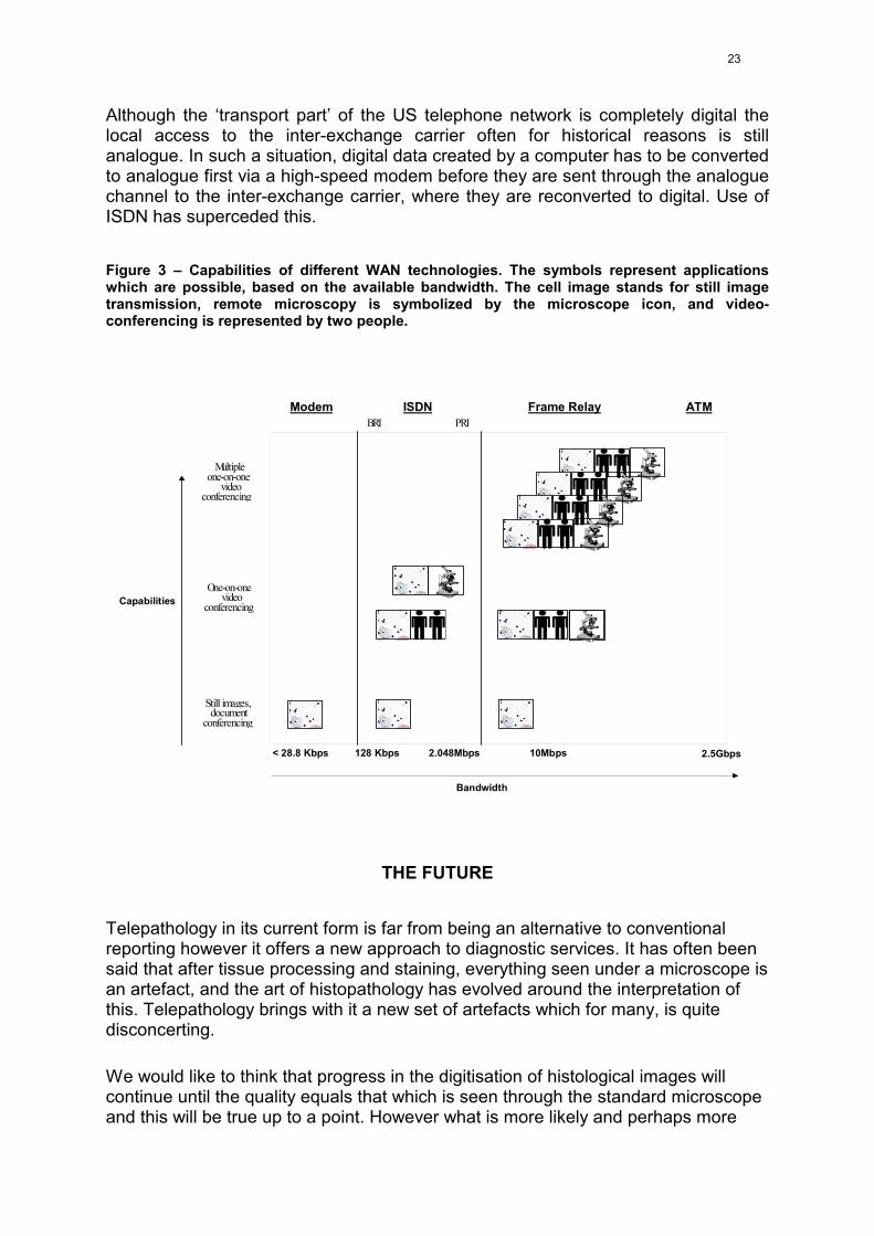
The bandwidth of different LAN technologies ranges from the Low-speed Token Ring (4 Mbps) through to Ethernet (10 Mbps), High-speed Token Ring (16 Mbps) to the high-end Fiber Distributed Data Interface (FDDI) technology with typical bandwidth values around 100 Mbps. Depending on the bandwidth, still image transmission, robotic telepathology, videoconferencing or even multiples of these applications is possible (Figure 3).
Wide Area Network (WAN) services can be broken up into two broad categories: access and transport. Contrary to LANs, the networks normally do not belong to the network user but are operated by inter-exchange carriers such as telephone companies. To create a WAN between two LANs, each must have a connection to their inter-exchange carrier. These access channels can be dedicated digital circuits with a high bandwidth. The advantage of getting a high data throughput has to be weighed versus the disadvantage of excluding all “external” participants who do not have the same dedicated access connections. Use of switched access services eliminates this problem. Instead of dedicated circuits, the services of a local exchange carrier are used to connect the user to whatever inter-exchange carrier he chooses.
The telephone network is probably the best known example of a WAN. In the US it is based on the North American digital hierarchy that defines packages of different bandwidth. DS-0 characterizes the lowest available bandwidth of 64 Kbps. This number is derived from converting the traditional analogue 4 KHz voice channel to digital using pulse code modulation. The next step in the hierarchy is DS-1, also called T-1 (already discussed), combining the bandwidth of 24 DS-0 channels to a total of 1.544 Mbps using time division multiplexing. Further steps up are DS-2, which combines 4 T-1 channels to a total of 6.312 Mbps and DS-3 which has the capacity of 28 T-1 channels (44.736 Mbps).
23
Although the ‘transport part’ of the US telephone network is completely digital the local access to the inter-exchange carrier often for historical reasons is still analogue. In such a situation, digital data created by a computer has to be converted to analogue first via a high-speed modem before they are sent through the analogue channel to the inter-exchange carrier, where they are reconverted to digital. Use of ISDN has superceded this.
Figure 3 – Capabilities of different WAN technologies. The symbols represent applications which are possible, based on the available bandwidth. The cell image stands for still image transmission, remote microscopy is symbolized by the microscope icon, and video-conferencing is represented by two people.
Capabilities
Bandwidth
Modem ISDN Frame RelayBRI PRI
ATM
Multipleone-on-one
videoconferencing
One-on-onevideo
conferencing
Still images,document
conferencing
< 28.8 Kbps 128 Kbps 2.048Mbps 10Mbps 2.5Gbps
THE FUTURE
Telepathology in its current form is far from being an alternative to conventional reporting however it offers a new approach to diagnostic services. It has often been said that after tissue processing and staining, everything seen under a microscope is an artefact, and the art of histopathology has evolved around the interpretation of this. Telepathology brings with it a new set of artefacts which for many, is quite disconcerting.
We would like to think that progress in the digitisation of histological images will continue until the quality equals that which is seen through the standard microscope and this will be true up to a point. However what is more likely and perhaps more

24
feasible, is that pathologists’ skill in interpreting digitised images will improve until it matches their skill with conventional images.
To nearly all pathologists, the idea of working without a microscope is inconceivable and to question that paradigm almost blasphemous. Could a possible future exist however, where not only has all optical equipment moved from the office into the laboratory, the use of glass slides is obsolete? Consider a future where a section need not leave the laboratory if it could be completely digitised then transmitted via local area network to the pathologist in his office. The microscope, would be moved to the laboratory and reduced to its optics, which would be integrated within the digitisation equipment. It is possible that the pathologists’ workstation would comprise an A3-size flatscreen monitor (employing a technology superior to the cathode ray tube), with links to information resources and automated reporting. Pyramidal layering of image resolution will show the image at various magnifications and focussing planes, effectively reproducing the views at each microscope objective magnification.
However, it is how we view technology as much as the technological developments themselves that will determine whether the Pathology specialty takes advantage of and develops existing tools; or disregards them, letting others be the innovators while we are left behind.

25
References
1. Eusebi V, Foschini L, Erde S, Rosai J. Transcontinental consults in surgical pathology via the internet. Hum Pathol 1997;28:13-16.
2. Eide TJ, Nordrum I. Frozen section service via the telenetwork in Northern Norway. Zentralbl Pathol 1992;138:409-412.
3. Ito H, Hironobu A, Kiyomi T, et al. Telepathology is available for transplant-pathology: Experience in Japan. Mod Pathol 1994;7:801-805.
4. Olsson S, Busch C. A national telepathology trial in Sweden: Feasibility and assessment. Arch Anat Cytol Pathol 1995;43:234-241.
5. Weinberg DS, Allaert F-A, Dusserre P, et al. Telepathology diagnosis by means of still digital images: An international validation study. Hum Pathol 1996;27:111-118.
6. Weinstein LJ, Epstein JI, Edlow D, Westra WH. Static image analysis of skin specimens: The application of telepathology to frozen section evaluation. Hum Pathol 1997;28:30-35.
7. Weinstein MH, Epstein JI. Telepathology diagnosis of prostate needle biopsies. Hum Pathol 1997;28:22-29.
8. Wold LE, Weiland LH. Telepathology at the Mayo. Clin Lab Managem Rev 1992;1:174-175.
9. Weinstein RS, Bloom KJ, Rozek LS. Telepathology and the networking of pathology diagnostic services. Arch Pathol Lab Med 1987;111:646-652.
10. Bird K. Telemedicine concept and practice. In: Bashshur E, Armstrong P, Yussel Z, eds. Telemedicine: Explorations in the Use of Telecommunications in Health Care. Springfield, IL: Charles C Thomas, 1975:89-112.
11. Weinstein RS, Bloom KJ, Rozek LS. Telepathology. Long distance diagnosis. Am J Clin Pathol 1989;91 (Suppl 1):S39-S42.
12. Saunders J. Personal communication
13. Riggs RS, Purtillo DT, Connor DH. Medical consultation via telecommunications. JAMA 1974;228:600-602.
14. Colburn D. The next best thing to being there. And now, diagnosis by satellite. Washington Post 1986;August 27:7.
26
15. Nordrum I, Engum B, Rinde E, et al. Remote frozen section service: A telepathology project to northern Norway. Hum Pathol 1991;22:514-518.
16. Nordrum I, Eide TJ. Remote frozen section service in Norway. Arch Anat Cytol Pathol 1995;43:253-256.
17. Kayser K, Oberholzer M, Weiss G, et al. Long distance image transfer: First results of its use in histopathological diagnosis. Acta Pathol Microbiol Immunol Scand 1991;99:808-814.
18. Kayser K, Drlicek M. Visual telecommunication for expert consultation of intraoperative sections. Zentralbl Pathol 1992;138:395-398.
19. Kayser K, Drlicek M, Rahn W. Aids of telepathology in intraoperative histomorphological tumour diagnosis and classification. In Vivo 1993;7:395-398.
20. Swartzmann P. Telemicroscopy: Design considerations for a key tool in telepathology. Zentralbl Pathol 1992;138:183-187.
21. Martin E, Dusserre P, Fages A, et al. Telepathology: A new tool for pathology? Presentation of a French national network. Zentralbl Pathol 1992;138:413-417.
22. Martin E, Dusserre P, Got CL, et al. Telepathology in France: Justifications and developments. Arch Anal Cytol Pathol 1995;43:191-195.
23. Leong FJW-M, McGee JO'D. Telepathology: Current status in tissue and cytopathology. J Pathol 1998;In press.
24. Graham AK, Schwarzmann P, Schmidt Y, McGee JO'D. Telepathology in breast screening quality assurance. J Pathol 1998;in press.
25. Weinstein RS, Bhattacharyya A, Halliday BE, et al. Pathology consultation services via the Arizona-International Telemedicine Network. Arch Anat Cytol Pathol 1995;43:219-226.
26. Halliday BE, Bhattacharyya AK, Graham AR, et al. Diagnostic accuracy of an international static-imaging telepathology consultation service. Hum Pathol 1997;28:17-21.
27. Shimosato Y, Yagi Y, Yamagishi K, et al. Experience and present status of telepathology in the National Cancer Center Hospital, Tokyo. Zentralbl Pathol 1992;138:413-417.
28. http://telepath.med.cornell.edu/pathmaker.html

27
29. http://www.uni-stuttgart.de/UNIuser/ipe/res/ip/histkome.html
30. Swartzmann P. Personal communication.
31. Weinstein RS. Telepathology comes of age in Norway (editorial). Hum Pathol 1991;22:511-513.
32. Dunn BE, Almagro UA, Choi H, et al. Dynamic robotic telepathology: Department of Veterans Affairs feasibility study. Hum Pathol 1997;28:8-12.
33. Wolf G, Petersen D, Dietel M, Petersen I. Telemicroscopy via the internet. Nature 1998;391:613-614.
34. http://www.leica.co.uk
35. http://info.isoc.org:80/guest/zakon/Internet/History/HIT.html
36. Furness PN. The use of digital images in pathology. J Pathol 1997;183:253-263.
37. Weinberg D, Doolittle M. Image management in pathology. Am J Clin Pathol 1996;105(Suppl 1):S54-S59.
38. Bloom KJ, Rozek LS, Weinstein RS. ROC curve analysis of super high resolution video for histopathology. SPIE Proceedings Visual Communications Image Processing II 1987;845:408-412.
39. Weinstein RS, Bloom RJ, Krupinski EA, et al. Human performance studies of the video microscopic component of a dynamic telepathology system. Zentralbl Pathol 1992;138:399-401.
40. Wald G. The receptors of color vision. Science 1964;145:1007-1016.
41. Stiles WS. Foveal threshold sensitivity on fields of different colors. Science 1964;145:1016-1017.
42. Doolittle MH, Doolittle KW, Winkelman Z, Weinberg DS. Color images in telepathology: How many colors do we need? Hum Pathol 1997;28:36-41.
43. http://ftp.nema.org/medical/temp/broch95.htm
44. http://www.cap.org

28
45. (DICOM) Digital Imaging and Communications in Medicine. NEMA PS 3 Supplement 15: Visible light image for endoscopy, microscopy and photography. The National Electrical Manufacturers Association. Rosslyn, VA, 1997.
46. http://idt.net/~dclunie/dicom-status/status.html
47. Leger A, Omachi T, Wallace GK. JPEG still picture compression algorithm. Optical Engineering 1991;30:947-954.
48. http://www.w3.org