tendons and ligaments in
TRANSCRIPT

TENDON AND LIGAMENT INJURIES: PART I 0749-0739/94 $0.00 + .20
FUNCTIONAL ANATOMY OF TENDONS AND LIGAMENTS IN
THE DISTAL LIMBS (MANUS AND PES)
Jean-Marie Denoix, DVM, PhD
Tendons and ligaments of the distal limbs of the horse have a prominent anatomic, functional, and clinical importance. During phylogenesis, equine limbs developed special adaptation for moving at higher speed, including simplification of the distal extremity to a single and strong digit, reduction of the muscle components in the distal limbs and development of accessory ligaments to reinforce the passive and automatic behavior of the limbs. Equine tendons and ligaments became very strong anatomic structures that sustain very high loads and strains, both while standing and moving; therefore, the function of this elastic and complex apparatus during weight bearing therefore is twofold-(l) to provide support to the fetlock and prevent hyperextension of the carpus, and (2) to restore the energy of impact and full weight bearing during propulsion and lift off. This functional importance is doubled by a great clinical interest because tendon and ligament injuries of the distal limbs are common problems and are detrimental to the horse industry. Furthermore, the development of new diagnostic methods, such as ultrasonography, have increased the need for a more detailed knowledge of tendon and ligament anatomy.56
This paper was supported by the Institut National de Recherche Agronomique, Department of Animal Pathology, and by the Service des Haras, des Courses et de l'Equitation, Paris, France.
From the Service d' Anatomie--Clinique Equine, Ecole Nationale Veterinaire d' Aifort, Cecex, France
VETERINARY CLINICS OF NORTH AMERICA: EQUINE PRACTICE
VOLUME 10 • NUMBER 2' AUGUST 1994 273

274 DENOIX
DESCRIPTIVE ANATOMY
The gross anatomy of the tendons and ligaments in the distal forelimb and hindlimb of the horse has been described and illustrated.1, 2, 5, 15, 16, 19, 27, 35, 37, 50, 51 All the anatomic terms used in this article conform with the international nomenclature,22 Tendon and ligament anatomy varies between the manus and the pes (distal thoracic and pelvic limbs), but is quite similar in the digit of the thoracic and pelvic limbs. In each limb (Figs. 1 and 2), three anatomofunctional entities can be described-the superficial digital flexor tendon (SOFT) and its accessory ligament (ALSOFT); the deep digital flexor tendor (OOFT) and its accessory ligament (AL-OOFT); and the suspensory apparatus composed by the third interosseous muscle (TIOM), the proximal scutum, and the distal sesamoidean ligaments (OSLs). The attachment sites of tendons and liga-
Figure 1. Medial aspect of the carpometacarpal area. 1, Radius; 2, third metacarpal bone; 3, second metacarpal bone; 4, extensor carpi radialis; 5, extensor carpi obliquus; 6, antebrachial fascia; 7, flexor retinaculum; 8, distal radial artery; 9, palmar annular ligament; 10, SDFT; 10a, manica flexoria; 11 , DDFT; 12, AL-DDFT; 13, TIOM; and 14, dorsal digital extensor tendon.

FUNCTIONAL ANATOMY OF TENDONS AND LIGAMENTS IN THE DISTAL LIMBS 275
Figure 2. Lateral aspect of the metacarpus. 1, Third metacarpal bone; 2, fourth metacarpal bone; 3, flexor retinaculum; 4, SDFT; 5, DDFT; 6, AL-DDFT; 7, TIOM; 7a, body; 7b, lateral branch; 7c, medial branch; 8, dorsal digital extensor tendon; 9, lateral digital extensor tendon; and 9a, carpal attachment.
ments to bone (entheses) are of particular interesr9, 4o and require further studies in horses because, at least in human medicine, the enthesis is considered to be the weakest point of the bone-tendon or bone-ligament functional unit.41
Superficial Digital Flexor Tendon and its Accessory Ligament
The body of the superficial digital flexor muscle is attached proximally to the medial epicondyle of the humerus, has strong tendinous intersections, and progressively continues to the SOFT in the distal forearm. This tendon also originates from an AL-SOFT inserted on the caudomedial aspect of the radius (Fig. 3), 7 to 11 cm proximal to the antebrachiocarpal joint. This strong fan-shaped fibrous band, previously

276 DENOIX
Figure 3. Palmaromedial aspect of the carpus. The carpal canal is opened. 1, Radius; 2, third metacarpal bone; 3, second metacarpal bone; 4, extensor carpi obliquus; 5, flexor carpi radialis ; 6, tendon sheath of the flexor carpi radialis within the junction between the flexor retinaculum and the antebrachial fascia; 7, flexor carpi ulnaris; 8, SDFT; 9, AL-SDFT; 10, SDF muscle body; 11, DDFT; 12, AL-DDFT; 13, common palmar ligament of the carpus; and 14, TIOM.
called radial, proximal, or superior check ligament,53 courses distocaudally and fuses with the tendon just proximal to the antebrachiocarpal joint (Fig. 3). Medially, it is in contact with the flexor carpi radialis tendon sheath. Knowledge of this relationship is important in view of the surgical technique of desmotomy of the AL-SOFT as a treatment for metacarpophalangeal flexural deformities23 and SOFT injuries.6
The fused rounded-shaped tendon passes distally through the carpal canal (discussed subsequently). In the metacarpus, the SOFT becomes flattened, with a half-moon shape. Its lateral border is sharp and its medial border round. Histologically, the lateral sharp border is more cellular than the broader medial part.62 Behind the palmar (intersesamoidean) ligament of the fetlock, its shape becomes symmetric and the tendon widens greatly. Proximal to the proximal sesamoid bones, a

FUNCTIONAL ANATOMY OF TENDONS AND LIGAMENTS IN THE DISTAL LIMBS 277
fibrous ring called the manica flexoria (Figs. 1 and 4) is attached to its borders and encircles the DDFT, which passes through it. Distal to the proximal sesamoid bones, the SDFT becomes progressively thinner in the sagittal plane and thicker abaxially. At the distal end of the proximal phalanx, the SDFT distal branches separate and become thicker distally on each side of the pastern (Fig. 5). They end between the axial and abaxial palmar ligaments of the proximal interphalangeal joint. All these anatomic elements insert distally on the scutum medium. This thick fibrocartilaginous structure is attached to the proximopalmar aspect of the middle phalanx and is in contact with the palmar aspect of the distal condyles of the proximal phalanx dorsally, and to the DDFT palmarly.
Figure 4. Dorsal aspect of the digital part of the isolated flexor tendons. 1, SDFT; 2, manica flexoria; 3, distal branches of the SDFT; 4, synovial fold between these branches; 5, DDFT; 5a, enlargement at the palmar aspect of the fetlock; 5b, size reduction at the mid-pastern; 5c, fibrocartilaginous pad palmar to the proximal part of the middle phalanx; 5d, widening at the palmar aspect of the distal sesamoid bone; and 6, distal attachment of the digital sheath synovial membrane.

278 DENorx
Figure 5. Palmomedial aspect of the digit. 1, Ungular cartilage (sectioned); 2, digital cushion (partially removed); 3, palmar annular ligament; 4, proximal digital annular ligament; 5, distal digital annular ligament; 6, SOFT distal branch; 7, OOFT; 8, mesotendon of the OOFT seen through the digital sheath wall ; 9, proper palmar digital artery; 10, ramus tori ; and 11, distal branch for the OOFT.
Deep Digital Flexor Tendon and its Accessory ligament
The deep digital flexor muscle consists of three heads. The principal one, the humeral head, contains collagenous intersections and is provided with a strong tendon. This tendon appears about 8 to 10 cm proximal to the antebrachiocarpal joint (see Fig. 3), but muscle bundles remain within it until the level of that joint, where it is joined by the tendons of the two other (ulnar and radial) heads. The broad, conjoined OOFT has a triangular cross-sectional shape in the carpal canal and becomes progressively narrower and round in the proximal metacarpus. At about the middle of the metacarpus, it blends with its AL-OOFT, previously called the subcarpal, distal , or inferior check ligamenP3 (see Figs. 1-3). This strong fibrous band is the direct continuation of the common palmar ligament of the carpus. Proximally, it is broad and has a rectangular shape; it becomes progressively narrower and thicker distally and then molded on the dorsal aspect of the OOFT. At the lateral and medial aspects of the OOFT, a member of fibrous bundles join the SOFT, predisposing to adhesions between the AL-OOFT and the SOFT in pathologic conditions. The corresponding ligament is thinner in the hindlimb.1, 53
The OOFT results from the union of a strong lateral digital flexor tendon and a thin medial digital flexor tendon. The lateral digital flexor tendon incorporates the caudal tibialis tendon and passes over the sustentaculum tali within the plantar tarsal sheath. The medial digital flexor tendon

FUNCTIONAL ANATOMY OF TENDONS AND LIGAMENTS IN THE DISTAL LIMBS 279
passes over the proximal tubercle of the talus, at the medial aspect of the tarsus in its own sheath. These two tendons fuse in the proximal metatarsus. In the distal third of the metacarpus (metatarsus), the DDFT has incorporated all the fibers of its accessory ligament, becomes oval, and passes through the manica flexoria (see Figs. 1,3,4).
Behind the proximal scutum, it becomes considerably wider, elliptic, and fibrocartilaginous. At the level of the proximal half of the proximal phalanx, the DDFT is divided into two round, symmetric parts in which its fibers adopt a spiral disposition. Its dorsopalmar thickness and its lateromedial width decrease distally (see Fig. 4). The narrowest crosssectional area is located in the middle of the proximal phalanx, where the DDFT passes between the two distal branches of the SOFT and becomes superficial (see Fig. 5). Palmar to the distal half of the proximal phalanx, the size of the DDFT increases in dorsopalmar and lateromedial directions. At the level of the proximal part of the middle phalanx, the dorsal half of the tendon becomes a distinct fibrocartilaginous pad proximal to the distal sesamoid bone (see Fig. 4). Distally, the DDFT is molded to the contour of the palmar aspect of the navicular bone. It is broad and forms a terminal fanlike expansion, containing cartilage, occupying the entire width of the space between the lateral and medial palmar processes of the distal phalanx. The DDFT ends on the facies flexoria (Fig. 6) of the distal phalanx, delineated dorsally by the semilunar line and the adjacent surface of the ungular cartilage.
Third Interosseous Muscle and Distal Sesamoidean Ligaments
The nOM is a strong tendinous band, long known for its role as the suspensory ligament (Fig. 7). In the forelimb, the nOM arises proximally from the distal row of the carpal bones and from the adjacent parts of the palmar aspect of the proximal extremity of the metacarpal bonesprincipally the third metacarpal bone. I. 2 The proximal part of the nOM is incompletely separated into two main divisions originating from the second and third carpal bones. Extension of palmar recesses of the carpometacarpal synovial cavity extend 1 to 2 cm distal to the origin of the nOM. In the hindlimb, the main area of attachment is at the proximal and plantaromedial aspect of the third metatarsal bone; there is less extensive attachment to the plantar tubercle of the first, third, and fourth tarsal bones. A distal recess of the tarsometatarsal joint is in contact with the lateral aspect of the proximal nOM in some horses.
The body of the nOM descends between the second and fourth metacarpal bones (see Fig. 7), in the metacarpal groove, and gradually is separated from the palmar aspect of the third metacarpal bone. The level at which it divides into two symmetric or asymmetric diverging branches varies among horses, but appears between mid-metacarpus and the distal fourth of this area (see Fig. 7).
The abaxial aspects of these two strong distal branches are easily

280 DENOIX
Figure 6. Magnetic resonance imaging sagittal section of the digit. 1, Proximal phalanx; 2, middle phalanx; 3, distal phalanx; 4, distal sesamoid bone; 5, dorsal digital extensor tendon; 6, SOFT; 7, OOFT; 7a, fibrocartilaginous pad palmar to the proximal part of the middle phalanx; 7b, distal part with a hypersignal; 8, oblique sesamoidean ligament; 9, straight sesamoidean ligament; 10, collateral sesamoidean ligaments; 11, distal sesamoidean ligament; and 12, digital cushion .
palpated on the live horse. These branches insert on the abaxial surface (interosseous face) of the corresponding proximal sesamoid bone. Each branch detaches a thin extensor branch dorsodistally that obliquely crosses the proximal phalanx axis and joins the dorsal digital extensor tendon (DDET) just above the proximal interphalangeal joint. Each extensor branch blends with the corresponding collateral sesamoidean ligament proximally, and there is a small subtendinous bursa between the extensor branch and the proximolateral aspect of the proximal phalanx.
In the forelimb, the nOM is rectangular, strong, and about 20 to 25 cm long. In the hindlimb, it is thinner, round, and 25 to 30 cm long. Although it is mainly collagenous, it contains variable amounts of striated muscular fibers,! ' 63 especially in the proximal and deep part. No significant difference occurs in the muscle content with age.63 The muscle fibers are organized into two longitudinal bundles within the proximal part and the body of the nOM.93 Standardbreds have 40% more muscle
FUNCTIONAL ANATOMY OF TENDONS AND LIGAMENTS IN THE DISTAL LIMBS 281
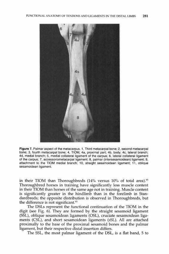
Figure 7. Palmar aspect of the metacarpus. 1, Third metacarpal bone; 2, second metacarpal bone; 3, fourth metacarpal bone; 4, TIOM; 4a, proximal part; 4b, body; 4c, lateral branch; 4d, medial branch; 5, medial collateral ligament of the carpus; 6, lateral collateral ligament of the carpus; 7, accessoriometacarpalligament; 8, palmar (intersesamoidean) ligament; 9, attachment to the TIOM medial branch; 10, straight sesamoidean ligament; 11, oblique sesamoidean ligament.
in their TIOM than Thoroughbreds (14% versus 10% of total area).93 Thoroughbred horses in training have significantly less muscle content in their TIOM than horses of the same age not in training. Muscle content is significantly greater in the hindlimb than in the forelimb in Standardbreds; the opposite distribution is observed in Thoroughbreds, but the difference is not significant.93
The DSLs represent the functional continuation of the TIOM in the digit (see Fig. 6). They are formed by the straight sesamoid ligament (SSL), oblique sesamoidean ligaments (OSL), cruciate sesamoidean ligaments (CSL), and short sesamoidean ligaments (sSL). All are attached proximally to the base of the proximal sesamoid bones and the palmar ligament, but their respective distal insertion differs .
The SSL, the most palmar ligament of the DSL, is a flat band, 5 to

282 DENOIX
9 mm thick, and wider proximally than distally (Fig. 8). Proximally, it has a trapezoidal cross-sectional shape, with its larger concave base facing palmarly. The SSL becomes oval and its thickness increases progressively distally as its size decreases in a lateromedial direction. Distally, it is attached to the scutum medium in the sagittal plane.
The OSLs represent the intermediate or middle ligaments of the DSL (see Fig. 8); on each palmoabaxial aspect of the proximal phalanx lies a strong ligament with a triangular cross-sectional shape and rounded margins. Between these two main parts, thicker abaxially, runs a thin sagittal portion. Sagittally, the thickness of the OSL increases progressively distally because of the convergence of the abaxial ligaments in the sagittal plane. The OSLs insert to the rough triangular area on the palmar aspect of the proximal phalanx and end between the proximal insertion of the two axial palmar ligaments of the proximal interphalangeal (PIP) joint at the distal third of the proximal phalanx. The palmar artery and vein of the proximal phalanx run between the SSL and the OSL in the middle third of the proximal phalanx (see Fig. 8).
The CSLs form the palmar wall of the distopalmar synovial recess of the metacarpophalangeal joint. They consist of two thin layers of fibers crossing each other and ending on the axial aspect of the proximopalmar tuberosity of the proximal phalanx. The sSLs are difficult to differentiate from the deep aspect of the OSL and are best seen by examining the sesamoidophalangeal space dorsally.
Figure 8. Palmar aspect of the digit. 1, Palmar (intersesamoidean) ligament; 2, straight sesamoidean ligament; 3, oblique sesamoidean ligament; 4, scutum medium; 5, SOFT distal branches; 6, palmar aspect of the proximal part of the middle phalanx; 7, OOFT reflected palmad; 8, distal recess of the digital sheath; 9, proper palmar digital artery and vein (the vein has been partially removed); 10, palmar branches of the proximal phalanx; and 11 , palmar branches of the middle phalanx.

FUNCTIONAL ANATOMY OF TEN DONS AND LIGAMENTS IN THE DISTAL LIMBS 283
Carpal Canal
The walls of the carpal canal are composed of several structures: (1) The dorsal wall is formed by the common palmar ligament of the carpus, which represents a thickened palmar part of the fibrous joint capsule; it continues distally as the AL-OOFT. (2) The palmaromedial wall is the flexor retinaculum stretched from the accessory carpal bone and its ligaments laterally to the distal radius, radial carpal, second (and first) carpal bones, proximal second metacarpal bone, and medial collateral ligament of the carpus medullae. Its proximal border continues the caudal antebrachial fascia and its distal border continues the palmar metacarpal fascia. Proximally, the AL-SOFT contributes to form the medial wall of the carpal canal (Fig. 9). (3) The lateral wall of the carpal canal is supported by the accessory carpal bone extended distally by the accessorioquartale and accessoriometacarpeum ligaments.
Carpal hyperextension is limited by the widespread fibrous tissue, including the retinaculum flexorum and its continuation in the distal antebrachial fascia and palmar metacarpal fascia (see Fig. 9). The same eccentric position of the ligaments that attach to the accessory bone also limits carpal hyperextension.
The SOFT and OOFT pass through the carpal canal enveloped in a common carpal synovial sheath (Fig. 10). This sheath extends from 7 to 10 cm proximally to the antebrachiocarpal joint until the proximal third or middle of the metacarpus distally. The proximal recess is wide and firmly covered medially by the strong antebrachial fascia; laterally, it covers the OOF muscle and extends between the ulnaris lateralis and lateral digital extensor muscle (Fig. 11). Synovial distention induces fluid accumulation between these muscles, proximal to the accessory carpal bone. The distal recess extends between the OOFT and the AL-OOFT. Synovial distention induces herniation at the medial or lateral aspect of the OOFT.
Digital Sheath
The palmar wall of the digital sheath is composed of two annular ligaments-the palmar annular ligament and the proximal digital annular ligament (see Fig. 4). The palmar annular ligament inserts on the palmar border of the proximal sesamoid bones. This strong transverse ligament binds down the flexor tendons and converts the proximal scutum (proximal sesamoid bones and palmar ligament) into a real canal. The proximal digital annular ligament is a thinner quadrilateral sheet lying immediately underneath the skin and, in great part, adherent to the SOFT. In sound limbs, it is very difficult to differentiate it from the SOFT itself at the palmar aspect of the digit. It is attached on either side, to the proximopalmar tuberosity of the proximal phalanx by a welldefined band; the distal attachment adheres to the distal branch of the SOFT and inserts with it on the distal part of the proximal phalanx.

284 DENOIX
6------..~
~ff_'H_-----7
r7Tffi7.r-------10
1~------~~~~
1-------2
~~~--------14
~::>------15
--------11
~~---------16
11:1"---------12
1~-------5
Figure 9. Palmaromedial view of the carpal sheath walls and accessory ligaments of the flexor tendons. 1, Radius; 2, accessory carpal bone; 3, third metacarpal bone; 4, second metacarpal bone; 5, fourth metacarpal bone; 6, flexor carpi radialis; 6a, flexor carpi radialis digital sheath; 7, flexor carpi ulnaris; 8, ulnaris lateralis; 9, extensor carpi obliquus; 10, ALSOFT; 11, AL-OOFT; 12, TIOM; 13, antebrachial fascia; 14, flexor retinaculum; 15, palmar metacarpal fascia, and 16, deep palmar metacarpal fascia.

FUNCTIONAL ANATOMY OF TENDONS AND LIGAMENTS IN THE DISTAL LIMBS 285
Figure 10. Palmaromedial aspect of the carpus. The retinaculum flexorum has been sectioned and reflected; therefore, the carpal canal is opened. 1, Radius; 2, third metacarpal bone; 3, second metacarpal bone; 4, extensor carpi obliquus; 5, flexor carpi radialis; 6, flexor carpi ulnaris; 7, SDFT; 8, DDFT; 9, AL-DDFT; 10, TIOM; 11, synovial sheath of the carpal canal; and 12, AL-SDFT.
Figure 11. Transverse cross-section in the distal forearm. 1, Radius; 2, craniolateral wall of the carpal canal; 3, ulnaris latera lis muscle; 4, flexor carpal ulnaris muscle; 5, flexor carpal radialis tendon; 6, proximal part of the flexor retinaculum; 7, superficial digital flexor tendon; 8, accessory ligament of the SDFT; 9, deep digital flexor tendon; 10, carpal sheath; 11, median artery, vein, and nerve; 12, distal radial artery and cephalic vein; and 13, ulnar nerve and collateral ulnar artery and vein.

286 DENOIX
The dorsal wall of the digital sheath is formed by the palmar aspect of the palmar (or intersesamoidean) ligament, DSL, scutum medium (thick fibrocartilaginous structure attached to the proximopalmar aspect of the middle phalanx), and middle phalanx.
The digital synovial sheath begins 4 to 7 cm proximal to the proximal sesamoid bones and extends distally to the half middle phalanx (Fig. 12). It has several recesses20 :
(1) The proximal recess is located proximal to the manica flexoria and palmar annular ligament in the distal fourth of the metacarpus. Between the distal branches of the nOM, it is in contact with digital veins and arteries and with the proximopalmar articular recess of the metacarpophalangeal joint.
(2) The collateral recesses are located on the lateral and medial aspects of the pastern, between the flexor tendons and the DSL. The proximal attachment of the proximal digital annular ligament separates a small proximal recess below the proximal sesamoid bone and a large distal recess behind the proximal half of the proximal phalanx. Behind the proximal sesamoid bones and proximal half of the proximal phalanx, the SDFT is widely adherent sagittally to the proximal digital annular ligament, so no fluid is present in this location.
(3) The distal recess extends between the middle phalanx and the dorsal aspect of the DDFT and is separated by a thin wall from the proximal recess of the podotrochlear bursa and the proxi-
Figure 12. Oorsopalmar contrast radiograph of the digital sheath 10 minutes after injection. 1, Proximal recess; 2, collateral recesses; 3, distal recess of the digital sheath; 4, OOFT radiolucent filling defect; 5, manica flexoria; 6, SOFT branch; 7, proximal attachment of the proximal digital annular ligament; 8, distal border of the proximal digital annular ligament; and 9, OOFT mesotendon filling defect.

FUNCTIONAL ANATOMY OF TENDONS AND LIGAMENTS IN THE DISTAL LIMBS 287
mopalmar recess of the distal interphalangeal (DIP) joint. At the palmar aspect of the DDFT, between the two digital annular ligaments, the distal recess presents a palmar pouch divided sagittally by the DDFT meso tendon. It is this palmar part that deforms the palmar profile of the pastern when the digital sheath is distended. The digital sheath facilitates displacement of the flexor tendons during flexion and extension movements of the metacarpophalangeal and interphalangeal joints. During metacarpophalangeal movements, the two flexor tendons slide together. During interphalangeal movements, the DDFT displacements are much greater than those of the SDFT. Sliding of the flexor tendons during digital joint movements can be imaged with ultrasonography.
The distal digital annular ligament is adherent to the palmar surface of the distal part of the DDFT and binds down the terminal expansion of this tendon. It is a crescentic fibrous sheet attached by a strong band, covering the distal branch of the SDFT, on either side of the middle of the proximal phalanx.
Podotrochlear Bursa
The podotrochlear bursa (navicular bursa) is between the DDFT and the distal sesamoid bone. Its proximal recess extends 1 to 2 cm proximal to the distal sesamoid bone and is applied closely to the proximopalmar recess of the DIP joint and the distal recess of the digital sheath. Its distal recess separates the distal sesamoidean ligament and the terminal part of the DDFT.
Vessels and Nerves
Vessels
Several arteries supply the SDFT.59 A "nutrient artery" coming from the median artery is described associated closely with the AL-SDFT30 and enters the tendon at the transition between muscle and tendon (at the proximal part of the tendon). Near the proximal border of the palmar annular ligament and on either side of the limb, passing within the mesotendon, the proper digital artery gives off an arterial branch (distal metacarpal branch) that carries blood to the SDFT (Fig. 13). Near the distal border of the palmar annular ligament, a proximal digital branch also reaches the SDFT.5, 34 These branches must be preserved during surgical interventions in this area. Complementary supply comes from the muscle body; the peritendon in the metacarpal region; the sagittal adhesion with the proximal digital annular ligament; and the periosteum, close to the distal insertion.59 All these branches contribute to supply an extensive intra tendinous arterial network and two major
288 DENOIX
vessels located at the medial and lateral aspects of the SOFT in the metacarpal area. Within the tendon, longitudinal arterioles course between fiber bundles and anastomize with fine perpendicular branches.3°
The vascularization of the normal DDFT has been studied through gross dissection and angiography within the digital sheath, which revealed three major sources.21 Proximal to the fetlock, near the proximal recess of the digital sheath, the common digital artery gives off a distal metacarpal branch running distally at the palmar aspect of the tendon. Distal to the fetlock, one or two vessels arising from the palmar branches of the proximal phalanx reach the dorsal aspect of the tendon and supply a dorsal sagittal artery. The terminal part of the tendon is supplied by two symmetric small vessels arising on each side of the tendon from the proper digital artery (Fig. 14). Microangiography reveals an extensive intra tendinous vascular network within the DDFT, except in the region palmar to the fetlock. 21 In that area, variable amounts of fibrocartilage can be found in the tendon, and vessels are scarce.
Nerves
The nOM is innervated by the palmar metacarpal nerves arising from the deep ramus of the palmar branch of the ulnar nerve. Because of a distal antebrachial communicating branch from the median nerve to the palmar branch of the ulnar nerve, the nOM is supplied partially by median nerve fibers.
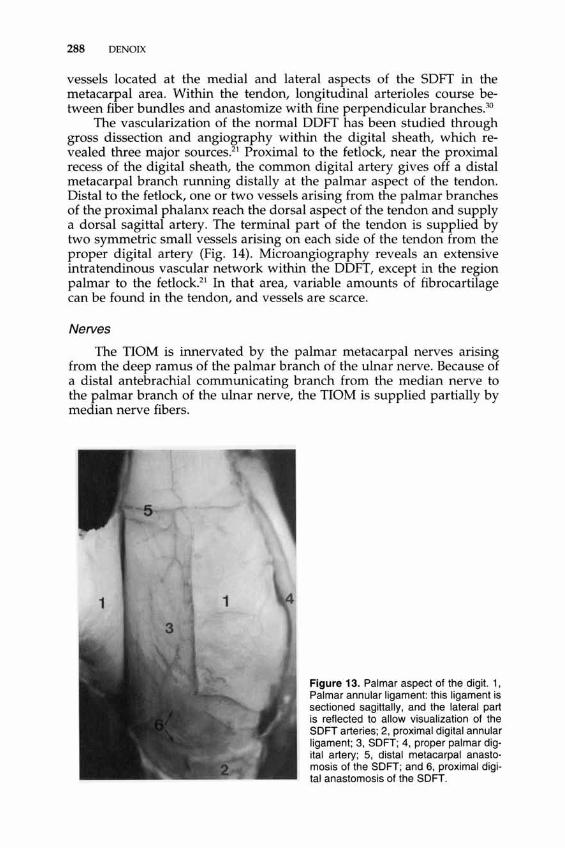
Figure 13. Palmar aspect of the digit. 1, Palmar annular ligament: this ligament is sectioned sagittally, and the lateral part is reflected to allow visualization of the SOFT arteries; 2, proximal digital annular ligament; 3, SOFT; 4, proper palmar digital artery; 5, distal metacarpal anastomosis of the SOFT; and 6, proximal digital anastomosis of the SOFT.

FUNCTIONAL ANATOMY OF TENDONS AND LIGAMENTS IN THE DISTAL LIMBS 289
Figure 14. Palmar aspect of the distal digit. 1, Distal digital annular ligament: this ligament is sectioned sagittally, and the lateral and medial parts are reflected to allow visualization of the DDFT; 2, 8DFT distal branches; 3, DDFT; 4, pal· mar wall of the digital sheath; 5, proper palmar digital artery; 6, sagittal palmar arteries of the DDFT; and 7, distal abax· ial branch of the proper digital artery for the terminal portion of the DDFT.
The metacarpal part of the flexor tendons is supplied by the palmar common digital nerves; the medial one is the direct continuation of the median nerve; the lateral one gets its fibers from the ulnar and median nerves.
A second large metacarpal communicating branch, coming from the medial (second) common palmar digital nerve to the lateral (third) common palmar digital nerve, obliquely crosses the palmar aspect of the SOFT, generally near the middle of the metacarpus.
The digital part of the flexor tendons is supplied by the proper digital nerves.
TOPOGRAPHIC ANATOMY AND CROSS-SECTIONS
Carpal Canal
The flexor tendons are included in the carpal synovial sheath, with the median artery and the medial palmar common digital nerve continuing the median nerve (Figs. 11, 14-17). All these structures are enveloped in the same synovial sheath inner wall, attached medially to the outer wall.
Metacarpus
In the proximal two thirds of the metacarpus, the following palmarodorsal sequence is found-SOFT, OOFT, AL-OOF, and TIOM body

290 DENOIX
Figure 15. Sagittal section of the carpus. 1, Radius; 2, radial carpal bone; 3, intermediate carpal bone; 4, third carpal bone; 5, third metacarpal bone; 6, common dorsal ligament; 7, extensor carpal radialis tendon; 8, common palmar ligament; 9, antebrachioradial ligament; 10, superficial digital flexor tendon; 11, accessory ligament of the SOFT; 12, deep digital flexor tendon; 13, accessory ligament of the OOFT; 14, third interosseous muscle; 15, flexor retinaculum; 16, flexor carpi ulnaris muscle; 17, lateral common digital vein and ulnar nerve; and 18, deep proximal metacarpal vascular anastomosis.
FUNCTIONAL ANATOMY OF TENDONS AND LIGAMENTS IN THE DISTAL LIMBS 291
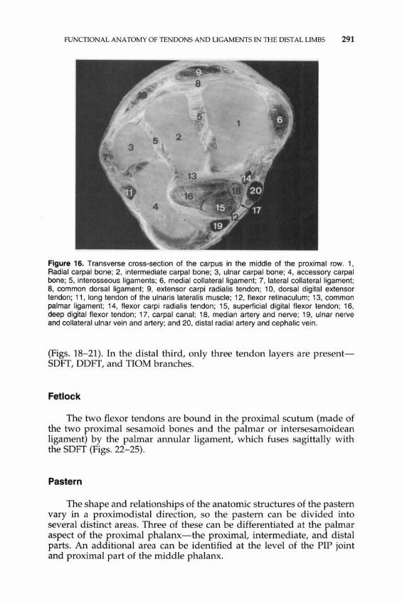
Figure 16. Transverse cross-section of the carpus in the middle of the proximal row. 1, Radial carpal bone; 2, intermediate carpal bone; 3, ulnar carpal bone; 4, accessory carpal bone; 5, interosseous ligaments; 6, medial collateral ligament; 7, lateral collateral ligament; 8, common dorsal ligament; 9, extensor carpi radialis tendon; 10, dorsal digital extensor tendon; 11, long tendon of the ulnaris lateralis muscle; 12, flexor retinaculum; 13, common palmar ligament; 14, flexor carpi radialis tendon; 15, superficial digital flexor tendon; 16, deep digital flexor tendon; 17, carpal canal; 18, median artery and nerve; 19, ulnar nerve and collateral ulnar vein and artery; and 20, distal radial artery and cephalic vein.
(Figs. 18-21). In the distal third, only three tendon layers are presentSDFT, DDFT, and nOM branches.
Fetlock
The two flexor tendons are bound in the proximal scutum (made of the two proximal sesamoid bones and the palmar or intersesamoidean ligament) by the palmar annular ligament, which fuses sagittally with the SDFT (Figs. 22-25).
Pastern
The shape and relationships of the anatomic structures of the pastern vary in a proximo distal direction, so the pastern can be divided into several distinct areas. Three of these can be differentiated at the palmar aspect of the proximal phalanx-the proximal, intermediate, and distal parts. An additional area can be identified at the level of the PIP joint and proximal part of the middle phalanx.

292 DENOIX
Figure 17. Transverse cross-section of the distal row. 1, Second carpal bone; 2, third carpal bone; 3, fourth carpal bone; 4, medial collateral ligament; 5, lateral collateral ligament; 6, palmolateral recess of the carpometacarpal joint; 7, extensor carpal radialis tendon; 8, dorsal digital extensor tendon; 9, common palmar ligament; 10, flexor retinaculum; 11, superficial digital flexor tendon; 12, deep digital flexor tendon; 13, carpal canal; 14, medial palmar common digital artery; 15, medial palmar common digital vein; and 16, lateral palmar common digital vein.
Proximal Part of the Proximal Phalanx
The palmar (metacarpophalangeal) annular ligament lies immediately underneath the skin and is fused with the SOFT (Figs. 26, 27). Beneath this tendon, the OOFT is separated from the OSL by the digital sheath. The different layers of the OSL cover the palmar aspect of the proximal part of the proximal phalanx.
Middle Part of the Proximal Phalanx
The wall of the digital sheath (proximal digital annular ligament) is very thin and adheres to the SOFT (Figs. 26, 28). The SOFT distal branches diverge progressively on each side of the pastern. The OOFT emerges between these branches. On the dorsal aspect of that tendon lies a thin synovial fold of the digital sheath. The SSL runs parallel to the OOFT. The OSLs end at the distal third of the proximal phalanx between the proximal insertion of the two palmar ligaments of the PIP joint. The palmar artery and vein of the proximal phalanx run between the SSLs and the OSLs in the middle third of the proximal phalanx.
Text continued on page 297
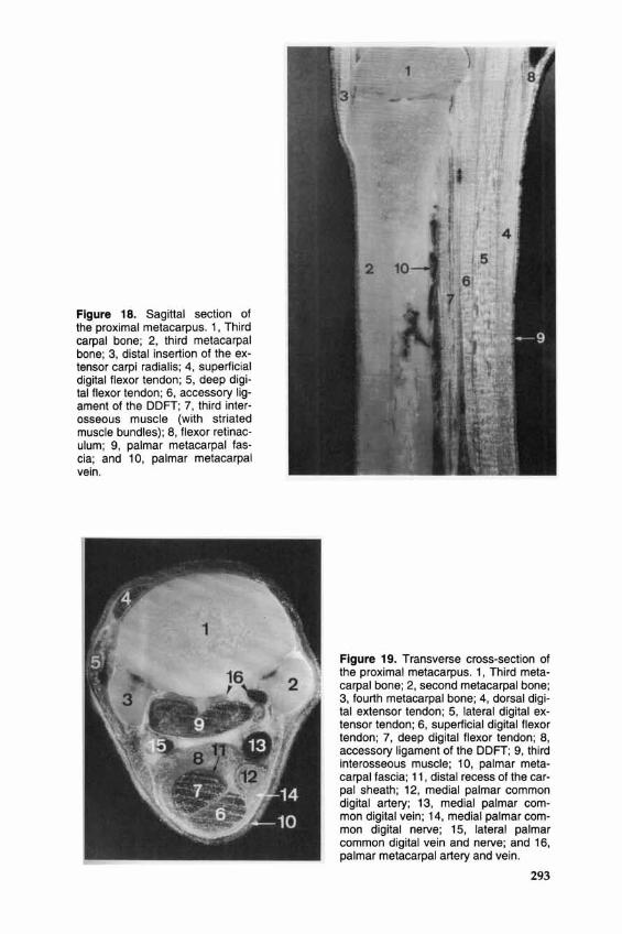
Figure 18. Sagittal section of the proximal metacarpus. 1, Third carpal bone; 2, third metacarpal bone; 3, distal insertion of the extensor carpi radialis ; 4, superficial digital flexor tendon; 5, deep digital flexor tendon; 6, accessory ligament of the DDFT; 7, third interosseous muscle (with striated muscle bundles); 8, flexor retinaculum; 9, palmar metacarpal fascia; and 10, palmar metacarpal vein.
Figure 19. Transverse cross-section of the proximal metacarpus. 1, Third metacarpal bone; 2, second metacarpal bone; 3, fourth metacarpal bone; 4, dorsal digital extensor tendon; 5, lateral digital extensor tendon; 6, superficial digital flexor tendon; 7, deep digital flexor tendon; 8, accessory ligament of the DDFT; 9, third interosseous muscle; 10, palmar metacarpal fascia; 11 , distal recess of the carpal sheath ; 12, medial palmar common digital artery; 13, medial palmar common digital vein; 14, medial palmar common digital nerve; 15, lateral palmar common digital vein and nerve; and 16, palmar metacarpal artery and vein.
293

294 DENOIX
Figure 21. Transverse cross-section in the distal metacarpus. 1, Third metacarpal bone; 2, second metacarpal bone; 3, fourth metacarpal bone; 4, dorsal digital extensor tendon; 5, lateral digital extensor tendon; 6, superficial digital flexor tendon; 7, deep digital flexor tendon; 8, third interosseous muscle medial branch; 9, third interosseous muscle lateral branch; 10, proximal recess of the digital sheath; 11, medial palmar common digital artery; 12, anastomosis of the common digital veins; and 13, palmar metacarpal artery and vein.
Figure 20. Transverse cross-section in the middle of the metacarpus. 1, Third metacarpal bone; 2, second metacarpal bone; 3, fourth metacarpal bone; 4, dorsal digital extensor tendon; 5, lateral digital extensor tendon; 6, superficial digital flexor tendon; 7, deep digital flexor tendon; 8, accessory ligament of the DDFT; 9, third interosseous muscle; 10, palmar metacarpal fascia; 11, medial palmar common digital artery; 12, medial palmar common digital vein; 13, medial palmar common digital nerve; 14, lateral palmar common digital vein and nerve; and 15, palmar metacarpal vein and artery.
FUNCTIONAL ANATOMY OF TENDONS AND LIGAMENTS IN THE DISTAL LIMBS 295
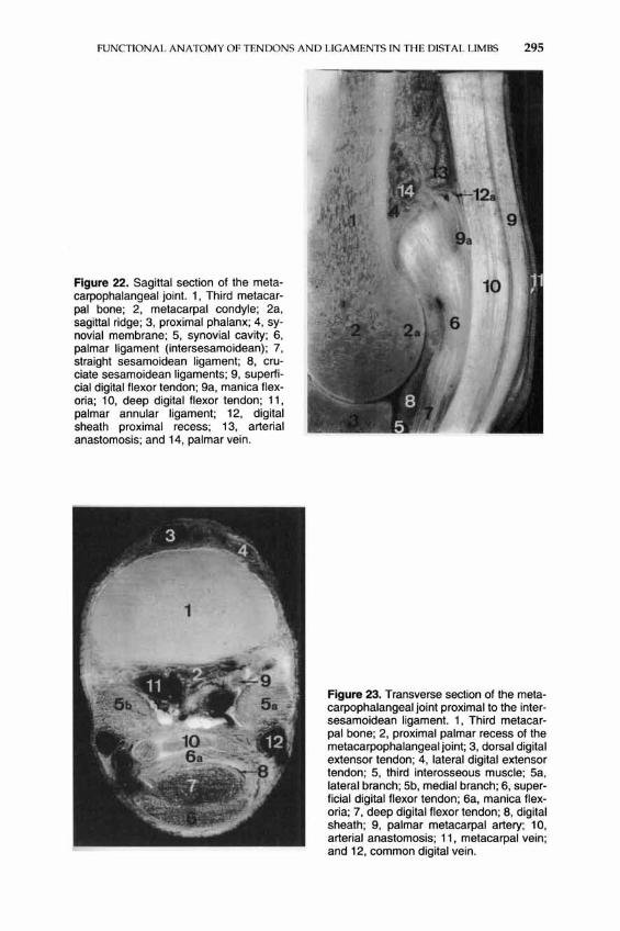
Figure 22. Sagittal section of the metacarpophalangeal joint. 1, Third metacarpal bone; 2, metacarpal condyle; 2a, sagittal ridge; 3, proximal phalanx; 4, synovial membrane; 5, synovial cavity; 6, palmar ligament (intersesamoidean); 7, straight sesamoidean ligament; 8, cruciate sesamoidean ligaments; 9, superficial digital flexor tendon; 9a, manica flexoria; 10, deep digital flexor tendon; 11, palmar annular ligament; 12, digital sheath proximal recess; 13, arterial anastomosis; and 14, palmar vein.
Figure 23. Transverse section of the metacarpophalangeal joint proximal to the intersesamoidean ligament. 1, Third metacarpal bone; 2, proximal palmar recess of the metacarpophalangeal joint; 3, dorsal digital extensor tendon; 4, lateral digital extensor tendon; 5, third interosseous muscle; Sa, lateral branch; 5b, medial branch; 6, superficial digital flexor tendon; 6a, manica flexoria; 7, deep digital flexor tendon; 8, digital sheath; 9, palmar metacarpal artery; 10, arterial anastomosis; 11, metacarpal vein; and 12, common digital vein.

296 DENOIX
Figure 24. Transverse section of the metacarpophalangeal joint proximal to the proximal sesamoid bones. 1, Third metacarpal bone; 2, joint capsule of the metacarpophalangeal joint; 3, proximal palmar recess of the synovial cavity; 4, palmar ligament; 5, medial collateral ligament; 6, dorsal digital extensor tendon; 7, lateral digital extensor tendon; 8a, third interosseous muscle lateral branch; 8b, third interosseous muscle medial branch; 9, Superficial digital flexor tendon; 9a, manica flexoria; 1 D, deep digital flexor tendon; 11 , digital sheath; 12, palmar proper digital artery; 13, palmar proper digital vein ; and 14, palmar proper digital nerve.
Figure 25. Transverse section of the metacarpophalangeal joint near the base of the proximal sesamoid bones. 1, Third metacarpal bone; 1 a, sagittal ridge; 2, proximal sesamoid bone; 3, palmar (intersesamoidean) ligament; 4, collateral sesamoidean ligament; 5, extensor branch of the TIOM; 6, superficial digital flexor tendon; 7, deep digital flexor tendon; 8, palmar annular ligament; 9, digital sheath cavity; 1 D, palmar proper digital artery; 11, palmar proper digital vein; and 12, palmar proper digital nerve.

FUNCTIONAL ANATOMY OF TENDONS AND LIGAMENTS IN THE DISTAL LIMBS 297
Figure 26. Sagittal section of the digit. 1, Third metacarpal bone; 2, proximal phalanx; 3, middle phalanx; 4, distal phalanx; 5, distal sesamoid bone; 6, intersesamoidean ligament; 7, cruciate sesamoidean ligament; 8, oblique sesamoidean ligament; 9, straight sesamoidean ligament; 10, dorsal articular capsule; 11 , scutum medium; 12, collateral sesamoidean ligament; 13, distal sesamoidean ligament; 14, dorsal digital extensor tendon; 15, deep digital flexor tendon; 16, superficial digital flexor tendon; 17, palmar annular ligament; 18, proximal digital annular ligament; 19, distal digital annular ligament; 20, digital sheath; 21, podotrochlear bursa; and 22, digital cushion.
Distal Part of the Proximal Phalanx
At this level, the synovial membrane is separated from the skin by a very thin palmar digital fascia (Figs. 26, 29). The OOFT is adjacent to the distal aspect of the SSL and then, distally, the scutum medium. The distal part of the proximal phalanx is covered axially by the distal part of the SSL. Laterally and medially, this structure is separated from the end of the distal branch of SOFT by the axial palmar ligament of the PIP joint. On each side of the pastern, the distal branch of the SOFT ends between the axial and abaxial palmar ligaments of the PIP joint. All these anatomic elements insert distally on the scutum medium.
298 DENOIX
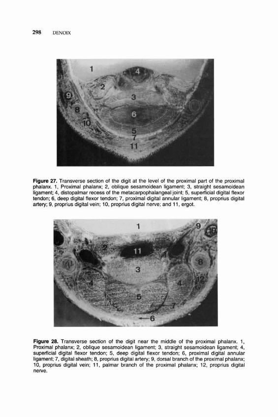
Figure 27. Transverse section of the digit at the level of the proximal part of the proximal phalanx. 1, Proximal phalanx; 2, oblique sesamoidean ligament; 3, straight sesamoidean ligament; 4, distopalmar recess of the metacarpophalangeal joint; 5, superficial digital flexor tendon; 6, deep digital flexor tendon; 7, proximal digital annular ligament; 8, proprius digital artery; 9, proprius digital vein; 10, proprius digital nerve; and 11, ergot.
Figure 28. Transverse section of the digit near the middle of the proximal phalanx. 1, Proximal phalanx; 2, oblique sesamoidean ligament; 3, straight sesamoidean ligament; 4, superficial digital flexor tendon; 5, deep digital flexor tendon; 6, proximal digital annular ligament; 7, digital sheath; 8, proprius digital artery; 9, dorsal branch of the proximal phalanx; 10, proprius digital vein; 11, palmar branch of the proximal phalanx; 12, proprius digital nerve.

FUNCTIONAL ANATOMY OF TENDONS AND LIGAMENTS IN THE DISTAL LIMBS 299
Figure 29. Transverse section of the digit at the level of the distal part of the proximal phalanx. 1, Proximal phalanx; 2, palmar recess of the proximal interphalangeal joint (PIP); 3, scutum medium; 4, distal attachment of the SOFT on the proximal phalanx; 5, deep digital flexor tendon; 6, palmar wall of the digital sheath; 7, proprius digital artery; 8, proprius digital vein; and 9, proprius digital nerve.
Figure 30. Frontal section of the digit passing through the palmar aspect of the proximal phalanx, middle phalanx, and distal sesamoid bone. 1, Proximal phalanx (proximopalmar tubercle); 2, middle phalanx (tuberositas flexoria); 3, distal phalanx (palmar process); 4, distal sesamoid bone (palmar cortex); 5, palmar (intersesamoidean) ligament; 6, scutum medium; 7, palmar recess of the DIP joint; 8, superficial digital flexor tendon distal branch; 9, deep digital flexor tendon; 10, digital sheath cavity; 11, podotrochlear bursa; 12, proprius digital artery; 13, digital cushion; and 14, ungular cartilage.

300 DENOIX
Proximal Interphalangeal Joint and Proximal Part of the Middle Phalanx
The palmar aspect of the distal condyles of the proximal phalanx is in contact with the scutum medium (Figs. 26, 30, 31). At the level of the proximal part of the middle phalanx, the DDFT is adjacent to the fibrocartilaginous surface of the tuberositas flexoria. It is separated from the skin by a thin distal annular ligament. Between these two structures, the synovial membrane is thicker sagittally and adheres to the palmar aspect of the DDFT.
Foot
The palmar surface of the distal digital annular ligament is widely covered by the digital cushion (Figs. 31-33). Dorsally, this ligament adheres to the DDFT, which is molded over the facies flexoria of the distal sesamoid bone.
Pes
The anatomy in the metatarsus is different from that in the metacarpus (Fig. 34). In the proximal metatarsus, the DDFT components are on the dorsomedial aspect of the SDFT, the lateral border of which is thicker. The nOM has a rounder shape than in the forelimb and is in contact with a large plantar metatarsal vein.
MECHANICAL PROPERTIES AND FUNCTIONAL ANATOMY
General Considerations
Tendons and ligaments have several important roles during locomotion and in the standing position: They act as force transmitters and joint coaptaters, with an especial involvement in fetlock suspension. As elastic structures, they have a major role as impact absorbers and have the ability to store and release energy, reducing the energy cost of locomotion, especially at high speeds.
In the hindlimbs of adult horses of various breeds, the cross-sectional area (CSA) of the SDTF, DDFT, and nOM varies considerably in a proximodistal direction and among horses.43 There is an inverse relationship between the cross-sectional area and the collagen content as well as tendon fibers, so the CSA is not representative of the local strength of equine tendons.43 Another study44 demonstrated that loaded equine hindlimb tendons are strained homogeneously. As a result, the modulus of elasticity is inversely proportional to the corresponding CSA and proportional to the collagen content. The increase in CSA by non-
FUNCTIONAL ANATOMY OF TENDONS AND LIGAMENTS IN THE DISTAL LIMBS 301
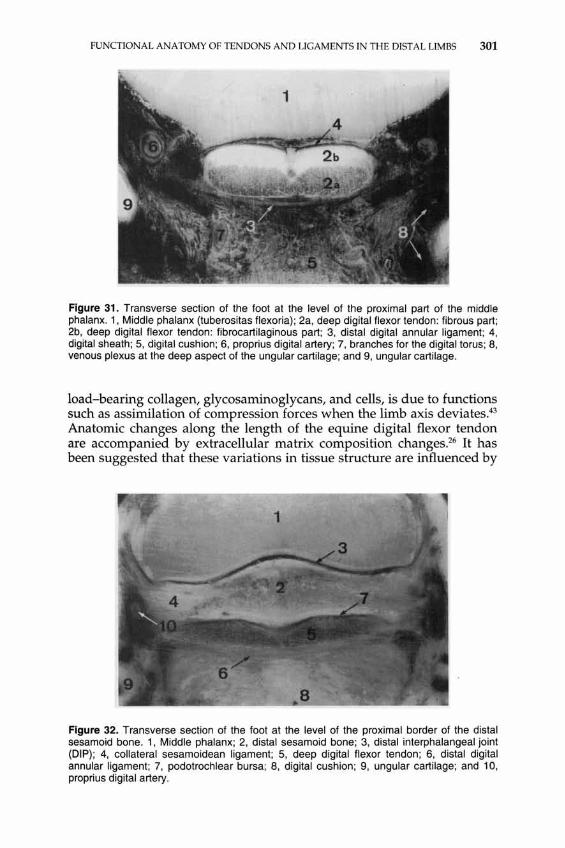
Figure 31. Transverse section of the foot at the level of the proximal part of the middle phalanx. 1, Middle phalanx (tuberositas flexoria); 2a, deep digital flexor tendon: fibrous part; 2b, deep digital flexor tendon: fibrocartilaginous part; 3, distal digital annular ligament; 4, digital sheath; 5, digital cushion; 6, proprius digital artery; 7, branches for the digital torus; 8, venous plexus at the deep aspect of the ungular cartilage; and 9, ungular cartilage .
load-bearing collagen, glycosaminoglycans, and cells, is due to functions such as assimilation of compression forces when the limb axis deviates.43
Anatomic changes along the length of the equine digital flexor tendon are accompanied by extracellular matrix composition changes.26 It has been suggested that these variations in tissue structure are influenced by
Figure 32. Transverse section of the foot at the level of the proximal border of the distal sesamoid bone. 1, Middle phalanx; 2, distal sesamoid bone; 3, distal interphalangeal joint (DIP); 4, collateral sesamoidean ligament; 5, deep digital flexor tendon; 6, distal digital annular ligament; 7, podotrochlear bursa; 8, digital cushion; 9, ungular cartilage; and 10, proprius digital artery.
302 DENOIX
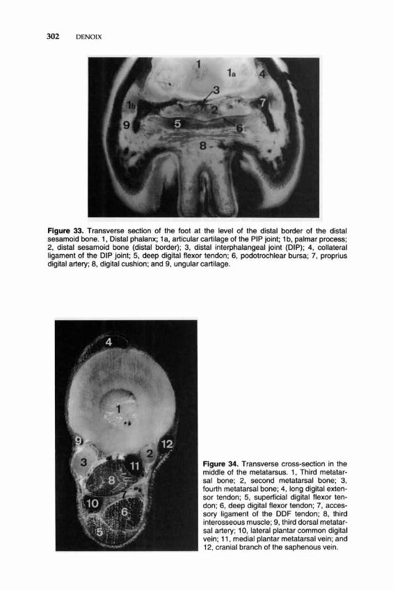
Figure 33. Transverse section of the foot at the level of the distal border of the distal sesamoid bone. 1, Distal phalanx; 1 a, articular cartilage of the PIP joint; 1 b, palmar process; 2, distal sesamoid bone (distal border); 3, distal interphalangeal joint (DIP); 4, collateral ligament of the DIP joint; 5, deep digital flexor tendon; 6, podotrochlear bursa; 7, proprius digital artery; 8, digital cushion; and 9, ungular cartilage.
Figure 34. Transverse cross·section in the middle of the metatarsus. 1, Third metatarsal bone; 2, second metatarsal bone; 3, fourth metatarsal bone; 4, long digital extensor tendon; 5, superficial digital flexor tendon; 6, deep digital flexor tendon ; 7, accessory ligament of the DDF tendon; 8, third interosseous muscle; 9, third dorsal metatarsal artery; 10, lateral plantar common digital vein; 11, medial plantar metatarsal vein; and 12, cranial branch of the saphenous vein.

FUNCTIONAL ANATOMY OF TENDONS AND LIGAMENTS IN THE DISTAL LIMBS 303
functional requirements of areas sustaining high tensional forces compared with pressure-supporting regions.26
Superficial Digital Flexor Tendon and its Accessory Ligament
Mechanical Properlies
The SOFT possesses a moderate modulus of elasticity (1096.5 MPa; mega Pascal: N mm - 2) and a relatively high strength to rupture (average, 1243 daN). The AL-SOFT has moderate strength to rupture (average, 905 daN)9,1O (Crevier, unpublished data, 1993), In the pony hindlimb, the modulus of elasticity of the SOFT ranges from 1000 MPa to 1282 MPa and the tendon ruptured at 12.3% strain,44
Function
Weight Bearing and Stance Phase. The SOFT actions are correlated to active contraction of the muscle belly and to the passive tension of its accessory ligament. Tension of the AL-SOFT is induced by extension of the metacarpophalangeal joint when weight is put on the limb. Because of the eccentric (palmar) position of the SOFT over the proximal scutum, metacarpophalangeal extension induces a wide distal sliding of the tendon (Fig. 35), limited proximally by the AL-SOFT. The AL-SOFT prevents overstretching of the SOF muscle belly by carrying the load during metacarpophalangeal overextension,53 especially at the end of a race, when fatigue weakens the muscle belly. Tension of the whole component, between the distal radial insertion and the proximal part of the middle phalanx insertion, contributes to limit both metacarpophalangeal and carpal extension. The role of the AL-SOFT has been investigated in equine cadaver forelimbs under static compression.54 Results showed that this ligament contributes to the support of the metacarpophalangeal joint under load. SOFT strains increased significantly after desmotomy of the AL-SOFT, which has been attributed to the change in the metacarpophalangeal joint angle, increasing the moment arm of the SOFT about this joint.54 Because of its proximal insertion on the medial humeral epicondyle, the muscle belly and its fibrous components also limit flexion of the elbow. Because of the location of the distal insertion on the palmar aspect of the PIP joint, tension of the SOFT during weight bearing is a limitation to PIP flexion. Ouring full weight bearing, high tension within the SOFT (and OOFT) is responsible for stabilization of the PIP joint. During propulsion, the PIP joint comes into extension and the distal condyles of the proximal phalanx slide palmarly on the proximal articular surface of the middle phalanx. Extension and palmar displacement are limited by the scutum medium, the palmar ligaments, and the SOFT.
The two distal branches of the SOFT inserted on the proximal sides of the middle phalanx are essential for the stabilization of the PIP joint

304 DENOIX
Figure 35. Isolated specimen placed on a hydraulic press under 1000 OaN used to evaluate the elongation and displacement of the tendons in the distal forelimb under experimental conditions that reproduce the stance phase. When pressure was applied on the limb to obtain a horizontal orientation of the foot , markers were drilled into the tendons at the level of the horizontal line drawn on the proximal metacarpus; distal markers were drilled 10 cm below in each tendon. Note that in this limb orientation when high loads are applied, the distal displacement of the SOFT is pronounced, and the elongation of the TIOM is marked. The OOFT undergoes the least deformation and distal displacement. Profound alterations are observed when the limb orientation mimics propulsion. Because of the distal displace· ment of the SOFT, the AL·SOFT is taut.
in the frontal plane (lateromedial displacements) and the transverse plane (rotation), especially because of their direct contact with the distal condyles of the proximal phalanxY
Swing Phase. At the beginning of the swing phase, tension and elasticity of SOF apparatus contribute passively to initiate flexion of the carpal, metacarpophalangeal, and PIP joints. Careful examination during gaits and slow-motion cinematography demonstrates that the SOFT suddenly becomes totally relaxed and undergoes vibrations just after the take-off. Then, flexion of the joints is increased by active muscle belly contraction, which induces total relaxation of the AL-SOFT. These move-

FUNCTIONAL ANATOMY OF TENDONS AND LIGAMENTS IN THE DISTAL LIMBS 305
ments are accompanied by a proximal sliding of the SOFT within the carpal canal. The respective displacements of the SOFT and palmar annular ligament during the stance phase require further investigations.
Bone Stresses. Extensor tendon tension has been correlated with bone strain and gait in the walking horse.4 When high compression is placed on the radius, the metacarpus and proximal phalanx undergo high compression on their dorsal cortices and tension on their palmar cortices.12 In those circumstances, the flexor tendons contribute prominently to limiting the palmar bending of the bones (Fig. 36), balancing the stresses on the different aspects of the third metacarpal bone and proximal phalanx, and preventing fractures.
Deep Digital Flexor Tendon and its Accessory Ligament
Mechanical Properties
The DDFT possesses a high modulus of elasticity-1585 MPa9, 10
(Crevier, unpublished data, 1993)-and a considerable strength to rupture (average, 1700 daN). The AL-DDFT has a low modulus of elasticity (490 MPa) and a moderate strength to rupture (average, 871 daN). In the pony hindlimb, the modulus of elasticity of the DDFT ranges from 738 to 1398 MPa and the tendon ruptures at 10% strain.42
Function
Weight-Bearing and Stance Phase. Tension of the AL-DDFT facilitates carpal extension when load is applied on a limb12,32 (Fig. 37). For high loads, the proximal DDFT limits the carpal extension, and the DDF apparatus limits metacarpophalangeal extension. Because of its proximal insertion on the medial humeral epicondyle and the fibrous content of its belly, the deep digital flexor muscle contributes to limit elbow flexion.
The DDFT undergoes mixed stresses in the fetlock area. It supports tension between its proximal and distal insertions and supports compression from the proximal scutum, widely applied by the palmar (in tersesamoidean) ligament on its dorsal aspect. This zone with mixed stresses has a fibrocartilaginous architecture with a lot of chondrocytes between the collagenous fibers. In the palmar aspect of the middle phalanx, the DDFT also has a dorsal fibrocartilaginous pad that supports pressure of the tuberositas flexoria of the middle phalanx. During DIP flexion (first parts of the stance phase) the DDFT slides proximally on this bone surface. During DIP extension (propulsion), because of the presence of the distal scutum, the DDFT undergoes a relative distal sliding on the tuberositas flexoria of the middle phalanx. These displacements take place within the distal recess of the digital sheath.
In the digit, the DDFT facilitates flexion of the PIP joint (in antagonism to the SOFT) during weight bearing. Its tension induces axial compression of the articular surfaces within the PIP and DIP joints, and
306 DENOIX
1 ....... -----1
n-+-----2
3---------------------r~~
4------------~~
1a
6-----;f1.
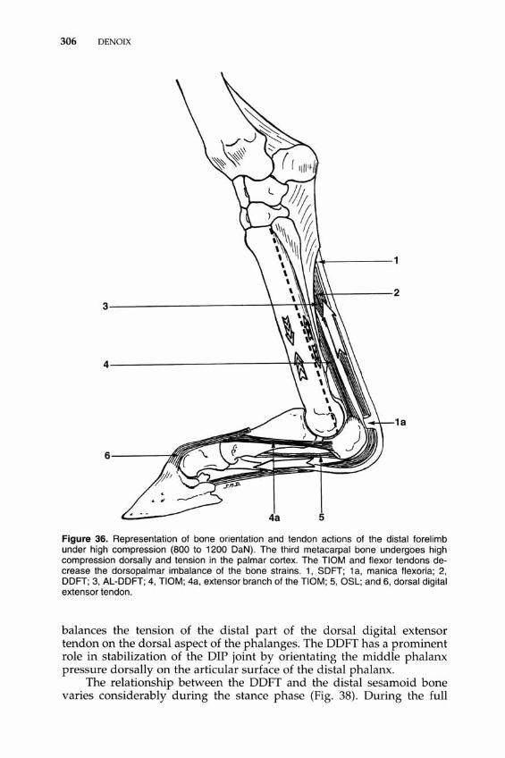
Figure 36. Representation of bone orientation and tendon actions of the distal forelimb under high compression (800 to 1200 DaN). The third metacarpal bone undergoes high compression dorsally and tension in the palmar cortex. The TIOM and flexor tendons decrease the dorsopalmar imbalance of the bone strains. 1, SOFT; 1 a, manica flexoria; 2, OOFT; 3, AL-OOFT; 4, TIOM; 4a, extensor branch of the TIOM; 5, OSL; and 6, dorsal digital extensor tendon.
balances the tension of the distal part of the dorsal digital extensor tendon on the dorsal aspect of the phalanges. The DDFT has a prominent role in stabilization of the DIP joint by orientating the middle phalanx pressure dorsally on the articular surface of the distal phalanx.
The relationship between the DDFT and the distal sesamoid bone varies considerably during the stance phase (Fig. 38). During the full

FUNCTIONAL ANATOMY OF TENDONS AND LIGAMENTS IN THE DISTAL LIMBS 307
Figure 37. Isolated specimen placed on a hydraulic press under 1000 DaN to evaluate the functional behavior of tendons and joints of the distal forelimb in experimental conditions that reproduce the stance phase. Note the spontaneous extension of the carpus when pressure is applied on the radius.
weight-bearing position, the DDFT is in close contact only with the distal border of the distal sesamoid bone. During propulsion, the DDFT bends over the distal scutum and comes in full contact with the distal sesamoid boneY Abnormal changes in angulation at the tendon insertion may result in uneven local stress distribution, which could precipitate failure.18 The role of the distal scutum is to prevent modifications of fiber orientation at the distal insertion during flexion and extension movements of the DIP joint46• 47 (see Fig. 38). Moreover, the distal scutum acts as a lever to facilitate foot rotation and heel take-off at the end of the stance phase.
During the last part of the stance phase, the active contraction of the muscle bellies and the elasticity of the tendon as well as the accessory ligament have a prominent role in inducing elevation of the fetlock and propulsion. The DDFT therefore is the most effective agent of DIP joint extension. It is during this part of the stance phase that the AL-DDFT is

(H o 00
[ == J Extension during propulsion
.. Flexion during weight bearing
/ ......... ;'
I ..... __ ,
1\ I \
I \ / \
I \ '- -, I , ....... i I i I
I I I I
I 'I
Figure 38. Role of the distal scutum to prevent modifications of fibers orientation at the distal insertion of the DDFT on the distal phalanx during flexion and extension movements of the distal interphalangeal joint. 1, DDFT.
FUNCTIONAL ANATOMY OF TENDONS AND LIGAMENTS IN THE mST AL LIMBS 309
stretched maximally24 and has the most efficient contribution to stabilization of the DIP joint. During propulsion, the increasing angulation of the DDFT on the distal sesamoid bone makes the pressure progressively higher on the facies flexoria of this bone. This was supported by a procedure for computing the internal forces of the digit.3,52 During propulsion, the proximal border of the sesamoid bone rotates dorsally. PIP joint extension induces contact between the tuberositas flexoria of the middle phalanx and the dorsal digital fibrocartilaginous pad of the DDFT.
Swing Phase. Immediately after take-off, sudden relaxation of the deep digital flexor apparatus induces vibrations during the swing phase. At the beginning of the swing phase, tension of the deep digital flexor apparatus contributes passively to initiate flexion of the interphalangeal, metacarpal and carpal joints. Then, the flexion of these joints is increased by active muscle contraction that induces total relaxation of the ALDDFT and proximal sliding of the tendon within the digital sheath and carpal canal.
Suspensory Apparatus (TIOM, Proximal Scutum, and DSL)
Mechanical Properties
The body of the nOM possesses a relatively high modulus of elasticity (1100 MPa) and considerable strength to rupture (average, 1715 daN). Strain at rupture reaches 10% to 12%9, 10 (Crevier, unpublished data, 1993). In the pony hindlimb, the modulus of elasticity of the nOM ranged from 576 MPa to 669 MPa and the tendon ruptured at 11% strain.44 On isolated anatomic specimens such as the one presented in Figure 37, the average maximum force applied on the limb to obtain failure of the suspensory apparatus was 1220 kg (between 918 and 1673 kg) and was significantly higher in trained horses than in resting horses?
Function
The main function of the nOM is to prevent excessive extension (dorsoflexion) of the metacarpophalangeal joint-i.e., to support the fetlock when weight is put on the limb during the standing position or stance phase (Fig. 39). In the weight-bearing position, tension within the nOM and flexor tendons regulates the amount and location of the stresses applied to the different aspects of the third metacarpal bone (see Fig. 36). For a moderate amount of load, tension within the tendinous structures on the palmar aspect of the metacarpus induces tension on the dorsal aspect of the third metacarpal bone and compression within the palmar cortex.12 With high compression on the radius, the metacarpus undergoes high compression on its dorsal cortex and tension on its palmar cortex. In those circumstances, the nOM and flexor tendons have
310 DENOIX
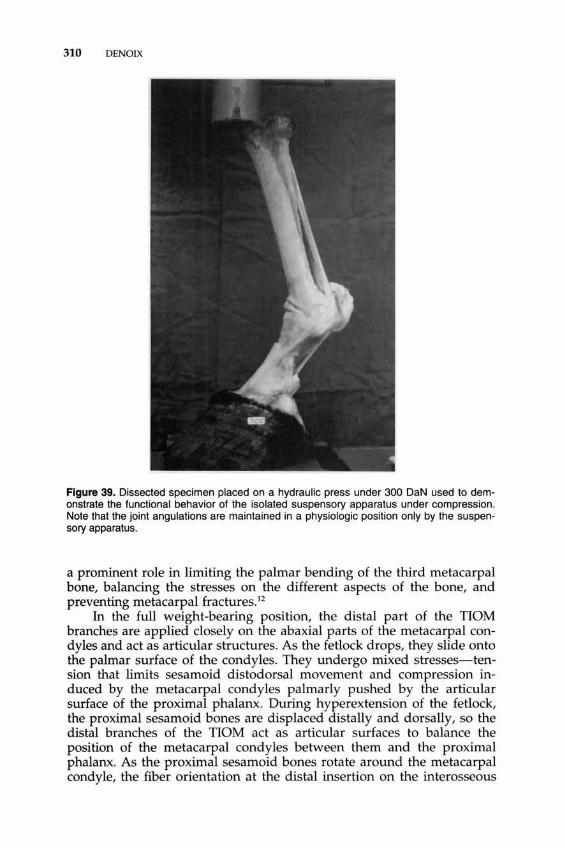
Figure 39. Dissected specimen placed on a hydraulic press under 300 DaN used to demonstrate the functional behavior of the isolated suspensory apparatus under compression. Note that the joint angulations are maintained in a physiologic position only by the suspensory apparatus.
a prominent role in limiting the palmar bending of the third metacarpal bone, balancing the stresses on the different aspects of the bone, and preventing metacarpal fracturesP
In the full weight-bearing position, the distal part of the nOM branches are applied closely on the abaxial parts of the metacarpal condyles and act as articular structures. As the fetlock drops, they slide onto the palmar surface of the condyles. They undergo mixed stresses-tension that limits sesamoid distodorsal movement and compression induced by the metacarpal condyles palmarly pushed by the articular surface of the proximal phalanx. During hyperextension of the fetlock, the proximal sesamoid bones are displaced distally and dorsally, so the distal branches of the nOM act as articular surfaces to balance the position of the metacarpal condyles between them and the proximal phalanx. As the proximal sesamoid bones rotate around the metacarpal condyle, the fiber orientation at the distal insertion on the interosseous

FUNCTIONAL ANATOMY OF TENDONS AND LIGAMENTS IN THE DISTAL LIMBS 311
surface is modified, and the body as well as branches of the nOM become applied closely to the palmar aspect of the metacarpus, compressing the metacarpal veins. In the weight-bearing position, the extensor branches of the nOM are stretched. Their proximal insertion is made taut by the tension of the nOM branches and their distal insertion is pulled by the dorsal digital extensor tendon, which is taut because of interphalangeal flexion. Elasticity of the nOM contributes to fetlock elevation during propulsion.
During asymmetric load bearing (due to limb obliquity or foot imbalance), the contribution of the TIOM distal branches and collateral ligaments to metacarpophalangeal joint stability is higher on the side opposite the compression (Fig. 40). This asymmetric pressure, combined with the shape and orientation of the articular surfaces, induces metacarpophalangeal rotation on the side opposite to the compressionY
...,...,f----3
Figure 40. Role of the suspensory apparatus in limiting the abaxial movements of the fetlock during asymmetrical weight bearing. The pressure of the proximal sesamoid bones limits the transverse sliding of the metacarpophalangeal articular surfaces. The abaxial displacement and rotation of the digit take place in opposite directions. 1, TIOM distal branch; 2, oblique sesamoidean ligament; and 3, short sesamoidean ligament.

312 DENOIX
During the swing phase, metacarpophalangeal joint flexion suddenly relaxes nOM and, because of the proximal displacement of the proximal scutum, the branches bend abaxially and separate proximally. At the beginning of the swing phase and stance phase, sudden modifications of tension and orientation of fibers induce vibrations within the nOM.
Because they join the wide distal part of the DDET, the extensor branches of the nOM are tightened by every interphalangeal flexion. During the stance phase, they limit the palmar flexion (and the dorsal subluxation) of the interphalangeal joints because of traction induced distally by the DDET and proximally by the nOM itself. When the fetlock drops, the dorsal angle between the distal branch and the extensor branch reduces. Desmopathy with elongation of the superficial part of the distal branches may induce dorsal subluxation of the PIP joint because of relaxation of the proximal attachment of the extensor branches of the nOM. These branches create tension on the dorsolateral and dorsomedial aspects of the digit, and contribute, with the collateralligaments, to limit movements of rotation and abaxial movements. This provides interphalangeal stability. During the second part of the swing phase (protraction), when the fetlock has become extended, the extensor branches contribute passively to interphalangeal extension.
Distal Sesamoidean Ligaments
The DSLs represent the digital component of the suspensory apparatus, the proximal sesamoid bones and palmar ligament being intercalated between the nOM and the DSL. Because of its insertions, the SSL participates in the sagittal stabilization of the metacarpophalangeal and PIP joint and has little action on rotation and abaxial movements. Conversely, the OSLs have a prominent role in the limitation of rotation and abaxial movements of the metacarpophalangeal joint when weight is put on the limb (see Fig. 40). In the weight-bearing position, the OSLs induce traction on the palmar cortex of the proximal phalanx (see Fig. 36) and compression on the dorsal aspect. l1 , 12 The SSL and the flexor tendons limit sagittal deformation of the proximal phalanx.
ROLES OF TENDONS AND LIGAMENTS DURING THE STANDING POSITION
The horse can remain in a standing position for a long time because most of the weight is supported by tendons and accessory ligaments, articular ligaments, fibrous intersections within the muscle bellies, and fascias.
Elbow
The bellies of the caudal antebrachial muscles contain a large quantity of tendinous tissue. Their proximal insertions are on the humeral

FUNCTIONAL ANATOMY OF TENDONS AND LIGAMENTS IN THE DISTAL LIMBS 313
epicondyles, caudal to the axis of rotation of the humeroantebrachial joint.50 In the standing position, suspension of the fetlock induces distal traction on the tendons. The continuing tendinous intersections in the digital flexor muscles are tense and therefore prevent flexion of the elbow joint. The triceps brachii and the coupled action of the ascendant pectoral and brachiocephalicus muscles also assist in preventing flexion of the humeroantebrachial joint.
Carpus
In a standing position, because of the shape of the distal articular surface of the radius with condylar facets located palmad, the carpus is spontaneously in extension without any muscle action. This is demonstrated on isolated limbs (see Fig. 37), with sections of all the extensor and flexor muscles placed under compressionY Extension also is partially due to the palmarodistal traction of the AL-DDF tendon and TIOM.J2, 32 Under clinical conditions, elongation of the AL-DDF tendon or TIOM often is accompanied by a lack of carpal extension and the knee deviates forward (the horse becomes buck kneed). Overextension is prevented by the palmar wall of the carpal canal (see Fig. 9) and accessory carpal bone ligaments, as well as the strong common palmar ligament caudally, and by close packing of the bearing facets of the carpal bones dorsally. Overextension also is limited by the tension of the flexor tendons and AL-SDFT. The extensor carpal radialis inserts on a large dorsomedial tubercle on the proximal metacarpus and has a passive extension action because of its relationship with the biceps brachii via the lacertus fibrosis, The biceps brachii is tensed in the standing horse, because of its action to prevent collapse of the shoulder joint.
Fetlock
The distal parts of the flexor tendons passively limit overextension of the metacarpophalangeal angle during weight bearing because of their proximal attachments to bone by their accessory ligaments. The suspensory apparatus, comprising the TIOM, proximal sesamoid bones, palmar ligament, and DSL, has a prominent role in the prevention of overextension or collapse of this joint. All these elements are tensed under load. Under pathologic conditions, section, rupture, or elongation of one of these components induces a lack of suspension of the fetlock Overextension of the fetlock joint also is limited by the deep palmar part of the collateral ligaments of the joint. High dorsal traction induced by the distal and extensor branches of the TIOM on the abaxial (interosseous) face of the distal sesamoid bones (see Fig. 36) is balanced by two structures-the strong palmar (intersesamoidean) ligament and the palmar annular ligament. This must be considered in the pathogenesis of palmar annular ligament desmopathy.

314 DENOIX
Modification of the foot or limb position induces asymmetric strain within the distal branches of the nOM and OSL. Stresses in elongation are higher on the opposite side of the compression, limit displacements in the frontal plane, and contribute to rotation (see Fig. 40). The proximal scutum is displaced toward the side of compression, and the contact between the sagittal ridge of the metacarpal condyle and the corresponding proximal sesamoid bone prevents the articular surfaces from sliding transversely (see Fig. 40).
PIP Joint
At rest or during the stance phase, flexion of the PIP joint is prevented by the SDFT, which inserts on the abaxial parts of the thick scutum medium. Tension of the oblique and straight sesamoidean ligaments and extensor branches of the nOM also limits the flexion (collapse) of the PIP joint.12. 13 When weight is put on the limb, all these anatomic elements are under tension and pull back on the PIP joint. Clinically, relaxation of the distal sesamoidean ligaments induced by rupture or elongation of the nOM is accompanied by a lack of extension (dorsal subluxation and flexion) of the PIP joint. Flexion of the PIP joint also is limited by the DDET tendon, which inserts on the dorsal aspect of the proximal phalanx and on the extensor process of the middle phalanx (see Fig. 26). The collateral sesamoidean ligaments inserted on the distal end of the abaxial aspect of the proximal phalanx, as well as collateral ligaments of the PIP joint, also limit PIP joint flexion.
Extension of the PIP joint is controlled by the DDFT and the palmar (axial and abaxial) ligaments of the joint. Overextension of this joint also is prevented by the straight sesamoidean ligament (if the nOM is taut) and the SDFT. During the stance phase and propulsion, the SDFT acts on the PIP joint as an extensor, a role opposite to its function during the swing phase. In combination with the collateral and palmar ligaments, the distal branches of the SDFT and the extensor branches of the nOM contribute to the stabilization of the PIP joint in the frontal plane. Modifications of the foot or limb position induce asymmetric passive strains within these elements that limit passive movements of abduction and adduction. Stresses in elongation are higher on the side opposite the compression, limit displacements in the frontal plane, and contribute to rotation. The middle scutum acts as the proximal scutum for the metacarpophalangeal joint. The extensor branches of the nOM also limit rotation between the proximal and middle phalanges.
DIP Joint
Stabilization of the DIP joint is the result of a complex balance of tensions. Flexion (collapse) of the joint is prevented by the stabilization

FUNCTIONAL ANATOMY OF TENDONS AND LIGAMENTS IN THE DISTAL LIMBS 315
of the fetlock. It also is limited by the wide dorsal digital extensor tendon, strongly attached on the dorsal aspect of the middle phalanx and on the extensor process of the distal phalanx (see Fig. 36). This system is reinforced by the extensor branches of the nOM. During full weight bearing, when the pastern is almost horizontal, the distal part of the DDET gets a large area of contact with the dorsal part of the distal articular surface of the middle phalanx and acts as an articular structure. The collateral ligaments of the joint also contribute to limiting DIP joint flexion. Conversely, distal interphalangeal flexion induces relaxation of the collateral and distal sesamoidean ligaments.
At the palmar aspect of the joint, in the weight-bearing position, tension of the DDFT has a prominent role in maintaining a horizontal orientation of the foot and preventing palmar subluxation of the middle phalanx. Experimental studies performed on isolated limbs12- 14 demonstrated that desmotomy of the AL-DDFT induces instability of the DIP joint, with disparity of contact between the articular surfaces of the middle and distal phalanges dorsally. Similar results were obtained with tenotomy of the DDFT. Clinical cases also demonstrate that rupture or elongation of the DDFT induces DIP joint instability and, in more severe cases, a functional inability to control the orientation of the foot during landing and to maintain a horizontal orientation of the foot during weight bearing. The tension generated within the tendons is parallel to the pastern axis. In the dynamic weight-bearing position, the pastern orientation is oblique, and experimental simulations on isolated limbs suggest that forces exerted by the middle phalanx on the articular surface of the distal phalanx are orientated dorsodistally during mid-stance, although the weight force component is vertical.
During propulsion, extension of the DIP joint accompanies fetlock elevation, bringing the pastern vertical. It also is induced directly by the tension of the DDFT, the role of which becomes opposite its function during the swing phase. Extension of the DIP joint induces traction in the collateral and distal sesamoidean ligaments of the joint, which contributes to limiting the movement. Pressure between the DDFT and the distal sesamoid bone increases4 because of the inflexion of the tendon on the distal scutum.
The collateral ligaments and collateral sesamoidean ligaments, the distal expansion of the DDFT palmarly, and the DDET, as well as the extensor branches of the nOM dorsally, contribute to the stabilization of the DIP joint in the frontal plane and limit passive movements of abduction and adduction. All these anatomic structures also limit DIP joint rotation. Modifications of the foot or limb position induce asymmetric strains within the distal parts of the DDFT and DDET, as well as collateral and collateral sesamoidean ligaments. Stresses in elongation are higher on the side opposite the compression, limit displacements in the frontal plane, and contribute to rotation. Desmotomy of the collateral sesamoidean ligaments on isolated limbs induces DIP joint instability during propulsion or an asymmetric weight-bearing position.12- 14

316 DENOIX
CHRONOLOGIC INTERVENTION OF THE TENDONS DURING THE STANCE PHASE AND MODIFICATIONS INDUCED BY TOE OR HEEL ELEVATION
Tendon Behavior During the Stance Phase
The functional anatomy of equine tendons and joints of the distal limbs was investigated a long time ago on isolated specimens placed under 10ad.58 Recent investigations have been performed under in vitro or in vivo experimental conditions, which have contributed to a better knowledge of the functional behavior of equine tendons,49 but a lot of data still are lacking for a complete understanding under all physiologic conditions of locomotion and sport exercises.
Just before landing, the correct orientation of the foot is controlled by the DDFT, which induces a distal interphalangeal joint flexion (Fig. 41A) to restore horizontal placement of the foot at the end of the swing phase. During this action, the dorsal and lateral digital extensor tendons maintain fetlock extension. On the SDFT of a pony at walk, muscle activity was recorded just preceding the foot's contact with the ground. 55
This muscle contraction may tense the tendon before application of high loads to prevent sudden elongation and vibrations of the tendon.49
The sudden hoof impact during landing leads to vibrations. These vibrations are limited by the palmar metacarpal fascia and annular ligaments, as well as by active muscle contraction, pre-stiffening the flexor tendons, and removing laxity from the joints.
During mid-stance (Fig. 41B), fetlock extension induces high tension in the SDFT and its accessory ligament. The whole suspensory apparatus undergoes high stresses: The fetlock extension induces high strains on the nOM, sesamoid bones, and distal sesamoidean ligaments. The DIP flexion is limited by tension of the distal part of the DDET and extensor branches of the nOM, which contribute to stabilize the PIP joint. Although it makes an important contribution to interphalangeal stability and fetlock suspension, the DDFT and its accessory ligament are relatively less stressed than the nOM and SDFT, because of the DIP joint flexion.
During the last period of the stance phase (Fig. 41C, propulsion), which brings the pastern vertical, fetlock elevation (metacarpophalangeal flexion) is induced by the passive elastic behavior of the suspensory apparatus, flexor tendons, and accessory ligaments, which are strongly elongated during the preceding mid-stance phase. Additional active contribution is provided by muscle contraction of the digital flexor muscle bellies. At the end of propulsion, fetlock flexion is accompanied by a relaxation of the suspensory apparatus. Because of the proximal displacement of the proximal sesamoid bones, the nOM becomes relaxed. The DIP joint extension induces relaxation of the extensor branches of the nOM. Fetlock flexion also induces an SDFT proximal displacement, inducing AL-SDFT relaxation. Tension within the SDFT is maintained by the muscle belly's active contraction. The DDFT makes a prominent contribution to the propulsion period of the stance phase. Before heel

<J> ...... ~
§l High tension o Moderate tension
A B c Figure 41. Functional anatomy of the TIOM, flexor tendons, and accessory ligaments during the stance phase. A, Propulsion; a, mid-stance phase; C, landing. For more complete explanations, see text.
318 DENOIX
take-off, the DIP extension maintains tension within the AL-DDFT and the distal part of the DDFT. At the end of the movement, this passive tension and the added traction of the muscle belly are responsible for heel take-off and the DIP flexion.
In vivo tendon forces were evaluated in the forelimb of ponies at the walk. 24 During the first part of the stance phase, forces peaked in the SDFT and DDFT and, in some individuals, in the nOM. The AL-DDFT was loaded during the second part of the stance phase. The total load was higher for the TIOM and AL-DDFT than for the flexor tendons.24
In vivo tension strain recorded during normal walking in the hindlimb of ponies demonstrated peaks of tension within the SDFT and nOM during the first half of the stance phase and peak tension within the DDFT and nOM during the second half of the stance phaseY, 45 It must be noted that the SDF muscle belly is reduced in the hindlimb, so the active traction on the tendon is limited during propulsion. Moreover, joint angle functional association by the reciprocal apparatus may induce differences compared with the similar musculotendinous structures of the forelimb. Measurements of nOM surface strains on horse forelimbs while walking28 were in agreement with the previous studt2 and indicated a biphasic peak of strain during the stance phase.
Continuous measurements of in vivo tendon strains in the forelimb SDFT of horses demonstrated the same kind of strain curve as in the hindlimb at the walk, but strain pattern changed between the walk and faster gaits.57 At the trot and gallop, the peak of the strain curve is higher and occurs near the mid-stance phase.
The load curve of an SDFT at the gallop was approximated using postmortem specimens placed on a testing machine.49 The conclusion of this in vitro study were consistent with in vivo strain investigations that demonstrated the prominence of the passive action of the flexor tendons with their accessory ligaments and suspensory apparatus during weight bearing.
Modifications Induced by Toe or Heel Elevation
Under static conditions, on live horses as well as on isolated limbs, modifications of the foot orientation in the sagittal plane induce distal joint displacements and rearrange the tensions within the flexor tendons and suspensory apparatusY, 12, 25, 31, 32, 36, 38, 48, 60
Elevation of the heel (Fig. 42A) provokes DIP joint flexion, inducing partial relaxation of the DDFT.8, 11, 12,25,32,60 The contribution of this tendon to fetlock suspension therefore decreases and the joint extends. This extension is responsible for a greater participation of the suspensory apparatusll , 12, 29, 60 in fetlock suspension. Evaluation of nOM strain at the walk with hoof-wall-angle manipulation demonstrates that the nOM strain is positively correlated with a hoof-wall angle of up to 65 degrees29 and the main increase in nOM strain per 10 degrees of hoof wall increase was 0.6%.29 Although it has not been established clearly, a positive

FUNCTIONAL ANATOMY OF TENDONS AND LIGAMENTS IN THE DISTAL LIMBS 319
r R
A B Figure 42. Modifications of the amount of tension within the TIOM and flexor tendons induced by elevation of the heels (A) or elevation of the toe (8).
correlation between the hoof angle and the SDFT apparatus strain is suspected.11 , 12 In the standing horse, elevating the heel by 10 degrees may change the fetlock angle by only 1 degree.61 Another studt indicated that a I-degree increase in hoof angle decreases the metacarpophalangeal joint angle 0.13 degree. An increase in hoof angle also releases tension on the collateral sesamoidean ligaments and is accompanied by PIP joint flexion.8, 12-14
Elevation of the toe (Fig. 42B) provokes DIP extension, inducing an increasing traction on the DDFT and its accessory ligament.II , 12, 25, 32, 48 The pastern becomes more vertical and the fetlock elevated, and the participation of the TIOM and SDFT in fetlock suspension is decreased.11, 12, 32
In vivo, measurement of tendon strains on adult horses in the standing position and while walking demonstrated that there was a decrease in strain for the DDFT with increases in hoof angle, but no appreciable change in strain for the SDFT and TIOM with changing hoof angle between 40 degrees and 70 degrees?3 In another study,57 in vivo continuous recording of forelimb SDFT strains showed that manipulation of the hoof angle by raising the heels by 5 to 10 degrees did not alter SDFT strains at the walk, but resulted in relative increases in weight-bearing SDFT strains at the trot.57 Conversely, elevation of the toe resulted in a small decrease in the magnitude of change in strain during weight bearing in the SDFT at the walk and the trot.S7

320 DENOIX
ACKNOWLEDGMENTS
Many thanks to Patricia Perrot and Benoit Bousseau for their help in the preparation of the manuscript.
References
1. Barone R: Anatomie Comparee des Mammiferes Domestiques, Tome 2: Arthrologie et Myologie, ed 2. Paris, Vigot, 1980, pp 187-217
2. Barone R: Anatomie Comparee des Mammiferes Domestiques, Tome I: Osteologie, ed 3. Paris, Vigot, 1986, p 296
3. Barnes GRG, Pinder ON: In vivo tendon tension and bone strain measurement and correlation. J Biomech 7:35-42, 1974
4. Bartel DL, Schryver HF, Lowe JE, et al: Locomotion in the horse: A procedure for computing the internal forces in the digit. Am J Vet Res 39:1721-1727,1978
5. Bouley MH: Traite du pied du cheval. In Labe (ed): Atlas. Paris, 1851, p 34 6. Bramlage LR: Superior check desmotomy as a treatment for superficial digital flexor
tendonitis: Initial report. Proc Am Assoc Equine Pract 33:365-369, 1987 7. Bukowiecki CF, Bramlage LR, Gabel AA: In vitro strength of the suspensory apparatus
in training and resting horses. Vet Surg 16:126-130, 1987 8. Bushe T, Turner TA, Poulos PW, et al: The effect of hoof angle on coffin, pastern and
fetlock joint angles. Proc Am Assoc Equine Pract 33:729-738,1988 9. Crevier N: Etude du comportement des tendons de la main du cheval soumis a des
contraintes en elongation. Memoire de DEA de Biomecanique et Physiologie du Mouvement; Universite de Paris Sud, 1990
10. Crevier N, Pourcelot P, Denoix JM, et al: Comparative analysis of mechanical behavior and histology of the three palmar tendons of the horse hand. In Proceedings of the Congress of the International Society of Biomechanics, Paris, France, 1993, pp 292-293
11. Denoix JM: Biomecanique de la region phalangienne chez Ie cheval. CEREOPA-ERPC, Proceedings, 11eme Journee d'Etudes, 1985, pp 60-75
12. Denoix JM: Etude biomecanique de la main du cheval: Extensometrie des rayons metacarpo-phalangiens et surfaces articulaires de contact (These de Doctorat). Universite Claude Bernard, Lyon I, 1987, P 565
13. Denoix JM, Berthelet MA: Etude biomecanique des articulations interphalangiennes du Cheval par la methode des surfaces de contact. CEREOPA-ERPC, Proceedings 13e me
Journee d'Etude, 1987, pp 145-164 14. Denoix JM, Berthelet MA: Biomechanical approach of the distal sesamoid bone with a
new method of contact study. In Proceedings of the International Congress on Equine Surgery and Equestrian Sports Medicine. Geneve, Swiss Equine Medical Association, 1987, pp 62-85
15. Dyce KM, Sack WO, Wensing CJG: Textbook of Veterinary Anatomy. Philadelphia, WB Saunders, 1987, pp 566-567
16. Ellenberger W, Baum M: Handbuch der Vergleichenden Anatomie der Haustiere, ed 13. Berlin, Verlog Von August Mirschwald, 1912, pp 158-163
17. Emery L, Miller J, Van Hoosen N: Horseshoeing Theory and Hoof Care. Philadelphia, Lea & Febiger, 1977, pp 39-63
18. Evans JH, Barbenel JC: Structural and mechanical properties of tendon related to function. Equine Vet J 7:1-8, 1975
19. Getty R: Sisson and Grossman's: The anatomy of domestic animals, ed 5. Philadelphia, WB Saunders, 1975, pp 358-362
20. Hago BED, Vaughan LC: Radiographic anatomy of the tendon sheaths and bursae in the horse. Equine Vet J 18:102-106, 1986
21. Hay-Kraus BL, Kirker-Head CA, Jakowski R, et al : The vascular anatomy of the equine deep digital flexor tendon [abstract 541. Vet Surg 21:395-396, 1992
22. International Committee on Veterinary Gross Anatomical Nomenclature: Nomina Anatomica Veterinaria, ed 3. Ithaca, NY, World Association of Veterinary Anatomists, 1983, p 37

FUNCTIONAL ANATOMY OF TENDONS AND LIGAMENTS IN THE DISTAL LIMBS 321
23. Jann HW, Beroza GA, Fackelman GE: Surgical anatomy for desmotomy of the accessory ligament of the superficial digital flexor (proximal check ligament) in horses. Vet Surg 15:378-382,1986
24. Janssen MO, Van den Bogert AJ, Riemersma DJ, et al: In vivo tendon forces in the forelimb of ponies at the walk, validated by ground reaction force measurements. Acta Anat 146:162-167,1993
25. Joly G, Tasset: Observations sur Ie travail des tendons flechisseurs du pied du cheval. Rev Gen Med Vet, vol IX:253-255, 1907
26. Jones AJ, Bee JA: Age- and position-related heterogeneity of equine tendon extracellular matrix composition. Res Vet Sci 48:357-364, 1990
27. Kainer RA: Functional anatomy of equine locomotor organs. In Stashak TS (ed): Adam's Lameness in Horses, ed 4. Philadelphia, Lea & Febiger, 1987, pp 1-70
28. Keegan KG, Baker GJ, Boero MJ, et al: Evaluation of support bandaging during measurement of proximal sesamoidean ligament strain in horses by use of a mercury strain gauge. Am J Vet Res 53:1203-1208, 1992
29. Keegan KG, Baker GJ, Boero MJ, et al: Measurement of suspensory ligament strain using a liquid mercury strain gauge: Evaluation of strain reduction by support bandaging and alteration of hoof wall angle. Proc Am Assoc Equine Pract 37:243-244,1991
30. Kraus-Hansen AE, Fackelman GE, Becker C, et al: Preliminary studies on the vascular anatomy of the equine superficial digital flexor tendon. Equine Vet J 24:46-51, 1992
31. Leach DH: Biomechanical considerations in raising and lowering the heel. Proc Am Assoc Equine Pract 29:333-342, 1983
32. Lienaux, Zwaenepoel: A propos d'une etude sur Ie mecanisme passif de la station chez Ie Cheval. Contribution a la physiologie des tendons flechisseurs du pied. Ann Med Vet 58:65-78, 1909
33. Lochner FK, Milne OW, Mills EJ, et al: In vivo and in vitro measurement of tendon strain in the horse. Am J Vet Res 41:1929-1937,1980
34. McIlwraith CW: Diseases of joints, tendons, ligaments, and related structures. In Stashak TS (ed): Adam's lameness in horses, ed 4. Philadelphia, Lea & Febiger, 1987, pp 339-485
35. Montane L, Bourdelle E: Anatomie regionale des animaux domestiques. In Bailliere (ed): vol 1, Paris, 1913, pp 662-709
36. Moyer W, Raker CW: Diseases of the suspensory apparatus. Vet Clin North Am Large Anim Pract 2(1):61-80, 1980
37. Nickel R, Schummer A, Seiferle E: Lehrbuch der Anatomie der haustiere, Band 1. Berlin und Hamburg, Verlag Paul Darcy, 1977, pp 213-215
38. Pader J: Precis theorique et pratique de marechalerie. Carre G (ed): Paris, 1892, p 393 39. Resnick D, Niwayama G: Diagnostic radiology. Entheses and Enthesopathy. Radiology
146:1-9,1983 40. Resnick D, Niwayama G: Supporting structures. In Diagnosis of Bone and Joint Disor
ders, ed 2, vol 2. Philadelphia, WB Saunders, 1988, pp 644-645 41. Resnick D, Niwayama G: Trauma to supporting structures, syndesmoses, and entheses.
In Diagnosis of Bone and Joint Disorders, ed 2, vol 5. Philadelphia WB Saunders, 1988, pp 2974-2977
42. Riemersma OJ: Kinetics and kinematics of the equine hind limb: In vivo tendon strain and joint kinematics. Am J Vet Res 49:1353-1359, 1988
43. Riemersma OJ, De Bruyn P: Variations in cross-sectional area and composition of equine tendons with regard to their mechanical function. Res Vet Sci 41:7-13,1986
44. Riemersma DJ, Schamhardt HC: In vitro mechanical properties of equine tendons in relation to cross-sectional area and collagen content. Res Vet Sci 39:263-270, 1985
45. Riemersma OJ, Schamhardt HC, Hartman W, et al: Kinetics and kinematics of the equine hind limb: In vivo tendon loads and force plate measurements in ponies. Am J Vet Res 49:1344-1352,1988
46. Rooney JR: Biomechanics of Lameness in Horses. Baltimore, Williams & Wilkins, 1969 47. Rooney JR: The Lame Horse. Causes, Symptoms and Treatment. New York, Millwool,
1974, P 237 48. Rooney JR: The angulation of the forefoot and pastern of the horse. J Equine Vet Sci
4:138-143,1984 49. Rooney JR, Quddus MA, Kingsbury HB: A laboratory investigation of the function of
the stay apparatus of the equine foreleg. J Equine Med Surg 2:173-180,1978

322 DENOIX
50. Sack WO: Guide to Dissection of the Horse. Ithaca, NY, Veterinary Text Books, 1977 51. Schmaltz R: Atlas der Anatomie des Pferdes, teil2. Berlin, Schoetz, 1909, pp 36-40 52. Schryver HF, Bartel DL, Langrana N, et al: Locomotion in the horse: Kinematics and
external and internal forces in the normal equine digit in the walk and trot. Am J Vet Res 39:1728-1733, 1978
53. Shively MJ: Functional and clinical significance of the check ligaments. Equine Pract 5:37-42, 1983
54. Shoemaker RS, Bertone AL, Mohammad LN, et al: Desmotomy of the accessory ligament of the superficial digital flexor muscle in equine cadaver limbs. Vet Surg 20:245-252, 1991
55. Silver IA, Brown PN, Goodship AE: Biomechanical assessment of locomotor performance in the horse. In Silver IA and Rossdale PO (eds): A clinical and experimental study of tendon injury, healing and treatment in the horse. Equine Vet J (suppl):23-35, 1983
56. Spaulding K: Ultrasonic anatomy of the tendons and ligaments in the distal metacarpalmetatarsal region of the equine limb. Vet RadioI25:155-166, 1984
57. Stephens PR, Nunamaker OM, Butterweck OM: Application of a Hall-effect transducer for measurement of tendon strains in horses. Am J Vet Res 50:1089-1095,1989
58. Straunard R: Les efforts des tendons. Ann Med Vet 84:49-67,1939 59. Stromberg B, Tufvesson G: Lesions of the superficial flexor tendon in race horses: A
microangiographic and histopathologic study. Clin Orthop 62:113-123,1969 60. Thompson KN, Cheung TK, Silverman BS: The influence of toe angle on strain charac
teristics of the deep digital flexor tendon, superficial flexor tendon, suspensory ligament, and hoof wall. Equine Athlete 5:1-7, 1992
61. Turner TA, Poulos PW, Harwell NM: The effect of hoof angle on coffin, pastern and fetlock joint angles. Proc Am Assoc Equine Pract 33:729-738,1987
62. Webbon PM: A histological study of macroscopically normal equine digital flexor tendons. Equine Vet J 10:253-259, 1978
63. Wilson DA, Baker GJ, Pijanowski GJ, et al: Composition and morphologic features of the interosseous muscle in Standardbreds and Thoroughbreds. Am J Vet Res 52:133-139, 1991
Address reprint requests to
Jean-Marie Denoix, DVM, PhD Clinique Equine
Ecole Nationale Veterinaire d'Alfort 7, Avenue du General de Gaulle
94704 Maisons-Alfort Cedex, France