testicular tumors - 12daysinmarch12daysinmarch.com/.../2017/10/testicular-tumors-pdf.pdftackling...
TRANSCRIPT

Testicular Tumors
Matthew Chabot and Kiran Mullur, Medical Students C/O 2019
www.12DaysinMarch.com

Closer look at the Testes
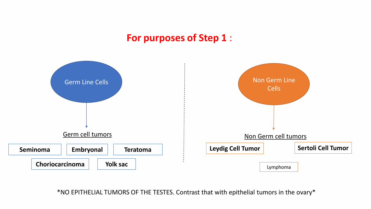
For purposes of Step 1 :
Germ Line Cells Non Germ Line Cells
Germ cell tumors Non Germ cell tumors
*NO EPITHELIAL TUMORS OF THE TESTES. Contrast that with epithelial tumors in the ovary*
Seminoma Teratoma Embryonal
Yolk sac Choriocarcinoma
Leydig Cell Tumor Sertoli Cell Tumor
Lymphoma

Tackling Testicular Tumors
These criteria will guide you to the promised land (aka right answer):
• Histology Histology Histology!
• Gross Appearance
• Tumor Markers
• Age
For example: Age old male presents with scrotal mass. Histological examination reveals pathological descriptor:
Question may ask:
• What is the most likely diagnosis?
• Which marker is tumor likely to secrete?
• What is the best treatment?
• Sites of metastasis?

Germ Cell Tumors
• 95% of ALL testicular tumors
• Almost always malignant (think they’re nasty and “germy”)
• Age: young men. 15-35 y/o
• Risk Factors: cryptorchidism
• Does not transilluminate • Contrast with hydrocele

Germ Cell Tumors
• 95% of ALL testicular tumors
• Almost always malignant (think they’re nasty and “germy”)
• Age: young men. 15-35 y/o
• Risk Factors: cryptorchidism
• Does not transilluminate • Contrast with hydrocele

Germ Cell Tumors
Seminoma: most common testicular tumor Age: young men 15-35 Histology: nests of large cells with clear cytoplasm - “fried egg appearance” with central nuclei Appearance: homogenous mass. Tumor Marker: may see elevated placental ALP and β-HCG - NOT most reliable markers Presentation: painless unilateral mass

Germ Cell Tumors
Yolk Sac Tumor:
Age: children < 3 y/o
Histology: Schiller-Duval bodies resembling primitive glomeruli
Appearance: yellow, mucinous
Tumor Marker: hallmark is increased AFP
Presentation: testicular mass in young child
α-fetoprotein

Germ Cell Tumors
Choriocarcinoma:
Age: young men
Histology: disordered syncytiotrophoblasts and cytotrophoblasts
Appearance: n/a
Tumor Marker: β-hCG
Presentation: testicular mass with gynecomastia and hyperthyroid; mets to the lungs
HcG mimics structure of TSH, LH, FSH
G β-hCG

Germ Cell Tumors
Choriocarcinoma:
Age: young men
Histology: disordered syncytiotrophoblasts and cytotrophoblasts
Appearance: n/a
Tumor Marker: BhCG
Presentation: testicular mass with gynecomastia and hyperthyroid; mets to the lungs
HcG mimics structure of TSH, LH, FSH
Cannonball Lesions

Germ Cell Tumors
Teratoma:
Age: young men or children (pure form in children)
Histology: cell types from all three germ layers
Appearance: large mass with hair, muscle, neural tissue
Tumor Marker: none
Malignant in adult males UNLIKE in adult females
Germ Cell Tumors
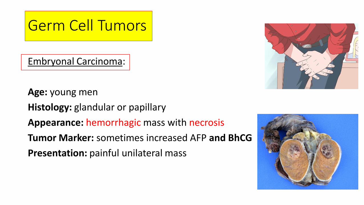
Embryonal Carcinoma:
Age: young men
Histology: glandular or papillary
Appearance: hemorrhagic mass with necrosis
Tumor Marker: sometimes increased AFP and BhCG
Presentation: painful unilateral mass

Germ Cell Tumors: Young lad with testicular mass PLUS
Seminoma
-homogenous
-”fried egg” and clear nuclei
Embryonal
-painful, necrotic mass
Yolk Sac
-children
-Schiller Duval Bodies
-AFP
Choriocarcinoma
-cyto/syncytiotrophoblasts
- Beta HcG marker
-don’t forget TSH, LH, FSH
-hematogenous spread
Teratoma
-three germ cell layers
-malignant in men only

Non Germ Cell Tumors
• Minority of testicular tumors (~5%)

Non Germ Cell Tumors
Leydig Cell Tumor: Androgen and Estrogen producing
Age: young men
Histology: Reinke crystals (eosinophilic cytoplasmic inclusions)
Appearance: golden brown color
Tumor Marker: none
Presentation: gynecomastia and early puberty

Non Germ Cell Tumors
Sertoli Cell Tumor: benign and don’t produce hormone
Testicular Lymphoma: testicular tumor of men >60. Usually bilateral NHL

Question Time:
25 year old chap comes in for evaluation of a painless right testicular mass. He also has complaints of weight loss, diaphoresis, palpitations and nervousness. Histological evaluation of his testicular mass is most likely to show:
A) Clear cytoplasm with central nuclei
B) Cells resembling primitive glomeruli
C) Normal testicular histology
D) Disordered cytotrophoblasts
E) Eosinophilic cytoplasmic inclusions