testosterone threshold levels and lean tissue mass targets needed to enhance skeletal muscle...
TRANSCRIPT
Journal of Gerontology: MEDICAL SCIENCES © The Author 2010. Published by Oxford University Press on behalf of The Gerontological Society of America.Cite journal as: J Gerontol A Biol Sci Med Sci. 2011 January;66A(1):122–129 All rights reserved. For permissions, please e-mail: [email protected]:10.1093/gerona/glq183� Advance Access published on November 8, 2010
122
LOSS�of�skeletal�muscle�mass�(sarcopenia)�contributes�to�declines�in�muscle�performance�and�physical�func-
tion�during�aging.�Substantial�losses�in�muscle�strength�may�result�in�difficulty�rising�from�a�chair,�climbing�stairs,�gen-erating� gait� speed,� maintaining� balance,� and� frailty� (1,2).�The�levels�of�endogenous�anabolic�hormones�also�decline�during� the� aging� process� (3).� Indeed,� 25%–30%� of� men�aged�older�than�60�years�have�low�levels�of�serum�testoster-one�levels�(4)�that�may�be�associated�with�sarcopenia�and�
muscle�weakness�(3,5).�Restoring�testosterone�to�youthful�levels� increases� synthesis� of� myofibrillar� proteins,� total�body� cell� mass,� and� muscle� strength� (6,7).� Declines��in� growth� hormone� (GH)� and� insulin-like�growth� factor� 1�(IGF-1)�may�also�contribute�to�comorbidities�in�older�men�with�normal�testosterone�levels�(3,8).
To�better�understand�the�relative�contributions�of�testos-terone�and�GH/IGF-1�axes�in�older�persons�at�risk�for�sarco-penia,�we�conducted�the�HORMA�(Hormonal�Regulators�of�
Testosterone�Threshold�Levels�and�Lean�Tissue�Mass�Targets�Needed�to�Enhance�Skeletal�Muscle�Strength�and�
Function:�The�HORMA�Trial
Fred�Sattler,1,2�Shalender�Bhasin,3�Jiaxiu�He,4�Chih-Ping�Chou,4�Carmen�Castaneda-Sceppa,5�Kevin�Yarasheski,6�Ellen�Binder,7�E.�Todd�Schroeder,2�Miwa�Kawakubo,4�Anqi�Zhang,3�
Ronenn�Roubenoff,5�and�Stanley�Azen4
1Department�of�Medicine�and�2Division�of�Biokinesiology,�University�of�Southern�California,�Los�Angeles,�California.�3Department�of�Medicine,�Boston�University,�Massachusetts.�
4Department�of�Preventive�Medicine,�University�of�Southern�California,�Los�Angeles,�California.�5Department�of�Medicine�and�Human�Nutrition�Research�Center�on�Aging�of�Tufts�University,�Boston,�Massachusetts.�
6Department�of�Medicine,�Washington�University,�St�Louis,�Missouri.�7Division�of�Geriatrics�and�Gerontology,�Washington�University�School�of�Medicine,�St�Louis,�Missouri.�
Address�correspondence�to�Fred�Sattler,�MD,�Department�of�Medicine,�University�of�Southern�California,�2020�Zonal�Avenue,�Room�434,��Los�Angeles,�CA�90033.�Email:�[email protected]
Background.� In�the�HORMA�(Hormonal�Regulators�of�Muscle�and�Metabolism�in�Aging)�Trial,�supplemental�testosterone�and�recombinant�human�growth�hormone�(rhGH)�enhanced�lean�body�mass,�appendicular�skeletal�muscle�mass,�muscle�performance,�and�physical�function,�but�there�was�substantial�interindividual�variability�in�outcomes.
Methods.� One�hundred�and�twelve�men�aged�65–90�years�received�testosterone�gel�(5�g/d�vs�10�g/d�via�Leydig�cell�clamp)�and�rhGH�(0�vs�3�vs�5�mg/kg/d)�in�a�double-masked�2�×�3�factorial�design�for�16�weeks.�Outcomes�included�lean�tissue�mass�by�dual�energy�x-ray�absorptiometry,� one-repetition�maximum�strength,�Margaria�stair�power,�and�activity�questionnaires.�We�used�pathway�analysis�to�determine�the�relationship�between�changes�in�hormone�levels,�muscle�mass,�strength,�and�function.
Results.� Increases�in�total�testosterone�of�1046�ng/dL�(95%�confidence�interval�=�1040–1051)�and�898�ng/dL�(95%�confidence�interval�=�892–904)�were�necessary�to�achieve�median�increases�in�lean�body�mass�of�1.5�kg�and�appendicular�skeletal� muscle� mass� of� 0.8� kg,� respectively,� which� were� required� to� significantly� enhance� one-repetition� maximum�strength�(≥30%).�Co-treatment�with�rhGH�lowered�the�testosterone�levels�(quantified�using�liquid�chromatography–tandem�mass�spectrometry)�necessary�to�reach�these�lean�mass�thresholds.�Changes�in�one-repetition�maximum�strength�were�associated�with�increases�in�stair�climbing�power�(r�=� .26,�p�=� .01).�Pathway�analysis�supported�the�model� that�changes�in�testosterone�and�insulin-like�growth�factor�1�levels�are�related�to�changes�in�lean�body�mass�needed�to�enhance�muscle�performance�and�physical�function.�Testosterone’s�effects�on�physical�activity�were�mediated�through�a�different�pathway�because�testosterone�directly�affected�Physical�Activity�Score�of�the�Elderly.
Conclusions.� To�enhance�muscle�strength�and�physical�function,�threshold�improvements�in�lean�body�mass�and�appendicular�skeletal�muscle�mass�are�necessary�and�these�can�be�achieved�by�targeting�changes�in�testosterone�levels.�rhGH� augments� the� effects� of� testosterone.� To� maximize� functional� improvements,� the� doses� of� anabolic� hormones�should�be�titrated�to�achieve�target�blood�levels.
Key Words: Testosterone—Growth�hormone—Lean�body�mass—Muscle�performance—Physical�function.
Received May 6, 2010; Accepted August 17, 2010
Decision Editor: Luigi Ferrucci, MD, PhD
at University of Sydney on Septem
ber 6, 2014http://biom
edgerontology.oxfordjournals.org/D
ownloaded from

DETERMINANTS OF MUSCLE STRENGTH AND PHYSICAL FUNCTION CHANGE 123
Muscle�and�Metabolism�in�Aging)�Trial�to�test�our�hypoth-esis�that�endogenous�testosterone�and�GH�are�important�in-dependent�but�complementary�regulators�of�skeletal�muscle�mass�and� function�even� into�advanced�age� (9).�Total� lean�body� mass� (LBM),� appendicular� skeletal� muscle� mass�(ASMM),� muscle� performance,� and� stair� climbing� power�increased� significantly� with� testosterone� and� changes� ap-peared�to�be�enhanced�by�recombinant�human�growth�hor-mone� (rhGH)� (9).� However,� there� was� considerable�variability� in� anabolic� responses� as� well� as� in� changes� in�testosterone� and� IGF-1� levels� during� treatment.�This� pro-vided� the�opportunity� to�examine�relationships�of�a�broad�range�of�hormone�changes,�including�declines�in�levels�as�may�occur�in�clinical�practice,�and�their�effects�on�changes�in� lean� tissue� mass,� muscle� strength,� performance,� and�physical�function.�We�used�pathway�analysis�to�test�the�hy-pothesis� that� testosterone�and�rhGH�affected�muscle�mass�directly�and�that�a�threshold�change�in�lean�tissue�mass�was�needed�to�generate�significant�improvements�in�muscle�per-formance� and� physical� function.� Additionally,� we� used�bootstrap�analysis�to�determine�target�hormone�levels�asso-ciated� with� threshold� changes� in� whole-body� and� appen-dicular� lean� mass� that� would� be� necessary� for� improving�muscle�performance�and�functional�outcomes.
Materials and Methods
Study DesignThe�HORMA�study� was�a�randomized�double-masked�in-
vestigation�of�testosterone�and�rhGH�supplementation�for�16�weeks� in� older� community-dwelling� men� with� testosterone�and� IGF-1�levels�typical�for�older�men�(9).�Eligible�partici-pants�were�randomized�(factorial�design)�to�one�of�two�physi-ological�doses�of�testosterone�during�a�Leydig�cell�clamp�as�well�to�placebo�or�one�of�two�physiological�doses�of�rhGH.
Study EligibilityParticipants�were� screened�at� the�University�of�Southern�
California,�Tufts�University,�and�Washington�University�after�providing�institutional�review�board–approved�informed�con-sent.�Men�were�aged�65–90�years�with�morning�total�testos-terone�in�the�lower�portion�of�the�adult�range�(150–550�ng/dL)�and�IGF-1�in�the�lower�adult�tertile�(<167�ng/mL).�For�screening,�total�testosterone�was�measured�by�automated�plat-form� immunoassays� in� the� local� clinical� laboratories� and�IGF-1�at�Quest�Diagnostics�(San�Juan�Capistrano,�CA).�Eligi-bility�criteria�included�prostatic�specific�antigen�≤4.0�ng/mL,�hematocrit�≤50%,�and�fasting�blood�glucose�<126�mg/dL�(9).
Study InterventionsParticipants� were� treated� monthly� for� 12� weeks� with��
a� long� acting� gonadotropin-releasing� hormone� agonist�(leuprolide� acetate� depot,� 7.5� mg� intramuscularly;�TAP�Pharmaceuticals,�Lake�Forest,�IL)�and�either�5�g�or�10�g/d�of�
1%�testosterone�transdermal�gel�(Solvay�Pharmaceuticals,�Marietta,�GA)�daily�for�16�weeks.�The�5�g�and�10�g�doses�of�testosterone�were�chosen�to�produce�a�spectrum�of�serum�levels�via�the�Leydig�cell�clamp�(to�fully�suppress�endoge-nous�production�of� testosterone)� that�would�be� in� the� low�normal� range� typical�of�older�men�or�mid-to-high�normal�levels� typical� of� younger� men,� respectively� (10).� Partici-pants� also� self-administered� 0,� 3,� or� 5� mg/kg� of� rhGH�(Genentech,�Foster�City,�CA)�each�evening.�The�3�mg/kg�dose�of�rhGH�was�chosen�because�3.3�but�not�2.0�mg/kg/d�increased�whole-body�protein�synthesis�in�GH-deficient�adults� (11).�The� 5�mg/kg/d�dose�was�chosen� to�produce�a�greater�anabolic�stimulus.
Outcome Measures
Hormone assays.—Testosterone� and� IGF-1� levels� were�determined�at�baseline�and�week�16.�Total�testosterone�was�measured�using�liquid�chromatography–tandem�mass�spec-trometry� (12),� free� testosterone� by� equilibrium� dialysis�(13),�and�IGF-1�by�a�chemiluminescence�immunoassay�(9).
Body composition.—Whole-body� and� regional� lean�mass�were�quantified�by�dual�energy�x-ray�absorptiometry,�calibrated�using�a�soft�tissue�phantom.�Scans�were�analyzed�at�the�USC�Reading�Center�by�a�dual�energy�x-ray�absorpti-ometry–certified�masked�bionutritionist.�Lean�mass�of� the�four�extremities�was�summed�to�obtain�ASMM.
Skeletal muscle performance and physical function.—Upper�and�lower�body�muscle�strength�was�determined�by�the�one-repetition�maximum�(1-RM)�method�for�the�bilat-eral� leg� press,� leg� extension,� leg� flexion,� latissimus� pull-down,� and� chest� press� (14).� Because� different� equipment�was�used�at�the�testing�centers,�changes�in�muscle�strength�are� presented� as� percentage� change� from� baseline� for� the�composite�(sum)�of�the�five�exercises.�Margaria�stair�climb-ing�power�was�calculated� from� time� (measured�by�photo-cells)�to�ascend�the�middle�four�steps�in�a�12-step�staircase�to�quantify�maximum�power�at�steady�state�because�there�is�an� early� acceleration� to�overcome� inertia� and�gravity� and�there�may�be�late�deceleration�due�to�fatigue�(15).�Physical�activity�was�assessed�with�the�Physical�Activity�Scale�of�the�Elderly� (PASE).�VO2peak� by� cycle� ergometry� was� deter-mined�during� the�baseline�electrocardiogram�stress� test� to�assure�that�it�was�safe�to�conduct�1-RM�testing.
Statistical ConsiderationsPaired� t� tests�were�used� to� assess�within�group�effects.�
Pathway�analyses�using�structural�equation�modeling�(16)�were�conducted�to�examine�the�direct�and�indirect�effects�of�the� changes� in�hormone� levels� (predictors)� on� changes� in�LBM,�ASMM,�and�1-RM�strength�(mediators),�Margaria�stair�climbing�power�(outcome),�and�physical�activity�by�
at University of Sydney on Septem
ber 6, 2014http://biom
edgerontology.oxfordjournals.org/D
ownloaded from
SATTLER ET AL.124
PASE� (outcome).�All� relationships� in� the� pathway� model�were�assumed�to�be�linear.
In� addition,� we� compared� the� average� change� in� total�LBM�and�in�ASMM�between�dichotomous�groups�defined�by�low�(below-median)�and�high�(above-median)�changes�in�total� testosterone,� free� testosterone,�and�IGF-1�for� the�par-ticipants�who�only�received�testosterone�and�those�who�re-ceived� both� testosterone� and� rhGH.� To� determine� the�combined�effect�of�change�in�testosterone�and�IGF-1�on�lean�mass,�changes�in�these�hormones�were�dichotomized�(low�vs�high)�at�their�medians�and�participants�were�categorized�into�four�groups:�low/low,�low/high,�high/low,�and�high/high,�re-spectively.�One-way�and�two-way�analysis�of�variance�was�used�to�examine�the�changes�and�the�potential�interaction�of�testosterone�and�IGF-1�levels�on�changes�in�total�LBM�and�ASMM.�Linear�trends�were�determined�by�Wald�analysis.
To� determine� the� magnitude� of� change� in� testosterone�with�and�without�rhGH�that�is�associated�with�1.5�kg�change�in�LBM�and�0.8�kg�change�in�ASMM,�we�used�the�bootstrap-ping� method� with� 1000� iterations� in� which� each� bootstrap�sample�contained�90%�of�the�original�sample�sets�(without�replacement)�for�the�39�men�receiving�only�testosterone�and�73�men�receiving�testosterone�plus�rhGH.�Statistical�analyses�were�carried�out�using�the�Statistical�Analysis�System�9.1�(Cary,�NC).
Results
Study PopulationOf�122� eligible� participants,� 112�were� randomized� and�
completed�16�weeks�of�study�medication.�For�testosterone�treatments,� 58� participants� were� randomized� to� 5� g� of��transdermal� gel� daily� and� 54� received� 10� g/d.� For� rhGH�treatments,� 39� participants� received� placebo,� 36� received�3.0�mg/kg,�and�37� received�5.0�mg/kg�daily.�Table�1�sum-marizes�baseline�characteristics�of�the�participants.
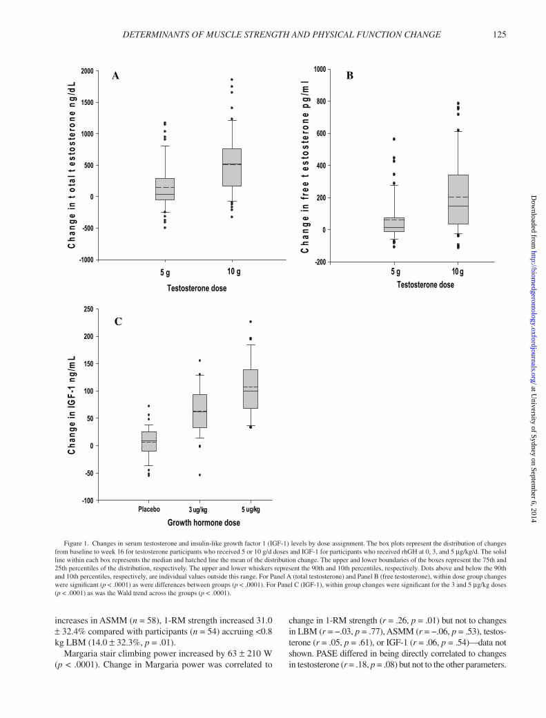
Changes in Serum Testosterone and IGF-1 LevelsTotal� testosterone� levels� by� liquid� chromatography–
tandem�mass�spectrometry�increased�by�143�±�379�ng/dL�(p�=�.006)�with�the�5�g�dose�and�by�510�±�503�ng/dL�(p�<�.0001)�with�the�10�g�dose�(for�between-dose�comparison�p�<� .0001;�Figure�1A).�Testosterone� levels�declined� in�21�participants�receiving�the�5�g�dose�and�in�eight�receiving�the�10�g�dose.�Free�testosterone�increased�by�60�±�136�pg/mL�(p�=� .001)�in�men�receiving�5�g/d�testosterone�gel�and�by�201�±�231�pg/mL�(p�<� .0001)� in� those�receiving�10�g/d�(for� between-dose�comparison�p�<� .0001;�Figure�1B);�31�participants�had�decreases�from�0�to�−111.5�pg/mL�com-pared�with�baseline.
Treatment� with� rhGH� (0,� 3,� and� 5� mg/kg/d)� increased�IGF-1�levels�by�6�±�28�(p�=�.16),�64�±�44�(p�<�.0001),�and�108� ±� 51� ng/mL� (p� <� .0001),� respectively� (Figure� 1C).�IGF-1�levels�declined�in�20�participants�receiving�placebo,�
three� receiving� 3� mg/kg/d,� but� none� receiving� 5� mg/kg/d�of�rhGH.
Primary OutcomesAfter�16�weeks�of�treatment,�total�LBM�increased�by�1.8�±�
1.9�kg�(interquartile�range�=�0.6–2.8�kg,�maximum�=�7.5�kg,�p�<�.0001)�and�ASMM�by�0.8�±�1.2�kg�(interquartile�range�=� 0.0–1.5� kg,� maximum� =� 4.4� kg,� p� <� .0001,� N� =� 112).�Composite�maximal�voluntary�1-RM�strength�increased�by�24�±�33%�(interquartile�range�=�2.3%–43.8%,�maximum�=�117%,�p�<�.0001,�N�=�95)�and�Margaria�stair�climbing�power�by�63�±�210�W�(interquartile�range�=�−11�to�+143�W,�maxi-mum�=�1248�W,�p�=�.003,�N�=�112).
Pathway AnalysisBefore�construction�of�the�pathway�model,�we�examined�
the�relationships�between�changes� in�hormone�levels,� lean�tissue�mass,�muscle�strength,�and�physical�function.�These�analyses� revealed� that� improvements� in�LBM�and�ASMM�were�correlated�with�increases�in�1-RM�strength�(LBM�r�=�.32,�p�=�.001;�ASMM�r�=�.30,�p�=�.003).�However,�change�in�1-RM�strength�was�not�related�to�changes�in�testosterone�(r�=�.12,�p�=�.24)�or�IGF-1�levels�(r�=�−.01,�p�=�.90).�Regression�analysis�indicated�that�LBM�had�to�increase�by�1.5�kg�and�ASMM�by�0.8�kg�to�achieve�meaningful�changes�in�muscle�strength�and�the�associated�improvements�in�physical�func-tion.�For�participants�achieving�≥1.5�kg�increases�in�LBM�(n�=�58),�1-RM�strength�increased�30.2�±�33.0%�compared�with�participants� (n�=�54)�accruing�<1.5�kg�LBM�(16.2�±�32.3%,�p�=�.04).�Similarly,�for�participants�achieving�≥0.8�kg�
Table�1.� Baseline�Characteristics�of�the�Study�Population�Prior�to�Treatment
Characteristic
N�=�112�Participants
Mean�±�SD�or�N�(%) Median�(range)
Age,�years 70.2�±�4.2 69.0�(64.0–85.0)BMI,�kg/m2 27.2�±�3.3 26.8�(20.1–34.8)Non-Hispanic�Caucasian 96�(86) N/AOn�treatment�for�hypertension 30�(27) N/AHistory�of�smoking 41�(37) N/AOn�treatment�for�dyslipidemia 39�(35) N/AHistory�ischemic�heart�events 13�(12) N/APASE 147�±�59 143�(30–369)Hemoglobin,�g/dL 14.6�±�0.9 14.8�(11.1–17.2)Serum�creatinine,�mg/dL 1.0�±�0.16 1.0�(0.7–1.5)Albumin,�g/dL 4.1�±�0.3 4.1�(3.5–5.4)Total�testosterone,�ng/dL* 363�±�97 361�(155–546)Total�testosterone,�ng/dL† 493�±�170 473�(111–961)IGF-1,�ng/mL 111�±�29 110�(31–167)Total�lean�body�mass,�kg 58.2�±�6.9 57.5�(41.6–78.0)Appendicular�lean�mass,�kg 25.5�±�3.3 25.5�(16.7–34.0)VO2peak�test,�mL/kg/min 24.6�±�4.9 24.3�(9.2–36.8)
Notes:� BMI� =� body� mass� index;� IGF-1� =� insulin-like� growth� factor� 1;�PASE�=�Physical�Activity�Scale�for�the�Elderly;�SD�=�standard�deviation.
*�By�automated�platform�immunoassays�for�screening.†�By�liquid�chromatography–tandem�mass�spectrometry�(batch�testing�after�
study).
at University of Sydney on Septem
ber 6, 2014http://biom
edgerontology.oxfordjournals.org/D
ownloaded from
DETERMINANTS OF MUSCLE STRENGTH AND PHYSICAL FUNCTION CHANGE 125
increases�in�ASMM�(n�=�58),�1-RM�strength�increased�31.0�±�32.4%�compared�with�participants�(n�=�54)�accruing�<0.8�kg�LBM�(14.0�±�32.3%,�p�=�.01).
Margaria�stair�climbing�power�increased�by�63�±�210�W�(p�<� .0001).�Change� in�Margaria�power�was�correlated� to�
change�in�1-RM�strength�(r�=�.26,�p�=�.01)�but�not�to�changes�in�LBM�(r�=�−.03,�p�=�.77),�ASMM�(r�=�−.06,�p�=�.53),�testos-terone�(r�=�.05,�p�=�.61),�or�IGF-1�(r�=�.06,�p�=�.54)—data�not�shown.�PASE�differed�in�being�directly�correlated�to�changes�in�testosterone�(r�=�.18,�p�=�.08)�but�not�to�the�other�parameters.
Ld/gnenoretsotsetlatot
niegnah
C
-1000
-500
0
500
1000
1500
2000
Testosterone dose
5 g 10 g
Lm/gn
1-FGI
niegnahC
-100
-50
0
50
100
150
200
250
Placebo 3 ug/kg 5 ug/kg
Growth hormone dose
lm/gp
enoretsotseteerf
niegnah
C
-200
0
200
400
600
800
1000
5 g 10 g Testosterone dose
C
BA
Figure�1.� Changes�in�serum�testosterone�and�insulin-like�growth�factor�1�(IGF-1)�levels�by�dose�assignment.�The�box�plots�represent�the�distribution�of�changes�from�baseline�to�week�16�for�testosterone�participants�who�received�5�or�10�g/d�doses�and�IGF-1�for�participants�who�received�rhGH�at�0,�3,�and�5�mg/kg/d.�The�solid�line�within�each�box�represents�the�median�and�hatched�line�the�mean�of�the�distribution�change.�The�upper�and�lower�boundaries�of�the�boxes�represent�the�75th�and�25th�percentiles�of�the�distribution,�respectively.�The�upper�and�lower�whiskers�represent�the�90th�and�10th�percentiles,�respectively.�Dots�above�and�below�the�90th�and�10th�percentiles,�respectively,�are�individual�values�outside�this�range.�For�Panel�A�(total�testosterone)�and�Panel�B�(free�testosterone),�within�dose�group�changes�were�significant�(p�<�.0001)�as�were�differences�between�groups�(p�<�.0001).�For�Panel�C�(IGF-1),�within�group�changes�were�significant�for�the�3�and�5�mg/kg�doses�(p�<�.0001)�as�was�the�Wald�trend�across�the�groups�(p�<�.0001).
at University of Sydney on Septem
ber 6, 2014http://biom
edgerontology.oxfordjournals.org/D
ownloaded from
SATTLER ET AL.126
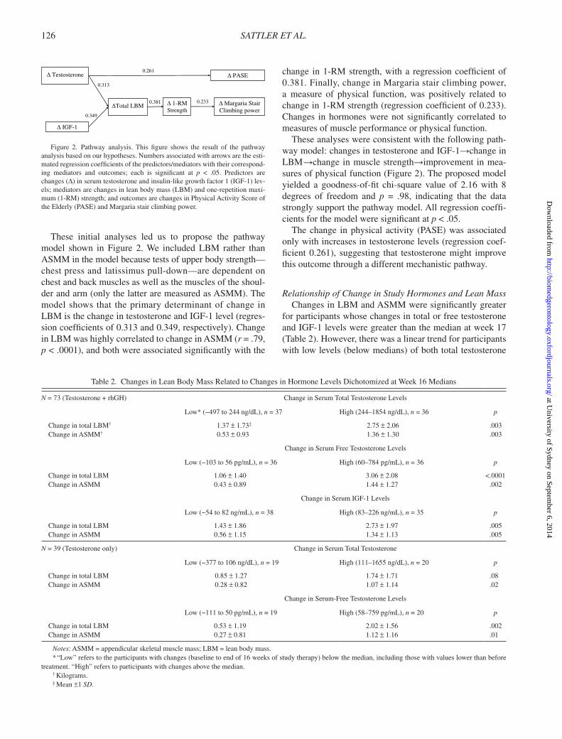
These� initial� analyses� led� us� to� propose� the� pathway�model� shown� in� Figure� 2.�We� included� LBM� rather� than�ASMM�in�the�model�because�tests�of�upper�body�strength—chest�press�and� latissimus�pull-down—are�dependent�on�chest�and�back�muscles�as�well�as�the�muscles�of�the�shoul-der�and�arm�(only�the�latter�are�measured�as�ASMM).�The�model�shows�that� the�primary�determinant�of�change�in�LBM�is�the�change�in�testosterone�and�IGF-1�level�(regres-sion�coefficients�of�0.313�and�0.349,�respectively).�Change�in�LBM�was�highly�correlated�to�change�in�ASMM�(r�=�.79,�p�<�.0001),�and�both�were�associated�significantly�with�the�
change� in�1-RM�strength,�with�a� regression�coefficient�of�0.381.�Finally,�change�in�Margaria�stair�climbing�power,�a� measure� of� physical� function,� was� positively� related� to�change�in�1-RM�strength�(regression�coefficient�of�0.233).�Changes� in�hormones�were�not� significantly� correlated� to�measures�of�muscle�performance�or�physical�function.
These�analyses�were�consistent�with�the�following�path-way�model:�changes�in�testosterone�and�IGF-1→change�in�LBM→change�in�muscle�strength→improvement� in�mea-sures�of�physical�function�(Figure�2).�The�proposed�model�yielded� a� goodness-of-fit� chi-square� value� of� 2.16� with� 8�degrees� of� freedom� and� p� =� .98,� indicating� that� the� data�strongly�support�the�pathway�model.�All�regression�coeffi-cients�for�the�model�were�significant�at�p�<�.05.
The�change� in�physical� activity� (PASE)�was�associated�only�with�increases�in�testosterone�levels�(regression�coef-ficient�0.261),� suggesting� that� testosterone�might� improve�this�outcome�through�a�different�mechanistic�pathway.
Relationship of Change in Study Hormones and Lean MassChanges�in�LBM�and�ASMM�were�significantly�greater�
for�participants�whose�changes�in�total�or�free�testosterone�and�IGF-1�levels�were�greater�than�the�median�at�week�17�(Table�2).�However,�there�was�a�linear�trend�for�participants�with�low�levels�(below�medians)�of�both�total�testosterone�
∆ enoretsotseT
∆ 1-FGI
∆ MBLlatoT ∆ MR-1htgnertS
∆ riatSairagraMnibmilC g p ewo r
∆ ESAP162.0
313.0
943.0
183.0 332.0
Figure� 2.� Pathway� analysis.� This� figure� shows� the� result� of� the� pathway�analysis�based�on�our�hypotheses.�Numbers�associated�with�arrows�are�the�esti-mated�regression�coefficients�of�the�predictors/mediators�with�their�correspond-ing� mediators� and� outcomes;� each� is� significant� at� p� <� .05.� Predictors� are�changes�(∆)�in�serum�testosterone�and�insulin-like�growth�factor�1�(IGF-1)�lev-els;�mediators�are�changes�in�lean�body�mass�(LBM)�and�one-repetition�maxi-mum�(1-RM)�strength;�and�outcomes�are�changes�in�Physical�Activity�Score�of�the�Elderly�(PASE)�and�Margaria�stair�climbing�power.
Table�2.� Changes�in�Lean�Body�Mass�Related�to�Changes�in�Hormone�Levels�Dichotomized�at�Week�16�Medians
N�=�73�(Testosterone�+�rhGH) Change�in�Serum�Total�Testosterone�Levels
Low*�(−497�to�244�ng/dL),�n�=�37 High�(244–1854�ng/dL),�n�=�36 p
� Change�in�total�LBM† 1.37�±�1.73‡ 2.75�±�2.06 .003� Change�in�ASMM† 0.53�±�0.93 1.36�±�1.30 .003
Change�in�Serum�Free�Testosterone�Levels
Low�(−103�to�56�pg/mL),�n�=�36 High�(60–784�pg/mL),�n�=�36 p
� Change�in�total�LBM 1.06�±�1.40 3.06�±�2.08 <.0001� Change�in�ASMM 0.43�±�0.89 1.44�±�1.27 .002
Change�in�Serum�IGF-1�Levels
Low�(−54�to�82�ng/mL),�n�=�38 High�(83–226�ng/mL),�n�=�35 p
� Change�in�total�LBM 1.43�±�1.86 2.73�±�1.97 .005� Change�in�ASMM 0.56�±�1.15 1.34�±�1.13 .005
N�=�39�(Testosterone�only) Change�in�Serum�Total�Testosterone
Low�(−377�to�106�ng/dL),�n�=�19 High�(111–1655�ng/dL),�n�=�20 p
� Change�in�total�LBM 0.85�±�1.27 1.74�±�1.71 .08� Change�in�ASMM 0.28�±�0.82 1.07�±�1.14 .02
Change�in�Serum-Free�Testosterone�Levels
Low�(−111�to�50�pg/mL),�n�=�19 High�(58–759�pg/mL),�n�=�20 p
� Change�in�total�LBM 0.53�±�1.19 2.02�±�1.56 .002� Change�in�ASMM 0.27�±�0.81 1.12�±�1.16 .01
Notes:�ASMM�=�appendicular�skeletal�muscle�mass;�LBM�=�lean�body�mass.*�“Low”�refers�to�the�participants�with�changes�(baseline�to�end�of�16�weeks�of�study�therapy)�below�the�median,�including�those�with�values�lower�than�before�
treatment.�“High”�refers�to�participants�with�changes�above�the�median.†�Kilograms.‡�Mean�±1�SD.
at University of Sydney on Septem
ber 6, 2014http://biom
edgerontology.oxfordjournals.org/D
ownloaded from
DETERMINANTS OF MUSCLE STRENGTH AND PHYSICAL FUNCTION CHANGE 127
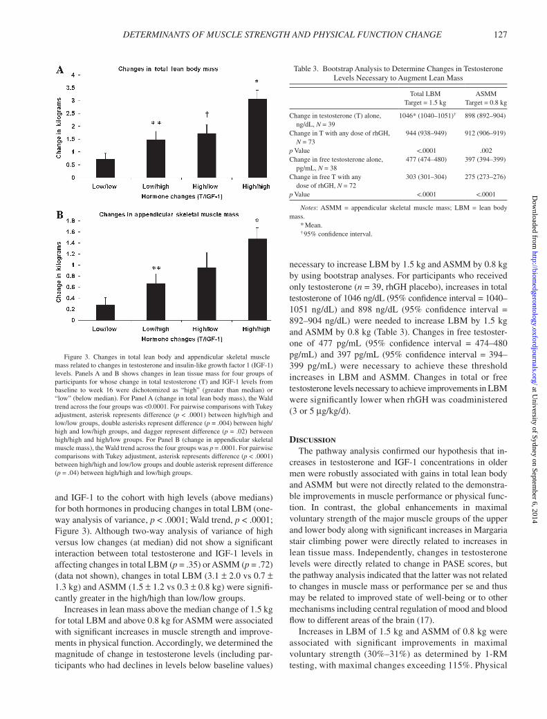
and�IGF-1�to�the�cohort�with�high�levels�(above�medians)�for�both�hormones�in�producing�changes�in�total�LBM�(one-way�analysis�of�variance,�p�<�.0001;�Wald�trend,�p�<�.0001;�Figure�3).�Although�two-way�analysis�of�variance�of�high�versus�low�changes�(at�median)�did�not�show�a�significant�interaction�between� total� testosterone�and� IGF-1� levels� in�affecting�changes�in�total�LBM�(p�=�.35)�or�ASMM�(p�=�.72)�(data�not�shown),�changes�in�total�LBM�(3.1�±�2.0�vs�0.7�±�1.3�kg)�and�ASMM�(1.5�±�1.2�vs�0.3�±�0.8�kg)�were�signifi-cantly�greater�in�the�high/high�than�low/low�groups.
Increases�in�lean�mass�above�the�median�change�of�1.5�kg�for�total�LBM�and�above�0.8�kg�for�ASMM�were�associated�with�significant�increases�in�muscle�strength�and�improve-ments�in�physical�function.�Accordingly,�we�determined�the�magnitude�of�change�in�testosterone�levels�(including�par-ticipants�who�had�declines�in�levels�below�baseline�values)�
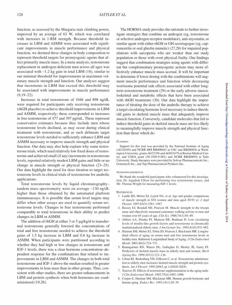
necessary�to�increase�LBM�by�1.5�kg�and�ASMM�by�0.8�kg�by�using�bootstrap�analyses.�For�participants�who�received�only�testosterone�(n�=�39,�rhGH�placebo),�increases�in�total�testosterone�of�1046�ng/dL�(95%�confidence�interval�=�1040–1051� ng/dL)� and� 898� ng/dL� (95%� confidence� interval� =�892–904�ng/dL)�were�needed� to� increase�LBM�by�1.5�kg�and�ASMM�by�0.8�kg�(Table�3).�Changes�in�free�testoster-one� of� 477� pg/mL� (95%� confidence� interval� =� 474–480�pg/mL)� and�397�pg/mL� (95%�confidence� interval�=�394–399� pg/mL)� were� necessary� to� achieve� these� threshold�increases� in� LBM� and�ASMM.� Changes� in� total� or� free�testosterone�levels�necessary�to�achieve�improvements�in�LBM�were�significantly�lower�when�rhGH�was�coadministered�(3�or�5�mg/kg/d).
DiscussionThe�pathway�analysis�confirmed�our�hypothesis�that�in-
creases� in� testosterone� and� IGF-1� concentrations� in� older�men�were�robustly�associated�with�gains�in�total�lean�body�and�ASMM �but�were�not�directly�related�to�the�demonstra-ble�improvements�in�muscle�performance�or�physical�func-tion.� In� contrast,� the� global� enhancements� in� maximal�voluntary�strength�of�the�major�muscle�groups�of�the�upper�and�lower�body�along�with�significant�increases�in�Margaria�stair� climbing� power� were� directly� related� to� increases� in�lean�tissue�mass.�Independently,�changes�in�testosterone�levels�were�directly�related�to�change�in�PASE�scores,�but�the�pathway�analysis�indicated�that�the�latter�was�not�related�to�changes�in�muscle�mass�or�performance�per�se�and�thus�may�be�related�to�improved�state�of�well-being�or�to�other�mechanisms�including�central�regulation�of�mood�and�blood�flow�to�different�areas�of�the�brain�(17).
Increases�in�LBM�of�1.5�kg�and�ASMM�of�0.8�kg�were�associated� with� significant� improvements� in� maximal�voluntary� strength� (30%–31%)� as� determined�by�1-RM�testing,�with�maximal�changes�exceeding�115%.�Physical�
Figure� 3.� Changes� in� total� lean� body� and� appendicular� skeletal� muscle�mass�related�to�changes�in�testosterone�and�insulin-like�growth�factor�1�(IGF-1)�levels.�Panels�A�and�B�shows�changes� in� lean� tissue�mass�for�four�groups�of�participants�for�whose�change�in�total�testosterone�(T)�and�IGF-1�levels�from�baseline� to� week� 16� were� dichotomized� as� “high”� (greater� than� median)� or�“low”�(below�median).�For�Panel�A�(change�in�total�lean�body�mass),�the�Wald�trend�across�the�four�groups�was�<0.0001.�For�pairwise�comparisons�with�Tukey�adjustment,� asterisk�represents�difference�(p�<� .0001)�between� high/high�and�low/low�groups,�double�asterisks�represent�difference�(p�=�.004)�between�high/high�and� low/high�groups,�and�dagger�represent�difference�(p�=�.02)� between�high/high�and�high/low�groups.�For�Panel�B�(change�in�appendicular�skeletal�muscle�mass),�the�Wald�trend�across�the�four�groups�was�p�=�.0001.�For�pairwise�comparisons�with�Tukey�adjustment,�asterisk�represents�difference�(p�<�.0001)�between�high/high�and�low/low�groups�and�double�asterisk�represent�difference�(p�=�.04)�between�high/high�and�low/high�groups.
Table�3.� Bootstrap�Analysis�to�Determine�Changes�in�Testosterone�Levels�Necessary�to�Augment�Lean�Mass
Total�LBM��Target�=�1.5�kg
ASMM��Target�=�0.8�kg
Change�in�testosterone�(T)�alone,��� ng/dL,�N�=�39
1046*�(1040–1051)† 898�(892–904)
Change�in�T�with�any�dose�of�rhGH,��� N�=�73
944�(938–949) 912�(906–919)
p�Value <.0001 .002Change�in�free�testosterone�alone,��� pg/mL,�N�=�38
477�(474–480) 397�(394–399)
Change�in�free�T�with�any��� dose�of�rhGH,�N�=�72
303�(301–304) 275�(273–276)
p�Value <.0001 <.0001
Notes:�ASMM� =� appendicular� skeletal� muscle� mass;� LBM� =� lean� body�mass.
*�Mean.†�95%�confidence�interval.
at University of Sydney on Septem
ber 6, 2014http://biom
edgerontology.oxfordjournals.org/D
ownloaded from
SATTLER ET AL.128
function,�as�assessed�by�the�Margaria�stair�climbing�power,�improved� by� an� average� of� 63� W,� which� was� correlated�with� increases� in� 1-RM� strength.� Because� threshold� in-creases�in�LBM�and�ASMM�were�associated�with�signifi-cant� improvements� in� muscle� performance� and� physical�function,�we�deemed�these�changes�in�body�composition�to�represent�threshold�targets�for�promyogenic�agents�that�af-fect�primarily�muscle�mass.�In�a�meta-analysis,�testosterone�replacement�in�androgen-deficient�men�across�all�ages�was�associated�with�~1.2�kg�gain�in�total�LBM�(18),�similar�to�our�minimal�threshold�for�improvements�in�maximum�vol-untary�muscle�strength�and�function.�Our�analyses�suggest�that� increments� in�LBM�that�exceed� this� threshold�may�be� associated� with� improvements� in� muscle� performance�(9,19–22).
Increases� in� total� testosterone� of� 1046� and� 898� ng/dL�were� required� for� participants� only� receiving� testosterone�(rhGH�placebo)�to�achieve�threshold�improvements�in�LBM�and�ASMM,�respectively;� these�corresponded�to� increases�in�free�testosterone�of�477�and�397�pg/mL.�These�represent�conservative� estimates� because� they� include� men� whose�testosterone� levels� declined,� as� may� occur� during� clinical�treatment� with� testosterone,� and� as� such� delineate� target��testosterone�levels�needed�to�sufficiently�enhance�LBM�and�ASMM�necessary�to�improve�muscle�strength�and�physical�function.�Our�data�may�also�help�explain�why�some�testos-terone�trials,�which�used�relatively�low�fixed�doses�of�testos-terone�and�achieved�small�(if�any)�increments�in�testosterone�levels,�reported�relatively�modest�LBM�gains�and�little�or�no�change� in� muscle� strength� or� physical� function� (23–25).�Our�data�highlight�the�need�for�dose�titration�to�target�tes-tosterone�levels�in�clinical�trials�of�testosterone�for�anabolic�applications.
Total� testosterone� levels� by� liquid� chromatography–tandem�mass�spectrometry�were�on�average�~130�ng/dL�higher� than� those� obtained� by� the� automated� platform�immunoassays.�It� is�possible� that�serum�level� targets�may�differ�when�other�assays�are�used�to�quantify�serum�tes-tosterone� levels.� Changes� in� free� testosterone� performed��comparable� to� total� testosterone� in� their� ability� to�predict�changes�in�LBM�or�ASMM.
The�addition�of�rhGH�at�either�3�or�5�mg/kg/d�to�transder-mal� testosterone� generally� lowered� the� concentrations� of�total�and�free�testosterone�needed�to�achieve�the�threshold�gains� of� 1.5� kg� increase� in� LBM� and� 0.8� kg� increase� in�ASMM.�When� participants� were� partitioned� according� to�whether�they�had�high�or�low�changes�in�testosterone�and�IGF-1�levels,�there�was�a�clear�hormone�concentration–de-pendent� response� for� the�combinations� that� related� to� im-provements�in�LBM�and�ASMM.�The�changes�in�both�total�testosterone�and�IGF-1�above�the�median�resulted�in�greater�improvements�in�lean�mass�than�in�other�groups.�Thus,�con-sistent�with�other�studies,�there�are�greater�enhancements�in�LBM�and�protein�synthesis�when�both�hormones�are�coad-ministered�(19,26).
The�HORMA�study�provides�the�rationale�to�further�inves-tigate�strategies�that�combine�an�androgen�(eg,�testosterone�or�selective�androgen�receptor�modulator),�anti-myostatin,�or�similar�agent�with�either�rhGH�or�GH�secretagogue�(eg,�cap-romorelin�or�oral�ghrelin�mimetic)�(27,28)�for�impaired�pop-ulations� with� sarcopenia� who� are� weaker� than� our� study�population�or�those�with�overt�physical�frailty.�Our�findings�suggest�that�combination�strategies�using�agents�with�differ-ent�but�complementary�promyogenic�actions�may�more�ef-fectively�enhance�muscle�mass�accrual.�It�will�be�important�to�determine�if�lower�dosing�with�the�combinations�will�aug-ment�muscle�performance�and�function�while�decreasing�worrisome�potential�side�effects�associated�with�either�long-term�testosterone�treatment�(29)�or�the�early�adverse�muscu-loskeletal� and� metabolic� effects� that� have� been� associated�with� rhGH� treatments� (30).� Our� data� highlight� the� impor-tance�of�titrating�the�dose�of�the�anabolic�therapy�to�achieve�a�target�circulating�hormone�level�necessary�to�induce�thresh-old�gains� in� skeletal�muscle�mass� that�adequately� improve�muscle�function.�Conversely,�candidate�molecules�that�fail�to�induce�threshold�gains�in�skeletal�muscle�mass�are�less�likely�to�meaningfully�improve�muscle�strength�and�physical�func-tion�than�those�which�do.
Funding
Support� for� this� trial�was�provided�by� the�National� Institute�of�Aging�(AG18169)�and�NCRR�M0I�RR000043�at�USC�and�RR000036�at�Wash-ington�University,�grants�AG22356�and�AG031679�at�Boston�Medical�Cen-ter,� and� USDA� grant� (58-1950-9-001)� and� NCRR� RR000054� at� Tufts�University.�Study�therapies�were�provided�by�Solvay�Pharmaceuticals�Inc.,�Genentech�Inc.,�and�Tap�Pharmaceutical�Products�Inc.
Acknowledgments
We�thank�the�wonderful�participants�who�volunteered�for�this�investiga-tion,�Dr.�Jagadish�Ulloor�for�performing�free� testosterone�assays,�and�Mr.�Thomas�Wright�for�measuring�IGF-1�levels.
References� 1.� Lindle�RS,�Metter�EJ,�Lynch�NA,�et�al.�Age�and�gender�comparisons�
of� muscle� strength� in� 654� women� and� men� aged� 20-93� yr.� J Appl Physiol.�1997;83(5):1581–1587.
� 2.� Bassey�EJ,�Bendall�MJ,�Pearson�M.�Muscle�strength� in� the� triceps�surae�and�objectively�measured�customary�walking�activity�in�men�and�women�over�65�years�of�age.�Clin Sci.�1988;74(1):85–89.
� 3.� Abbasi�AA,� Drinka� PJ,� Mattson� DE,� Rudman� D.� Low� circulating�levels�of�insulin-like�growth�factors�and�testosterone�in�chronically�institutionalized�elderly�men.�J Am Geriatr Soc.�1993;41(9):975–982.
� 4.� Harman�SM,�Metter�EJ,�Tobin�JD,�Pearson�J,�Blackman�MR.�Longitu-dinal� effects� of� aging�on� serum� total� and� free� testosterone� levels� in�healthy�men.�Baltimore�Longitudinal�Study�of�Aging.�J Clin Endocrinol Metab.�2001;86(2):724–731.
� 5.� Baumgartner� RN,� Waters� DL,� Gallagher� D,� Morley� JE,� Garry� PJ.��Predictors�of�skeletal�muscle�mass�in�elderly�men�and�women.�Mech Ageing Dev.�1999;107(2):123–136.
� 6.� Urban�RJ,�Bodenburg�YH,�Gilkison�C,�et�al.�Testosterone�administra-tion�to�elderly�men�increases�skeletal�muscle�strength�and�protein�syn-thesis.�Am J Physiol.�1995;269(5,�pt�1):E820–E826.
� 7.� Tenover�JS.�Effects�of�testosterone�supplementation�in�the�aging�male.�J Clin Endocrinol Metab.�1992;75(4):1092–1098.
� 8.� Corpas�E,�Harman�SM,�Blackman�MR.�Human�growth�hormone�and�human�aging.�Endocr Rev.�1993;14(1):20–39.
at University of Sydney on Septem
ber 6, 2014http://biom
edgerontology.oxfordjournals.org/D
ownloaded from
DETERMINANTS OF MUSCLE STRENGTH AND PHYSICAL FUNCTION CHANGE 129
� 9.� Sattler�FR,�Castaneda-Sceppa�C,�Binder�EF,� et� al.�Testosterone�and�growth�hormone�improve�body�composition�and�muscle�performance�in�older�men.�J Clin Endocrinol Metab.�2009;94(6):1991–2001.
�10.� Swerdloff�RS,�Wang�C,�Cunningham�G,�et�al.�Long-term�pharmacoki-netics�of�transdermal�testosterone�gel�in�hypogonadal�men.�J Clin Endocrinol Metab.�2000;85(12):4500–4510.
�11.� Lucidi�P,�Lauteri�M,�Laureti�S,�et�al.�A�dose-response�study�of�growth�hormone�(GH)�replacement�on�whole�body�protein�and�lipid�kinetics�in�GH-deficient�adults.�J Clin Endocrinol Metab.�1998;83(2):353–357.
�12.� Sir-Petermann�T,�Codner�E,�Perez�V,�et�al.�Metabolic�and�reproductive�features�before�and�during�puberty�in�daughters�of�women�with�polycys-tic�ovary�syndrome.�J Clin Endocrinol Metab.�2009;94(6):1923–1930.
�13.� Sinha-Hikim�I,�Arver�S,�Beall�G,�et�al.�The�use�of�a�sensitive�equilib-rium�dialysis�method�for�the�measurement�of�free�testosterone�levels�in�healthy,�cycling�women�and� in�human� immunodeficiency�virus-infected�women.�J Clin Endocrinol Metab.�1998;83(4):1312–1318.
�14.� Schroeder� ET,� Wang� Y,� Castaneda-Sceppa� C,� et� al.� Reliability� of��maximal�voluntary�muscle�strength�and�power� testing�in�older�men.��J Gerontol A Biol Sci Med Sci.�2007;62(5):543–549.
�15.� Margaria�R,�Aghemo�P,�Rovelli�E.�Measurement�of�muscular�power�(anaerobic)�in�man.�J Appl Physiol.�1966;21(5):1662–1664.
�16.� Kline�RB.�Principles and Practice of Structural Equation Modeling.�2nd�ed�New�York:�The�Guilford�Press;�2005.
�17.� Azad� N,� Pitale� S,� Barnes�WE,� Friedman� N.�Testosterone� treatment�enhances�regional�brain�perfusion�in�hypogonadal�men.�J Clin Endo-crinol Metab.�2003;88(7):3064–3068.
�18.� Bhasin�S,�Calof�OM,�Storer�TW,�et�al.�Drug� insight:� testosterone�and�selective�androgen�receptor�modulators�as�anabolic�therapies�for�chronic�illness�and�aging.�Nat Clin Pract Endocrinol Metab.�2006;2(3):146–159.
�19.� Blackman�MR,�Sorkin�JD,�Munzer�T,�et�al.�Growth�hormone�and�sex�steroid�administration�in�healthy�aged�women�and�men:�a�randomized�controlled�trial.�JAMA.�2002;288(18):2282–2292.
�20.� Ferrando�AA,� Sheffield-Moore� M,�Yeckel� CW,� et� al.� Testosterone�administration�to�older�men�improves�muscle�function:�molecular�and�physiological� mechanisms.� Am J Physiol Endocrinol Metab.� 2002;282(3):E601–E607.
�21.� Page�ST,�Amory�JK,�Bowman�FD,�et�al.�Exogenous�testosterone�(T)�alone�or�with�finasteride�increases�physical�performance,�grip�strength,�and�lean�body�mass�in�older�men�with�low�serum�T.�J Clin Endocrinol Metab.�2005;90(3):1502–1510.
�22.� Bhasin�S,�Woodhouse�L,�Casaburi�R,�et�al.�Older�men�are�as�respon-sive�as�young�men�to�the�anabolic�effects�of�graded�doses�of�testoster-one� on� the� skeletal� muscle.� J Clin Endocrinol Metab.� 2005;90(2):678–688.
�23.� Nair�KS,�Rizza�RA,�O’Brien�P,�et�al.�DHEA�in�elderly�women�and�DHEA�or�testosterone�in�elderly�men.�N Engl J Med.�2006;355(16):1647–1659.
�24.� Emmelot-Vonk�MH,�Verhaar�HJ,�Nakhai�Pour�HR,�et�al.�Effect�of�tes-tosterone�supplementation�on�functional�mobility,�cognition,�and�other�parameters� in� older� men:� a� randomized� controlled� trial.� JAMA.�2008;299(1):39–52.
�25.� Giannoulis�MG,�Sonksen�PH,�Umpleby�M,�et�al.�The�effects�of�growth�hormone� and/or� testosterone� in� healthy� elderly� men:� a� randomized�controlled�trial.�J Clin Endocrinol Metab.�2006;91(2):477–484.
�26.� Huang� X,� Blackman� MR,� Herreman� K,� Pabst� KM,� Harman� SM,��Caballero�B.�Effects�of�growth�hormone�and/or�sex�steroid�administra-tion�on�whole-body�protein�turnover�in�healthy�aged�women�and�men.�Metabolism.�2005;54(9):1162–1167.
�27.� White�HK,�Petrie�CD,�Landschulz�W,�et�al.�Effects�of�an�oral�growth�hormone�secretagogue�in�older�adults.�J Clin Endocrinol Metab.�2009;94(4):1198–1206.
�28.� Nass�R,�Pezzoli�SS,�Oliveri�MC,�et�al.�Effects�of�an�oral�ghrelin�mi-metic� on� body� composition� and� clinical� outcomes� in� healthy� older�adults:�a�randomized�trial.�Ann Intern Med.�2008;149(9):601–611.
�29.� Bhasin� S,� Cunningham� GR,� Hayes� FJ,� et� al.� Testosterone� therapy��in�adult�men�with�androgen�deficiency�syndromes:�an�endocrine�society�clinical� practice� guideline.� J Clin Endocrinol Metab.� 2006;91(6):1995–2010.
�30.� Molitch� ME,� Clemmons� DR,� Malozowski� S,� et� al.� Evaluation� and�treatment�of�adult�growth�hormone�deficiency:�an�Endocrine�Society�Clinical� Practice� Guideline.� J Clin Endocrinol Metab.� 2006;91(5):1621–1634.
at University of Sydney on Septem
ber 6, 2014http://biom
edgerontology.oxfordjournals.org/D
ownloaded from