textbook of - postgraduate bookspostgraduatebooks.jaypeeapps.com/pdf/orthopedics/textbook_of... ·...
TRANSCRIPT


Textbook ofILIZAROV SURGICAL TECHNIQUES
Bone Correction and Lengthening
VLADIMIR GOLYAKHOVSKY MD, PhD, DSc
Attending PhysicianNew York University Hospital for Joint Diseases Orthopaedic Institute
Clinical Associate Professor of Orthopaedic SurgeryNew York University School of Medicine
New York, NY 10003Formerly Professor and Chairman of Orthopaedics and Traumatology
Moscow Institute of Medicine and DentistryMoscow, Russia
VICTOR H FRANKEL MD, PhD
President EmeritusNew York University Hospital for Joint Diseases Orthopaedic Institute
Professor of Orthopaedic SurgeryNew York University School of Medicine
New York, NY
Illustrations and Photos by VLADIMIR GOLYAKHOVSKY
JAYPEE BROTHERS MEDICAL PUBLISHERS (P) LTDNew Delhi • St Louis (USA) • Panama City (Panama) • London (UK) • AhmedabadBengaluru • Chennai • Hyderabad • Kochi • Kolkata • Lucknow • Mumbai • Nagpur
®Jayp
ee B
rothe
rs

Published byJitendar P VijJaypee Brothers Medical Publishers (P) Ltd
Corporate Office4838/24 Ansari Road, Daryaganj, New Delhi - 110002, IndiaPhone: +91-11-43574357, Fax: +91-11-43574314
Registered OfficeB-3 EMCA House, 23/23B Ansari Road, Daryaganj, New Delhi - 110 002, IndiaPhones: +91-11-23272143, +91-11-23272703, +91-11-23282021, +91-11-23245672Rel: +91-11-32558559, Fax: +91-11-23276490, +91-11-23245683e-mail: [email protected], Website: www.jaypeebrothers.com
Offices in India• Ahmedabad, Phone: Rel: +91-79-32988717, e-mail: [email protected]
• Bengaluru, Phone: Rel: +91-80-32714073, e-mail: [email protected]
• Chennai, Phone: Rel: +91-44-32972089, e-mail: [email protected]
• Hyderabad, Phone: Rel:+91-40-32940929, e-mail: [email protected]
• Kochi, Phone: +91-484-2395740, e-mail: [email protected]
• Kolkata, Phone: +91-33-22276415, e-mail: [email protected]
• Lucknow, Phone: +91-522-3040554, e-mail: [email protected]
• Mumbai, Phone: Rel: +91-22-32926896, e-mail: [email protected]
• Nagpur, Phone: Rel: +91-712-3245220, e-mail: [email protected]
Overseas Offices• North America Office, USA, Ph: 001-636-6279734
e-mail: [email protected], [email protected]
• Central America Office, Panama City, PanamaPh: 001-507-317-0160, e-mail: [email protected]: www.jphmedical.com
• Europe Office, UK, Ph: +44 (0) 2031708910e-mail: [email protected], [email protected]
Textbook of Ilizarov Surgical Techniques: Bone Correction and Lengthening
© 2010, Jaypee Brothers Medical Publishers
All rights reserved. No part of this publication should be reproduced, stored in a retrieval system, or transmittedin any form or by any means: electronic, mechanical, photocopying, recording, or otherwise, without the priorwritten permission of the authors and the publisher.
This book has been published in good faith that the material provided by authors is original. Every effort ismade to ensure accuracy of material, but the publisher, printer and authors will not be held responsible forany inadvertent error(s). In case of any dispute, all legal matters are to be settled under Delhi jurisdictiononly.
First Edition: 2010
ISBN 978-81-8448-
Typeset at JPBMP typesetting unitPrinted at Ajanta Offset
Jayp
ee B
rothe
rs

To the memory ofProfessor Gavriil Abramovich Ilizarov
Portrait of Dr Ilizarov (Right) with the authorDr Vladimir Golyakhovsky, 1988, New York
Sant Yago, Chily
Jayp
ee B
rothe
rs

Preface
Seventeen years passed since publication, in1993, the “Operative Manual of Ilizarov Techniques”, a richly illustrated firstAmerican book about the Ilizarov techniques and methods. Utilizing the techniques developed by him, a circular externalfixator, a Russian Doctor Gavriil Ilizarov, in 1950-1960s introduced principally new techniques for treatment bonefractures and deformities. With this innovation, he opened a new chapter in the subject of the whole orthopedicsurgery—the minimally invasive methods of bone deformities correction. For many years, his name and work were notknown in the Western countries because the communist Soviet Russia was isolated behind the so-called “Iron Curtain”.With the fall of communism, in 1990s, the great interest and enthusiasm developed toward the Ilizarov work amongEuropean and American orthopedic communities. The “Operative Manual” was written at that time with the purposeto instruct the surgeons how to assemble and apply the configurations of a circular fixator for the bone correction andlengthening. In several years there were new editions of this book translated and published in Brazil and Russia. In thisway the “Operative Manual” has served thousands of orthopedic surgeons as a guide to master Ilizarov’s methods.
In the past 17 years the professional market of the books and articles on the subject of the Ilizarov methods had aplenitude of new ideas, and the “Operative Manual” was almost forgotten. In 2009, the Indian publisher company “JaypeeBrothers Medical Publishers”, based in New Delhi, came across the “Operative Manual”, which was not available anymore. The Indian publisher discovered it and considered a classic book and offered to one of the authors (VladimirGolyakhovsky) to have a new edition with revision of the recent advances happened during last several years. Indeed,there are new modifications of the Ilizarov fixator and the Ilizarov techniques developed in Italy, Germany, the US, andother countries. The author gladly agreed to have a new Indian edition, and to include the recent developments, togetherwith the new illustrations, in the book. Authors also offered to the Publisher to write new chapters:
• Gavriil Abramovich Ilizarov—The Man, The Creator, The Teacher• Biomechanics of the Ilizarov external fixator• Ilizarov frame assemblage• Biomechanics of the Ilizarov method• Biomechanic considerations for the fracture treatment• Basics of the method of distraction osteogenesis• Basics of the method of compression osteogenesis• Criteria of bone healing and fixator removal.These chapters, composed together, have a purpose to serve as an introduction for the description of the Ilizarov
techniques and methods. Some of these materials were scattered in the chapters of “Operative manual” but the authorconsiders expedient for the reader to get to know these particular subjects before learning how to assemble and usethe fixator.
The last chapter of the book “The New Modifications of the Ilizarov Technique” reflects the development of newinnovations which were developed since circulation of the original Ilizarov’s ideas in orthopedic community around the
Jayp
ee B
rothe
rs

viii Textbook of Ilizarov Surgical Techniques: Bone Correction and Lengthening
world. The apparent simplicity and clarity of the Ilizarov technique provoked many orthopedic surgeons to offer theirown modifications, as it always happened to the great inventions. Some of the new modifications are based on advancedtechnology of new materials for fixator parts, there are introductions of aluminum rings, and carbon rings, and titaniumrods. Some modifications consist of small technical innovations. All these modifications are governed by the principlesdiscovered and technique developed by Dr Ilizarov.
The most essential innovation of the Ilizarov treatment is an addition of the computer programming to thecalculation of bone deformity correction. The computer assisting control helps doctors and the patients to produceprecalculated bone reduction, correction, compression and lengthening. Several special software programs includingInternet-based programs are available in the US, Germany and other countries. But, as Professor Ilizarov was fond ofsaying: “To perform the treatment properly and successfully, one must know the method as well as the apparatus.”
Dr Gavriil Ilizarov was in India, in 1983, with lectures and demonstrations. One of the authors (VladimirGolyakhovsky) also was in India, in 1994, and gave lectures and performed surgeries at the hospitals in Mumbai andNew Delhi. He was impressed by the great interest developed by the Indian orthopedic surgeons toward the Ilizarov’smethods. He also traveled the country and became a fan of India and Indian people. We present the new edition ofthe book with a hope that this will make contribution to the practice of Ilizarov techniques.
Vladimir GolyakhovskyVictor H Frankel
Jayp
ee B
rothe
rs

The authors, with Professor Ilizarov, at the Hospital for Joint Diseases in New York City,
December 1991
Jayp
ee B
rothe
rs

Acknowledgments
The authors wish to thank Dr Svetlana Ilizarov (a daughter of Professor Ilizarov) for a help with the recommendationin writing the chapters of her father biography and of New Modifications of the Ilizarov Technique. Svetlana worked forseveral years in the beginning of her professional life as a doctor and researcher at the Kurgan Medical Center andInstitute, in Russia, under the guidance of her father and is very much familiar with the main subject of this book. Sheis now working in New York City and is familiar with some of the new modifications of the Ilizarov technique. We alsooffer our gratitude to Mrs Irina Golyakhovsky, the wife of Dr Vladimir Golyakhovsky, who word–processed theintroductory chapters with the patience and dedication. And we want to express our thanks to Shri Jitendar P Vij(Chairman and Managing Director) of Jaypee Brothers Medical Publishers (P) Ltd., for his high appraisal of the originalbook of “Operative Manual of Ilizarov Techniques”, for offering us a new publication of the book, and his kind attentionand support during our work of preparation of the manuscript.
Jayp
ee B
rothe
rs

Contents
Part One: INTRODUCTORY CHAPTERS
1. Biomechanics of the Ilizarov External Fixator 32. Ilizarov Frame Assemblage 73. Biomechanics of the Ilizarov Method 94. Biomechanic Considerations for the Fracture Treatment 135. Basics of the Method of Distraction Osteogenesis 156. Basics of the Method of Compression Osteogenesis 177. Criteria of Bone Healing and Fixator Removal 19
Part Two: ASSEMBLY OF THE CIRCULAR FIXATOR
8. Rings and Arches 23Half-Ring 25Five-Eighths Ring 34Half-Ring With Curved Ends 36Arches 37
9. Ring Connections 39Bolts and Nuts 39Rods and Plates 49Threaded Sockets and Bushings 58Supports and Posts, and Half-Hinges 60Wire-Fixation Bolts 64Wire-Fixation Buckles 66Washers 68Wrenches 69
10. Frame Assemblage 71General Considerations 71Ring Positioning 73Ring Level 74Ring Inclination 76Space Between Skin and Ring 80
Jayp
ee B
rothe
rs

xiv Textbook of Ilizarov Surgical Techniques: Bone Correction and Lengthening
Technical Hint: Two-Fingers Breadth Rule 81Ring Positioning at Osteotomy/Corticotomy, Nonunion and/or Fracture Sites 82Ring Orientation 83
11. Wires: Types and Utilization 85General Considerations 85Technique of K-Wire Introduction 89Wire Positioning on the Same Ring 97Offset Wire Positioning 100Proper Distance of Wires From Joints, and Direction of Introduction 101Wires With Stoppers 103Wire Tensioning 104Affixing Wire to Ring 109Technical Hint: Wire Tensioning 111Reducing (Correcting) Wire 112Wire Retensioning Technique 114Wire Cutting and Bending 114Guidewire 115Pulling or Traction Wire 117Bone Fixation With Half-Pins 118
Part Three: CLINICAL TECHNIQUES
12. Ilizarov Corticotomy (Compactotomy) Technique 125Anatomic and Physiologic Considerations 125Corticotomy Technique 126Level of Corticotomy 136Monofocal and Bifocal Corticotomy 137Radial Bone Cut and Fibula Resection 137Corticotomy or Osteotomy for Partial Bone Defect Replacement 138S-Shaped Osteotomy for Purulent Osteomyelitic Cavities 139Corticotomy for Transverse Shifting and Bone Widening 140
13. Hinges 143Positioning of Hinges 145Opening Wedge Hinge 150Distraction Hinge 151Compression Hinge 152Translation Hinge 153Translation Correction Device 154Rotation Correction Device 156Derotation Maneuver 156Derotation Combined With Lengthening 157Speed of Correction with Hinges (Rule of Triangles) 158Two-Axis Hinges 159
Jayp
ee B
rothe
rs

Contents xv
14. General Principles of Ilizarov Technique 161Technique of Bone Distraction 161Technique of Bone Compression 165Correction of Joint Contractures 169Case Illustrations 173
15. Segmental Bone Transport in Large Bone Loss and in Severe Infection 187General Considerations 187External Bone Transport Technique 187Internal Bone Transport Technique 188Combined External-Internal Transport Technique 190Advantages and Disadvantages of the Techniques 191Special Considerations in Segmental Bone Transport Techniques 191Biomechanics of Ilizarov External Fixator Relating to Frame Construction in
Large Bone Loss and Distraction and to Anatomic Factors 191Technique of Segmental Bone Deviation Correction 194Combined Distraction With Correction of Incongruency and Rotation Deformity 197Case Illustrations 198
16. Ilizarov Fracture Management; Treatment of Foot and Hand; and Arthrodesis 207Indications for Ilizarov Fracture Treatment 210Ilizarov Technique for Correcting Foot Deformities 218Ilizarov Technique for Corrective Hand Procedures 225Ilizarov Technique for Compressive Arthrodesis 227Ilizarov Technique for Stump Lengthening 229Case Illustrations 230
17. Fixator Removal and Complications of Ilizarov Technique 233Criteria for Fixator Removal 233Technique of Ilizarov Fixator Removal 235Complications 236
Part Four: THE NEW DEVELOPMENT OF THE METHOD
18. The New Modifications of the Ilizarov Technique 243The Conclusion 251
Selected Bibliography 253
Index 255Jayp
ee B
rothe
rs

Gavriil Abramovich Ilizarov—The Man, The Creator, The Teacher
The progress of science almost always is created by two types of geniuses: the first type brings about the conception ofa new idea, this is the genius-discoverer; and the second type develops the existing idea, this is the genius of completion.Only in a very rare occasion the same scientist can become both the originator and developer of an idea. This rare typeof genius creates a revolution in science.
Two names those of John Charnley (1911-1982) and Gavriil Ilizarov (1921-1992) are especially prominent in thehistory of Orthopedics of the 20th century. The success of Charnley’s innovation of low friction arthroplastyrevolutionized replacement of total joint in all of the major joints. The success of Ilizarov’s circular fixator withmultiplanar transfixation revolutionized the fracture, bone deformities, nonunion and other bone pathology treatment,leading to use of tissue generation for the lengthening and correction of all tubular bones.
Dr Charnley belongs to the second type of geniuses—he developed and completed the existed idea of jointreplacement. Professor Ilizarov belongs to the rare type of geniuses who brought about the concept of a new idea andcompleted it by himself, developing the new chapter in orthopedic science. He was the first one to introduce a newtype of the circular external fixator, in 1950s, and the first one to develop the Tension Stress concept for the directionand control over the tissue growth, and on the way he developed a new doctrine of tissue generation under the influenceof bone distraction. The most surprising fact of his achievements is that he accomplished them under very poor andprimitive conditions in a small and remote Siberian corner of Russia. In his creative work he encountered many peoplewho did not believe the evidence of their own eyes. He had a very interesting life story full of numerous obstacles andheroic achievements.
Gavriil Abramovich Ilizarov was born in the small town of Beloveghsk in the Western Part of Whit Russia (“Gavriil”is the Russian interpretation of the biblical name Gabriel, “Abramovich” is the Russian patronymic name after Abraham).His father belonged to the little known Jewish community called Tats, “the Mountain Jewish” of the Caucasus Mountains.The family was extremely poor and to survive they moved to the father’s native place the Caucasian village of Kussary,in the southern Republic of Russia Dagestan, Young Gavriil, the oldest of six children, had to help his mother to takecare of his siblings, and he could not attend the school until he was eleven. Very early, however, he displayed greatintellectual curiosity toward natural phenomena and science. The hard life of his boyhood gave him strong character,a desire to explore the possible, and an ability of great persistence. All these qualities would help him in the future.Moving forward through the ordeals, he took the school course just in five years, and then the three-year Rabfack course(Russian equality of the American College) in two years.
In 1939, he was accepted to the Simpheropol Medical School in Russian region of Crimea. In two years, in 1941,the War between fascist’s Germany and communist’s Russia erupted, and his Medical School was evacuated to the smalltown of Kzyl-Orda in the Soviet Middle East (Kazakhstan). The conditions of life and education there were extremelypoor; most of the teaching staff went to the war, there was no clinical training at all. Nevertheless, the doctors were ingreat need in the country, and young Dr Ilizarov graduated from the school in 1944. Without any practical training,he was sent to work in the village of Dolgovka in the Kurgan district of Western Siberia. One has to imagine all
Jayp
ee B
rothe
rs
xviii Textbook of Ilizarov Surgical Techniques: Bone Correction and Lengthening
difficulties at that war time in Russia. The working conditions for a doctor there were very primitive; he lacked muchnecessary medicine and essential equipment. During the five years of his sole practice there, Dr Ilizarov had noalternative but to overcome all the difficulties, and to become a self-trained internist, surgeon, obstetrician, pediatrician,and so on. There he developed some skill in conservative treatment of fractures.
In 1950, he was promoted to the position of a staff physician in the Hospital for the War Invalids in the small Siberiancity of Kurgan, over 2000 km east of Moscow. At that time, there were thousands of wounded World War II veteranswith bone nonunion, deformities and osteomyelitis. The small provincial Hospital had the shortage of vital equipmentfor such patients; the treatment was usually performed either by plaster casts application or by skeletal traction, andthe results were unsatisfactory. Dr Ilizarov was determined to overcome that routine ways and to break throughstagnation of the old conservative principles of treatment. The necessity is the mother of his first invention, and necessitycombined with extraordinary talent helped him to make something from nothing: in 1951, he developed the ring anddevice for stable transosseus fixation. The Ilizarov’s ring was first in the World with two K-wires in crossing directions.1
He himself told us the story how once he woke up during the night inspired by this idea. He had to immediatelycheck up the idea. The only piece of wood to imitate the bone within easy reach was the handle of a spade. He used itas the model of a bone, introduced the two wires crosswise and fixed them to the improvised ring. This made theposition of ring very stable. The idea behind was to use two rings connected to the bone above and below the fractureor nonunion, and then to connect to each other by the metal rods alongside the bone. In this way he developed a framefor a circular external fixator capable to bring about compression, distraction and correction of the bone fragments.
Dr Ilizarov did not have any training in engineering and did not have anybody to help him perfecting the modelof a two-ring frame. Adding the dozens of new parts to the original construction, he overcame many difficulties byhimself. Initially, for the purpose of support and stable position of the rings, he combined circular frame with the Böhlersplint for skeletal traction (a popular 20th century tool for fracture treatment), then gradually he perfected the frameutilizing it for multiple purposes. Despite all his efforts, he did not get any respect and help from the bureaucraticorganizations and manufacturing companies. All the frame parts were made by his grateful patients at the small localfactory for repairing bus parts, and smuggled out a part at a time. Ironically, the officials in the Ministry of Healthignored the very existence of Dr Ilizarov, they called his innovations “a locksmith’s approach to the surgery”.
With enormous efforts, he was able to achieve good results from the treatment and, in 1958, he brought his fixatorto Moscow, the capital of Russia, to present his invention to the attention of the leading orthopedists. He was allowedto demonstrate his fixator in action just in one experimental surgery at the Botkin General Hospital. At that time, thefixator still was the circular frame supported by the Böhler splint. The application of frame was combined withsimultaneous regimen of skeletal traction, and to assist him was assigned a young resident, Dr Vladimir Golyakhovsky.Surgery was a success and provoked the impression of astonishment and great interest. But none of the leading professorspaid much attention to the ideas of the unknown provincial physician from a remote Siberian town. He left Moscowvery disappointed.
Fortunately, Ilizarov had a great amount of energy and determination. He continued perfecting the fixator and thetechnique of different methods of treatment, expanding the indications for fixator use. Over the years, his fame hadspread and people throughout Russia learned of his existence only from the “grapevine”, swapped stories about themiracles he performed in treating the hopeless invalids. His reputation for successful treatment slowly spread, and thepatients from over the country traveled to the city of Kurgan, seeking relief from their misery. They started to call him“The Magician from Kurgan”.
1The ring fixators were not widely known at that time although Dr Bittner, an American, introduced similar ring in 1934. But
his invention had no farther practical development and is virtually unknown by the clinicians even now. In no way could Dr Ilizarov
know about it in Russian Siberia.
Jayp
ee B
rothe
rs

Graviil Abramovich Ilizarov—The Man, The Creator, The Teacher xix
Most of his brilliant findings came to him empirically. A particular anecdotal case illustrates this well: one of hispatients, the World War II invalid, had a very short stump of the leg below the knee, and he suffered from inability towear prosthesis. This man has begged Dr Ilizarov to do something for him just because he considered be a surgeon-magician. But the bone was too weak and even this “magician” did not know how to help him. At that time, he didnot yet develop his well-balanced concept of the distraction osteogenensis. Yielding to that insisting patient, hereluctantly introduced through a short stamp of tibia two K-wires connected to the rings for gradual distraction. Hemade a careful cut through the bone, preserving medullar blood supply. His idea was to create a gap for filling it witha bone transplant. In three months, he ordered X-ray to calculate the size of a transplant for implantation. To his greatastonishment, the traction gap was replaced by the newly formed bone. Dr Ilizarov realized that the new bone was grownin result of a slow gradual traction. Any doctor could be surprised by such observation and then forget it. But Ilizarovimmediately recognized the great potentials of this observation: the distraction can produce osteogenesis.
He continued clinical experiments of the use of a careful bone cut with slow gradual traction, and almost alwaysthere was a new bone formation. This single empirical observation brought about the discovery of tension-stressphenomena and development of a new technique, the corticotomy or compactotomy. At that time, he put together atiny experimental laboratory in which he performed solo research. Through his experimental work, he was the first toshow that distraction rather than compression causes the healing of non-unions and pseudoarthrosis. This was thebeginning of a new concept of conservation and exploitation of the bone’s natural unlimited plasticity. In result, heintroduced the concept of tension-stress effect of gradual tissue distraction.
Early in the 1960s, Dr Ilizarov reported the first successful lengthening of up to 25 cm of the shortened lowerextremities. Nobody in the world could achieve such results. Undeterred, he continued to pester the professors andbureaucrats of various institutions in the attempt to gain recognition for his fixator and technique he had developed.The essence of his research and clinical findings—that the distraction but not compression is a mode of treatment—was disparaged as representing a contradiction of the fundamental principles of orthopedics. Dr Ilizarov seems to beupsetting those concerned with maintaining conventional levels of orderliness and humbleness. The envy also playedan important part in this wrong attitude. As a result, his accomplishments were never reproduced in any of the majormedical centers.
For 20 years, Dr Ilizarov continued to work in the same tiny hospital in Kurgan, Siberia, without support andunderstanding from the bureaucrats of science. He was not a communist party member in the society dominated bythe communist doctrine. In 1965, at last his perseverance bore fruit: a decision was made by Moscow orthopedic bossesto send someone to Kurgan to see what that insistent Dr Ilizarov was doing and check his results. The trip to Kurganfell on Dr Vladimir Golyakhovsky, the senior attending at the Central Institute of Traumatology and Othopaedics, andalso not a communist. At that time, the Moscow orthopedic community had known practically nothing of the Ilizarovimprovements in his methods. Nevertheless, the Golyakhovsky’s bosses gave him this parting briefing: “Don’t trust theIlizarov stories one bit: he’s a crook. Go to the Hospital and personally examine every patient, writing down notes ofeach case. Back to Moscow you’ll present an annihilating report. It’s high time that the con artist was exposed”.
At the arrival to Kurgan, Golyakhovsky was astonished to discover that the city looked as Mecca for the crippledinvalids, everywhere there were limping people with the crutches who came to seek healing by Ilizarov. The line ofwaiting Ilizarov surgery was more than 1000 patients. For two cold winter months, Golyakhovsky worked side-by-sidewith Ilizarov at rather primitive operating room, learning the techniques from that outstanding surgeon. He found thatthe fixator and the methods were perfected very much comparing to what he had seen 7 years before in Moscow. Theconditions at the old small hospital were extremely difficult even by the Russian backward medicine: the dark roomswere crowded with a dozen or more cramped patients with the Ilizarov frames on their legs or arms, it was almostimpossible to walk between the beds, the air was stuffy and stinking. But the results of the treatment were as incredibleas the Ilizarov’s surgical brilliance. He could gradually stretch the bone as much as twenty-five cm. Golyakhovsky was
Jayp
ee B
rothe
rs

xx Textbook of Ilizarov Surgical Techniques: Bone Correction and Lengthening
full of admiration for “Magician from Kurgan”, and eventually they established a warm friendship. He wrote back tohis Moscow bosses that Ilizarov methods are the new progressive development. There he made his first sketches of thetechniques which made the framework of the illustration for this book, and he brought the sketches back to Moscow.He also brought with him an Ilizarov present—the two sets of his fixator to start to use them in Moscow.Dr Golyakhovsky delivered a report which was exactly opposite to the one expected from him. Presenting hisimpressions and findings, he tried to persuade his superiors that the Kurgan surgeon deserved all the support possiblefor development of his ideas and for spreading his methods. He described them as the original Russian discoverydeserved to be widespread abroad, and he even predicted that Dr Ilizarov deserves the world fame. But all was in vain,for his “tactical mistake” Golyakhovsky was punished by a demotion and prohibited to do the Ilizarov surgery.
In 1967, the No. 1 athlete of the world, an Olympic champion, high-jumper, Valery Brumel sustained a compoundleg fracture subsequently plagued with persistent nonunion and osteomyelitis. Millions of his fans followed his conditionwith great attention. Moscow professors recommended amputation of the leg. Dr Golyakhovsky referred Mr Brumelto Dr Ilizarov, and the Ilizarov’s procedure saved his leg and his ability to jump came back again. The recovery of thisprominent patient brought an excellent publicity to Ilizarov and this helped him to overcome the resistance ofbureaucrats. After 20 years of persistent efforts, he was fully recognized in his country Russia, became a member ofthe Academy of Science and a member of Russian Parliament. He was granted permission and given sufficient statefunds to build the new Institute of Experimental and Clinical Orthopedic Surgery in the city of Kurgan. There heestablished the largest medical center for the treatment of bone pathology by his own methods: 800 beds, hugerehabilitation clinic and large research department with dozens of laboratories. In Moscow, Dr Golyakhovsky was finallygiven permission to start using Ilizarov fixator for surgeries and the methods became popular all over Russia. Later on,Golyakhovsky visited his old friend Ilizarov now the Director of the Kurgan Institute and a professor. They publishedtogether several scientific papers. Dr Golyakhovsky became a professor in Moscow and soon after, in 1978, heimmigrated to the United States. There he had to overcome all the immigrant’s difficulties, and to start from the scratcha new surgical career. In New York City, he unsuccessfully tried to excite the curiosity of American doctors telling themof the Ilizarov techniques. But even though Ilizarov’s work became highly recognized in Russia, the rest of the worldknew very little about him and neither the method nor the Ilizarov name were known to the American orthopedicsurgeons until the 1980s.
Meanwhile, in 1980, internationally-known Italian author and explorer, Mr Carlo Mauri, from the city Lecco, themember of the famous Kon-Tiki and Ra expeditions, went to Kurgan seeking Prof Ilizarov’s help. Mr Mauri had a severepersistent nonunion and osteomyelitis of the leg. All prominent European orthopedic surgeons tried to help him butwithout success, and he was recommended to have an amputation. Ilizarov performed on him one of his trademarkprocedures of distraction for development of local osteogenesis, the bone healed and the leg was saved. Mr Mauri calledIlizarov “the Michelangelo of Orthopaedics”. Impressed by the miracle of his recovery, the team of Italian orthopedicsurgeons from the city of Lecco went to Kurgan to learn the technique. They invited Ilizarov to conduct a symposiumin Italy, and they secured a license to manufacture his fixator. This catapulted Ilizarov to international fame, he wasinvited to visit many countries including India, in 1983. In 1985, Dr Victor Frankel, the President of the New YorkHospital for Joint Diseases, has seen the Ilizarov procedure in Spain and Italy and developed a great interest in thispioneering technique. He brought the fixator to New York and performed the first Ilizarov surgery in America, inDecember 1986. At the same time, another American surgeon, Dr Stuart Green, of California, was also bringing theIlizarov technique to America. In 1987, Drs Frankel and Green went to Kurgan and invited Professor Ilizarov to NewYork. At the same time, Dr Dror Paley, from Canada, also went to Kurgan, he learned the technique there and performedthe first Ilizarov surgery in Canada, in 1987.
The first international Symposium of the Ilizarov technique was organized in New York, in October 1987, by theHospital for Joint Diseases together with the Smith and Nephew Richards Medical Company. The symposium was a
Jayp
ee B
rothe
rs

Graviil Abramovich Ilizarov—The Man, The Creator, The Teacher xxi
real triumph of Prof Ilizarov. More than 300 American orthopedic surgeons came to listen to his lectures and to starttraining in the techniques. The Smith and Nephew company started to produce the Ilizarov apparatus in America. Atthe Symposium, the first meeting in ten years between the two old friends professors Ilizarov and Golyakhovskyoccurred.
Golyakhovsky was given by Dr Frankel a position at the Hospital, and they started to work together. The result oftheir joint work was the first American Manual of the Ilizarov technique based on 300 first surgeries, which is also thebasis of this book.
Professor Ilizarov has published more than 600 scientific papers and the monographic creation “TransosseousOsteosynthesis”, and he received almost 300 patents from many countries. He became a well-known scientist and surgeonaround the world and was invited to lecture to dozens of countries. Dr Golyakhovsky’s early prediction proved finallyto be right.
Thousands of orthopedic surgeons around the world use now the Ilizarov methods and Ilizarov fixator performingthe Ilizarov procedures. Even the title of procedure “Ilizarov” became itself the common noun. In many countries theASAMI (Association for Study of the Application of the Method of Ilizarov) chapters have been organized.
On July 24, 1992, Professor Gavriil Abramovich Ilizarov suddenly passed away from heart failure, at the age of 71.The world lost the remarkable Man, the Creator, and the Teacher.
Vladimir Golyakhovsky
Jayp
ee B
rothe
rs

Professor Gavriil A. IlizarovReceiving
Inspiration for His External Fixator
Caricature by Dr Vladimir Golyakhovsky presenting Dr Ilizarov in a pose and costume of Sir IsaacNewton at the historical moment of his Inspiration for Discovery from the falling apple. Dr Ilizarov likedthis drawing very much because there were real similarities in both discoveriesJa
ypee
Brot
hers

TECHNIQUE OF BONE DISTRACTION
Distraction is used for bone lengthening (primarily), forcorrection of bone deformities, for bone segment trans-port, as a stimulus for nonunion and pseudoarthrosishealing, as a stimulus for neovascularization, and for jointcontracture correction.
Motor forces of distraction are achieved by movementof the nuts fixed to the ring or by turning of the gradua-ted telescopic rod, and are translated to the tensionedwires introduced into the bone. Three parameters ofthese forces produce the full effect of distraction: speed,rhythm, and distribution on the bone circumference.
The optimum speed is 1 mm/day, and the optimalrhythm is four times per day. Thus there must be fourdistraction adjustments daily, at intervals of 6 hours, witheach adjustment being 0.25 mm, or one-fourth of thethread pitch.
The speed and rhythm of distraction are adjustable; insome cases they must be increased, in others reduced.
The chief indications for increase in distraction speedand rhythm include: (1) young age of the patient, usuallychildren up to 12 to 14 years; (2) when the X-ray controlimage indicates a tendency to premature bone consoli-dation at the site of distraction; and (3) when the X-raycontrol image indicates an uncompleted bone cut at thesite of corticotomy.
The chief indications for reduction of the distractionspeed and rhythm include: (1) severe pain at the site ofdistraction, especially after creating a 3- to 4-cm gap; (2)clinical signs of peripheral vascular and/or neurologic
deficiency; (3) X-ray control image indicates slowdevelopment of bone regeneration.
In any event, the increase in distraction speed andrhythm cannot exceed 1.5 mm/day, in increments of sixturns of 0.25 mm. The reduction of distraction cannotbe less than 0.25 to 0.50 mm/day. In some cases asituation may arise in which distraction may be haltedfor 2 to 3 days, and even may be reversed with a slowerspeed (see compression technique description, later inthis chapter).
The motor forces must be distributed evenly on themoving bone fragment circumference. Even distributionrequires that these forces be applied with consideration tothe number and positioning of wires attached to the ring.The resistance of the surrounding soft tissue to distrac-tion also is easier to overcome with the even distributionof forces. Undesirable bone angulation also may developbecause of asymmetric distraction.
Provided that on average two wires are introduced intothe bone at a 60- to 90-degree angle to one another, theoptimal distribution of distraction forces is four points ofapplication with 90-degree angles between them(Fig. 14.1).
It is not recommended that distractional forces beapplied to the ring fixed to the bone with half-pins. Fordistraction of more than 2 to 3 cm there are two reasonsto avoid exertion of forces on the half-pin ring. First, thehalf-pins cut the skin, leaving gross scars. Second, thehalf-pins do not allow micromotion at the site of distrac-tion, which is an important component of the regeneratebone stimulation.
14General Principles of
Ilizarov Technique
Jayp
ee B
rothe
rs

162 Part Three: Clinical Techniques
Distraction Technique
There are three ways to perform distraction with theIlizarov apparatus:1. With the wrenches, by turning the nuts attached to
the ring. This is done by loosening the nut at the ringwall situated farthest from the distraction site. Thenthe opposite nut is tightened to the ring wall, turned
exactly one-fourth of the full turn. This produces0.25 mm movement of the ring to the side oppositethe corticotomy site and moves the bone. After this,the first nut must be tightened firmly to the ring wallto secure the new ring position (Fig. 14.2).
To be precise in turning the hexagonal nut forone-fourth of a turn, it is useful to compare the
Figure 14.2: Distraction technique using the nuts. Segment of the pushing-pulling ring of a frame is shown with a threaded rod
and nuts attached to the ring. Curved arrows indicate direction in which nuts are turned; straight arrow indicates direction of
ring movement. A, nut at the wall opposite the distraction site is loosened. B, nut at the distraction site is tightened one-fourth
turn, producing exactly O.25-mm movement of the ring. C, first nut then is retightened to secure the new (moved) ring position
Figure 14.1: Three optimum parameters for distraction forces. Segments of the frame and the bone are shown with the
corticotomy site to the right and the ring to the left. Large arrow indicates direction of distraction; small solid arrows indicate
sites of application and direction of distraction forces. Four evenly distributed forces are applied to the ring four times a day,
each producing 0.25 mm of ring motion each time. Together they produce 1 mm of bone distraction per day
A B C
Jayp
ee B
rothe
rs

General Principles of Ilizarov Technique 163
wrench position with the position of a clock hand.This simplifies the procedure, especially for non-trained patients or for parents who must perform thisfor their children. Imagine the wrench as if it was aclock hand against a dial; the nut must be advanced3 hours each time (Fig. 14.3). Another way to simplifythe precise turning of the nut for one-fourth of a turnis to color one of its six sides (e.g. with nail polish orcorrection fluid) and to advance the marked side tothe 12-, 3-, 6-, and 9-o'clock positions, respectively,with each turn. There also is a special combined nutfor the same purpose.
Because the threaded rods are flexible, thistechnique is recommended only for short rods used
between the supporting and pushing-pulling movablerings. It is useful for 7- to 10-cm rods, and for thedistraction device.
2. With the increased length of threaded rods up to 12to 20 cm it is expedient to use the hollow aluminumtelescopic rod technique. These rods increase framestability by increasing the outer rod diameter, and alsoallow frame expansion from within.
To carry out distraction, it is necessary first toloosen the bolt tightening the head of the telescopicrod, then to turn the nut fixing this head on thethreaded rod. The one-quarter turn will produce0.25 mm of distraction. The bolt must be tightenedafter each nut turn (Fig. 14.4).
Figure 14.3: Wrench position in technique of distraction by turning
of the nuts. The wrench is positioned according to the hands of a
clock, and is turned "3 hours" with each turn. This produces
0.25 mm of distraction
Figure 14.4: Distraction technique with
the aluminum telescopic rod. Straight
arrow indicates direction of distraction;
curved arrows indicate directions in
which the tightened bolt and fixing nut are
turned
Jayp
ee B
rothe
rs

164 Part Three: Clinical Techniques
3. The most reliable, and the recommended method ofperforming distraction is by using the graduatedtelescopic rod. This device has a rotating head with aratchet mechanism. This mechanism is released bydepressing the lever, and it stops by itself after one-fourth revolution. With movement of the levers intolocking position, a clicking sound is produced.Because of this sound the rod often is called the"clicker."
The advantage of this technique is that the clickermay be turned with the fingers or with the special19-mm wrench for telescopic rod, applied to the
square part whose sides are calibrated in incrementsfrom 1 to 4 (Fig. 14.5).
4. In the 1980s Ilizarov developed the autodistractor, amechanism programmed to produce a 1-mm revolu-tion of the threaded rod, in 64 micromotions per day.In essence, this is a nonstop motor. Four autodistrac-tors are attached to the threaded rods, and arepowered by batteries carried on the patient's belt.
This motor produces smooth distraction and isfree of the constant need to be turned. Severalcompanies in the United States now are trying todevelop an analog of this system.
Figure 14.5: Distraction technique with the
graduated telescopic rod. Curved arrow indicates
direction in which the movable head is turned;
straight arrow indicates the direction of
distraction
Figure 14.6: Three optimum parameters for compression forces. Segment of the frame and the bone are shown with the
nonunion site on the right and the ring on the left. Large arrow indicates compression direction; small arrows indicate sites of
application and direction of compression forces. Four evenly distributed forces are applied to the ring four times a day, each of
them producing 0.25 mm of ring movement each time. Together they produce 1 mm of bone compression per day
Jayp
ee B
rothe
rs

General Principles of Ilizarov Technique 165
TECHNIQUE OF BONE COMPRESSION
General Considerations
Compression is used in treatment of bone nonunion(primarily), correction of bone deformities, bone segmenttransport, arthrodesis technique, and reversing distraction.
The motor forces of compression are produced by thethreaded rods fixed to the ring and are translated to thetensioned wires introduced into the bone or to the half-
pins. Use of the thick half-pins for compression can beindicated in some cases of arthrodesis and for shortcompression (< 2 cm), but is not recommended for alarge gap (> 3 cm) nonunion treatment for the samereasons that it is not recommended in distraction.
In most situations compression is produced in thesame manner as distraction, but in the opposite direction.The same three parameters of operation effect comp-ression: speed, rhythm, and distribution of forces on thebone circumference (Fig. 14.6).
Figure 14.7: Compression-distraction technique (stage 1). Femoral bone pseudoarthrosis with angulation and shortness. The
frame with the pushing component and multiple hinges is applied. The main supporting element of the frame consists of two
connected long plates; additional support is provided by the telescopic rod. Pushing component consists of two half-rings
connected with hinged rods and connected to supporting plates by hinged pushing devices. Straight arrow indicates direction
of correcting forces; curved arrows indicate movement of the hinges. Note that the distal ring is fixed by two half-pins. This
decreases risk of knee flexion contracture and is acceptable for a short distraction
Jayp
ee B
rothe
rs
166 Part Three: Clinical Techniques
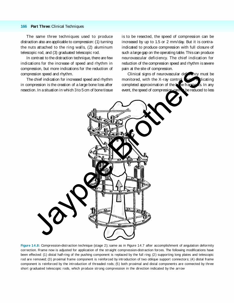
Figure 14.8: Compression-distraction technique (stage 2); same as in Figure 14.7 after accomplishment of angulation deformity
correction. Frame now is adjusted for application of the straight compression-distraction forces. The following modifications have
been effected: (1) distal half-ring of the pushing component is replaced by the full ring; (2) supporting long plates and telescopic
rod are removed; (3) proximal frame component is reinforced by introduction of two oblique support connectors; (4) distal frame
component is reinforced by the introduction of threaded rods; (5) both proximal and distal components are connected by three
short graduated telescopic rods, which produce strong compression in the direction indicated by the arrow
The same three techniques used to producedistraction also are applicable to compression: (1) turningthe nuts attached to the ring walls, (2) aluminumtelescopic rod, and (3) graduated telescopic rod.
In contrast to the distraction technique, there are fewindications for the increase of speed and rhythm incompression, but more indications for the reduction ofcompression speed and rhythm.
The chief indication for increased speed and rhythmin compression is the creation of a large bone loss afterresection. In a situation in which 3 to 5 cm of bone tissue
is to be resected, the speed of compression can beincreased by up to 1.5 or 2 mm/day. But it is contra-indicated to produce compression with full closure ofsuch a large gap on the operating table. This can produceneurovascular deficiency. The chief indication forreduction of the compression speed and rhythm is severepain at the site of compression.
Clinical signs of neurovascular deficiency must bemonitored, with the X-ray control image indicatingcompleted approximation of the bone fragments. In anyevent, the speed of compression cannot be reduced to less
Jayp
ee B
rothe
rs