th17 cells require ongoing classic il-6 receptor signaling
TRANSCRIPT

This is an Open Access document downloaded from ORCA, Cardiff University's institutional
repository: http://orca.cf.ac.uk/131291/
This is the author’s version of a work that was submitted to / accepted for publication.
Citation for final published version:
Harbour, Stacey N., DiToro, Daniel F., Witte, Steven J., Zindl, Carlene L., Gao, Min, Schoeb,
Trenton R., Jones, Gareth W., Jones, Simon, Hatton, Robin D. and Weaver, Casey T. 2020. Th17
cells require ongoing classic IL-6 receptor signaling to retain transcriptional and functional identity.
Science Immunology 5 (49) , eaaw2262. 10.1126/sciimmunol.aaw2262 file
Publishers page: http://dx.doi.org/10.1126/sciimmunol.aaw2262
<http://dx.doi.org/10.1126/sciimmunol.aaw2262>
Please note:
Changes made as a result of publishing processes such as copy-editing, formatting and page
numbers may not be reflected in this version. For the definitive version of this publication, please
refer to the published source. You are advised to consult the publisher’s version if you wish to cite
this paper.
This version is being made available in accordance with publisher policies. See
http://orca.cf.ac.uk/policies.html for usage policies. Copyright and moral rights for publications
made available in ORCA are retained by the copyright holders.

Th17 Cells Require Ongoing Classic IL-6 Receptor Signaling
to Retain Transcriptional and Functional Identity
Authors:
Stacey N. Harbour1, Daniel F. DiToro1, Steven J. Witte1, Carlene L. Zindl1, Min Gao2,3,
Trenton R. Schoeb2, Gareth W. Jones4,5, Simon A. Jones4,5, Robin D. Hatton1,
and Casey T. Weaver1*
Affiliations:
Departments of 1Pathology and 2Genetics, University of Alabama at Birmingham, Birmingham
AL 35294, USA
3Informatics Institute, University of Alabama at Birmingham, Birmingham AL 35294, USA
4Systems Immunity University Research Institute, Cardiff University
5Division of Infection and Immunity, School of Medicine, Cardiff University, Cardiff CF14
4XN, Wales, U.K.
* Correspondence: [email protected] (C.T.W.)
Harbour et al.
2
One Sentence Summary:
We report here that, in addition to its role in the induction of Th17 cell development, ongoing
classic IL-6 receptor signaling is indispensable for maintenance of the Th17 program, identifying
a major mechanism by which Th17 cells are retained despite their tendency to transdifferentiate
into Th1-like cells that drive immune-mediated disease.
Abstract:
Acting in concert with TGF-b, IL-6 signaling induces Th17 cell development by programming
Th17-related genes via STAT3. A role for IL-6 signaling beyond the inductive phase of Th17 cell
development has not been defined, as IL-23 signaling downstream of Th17 cell induction also
activates STAT3 and is thought responsible for Th17 cell maintenance. Here, we find that IL-6
signaling is required for both induction and maintenance of Th17 cells; IL-6Ra–deficient Th17
cells rapidly lost their Th17 phenotype and did not cause disease in two models of colitis. Co-
transfer of WT Th17 cells with IL-6Ra–deficient Th17 cells induced colitis but was unable to
rescue phenotype loss of the latter. High IL-6 in the colon promoted classic, or cis, rather than
trans receptor signaling that was required for maintenance of Th17 cells. Thus, ongoing classic IL-
6 signaling underpins the Th17 program and is required for Th17 cell maintenance and function.
Harbour et al.
3
Main Text:
Introduction
Distinct subsets of effector CD4+ T cells differentiate from multipotent naïve precursors under
control of antigen- and cytokine-induced signals. Th17 cells arise from antigen-activated naïve
CD4+ T cells in response to TGF-b and IL-6 (1-4), the latter inducing a signaling cascade that
recruits Janus kinases JAK1, JAK2 and TYK2. These in turn induce tyrosine phosphorylation of
both STAT3 and STAT1 (5). STAT3 induces the expression of Th17-related genes including
Il17a, Il17f, Il22, Il23r and the master transcription factor Rorc (6-8), which itself is a positive
regulator of key Th17 genes and a negative regulator of alternative lineage fates (9-12). Expression
of Il23r as well as other Th17-specific genes is required for several Th17-mediated diseases,
including inflammatory bowel disease (IBD) (13-15), psoriasis (16) and ankylosing spondylitis
(17), and exposure of Th17 cells to IL-23 is critical for Th17 maintenance and pathogenesis (15,
18-22).
Th17 cells are highly plastic (19, 23). Under the influence of IL-12 most Th17 cells rapidly
extinguish RORgt expression up-regulate T-bet and a Th1-like program (19, 24, 25). Although IL-
23 has been shown to important for Th17 cell maintenance due to its activation of STAT3,
reiterative IL-23 signaling also induces Th17 cells to extinguish Il17a and Rorc in favor of Th1-
related genes (19, 24, 25). This transdifferentiation of Th17 cells is dependent on both STAT4 and
T-bet (19, 23, 26, 27). Thus, the balance of cytokines in the local environment play a critical role
in Th17 maintenance and function. Although the role of IL-6 in Th17 cell induction is well
characterized (2, 4), it is unknown whether ongoing IL-6 signaling contributes to Th17
maintenance, or whether there are additional factors that antagonize lineage-destabilizing signals
such as IL-12– and IL-23–induced STAT4 to preserve the Th17 phenotype.
IL-6 is produced by a wide range of cells, including hematopoietic-derived monocytes,
macrophages and dendritic cells, and non-hematopoietic stromal cells such as fibroblasts and
endothelial cells(28). Various inflammatory cytokines (e.g., IL-1 and TNFa) and innate sensing

Harbour et al.
4
mechanisms contribute to the generation of IL-6, which elicits a broad range of biological
functions on target cells (29). Effects range from control of organ development, the regulation of
the acute-phase response and immune-related functions such as cell survival, differentiation,
proliferation and apoptosis (30). The pleiotropic effects of IL-6 are transmitted through a tightly
controlled, variable mode of signaling (31). Classic, or cis, IL-6 receptor signaling occurs in cells
that express both membrane-bound IL-6Ra and the signal transducing component, gp130, which
bind IL-6 in a hexameric complex where IL-6, IL-6Ra and gp130 exist in a 2:2:2 stoichiometry
(32). While gp130 is ubiquitously expressed (33), IL-6Ra expression is restricted to hepatocytes
(34) and subsets of leukocytes, including naïve T cells (35). In activated cells that express IL-6Ra,
particularly activated neutrophils, monocytes and T cells (36), the extracellular domain can be
shed by proteolysis to liberate a functional soluble IL-6Ra (sIL-6Ra)(37, 38). Soluble IL-
6Ra binds IL-6 to form an agonistic dimer that can bind gp130 to induce IL-6 signaling through a
mechanism termed IL-6 trans-signaling. A soluble isoform of gp130 (sgp130) buffers the activity
of IL-6 trans-signaling and restricts the bioavailability of IL-6–sIL-6Ra complexes for signaling
through membrane-bound gp130 (39). Given that gp130 is ubiquitously expressed, the local
balance between IL-6, sIL-6Ra and sgp130 IL-6 responsive target cells thus determines the
prominent mode of IL-6 signaling acting on any cell type (40). A additional form of IL-6 signaling,
referred to as trans-presentation, involves binding of the cell suface-asscociated IL-6–sIL-6Ra
complexes on a subset of dendritic cells to gp130 expressed on naive T cells, and has been
proposed as a mechanism that promotes the development of pathogenic Th17 cells (41).
An excess of IL-6 relative to sIL-6Ra in acute injury leads to classic signaling, which is
believed to have more homeostatic effects such as the acute phase response (42). An excess of sIL-
6Ra, however, is a marker of active inflammation (43) and allows for trans-signaling to a wide
variety of cell targets, and amplifies inflammatory responses through the recruitment and
activation of mononuclear cells (44, 45). IL-6 trans-signaling has been shown to affect the
recruitment of Th17 cells in model of peritoneal inflammation (46), although it is unknown
whether these effects were direct or indirect.

Harbour et al.
5
Here, we have used a mouse model of Th17 cell-induced colitis (19) to examine a possible
contribution of IL-6 to Th17 biology beyond the early phase of Th17 induction. We find that
expression of IL-6Ra by mature Th17 cells is indispensable for maintenance of the Th17
phenotype and a pathogenic response. Classic IL-6 receptor signaling was dominant over IL-6
trans-signaling in the colon due to the high levels of IL-6 produced there. Our findings establish
that ongoing IL-6 is required for maintenance of Th17 cells and suggest that a major contributor
to Th17 plasticity is the loss of IL-6 signaling.
Results
Naïve CD4+ T cells require IL-6 signaling for development of colitis
The Th17 pathway is critical to IBD pathogenesis. In view of the requirement for IL-6 in Th17
cell development, we examined whether naïve CD4+ T cells deficient in IL-6Ra were able to
develop into Th17 cells and drive intestinal inflammation in vivo. WT and Il6ra-/- naïve T cells
were used as donors in the CD4+ CD45RBHi T cell transfer colitis model (47). Eight weeks after
transfer into Rag1-/- recipients, T cells from WT mice were found to induce substantial disease.
This was reflected by a significant loss of weight (Fig. 1A), as well as an increase in CD4+ cells
recovered from the colonic lamina propria (CLP) of colitic mice (Fig. 1B). While the transfer of
WT CD4+ cells resulted in pronounced pathology (Fig. 1C and D), the transfer of Il6ra-/- CD4+ cells
failed to induce weight loss or significant colonic inflammation.
Consistent with previous reports, flow cytometric analysis of T cells recovered from the
inflamed CLP and mesenteric lymph node (MLN) identified four populations of IL-17A and IFN-
g expressing cells in WT recipients (Fig. 1G and Supplementary Fig. 1)(19). In contrast, there
was a significant reduction in the percentage and number of IL-17A+ and IL-17A+IFN-g+ cells in
the CLP (Fig. 1G,H) and MLN (Supplementary Fig. 1) of recipients of Il6ra-/- T cells.
Interestingly, the relative percentage of IFN-g producing cells in Il6ra-/- recipient T cells was
unchanged in the CLP (Fig. 1H) and increased in the MLN (Supplementary Fig. 1), but their

Harbour et al.
6
numbers were not increased. Thus, IL-6 receptor signaling in these cells is not required for
development of an intact Th1 pathway but is essential for control of pathogenic T cells displaying
a Th17 program (15, 48). The numbers of CD4+ Foxp3+ cells in the CLP (Fig. 1E,F) and MLN
(Supplementary Fig. 1) remained unchanged between recipients of WT and Il6ra-/- cells,
suggesting that reduction in disease in Il6ra-/- recipients was not due to an increase in Treg cells
and suppression of an inflammatory response in the absence of IL-6. Together, these results
indicate that IL-6 signaling in naïve CD4+ T cells is required for the pathogenesis of IBD and that
trans-signaling in the absence of membrane bound IL-6Ra is not sufficient to induce development
of a pathogenic Th17 response. These findings are in agreement with other studies that showed
defective Th17 responses in the absence of IL-6Ra (49).
Maintenance and pathogenesis of Th17 cells is dependent on ongoing IL-6 signaling
Previous results using IL-6–deficient animals or blocking IL-6 interventions suggest that IL-6 is
required to promote Th17-induced disease (12, 50, 51). However it is currently unknown whether
on-going IL-6 signaling is required to maintain Th17 responses. To investigate this, we used a
Th17 transfer model of colitis (19), wherein Th17 polarized cells are cultured ex vivo from naïve
CD4+ T cells of Il17fThy1.1 reporter mice (Fig. 2A) and sorted on the basis of Thy1.1 (IL-17F)
expression (Fig. 2B). Transfer of Thy1.1+ (IL-17F+) Th17 cells into Rag1-/- mice induces a rapid
onset of disease that is equivalent in severity to that caused by transfer of CD4+ CD45RBHi T cells
(19).
Isolated Th17 cells were transferred into groups of Rag1-/- or Il6-/-.Rag1-/- mice. When compared
to Il6-/-.Rag1-/- recipients, Rag1-/- recipients showed significantly elevated disease activity (Fig. 2C,
D). In this regard, the absence of IL-6 lead to a reduction in the percentage and number of IL-17A+
CD4+ cells recovered from the CLP of colitic mice (Fig. 2E, F). However, the percentage and
numbers of double IL-17A+IFN-g+ and single IFN-g+ cells in Il6-/-.Rag1-/- recipients was comparable
to that seen in Rag1-/- recipients. The percentage of CD4+ Foxp3+ cells in the CLP of recipient mice
was also unchanged (Supplementary Fig. 2), suggesting that disease disparity was not due to

Harbour et al.
7
elevated Treg numbers in the absence of IL-6. While IL-6 signaling is known to promote anti-
apoptotic mechanisms (44), Annexin V staining of CD4+ cells recovered from the CLP of colitic
Rag1-/- and Il6-/-.Rag1-/- recipients showed no difference in the percentage of apoptotic (Annexin V+
Propidium Iodide+) cells (Supplementary Fig. 2), suggesting that the differences observed was
not due to a loss of IL-6-mediated T cell survival at this stage of disease. While IL-6 deficient mice
are known to have other defects that may inhibit an immune response, particularly in the innate
immune compartment, these findings support a role for IL-6 in the maintenance of Th17 responses
required for chronic disease progression. We therefore considered the mode of IL-6 receptor
signaling responsible for this outcome.
Classic IL-6 signaling is required for maintenance of pathogenic Th17 cells
The previous experiments suggested that ongoing IL-6 signaling is required to maintain a
pathogenic Th17 response, but could not discern whether IL-6 was acting via classic versus trans
IL-6 signaling to maintain Th17 cells. Because naïve CD4+ T cells from Il6ra-/- mice are not
responsive to direct IL-6 activation (Fig. 3A), we used Th17 cells from Il6ra-/- mice as donors in
the Th17 transfer colitis model to interrogate the requirement for classic IL-6 receptor signaling.
Consistent with our previous work (46), under Th17 polarizing conditions, Il6ra-/- CD4+ T cells
could not produce IL-17A in response to TGF-b + IL-6. However, this lack of IL-6–induced Th17
differentiation was restored by using a recombinant IL-6-sIL–6Ra fusion protein (Hyper-DS-sIL-
6R; HDS), which acts exclusively via IL-6 trans-signaling (Fig. 3B). Importantly, both IL-6 and
HDS generated a similar proportion of Th17 cells when combined with TGF-b (Fig. 3B,
Supplementary Fig. 3). There was no difference in the proliferative capacity of WT and Il6ra-/-
Th17 cells in vitro as measured by vital dye dilution, and RNA-seq analysis of WT (IL-6), WT
(HDS), and Il6ra-/- Th17 precursors showed a very high degree of similarity by principal component
analysis of gene expression, with only 3 genes being > 1.5 fold differentially expressed with an
FDR of < 0.05 (Serpine2, Kit, Nav1) (Supplementary Fig. 3). In addition, pSTAT3 levels were
equivalent between Th17 cells restimulated with IL-6 or HDS in vitro (Supplementary Fig. 3).

Harbour et al.
8
Thus, naïve CD4+ T cells display a comparable capacity to differentiate into Th17 cells in response
to either classic or trans IL-6 receptor signaling.
Exploiting this dual form of Th17 cell regulation, we next considered the relative importance
of these IL-6 receptor signaling mechanisms in maintaining local Th17 responses. Thy1.1+ Th17
precursors from WT (IL-6 polarized), WT (HDS polarized) and Il6ra-/- (HDS polarized) CD4+ T-
cells were transferred into Rag1-/- mice. Disease activity and T cell phenotype were assessed four
weeks after transfer. Notably, while Th17 cells from WT mice caused severe disease in Rag1-/-
recipients, Th17 cells from Il6ra-/- mice induced significantly less pathology (Fig. 3C,
Supplementary Fig. 3). Importantly, there was no difference in the ability of WT (IL-6) versus
WT (HDS) Th17 cells to induce disease, suggesting that these cells are functionally comparable.
The reduced disease observed in recipients of Il6ra-/- Th17 cells was associated with a significant
loss of IL-17A+ and IL-17A+IFN-g+ T cells recovered from the CLP, with a concomitant increase in
the percentage of IFN-g+ T cells when compared to recipients of WT Th17 cells (Fig. 3E,G). As
with disease scores, there were no detectable differences between phenotypes of recovered CD4+
cells from WT (IL-6) and WT (HDS) recipients. Thus, in the absence of IL-6 sustained signaling,
Th17 cells were unstable, indicating that ongoing, classic IL-6 signal is required to maintain a
Th17 response.
Additionally, there were no differences in the percentage or number of CD4+ Foxp3+ cells in
the CLP of Il6ra-/- versus WT recipients (Fig. 3F,H-I), suggesting that loss of Th17 phenotype is
not due to conversion to Treg in the absence of IL-6 signaling. Serum sIL-6Ra, which is elevated
during inflammation due to shedding from membrane bound IL-6Ra–expressing cells, was
significantly increased in recipients of WT Th17 cells. This was in contrast to recipients of Il6ra-/-
Th17 cells (Fig. 3D), in which serum sIL-6Ra levels were equivalent to untreated Rag1-/- mice,
suggesting that sIL-6Ra is a marker of disease activity in this model. Importantly, colon explant
cultures showed a significant reduction in IL-17A in Il6ra-/- Th17 cell recipients, despite showing
an increase in IL-23 and IL-21 levels (Supplementary Fig. 3), suggesting that these STAT3
inducing cytokines were present but unable to compensate for the loss of IL-6 responsiveness in

Harbour et al.
9
CD4+ cells. Together, these results suggest that continued classic IL-6 receptor signaling, rather
than IL-6 trans-signaling is required to maintain the pathogenic phenotype of Th17 cells in vivo,
and that the Th17 phenotype is unstable in the absence of classic signaling.
Loss of Th17 phenotype in Il6ra-/- mice cannot be rescued by co-transfer of WT Th17 cells
Coordinate increases of sIL-6Ra and IL-6 potentiate IL-6 trans-signaling. To rule out the
possibility that reduced disease in recipients of Il6ra-/- Th17 cells (Fig. 3C) was due to lack of
availability of sIL-6Ra, co-transfer experiments were performed to assess the ability of Il6ra-/-
Th17 cells to maintain their phenotype in the presence of inflammation induced by WT Th17 cells.
Th17 precursors from congenically marked WT and Il6ra-/- mice were transferred into Rag1-/-
recipients either individually or together (WT + Il6ra-/- 4 wks), with a fourth group receiving Il6ra-
/- Th17 cells 2 weeks after WT Th17 cell transfer (WT + Il6ra-/- (2 wks)) (Fig. 4A).
Four weeks after initial transfer, WT and Il6ra-/- Th17 cells in recipients of both (WT + Il6ra-/-
4 wks) had repopulated the colon in equivalent numbers (Supplementary Fig. 4), suggesting there
was no survival advantage by population. Disease severity trended with the total number of
recovered CLP CD4+ T cells (Supplementary Fig. 4). For example, disease was significantly
ameliorated in Rag1-/- mice that received single Il6ra-/- Th17 cell transfers when compared to
recipients of WT Th17 cells (Fig. 4B, Supplementary Fig. 5). Both co-transfer groups developed
disease that was equivalent to WT recipients, reflecting the increased total number of pathogenic
CD4+ T cells in these mice. CD4+ T cells recovered from the CLP of recipient mice showed that
regardless of whether they were transferred individually or with WT cells, Il6ra-/- Th17 cells failed
to retain their Th17 identity, as evidenced by a significant reduction in the percentage of IL-17A+
and IL-17A+IFN-g+, as well as IL-17A+TNF-a+ and IL-17A+GM-CSF+ CD4+ T cells compared to
recipients of WT cells (Fig. 4C-D and Supplementary Fig. 4). This correlated with a significant
reduction in the numbers of IL-17A+ and IL-17A+IFN-g+ T cells recovered from the majority of
Il6ra-/- transfer recipients (Fig. 4C).
Harbour et al.
10
Importantly, loss of the Th17 phenotype by Il6ra-/- cells occurred as early as 2 weeks post
transfer (Fig. 4D, bottom panel, and Supplementary Fig. 4). And while the percentage of IFN-
g+ cells was increased (and IL-17A+ cells lost) in the CLP of Il6ra-/- Th17 cell recipients in both co-
transfer groups, the number of IFN-g+ cells was significantly decreased compared to WT cells in
all groups except that of WT + Il6ra-/- cell recipients (4 wks).
The balance between classic IL-6 receptor signaling and IL-6 trans-signaling is tightly
controlled by the bioavailability of IL-6, sIL-6Ra and sgp130. These were therefore quantified in
serum and colon explant cultures from recipient mice (Supplementary Fig. 4). Significant
differences were found: Molar ratios of each analyte showed that systemic sIL-6Ra was in excess
of both IL-6 and sgp130 in all recipient groups (Fig. 4E-F)—a stoichiometry that favors IL-6 trans-
signaling. Conversely, colon explant cultures showed that IL-6 was in molar excess relative to sIL-
6Ra in all groups (Fig. 4E), while sIL-6Ra was again in excess of sgp130 (Fig. 4F)—enabling
both classic and trans-signaling in the colon microenvironment.
IL-6 signaling promotes T cell proliferation and inhibits apoptosis via its actions to increase
Bcl-2(52). We therefore measured cell cycling in WT and Il6ra-/- Th17 cells following transfer.
There were no differences in either BrdU+ or Ki67+ T cells isolated from the spleens and MLNs
(Supplementary Fig. 5) of recipient Rag1-/- mice 7 days after transfers of WT, Il6ra-/- or co-
transferred WT + Il6ra-/- Th17 cells. From this we inferred that there was no difference in the
proliferative capacity of Il6ra-/- Th17 cells, whether transferred individually or co-transferred with
WT Th17 cells. Similarly, there were equivalent percentages of CD4+ T cells from the CLP and
MLN (Supplementary Fig. 5) that were BrdU+ and Ki67+ four weeks after transfer, suggestive of
no defect in the proliferative capacity of Il6ra-/- Th17 cells in active colitis. Interestingly, nearly
70% of CLP T cells from Il6ra-/- Th17 recipients were BrdU+ four weeks after transfer, even though
these cells were not pathogenic. Immunofluorescent staining of colon sections from WT + Il6ra-/-
recipients showed that WT and Il6ra-/- Th17 cells repopulate the colon equivalently at 4 weeks post
transfer (Fig. 4G-H), indicating no defect in the ability of Il6ra-/- Th17 cells to traffic to the colon.

Harbour et al.
11
Together, these findings establish that persistent IL-6 signaling is required to maintain the
Th17 phenotype in vivo, and that classic rather than trans IL-6 receptor signaling is favored for
maintenance of the Th17 program in the colon microenvironment; the requirement for IL-6
signaling is not related to effects on cell cycle or cell survival, but instead appears to be intrinsic
to Th17 lineage maintenance.
IL-6 trans-signaling activates STAT1 and STAT3 in IL-6Ra-deficient Th17 cells ex vivo
IL-6 signaling activates the transcription factors STAT3 and STAT1 in naive CD4+ T cells, and
studies have shown that CD4+ T cell activation status can alter the balance of STAT3/STAT1
activation downstream of IL-6 signaling (53, 54). Il6ra-/- Th17 cells appeared to be either
unresponsive or unable to receive IL-6 trans-signaling in vivo (Fig. 4). To determine whether this
might be due to a lack of inflammation-driven availability of the IL-6–IL-6Ra complex or
unresponsiveness of Th17 cells to this complex, we co-transferred congenically marked Thy1.1+
Th17 cells from WT and Il6ra-/- mice into Rag1-/- recipients. Four weeks after transfer, we examined
the responsiveness of recovered MLN T cells to IL-6 or HDS ex vivo.
Overall, responsiveness to IL-6 signaling was lower in ex vivo T cells compared to naïve T
cells (Fig. 5A,B), due at least in part to reduced expression of IL-6Ra and gp130 on recovered
cells (Supplementary Fig. 6). IL-6 trans-signaling mediated by HDS induced both pY-STAT3
(Fig. 5A,B) and pY-STAT1 (Fig. 5C,D) in a small but significant fraction of both WT and Il6ra-/-
T cells stimulated ex vivo, although the percentage of cells positive for pY-STAT3 was
substantially higher than those positive for pY-STAT1. In contrast, only WT CD4+ T cells were
responsive to classic IL-6 signaling ex vivo. IL-6 stimulation induced pY-STAT3 in a significant
fraction of WT cells, but, interestingly, did not induce pY-STAT1 in the same population,
indicating that, unlike naïve precursors, coupling of the IL-6 receptor to STAT1 via classic
signaling is lost in Th17 cells.
Collectively, these data show that both WT and Il6ra-/- CD4+ T cells are responsive to trans-
signaling ex vivo, but this is limited by diminished gp130 expression, consistent with activation-
Harbour et al.
12
induced downregulation of gp130 in CD4+ T cells (53, 55, 56). Moreover, classic IL-6 receptor
signaling in WT cells, which promotes retention of the Th17 phenotype, preferentially induces pY-
STAT3 without pY-STAT1. Therefore, ongoing classic IL-6 signaling in Th17 cells is required to
maintain tyrosine phosphorylation of STAT3, and thus IL-6-induced STAT3 activation is critical
for both the development and maintenance of Th17 cells.
Maintenance of the Th17 transcriptional program is dependent on ongoing IL-6 receptor
signaling
To further characterize the phenotypic divergence of Il6ra-/- Th17 cells, we performed RNA-
sequencing analysis of T cells that were isolated from the CLP of Rag1-/- mice that received co-
transfers of congenically marked WT and Il6ra-/- Th17 cells (Fig. 6A). Recipient mice were
repopulated with equivalent percentages (Fig. 6B) and numbers (Fig. 6C) of Th17 cells in the CLP
four weeks after transfer. Transcriptome analysis revealed a number of differences in gene
expression in recovered WT and Il6ra-/- CD4+ T cells ex vivo (Fig. 6D). Gene set enrichment
analysis (GSEA) using a previously defined Th17–Th1 gene set (57) identified significant
enrichment of Th1-related genes in Il6ra-/- T cells ex vivo (Fig. 6E, right and Supplementary Fig.
7). Conversely, and in accord with our previous findings, the transcriptome of WT T cells was
enriched for a subset of genes related to Th17 cells ex vivo (Fig. 6E, left and Supplementary Fig.
7). Signature Th17 related genes that were retained in WT but not Il6ra-/- T cells ex vivo included
Il17a, Il17f, Il21, Il22, Il23r, Rorc and Ccr6 (Fig. 6F). A number of Th1-related genes was
enriched in Il6ra-/- T cells, including Ifng, Tbx21 and Il12rb2, indicative of a molecular program
more similar to Th1 cells than the Th17 precursors. GSEA using a dataset derived from colonic
CD4+ T cells (GSE58147)(58) showed the expression pattern of WT and Il6ra-/- CD4+ T cells was
significantly related to that of WT and Il23r-/- CD4+ T cells, respectively (Supplementary Fig. 7).
Likewise, a subset of genes induced by Tbx21 was significantly associated with Il6ra-/- T cells
(Supplementary Fig. 7), consistent with the transdifferentiation of these cells to a Th1-like
program.

Harbour et al.
13
Using a previously published dataset defining the transcriptional program of Th17 cells (9),
significant enrichment of Stat3- and Rorc-induced genes was found in WT T cells ex vivo, as
expected, whereas Stat3- and Rorc-repressed genes were significantly enriched in Il6ra-/- T cells
(Fig. 6G-J). In addition, expression of genes regulated by other transcription factors associated
with the Th17 lineage, including Batf, Irf4, Fosl2 and Hif1a, was significantly reversed in Il6ra-/-
T cells ex vivo (Supplementary Fig. 7), in fitting with the loss of Th17 phenotype in these cells.
As expected, the Stat3- and Rorc-induced genes enriched in WT cells contained some overlap,
including Il17a, Il17f, Il23r, Il1r1 and Lif (Fig. 6H,J). Interestingly, there was little overlap in
genes repressed by Stat3 and Rorc in Il6ra-/- CLP T cells. Targets of Rorc included Ahr, Ccr5 and
Il12rb2 (Fig. 6H), while Stat3 repressed genes included Fasl, and the Th1-related transcription
factors Tbx21 and Irf1 (Fig. 6J). This is consistent with data suggesting that Stat3 is a key
transcriptional activator in Th17 cells, whereas Rorc is thought to be an expression modulator (9).
Our previous work has shown that Th17 cells develop divergent gene expression profiles under
the influence of IL-23 signaling (19), such that an increase in expression of Tbx21, Ifng, Ccr5 and
Fasl is correlated with a significant portion of IFN-g–producing ex-Th17 cells, whilst a decrease
in the expression of Il17a, Il17f, Ccr6 and Ccl20 is correlated with a reduction in IL-17A+ Th17
cells. We find here that Il6ra-/- CLP CD4+ T cells were enriched for the aforementioned Th1 genes.
Conversely, expression of Th17-related genes was associated with WT, rather than Il6ra-/- CLP
CD4+ T cells, further indicating that ongoing IL-6 signaling is required to maintain Th17 program
stability. Taken together, these data confirmed our finding that Il6ra-/- Th17 cells rapidly lose their
Th17 phenotype in vivo, and extended them to show that, absent classic ongoing IL-6 signaling,
the entire Th17 gene expression program is downregulated, favoring the transition of many cells
to a Th1-like program.
Enforced RORgt expression in IL-6R-deficient Th17 cells supports maintenance of the Th17
program but does not completely prevent Th1 transdifferentiation
Harbour et al.
14
A critical gene target of IL-6 signaling in Th17 cells is Rorc, which underpins the Th17 gene
expression program while inhibiting alternative programs. Important functions of IL-6/STAT3-
induced RORgt include inhibition of IFN-g/STAT1 and IL-12/STAT4 via suppression of T-bet
(61), although RORgt also sustains expression of Il23r, which has been shown to be central to the
transdifferentiaiton of Th17 cells into Th1-like cells in vivo via STAT4 signaling (19, 26, 73). Our
gene expression analysis showed that in IL-6Ra-deficient Th17 cells, Rorc and Il23r expression
were rapidly lost along with other genes of the Th17 program following adoptive transfers (Fig.
6F). To determine whether loss of RORgt was responsible for loss of the Th17 phenotype, we
polarized congenically marked naïve WT and Il6ra-/-.Il17fThy1.1 T cells under Th17 conditions with
HDS and transduced cells with retroviruses that expressed GFP alone or GFP plus RORgt at 24
hrs of a 5 day culture (Fig. 7A). As expected, Th17 precursors from both WT and Il6ra-/- mice
expressed a higher percentage of IL-17A after enforced RORgt expression (Fig. 7B, right).
GFP+Thy1.1+ Th17 cells were sorted 4 days after transduction and WT Th17 cells expressing GFP
alone (WT pMIG) were transferred together with either Il6ra-/- cells expressing GFP alone (Il6ra-/-
pMIG) or Il6ra-/- cells expressing both GFP and RORgt (Il6ra-/- pMIG-RORgt) into Rag1-/-
recipients.
Three weeks after transfer, both co-transfer groups had developed comparable disease (Fig. 7C,
7F). CD4+ T cells recovered from the CLP showed that, consistent with previous results (Fig. 4),
Th17 cells transfected with empty vector (Il6ra-/- pMIG) rapidly lost their Th17 phenotype as shown
by a significant reduction in IL-17A+ cells compared to co-transferred WT (WT pMIG) Th17 cell
controls (Fig. 7D and E), with a concomitant increase in the relative frequency of IFN-g+ cells. In
contrast, Il6ra-/- Th17 cells transfected with RORgt (Il6ra-/- pMIG-RORgt) had a much greater
propensity to retain their Th17 phenotype even in the absence of classical IL-6 signaling. Thus,
the frequencies of both IL-17A+ and IL-17A+ IFN-g+ cells from recipients of RORgt-transfected
Il6ra-/- Th17 cells were markedly increased in comparison to those transduced with vector alone
(Fig. 7D and E). Moreover, RORgt-transfected Th17 cells had significantly greater retention of
IL-17 expression compared to WT controls, reflecting the role of RORgt in promoting Th17

Harbour et al.
15
cytokine production (12). Interestingly, although RORgt-transfected Il6ra-/- Th17 cells had
significantly decreased expression of IFN-g compared to Il6ra-/- Th17 cells transfected with empty
vector, they showed a modest, but significant increase in the fraction of IFN-g+ cells compared to
WT Th17 cells. This is consistent with the development of Th1-like cells from the larger frequency
of Th17 precursors maintained by exogenous RORgt (19), but also suggested that enforced RORgt
expression did not entirely repress transdifferentiation of Th17 cells into Th1-like cells.
In accord with the phenotypic data, gene expression analysis of CD4+ T cells recovered from CLP
showed that RORgt-transfected Il6ra-/- Th17 cells had a significant increase in expression of genes
of the Th17 program compared to empty-vector Il6ra-/- Th17 cells, including Il17a, Rorc, Il17f and
Il23r (Fig. 7G). Further, there was decreased Il12rb but not Tbx21, suggesting differential
sensitivities of these two genes to RORgt-mediated repression. Notably, expression of Rorc was
not significantly increased over that of recovered WT controls, indicating the levels of RORgt
driven by retroviral transduction were physiologic. As anticipated, Il23r was significantly elevated
in RORgt-transfected Il6ra-/- Th17 cells compared to both empty-vector Il6ra-/- cells and co-
transferred WT control cells, consistent with an central role for IL-6–induced RORgt in the
maintenance of Il23r expression. As IL-23 signaling via STAT4 is known to be a principal pathway
mediating the transdifferentiation to Th1-like cells in vivo (19, 26), it is likely that the elevated
expression of IL-23R by RORgt-transfected Il6ra-/- Th17 cells contributed to the modest increase in
expression of IFN-g by these cells compared to WT controls, despite on-going RORgt expression.
Together, these results suggest that ongoing classical IL-6 signaling supports the Th17 program
by sustaining RORgt expression that contributes to the maintenance of Th17 cells, which provide
a reservoir of cells that can transdifferentiate into pathogenic Th1-like cells that are essential to
colitogenesis.

Harbour et al.
16
Discussion
IL-6 is required for lineage specification of Th17 cells through its activation of STAT3, which
antagonizes the IL-2/STAT5-driven specification of induced Treg cells with which Th17 cells
share TGF-b–dependent developmental programming (62-64). Here, we report that in addition to
its key role in Th17 cell induction (3, 4), there is an absolute requirement for persistent IL-6
signaling to maintain the Th17 genetic program, and thus Th17 cells. Although it has been reported
that IL-23 signaling is important for the maintenance of Th17 cells (18, 19, 21), we find that
ongoing IL-6 signaling is indispensable and foundational, acting upstream of IL-23 via its unique
function to sustain RORgt expression and thereby IL-23R expression. Importantly, although Th17
cells were fully competent to receive IL-6 trans-signaling, classic IL-6 signaling proved necessary
for Th17 maintenance in the uninflamed and inflamed intestines. This appears to be due to the
combined effects of high local production of IL-6, which was incompletely buffered by soluble
IL-6Ra, and the abundance of gp130-expressing non-T cells (e.g.., intestinal epithelial cells),
which act as a sink for trans-signaling. Possible differences between the classic and trans-signaling
modes may also be contributory and deserve further study.
In contrast to Th1 and Th2 cells, Th17 cells show substantially greater plasticity (19, 23, 65).
This reflects intrinsic instability in the transcriptional circuit upon which Th17 cell development
is based. Whereas the Th1 and Th2 programs are reinforced by cell-extrinsic and -intrinsic
mechanisms that enhance expression of T-bet and GATA-3, respectively(66), mechanisms by
which RORgt expression is sustained and the Th17 program maintained have been unclear. IL-23
has been reported to be a trophic factor for Th17 cells (19, 23, 65), but IL-23 signaling also
promotes the transdifferentiation of Th17 cells to Th1-like cells (19, 26), thereby contributing to
the instability of Th17 cells. Despite its dominant activation of STAT3, IL-23 signaling also
activates STAT4, leading to expression of T-bet, which represses RORgt via direct binding to the
Rorc locus (26, 61). Our finding that Th17 cells require tonic or intermittent classic IL-6 signaling,
which shifts from a STAT3–STAT1 output during early development to a pure STAT3 output in
mature cells, indicates that this pathway of STAT3 activation may be essential to counterbalance

Harbour et al.
17
the STAT4 output of the IL-23 (or IL-12) receptor to sustain RORgt and the Th17 program.
However, it also appears that the IL-6–dependent maintenance of a pool of Th17 cells provides
precursors from which pathogenic Th1-like cells can arise. In view of the findings herein, it would
appear that stable expression of RORgt does not completely block Th1 reprogramming, consistent
with a dominant role of T-bet in overriding the Th17 program.
In view of the role for STAT1 in modulating the STAT3-dominant output of the IL-6 receptor
via formation of STAT3-STAT1 heterodimers (67), results herein suggest that the maintenance of
mature Th17 cells via classic signaling may be strictly dependent on STAT3 homodimers.
Moreover, as STAT1 signaling impairs Th17 programming (59), the loss of STAT1 activation in
Th17 cells receiving classic IL-6 signaling may further reinforce maintenance of the Th17
program. Because trans IL-6 signaling in Th17 cells retained a STAT1 output, which can also
induce T-bet expression, it is possible that Th17 cells exposed to this mode of IL-6 signaling may
be less resistant to transdifferentiaion mediated by Th1-promoting cytokines that preferentially
activate STAT1 (e.g. IFNs) or STAT4 (e.g. IL-12). This will require further study. Notably, despite
its elevated expression in the colons of recipients of IL-6Ra-deficient Th17 cells, IL-21, which
also activates STAT3 and has been implicated as an autocrine factor for Th17 cells (8, 68), was
also unable to compensate for deficiency of IL-6 signaling in maintenance of the Th17 program.
The basis for the developmentally-linked shift in STAT activation by the IL-6 receptor and
insufficiency of autocrine IL-21 to substitute for IL-6 signaling will also require further study. In
any case, collectively, these findings suggest that the local balance of IL-6 and IL-23 is critical in
sustaining or destabilizing the Th17 program, resulting a protective versus pathogenic Th17
response.
The finding of a critical requirement for paracrine IL-6 in both the induction and maintenance
of Th17 cells draws a further parallel between Th17 cells and Treg cells (62). Treg cells require
IL-2 for both their development and maintenance (69), but are unable to produce IL-2 themselves.
Instead, they are thought to rely on production of IL-2 by activated naive or other effector T cells
(69), akin to the reliance of Th17 cells on IL-6 produced by innate immune cells. For both lineages,

Harbour et al.
18
the dependence on a single cytokine as both developmental inducer and maintenance factor renders
them particularly susceptible to lineage instability. Moreover, as these two cytokines are cross-
inhibitory—IL-6 promotes the development of Th17 cells and inhibits Tregs, while IL-2 promotes
the development of Tregs and inhibits Th17 cells—this suggests that competition for their
respective inductive/maintenance cytokine is a major contributor to Th17 versus Treg pool size
both at homeostasis and during the emergence and subsidence of an antigen-driven inflammatory
response. This is of particular importance in intestinal tissues, which are enriched in both Th17
and Treg cells and are subject to ongoing antigenic stimulation from the intestinal microbiome
(70). In view of the current data that implicate importance of the balance of these cytokines in
controlling Th17–Treg cell balance during both an evolving and waning antigenic response, it will
be important to better define the precise sources and dynamics of IL-2 and IL-6 production.
The relative contribution of Th17 and Th1 cells to immune-mediated diseases has been an area
of considerable interest. Early seminal studies identified a definitive link between Th1 cells and
colitis (47, 71), whereas subsequent studies established an essential role for the IL-23–Th17 axis
in disease pathogenesis (15, 72). A recent study from our group reconciled these findings by
demonstrating that Th1-like cells, which derive from Th17 precursors contingent on intact IL-23-
induced STAT4 activation and T-bet expression, were critical for colitis development (73). A
notable feature of our current findings is that IL-6Ra-deficient Th17 cells more readily transitioned
to IFN-g–producing Th1 cells in vitro yet failed to cause colitis in vivo. Although this appears to
contradict a requirement for Th1 cells in the pathogenesis of IBD, our previous studies showed
that Th17 cells assisted the development and/or survival of pathogenic Th1 cells that were unable
to drive disease on their own (73). While the basis for Th17-mediated support for de novo Th1
development remains to be defined, a trivial explanation for the observed lack of disease may be
that the rapid loss of Th17 cells unable to receive IL-6 signaling resulted in an inadequate number
of Th17 precursors to give rise to sufficient Th1-like cells or promote their de novo development.
In light of a critical role for ongoing, classic IL-6 signaling in Th17 maintenance, a more
detailed understanding of the dynamics of IL-6Ra and gp130 expression by Th17 cell populations
Harbour et al.
19
is needed, as is a better understanding of factors regulating the local concentrations of sIL-6Ra,
IL-6 and sgp130 in specific tissue microenvironments such as the intestines. While we and others
have found that IL-6Ra and gp130 are downregulated upon T cell activation (31), the mechanisms
are different. IL-6Ra appears to be downregulated primarily by cleavage of the extracellular
domain by the metalloproteinases ADAM10 and ADAM17 in humans (74), or only ADAM 10 in
mice (75), which would appear to favor a shift from classic to trans IL-6 signaling in developing
Th17 cells. Surface expression of gp130 is downregulated by internalization, alternative splicing
of the encoding mRNA to favor the secreted form, and/or other unidentified mechanisms (31).
While IL-2 signaling has been shown to inhibit expression of Il6ra and Il6st (gp130)(76), little
else is known about conditions that promote retention or re-expression of IL-6Ra and gp130 by T
cells, which now appear to be key in determining which cells are competent to retain the Th17
program and by what mode of IL-6R signaling. Although IL-6 trans-signaling was intact and
comparable in WT and IL-6Ra-deficient T cells recovered from colitic mice, it was substantially
diminished, reflecting diminished gp130 expression and/or signaling competency. This impacted
classic signaling in WT cells as well and suggests that mechanisms controlling the expression of
gp130 by effector T cells are limiting for Th17 maintenance—irrespective of the expression of IL-
6Ra and the local balance of free versus sIL-6Ra-bound IL-6.
Although IL-6 trans-signaling was fully sufficient to differentiate Th17 cells in vitro, it was
insufficient for either Th17 development or maintenance in vivo, despite elevated sIL-6Ra. The
reasons for this are unclear, but are unlikely to involve complete buffering by sgp130, as sIL-6Ra
was in substantial excess in colitic mice. Rather, competition from intestinal innate, epithelial and
stromal cells, all of which can express gp130 and can signal in trans, appeared to prevent IL-6
trans-signaling in Th17 cells. At least in the colon, a high IL-6–sIL-6Ra ratio due to high local
production of IL-6 favored sufficiently high concentrations of free IL-6 to enable classic signaling
to the relatively small percentage of T cells co-expressing membrane bound IL-6Ra and gp130.
Interestingly, the stoichiometry of IL-6–IL-6Ra was comparable in both the homeostatic and
inflamed intestines, consistent with a dominant role for classic signaling in Th17 maintenance in
Harbour et al.
20
this tissue both at baseline and in the diseased state. Whether this is a common feature of mucosal
and non-mucosal barrier tissues is unknown.
An association between polymorphisms in the IL6RA gene and susceptibility to both RA and
IBD have been found (77, 78). The IL6RA variant rs2228145, which leads to increased proteolytic
cleavage of IL-6Ra and thus a reduction in membrane-bound IL-6Ra in favor of increased serum
levels (79) is associated with reduced risk of both RA (77) and IBD (78). Although the mechanism
of protection remains to be characterized, it is possible that it involves both increased buffering of
free IL-6 as well as reduced expression of cell surface IL-6Ra, such that classic signaling required
for Th17 development, and thus disease induction, is impaired. Given the findings herein, it is
possible that this variant also compromises the maintenance of Th17 cells.
Our finding that ongoing IL-6 signaling is required for Th17 maintenance has implications for
therapeutics that target Th17-mediated diseases. In systemic Th17-related autoimmune diseases
such as rheumatoid arthritis, juvenile idiopathic arthritis, and systemic sclerosis, treatment with
biologics that inhibit IL-6 is an effective or promising therapy (80-82). This is in contrast to tissue-
specific immune-mediated diseases, including IBD, where therapies that inhibit IL-6 signaling
have not gained clinical use despite evidence that antibodies targeting IL-6 or IL-6Ra may have
efficacy (83). Given that IL-17A- and IL-22-producing Th17 cells appear to be protective rather
than pathogenic in the intestines (84), persistent classic IL-6 receptor signaling that sustains
intestinal Th17 responses may enhance barrier function to restrain disease. This is in line with the
view that classic and trans-signaling promote anti- and pro-inflammatory responses, respectively
(85), and suggests that inhibiting classic signaling and thus the protective effects of IL-6 may
exacerbate disease. Given that IL-6 can amplify inflammation-induced injury to a variety of cells
and tissues, it remains a valid therapeutic target (31), although it may be important to selectively
target the pro-inflammatory effects of IL-6 while sparing its protective effects. The development
of inhibitors that specifically target trans-signaling by inhibiting the docking of the IL-6–IL-6Ra
complex to gp130 may offer a clinical advantage in selectively targeting the deleterious effects of

Harbour et al.
21
IL-6 without affecting the more beneficial effects, including the maintenance of protective Th17
cells.
Materials and Methods
Study Design
This study was designed to investigate whether ongoing IL-6 is required for Th17 function, and
whether classical or trans signaling was more important for maintenance of Th17 cells.
Experiments consisted of analyzing T cell populations in the inflamed intestines of
immunodeficient recipients as part of two established T cell transfer models of colitis. All animals
were bred and maintained in accordance with institutional animal care and use committee
regulations. Littermate comparisons were used for all experiments where possible. Gender and
aged matched male and female mice were used, and animals were randomly assigned into different
groups. Group sizes were the minimum required to detect statistically significant differences based
on previous experience. Experiments were not blinded to investigators, but histopathological
samples were blinded prior to scoring. All experiments were replicated as presented in the figure
legends.
Mice
The following mice were purchased from the Jackson Laboratories: C57BL/6.CD45.1, Il6-/-,
B6.FVB-Tg(EIIaCre), and Rag1-/-. The generation of Il17fThy1.1/Thy1.1 reporter mice has been described
previously (19). Il17fThy1.1 mice were crossed to B6.CD45.1 to create Il17fThy1.1(CD45.1). Il6rafl/fl mice
were a gift from A. Drew (Univ. of Cincinnati) and were crossed with EIIaCre mice to generate Il6ra-
/-. Where indicated, crosses between Il17fThy1.1/Thy1.1 reporter mice and individual knockout mice were
made in our facility to generate mice homozygous for Il17fThy1.1 and respective gene deletion alleles
(eg. Il6ra-/-). All mice were on a C57BL/6 background.

Harbour et al.
22
CD4 T cell preparation and culture
CD4+ T cells were purified from pooled spleen and lymph nodes by Dynabeads Mouse CD4,
followed by DETACHaBEAD Mouse CD4, according to the manufacturer’s instructions
(ThermoFisher). Naïve CD4+ CD62L+ CD25- T cells were obtained by FACS sorting. CD4+ T cells
were cultured at a ratio of 1:5 with irradiated splenic feeder cells (or, where noted, Dynabeads T-
Activator CD3/CD28 beads (ThermoFisher)) for 7 d in RPMI containing 10% FBS, 100 IU/mL
penicillin, 100 µg/mL streptomycin, 1 µM sodium pyruvate, 1x non-essential amino acids, 2.5 µM
b-mercaptoethanol, and 2 µM L-glutamine (R10 medium). Cells were stimulated with 2.5 µg/mL
anti-CD3 (clone 145-11), and for Th17 polarizations were supplemented with 2.5 ng/mL rhTGF-
b1 (R&D systems), 10 ng/mL rmIL-6 (R&D systems) or 10 ng/mL hyper-IL-6 (S. Jones, Cardiff
University), 10 µg/mL anti-IFN-g (clone XMG1.2), and 10 ug/mL anti-IL-4 (clone 11B11). In
restimulation cultures, viable T cells were recovered on day 7 of initial cultures and activated with
fresh splenic feeders or CD3/CD28 activator beads, cytokines and antibody mixtures as indicated.
Unless otherwise noted, rmIL-23 or rmIL-12 (R&D systems) were added at 20 ng/mL and 2.5
ng/ml respectively.
CD4 adoptive transfer model of colitis
CD4 cells were cultured under Th17 polarizing conditions for 7 d. Viable cells were recovered
(Ficoll) and Thy1.1+ cells were isolated by magnetic sorting (Miltenyi Biotec). A total of 4 x 10⁵
Thy1.1+ Th17 cells were injected i.p into age and sex matched Rag1-/- recipients. For co-transfer
experiments, 2 x 10⁵ each of WT and Il6ra-/- Th17 cells were transferred. For the naïve CD4 transfer
model, CD4+ CD45RBHi cells were FACS sorted from pooled spleen and lymph nodes and a total
of 4 x 10⁵ cells were injected into age matched Rag1-/- recipients. Mice were monitored regularly
for signs of disease and weighed weekly. At 4 or 8 weeks after transfer, mice were sacrificed and
MLNs and colons were recovered. Colons were cut longitudinally, and small lengths of tissue were
obtained from the proximal, middle and distal portions of the colon, fixed in 10% formalin, and
Harbour et al.
23
processed for histopathological analyses. Samples were scored by a pathologist in a blinded
fashion as previously described (86).
Isolation of colonic lymphocytes
Lamina propria lymphocytes from the colon (CLP) were isolated as described previously(86).
Briefly, colons were cut longitudinally, and were cut into 1 cm long strips. Tissues were washed
in cold 1 x HBSS containing 2% (vol/vol) FBS plus 100 IU/mL penicillin and 100 µg/mL
streptomycin. After being washed, tissues were digested at 37 C for 45 min with gentle stirring in
R10 medium containing Collagenase D (100 U/mL) and DNase I (20 mg/mL) (Sigma). Lamina
propria lymphocytes were purified on a 40%/75% (vol/vol) Percoll gradient by centrifugation for
20 min at 25 °C.
Flow cytometric analysis
Where indicated, CD4+ cells were stimulated with PMA (50 ng/mL; Sigma) and Ionomycin (750
ng/mL; EMD Millipore) for 5 h in the presence of GolgiPlug (BD Biosciences). Intracellular
staining was performed as previously described(86). LIVE/DEAD Fixable near-IR Dead Cell Stain
(Invitrogen) was used to exclude dead cells. Staining of CD4+ T cells was performed with
antibodies to CD4 (RM4-5), IL-6Ra (D7715A7), IL-17A (TC11-18H10), TCR-b (H57-597) and
Foxp3 (FJK-16s) from BD Biosciences, IFN-g (XMG1.2), TCR-b (H57-597), gp130 (KGP130),
and Thy1.1 (His51) from eBioscience, and Thy1.1 (OX-7) from Biolegend. For apoptosis analysis,
Annexin V and Propidium Iodide staining was performed using an Annexin V Apoptosis Detectin
Kit (eBioscience) according to manufacturer’s instructions. Samples were acquired on an LSRII
or Attune NxT flow cytometer and data were analyzed with FlowJo software (Tree Star).
Phospho-STAT staining
CD4+ T cells from in vitro cultures or MLN cells from recipient Rag1-/- mice were rested for 4 h at
37 °C in RPMI, then stimulated for 15 min with 0.95 nM IL-6 or HDS. Cells were fixed for 15
Harbour et al.
24
min at 37 °C with 3% paraformaldehyde (PFA), then washed and permeabilized for 30 min at 4
°C with 90% methanol. Following two washes, cells were stained for 1 h at room temp with
antibodies against CD4 (clone RM4-5), pSTAT3 (Y705; clone 4/P-STAT3) and pSTAT1 (Y701;
clone 4a), all from BD Biosciences.
BrdU staining
Where noted, CD4+ T cell recipient Rag1-/- mice were fed with 1 mg/mL Bromodeoxyuridine
(BrdU; Sigma) in tap water with 1% sucrose (w/vol) for 7 d prior to sacrifice. At 1 and 4 weeks
after transfer, CD4+ cells from spleen, MLN and CLP were harvested and BrdU was detected using
the BrdU Flow Kit (BD Biosciences) according to manufacturer’s instructions. CD4+ cells were
co-stained with CD4 (RM4-5), LIVE/DEAD Fixable near-IR Dead Cell Stain, TCR-b (H57-597)
and Ki67 (eBioscience, SolA15).
Measurement of IL-6, sIL6Ra, sgp130, and other analytes
Blood was collected from mice upon euthanasia, and serum obtained by centrifugation. For colon
explant cultures, colon tissue was flushed, cut into three equal sections and then opened
longitudinally. Individual tissue segments were minced and placed in 0.4 ml of R10 medium for 2
d, then supernatant collected by centrifugation. Cytokines and other analytes were measured by
Milliplex assay (Millipore) following manufacturer’s instructions.
Immunohistology
For immunostaining experiments, colons were fixed in 2% (vol/vol) PFA for 2 h at room
temperature, washed in PBS, and then set overnight at 4 ºC in 20% (wt/vol) sucrose solution.
Tissue was then rinsed in PBS, cut into equal segments, embedded in O.C.T. (Tissue-Tek), and
snap frozen. Tissue sections (6-8 µm) were stained with biotinylated anti-CD45.1 (clone A20,
eBioscience) followed by Streptavidin-Alexa Fluor 488 (Invitrogen), APC-conjugated anti-
EpCAM1 (G8.8, eBioscience), and Alexa Fluor 594-conjugated anti-CD3 (17A2, eBioscience)

Harbour et al.
25
followed by donkey anti-goat IgG Alexa Fluor 594 to amplify the signal. Images were obtained
using a Nikon A1R confocal microscope and processed with Nikon Elements software.
RNA-seq analysis
Adaptors were trimmed and aberrant reads were removed using TrimGalore (version 0.4.5). The
quality controlled reads were mapped onto the mouse genome build GRCm38
(ENSEMBL.mus_musculus.release-75)(87) using STAR (version 2.5.3)(88). BAM files were
sorted using SAMtools (version 0.1.18)(89), and reads were counted for each gene using HTSeq
(version 0.7.2)(90). Differential expression analysis was performed using DESeq2 (version
1.18.1)(91) using R (version 3.4.3). Dispersion shrinkage of fold changes was performed with the
ASHR algorithm (92). To prepare data for gene set enrichment analysis, results of differential gene
expression analysis were ranked by signed p-value. Gene lists were ranked by p-value of Wald test
multiplied by sign of fold-change and analyzed for enrichment of curated query gene sets using
Fast Gene Set Enrichment Analysis (fgsea, version 1.4.0) with 1 million permutations. Gene set
enrichment p-values of Normalized Enrichment Scores (NES) were corrected with the Benjamini-
Hochberg procedure. When available, gene sets were made from reported results of transcriptome
analysis. However, for many published data sets, only raw data were available, and gene sets were
created using in-house scripts. Query gene sets were created from publicly available data. Raw
data was obtained from the gene expression omnibus (93). Bulk RNA-seq data was analyzed as
described above and gene sets were made by selecting the top 250 up and down regulated genes
by p-value with a fold change of at least 1.5. Microarray data was analyzed using the limma R
package (94). The functions model.matrix and lmFit were used to create and fit linear models,
respectively. Due to the increased noise in microarray data, the top 125 up and down regulated
genes were selected by p-value with a fold change of at least 1.5 to create gene sets. All query gene
sets used are available in Supplemental Table 1.

Harbour et al.
26
The Jaccard indexes were calculated for each leading edge gene set versus all others by dividing
the number of genes in the intersection by the number of genes in the union of each comparison.
The resulting matrix underwent dimension reduction by using t-Stochastic Neighbor Embedding
(t-SNE) as implemented in the R package Rtsne (version 0.13, with settings dim=2, perplexity=5,
and max_iter = 50,000).
Gene set enrichment analysis
Gene set enrichment analysis was performed using software from the Broad Institute(95). A pre-
ranked analysis was performed on a gene list ordered by signed p-value using Classic enrichment
statistic, 10,000 permutations, and meandiv Normalization mode. Th17 and Th1 associated gene
sets were created from publicly available microarrays (GEO GSE14308)(57). Limma (3.32.8) was
used to calculate differential expression by comparing Th1 samples to Th17 samples. The top 250
genes by most significant adjusted p-value were separated by fold change. Those with a log2
transformed fold change greater than zero generated the Th1 gene set, those with fold changes less
than zero generated the Th17 gene set. Hallmark gene sets were obtained from the MSigDB (96).
Heatmaps were created using R 3.0.1 and the function heatmap.2 from the ggplot2 package (97).
Retrovirus production and CD4+ T cell transduction
GFP and RORgt GFP retroviruses were produced by transfecting Platinum-E packaging cells (Cell
Biolabs) with pMIG (Addgene #9044) or MIGR-RORgt (Addgene #24069) retroviral plasmids
using Lipofectamine 2000 (ThermoFisher), and collecting supernatant 24 and 48 hours later.
To transduce Th17 cells, FACS-sorted naïve CD4+ T cells from Il17fThy1.1 mice were cultured in R10
medium with Dynabeads Mouse T-activator CD3/CD28 beads (ThermoFisher) at a ratio of 5 beads
per CD4+ cell. After 24 hours, cells were spin infected with retrovirus containing media for 90 min
(2500 rpm, 30 ºC). Cells were then washed with R10 medium and cultured for a further 4 d under
Th17 polarizing conditions. For transfers, CD4+ Thy1.1+ GFP+ cells were FACS sorted and injected
i.p into age and sex matched Rag1-/- recipients.

Harbour et al.
27
Real-time PCR
Congenically marked CD4+ TCR-b+ cells were sorted from the CLP of Rag1-/- recipients. Total RNA
was isolated using an miRNeasy Isolation kit (Qiagen) according to manufacturer’s instructions.
cDNA was synthesized with the iScript Reverse Transcription Supermix (BioRad), and real-time
PCR was performed with SsoAdvanced Universal Probes Supermix or SsoAdvanced Universal
SYBR Green Supermix (BioRad) for 35-40 cycles. Reactions were run in duplicate and normalized
to B2m.
Statistical analysis
Statistical significance was calculated using Prism software (GraphPad). The nonparametric two-
tailed Mann Whitney test or Kruskal Wallis test was used to determine significance for pathology
data, all other data was analyzed using unpaired two-tailed Student’s t-test or one-way ANOVA
as appropriate. All P values ≤ 0.05 were considered significant.
References and Notes:
1. P. R. Mangan et al., Transforming growth factor-β induces development of the TH17
lineage. Nature. 441, 231–234 (2006).
2. C. T. Weaver, R. D. Hatton, P. R. Mangan, L. E. Harrington, IL-17 family cytokines and
the expanding diversity of effector T cell lineages. Annu. Rev. Immunol. 25, 821–852
(2007).
3. M. Veldhoen, R. J. Hocking, C. J. Atkins, R. M. Locksley, B. Stockinger, TGFβ in the
Context of an inflammatory cytokine milieu supports de novo differentiation of IL-17-
producing T cells. Immunity. 24, 179–189 (2006).
4. E. Bettelli et al., Reciprocal developmental pathways for the generation of pathogenic
Harbour et al.
28
effector TH17 and regulatory T cells. Nature. 441, 235–238 (2006).
5. P. C. Heinrich et al., Principles of interleukin (IL)-6-type cytokine signalling and its
regulation. Biochem. J. 374, 1–20 (2003).
6. L. Durant et al., Diverse targets of the transcription factor STAT3 contribute to T cell
pathogenicity and homeostasis. Immunity. 32, 605–615 (2010).
7. X. O. Yang et al., STAT3 regulates cytokine-mediated generation of inflammatory helper
T cells. J. Biol. Chem. 282, 9358–9363 (2007).
8. L. Zhou et al., IL-6 programs TH-17 cell differentiation by promoting sequential
engagement of the IL-21 and IL-23 pathways. Nat Immunol. 8, 967–974 (2007).
9. M. Ciofani et al., A validated regulatory network for Th17 cell specification. Cell. 151,
289–303 (2012).
10. N. Yosef et al., Dynamic regulatory network controlling TH17 cell differentiation. Nature.
496, 461–468 (2013).
11. K. Ichiyama et al., Foxp3 inhibits RORgt-mediated IL-17A mRNA transcription through
direct interaction with RORgt. J. Biol. Chem. 283, 17003–17008 (2008).
12. I. I. Ivanov et al., The orphan nuclear receptor RORgt directs the differentiation program
of proinflammatory IL-17+ T helper cells. Cell. 126, 1121–1133 (2006).
13. R. H. Duerr et al., A genome-wide association study identifies IL23R as an inflammatory
bowel disease gene. Science. 314, 1461–1463 (2006).
14. B. Khor, A. Gardet, R. J. Xavier, Genetics and pathogenesis of inflammatory bowel
disease. Nature. 474, 307–317 (2011).
Harbour et al.
29
15. P. P. Ahern et al., Interleukin-23 drives intestinal inflammation through direct activity on
T cells. Immunity. 33, 279–288 (2010).
16. R. P. Nair et al., Genome-wide scan reveals association of psoriasis with IL-23 and NF-
kB pathways. Nat Genet. 41, 199–204 (2009).
17. Australo-Anglo-American Spondyloarthritis Consortium (TASC) et al., Genome-wide
association study of ankylosing spondylitis identifies non-MHC susceptibility loci. Nat
Genet. 42, 123–127 (2010).
18. C. O. Elson et al., Monoclonal anti–interleukin 23 reverses active colitis in a T cell–
mediated model in mice. 132, 2359–2370 (2007).
19. Y. K. Lee et al., Late developmental plasticity in the T helper 17 lineage. Immunity. 30,
92–107 (2009).
20. M. J. McGeachy et al., The interleukin 23 receptor is essential for the terminal
differentiation of interleukin 17–producing effector T helper cells in vivo. 10, 314–324
(2009).
21. G. L. Stritesky, N. Yeh, M. H. Kaplan, IL-23 promotes maintenance but not commitment
to the Th17 lineage. J. Immunol. 181, 5948–5955 (2008).
22. C. L. Langrish et al., IL-23 drives a pathogenic T cell population that induces autoimmune
inflammation. J Exp Med. 201, 233–240 (2005).
23. K. Hirota et al., Fate mapping of IL-17-producing T cells in inflammatory responses. Nat
Immunol. 12, 255–263 (2011).
24. M. H. Lexberg et al., Th memory for interleukin-17 expression is stable in vivo. Eur. J.
Immunol. 38, 2654–2664 (2008).

Harbour et al.
30
25. P. J. Morrison et al., Th17-cell plasticity in Helicobacter hepaticus-induced intestinal
inflammation. Mucosal Immunol. 6, 1143–1156 (2013).
26. R. Mukasa et al., Epigenetic instability of cytokine and transcription factor gene loci
underlies plasticity of the T helper 17 cell lineage. Immunity. 32, 616–627 (2010).
27. Y. Lee et al., Induction and molecular signature of pathogenic TH17 cells. Nat Immunol.
13, 991–999 (2012).
28. T. Tanaka, M. Narazaki, T. Kishimoto, IL-6 in inflammation, immunity, and disease. Cold
Spring Harb Perspect Biol. 6, a016295–a016295 (2014).
29. J. Scheller, J. Grötzinger, S. R. John, Updating interleukin-6 classic- and trans-signaling.
Signal Transduction. 6, 240–259 (2006).
30. T. Hirano, Interleukin 6 and its receptor: ten years later. Int. Rev. Immunol. 16, 249–284
(1998).
31. C. A. Hunter, S. A. Jones, IL-6 as a keystone cytokine in health and disease. 16, 448–457
(2015).
32. M. J. Boulanger, D. C. Chow, E. E. Brevnova, K. C. Garcia, Hexameric structure and
assembly of the interleukin-6/IL-6 alpha-receptor/gp130 complex. Science. 300, 2101–
2104 (2003).
33. M. Saito, K. Yoshida, M. Hibi, T. Taga, T. Kishimoto, Molecular cloning of a murine IL-
6 receptor-associated signal transducer, gp130, and its regulated expression in vivo. The
Journal of Immunology. 148, 4066–4071 (1992).
34. J. V. Castell et al., Plasma clearance, organ distribution and target cells of interleukin-
6/hepatocyte-stimulating factor in the rat. Eur. J. Biochem. 177, 357–361 (1988).

Harbour et al.
31
35. H. H. Oberg, Differential expression of CD126 and CD130 mediates different STAT-3
phosphorylation in CD4+CD25- and CD25high regulatory T cells. 18, 555–563 (2006).
36. E. M. Briso, O. Dienz, M. Rincon, Cutting edge: soluble IL-6R is produced by IL-6R
ectodomain shedding in activated CD4 T cells. The Journal of Immunology. 180, 7102–
7106 (2008).
37. J. Müllberg et al., A metalloprotease inhibitor blocks shedding of the IL-6 receptor and
the p60 TNF receptor. The Journal of Immunology. 155, 5198–5205 (1995).
38. J. Müllberg et al., The soluble interleukin-6 receptor is generated by shedding. Eur. J.
Immunol. 23, 473–480 (1993).
39. T. Jostock et al., Soluble gp130 is the natural inhibitor of soluble interleukin-6 receptor
transsignaling responses. Eur. J. Biochem. 268, 160–167 (2001).
40. G. Müller-Newen et al., Soluble IL-6 receptor potentiates the antagonistic activity of
soluble gp130 on IL-6 responses. The Journal of Immunology. 161, 6347–6355 (1998).
41. S. Heink et al., Trans-presentation of IL-6 by dendritic cells is required for the priming of
pathogenic TH17 cells. Nat Immunol. 18, 74–85 (2017).
42. P. C. Heinrich, J. V. Castell, T. Andus, Interleukin-6 and the acute phase response.
Biochem. J. 265, 621–636 (1990).
43. T. Hosokawa et al., Interleukin-6 and soluble interleukin-6 receptor in the colonic mucosa
of inflammatory bowel disease. J. Gastroenterol. Hepatol. 14, 987–996 (1999).
44. R. Atreya et al., Blockade of interleukin 6 trans signaling suppresses T-cell resistance
against apoptosis in chronic intestinal inflammation: evidence in crohn disease and
experimental colitis in vivo. Nat Med. 6, 583–588 (2000).

Harbour et al.
32
45. S. M. Hurst et al., Il-6 and its soluble receptor orchestrate a temporal switch in the pattern
of leukocyte recruitment seen during acute inflammation. Immunity. 14, 705–714 (2001).
46. G. W. Jones et al., Loss of CD4+ T cell IL-6R expression during inflammation underlines a
role for IL-6 transsignaling in the local maintenance of Th17 cells. The Journal of
Immunology. 184, 2130–2139 (2010).
47. F. Powrie, M. W. Leach, S. Mauze, L. B. Caddle, R. L. Coffman, Phenotypically distinct
subsets of CD4+ T cells induce or protect from chronic intestinal inflammation in C. B-17
scid mice. International Immunology. 5, 1461–1471 (1993).
48. M. Leppkes et al., RORg-expressing Th17 cells induce murine chronic intestinal
inflammation via redundant effects of IL-17A and IL-17F. Gastroenterology. 136, 257–
267 (2009).
49. S. A. Nish et al., T cell-intrinsic role of IL-6 signaling in primary and memory responses.
eLife. 3, e01949–19 (2014).
50. T. Feng, L. Wang, T. R. Schoeb, C. O. Elson, Y. Cong, Microbiota innate stimulation is a
prerequisite for T cell spontaneous proliferation and induction of experimental colitis. J
Exp Med. 207, 1321–1332 (2010).
51. M. Fujimoto et al., Interleukin-6 blockade suppresses autoimmune arthritis in mice by the
inhibition of inflammatory Th17 responses. Arthritis Rheum. 58, 3710–3719 (2008).
52. T. K. Teague, P. Marrack, J. W. Kappler, A. T. Vella, IL-6 rescues resting mouse T cells
from apoptosis. The Journal of Immunology. 158, 5791–5796 (1997).
53. U. A. Betz, W. Müller, Regulated expression of gp130 and IL-6 receptor alpha chain in T
cell maturation and activation. International Immunology. 10, 1175–1184 (1998).
Harbour et al.
33
54. T. K. Teague et al., Activation-induced inhibition of interleukin 6-mediated T cell survival
and signal transducer and activator of transcription 1 signaling. 191, 915–926 (2000).
55. G. Perona-Wright et al., Persistent loss of IL-27 responsiveness in CD8+ memory T cells
abrogates IL-10 expression in a recall response. Proc. Natl. Acad. Sci. U.S.A. 109, 18535–
18540 (2012).
56. X. J. Wang et al., gp130, the cytokine common signal-transducer of interleukin-6 cytokine
family, is downregulated in T cells in vivo by interleukin-6. Blood. 91, 3308–3314 (1998).
57. G. Wei et al., Global mapping of H3K4me3 and H3K27me3 reveals specificity and
plasticity in lineage fate determination of differentiating CD4+ T cells. Immunity. 30, 155–
167 (2009).
58. C. Schiering et al., The alarmin IL-33 promotes regulatory T-cell function in the intestine.
Nature. 513, 564–568 (2014).
59. L. E. Harrington et al., Interleukin 17–producing CD4+ effector T cells develop via a
lineage distinct from the T helper type 1 and 2 lineages. Nat Immunol. 6, 1123–1132
(2005).
60. B. Guo, E. Y. Chang, G. Cheng, The type I IFN induction pathway constrains Th17-
mediated autoimmune inflammation in mice. J. Clin. Invest. 118, 1680–1690 (2008).
61. V. Lazarevic et al., T-bet represses TH17 differentiation by preventing Runx1-mediated
activation of the gene encoding RORgt. Nat Immunol. 12, 96–104 (2011).
62. C. T. Weaver, L. E. Harrington, P. R. Mangan, M. Gavrieli, K. M. Murphy, Th17: an
effector CD4 T cell lineage with regulatory T cell ties. Immunity. 24, 677–688 (2006).
63. X.-P. Yang et al., Opposing regulation of the locus encoding IL-17 through direct,
Harbour et al.
34
reciprocal actions of STAT3 and STAT5. Nat Immunol. 12, 247–254 (2011).
64. A. Laurence et al., Interleukin-2 signaling via STAT5 constrains T helper 17 cell
generation. Immunity. 26, 371–381 (2007).
65. K. M. Murphy, B. Stockinger, Effector T cell plasticity: flexibility in the face of changing
circumstances. Nat Immunol. 11, 674–680 (2010).
66. K. M. Murphy, S. L. Reiner, The lineage decisions of helper T cells. Nature Reviews
Immunology. 2, 933–944 (2002).
67. K. Hirahara et al., Asymmetric Action of STAT Transcription Factors Drives
Transcriptional Outputs and Cytokine Specificity. Immunity. 42, 877–889 (2015).
68. T. Korn et al., IL-21 initiates an alternative pathway to induce proinflammatory T(H)17
cells. Nature. 448, 484–487 (2007).
69. T. R. Malek, The biology of interleukin-2. Annu. Rev. Immunol. 26, 453–479 (2008).
70. C. L. Maynard, C. O. Elson, R. D. Hatton, C. T. Weaver, Reciprocal interactions of the
intestinal microbiota and immune system. Nature. 489, 231–241 (2012).
71. F. Powrie, R. Correa-Oliveira, S. Mauze, R. L. Coffman, Regulatory interactions between
CD45RBhigh and CD45RBlow CD4+ T cells are important for the balance between protective
and pathogenic cell-mediated immunity. J Exp Med. 179, 589–600 (1994).
72. S. Hue et al., Interleukin-23 drives innate and T cell–mediated intestinal inflammation. J
Exp Med. 203, 2473–2483 (2006).
73. S. N. Harbour, C. L. Maynard, C. L. Zindl, T. R. Schoeb, C. T. Weaver, Th17 cells give
rise to Th1 cells that are required for the pathogenesis of colitis. Proc. Natl. Acad. Sci.
U.S.A. 112, 7061–7066 (2015).

Harbour et al.
35
74. V. Matthews et al., Cellular cholesterol depletion triggers shedding of the human
interleukin-6 receptor by ADAM10 and ADAM17 (TACE). J. Biol. Chem. 278, 38829–
38839 (2003).
75. C. Garbers et al., Species specificity of ADAM10 and ADAM17 proteins in interleukin-6
(IL-6) trans-signaling and novel role of ADAM10 in inducible IL-6 receptor shedding. J.
Biol. Chem. 286, 14804–14811 (2011).
76. W. Liao, J.-X. Lin, L. Wang, P. Li, W. J. Leonard, Modulation of cytokine receptors by
IL-2 broadly regulates differentiation into helper T cell lineages. Nat Immunol. 12, 551–
559 (2011).
77. S. Eyre et al., High-density genetic mapping identifies new susceptibility loci for
rheumatoid arthritis. Nat Genet. 44, 1336–1340 (2012).
78. C. A. Parisinos et al., Variation in interleukin 6 receptor gene associates with risk of
Crohn’s disease and ulcerative colitis. YGAST, 1–17 (2018).
79. C. Garbers et al., The interleukin-6 receptor Asp358Ala single nucleotide polymorphism
rs2228145 confers increased proteolytic conversion rates by ADAM proteases. Biochim.
Biophys. Acta. 1842, 1485–1494 (2014).
80. P. Emery et al., IL-6 receptor inhibition with tocilizumab improves treatment outcomes in
patients with rheumatoid arthritis refractory to anti-tumour necrosis factor biologicals:
results from a 24-week multicentre randomised placebo-controlled trial. Ann Rheum Dis.
67, 1516–1523 (2008).
81. D. Khanna et al., Safety and efficacy of subcutaneous tocilizumab in systemic sclerosis:
results from the open-label period of a phase II randomised controlled trial (faSScinate).
Ann Rheum Dis. 77, 212–220 (2018).

Harbour et al.
36
82. S. Yokota et al., Efficacy and safety of tocilizumab in patients with systemic-onset
juvenile idiopathic arthritis: a randomised, double-blind, placebo-controlled, withdrawal
phase III trial. Lancet. 371, 998–1006 (2008).
83. H. Ito et al., A pilot randomized trial of a human anti-interleukin-6 receptor monoclonal
antibody in active Crohn's disease. YGAST. 126, 989–96– discussion 947 (2004).
84. W. O'Connor et al., A protective function for interleukin 17A in T cell-mediated intestinal
inflammation. Nat Immunol. 10, 603–609 (2009).
85. S. Rose-John, IL-6 Trans-Signaling via the Soluble IL-6 Receptor: Importance for the Pro-
Inflammatory Activities of IL-6. Int. J. Biol. Sci. 8, 1237–1247 (2012).
86. C. L. Maynard et al., Regulatory T cells expressing interleukin 10 develop from Foxp3+
and Foxp3- precursor cells in the absence of interleukin 10. Nat Immunol. 8, 931–941
(2007).
87. T. Hubbard et al., The Ensembl genome database project. Nucleic Acids Res. 30, 38–41
(2002).
88. A. Dobin et al., STAR: ultrafast universal RNA-seq aligner. Bioinformatics. 29, 15–21
(2013).
89. H. Li et al., The Sequence Alignment/Map format and SAMtools. Bioinformatics. 25,
2078–2079 (2009).
90. S. Anders, P. T. Pyl, W. Huber, HTSeq--a Python framework to work with high-
throughput sequencing data. Bioinformatics. 31, 166–169 (2015).
91. M. I. Love, W. Huber, S. Anders, Moderated estimation of fold change and dispersion for
RNA-seq data with DESeq2. Genome Biol. 15, 550 (2014).

Harbour et al.
37
92. M. Stephens, False discovery rates: a new deal. Biostatistics. 18, 275–294 (2017).
93. R. Edgar, M. Domrachev, A. E. Lash, Gene Expression Omnibus: NCBI gene expression
and hybridization array data repository. Nucleic Acids Res. 30, 207–210 (2002).
94. M. E. Ritchie et al., limma powers differential expression analyses for RNA-sequencing
and microarray studies. Nucleic Acids Res. 43, e47 (2015).
95. A. Subramanian et al., Gene set enrichment analysis: a knowledge-based approach for
interpreting genome-wide expression profiles. Proceedings of the National Academy of
Sciences. 102, 15545–15550 (2005).
96. A. Liberzon et al., The Molecular Signatures Database (MSigDB) hallmark gene set
collection. Cell Syst. 1, 417–425 (2015).
97. H. Wickham, ggplot2: Elegant Graphics for Data Analysis Springer-Verlag. New York
(2009).
Acknowledgments:
The authors are grateful to members of the Weaver laboratory for helpful comments and
suggestions. We thank D. O’Quinn, D.J. Wright and H. Turner for technical assistance. We also
thank A. Drew (University of Cincinnati) for the provision of Il6rafl/fl mice. We gratefully
acknowledge the UAB Comparative Pathology laboratory for histology, the UAB High Resolution
Imaging Facility (NIH P30 AR048311) for confocal microscopy images, the UAB Epitope
Recognition and Immunoreagent Core Facility (NIH P30 AR048311) for antibody preparations,
and the CFAR Basic and Translational Sciences Core Facility (NIH P30 AI27667) for FACS
sorting.
Harbour et al.
38
Funding
This work was supported by NIH R01 DK093015, NIH R01 DK103744, grants from the Crohns
and Colitis Foundation of America, and UAB Institutional Funds to CTW, as well as Versus
Arthritis (Ref 20770, 19796) and Wellcome Trust (Ref 079044) to GWJ and SAJ.
Author contributions
SNH and CTW designed the research strategy and wrote the manuscript. SNH, DDT, CLZ, RDH
performed experiments. SNH, SJW, TRS and CTW analyzed experiments. SAJ and GWJ provided
key reagents and made helpful suggestions.
Data and materials availability
Gene expression data have been deposited in the NCBI Gene Expression Omnibus with the
accession number GSE115141.

Harbour et al.
39
Figures
Fig. 1. Naïve CD4+ T cells require IL-6 for development and colitis pathogenesis. FACS sorted
CD4+ CD45RBHi T cells isolated from WT and Il6ra-/- mice were transferred into Rag1-/- recipients
(4 x 10⁵/mouse)(n = 5 per group) and analyzed for colitis 8 weeks after transfer. (A) Weight loss
Harbour et al.
40
expressed as a percentage of starting weight (mean ± SEM). * P < 0.05, ** P < 0.01. (B) Total
numbers of CD4+ T cells recovered from the CLP of colitic mice (mean + SEM). ** P = 0.0044.
(C) Histological scores (mean + SEM). ** P = 0.005 (E) Representative H&E stained sections of
colon from recipients of WT (upper panel) or Il6ra-/- CD4+ T cells. H&E stain; original
magnification 10x; scale bars 100 µm (E) Representative FACS plots showing Foxp3 expression
in CLP CD4+ T cells ex vivo from WT or Il6ra-/- recipients after development of colitis. (F) Relative
frequencies (upper) and total numbers (lower) of Foxp3+ CD4+ T cells from the CLP (mean ± SEM).
P = 0.1724 (upper), P = 0.054 (lower) (G) Representative FACS plots showing IL-17A and IFN-
g in CLP CD4+ T cells ex vivo after development of colitis. (H) Relative frequencies (upper) and
total numbers (lower) of IL-17A+, IL-17A+IFN- g +, and IFN- g + CD4+ T cells in the CLP of colitic
mice (mean ± SEM). * P = 0.0268, * P = 0.0121, P = 0.0803 (upper IL-17A+, IL-17A+IFN- g +, and
IFN- g + CD4+ T cells); ** P = 0.0023, *** P = 0.0008, * P = 0.0191 (lower IL-17A+, IL-17A+IFN-
g +, and IFN- g + CD4+ T cells) Data are representative of at least three independent experiments (A,
D-H; n = 5 per group), or pooled from two independent experiments (B, C; n = 10 per group). P
values; two-way ANOVA with Bonferroni post-test (a) or unpaired two-tailed Student’s t-test (B-
C, F, H).

Harbour et al.
41
Fig. 2. Maintenance and pathogenesis of Th17 cells is dependent on persistent IL-6 signaling.
CD4+ T cells from Il17fThy1.1 mice were cultured with splenic feeder cells under Th17 polarizing
conditions (2.5 µg/ml anti-CD3, 2.5 ng/ml TGF-b, 10 ng/ml IL-6, 10 µg/ml anti-IFN-g, 10 µg/ml
anti-IL-4) for 7 d. (A) Expression of IL-17A, IFN-g and Thy1.1 in a fraction of in vitro cultured
CD4+ T cells after PMA/Ionomycin stimulation for 5 h in the presence of monensin. (B) Thy1.1
expression in CD4+ T cells after purification of Thy1.1+ (IL-17F+) Th17 cells. 4 x 10⁵ cells Th17
cells per mouse were transferred into Rag1-/- (n = 4) or Il6–/–. Rag1-/- (n = 8) mice. Four weeks after
transfer, mice were analyzed for development of colitis. (C) Histological scores (mean + SEM). *
P = 0.0264. (D) Representative H&E stained sections of colon from Rag1-/- (left) or Il6-/-. Rag1-/-
(right) recipients of WT Th17 cells. H&E stain; original magnification 10x; scale bars 100 µm.
(E) Representative FACS plots showing IL-17A and IFN-g in CLP CD4+ T cells ex vivo. (F)
Relative frequencies (upper) and total numbers (lower) of IL-17A+, IL-17A+ IFN-g+, and IFN-g+
eviti s
op
eni
kot
yC
4D
C lat
ot %(
+)
4D
C fo r
eb
mu
N+
e
viti so
p e
nik
otyc
01 x( sll
ec4
)
IFN-γ
A7
1-LI
IL-17A+ IFN-γ- IL-17A- IFN-γ+IL-17A+ IFN-γ+
*
erocs
esa
esiD
Il6-/-.Rag1-/-Rag1-/-
Il6-/-.Rag1-/-
Rag1 -/-
Il6-/-.Rag1-/-
Rag1-/-
Il6-/-.Rag1-/-
Rag1-/-
0
5
10
15
20
25
0
5
10
15
20
25
0
2
4
6
8
10
0
10
20
30
40
**
00 0
5
10
15
20
25
5
10
15
10
20
30
*
11.2 7.3
21.8
5.3 4.9
21.1
c d
a b32.8 0.1
0.4
14.9 18.2
21.6
0.5 0.2
39.9
98.8
IFN-γ
IL-1
7A
Thy1.1
A7
1-LI
Thy1.1
IFN
-γ
Thy1.1
e
100 μm100 μm
Il6-/-
.Rag1-/-
Rag1-/-
f
Harbour et al.
42
CD4+ T cells in the CLP of colitic mice (mean ± SEM). ** P = 0.0099, * P = 0.0302. Data are
representative of at least two independent experiments. P values; unpaired two-tailed Mann-
Whitney test (C) or unpaired two-tailed Student’s t-test (F).
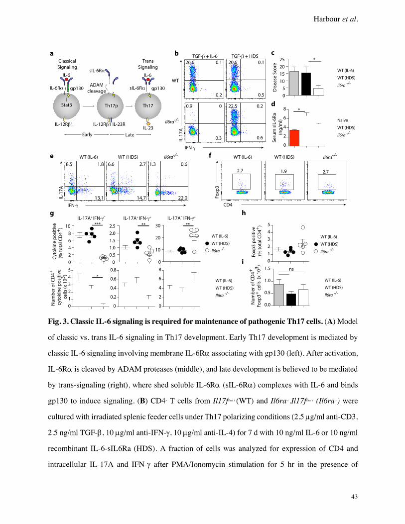
Harbour et al.
43
Fig. 3. Classic IL-6 signaling is required for maintenance of pathogenic Th17 cells. (A) Model
of classic vs. trans IL-6 signaling in Th17 development. Early Th17 development is mediated by
classic IL-6 signaling involving membrane IL-6Ra associating with gp130 (left). After activation,
IL-6Ra is cleaved by ADAM proteases (middle), and late development is believed to be mediated
by trans-signaling (right), where shed soluble IL-6Ra (sIL-6Ra) complexes with IL-6 and binds
gp130 to induce signaling. (B) CD4+ T cells from Il17fThy1.1 (WT) and Il6ra-/-.Il17fThy1.1 (Il6ra-/-) were
cultured with irradiated splenic feeder cells under Th17 polarizing conditions (2.5 µg/ml anti-CD3,
2.5 ng/ml TGF-b, 10 µg/ml anti-IFN-g, 10 µg/ml anti-IL-4) for 7 d with 10 ng/ml IL-6 or 10 ng/ml
recombinant IL-6-sIL6Ra (HDS). A fraction of cells was analyzed for expression of CD4 and
intracellular IL-17A and IFN-g after PMA/Ionomycin stimulation for 5 hr in the presence of
Cy
tok
ine
po
siti
ve
4D
C lat
ot %(
+)
Nu
mb
er
of
CD
4+
eviti s
op
eni
kot
yc0
1 x( sllec
3)
Se
rum
sIL
-6R
a
(n
g/m
l)
WT
Il6ra-/-
TGF-β + IL-6
IFN-γ
A7
1-LI
26.6 0.1
0.2
20.6 0.1
0.5
0.9 0
0.3
22.5 0.2
0.6
0
5
10
15
20
25 *
eroc
S es
aesi
D
IL-17A+ IFN-γ- IL-17A- IFN-γ+IL-17A+ IFN-γ+
a b
f
g
c
0
2
4
6
8
10
0
0.5
1.0
1.5
2.0
2.5
0
10
20
30*** ** **
0
1
2
3
4
5
0
0.2
0.4
0.6
0.8
0
2
4
6
8
*
0
1
2
3
4
5
0.0
0.5
1.0
1.5
0
2
4
6
8 *
TGF-β + HDS
8.5 1.8
13.1
6.6 2.7
14.7
1.3 0.6
22.0
IFN-γ
IL-1
7A
e
2.7 1.9 2.7
CD4
3px
oF
Il6ra-/-
h
i
eviti s
op
3px
oF4
DC l
atot
%( +
)
4D
C fo r
eb
mu
N+
Foxp
3+
01 x( sll
ec 3
)
Stat3
ADAM
cleavage
sIL-6Rα
Th17p
Classical
Signaling
Trans
Signaling
IL-6
IL-6Rα gp130
IL-6
sIL-6Rα gp130
Th17
IL-12Rβ1 IL-23RIL-12Rβ1
d
IL-23
Early Late
ns
WT (IL-6) WT (HDS)WT (IL-6) WT (HDS) Il6ra-/-
Il6ra-/-
WT (IL-6)
WT (HDS)
Il6ra-/-
Naive
WT (HDS)
Il6ra-/-
WT (IL-6)
WT (HDS)
Il6ra-/-
WT (IL-6)
WT (HDS)
Il6ra-/-
WT (IL-6)
WT (HDS)
Il6ra-/-
WT (IL-6)
WT (HDS)

Harbour et al.
44
monensin. FACS plots are gated on CD4+ cells. (C) WT (IL-6), WT (HDS) and Il6ra-/- Thy1.1+ Th17
cells obtained in (B) were transferred into Rag1-/- mice (4 x 10⁵/mouse). Histological scores were
analyzed four weeks after transfer (mean + SEM)(WT (IL-6) n = 8, WT (HDS) n = 13, Il6ra-/- n =
12). * P = 0.0305. (D) Serum sIL-6Ra levels from naïve Rag1-/- mice as well as colitic recipients
of WT (hIL-6) and Il6ra-/- Th17 cells at 4 weeks post transfer (n = 5 per group) were quantified by
Milliplex assay (mean + SEM). * P <0.05. (E) Representative FACS plots showing IL-17A and
IFN-g in CLP CD4+ T cells ex vivo. (F) Representative FACS plots showing Foxp3 in CLP CD4+
T cells ex vivo. (G) Relative frequencies (upper) and total numbers (lower) of IL-17A+, IL-17A+
IFN-g+, and IFN-g+ CD4+ T cells in the CLP of colitic mice (mean ± SEM)(n = 5 per group). (H)
Relative frequencies and (I) total numbers of CD4+ Foxp3+ T cells in the CLP of colitic mice (mean
+/- SEM). Data are pooled from (C) or representative of (E – I) three independent experiments. P
values; non parametric Kruskal-Wallis with Dunn’s post comparison test (C), one-way ANOVA
with Bonferroni post test (D) or Tukey’s Multiple Comparison test (G – I).
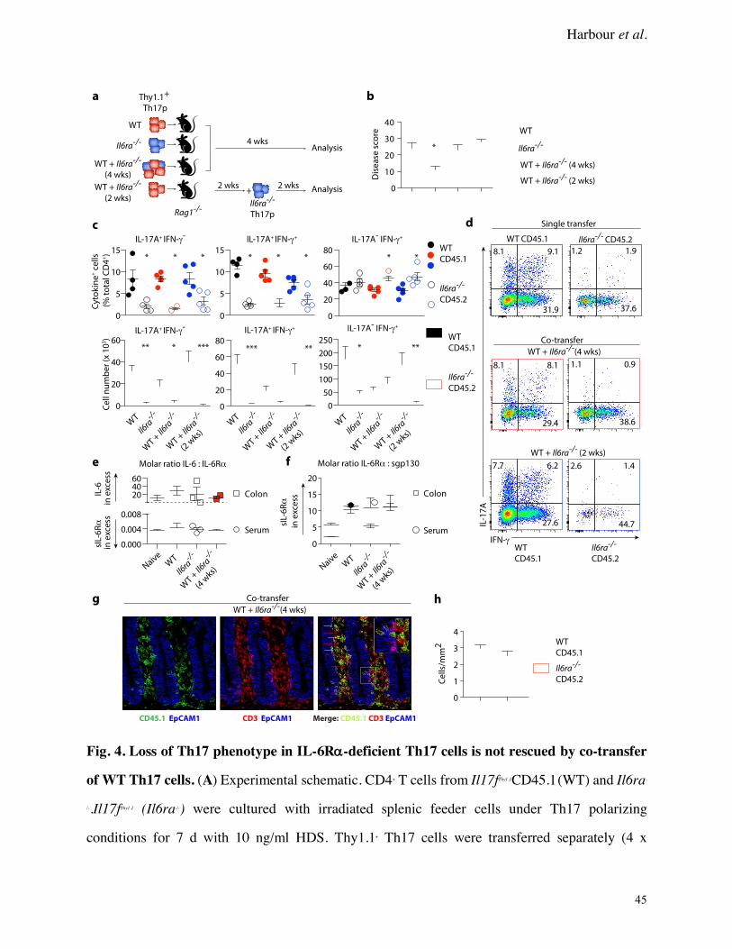
Harbour et al.
45
Fig. 4. Loss of Th17 phenotype in IL-6Ra-deficient Th17 cells is not rescued by co-transfer
of WT Th17 cells. (A) Experimental schematic. CD4+ T cells from Il17fThy1.1CD45.1 (WT) and Il6ra-
/-.Il17fThy1.1 (Il6ra-/-) were cultured with irradiated splenic feeder cells under Th17 polarizing
conditions for 7 d with 10 ng/ml HDS. Thy1.1+ Th17 cells were transferred separately (4 x
WT
CD45.1
WT
CD45.1
Il6ra-/-
CD45.2
Cyt
ok
ine
+ c
ell
s
(%
to
tal C
D4
+)
Ce
ll n
um
be
r (x
10
3)
0
5
10
15
0
5
10
15
0
20
40
60
80
0
20
40
60
0
20
40
60
80
0
50
100
150
200
250
IL-17A+ IFN-γ- IL-17A- IFN-γ+IL-17A+ IFN-γ+
* * * * * * * *
** * *** *** ** * **
WT
Il6ra-/-
WT +
Il6ra
-/-
WT +
Il6ra-/-
(2 w
ks)
Il6ra-/-
CD45.2
a
8.1 9.1
31.9
8.1 8.1
29.4
7.7 6.2
27.6
1.2 1.9
37.6
1.1 0.9
38.6
2.6 1.4
44.7
IFN-γ
IL-1
7A
WT CD45.1
WT + Il6ra-/-(4 wks)
WT + Il6ra-/- (2 wks)
WT
CD45.1
Il6ra-/-
CD45.2
Il6ra-/- CD45.2
Single transfer
Co-transfer
b
IL-17A+ IFN-γ- IL-17A- IFN-γ+IL-17A+ IFN-γ+
0
10
20
30
40
Dis
ea
se s
core
*
e
Colon
Serum
WT
Il6ra-/-
WT +
Il6ra
-/-
(4 w
ks)
Naive
IL-6
in
exc
ess
sIL-
6R
α
in e
xce
ss
Molar ratio IL-6 : IL-6Rα
WT
Il6ra-/-
WT +
Il6ra
-/-
WT +
Il6ra-/-
(2 w
ks) W
T
Il6ra-/-
WT +
Il6ra
-/-
WT +
Il6ra-/-
(2 w
ks)
WT
Il6ra-/-
WT + Il6ra-/- (4 wks)
WT + Il6ra-/- (2 wks)
Thy1.1+
Th17p
WT
Il6ra-/-
WT + Il6ra-/-
(4 wks)
WT + Il6ra-/-
(2 wks)
Rag1-/-
4 wks
2 wks 2 wks
Il6ra-/-
Th17p
+
Analysis
Analysis
c d
f
WT
Il6ra-/-
WT +
Il6ra
-/-
(4 w
ks)
Naive
Molar ratio IL-6Rα : sgp130
sIL-
6R
α
in e
xce
ss
g
0
1
2
3
4WT
CD45.1
Il6ra-/-
CD45.2Ce
lls/
mm
2
WT + Il6ra-/-(4 wks)
Co-transfer h
Colon
Serum
0.000
0.004
0.008
204060
0
5
10
15
20
Merge: CD45.1 CD3 EpCAM1CD45.1 EpCAM1 CD3 EpCAM1
Harbour et al.
46
105/mouse) or together (2 x 10⁵ each/mouse) into Rag1-/- recipients (WT n = 4, Il6ra-/- n = 4, WT +
Il6ra-/- n = 5, WT + Il6ra-/- (2 wks) n = 5). (B) Histological scores were analyzed four weeks after
transfer (mean + SEM). * P < 0.05 (C) Relative frequencies (upper) and total numbers (lower) of
IL-17A+, IL-17A+ IFN-g+, and IFN-g+ CD4+ T cells in the CLP of colitic mice (mean ± SEM). * P <
0.05, ** P < 0.01, *** P < 0.001. (D) Representative FACS plots showing IL-17A and IFN-g in
CLP CD4+ T cells from recipients of single or co-transfers ex vivo after development of colitis. (E-
F) Levels of sIL-6Ra, sgp130 and IL-6 in colon explants and serum of naïve Rag1-/- mice as well
as colitic recipients of single or co-transfer Th17 cells were quantified by Milliplex assay. Molar
ratio of IL-6–IL-6Ra (E) and IL-6Ra–sgp130 (F) in colon explants (squares) and serum (circles)
from colitic mice are shown. (G) Colon tissue from WT + Il6ra-/- Th17 co-transfers were stained
with anti-EpCAM1 (blue), anti-CD45.1 (green) and anti-CD3 (red). Left panel shows anti-CD45.1
(WT), middle panel shows anti-CD3 (total T cells), right panel shows merged image (red arrows,
Il6ra-/- CD4+ T cells ; green arrows, WT CD4+ T cells). Scale bars 20 µm. (H) Quantification of WT
and Il6ra-/- CD4+ T cells from (G). Data are representative of three independent experiments. P
values; non parametric Kruskal-Wallis with Dunn’s post comparison test (B), one-way ANOVA
with Bonferroni post test (C).

Harbour et al.
47
Fig. 5. IL-6Ra-deficient Th17 cells are responsive to trans-signaling ex vivo. Thy1.1+ Th17
cells from Il17fThy1.1CD45.1 (WT) and Il6ra-/-.Il17fThy1.1 (Il6ra-/-) were adoptively transferred together (2
x 105 each/mouse) into Rag1-/- recipients (n = 4 mice). Four weeks after transfer, CD4+ cells from
the MLN of recipient mice or naïve CD4+ cells from the MLN of WT mice were stimulated with
equimolar concentrations of IL-6 or HDS (0.95nM) for 15 min. (A) Representative FACS plots
showing pSTAT3 (Y705) in MLN CD4+ T cells from naïve or colitic mice. (B) Representative
FACS plots showing pSTAT1 (Y701) in MLN CD4+ T cells from naïve or colitic mice. (C).
Relative frequencies of pSTAT3+ (upper) and pSTAT1+ CD4+ T cells (lower) in the MLN of colitic
mice (mean + SEM). * P < 0.05, ** P < 0.01. Data are representative of two independent
experiments. P values; two-way ANOVA with Bonferroni post-test (C).
(-) IL-6 HDS
0
2
4
6pStat3 (Y705)
IL-6 HDS(-)
* *
CD4
pS
tat3
(Y
70
5)
2.5
2.0
5.2
2.0
8.5
11.8
WT
Il6ra-/-
a
0.1 80.6 79.2
0
0.5
1.0
1.5
2.0
*
4D
C e
vil fo t
necr
eP
+
RC
Tβ+
pStat1 (Y701)
<0.1 33.6 41.9
0.4 2.1
0.4 0.4 1.7
0.5
(-) IL-6 HDS
WT
Il6ra-/-
CD4)
10
7Y(
1tat
Sp
b
* **
WT Il6ra-/-
WT + Il6ra-/-(4 wks)
Naive CD4+
WT + Il6ra-/-(4 wks)
Naive CD4+
c

Harbour et al.
48
Fig. 6. IL-6 is required to maintain a Th17 gene program in vivo. (A) Experimental schematic.
Thy1.1+ Th17 cells from Il17fThy1.1CD45.1 (WT) and Il6ra-/-.Il17fThy1.1 (Il6ra-/-) were adoptively
transferred together (2 x 10⁵ each/mouse) into Rag1-/- recipients (n = 4). Four weeks after transfer,
congenically marked CD4+ cells from the CLP of recipient mice were isolated and gene expression
was assessed by RNA-seq. (B) Recovery of CLP CD4+ cells from Rag1-/- recipients of WT and
Il6ra-/- Th17 cells (mean ± SEM). (C) Total number of CLP CD4+ T cells recovered from Rag1-/-
recipients (mean + SEM). (D) MA plot displaying differentially expressed genes in WT (lower)
and Il6ra-/- (upper) CD4+ T cells ex vivo. Red circles indicate genes with false discovery rate (FDR)
<0.05. (E) Barcode plots of gene set enrichment results obtained using a set of genes previously
a
b c
d
f
0.0
0.5
1.0
1.5
2.0
e
vil fo .
oN
CD
4+
R
CT
β+0
1 x(6
)
WT Il6ra-/-
Thy1.1+
Th17pWT
Il6ra-/-WT +
Il6ra-/-Rag1-/-
4 wksRNA-seq
analysis
CLP
CD4+ T cells
g
0
20
40
60
80
100
Pe
rce
nt
of
liv
e
CD
4+
TC
Rβ+
WT Il6ra-/-
Ccr
9C
cr6
WT
Il6ra-/-
Tb
x21
Ro
rcN
av2
Il6ra
Il23
rIl2
2Il2
1
Il1r2
Il1r1
Il17
re
Il12
rb2
Igf1
r
Ifn
g
Gzm
bG
zma
Fasl
Csf
1C
cl5
Il17
a
Enriched in Th17 cells Enriched in Th1 cells
0
2
-2
z score
Rorc induced genes
-0.4
-0.2
0
-0.6FDR q<10-3NES -1.98
WT Il6ra-/-
Rorc repressed genes
0
0.2
0.4
0.6FDR q<10-3NES 1.98
WT Il6ra-/-
-0.4
-0.2
0
-0.6
FDR q<10-3NES -1.78
WT Il6ra-/-
Il7r
Il12
rb2
Il10
Gp
r18
Gp
r15
Cts
wC
tla
2b
Ccr
5C
cr2
Ah
rN
av2Li
fIt
ga
3Il2
3r
Il22
Il1r1
Il17
fIl1
7a
Igfb
p7
Cxc
l5C
xcl3
Csf
2C
cr6
Ccl
20
Bcl
6
WT
Il6ra-/-
En
rich
me
nt
sco
re
En
rich
me
nt
sco
re
En
rich
me
nt
sco
re
En
rich
me
nt
sco
re
Tn
frsf
8R
un
x1R
orcLi
fIt
ga
3Il2
3r
Il21
Il1r1
Il17
fIl1
7a
Ifn
gr2
Ba
tf
Tn
fsf4
Tn
fsf1
0T
nfr
sf1
bT
bx2
1S
ocs
2N
od
1Ir
f8Ir
f7Ir
f1Ik
zf2
Fasl
Cts
wC
d1
64
Ccl
4C
cl3
WT
Il6ra-/-
0
2
-2
z score
Stat3 induced genes Stat3 repressed genes
h
i j
Rorc induced genes Rorc repressed genes
Stat3 induced genes Stat3 repressed genes
Il17
f
Ctse
Nav1
Il17f
Il1r1Il1r2
Il12rb2
Rorc
Il1rl1
Il10ra
Il17re
-5
0
5
0 5 10 15
Mean expression (log2)
Fold
ch
an
ge
(lo
g2
)
Il17a
Il18rapCtsw
Ccr5Il12rb1Csf1
Ccr4
Ccr6Il22Il23rWT
Il6ra-/-
0
0.25
0.50FDR q<10-3NES 2.36
WT Il6ra-/-
e
0
0.25
0.50
Enriched in Th1 cells
En
rich
me
nt
sco
re FDR q<10-2NES 2.20
-0.4
-0.2
0
-0.8
-0.6
Enriched in Th17 cells
En
rich
me
nt
sco
re
FDR q<10-3NES -2.16
WT Il6ra-/-WT Il6ra-/-
0
2
-2
z score

Harbour et al.
49
reported to be differentially expressed between Th17 cells (left) or Th1 cells (right) (data from
GSE 14308) (NES, normalized enrichment score). (F) Expression of representative Th1 and Th17
cell associated genes identified by gene set enrichment in (E) are depicted by heat map with z-
scored expression values. (G) Barcode plots of gene set enrichment results obtained using a set of
genes previously reported to be regulated by Rorc in Th17 cells (GSE 40918). (H) Expression of
representative genes identified by gene set enrichment in (G) are depicted by heat map with z-
scored expression values. (I) Barcode plots of gene set enrichment results obtained using a set of
genes previously reported to be regulated by Stat3 in Th17 cells (GSE 40918) (J). Expression of
representative genes identified by gene set enrichment in (I) are depicted by heat map with z-
scored expression values. Data are from two independent experiments.

Harbour et al.
50
Fig. 7. Enforced RORgt expression represses transition to Th1-like cells by IL-6Ra-deficient
Th17 precursors in vivo. (A) CD4+ T cells from Il17fThy1.1 CD45.1 (WT) and Il6ra-/-.Il17fThy1.1 (Il6ra-/-)
pMIG
Retrovirus:
pMIG-RORγt
WT
GF
P
Thy1.1
IL-1
7A
IFN-γ
WT CD45.1 Il6ra-/- CD45.2
WT pMIG + Il6ra-/- pMIG
WT pMIG + Il6ra-/- pMIG-RORγt
WT CD45.1 Il6ra-/- CD45.2
Cy
tok
ine
+ c
ell
s
(%
to
tal C
D4
+)
IL-17A+ IFN-γ- IL-17A+ IFN-γ+ IL-17A- IFN-γ+
WT pMIG
Il6ra-/- pMIG
WT pMIG
Il6ra-/- pMIG-RORγt
0
5
10
15 * *
0
1
2
3
4
0
20
40
60* ** *** ***2.3 1.8
26.1
0.6 1.2
49.0
6.9 3.1
31.1
2.7 1.4
21.0
23.7 69.7
3.7
34.0 60.2
3.2
6.8 56.9
26.5
6.9 55.1
25.9
33.3 0.5
0.3
28.3 0.6
0.9
51.3 0.6
0.4
46.2 0.6
0.4
pMIG
Retrovirus:
pMIG-RORγt
IL-1
7A
IFN-γ
Il17a
0.00
0.01
0.02
0.03
0.04
Il17f
0.000
0.005
0.010
0.015
Rorc
0.00
0.01
0.02
0.03
0.04
Tbx21
0.00
0.01
0.02
0.03
Il12rb2
0.000
0.005
0.010
0.015
0.020
0.025
Il23r
0.000
0.002
0.004
0.006* ****
***
** ** **
WT pMIG WT pMIG
Il6ra-/-
WT
Il6ra-/-
2^
(CT
B2
M-C
TG
OI)
2^
(CT
B2
M-C
TG
OI)
a b c
d e
f
g
ns
0
5
10
15
Dis
ea
se S
core
WT pMIG + Il6ra-/- pMIG
WT pMIG + Il6ra-/- pMIG-RORγt
ns
WT pMIG + Il6ra-/- pMIG WT pMIG + Il6ra
-/- pMIG-RORγt
100 μm 100 μm
Il6ra-/- pMIG Il6ra
-/- pMIG-RORγt

Harbour et al.
51
were cultured under Th17 polarizing conditions (2.5 ng/ml TGF-b, 10 µg/ml anti-IFN-g, 10 µg/ml
anti-IL-4, 10 ng/ml HDS) with CD3/CD28 expander beads (ThermoFisher) and transduced with
retroviruses encoding GFP alone (pMIG) or GFP with RORgt (pMIG- RORgt). FACS plots show
GFP and Thy1.1 expression on D6 and are gated on CD4+ T cells. (B) Th17p were stained
intracellularly for IL-17A and IFN-g after PMA/Ionomycin activation for 5 h in the presence of
monensin. FACS plots are gated on CD4+ GFP+ Thy1.1+ cells. (C) GFP+ Thy1.1+ Th17 cells from
(A) were FACS purified and transferred together (2 x 10⁵ each/mouse) into Rag1-/- recipients (WT
pMIG + Il6ra-/- pMIG n = 7, WT pMIG + Il6ra-/- pMIG-RORgt n = 7). Histological scores were
analyzed three weeks after transfer (mean + SEM). (D) Representative FACS plots showing IL-
17A and IFN-g in CLP CD4+ T cells from recipients of co-transfers ex vivo after development of
colitis. (E) Relative frequencies of IL-17A+, IL-17A+ IFN-g+, and IFN-g+ CD4+ T cells in the CLP of
colitic mice (mean ± SEM). * P < 0.05, ** P < 0.01, *** P < 0.001. (F) Representative H&E
stained sections of colon from recipients of WT pMIG + Il6ra-/- pMIG or WT pMIG + Il6ra-/- pMIG-
RORgt CD4+ T cells. H&E stain; original magnification 10x; scale bars 100 µm. (G) RNA isolated
from FACS purified WT and Il6ra-/- CLP CD4+ T cells from co-transfer recipients was analyzed ex
vivo by qPCR for expression of Il17a, Il17f, Rorc, Tbx21, Il12rb2 and Il23r (mean + SEM of three
biological replicates). * P < 0.05, ** P < 0.01. Data are representative of three experiments. P
values; one-way ANOVA with Tukey’s multiple comparison test (G) or one-way ANOVA with
Bonferroni post test (E) .
Supplementary Materials:
Supplementary Fig. 1. Classical IL-6 signaling is required for development of colitis.
Supplementary Fig. 2. Innate derived IL-6 is required for Th17 pathogenicity and maintenance.

Harbour et al.
52
Supplementary Fig. 3. Classical IL-6 signaling is required for maintenance of Th17 cells.
Supplementary Fig. 4. Co-transfer of Il6ra-/- with WT Th17 cells is unable to rescue their
phenotype.
Supplementary Fig. 5. Co-transfer of Il6ra-/- with WT Th17 cells is unable to rescue their Th17
phenotype.
Supplementary Fig. 6. IL-6Ra and gp130 expression on Th17 cells ex vivo.
Supplementary Fig. 7. Il6ra-/- Th17 cells lose their Th17 gene expression signature in vivo.
Supplementary Table 1. Full gene sets used for GSEA analysis.

Harbour et al.
Supplemental Figures
Supplementary Fig. 1. Classical IL-6 signaling is required for development of colitis. FACS
sorted CD4+ CD45RBHi T cells isolated from WT and Il6ra-/- mice were transferred into Rag1-/-
recipients (4 x 10⁵ per mouse)(WT n = 5 per group, Il6ra-/- n = 6 per group) and analyzed for colitis
8 weeks after transfer. (A) Representative FACS plots showing IL-17A and IFN-g in MLN CD4+
T cells ex vivo after development of colitis. (B) Representative FACS plots showing Foxp3
expression in MLN CD4+ T cells ex vivo from WT or Il6ra-/- recipients after development of colitis.
(C) Relative frequencies (upper) and total numbers (lower) of IL-17A+, IL-17A+IFN-g+, and IFN-g+
CD4+ T cells in the MLN of colitic mice (mean ± SEM). *** P <0.0001, *** P <0.0001, ** P =
0.0045 (upper IL-17A+, IL-17A+IFN- g +, and IFN- g + CD4+ T cells). ** P = 0.002, ** P = 0.0046, P
= 0.3380 (lower IL-17A+, IL-17A+IFN- g +, and IFN- g + CD4+ T cells) (D) Relative frequencies
(upper) and total numbers (lower) of Foxp3+ CD4+ T cells from the MLN (mean ± SEM). P =
0.2940 (upper), P = 0.0887 (lower). P values; unpaired two-tailed Student’s t-test (C - D).
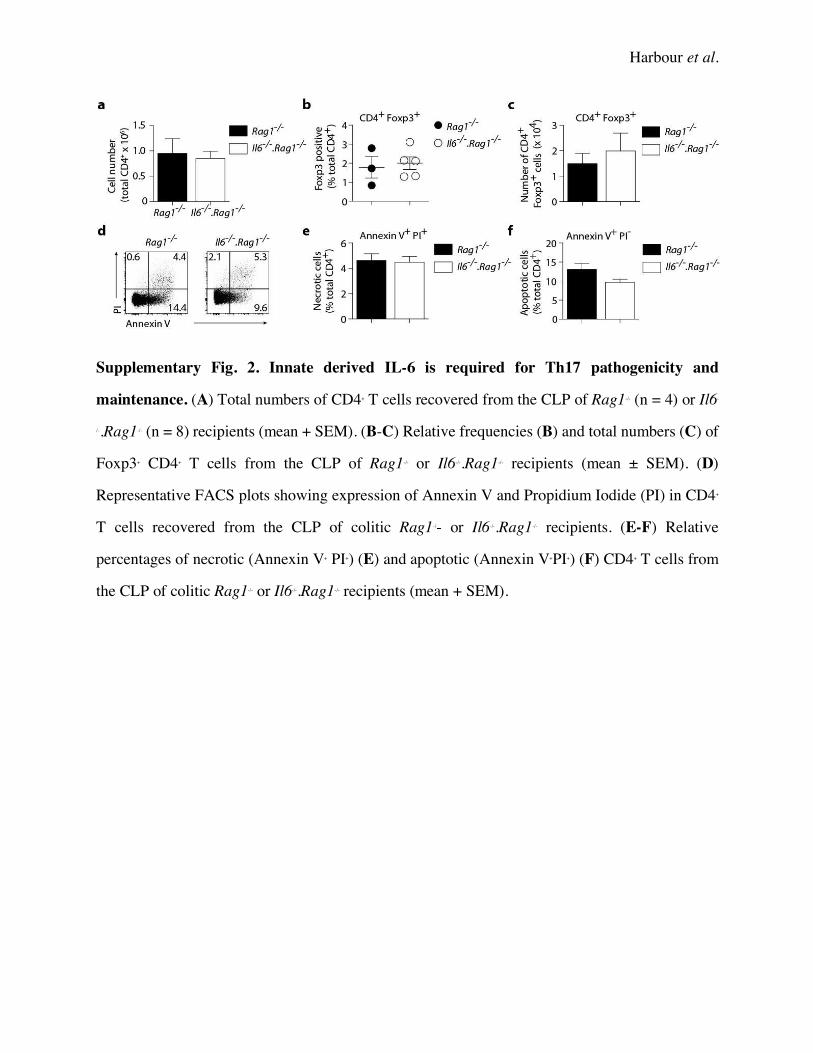
Harbour et al.
Supplementary Fig. 2. Innate derived IL-6 is required for Th17 pathogenicity and
maintenance. (A) Total numbers of CD4+ T cells recovered from the CLP of Rag1-/- (n = 4) or Il6-
/-.Rag1-/- (n = 8) recipients (mean + SEM). (B-C) Relative frequencies (B) and total numbers (C) of
Foxp3+ CD4+ T cells from the CLP of Rag1-/- or Il6-/-.Rag1-/- recipients (mean ± SEM). (D)
Representative FACS plots showing expression of Annexin V and Propidium Iodide (PI) in CD4+
T cells recovered from the CLP of colitic Rag1-/- or Il6-/-.Rag1-/- recipients. (E-F) Relative
percentages of necrotic (Annexin V+ PI+) (E) and apoptotic (Annexin V+PI+) (F) CD4+ T cells from
the CLP of colitic Rag1-/- or Il6-/-.Rag1-/- recipients (mean + SEM).
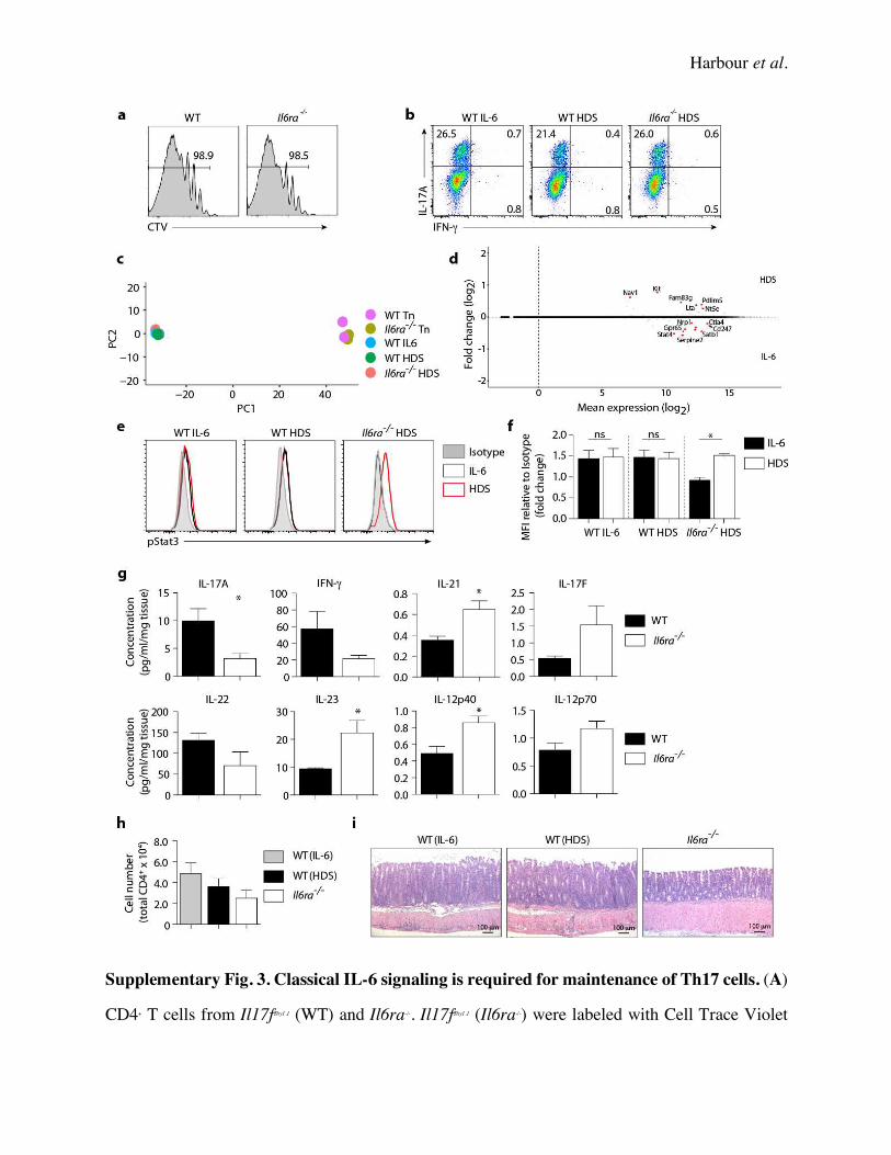
Harbour et al.
Supplementary Fig. 3. Classical IL-6 signaling is required for maintenance of Th17 cells. (A)
CD4+ T cells from Il17fThy1.1 (WT) and Il6ra-/-. Il17fThy1.1 (Il6ra-/-) were labeled with Cell Trace Violet
Harbour et al.
(CTV; ThermoFisher) and cultured with irradiated splenic feeder cells under Th17 polarizing
conditions for 7 d with 10 ng/ml recombinant hIL-6. A fraction of cells were analyzed for
expression of CD4 and CTV dilution. FACS plots are gated on CD4+ Thy1.1+ Th17 cells. (B). CD4+
T cells from Il17fThy1.1 (WT) and Il6ra-/-. Il17fThy1.1 (Il6ra-/-) were cultured with irradiated splenic feeder
cells under Th17 polarizing conditions with 10 ng/ml IL-6 or 10 ng/ml recombinant IL-6-sIL6Ra
(HDS) for 7 d. A fraction of cells was analyzed for expression of CD4 and intracellular IL-17A
and IFN-g after PMA/Ionomycin stimulation for 5 hr in the presence of monensin. FACS plots are
gated on CD4+ cells. (C) 6 days after culture, Thy1.1+ Th17 precursors were isolated and gene
expression analyzed by RNA seq. Plot shows PCA analysis of mRNA transcriptome in naïve and
in vitro Th17 polarized WT (IL-6), WT (HDS) and Il6ra-/- (HDS) CD4+ T cells by RNA-seq
analysis. Each dot represents an individual mouse. Data are from two independent experiments.
(D) MA plot displays differentially expressed genes in WT (HDS)(upper) and WT (IL-6)(lower)
CD4+ T cells ex vivo. Red circles indicate genes with false discovery rate (FDR) <0.05. (E) CD4+
T cells from Il17fThy1.1 (WT) and Il6ra-/-. Il17fThy1.1 (Il6ra-/-) were cultured with irradiated splenic feeder
cells under Th17 polarizing conditions with 10 ng/ml IL-6 or 10 ng/ml recombinant IL-6-sIL6Ra
(HDS). On day 5, Th17 cells were stimulated with equimolar concentrations of IL-6 or HDS
(0.95nM) for 15 min. Representative FACS plots show pSTAT3 (Y705) in in vitro polarized Th17
cells. (F) Fold change of pSTAT3 (Y705) MFI (GeoMean) relative to isotype control (mean +
SEM) from three independent experiments. * P < 0.05, one-way ANOVA with Bonferroni post-
test. (G) Colon culture explant supernatants from Rag1-/- recipients of WT or Il6ra-/- Th17 cells (n
= 3 per group) were assayed for select cytokines by Luminex assay (Millipore) four weeks after
transfer. Data are expressed as pg/mg/ml tissue (mean + SEM) and represent one of two
independent experiments. IL-17A * P = 0.048, IFN-g P = 0.1742, IL-21 * P = 0.0303, IL-17F P
= 0.1573, IL-22 P = 0.1366, IL-23 * P = 0.0465, IL-12p40 * P = 0.0328, IL-12p70 P = 0.0997. P
values; unpaired two-tailed Student’s t-test.
(H) Total number of CD4+ T cells isolated from the CLP of Rag1-/- recipients of WT (IL-6), WT
(HDS) or Il6ra-/- Th17p cells four weeks after transfer (mean + SEM). (I) Representative H&E

Harbour et al.
stained sections of colon from Rag1-/- recipients of WT (IL-6)(left), WT (HDS)(middle), or Il6ra-
/- Th17p (right) WT Th17 cells. H&E stain; original magnification 10x; scale bars 100 µm.

Harbour et al.
Harbour et al.
Supplementary Fig. 4. Co-transfer of Il6ra-/- with WT Th17 cells is unable to rescue their
phenotype. (A) Relative percentage (upper) and total numbers (lower) of CD4+ T cells recovered
from the CLP of colitic recipients of WT, Il6ra-/- or WT + Il6ra-/- co-transfers of Th17 cells (mean
+ SEM). (B) Representative FACS plots showing expression of Foxp3 in recovered CD4+ T cells
from the CLP of colitic mice. (C) Relative frequencies (upper) and total number (lower) of CD4+
Foxp3+ cells in the CLP of colitic recipients of WT, Il6ra-/- or WT + Il6ra-/- Th17 co-transfers (mean
± SEM). (D) Representative FACS plots showing expression of IL-17A, TNF-a and GM-CSR in
recovered CD4+ T cells from the CLP of colitic mice. (E) Relative percentage of IL-17A+GM-
CSF+, GM-CSF+ (upper), IL-17A+TNF-a+, and TNF-a+ CD4+ T cells in the CLP of colitic mice
(mean ± SEM). ** P < 0.01 (unpaired two-tailed Student’s t-test). (F-G) Levels of sIL-6Ra, sgp130
and IL-6 in colon explants (F) and serum (G) of naive Rag1-/- mice as well as colitic recipients of
single or co-transfer Th17 cells were quantified by Milliplex assay (mean + SEM). Data are
representative of two independent experiments (WT n = 4, Il6ra-/- n = 4, WT + Il6ra-/- n = 5, WT +
Il6ra-/- (2 wks) n = 5). * P < 0.05, ** P < 0.01, *** P < 0.001 (one-way ANOVA with Bonferroni
post-test).
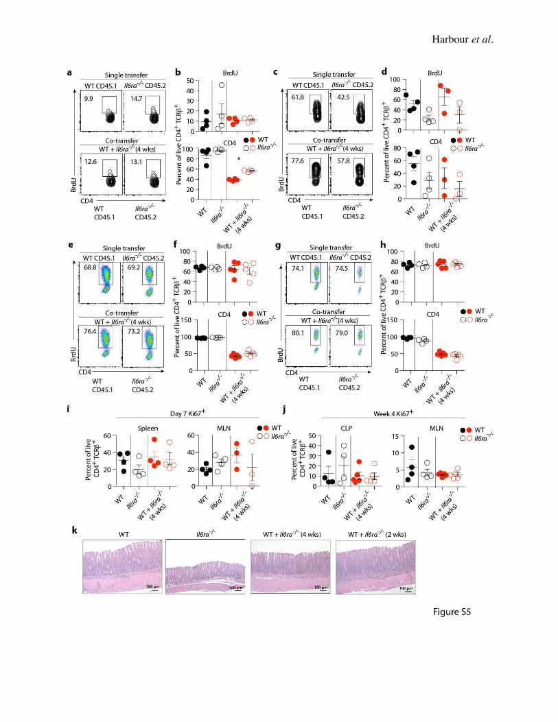
Harbour et al.
Harbour et al.
Supplementary Fig. 5. Co-transfer of Il6ra-/- with WT Th17 cells is unable to rescue their
Th17 phenotype. Rag1-/- mice (n = 4 per group) were adoptively transferred with 4 x 10⁵ WT or
Il6ra-/- Th17 cells or co-transferred with 2 x 10⁵ WT + Il6ra-/- Th17 cells. Recipient mice were fed
with water containing 1 mg/ml Bromodeoxyuridine (BrdU) for 7 days. (A and C). Representative
FACS plots show BrdU staining of CD4+ T cells recovered from the spleens (A) and MLN (C) of
single and co-transfer Th17 cell recipients 7 days after transfer. (B and D) Relative percentage of
BrdU+ CD4+ cells (upper) and total CD4+ cells (lower) in the spleen (B) and MLN (D) of single and
co-transfer Th17 recipients 7 days after transfer (mean ± SEM). (E and G) Representative FACS
plots showing BrdU staining of CD4+ T cells recovered from the CLP (E) and MLN (G) of single
and co-transfer Th17 cell recipients 4 weeks after transfer. (F and H) Relative percentage of BrdU+
CD4+ cells (upper) and total CD4+ cells (lower) in the CLP (F) and MLN (H) of single and co-
transfer Th17 recipients 4 weeks after transfer (mean ± SEM). (I) Relative percentages of CD4+
Ki67+ cells in the spleen (left) and MLN (right) of recipient mice 7 days after transfer (mean ±
SEM). (J) Relative percentages of CD4+ Ki67+ cells in the CLP (left) and MLN (right) of recipient
mice 4 weeks after transfer (mean ± SEM). (K) Representative H&E stained sections of colon
from recipients of (left to right) WT, Il6ra-/-, WT + Il6ra-/- (4 wks) and WT + Il6ra-/- (2 wks) CD4+
T cells. H&E stain; original magnification 10x; scale bars 100 µm. Data are representative of two
independent experiments. * P < 0.05 (one-way ANOVA with Bonferroni post-test).
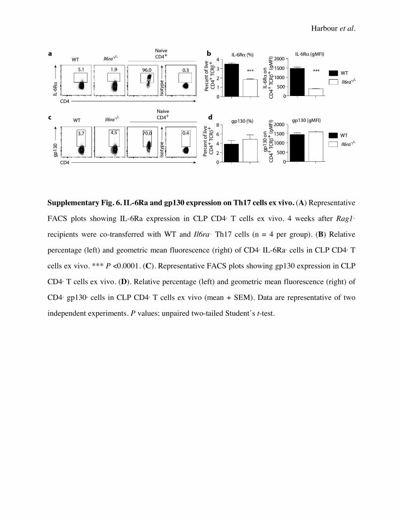
Harbour et al.
Supplementary Fig. 6. IL-6Ra and gp130 expression on Th17 cells ex vivo. (A) Representative
FACS plots showing IL-6Ra expression in CLP CD4+ T cells ex vivo, 4 weeks after Rag1-/-
recipients were co-transferred with WT and Il6ra-/- Th17 cells (n = 4 per group). (B) Relative
percentage (left) and geometric mean fluorescence (right) of CD4+ IL-6Ra+ cells in CLP CD4+ T
cells ex vivo. *** P <0.0001. (C). Representative FACS plots showing gp130 expression in CLP
CD4+ T cells ex vivo. (D). Relative percentage (left) and geometric mean fluorescence (right) of
CD4+ gp130+ cells in CLP CD4+ T cells ex vivo (mean + SEM). Data are representative of two
independent experiments. P values; unpaired two-tailed Student’s t-test.
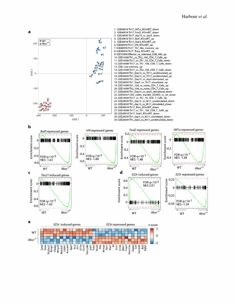
Harbour et al.
Harbour et al.
Supplementary Fig. 7. Il6ra-/- Th17 cells lose their Th17 gene expression signature in vivo.
(A) Similarity of gene sets calculated by the Jaccard index. Dimensions were reduced to two with
t-SNE and visualized with a scatterplot. (B) Barcode plots of gene set enrichment results obtained
using sets of genes previously reported to be regulated by several Th17 transcription factors (GSE
40918). (C) Barcode plots of gene set enrichment results obtained using a set of genes regulated
by Il23r in Th17 cell (GSE 58147). (D) Expression of representative genes identified by gene set
enrichment in (C) are depicted by heat map with z-scored values. Data are from two independent
experiments.