the “road map” - university of the...
TRANSCRIPT
PRACTICAL ROADMAP OSTEOGENESIS TN Augustine
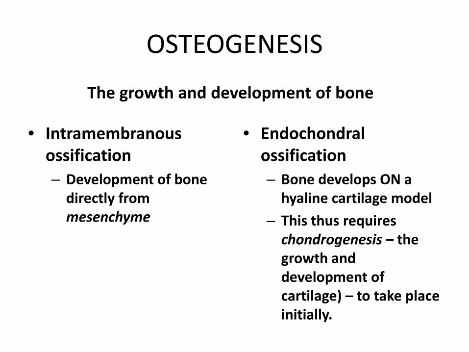
OSTEOGENESIS
• Intramembranous ossification – Development of bone
directly from mesenchyme
• Endochondral ossification – Bone develops ON a
hyaline cartilage model – This thus requires
chondrogenesis – the growth and development of cartilage) – to take place initially.
The growth and development of bone
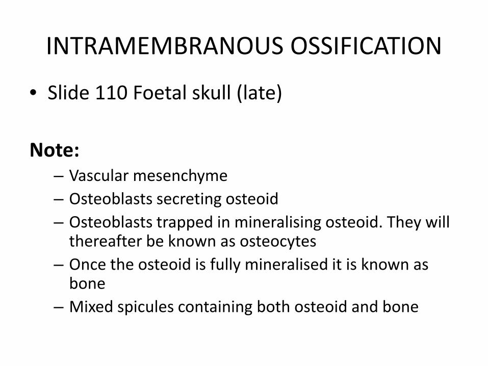
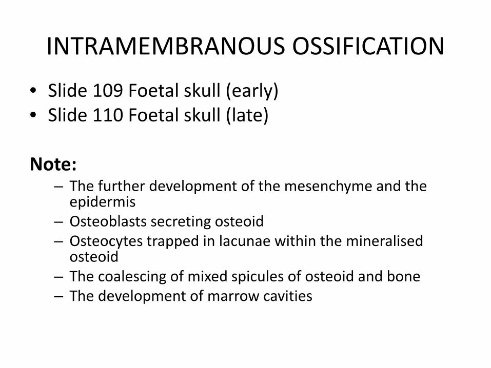
INTRAMEMBRANOUS OSSIFICATION
• Slide 110 Foetal skull (late)
Note: – Vascular mesenchyme – Osteoblasts secreting osteoid – Osteoblasts trapped in mineralising osteoid. They will
thereafter be known as osteocytes – Once the osteoid is fully mineralised it is known as
bone – Mixed spicules containing both osteoid and bone
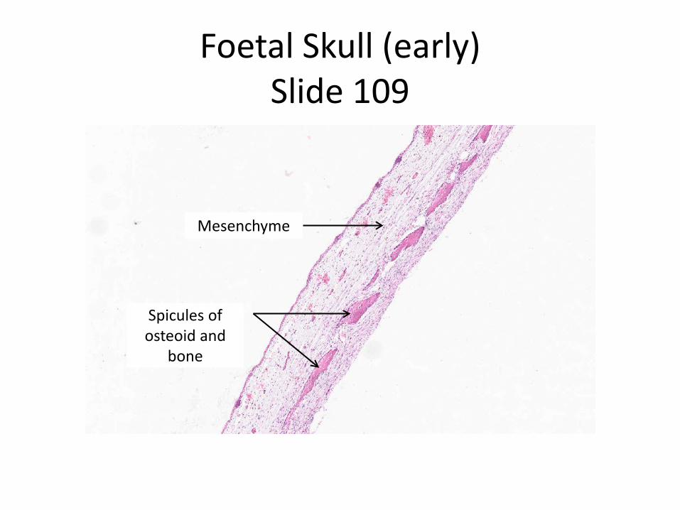
Foetal Skull (early) Slide 109
Spicules of osteoid and
bone
Mesenchyme
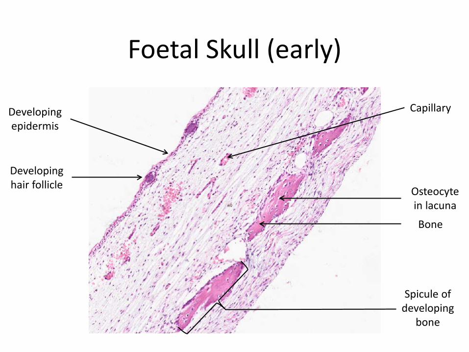
Foetal Skull (early)
Developing epidermis
Capillary
Osteocyte in lacuna
Developing hair follicle
Bone
Spicule of developing
bone
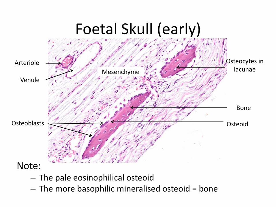
Foetal Skull (early)
Arteriole Mesenchyme
Osteocytes in lacunae
Venule
Bone
Osteoid Osteoblasts
Note: – The pale eosinophilical osteoid – The more basophilic mineralised osteoid = bone
INTRAMEMBRANOUS OSSIFICATION • Slide 109 Foetal skull (early) • Slide 110 Foetal skull (late)
Note:
– The further development of the mesenchyme and the epidermis
– Osteoblasts secreting osteoid – Osteocytes trapped in lacunae within the mineralised
osteoid – The coalescing of mixed spicules of osteoid and bone – The development of marrow cavities
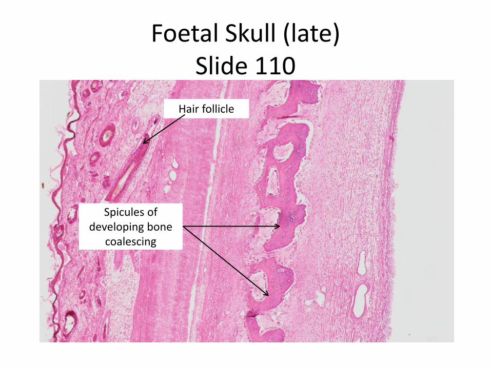
Foetal Skull (late) Slide 110
Spicules of developing bone
coalescing
Hair follicle
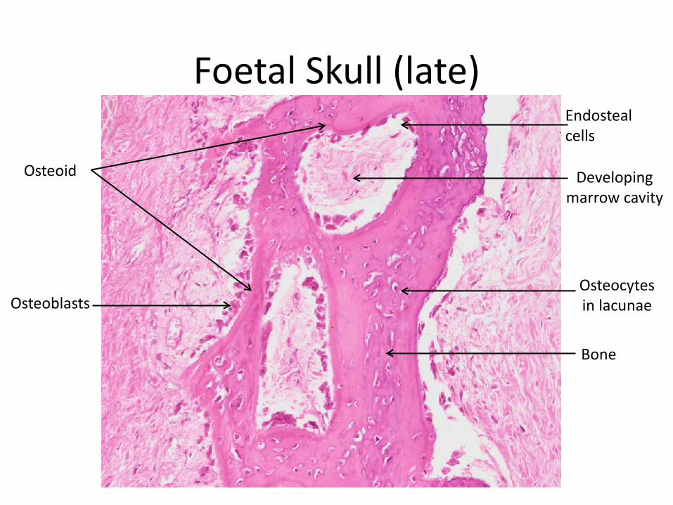
Endosteal cells
Osteoid Developing marrow cavity
Foetal Skull (late)
Osteocytes in lacunae Osteoblasts
Bone
ENDOCHONDRAL OSSIFICATION
Note: – Requires a hyaline cartilage model upon which osteoid
is laid down and mineralised • The hyaline cartilage model develops via chondrogenesis
ENDOCHONDRAL OSSIFICATION
• Slide 52 Foetal knee Note:
– The developing femur, patella and tibia. – The developing synovial cavity – The surrounding tissues
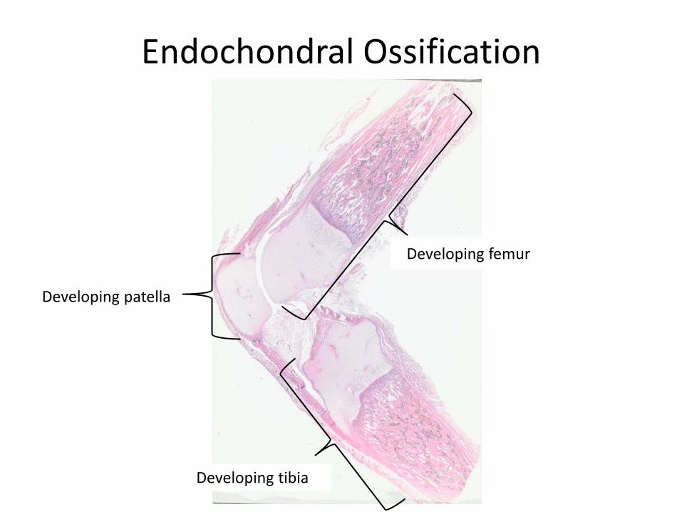
Endochondral Ossification
Developing femur
Developing tibia
Developing patella
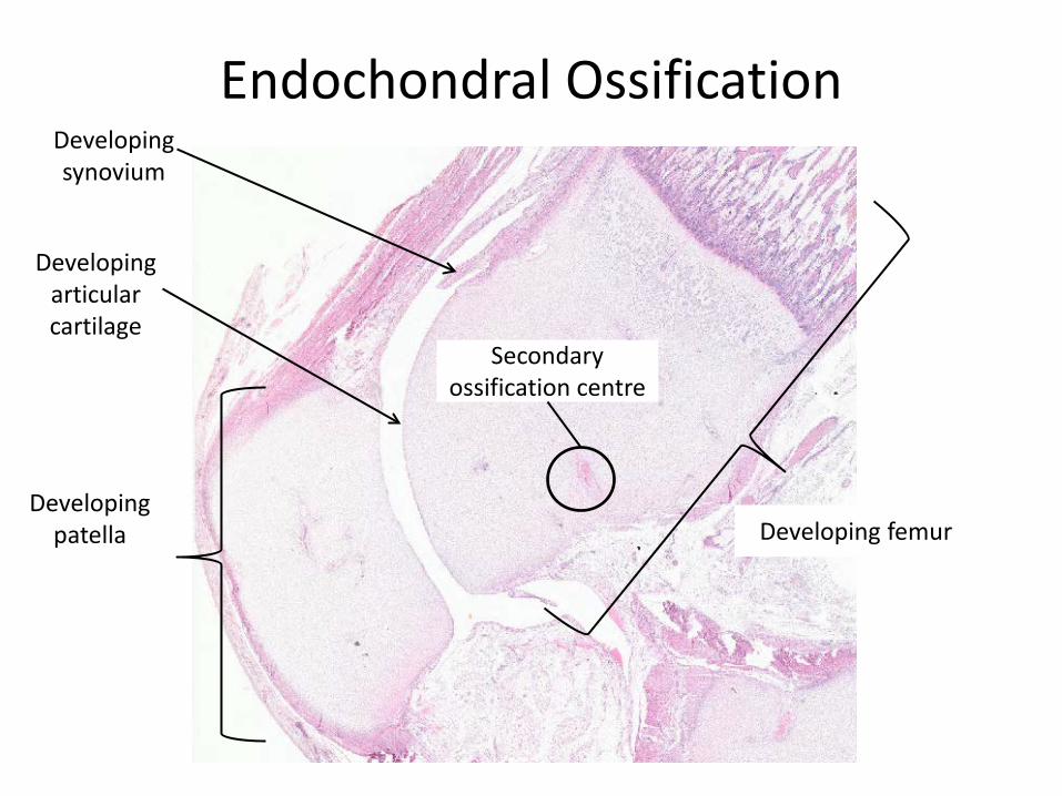
Developing synovium
Developing articular cartilage
Endochondral Ossification
Developing femur Developing
patella
Secondary ossification centre
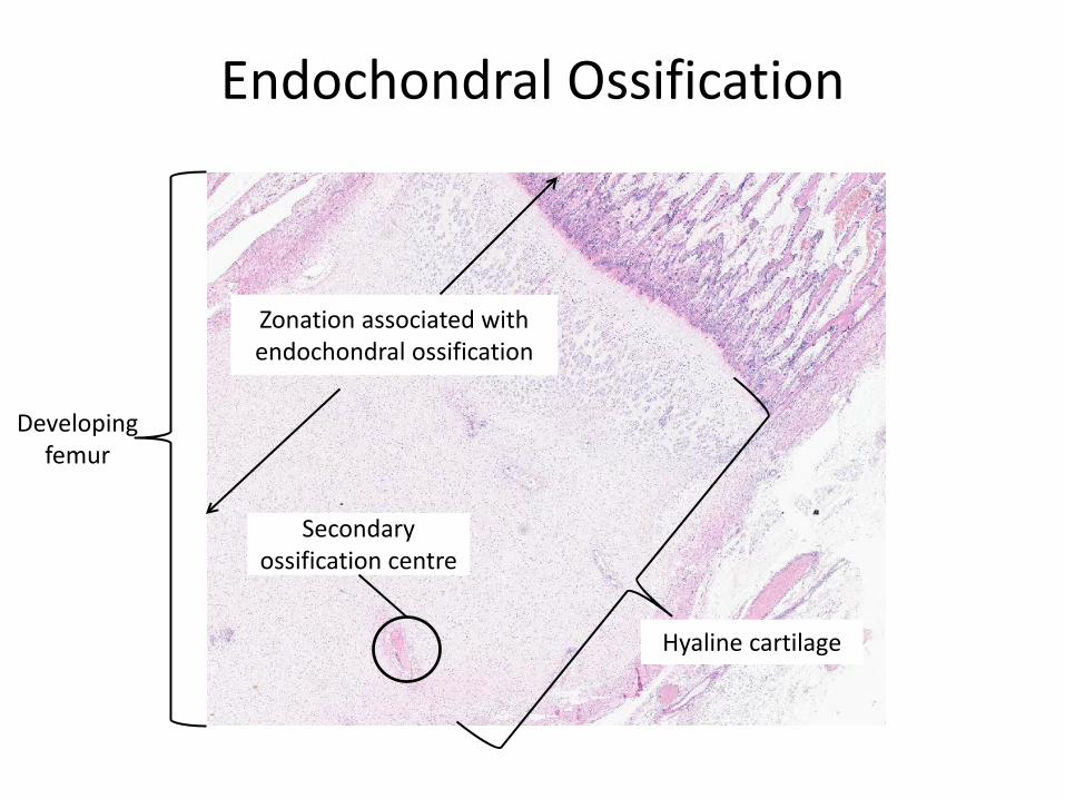
Endochondral Ossification
Developing femur
Secondary ossification centre
Hyaline cartilage
Zonation associated with endochondral ossification
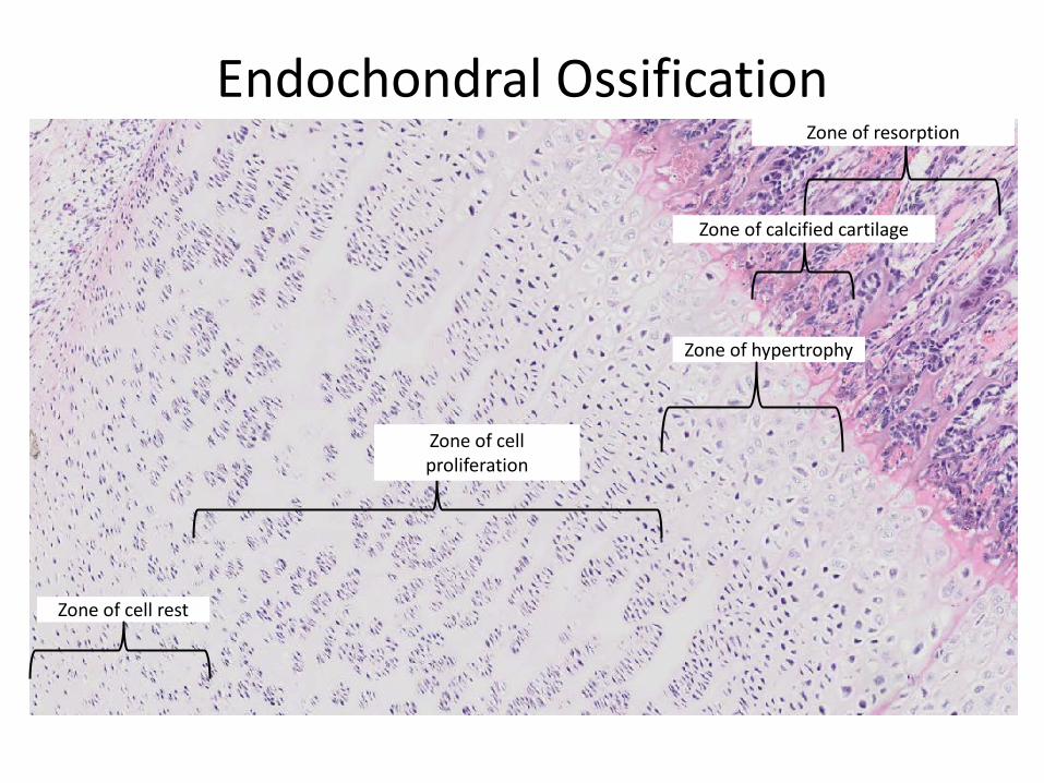
Endochondral Ossification
Zone of cell rest
Zone of hypertrophy
Zone of cell proliferation
Zone of calcified cartilage
Zone of resorption
Osteoid
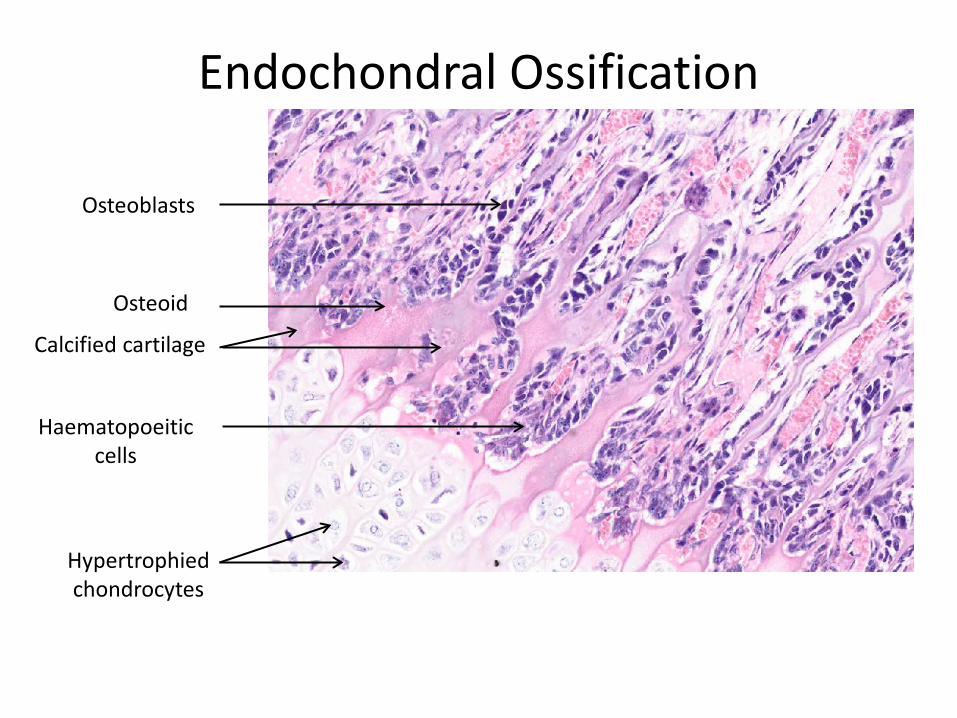
Endochondral Ossification
Hypertrophied chondrocytes
Calcified cartilage
Haematopoeitic cells
Osteoblasts
Calcified cartilage
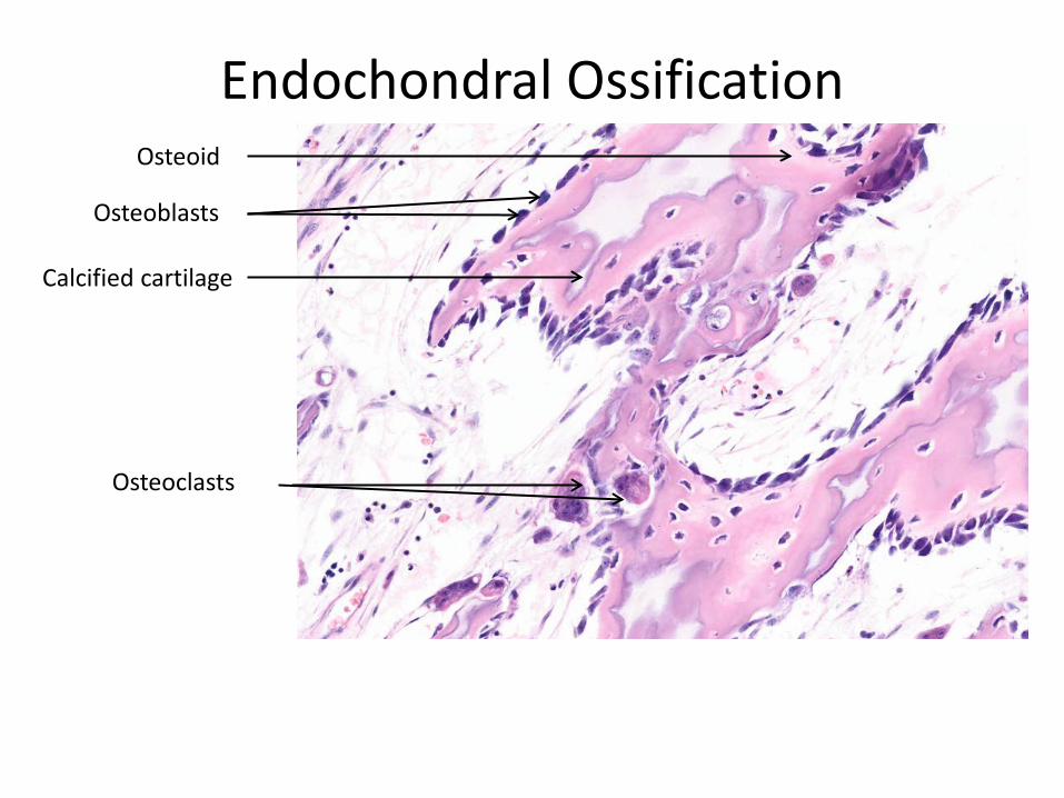
Endochondral Ossification
Osteoblasts
Osteoid
Osteoclasts
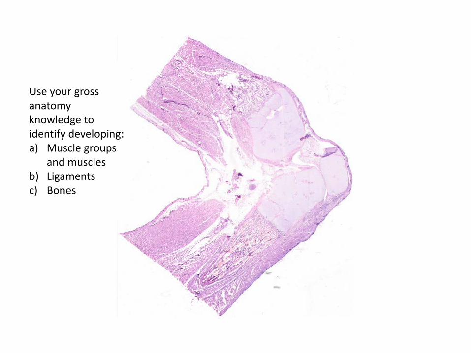
Use your gross anatomy knowledge to identify developing: a) Muscle groups
and muscles b) Ligaments c) Bones