the ap-1 site is required for basal expression but is not necessary
TRANSCRIPT

© 1991 Oxford University Press Nucleic Acids Research, Vol. 19, No. 13 3723-3731
The AP-1 site Is required for basal expression but is notnecessary for TPA-response of the human stromelysingene
Giovanna Buttice, Susan Quinones and Markku KurkinenDepartment of Medicine, University of Medicine and Dentistry of New Jersey, Robert Wood JohnsonMedical School, Piscataway, NJ 08854, USA
Received January 2, 1991; Revised and Accepted May 15, 1991
ABSTRACT
We have studied the activity of the AP-1 site, a targetfor the Fos and Jun family of transcription factors, inthe context of the human stromelysin promoter (-1303to + 4). In transiently transfected human HepG2, HeLaand fibroblast cell cultures, point-mutations in anyposition of the stromelysin AP-1 sequence TGAGTCA( - 70 to - 64) reduced both the basal level and TPA-Induced expression from the stromelysin promoter.TPA-induction fold of the mutant promoters, however,was comparable to that of the wild-type promoter.Similarly, antlsense c-Fos mRNA expression reducedbasal activity but had no significant effect on therelative TPA-response of the stromelysin promoter.Further, in mouse F9 cells cotransfected with c-Fos andc-Jun expression plasmlds, the transfected wild-typestromelysin promoter activity was increased 57-foldwhereas no transactivation was detected for an AP-1mutant stromelysin promoter. In gelshift assays,stromelysin promoter fragments ( -101 to -11) ,containing the mutated AP-1 site, all failed to bind orcompete for the In vitro synthesized Fos and Junproteins. We Interpret these data to suggest that theFos and Jun proteins,or similar activity, and the AP-1site are required for the basal level expression of thehuman stromelysin gene. Strikingly, these data alsosuggest that the stromelysin AP-1 site is not necessaryfor the TPA-response.
INTRODUCTION
Phorbol esters such as 12-0-Tetradecanoyl-Phorbol-13-Acetate(TPA) have pleiotropic effects on cells and gene expression (1,for review). These effects are initiated and sustained by thetransient stimulation of protein kinase C enzymes (2). Studieson the regulation of gene expression by TPA have recentlyidentified a number of transcription factors and nucleotidesequences that are involved in TPA responses, including factorsand DNA-binding sites for AP-1 and NF-kB. The AP-1(Activator Protein-1) binding site is a highly conserved enhancer-like element present in many viral and cellular genes that isrecognized by the Fos and Jun family of oncoproteins (3-7) .
Of interest is that the effects on gene regulation of many growthfactors and cytokines including interleukin-1 (8) and tumornecrosis factor (9), are thought to be mediated through the AP-1site by the Fos and Jun proteins.
The sequence-specific DNA binding activity of the Fos andJun proteins requires dimerization. Unlike the Fos proteins, Junproteins (c-Jun, JunB and JunD) can form homo- andheterodimers with an equal affinity to the AP-1 site. Each memberof the Jun family also forms heterodimers with the Fos proteinleading to enhanced binding to the AP-1 site in DNA (10—14).Of interest is that JunB does not activate target genes regulatedthrough the AP-1 site but rather is involved in the repressionof c-Jun mediated gene activation (15, 16).
Several studies have examined the AP-1 sequence requirementsfor the binding of the Fos-Jun heterodimer (10—14, 17). Insummary, mutations that can prevent Fos-Jun binding to the AP-1site can also inactivate or reduce its effect on the basal and TPA-induced promoter activity. Recently, Risse et al. (18) made allpossible point, mutations in the AP-1 consensus TGAGTCAsequence and demonstrated that, in contrast to the TCA halfsequence, many mutations in the TGAG portion of the sequenceshowed normal or significant binding of the Fos and Jun proteins.These results suggest that the palindromic AP-1 site isasymmetrical for Fos and Jun binding (18).
Collagenase and stromelysin are two major proteolytic enzymesthat degrade components of the connective tissue matrix (19).Their controlled expression represents an important cellularresponse of the immune and inflammatory systems and is vitalfor tissue remodeling during development, growth, wound repairand inflammatory reactions. Of importance is that many cytokinesincluding interleukin-1 and tumor necrosis factor can stimulatecollagenase and stromelysin transcription and, conversely,retinoid and glucocorticoid hormones can repress theirtranscription (20-22, unpublished). Recently, several papersreported that the AP-1 site has a key role in the induction andrepression of transcription from the collagenase promoter(23-25).
We are studying the role and interactions of the AP-1 site inan authentic promoter context, the human stromelysin promoter(-1303 to +4). By mutational analysis and transient transfectionof cultured cells we demonstrate here that the stromelysin AP-1site and the c-Fos and c-Jun proteins, or similar activity, are
Downloaded from https://academic.oup.com/nar/article-abstract/19/13/3723/2387860by gueston 16 March 2018

3724 Nucleic Acids Research, Vol. 19, No. 13
necessary for the basal level gene expression. In contrast to theseresults, the AP-1 site mutants or expression of antisense c-FosmRNA had no significant effect on the relative TPA-inducedstromelysin promoter activity. Interestingly, many promotermutants showed increased relative TPA-response as comparedto that of the wild-type stromelysin promoter.
MATERIALS AND METHODSCell cultureHela (ATCC CCL2), HepG2 (ATCC HB8065) and primaryforeskin fibroblasts (21) were maintained and subcultured inDulbecco's Modified Eagle's Medium (DMEM) containing 10%fetal calf serum (FCS), streptomycin (100 ug/ml) and penicillin(100 units/ml). F9 (ATCC CRL1720) cells were grown ongelatin-treated dishes in DMEM containing 10% FCS andantibiotics.
Transient transfection and reporter gene assayWe use the human growth hormone as a reporter gene to studythe control of expression from the linked human stromelysinpromoter. Because the reporter gene product, growth hormone,is secreted into the cell culture medium, its level of expressioncan be measured by a sensitive radioimmunoassay. Accordingly,we routinely use a transfection protocol for cells grown on 96-wellmicrotiter plates (26). For transient transfection experiments,human skin fibroblasts and mouse F9 embryonal carcinoma stemcells were plated on 24 or 48-well plates and human HeLa andHepG2 cells were plated on 96-well plates. When indicated,before transfection, F9 cells were treated with retinoic anddibutyryl cyclic AMP (36) for three days to induce cellulardifferentiation. For the skin fibroblasts, transfections were carriedout overnight using calcium phosphate as described (21); all othercells were transfected overnight with the cell transphect kit(Pharmacia). Cultures were then rinsed twice and furtherincubated in DMEM containing 10% FCS plus antibiotics fortwo days after which the culture medium was removed andassayed for secreted growth hormone using a solid phaseradioimmunoassay kit (Nichols Institute). Experiments werecarried out in triplicate or quadruplicate wells for given samplesusing two different plasmid preparations and repeated at leastthree times for each cell line. The amount of transfected DNAwas adjusted according to the well-surface so that 50 ng per wellwas routinely used on 96-well plates. No carrier DNA was usedexcept for the skin fibroblasts where the transfected DNA wascomposed of 50% reporter and 50% calf thymus DNA. All DNApreparations were purified by centrifugation twice through a CsClgradient.
Oligonucleotide site-directed mutagenesisPoint mutations M1-M7 of the AP-1 site (Table I), the AP-1deletion mutant, and the insertion of unique Smal site (+4) forthe stromelysin promoter were made by site-directed mutagenesisof single stranded template essentially as described in theMutaGene kit (Bio Rad) using T4 or T7 DNA polymerase (27,28). All mutants were isolated and characterized byoligonucleotide screening and DNA sequencing. Oligonucleotideswere synthesized on a Biosearch Cyclone DNA synthesizer andused without further purification. In all mutant oligonucleotidesthe target site was in the middle of the sequence. To constructa unique Sma I site (+4) in the human stromelysin promoter,
a plasmid derived from a Sstl/partial Xbal digest of a humanstromelysin gene clone (21) was cut with SstI and Hind m (whichcuts in the vector poly linker) to isolate a 2.1 kb fragmentcontaining promoter region, the first exon and first intron of thegene. This was cloned into M13mpl8 vector and mutated witha 34-mer oligonucleotide that inserted four nucleotides (CCCG)after the +4 base of the human stromelysin gene (21). Themutated DNA was cut with SstI (-1303) and Sma I (+4) and,using Hind HI (5') and Bam HI (3') octamer linkers, was insertedinto the human growth hormone reporter gene p̂ >GH (29) andinto M13mpl9 vector to construct the stromelysin promotermutants. In mutants M1-M7, each nucleotide in the AP-1sequence TGAGTCA was changed (see Table I) using 17-meroligonucleotides. To construct the mutant in which the AP-1 siteis absent, the AP-1 sequence was removed using a 44-meroligonucleotide.
Plasmid constructsThe stromelysin promoter linked to chloramphenicolacetyltransferase (CAT)-gene was created by inserting the wild-type or Ml mutant fragment Sst I (-1303) to Sma I (+4) intopGCAT-C vector (34). Col-TREIX/TK-CAT (30) is derivedfrom pBLCAT2 (35) and contains the AP-1 site from humancollagenase gene (-73 to -65) inserted upstream of thethymidine kinase (TK) gene (-105 to +51). plXBLGH andpBLGH were derived from Col-TREIX/TK-CAT andpBLCAT2, respectively, by EcoRI digestion and replacement ofthe CAT gene with human growth hormone (GH) gene. Theplasmids pSVsof, expressing antisense mouse c-Fos RNA (30),pSV-c-Jun, expressing mouse c-Jun (33), pBK28, expressinghuman c-Fos (32) and rat cDNA clones for c-Fos and c-Jun (11)have been described.
Chloramphenicol acetyl transferase (CAT) assayFor CAT assay, 7x 10⁵ cells were transiently transfected in 100mm plates with 20 ug reporter and 20 ug carrier DNA and treatedas above. Aliquots of cell extract containing equal amounts oftotal protein (32 ug) were assayed in a cocktail containing 400nCi I4C-chloramphenicol, 5.3 mM acetyl CoA and 250 raMTris-Cl, pH 7.5. After an incubation of 45 minutes at 37°C, theentire reaction mixture was extracted with ethyl acetate andanalyzed by thin layer chromatography (33).
RNase protection assayThe fragment of the stromelysin promoter-growth hormonereporter gene construct extending from —70 to the end of thefirst exon of growth hormone gene (see Fig. 2) was amplifiedby polymerase chain reaction and cloned into sp73 vector(Promega). The construct was characterized by nucleotide
WTMlM2M3M4M5M6M7
TGAGTCA©GAGTCATSAGTCATGgGTCATGASTCATGAG®CATGAGT&ATGAGTC©
Table I. AP-1 site mutations. M1-M7 refer to the point mutations (highlighted)in the wild-type (WT) stromelysin AP-1 site ( -70 to -64).
Downloaded from https://academic.oup.com/nar/article-abstract/19/13/3723/2387860by gueston 16 March 2018

Nucleic Acids Research, Vol. 19, No. 13 3725
sequencing. Labeled antisense RNA was generated using T7RNA polymerase and 32P-UTP (800 Ci/mmol, NEN) accordingto the protocol provided by Pharmacia. The labeled RNA(approx. 2 x 10⁵ counts) was then hybridized with 20 ug of totalRNA from transiently transfected HepG2 cells and treated withRNase A and Tl as described (33). The reaction products wereanalyzed on 8% acrylamide-7M urea sequencing gel.
In vitro synthesis of Fos and JunRat cDNAs for c-Fos and c-Jun were linearized with Bam HIin the polylinker site and transcribed in vitro using SP6polymerase and m7 'GpppG' as recommended by the supplier
Pig. 1. Basal level and TPA-induced stromelysin promoter activity. Humanstromelysin promoter (-1303 to +4), the AP-1 site mutants M1-M7 and thepromoter lacking the AP-1 site (A) were inserted into the human growth hormone(hGH) reporter gene and transiently transfected into HepG2, fibroblast and HeLacells. One set of cultures was treated with TPA (100 ng/ml) for two days. Thesuperimposed bars show the activity of the wild-type (wt) and mutant (M1-M7and A) stromelysin promoters for the basal level (filled bars) or TPA-inducedexpression (open bars). Numbers refer to the fold-increase in the promoter activityinduced by TPA. The promoter activity is expressed as c.p m. of secreted growthhormone as determined by radioimmunoassay. 10.000 c.p.m. corresponds to 5ng/ml of hGH in the medium. In HeLa transfections we noted large variationin the TPA-response (up to 77-fold, see Table IT). However, these circumstancesdo not affect the suggestions and conclusions presented in this paper. The levelof expression of the promoterless growth hormone gene (p#GH), indicated bythe left hand bar, has not been subtracted from the experimental values. Notethat in many cases this activity exceeds the basal level expression from the mutantpromoters. Further, this activity is increased in TPA treated cells, presumablydue to increased random transcription of the promoterless growth hormone gene(see text). Due to different transfection efficiencies of the cells, promoter activitiesare not comparable between the different cell types. Data are from onerepresentative set of experiments.
(Promega). Synthesis of the mRNA was monitored by agarosegel. In vitro translation of the Fos and Jun mRNAs (3 /ig) wascarried out in nuclease treated rabbit reticulocyte lysate (Promega)using 35S-methionine. The translation products werecharacterized and assayed by SDS-gel electrophoresis and byTCA precipitation.
GelshiftThe Avail-Aval (-101 to -11) fragment of stromelysinpromoter was isolated by agarose gel electrophoresis and end-labeled using Klenow DNA polymerase and [32P]dCTP. Thebinding reaction mixture (20 /il) contained 0.4 ng of probe (8000c.p.m.), 1—3 ul of the in vitro synthesized Fos and Jun withoutpreincubation and 17 ul of binding reaction buffer (Stratagene).In competing reactions double-stranded 22-mer oligonucleotidescontaining the SP-1 and AP-1 sites (Stratagene) were used. Forgreater resolution, protein-DNA complexes were resolved on 6%polyacrylamide gel in 1 Xgelshift buffer (Stratagene) at 4°C andconstant current of 25 mA. Autoradiography was performed ondried gels using an extra film to quench radioactivity arising fromthe 35S-labelled Fos and Jun proteins.
hGH iion
I ™-nt I
- RNA probe
protected fragment
- + TPA
216nt probe-
105nt -
65nt -
protected probe
Fig. 2. RNase protection assay. Twenty mkrograms of cytoplasmic RNA isolatedfrom transfected HepG2 cells, was hybridized with antisense RNA probe (2 x 10⁵
cpm) and digested as described in Materials and Methods. Cells on 15 cm plateswere transiently transfected with 20 ug of promoterless growth hormone genepoJGH (lane 3) or with stromelysin M1 mutant promoter linked to the reportergene of human growth hormone (lanes 4 and 5). The size of the protected fragmentis illustrated on top of the figure and shown by the arrow. The 216 nt RNA probefrom T7 transcription covering the first exon of growth hormone gene is alsoillustrated and shown in lane 2. The dark spot halfway on the lane is an artifactof the autoradiogram. The size of the RNA markers is shown in lane 1. ' - 'and ' + ' refer to TPA treatment (100 ng/ml) of the transfected cells for 18 hrs.
Downloaded from https://academic.oup.com/nar/article-abstract/19/13/3723/2387860by gueston 16 March 2018
3726 Nucleic Acids Research, Vol. 19, No. 13
RESULTS
The AP-1 site is required for basal level promoter activitybut it is not necessary for TPA-inductionTo study the transcriptional role of the AP-1 site in an authenticpromoter context, the TGAGTCA sequence ( -70 to -64) ofthe human stromelysin promoter (21) was mutated in all sevenpositions to generate the point mutants M1-M7 (Table 1). Thesemutant promoters (—1303 to +4) were then inserted into thereporter gene of human growth hormone, p<£GH (29). Usingtransiently transfected human HepG2, HeLa and fibroblast cellcultures, we men assayed their basal level and TPA-inducedactivity. Fig. 1 represents results from a typical set of experimentsand shows that in all cell lines tested the basal level expressionof the mutant promoters was reduced in comparison with the wild-type activity. This result was most pronounced in HepG2 cellswhere the basal level activity of all the mutant promoters wasonly 5 %-10% of the wild-type activity. In fibroblasts and HeLacells, depending on the AP-1 site mutation, the mutant promotersshowed about 10-50% of the wild-type expression.
Treatment of the transiently transfected cells with the phorbolester TPA increased the wild-type stromelysin promoter activityabout 2 to 5 fold depending on the cell line used. Interestingly,the TPA-response of the mutant promoters appeared to becomplex and cell type-specific. In comparison with the wild-typepromoter, all the mutant promoters showed an absolute reductionin their response to TPA treatment. However, taking into accountthe lower basal level expression of the mutant promoters, thefold of TPA-induction (relative TPA-response) was generally notreduced (Fig. 1). In fact, the relative TPA-response for manyof the mutant promoters exceeded that of the wild-type promoter.
For example, in HepG2 cells and fibroblasts, the promoteractivity of the mutants M6 and M7 was highly inducible by TPAcompared to that of the wild-type promoter. In contrast, thestromelysin promoter mutant M5 showed little or no TPA-response in any of the cell lines tested. These results wereconsistently seen in three separate transfection experiments donein triplicate or quadruplicate samples and with two differentpreparations of DNA. Finally, the data shown in Fig. 1demostrate that only in HeLa cells, all the AP-1 site mutationsreduced both the basal expression and relative TPA-response ofthe stromelysin promoter.
We have shown elsewhere (26) that when measured from thesame transiently transfected HepG2 cell culture, the growthhormone gene expression as measured by radioimmunoassay andby RNase protection assay (using total cellular RNA) givecompatible data for the stromelysin promoter activity and reflectscorrectly initiated transcripts from the transfected promoter. Asshown in Fig. 2, using HepG2 cells transfected with the Mlstromelysin promoter mutant, RNase protection assay of theuntreated cells produced a distinct band of 79 nucleotides inlength. The intensity of this protected band was significantlyincreased in cells treated with TPA. These data clearlydemonstrate that the growth hormone gene expression from thelinked stromelysin mutant promoter, albeit low, represents realand correctly initiated transcripts in the transfected cells. Insimilar experiments using HepG2 cells transfected with thepromoterless growth hormone reporter gene we consistently sawa very faint RNase protected signal corresponding to the size ofthe first exon of the growth hormone gene (not visible in Fig. 2).This result, presumably due to a random initiation oftranscription, could explain the low level of growth hormoneexpression of the cells transfected with the promoterless reportergene (Fig. 1).
I
WT free probe
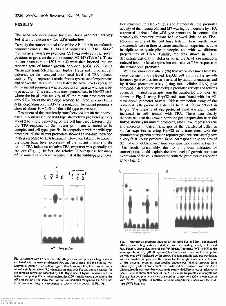
Fig. 3. Gelshift with Fos and Jun. The 90-bp stromelysin promoter fragment wasincubated with in vitro synthesized Fos and Jun proteins and the binding wasassayed by gelshift. Left side of figure: Reactions with Jun, Fos, Fos + Jun orreticulocyte lysate alone (RL) demonstrate that both Fos and Jun are needed forthe complex formation (denoted by FJ). Right side of figure: Reaction with orwithout competing 22-mer oligonucleotides (250 x molar excess) containing theAP-1 or the SP-1 site show that Fos and Jun complex recognized the AP-1 sitein the promoter fragment (sequence is shown on the bottom of Fig. 3).
H
B
Competitors . WT Ml M2 M3 M4 MS U6 M7
WT Ml M2 M3 M4 M5 M6 M7» P Labeled Fragments WT " P Labeled F'agm«m«
Fig. 4. Stromelysin promoter mutants do not bind Fos and Jun. The mutated90-bp promoter fragments are tested here for their binding activity to Fos andJun. Panel A shows that none of the 32P labeled fragments (WT to M7) at thesame specific activity (20.000 cpm/ng) bind to Fos and Jun complex except forthe wild-type (WT) indicated by the arrow. The faint gelshift band that comigrateswith the Fos-Jun complex, and the low molecular weight bands seen with someof the mutants, represent non-specific endogenous binding proteins fromreticulocyte lysate. These complexes could not be competed with the AP-1oligonucleotide nor were they consistently seen with different lots of reticulocytelysate. Panel B shows that none of the AP-1 mutant fragments can compete forFos and Jun complex when they are used as competitors (50 x molar excess)with 32P WT fragment. In contrast, efficient competition is seen with the wild-type (WT) fragment.
Downloaded from https://academic.oup.com/nar/article-abstract/19/13/3723/2387860by gueston 16 March 2018

Nucleic Acids Research, Vol. 19, No. 13 3727
We interpret the above results to suggest that, in the humanstromelysin promoter context (-1303 to +4), the AP-1 site isrequired for basal level expression but, apparently, is not essentialin the mechanism of TPA-induction. In order to study this notionfurther, we removed the AP-1 site (TGAGTCA) from thestromelysin promoter by oligonucleotide site-directed mutagenesisand studied the expression and TPA-response of such anAP-1-less promoter in transiently transfected cells in culture. Asshown in Fig. 1, the activity of this mutant promoter was similarto that of the M1-M7 mutants in all cell lines tested. Althoughits basal level expression was reduced, its relative TPA-responsein HepG2 cells and fibroblasts was about the same or higher,respectively, than that of the wild-type stromelysin promoter.Again, only in HeLa cells was the TPA-inducibility of thispromoter mutant reduced. These results clearly indicate that TPAcan stimulate human stromelysin gene expression by a mechanismindependent of the AP-1 site.
DNA binding of the Fos and Jun proteinsIn order to examine the interaction of Fos and Jun proteins withthe wild-type and mutant stromelysin promoters, we assayed bygelshift the binding of in vitro synthesized Fos and Jun1 proteinsto a 90-bp Avail-Aval restriction fragment (-101 to -11)containing the AP-1 site. Results of these experiments are shownin Figs. 3 and 4. In line with several earlier reports (10-14),both the Fos and Jun proteins (Fos-Jun heterodimer) wererequired for efficient binding to the AP-1 site (Fig. 3). Thisbinding apparently is specific because it could be competed witha 22-bp double stranded oligonucleotide containing the AP-1 sitebut not with a similar oligonucleotide containing the SP-1 site(Fig. 3). In order to test whether or not the mutant promoterfragments can bind the Fos-Jun complex, we used two differentapproaches. First, in the binding assay (Fig. 4A) we used thewild type and mutant fragments, labeled to the same specificactivity, to assay for their binding to the Fos and Jun proteincomplex. As shown, none of the AP-1 mutant fragments couldbind to the in vitro synthesized Fos-Jun heterodimer whereaseffective binding was observed with the wild-type promoterfragment. Second, in the competing binding assay (Fig. 4B) weused the mutant promoter fragments (50 X molar excess) tomeasure their capacity to inhibit the Fos and Jun complexformation with the wild-type fragment. This assay indicated that,in comparison with the wild-type fragment, all seven fragmentsfrom M1-M7 mutant promoters showed little or no competitionfor the Fos-Jun heterodimer complex. These data, taken togetherwith the results of the above transient gene expression studies,suggest that the low basal level activity of the stromelysinpromoter mutants is due to the lack of Fos-Jun binding to themutated AP-1 site.
Antisense Fos RNA expressionTo further study the involvement of the Fos and Jun proteinsin the basal level or TPA-induced expression of the stromelysinpromoter, the wild-type promoter was cotransfected into HepG2,HeLa and fibroblast cells with control plasmid or a plasmidexpressing antisense c-Fos mRNA. As shown in Table II, in thepresence of antisense Fos mRNA, basal stromelysin promoterexpression was reduced to 67% (HepG2), 57% (HeLa) and 56%(fibroblasts) of the control activity. Strikingly, however, in
1 In all of the experiments reported in this paper we used c-Fos and c-Jun.
HepG2 cells and fibroblasts the relative TPA-response of thestromelysin promoter was not affected by antisense Fosexpression. Of interest and consistent with the other data aboveis that only in Hela cells was the relative TPA-response reduced.These results, therefore, provide independent support for the viewthat c-Fos protein and, by implication the AP-1 site, is involvedin the basal level expression but is not essential for the mechanismof TPA-induced promoter activity.
Transactivation by the Fos and Jun proteinsIf the AP-1 site and the Fos and Jun proteins are required forthe basal level activity of the stromelysin promoter, thenoverexpression of the Fos and Jun proteins should transactivatethe wild-type promoter but not the AP-1 site mutant promoters.
Table II. Antisense Fos mRNA inhibits the basal level but not TPA-inducedstromelysin promoter activity. Cells were transiently transfected with the growthhormone reporter gene containing human stromelysin promoter (-1303 to +4)together with the plasmid KSV10 (control) or a plasmid expressing antisense mousec-Fos mRNA (sof). One set of cultures was treated with TPA (100 ng/ml) fortwo days before assaying promoter activity. Numbers on the left hand columnindicate the decrease of promoter activity in comparison with the control basallevel (normalized to value 1) upon transfection of antisense Fos (sof). Numberson the right hand columns refer to the promoter activity and fold of inductionafter TPA treatment. Data are from one representative experiment and an averageof four separate transfections.
H»pQ2
HeL«
Flbroblait
Ptasnild
Control
Sof
Control
Sof
Control
Sof
-
1
0 .
1
0.
1
0 .
TPA
( 7
37
56
+
14
10
77
18
3.4
2.0
Induction
14
15
77
32
3.4
3.6
111
1UJ
a.
u.O
5o
50000
40000-
30000"
20000-
10000-
F9 5 7
0 .6
pOGH
- +WT
- +M1
FJ
Fig. 5. Fos and Jun transactivate the stromelysin promoter in F9 cells. Wiki-type or the Ml mutant stromelysin promoters in the growth hormone reportergene were cotransfected into F9 embryonal carrinoma stem cells with equalamounts (0.5 /ig/well) of KSV10 control plasmid ( - ) or with plasmids pSV-C-Jun and pBK-28 expressing mouse c-Jun and human c-Fos proteins, ( + FJ),respectively. The activity of promoterless growth hormone gene (p«£GH) has notbeen subtracted from experimental values. Numbers refer to the fold oftransactivation by Fos and Jun.
Downloaded from https://academic.oup.com/nar/article-abstract/19/13/3723/2387860by gueston 16 March 2018

3728 Nucleic Acids Research, Vol. 19, No. 13
60000c
_onto
Ig 60000
40000
I20000
cJ
JB
JD
Undiflerentiated
F9
control TPA
Differentiated
control TPA
WT+FJ
WT
I CF
z
Fig. 6. Transcriptional activation of stromelysin gene in F9 cells. Undifferentiatedand differentiated F9 cells were transfected with WT stromelysin promoter (filledbars) and cotransfected with c-Jun and c-Fos expression vectors (open bars). Oneset of each culture was treated with TPA for two days after transfection. Theinduction-fold following TPA treatment is indicated in the text. The transactivationfold in different experiments was between 20 and 100-fold. A table showing TPA-response and differential mRNA expression for Fos and Jun proteins indifferentiated and undifferentiated F9 cells is based on the Northern hybridizationdata of Yang-Yen el al. (36). The three levels of expression are low (+), medium( + + ) and high (+ + +).
Accordingly, the wild-type stromelysin promoter was transfectedinto mouse F9 embryonal carcinoma stem cells together with acontrol plasmid or plasmids expressing human Fos and mouseJun proteins. As demonstrated previously, undifferentiated F9cells have no endogenous c-Fos or c-Jun proteins (15,16,36).As shown in Fig. 5, cotransfection of the Fos and Jun expressionplasmids resulted in a dramatic increase (57-fold) in the basallevel stromelysin promoter activity. In contrast, and consistentwith our other data reported here, Fos and Jun could nottransactivate the stromelysin promoter mutant Ml.
To study further the role of Fos and Jun in the stromelysingene regulation, we carried out transfection experiments indifferentiated and undifferentiated F9 cells in the presence orabsence of TPA. As shown in Fig. 6, very little stromelysinexpression is detected in both cell types and, significantly, thepromoter response to TPA-treatment appears minimal. Inundifferentiated F9 cells, TPA induces the stromelysin expression1.9 fold and in differentiated F9 cells the induction is 1.4 fold.Again, a striking transactivation of the stromelysin promoteroccurs in the presence of cotransfected c-Fos and c-Jun expressionplasmids. The effect of TPA in these experiments is intriguing.Taking into account the minimal TPA-response of the stromelysinpromoter alone, TPA seems to inhibit the relative transactivationby Fos and Jun. In the experiment shown in Fig. 6 this inhibitionis about 50% in both types of F9 cells (see Discussion).
As shown in Fig. 7, the Fos and Jun proteins can alsotransactivate the stromelysin promoter in HepG2 cells. In theseexperiments, cotransfection with Fos and Jun expression plasmidsresulted in a consistent 3-fold increase in the basal level promoter
H.pO2
pOOH TPA
FJ
Fig. 7. Fos and Jun transactivate the stromelysin promoter in HepG2 cells. HepG2cells were cotransfected with the wild-type stromelysin promoter and either thecontrol plasmid or the Fos and Jun expression plasmids ( + FJ) as in Fig. 5. Oneset of cultures was treated (+) with TPA (100 ng/ml) for two days before assayingthe promoter activity. Numbers refer to the induction fold by TPA and the foldof transactivation by Fos and Jun. The activity of promoterless reporter gene(p4>GH) has not been subtracted from experimental values.
TPApGCat C-
TJ.047
+
f0.045
WTCit-
fo.6a
+
t
t2.8
4 . J
U1Cat-
f0.21
0.29
+
f0 61
3.S
% AcetytaUon
told Induction
Fig. 8. TPA-induced CAT expression from wild-type and mutant stromelysinpromoters. Primary human skin fibroblasts were transiently transfected withpGCAT-C (promoterless CAT reporter gene), WTCAT (CAT gene with wild-type stromelysin promoter) or Ml CAT (CAT gene containing promoter withmutant AP-1 site) and treated with TPA 24h post-transfection for an additional48h. CAT activity and the percentage of acetylation was determined as in Materialsand Methods. Induction fold was determined after subtracting from theexperimental values the background% acetylation contributed by pGCAT-C alone.Basal wild-type CAT activity was assigned a value of 1 to facilitate comparisons.The value of 4.3 refers to the induction fold relative to basal wild-type activity.The value of 3.5 refers to the induction fold relative to basal mutant activity.Data are from one representative experiment.
activity. A similar increase in the promoter expression wasobtained by TPA-treatment of control cells that were transfectedwith the stromelysin promoter alone. Interestingly, TPA treatment of the Fos and Jun transactivated HepG2 cells neitherinhibited nor enhanced the basal level stromelysin promoteractivity.
Reporter gene contextWe were concerned about the possibility that the results for thestromelysin AP-1 site presented here may have reflected some'cryptic' contribution of the hGH reporter gene used. Forexample, the hGH gene contains elements that can allow responseto glucocorticoids in certain reporter gene constructs (29).Therefore, the wild-type and Ml mutant stromelysin promoters(—1303 to +4) were inserted into the reporter gene ofchloramphenicol acetyl transferase, GCAT-C, (34). As shown
Downloaded from https://academic.oup.com/nar/article-abstract/19/13/3723/2387860by gueston 16 March 2018

Nucleic Acids Research, Vol. 19, No. 13 3729
aooo
4SOO
1000
0
B •
•control
TPA
IPOGH pDBUX
Fig. 9. The role of AP-1 in minimal TK promoter. Panel A shows the TPAresponse of the CAT reporter gene (Col-TREIX/TK-CAT) containing minimalTK promoter with the AP-1 site from human collagenase gene ( -73 to -65).pGCAT-C is promoterless CAT gene. HepG2 cells were transiently transfectedwith the reporter genes and cultured with or without TPA for 48 hr before CATassay. Panel B shows the TPA response of the growth hormone reporter genecontaining minima] TK promoter with (plXBLGH) or without (pBLGH) the AP-1site from human collagenase gene. p</>GH is promoterless growth hormone gene.Human skin fibroblasts were transiently transfected with the reporter genes andcultured with or without TPA for 48 hr before growth hormone assay.
in Fig. 8, in transiently transfected skin fibroblasts, the relativeTPA-responses of the wild-type (4.3-fold induction) and the Mlmutant (3.5-fold) promoter were comparable. We also saw theseresults using similarly transfected HepG2 cells (data not shown).Based on the results of these experiments we conclude that thesequence contribution of the reporter gene is insignificant. Rather,the transient gene expression in the experiments reported hereis due to the transcriptional activity of the stromelysin promoter.
We have studied here the transcriptional activity of AP-1 sitein the context of the human stromelysin promoter. Therefore,it was of interest to study the activity of one copy of the AP-1site inserted into an unrelated gene promoter, the minimalpromoter (-105 to + 51) of the thymidine kinase (TK) gene.The AP-1 site linked to the TK promoter was used in two differentreporter genes, CAT and hGH, and its role in basa] expressionand TPA-response was analyzed in transiently transfected skinfibroblasts and HepG2 cells in culture. As shown in Fig. 9B,the AP-1 site could only marginally stimulate basal expressionfrom the TK promoter. Its role as a TPA response elementappeared to be marginal as well. In several experiments usingthe reporter gene of growth hormone, one copy of the AP-1 siteincreased the relative TPA-response of the TK promoter about1.4-fold. Furthermore, this result is in line with the minimal TPA-response (Fig. 9A) seen using the CAT reporter gene.
DISCUSSION
Originally, AP-1 was defined as a DNA-binding activity in HeLacell extracts that recognized sequences in the basal level enhancerof the metallothionein HA gene and the 72-bp enhancer ofSV40-virus (4). These sequences or AP-1 sites as they are nowknown in many cellular and viral genes play a role in theactivation or repression of gene expression (3). The AP-1 siteis one of the TPA response elements (TRE) and a major targetfor the binding of the Fos and Jun family of transcription factors(5-7) .
We have studied here the transcriptional role of the AP-1 sitein the context of the human stromelysin promoter (—1303 to +4).
Our results from transient transfection experiments show that thestromelysin AP-1 site ( -70 to -64) is required for basalexpression of the promoter. Because the promoter with mutatedAP-1 sites showed reduced transcriptional activity and did notbind or compete for in vitro synthesized c-Fos and c-Jun proteins(Figs. 1 -4 ) it seems likely that the basal expression is regulatedby Fos and Jun. In support of this view, we have shown herethat c-Fos and c-Jun expression in transiently transfected mouseF9 cells transactivates the wild-type stromelysin promoter butnot the AP-1 site mutant promoter tested.Furthemore, wespeculate that if the Fos and Jun proteins are needed for basalexpression of the stromelysin gene then the heterodimer of thec-Fos and c-Jun proteins should play a major role in theregulation. This notion is based on the data shown here and thatreported by Yang-Yen et al. (36) as summarized in Fig. 6. Inthe F9 embryonal carcinoma stem cells only Jun D isconstitutively expressed and at increased levels in differentiatedF9 cells. Upon cell differentiation c-Jun becomes expressed aswell. It is interesting to note that, although TPA induces theexpression of c-Fos and Jun B in undifferentiated F9 cells, inno circumstances are c-Fos and c-Jun coexpressed. These dataare in line with the low level of stromelysin expression intransfected F9 cells as well as with the minimal increase ofpromoter activity in the TPA treated F9 cells (see Fig. 6 ).
An intriguing result of the present study was the observationthat a functional AP-1 site apparently is not necessary for therelative TPA-response of the stromelysin promoter. First of all,Fig. 1 shows that the TPA-response of the AP-1 site mutantstromelysin promoters and, strikingly, the promoter mutantlacking the AP-1 site is similar to that of the wild-type promoterin transfected fibroblasts and HepG2 cells. However, it isimportant to point out that in HeLa cells both the basal expressionand TPA-response apparently are affected by the AP-1 sitemutations. It is also intriguing that one of the AP-1 site mutations(M5) almost completely abolished the TPA-response of thestromelysin promoter in all cell lines tested (Fig.l).Understanding these differences and exceptions in results mayprovide insight into the mechanism of TPA-response.
Using the terminology for strong versus weak promoters, theabove results can be restated as follows. If the wild-typestromelysin promoter is considered as a strong promoter thenthe AP-1 site mutant promoters represent weak promoters becauseof their reduced basal activity in the transfected cells. As we haveshown here, both of these promoter types, however, can besimilarly induced by TPA treatment. Importantly, since in thesecases the only difference between the strong and weak promotersis the presence or absence of a functional AP-1 site, respectively,we conclude here that the AP-1 site is not necessary for TPA-response. Results from cotransfection experiments with antisensec-Fos mRNA expression plasmid are in line with this notion.As shown in Table II, antisense c-Fos transfection reduces thebasal level stromelysin expression but has little or no effect onthe relative TPA-response of the stromelysin promoter.Accordingly, these results provide totally independent evidencefor the idea that the Fos protein and, by implication, the AP-1site is not necessary for TPA-induction mechanism.
We have recently identified, in the human stromelysinpromoter, three additional AP-1 like sites (B. G., unpublished).In gelshift experiments, in comparison with the proximal AP-1site, these elements display weak or moderate binding for thein vitro synthesized Fos-Jun heterodimer. Our preliminary resultsfrom mutational analysis and transient gene expression studies
Downloaded from https://academic.oup.com/nar/article-abstract/19/13/3723/2387860by gueston 16 March 2018

3730 Nucleic Acids Research, Vol. 19, No. 13
suggest that none of these three AP-1 like elements affect thelevel of TPA-induced stromelysin promoter activity. However,results from similar experiments indicate that each one of themcan regulate the basal promoter activity. Irrespective of theirputative role in the stromelysin gene regulation we conclude fromthe above data that these elements for the Fos-Jun binding cannot explain the TPA-response of the stromelysin promotermutants studied here.
We also tested the effect of one copy of the AP-1 site on theheterologous thymidine kinase (TK) gene promoter. In transfectedfibroblasts and HepG2 cells the AP-1 site had little or no effecton basal expression or TPA-response of the TK promoter.Furthermore, we have consistently seen these results using twodifferent reporter genes, the CAT and hGH genes. These datawhich have been reported by others as well (15,30,36) suggestthat the AP-1 site alone is not sufficient for transcriptional controlof genes. Yet significant TPA-response and stimulation oftranscription is obtained with promoter constructs containingmultimerized (three or five copies) AP-1 sites (15,30,36). Inrespect of promoter structure and mechanism of gene regulation,these observations, however, are difficult to interpret.Presumably, the true organization of regulatory elements, suchas the AP-1 site, plays an important role in gene expressioncomparable to that of the elements themselves. It follows thatthe transcriptional activity of the AP-1 site may change dependingon its precise DNA sequence environment (see below).
Recently, three papers have reported data which agree withthe results and conclusions presented here. First, Timmers et al.(38) identified an AP-1 like site in the rat JE gene that wasessential for the basal expression but not for TPA-response.Mutations on the JE AP-1 site (TGACTCC - 5 2 to -46)significantly reduced the basal activity of the promoter (—543to +54) and interestingly, resulted in increased relative TPA-response as compared to that of the wild-type promoter. Theirresults also indicate that the JE AP-1 site is not sufficient forthe basal level expression because minimal promoter constructswith the JE AP-1 site showed little or no basal level expressionor TPA induced activity. It is unclear, however, whether the Fosand Jun proteins are involved in the activation of the JE AP-1site because the sequence, which is similar to the M7 mutantdescribed here, does not bind in vitro synthesized Fos and Junproteins. Second, by studying the effects of antisense Fos mRNAexpression in NIH 3T3 cells, Kerr et al. (39) suggested that thegrowth factors PDGF (platelet derived growth factor) and EGF(epidermal growth factor) induce rat stromelysin gene expressionby Fos-dependent and Fos-independent pathways, respectively.Significantly, however, the stimulatory effect of both PDGF andEGF on stromelysin transcription seemed to involve factorsrecognizing the AP-1 site. In addition, their data indicated thatin cells expressing antisense c-Fos mRNA the basal level activityof transfected stromelysin promoter (-751 to — 1) was reducedto 65% of the control promoter activity. However, in these cellsno significant effect was seen on the relative TPA-response ofthe stromelysin promoter. Third, as shown by van Dam et al.(40), the AP-1 site of the c-Jun gene is important for basalexpression but does not affect the TPA-response of the gene.Similar to our results, mutational inactivation of the c-Jun AP-1site abolished stimulation by TPA of the - 7 9 to +170 geneconstruct, whereas this did not affect the TPA inducibility of the—1600 to +740 gene for c-Jun. Thus, the AP-1 site functionsdifferently in the contexts of the full-length c-Jun promoter andof smaller promoter fragments (40).
Furthermore, we note here that the AP-1 site sequence, TTA-GTCA, of the stromelysin promoter mutant M2 is the same asthat in the P motif of SV40 enhancer (41). While mutation ofthis sequence completely abolished the AP-1 site DNase footprintit, remarkably, had no effect either on the basal expression fromthe linked B-globin reporter gene or its induction by TPA. Fromthe results of these experiments it seems that the P motif AP-1binding site is not required for induction of SV40 enhanceractivity by TPA (41). Interestingly, however, five copies of theP motif alone was as active and displayed similar transcriptionalresponse to TPA as the complete SV40 enhancer. It is temptingto speculate that these results may be due to sequence contexteffects so that different or additional transcription factors areneeded to distinguish the P motif when it is part of the SV40enhancer or when it is alone. Alternatively, these data may reflectother interacting elements in the enhancer sequence or in the B-globin reporter gene used in these studies. These notions,furthermore, are consistent with the recent data on the differentialeffects of the adenovirus El A oncoprotein on the transcriptionalactivity of the c-Jun and collagenase AP-1 sites (42). Indeed,Deutsch et al. (43) have presented direct evidence for the roleof sequence context on the transcriptional activity of the AP-1site. For example, different sequence contexts of only a few basesflanking the AP-1 site had pronounced effects on the TPA-response of the human gonadotropin gene in transientlytransfected JEG-3 cells. Finally, Schule et al. (44) havedemonstrated the importance of promoter architecture in thetranscriptional control of genes. The results of their experimentssuggest that the glucocorticoid receptor can interact with manydifferent transcriptional factors and synergistically activate geneexpression.
We also note here that the stromelysin promoter mutants M2and M3 contain mutations in the AP-1 site (TTAGTCA andTGCGTCA, mutations underlined) identical to those preparedby Risse et al. (18). In their studies, in comparison with the wild-type sequence, these point mutations had little or no effect onthe Fos-Jun heterodimer binding to a double stranded 17-meroligonucleotide. In contrast, when we examined these sequencesin the context of 90-bp fragment from the stromelysin promoter,we could not detect any binding or competition for the Fos-Junheterodimer. Importantly, in transient gene expression studiesthese same mutants showed reduced basal level activity similarto that of the other promoter mutants studied here. At presentwe have no explanation for this striking variation in results butdifferent sequence contexts may be important for the Fos-Junbinding.
The AP-1 site mutants studied here were constructed to besymmetrical in the palindromic sequence. As an example, theM3 mutant reads TGCGTCA on the top strand. Itscomplementary sequence on the bottom strand is TGACGCAwhich is similar to that of the M5 mutant top strand sequenceTGAGGCA. Therefore, the M3 mutant is analogous to the M5mutant when turned upside down. Note that in many genes thecentral nucleotide of the AP-1 site is either C or G. Because ofthese considerations we expected to see similar responses for theM3 and M5 stromelysin promoter mutants. Thus, it wasintriguing to find out that the M5 mutant promoter consistentlyshowed little or no TPA-response whereas the M3 mutant couldbe induced by TPA in relative terms similarly to that of the wild-type promoter (Fig.l). We interpret these data to suggest thatthe AP-1 site activity in vivo can be polarized to reflect thecontribution of the sequence context of the gene.
Downloaded from https://academic.oup.com/nar/article-abstract/19/13/3723/2387860by gueston 16 March 2018

Nucleic Acids Research, Vol. 19, No. 13 3731
ACKNOWLEDGEMENT
We are grateful to Helmut Ponta, Peter Angel, Michael Karin,Inder Verma and Tom Curran for the many DNA constructs usedin this work. Further, we thank Thierry Frebourg and BrunoLuckow for the GCAT-C and BLCAT2 plasmids. We also thankTom Curran for his encouragement and comments on themanuscript. We thank Elizabeth Lesko for preparing the plasmidDNA, Maozhou Yang for help with the photography andFrancine Mittleman for typing this manuscript. This work wassupported by grants from the N.I.H. (AR 33714, HD 24442).
REFERENCES
1. Hollstein, M. and Yamasaki, H. (1987)in: 'Tumor Cell Differentiation1 (J.Aarbaklce, P.K. Chiang, and H.P. Koeffler, eds.,) The Humana Press,Clifton, New Jersey.
2. Nishizuka, Y. (1988) Nature 334, 661-665.3. Curran, T. and Franza, B.R., Jr. (1988) Cell 55, 395-397.4. Lee, W., Haslinger, A., Karin, M. and Tjian, R. (1987) Nature 325,
368-372.5. Angel, P., Imagawa, M., Chiu, R., Stein, B., Imbra, R.J., Rahmsdorf, H.J.,
Jonat, C , Herrlich, P. and Karin, M. (1987) Cell 49,729-739.6. Lee, W., Mitchell, P. and Tjian, R. (1987) Cell 49, 741-752.7. Rauscher, FJ. m, Sambucetti, L.C., Curran, T., Distel, R.J. and Spiegelman,
B.M. (1988) Cell 52, 471-480.8. Muegge, K., Williams, T.M., Kant, J., Karin, M., Chiu, R., Schmidt, A.,
Siebenlist, V., Young, H.A. and Purim, S.K. (1989) Science 246, 249-251.9. Brenner, D.A., O'Hara, M., Angel, P., Chojkier, M. and Karin, M. (1989)
Nature 337, 661-663.10. Kouzarides, T. and Ziff, E. (1989) Nature 340, 568-571.11. Rauscher, F.J. Ill, Voulalas, P.J., Franza, B.R., Jr. and Curran, T. (1988)
Genes Dev., 2, 1687-169912. Turner, R. and Tjian, R. (1989) Science 243, 1689-1694.13. Nakabeppu, Y., Ryder, K. and Nathans, D. (1988) Cell 55, 907-915.14. Halazonetis, T.D., Georgopoulos, K., Greenberg, M.E. and Leder, P. (1988)
Cell 55, 917-924.15. Chiu, R., Angel, P. and Karin, M. (1989) Cell 59, 979-986.16. Schutte, J., Viollet, J., Neu, M., Segal, S., Feokorko, J. and Minna, J.
(1989) Cell 59, 987-997.17. Nakabeppu, Y. and Nathans, D. (1989) EMBO J., 8, 3833-3841.18. Risse, G., Jooss, K, Neuberg, M., Bruller, H-J. and Muller, R. (1989) EMBO
J., 8, 3825-3832.19. Matrisian, L.M. (1990) Trends Genet. 6: 121-125.20. Frisch, S.M. and Ruley, H.E. (1987) J. Biol. Chem. 262: 16300-16304.21. Quinones, S.,Saus, J.,Otani, Y., Harris, E.D.,Jr. and Kurkinen, M. (1989)
J. Biol. Chem., 264, 8339-8344.22. Lee, T.H., Lee, G.W., Ziff, E.B. and Vilcek, J. (1990) Mol. Cell. Biol.
10: 1982-1988.23. Jonat, C , Rahmsdorf, H.J., Park, K.-K, Cato, A.C.B., Gebel, S., Ponta,
H. and Herrlich, P. (1990) Cell 62: 1189-1204.24. Yang-Yen, H.-F, Chambard, J.-C., Sun, Y.-L, Smeal, T., Schmidt, T.J.,
Drouin, J. and Karin, M. (1990) Cell 62: 1205-1215.25. Schule, R., Rangaran, P., Kliewer, S., Ransone, L.J., Bolado, J., Yang,
N., Verma, I.M. and Evans, R.M. (1990) Cell 62: 1217-1226.26. Buttice, G. and Kurkinen, M. (1990) Nucleic Acids Res., 18, 3079.27. Kunkel, T.A. (1985) Proc. Natl. Acad. Sci. USA 82, 488-492.28. Venkitaraman, A.R. (1989) Nucleic Acids Res., 17, 3314.29. Selden, R.F., Howie, K.B., Rowe, M.E., Goodman, H.M. and Moore, D.D.
(1986) Mol. Cell. Biol., 6, 3173-3179.30. Schonthal, A., Herrlich, P., Rahmsdorf, HJ. and Ponta, H. (1988) Cell
54, 325-334.31. Lamph, W.W., Wamsley, P., Sassone-Corsi, P. and Verma, I.M. (1988)
Nature 334, 629-631.32. Sassone-Corsi, P., Lamph, W.W., Kamps, M. and Verma, I. (1988) Cell
54, 553-560.33. Ausubel, M.F., Brent, R., Kingston, R.E., Moore, D.D., Seidman, J.G.,
Smith, J.A. and Struhl, K. (1988) Current Protocols in Molecular Biology,John Wiley & Sons, New York.
34. Frebourg, T. and Brison, O. (1988) Gene, 65, 315-318.35. Luckow, B. and Schutz, G. (1987) Nucleic Acids Res. 115, 5490.36. Yang-Yen, H.-F., Chiu, R. and Karin, M. (1990) New Biologist 2, 351 -361.
37. Ryder, K., Lanahan, A., Perez-Albuerne, E. and Nathans, D. (1989) Proc.Natl. Acad. Sci. USA 86, 1500-1503.
38. Timmers, H.T.M., Pronk, G.J., Bos, J. L. and van der Eb, A.J. (1990)Nucleic Acids Res., 18, 23-34.
39. Kerr, L.D., Holt, J. and Matrisian, L.M. (1988) Science 242, 1424-1427.40. van Dam, H., Offringa, R., Meijer, I., Stein, B., Smits, A.M., Heniich,
P., Bos, J.L. and van der Eb, A.J. (1990) Mol. Cell.Biol. 10, 5857-5864.41. Chiu, R., Imagawa, M., Imbra, R.J., Bockoven, J.R. and Karin, M. (1987)
Nature 329, 648-651.42. Offringa, R., Gebel, S., Van Dam, H., Timmers, M., Smits, A., Zwart,
R., Stein, B., Bos, J.L., Van der Eb, A. and Herrlich, P. (1990) Cell, 62,527-538.
43. Deutsch, P.J., Hoeffer, J.P., Jameson, J.L.and Habener, J.F. (1988) Proc.Natl. Acad. Sci. USA 85, 7922-7926.
44. Schule, R., Muller, M.,Kaltschmidt, C. and Renkawitz, R. (1988) Science242, 1418-1420.
Downloaded from https://academic.oup.com/nar/article-abstract/19/13/3723/2387860by gueston 16 March 2018