the apurinic/apyrimidinic endodeoxyribonuclease of rat-liver chromatin
TRANSCRIPT
Eur. J. Biochem. 129, 509-517 (1983) 0 FEBS 1983
The Apurinic/Apyrimidinic Endodeoxyribonuclease of Rat-Liver Chromatin Regis CESAR and Walter G. VERLY
Supplementary Material Suzanne BRICTEUX-GREGOIRE, Yvette HABRAKEN, and Walter G. VERLY Biochimie, Faculte des Sciences, Universite de Liege
(Received June 30/September 29, 1982)
The extract from rat liver chromatin contains two apurinic/apyrimidinic (AP) endodeoxyribonucleases named 0.2 M and 0.3 M isozymes according to the phosphate concentration necessary to elute them from an hydroxyapatite column. The 0.3 M isozyme is the main and perhaps the only chromatin AP endodeoxyribonuclease in the living cell. This 0.3 M isozyme was purified by successive chromatographies on hydroxyapatite, phosphocellulose, heparin- Sepharose and alkylated-depurinated DNA-cellulose. It has a molecular weight of approximately 39000 ; its optimum pH is around 8.0; it needs Mg2+ or Mn2+ to be active and the optimum concentration for Mg2 + is between 5 mM and 10 mM. The 0.3 M isozyme has no action on intact DNA strands or on alkylated sites; it cuts the phosphodiester bridge which is the immediate neighbour of the AP site on its 5' side leaving 3'-hydroxyl and 5'- phosphate ends. It has no associated exonuclease activity. To hydrolyze the phosphoester bond near the AP site, the enzyme makes a close contact with three base residues in the large groove of the DNA molecule.
More than 90 % of the AP (apurinic/apyrimidinic) endo- deoxyribonuclease activity in rat liver cells are in chromatin. Native chromatin has little action on an added foreign DNA containing AP (apurinic or apyrimidinic) sites, but the activity is found in the non-histone proteins after dissociation of the chromatin [l]. Chromatography of the chromatin proteins on DEAE-cellulose separates two AP endodeoxyribonucleases. In the initial work done in this laboratory, the spxies emerging at the lower ionic strength was the most abundant; it was completely purified and its properties were studied [2,3].
In more recent experiments, it was found convenient to start the purification of the chromatin proteins with an hydroxyapatite chromatography which also separates the two AP endodeoxyribonuclease isozymes : the species which had been completely purified is eluted with 0.2 M potassium phosphate whereas the other species emerges with 0.3 M potassium phosphate. These two species were called 0.2 M and 0.3 M isozymes respectively. The ratio of the two isozymes varies according to details in the procedure followed before they are separated. The 0.2 M isozyme is brought below 10 % when 0.5 mM phenylmethylsulfonyl fluoride is used in all solutions and the time for completion of the hydroxyapatite chromatography is kept to a minimum (see miniprint supple- ment). It seems finally that most, if not all, of the 0.2 M isozyme is an artifact and that the important AP endodeoxy- ribonuclease in the living cell chromatin is the 0.3 M isozyme. This paper deals with the purification and properties of the 0.3 M AP endodeoxyribonuclease isozyme of rat liver chromatin. ~~
Abbreviations. AP, apurinic or apyrimidinic; PMSF, phenylmethyl- sulfonyl fluoride.
Enzymes. Apurinic/apyrimidinic (AP) endodeoxyribonucleases (EC 3.1.25.2); pancreatic deoxyribonuclease (EC 3.1.21.1); alkaline phos- phatase (EC 3.1.3.1); polynucleotide 5'-hydroxyl-kinase (EC 2.7.1.78); snake venom phosphodiesterase (EC 3.1.4.1).
MATERIALS AND METHODS
NaCl/Cit Buffers
NaCl/Cit = 150 mM NaC1,15 mM sodium citrate; the pH was 7.0 or 8.0 as specified. It was sometimes diluted 10-fold (0.1 NaCl/Cit).
Substrates
Untreated, Alkyluted andAlkyl~ted-depurinuted[~ HjDNAs. DNA, prepared from Escherichia coli B41 grown in the presence of [3H]thymidine (20 Ci/mmol) had a specific radioactivity of about 70000 dis. min-' + pg-l. Alkylation with methyl methanesulfonate and partial depurination were carried out as previously described [4]. The alkylated [3H]DNA contained about 140 methyl groups and the al- kylated-depurinated [3H]DNA about 50 AP sites/103 nucleo- tides. The labelled double-stranded DNA (untreated, al- kylated or alkylated-depurinated) was dissolved in 0. l NaCl/Cit, 20 mM MgC12, pH 8.0, to a concentration of 20 pg . ml-'. Acid-depurinated r3H]DNA. To 1 vol. of untreated [3H]DNA solution (200 pg . ml-') was added 1 vol. of 0.5 M sodium acetate buffer, pH 3.7. After an 120-h incubation at 37 "C, it was dialyzed against 0.1 NaCl/Cit, 20 mM MgC12, pH 8.0. The depurinated [3H]DNA contained 13 AP sites/103 nucleotides.
Nicked r3H]DNA. [3H]DNA (16 pg) in 800 bl of 0.1 NaCl/Cit, 20 mM MgCl,, pH 8.0, was mixed with 80 p10.15 M KCl, 0.05 %bovine serum albumin, 0.01 % 2-mercaptoethanol containing 0.04 unit of pancreatic deoxyribonuclease (Boeh- ringer) and incubated 15 min at 37 "C. The nuclease was then inactivated by heating 10 min at 77°C to obtain double- stranded nicked [3H]DNA. This substrate contained about 11 breaks/l O3 nucleotides.
Nicked (3 H j D N A with 5'-(32 Pjphosphate Ends. Nicked [3H]DNA prepared as in the previous section was successively
510
treated with alkaline phosphatase and polynucleotide 5'- hydroxyl-kinase together with [Y-~~P]ATP following the method of Weiss et al. [5]. After phenol deproteinization and precipitation with ethanol, the double-labelled DNA (70000 dis. 3H min-'.pg-'; 200000 dis. 32P min-'.pg-') was dissolved in 10 mM Tris/HCl, 0.1 mM EDTA, pH 8.0.
Doubly-labelled Uracil-containing Poly(dA-dT). The de- tails of the preparation were given previously [3,6]. The polymer was labelled with 3H in the adenine residues and with 32P 5' to the uracil residues.
Enzyme Assay
To 20 p1 of alkylated-depurinated [3H]DNA (0.4 pg) solution were added 20 pl of enzyme preparation. After a 15- min incubation at 37 "C, the tubes were cooled in ice, then 100 pl NaCl/Cit, pH 7.0, containing 200 pg of unlabelled DNA and 860 pl5.8 % perchloric acid were added successively. The tubes were shaken vigorously, then centrifuged at 12000 x g for 10 min; 500 pl from the supernatants were used to measure the acid-soluble radioactivity. Break frequencies were calculated from the acid-soluble fractions (unpublished). Results were corrected for controls without enzyme which always contained very few breaks.
One unit (U) of AP endodeoxyribonuclease is the enzyme activity which hydrolyzes 1 pmol phosphoester bonds near AP sites/min in the conditions described above [7].
Assay for Protein
of Peterson [8] using bovine serum albumin as standard. Protein concentration was measured by the micromethod
Chromatography on Bio-Gel P-I00
A column (1.6 x 95 cm) of Bio-Gel P-100 was equilibrated with 50 mM Tris/HCl, 0.1 M ammonium sulfate, pH 7.5. The void volume ( Vo) was measured with blue dextran, the total volume (6) with riboflavin, and the column was calibrated with four proteins of known molecular weights : cytochrome c (12 SOO), chymotrypsinogen (25 000), ovalbumin (45 000) and bovine serum albumin (67000). Each of these proteins or the enzyme was dissolved in 2 ml of buffer and applied on the column. The elution was carried out at a rate of 8 ml . h-' ; 3.1- ml fractions were collected and the absorbance at 280 nm or the activity on alkylated-depurinated [3H]DNA was measured to determine the elution volume ( Ve).
Acid-Solubility
This always refers to solubility in 5 % perchloric acid.
EXPERIMENTS AND RESULTS
Preparation of Chromatin and Purification of the 0.3 M Isozyme
All the solutions used to prepare the chromatin extract contained 0.5 mM phenylmethylsulfonyl fluoride (PMSF). The nuclei were isolated from rat liver and chromatin prepared from the purified nuclei as described by Thibodeau and Verly [ 11. The chromatin was dissociated with heparin-Ultrogel (LKB) and the heparin-Ultrogel-DNA-protein complex was extracted twice with 0.5 M KC1,lO mM potassium phosphate,
300
200
100 0 P c
L . 2 0
g E 2
7- v
- m
W
LO
20
0
0.2M 0.3M @ 1 1
0.2M 0.5M
1 1 1.0 {
Fraction number
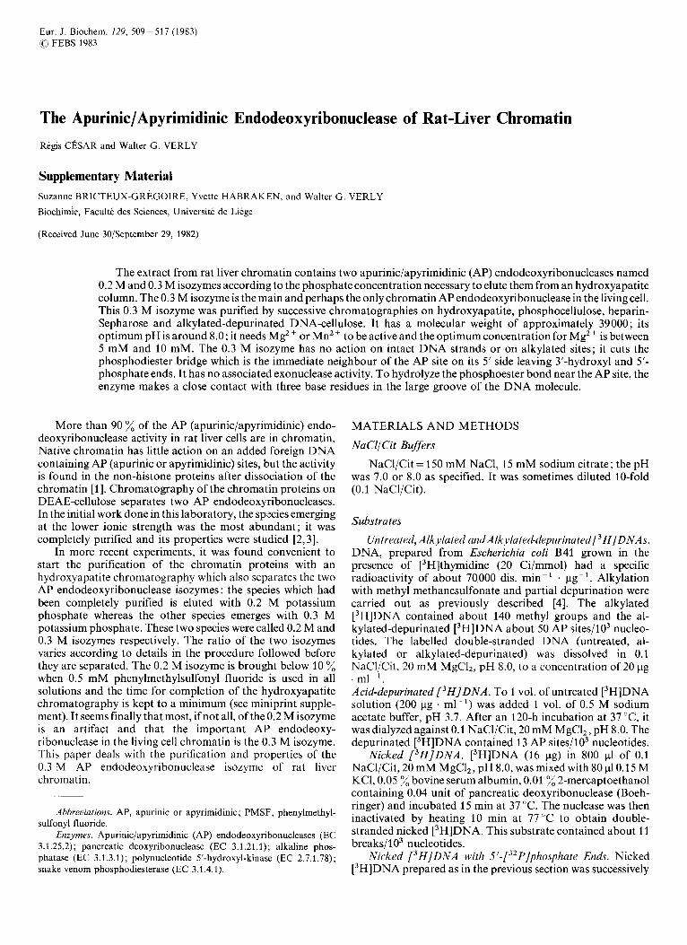
Fig. 1. Purification of the 0.3 M isozyme of A P endodeoxyribonuclease. (A) Hydroxyapatite chromatography. The 0.5 M KCI extract from rat liver chromatin was poured on a column (1.6 x 15 cm) of hydroxyapatite. Elution was carried out with 100ml of 0.2 M and 100 ml of 0.3 M potassium phosphate, pH 6.80; 9-ml fractions were collected. The arrows indicate when the eluent emerging from the column had reached 0.2 M and 0.3 M respectively (refractory index determination). (B) Phosphocellulose chro- matography. Fractions 39 -43 from the hydroxyapatite chromatography were pooled and dialyzed against buffer B. The solution was poured on a column (1 x 8 cm) of phosphocellulose. After washing with 40 ml of buffer B, the column was eluted with 100 ml of a 0 - 1 M KC1 linear gradient in buffer B; 3.1-ml fractions were collected. The straight line represents the gradient as it emerged from the column. (C) Heparin-Sepharose chromat- ography. Fractions 44 and 45 from the phosphocellulose chromatography were pooled and dialyzed against 10 mM phosphate, pH 7.30. The solution was poured on a column (1 x 6 cm) of heparin-Sepharose. After a 65-ml washing, the column was eluted by steps with 20 ml each of 0.2 M, 0.5 M, and 1.0 M KCl in the same buffer; 3-ml fractions were collected. The arrows indicate when the eluent emerging from the column had reached the indicated KC1 concentrations (refractory index determination). (D) Alkylated-depurinated DNA-cellulose chromatography. Fractions 31 - 33 from the heparin-Sepharose chromatography were pooled and dialyzed against buffer D. The protein solution was poured on a column (1 x 10 cm) of alkylated-depurinated DNA-cellulose. After washing with 25 ml of buffer D, the column was eluted with 100 ml of a 0-1 M KCl linear gradient in buffer D ; 3-ml fractions were collected. The straight line represents the gradient as it emerged from the column. An aliquot of each fraction, after dilution with buffer E, was used to measure the activity on alkylated-depurinated [3H]DNA. The values are the minimum since the determinations were not always done under maximal conditions
10 mM Tris/HCl, pH 8.0, as described in the miniprint supplement (see also [9]). The pooled extracts (Prep I ; 237 ml) contained 100 mg protein and 3610 kU of AP endodeoxy- ribonuclease activity.
Prep I was directly poured onto a column (1.6 x 15 cm) of hydroxyapatite equilibrated with 5 mM potassium phosphate, pH 6.8. The column was eluted by steps with 100 ml of 0.2 M and 100 ml of 0.3 M potassium phosphate, pH 6.8, at a rate of 15 ml . h-'. Absorbance at 230 nm and activity on alkylated- depurinated [3H]DNA were measured on the collected 9-ml
51 1
Table 1. Purification of the 0.3 M isozyme of AP endodeoxyribonuclease from rat liver chromatin Prep I refers to the proteins extracted from the heparin-Ultrogel-DNA-protein complex with 0.5 M KC1; Prep I1 is the second peak of the hydroxyapatite chromatography (0.3 M isozyme) after dialysis ; Prep I11 is the active fractions from the phosphocellulose chromatography after dialysis; Prep IV is the second peak from the heparin-Sepharose chromatography after dialysis ; Prep V is the single peak from alkylated-depurinated DNA-cellulose chromatography. The 0.3 M isozyme activity in Prep I has been calculated taking into account the ratio of 0.2 M to 0.3 M isozyme found in the hydroxyapatite chromatographies and assuming the same yields for the two isozymes
Prep Enzyme activity Yield Protein Specific activity Purification
total 0.3 M total 0.3 M isozyme isozyme
kU % mg kU/mg protein -fold
I 3610 2635 100 99.8 36.2 26 1 I1 1374 52 7.9 174 7 I11 620 23 1.8 344 13 IV 546 21 0.94 560 22 V 496 19 0.32 1550 59
fractions. Fig. 1A shows that two peaks of activity were separated: the 0.2 M isozyme represented 27 % and the 0.3 M isozyme 73 % of the recovered activity; the overall yield was 52 %. Only the 0.3 M isozyme was processed further.
Fraction 39 - 43 from the hydroxyapatite chromatography were pooled and dialyzed over night against 4 1 of buffer B (1 5 mM potassium phosphate, 0.1 mM EDTA, 0.1 mM 2- mercaptoethanol, pH 7.2). A small precipitate which formed in the dialysis bag was discarded by centrifugation. The 47-ml supernatant (Prep 11) containing 7.9 mg protein and 1370 kU enzyme was poured at a rate of 20 ml . h-' onto a column (1 x 8 cm) of phosphocellulose equilibrated in buffer B. After washing with 40 ml of the same buffer, the column was eluted, a t a r a t e o f 8 m l ~ h - ' , w i t h 1 0 0 m l o f a 0 - 1 MKCllinear gradient in buffer B. Absorbance at 230 nm and activity on alkylated-depurinated [3H]DNA were measured on each 3.1-ml fraction. Fig. 1B shows a protein peak eluted at 0.5 M KCl corresponding to the single peak of enzyme activity.
Fractions 44 and 45 from the previous chromatography were pooled and dialyzed three times against 1 1 of buffer C (10 mM potassium phosphate, pH 7.3). The small precipitate which appeared in the dialysis bag was discarded by centri- fugation. The 6-ml supernatant (Prep 111), containing 1.8 mg protein and 620 kU enzyme, was poured onto a column (1 x 6 cm) of heparin-Sepharose (Pharmacia) equilibrated with buf- fer C. The column was washed with 65 ml of the same buffer, then eluted by steps with 20 ml of 0.2 M, 20 ml of 0.5 M and 20 ml of 1.0 M KCl in buffer C at a rate of 8 ml . h-'. Absorbance at 230 nm and activity on alkylated-depurinated [3H]DNA were measured on each 3-ml fraction. Fig. 1C shows two peaks: a minor one eluted with 0.2 M potassium phosphate which is an unexplained accident since it was never observed in any other heparin-Sepharose chromatography, and the major one eluted with 0.5 M potassium phosphate.
Fractions 31 - 33 from the heparin-Sepharose chromatog- raphy were pooled and dialyzed three times against 11 of buffer D (20 mM Tris/HCl, 1 mM EDTA, 0.2 mM dithio- threitol, pH 8.1, containing 10 glycerol). The dialyzed solution (Prep IV; 6.3 ml), containing 0.94 mg protein and 546 kU enzyme, was poured onto a column (1 x 10 cm) of alkylated-depurinated DNA cellulose equilibrated with buffer D. After washing with 25 ml of the same buffer, the column was eluted with 100 ml of a 0 - 1 M KCl linear gradient in buffer D at a rate of 8 ml . h-'. The proteins were undetectable at 230 nm. The activity on alkylated-depurinated [3H]DNA
I
10 , 1 , 0 0.2 0.L
K,"
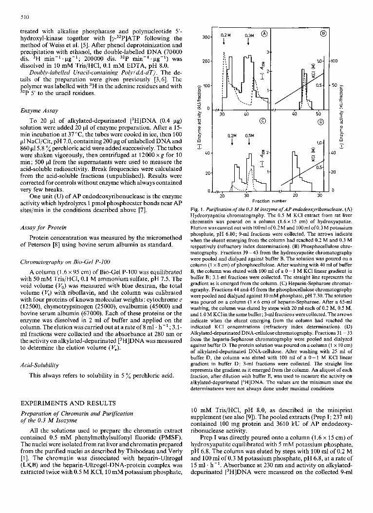
Fig. 2. Molecular weight of the 0.3 M isozyrne of AP endodeoxyribonuclease. A BioGel P-100 column (1.6 x 95 cm) was calibrated with four proteins of known molecular weights (cytochrome c= 12500; chymotrypsinogen = 25000; ovalbumin =45000; bovine serum albumin = 67000). The K,, values were calculated from the elution volumes in each case and plotted against the logarithm of the molecular weights (black dots). The K,, for the AP endodeoxyribonuclease was also calculated and its position (open dot) on the best straight line passing through the black dots indicated a molecular weight of about 39000
was measured on the 3-ml fractions. Fig. 1D shows a single peak of enzyme activity, eluted with 0.3 M KC1, containing 496 kU of AP endodeoxyribonuclease activity (Prep V). After addition of 0.023 % bovine serum albumin (Boehringer), Prep V was shared between several tubes which were stored at 4 "C.
The results of the four purification steps are summarized in Table 1.
Molecular Weight of the Enzyme
Prep V was 10-fold diluted with the buffer used to equilibrate the BioGel P-100 column. A 2-ml aliquot of the diluted enzyme solution was placed on the column and the elution was carried out as described in Materials and Methods ; the AP endodeoxyribonuclease activity was mea- sured on each fraction to determine V,. In Fig. 2, Kav = (V, - V,)/(V, - V,) is plotted against the logarithm of the molecular weight; the value for the enzyme as measured on the straight line obtained from the standards gives a molecular weight of 39 000.
512
Specificity of the Endonuclease Activity
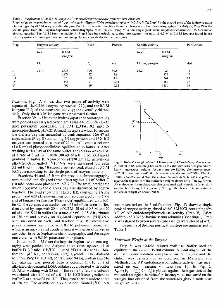
Prep V was diluted 30-fold with buffer E (50 mM Tris/HCl, 50 mM NaCl, pH 8.0). Aliquots (20 pl) of the diluted enzyme solution (37 units) or the same volumes of buffer E were mixed with 20 pl of solutions containing 0.4 pg of untreated, alkylated or alkylated-depurinated [3H]DNA and incubated at 37 "C for 15 min before measuring the acid-soluble fraction. In half of the tubes, the incubation with or without enzyme was followed by addition of 40 p1 of 0.4 M NaOH and a further 15-min incubation at 37 "C (a treatment known to produce a break near each AP site [4]), before measuring the acid-soluble fraction. Break frequencies were calculated from the acid- soluble fractions. Table 2 shows that the enzyme had no action on untreated DNA; its action on alkylated or alkylated- depurinated DNA was restricted to alkali-labile sites, i.e. AP sites, since a preliminary incubation with a large amount of enzyme did not significantly increase the break frequency given by an exposure to NaOH.
Table 2. Action of the 0.3 M isozyme of AP endodeoxyribonuclease on untreated, ulkylated or alkyluted-depurinated D N A [3H]DNA (0.4 pg) (untreated, alkylated or alkylated-depurinated) was incubated for 15 min at 37 "C with or without 37 units of Prep V in 40 pl of solution. The acid-soluble fraction was measured either directly or after a 15-min treatment at 37 "C with NaOH (0.2 M final concentration). Break frequencies were calculated from the acid-soluble fractions
DNA Enzyme NaOH Breaks/103 nucleotides
Untreated - - 3.8 3.8 +
- + 4.5 + + 5.3
5.3 18.5 +
- + 18.0 + + 19.0
7.0 depurinated + - 47.0
- + 54.0 + + 55.0
-
Alkylated - -
-
Alkylated- - -
Absence of Exonuclease Activity
Prep V was 40-fold diluted with buffer E. Two different experiments were carried out.
In the first experiment, 50-p1 aliquots of the diluted enzyme solution were mixed with 50 pl of nicked [3H]DNA solution (1.0 pg) and incubated for various times up to 90 min. The measured acid-soluble radioactivity was not greater than in the controls incubated without enzyme for the same time.
In the second experiment, 25-pl aliquots of the diluted enzyme solution were mixed with 25 pl of nicked [3H]DNA labelled with 32P at the 5'-phosphate ends (3.8 pg) solution and incubated for various times up to 90 min. The 32P released in the acid-soluble fraction was the same as in the controls incubated without enzyme for the same time.
Position of the Phosphodiester Bridge Cut by the 0.3 M Isozyrne of A P Endodeoxyribonuclease
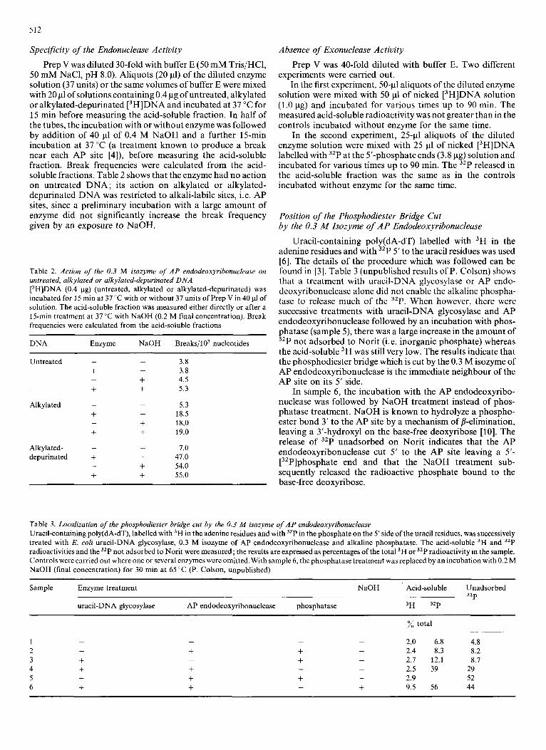
Uracil-containing poly(dA-dT) labelled with 3H in the adenine residues and with 32P 5' to the uracil residues was used [6]. The details of the procedure which was followed can be found in [3]. Table 3 (unpublished results of P. Colson) shows that a treatment with uracil-DNA glycosylase or AP endo- deoxyribonuclease alone did not enable the alkaline phospha- tase to release much of the 32P. When however, there were successive treatments with uracil-DNA glycosylase and AP endodeoxyribonuclease followed by an incubation with phos- phatase (sample 5), there was a large increase in the amount of 32P not adsorbed to Norit (i. e. inorganic phosphate) whereas the acid-soluble 3H was still very low. The results indicate that the phosphodiester bridge which is cut by the 0.3 M isozyme of AP endodeoxyribonuclease is the immediate neighbour of the AP site on its 5' side.
In sample 6, the incubation with the AP endodeoxyribo- nuclease was followed by NaOH treatment instead of phos- phatase treatment. NaOH is known to hydrolyze a phospho- ester bond 3' to the AP site by a mechanism of p-elimination, leaving a 3'-hydroxyl on the base-free deoxyribose [lo]. The release of 32P unadsorbed on Norit indicates that the AP endodeoxyribonuclease cut 5' to the AP site leaving a 5'- [32P]phosphate end and that the NaOH treatment sub- sequently released the radioactive phosphate bound to the base-free deoxyribose.
Table 3. Localization of the phosphodiester bridge cut by ihe 0.3 M isozyme of A P endodeoxyribonuciease Uracil-containing poly(dA-dT), labelled with 3H in the adenine residues and with 3zP in the phosphate on the 5'side of the uracil residues, was successively treated with E. coli uracil-DNA glycosylase, 0.3 M isozyme of AP endodeoxyribonuclease and alkaline phosphatase. The acid-soluble 3H and 3zP radioactivities and the 32P not adsorbed to Norit were measured; the results are expressed as percentages of the total 3H or 32P radioactivity in the sample. Controls were carried out where one or several enzymes were omitted. With sample 6, the phosphatase treatment was replaced by an incubation with 0.2 M NaOH (final concentration) for 30 min at 65 "C (P. Colson, unpublished)
Sample Enzyme treatment NaOH Acid-soluble Unadsorbed 32P
uracil-DNA glycosylase AP endodeoxyribonuclease phosphatase 3H 3zP
% total ~~ ~~ ~
- - 2.0 6.8 4.8 1 2 - + + - 2.4 8.3 8.2 3 + - + - 2.7 12.1 8.7 4 + + - - 2.5 39 29 5 + + + - 2.9 52 6 + + - + 9.5 56 44
- -
513
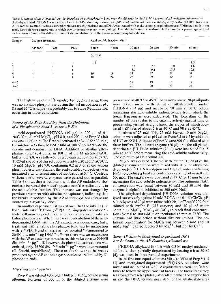
Table 4. Nature of the 3' ends left by the hydrolysis of a phosphoester bond near the A P sites by the 0.3 M isozyme of A P endodeoxyribonuclease Acid-depurinated [3H]DNA was incubated with the AP endodeoxyribonuclease (AP endo) and the solution was subsequently heated at 100 "C for 2 rnin. After another treatment with alkaline phosphatase (Pase), the denaturated DNA was treated with snake venom phosphodiesterase (PDE) (see details in the text). Controls were carried out in which one or several enzymes were omitted. The table indicates the acid-soluble fraction (as a percentage of total radioactivity) found after different times of the incubation with the snake venom phosphodiesterase
~~
Sample Enzyme treatment Acid-soluble fraction after -
APendo Pase PDE 3 min 7 min 10 min 20 min 30 min 60 min
% total
1 + - - 1.1 1.3 2 - + + 6.8 1.6 9.0 11.6 3 - - + 5.7 9.1 10.5 10.8 4 + + + 19 19 24 27 28 31 5 + - + 18 24 26 29 30 35
4 -2 17 19 19 19 5 -3 20 20 20 24
The high value of the 32P unadsorbed by Norit when there was no alkaline phosphatase during the last incubation at pH 8.8 and 65 "C (sample 4) is probably due to some j?-elimination occurring in those conditions.
Nature of the Ends Resulting from the Hydrolysis of a Phosphoester Bond 5 ' to the A P Site
Acid-depurinated [3H]DNA (10 pg) in 200 pl of 0.1 NaCl/Cit, 20 mM MgCI,, pH 8.0, and 200 pl of Prep V (400 enzyme units) in buffer E were incubated at 37 "C for 30 min; the mixture was then heated 2 rnin at 100 "C to inactivate the enzyme and denature the DNA. Addition of alkaline phos- phatase (Sigma; 4 units) in 100 pl of 0.3 M glycine/NaOH buffer, pH 8.8, was followed by a 30-min incubation at 37 "C. To 20-11 aliquots of this solution were added 20 p1 of NaCl/Cit, 10 mM MgCl,, pH 7.0, containing 0.2 mU of snake venom phosphodiesterase (Sigma) ; the acid-soluble radioactivity was measured after different times of incubation at 37 "C. Controls without one or several enzymes were carried out in parallel. Table 4 shows that a treatment with the AP endodeoxyribo- nuclease increased the rate of appearance of the radioactivity in the acid-soluble fraction. This increase was not changed by previous treatment with alkaline phosphatase, indicating that the breaks introduced by the AP endodeoxyribonuclease are limited by 3'-hydroxyl ends.
In another experiment, it was shown that the labelling of the 5' ends with 32P from [Y-~~PIATP using polynucleotide 5'- hydroxylkinase depended on a previous treatment with al- kaline phosphatase. When there was no incubation of the acid- depurinated DNA with the AP endodeoxyribonuclease, but a treatment with alkaline phosphatase followed by incubation with [p3'P]ATP and kinase, the incorporated 32P amounted to 26400 dis. min-' pg DNA-'. When there was an incubation with the AP endodeoxyribonuclease, this value rose to 204000 dis . min-' . pg-'. If, however, thephosphatase treatment was omitted, only 76300 dis . 32P min-' pg-' were incorporated (G. Zocchi, unpublished). These results show that the breaks produced by the AP endodeoxyribonuclease are limited by 5'- phosphate ends.
Miscellaneous Properties
Prep V was diluted 400-fold in buffer E, 0.2 % bovine serum albumin. Portions of 300 p1 of the diluted enzyme were
prewarmed at 40 "C or 45 "C for various times; 2O-pl aliquots were taken, mixed with 20 p1 of alkylated-depurinated [3H]DNA (0.4 pg) and incubated 10 rnin at 30°C before measuring the acid-soluble radioactivities from which the break frequencies were calculated. The logarithm of the number of breaks due to the enzyme activity against time of prewarming yielded straight lines, the slopes of which indi- cated half-lives of about 2 h at 40 "C and 90 s at 45 "C.
Portions of 25 mM Tris, 25 mM Hepes, 10 mM MgCl, solution were adjusted to pH values from 6.5 to 9.5 by addition of HCI or KOH. Aliquots of Prep V were 800-fold diluted with these buffers. The diluted enzyme (20 pl) and the alkylated- depurinated [3H]DNA solution (20 pl) were incubated for 15 rnin at 37 "C before measuring the acid-soluble radioactivity. The optimum pH is around 8.0.
Prep V was diluted 100-fold with buffer D; 20 pl of the diluted enzyme solution were mixed with 20 p1 of alkylated- depurinated [3H]DNA solution and 10 p1 of water containing NaCl to produce a final concentration varying between 0 and 500 mM. The mixture was incubated at 37 "C for 15 rnin before measuring the acid-soluble radioactivity. The optimum NaCl concentration was found between 30 mM and 50 mM; the enzyme is eightfold inhibited at 500 mM NaCI.
The alkylated-depurinated [3H]DNA solution was dia- lyzed successively against 10 mM EDTA and 0. l NaCl/Cit, pH 8.0. Aliquots of 20 p1 were mixed with 20 pl of Prep V 200-fold diluted with buffer E (2U enzyme) and 10 p1 of water containing MgCI2, MnC1, or CaC12 to reach final concentra- tions from 0 to 100 mM, then incubated 15 min at 37 "C. The enzyme had little action without divalent cations. The op- timum concentration for MgCl, was between 5 mM and 10 mM; Mg2+ can be replaced by Mn2+, but not by Ca2+.
Some A P Sites in Methylated-Depurinated DNA Are Resistant to the A P Endodeoxyribonuclease
[3H]DNA alkylated for 1 h with 0.3 M methyl methane- sulfonate, then partially depurinated by heating 6 h at 50 "C [4], was used in three parallel experiments.
In the first one, equal volumes (350 pl) of diluted Prep V (1 5 U) and methylated-depurinated [3H]DNA solutions were mixed and incubated at 37 "C. Aliquots were taken at different times to follow the appearance of breaks. The break frequency was found to reach a plateau after 60 rnin when the enzyme had nicked the DNA strands near 70 % of the alkali-labile sites
514
1
L 100. m
80.
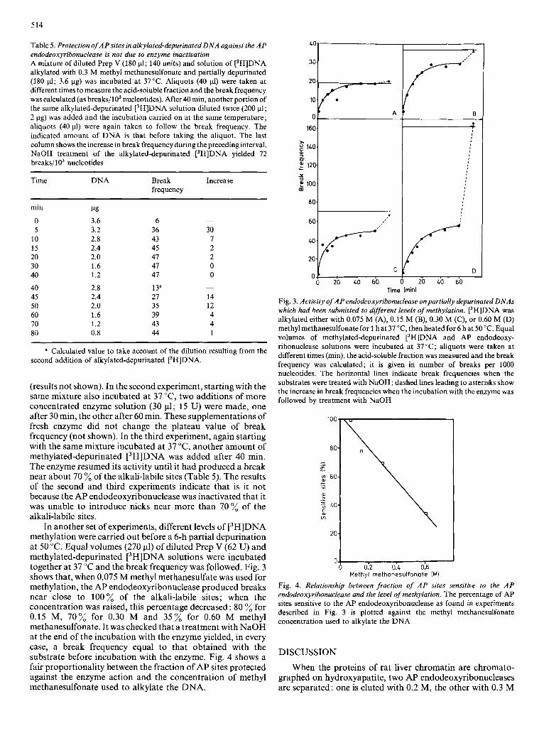
Table 5. Protection of AP sites in alkylated-depurinated DNA against the AP endodeoxyribonuclease is not due to enzyme inactivation A mixture of diluted Prep V (180 pl; 140 units) and solution of [3H]DNA alkylated with 0.3 M methyl methanesulfonate and partially depurinated (180 pl; 3.6 pg) was incubated at 37°C. Aliquots (40 pl) were taken at different times to measure the acid-soluble fraction and the break frequency was calculated (as breaks/103 nucleotides). After 40 min, another portion of the same alkylated-depurinated [3H]DNA solution diluted twice (200 pl; 2 pg) was added and the incubation carried on at the same temperature; aliquots (40 pl) were again taken to follow the break frequency. The indicated amount of DNA is that before taking the aliquot. The last column shows the increase in break frequency during the preceding interval. NaOH treatment of the alkylated-depurinated [3H]DNA yielded 72 breaks/103 nucleotides
Time DNA Break Increase frequency
min pg 0 3.6 6 5 3.2 36 30
10 2.8 43 7 15 2.4 45 2 20 2.0 47 2 30 1.6 41 0 40 1.2 47 0
40 2.8 13" ~
45 2.4 27 14 50 2.0 35 12 60 1.6 39 4 70 1.2 43 4 80 0.8 44 1
~
a Calculated value to take account of the dilution resulting from the second addition of alkylated-depurinated [3H]DNA.
(results not shown). In the second experiment, starting with the same mixture also incubated at 37 "C, two additions of more concentrated enzyme solution (30 pl; 15 U) were made, one after 30 min, the other after 60 min. These supplementations of fresh enzyme did not change the plateau value of break frequency (not shown). In the third experiment, again starting with the same mixture incubated at 37 "C, another amount of methylated-depurinated [3H]DNA was added after 40 min. The enzyme resumed its activity until it had produced a break near about 70 % of the alkali-labile sites (Table 5). The results of the second and third experiments indicate that is it not because the AP endodeoxyribonuclease was inactivated that it was unable to introduce nicks near more than 70% of the alkali-labile sites.
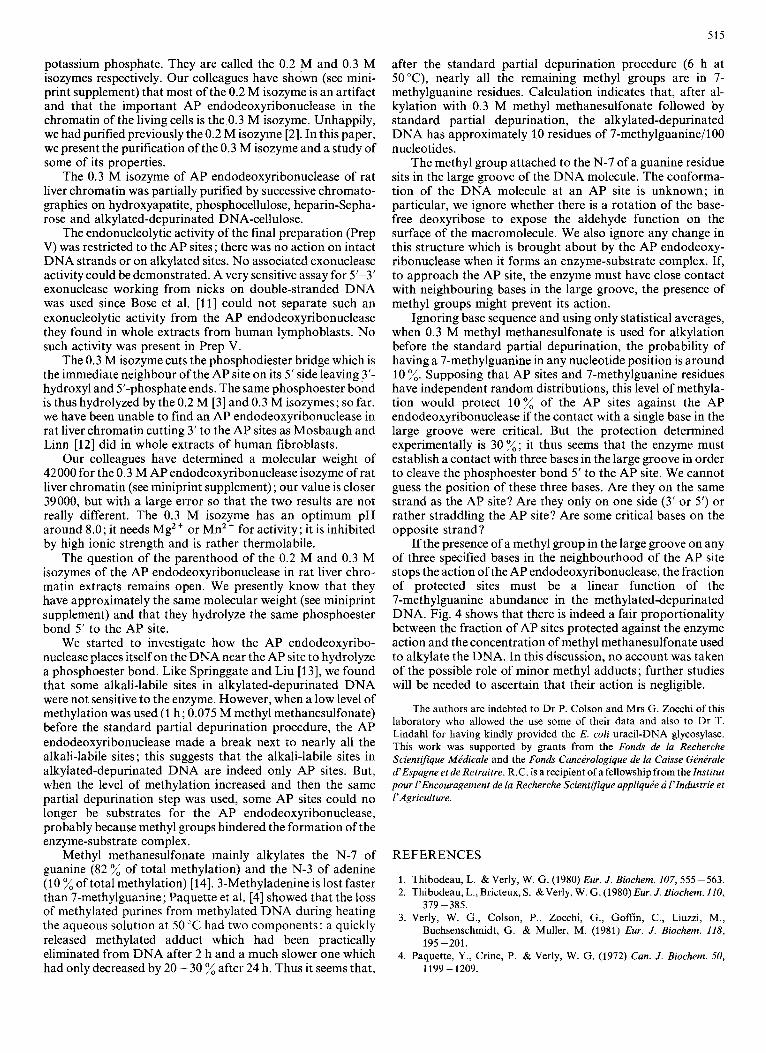
In another set of experiments, different levels of [3H]DNA methylation were carried out before a 6-h partial depurination at 50 "C. Equal volumes (270 11) of diluted Prep V (62 U) and methylated-depurinated [3H]DNA solutions were incubated together at 37 "C and the break frequency was followed. Fig. 3 shows that, when 0.075 M methyl methanesulfate was used for methylation, the AP endodeoxyribonuclease produced breaks near close to 100% of the alkali-labile sites; when the concentration was raised, this percentage decreased : 80 % for 0.15 M, 70% for 0.30 M and 35% for 0.60 M methyl methanesulfonate. It was checked that a treatment with NaOH at the end of the incubation with the enzyme yielded, in every case, a break frequency equal to that obtained with the substrate before incubation with the enzyme. Fig. 4 shows a fair proportionality between the fraction of AP sites protected against the enzyme action and the concentration of methyl methanesulfonate used to alkylate the DNA.
2ot7?==-- 10
160 OI------
60 I....' 20
0 20 40 60
D 20 LO 60
Time (mid
Fig. 3. Activity of A P endodeoxyribonuclease on partially depurinated DNAs which had been submitted to different levels of methylation. [3H]DNA was alkylated either with 0.075 M (A), 0.15 M (B), 0.30 M (C), or 0.60 M (D) methyl methanesulfonate for 1 hat 37 "C, then heated for 6 hat 50 "C. Equal volumes of methylated-depurinated [3H]DNA and AP endodeoxy- ribonuclease solutions were incubated at 37 "C; aliquots were taken at different times (min), the acid-soluble fraction was measured and the break frequency was calculated; it is given in number of breaks per 1000 nucleotides. The horizontal lines indicate break frequencies when the substrates were treated with NaOH; dashed lines leading to asterisks show the increase in break frequencies when the incubation with the enzyme was followed by treatment with NaOH
80
2oL 0 0 0.2 0.L 0.6
Methyl rnethanesulfonhte IM)
Fig. 4. Relationship between fraction of A P sites sensitive to the AP endodeoxyribonuclease and the level of methylation. The percentage of AP sites sensitive to the AP endodeoxyribonuclease as found in experiments described in Fig. 3 is plotted against the methyl methanesulfonate concentration used to alkylate the DNA
DISCUSSION
When the proteins of rat liver chromatin are chromato- graphed on hydroxyapatite, two AP endodeoxyribonucleases are separated: one is eluted with 0.2 M, the other with 0.3 M
515
potassium phosphate. They are called the 0.2 M and 0.3 M isozymes respectively. Our colleagues have shown (see mini- print supplement) that most of the 0.2 M isozyme is an artifact and that the important AP endodeoxyribonuclease in the chromatin of the living cells is the 0.3 M isozyme. Unhappily, we had purified previously the 0.2 M isozyme [2]. In this paper, we present the purification of the 0.3 M isozyme and a study of some of its properties.
The 0.3 M isozyme of AP endodeoxyribonuclease of rat liver chromatin was partially purified by successive chromato- graphies on hydroxyapatite, phosphocellulose, heparin-Sepha- rose and alkylated-depurinated DNA-cellulose.
The endonucleolytic activity of the final preparation (Prep V) was restricted to the AP sites; there was no action on intact DNA strands or on alkylated sites. No associated exonuclease activity could be demonstrated. A very sensitive assay for 5'-3' exonuclease working from nicks on double-stranded DNA was used since Bose et al. [ l l] could not separate such an exonucleolytic activity from the AP endodeoxyribonuclease they found in whole extracts from human lymphoblasts. No such activity was present in Prep V.
The 0.3 M isozyme cuts the phosphodiester bridge which is the immediate neighbour of the AP site on its 5' side leaving 3'- hydroxyl and 5'-phosphate ends. The same phosphoester bond is thus hydrolyzed by the 0.2 M [3] and 0.3 M isozymes; so far, we have been unable to find an AP endodeoxyribonuclease in rat liver chromatin cutting 3' to the AP sites as Mosbaugh and Linn [12] did in whole extracts of human fibroblasts.
Our colleagues have determined a molecular weight of 42000 for the 0.3 M AP endodeoxyribonuclease isozyme of rat liver chromatin (see miniprint supplement); our value is closer 39000, but with a large error so that the two results are not really different. The 0.3 M isozyme has an optimum pH around 8.0; it needs Mg2+ or MnZf for activity; it is inhibited by high ionic strength and is rather thermolabile.
The question of the parenthood of the 0.2 M and 0.3 M isozymes of the AP endodeoxyribonuclease in rat liver chro- matin extracts remains open. We presently know that they have approximately the same molecular weight (see miniprint supplement) and that they hydrolyze the same phosphoester bond 5' to the AP site.
We started to investigate how the AP endodeoxyribo- nuclease places itself on the DNA near the AP site to hydrolyze a phosphoester bond. Like Springgate and Liu [13], we found that some alkali-labile sites in alkylated-depurinated DNA were not sensitive to the enzyme. However, when a low level of methylation was used (1 h; 0.075 M methyl methanesulfonate) before the standard partial depurination procedure, the AP endodeoxyribonuclease made a break next to nearly all the alkali-labile sites ; this suggests that the alkali-labile sites in alkylated-depurinated DNA are indeed only AP sites. But, when the level of methylation increased and then the same partial depurination step was used, some AP sites could no longer be substrates for the AP endodeoxyribonuclease, probably because methyl groups hindered the formation of the enzyme-substrate complex.
Methyl methanesulfonate mainly alkylates the N-7 of guanine (82 % of total methylation) and the N-3 of adenine (10 %of total methylation) [14]. 3-Methyladenine is lost faster than 7-methylguanine; Paquette et al. [4] showed that the loss of methylated purines from methylated DNA during heating the aqueous solution at 50 "C had two components : a quickly released methylated adduct which had been practically
after the standard partial depurination procedure (6 h at 50°C), nearly all the remaining methyl groups are in 7- methylguanine residues. Calculation indicates that, after al- kylation with 0.3 M methyl methanesulfonate followed by standard partial depurination, the alkylated-depurinated DNA has approximately 10 residues of 7-methylguanine/lOO nucleotides.
The methyl group attached to the N-7 of a guanine residue sits in the large groove of the DNA molecule. The conforma- tion of the DNA molecule at an AP site is unknown; in particular, we ignore whether there is a rotation of the base- free deoxyribose to expose the aldehyde function on the surface of the macromolecule. We also ignore any change in this structure which is brought about by the AP endodeoxy- ribonuclease when it forms an enzyme-substrate complex. If, to approach the AP site, the enzyme must have close contact with neighbouring bases in the large groove, the presence of methyl groups might prevent its action.
Ignoring base sequence and using only statistical averages, when 0.3 M methyl methanesulfonate is used for alkylation before the standard partial depurination, the probability of having a 7-methylguanine in any nucleotide position is around 10 %. Supposing that AP sites and 7-methylguanine residues have independent random distributions, this level of methyla- tion would protect 10% of the AP sites against the AP endodeoxyribonuclease if the contact with a single base in the large groove were critical. But the protection determined experimentally is 30%; it thus seems that the enzyme must establish a contact with three bases in the large groove in order to cleave the phosphoester bond 5' to the AP site. We cannot guess the position of these three bases. Are they on the same strand as the AP site? Are they only on one side (3' or 5') or rather straddling the AP site? Are some critical bases on the opposite strand?
If the presence of a methyl group in the large groove on any of three specified bases in the neighbourhood of the AP site stops the action of the AP endodeoxyribonuclease, the fraction of protected sites must be a linear function of the 7-methylguanine abundance in the methylated-depurinated DNA. Fig. 4 shows that there is indeed a fair proportionality between the fraction of AP sites protected against the enzyme action and the concentration of methyl methanesulfonate used to alkylate the DNA. In this discussion, no account was taken of the possible role of minor methyl adducts; further studies will be needed to ascertain that their action is negligible.
The authors are indebted to Dr P. Colson and Mrs G. Zocchi of this laboratory who allowed the use some of their data and also to Dr T. Lindahl for having kindly provided the E. coli uracil-DNA glycosylase. This work was supported by grants from the Fonds de la Recherche ScientiFque MPdicale and the Fonds Cancirologique de la Caisse GPnPrale d'Espagne et de Retraitre. R.C. is a recipient of a fellowship from the Institut pour /'Encouragement de la Recherche Scientifique appliquee a l'lndustrie et I' Agriculture.
REFERENCES
1. Thibodeau, L. & Verly, W. G. (1980) Eur. J . Biochem. 107,555 -563. 2. Thibodeau, L., Bricteux, S . &Verly, W. G. (1980) Eur. J . Biochem. 110,
3. Verly, W. G., Colson, P., Zocchi, G., Goffin, C., Liuzzi, M., Buchsenschmidt, G. & Muller, M. (1981) Eur. J . Biochem. 118,
379 -385.
195 -201. eliminated from DNA after 2 h and a much slower one which 4. Paquette, Y., Crine, P. & Verly, W. G. (1972) Can. J. Biochem. 50, had only decreased by 20 - 30 % after 24 h. Thus it seems that, 1199-1209.
516
5. Weiss, B., Live, T. R. & Richardson, C. C. (1968) J . Biol. Chem. 243,
6 . Clements, J. E., Rogers, S. G. & Weiss, B. (1978) J. Biol. Chem. 253,
7 . Verly, W. G. (1980) in Techniques in D N A Repair Research (Friedberg, E. C. & Hanawalt, P., eds) ch. 20, p. 240, Marcel Dekker, New York.
8. Peterson, G. L. (1977) Anal. Biochem. 83, 346-356. 9 . Renard, A. & Verly, W. G. (1980) FEBS Lett. 114, 98 -102.
10. Bayley, C. R., Bramrner, K. W. &Jones, A. S. (1961)J. Chem. SOC. 368,
1 1 . Bose, K., Karran, P. & Straws, B. (1978) Proc. Nut1 Acad. Sci. USA,
12. Mosbaugh, D. W. & Linn, S. (1980) J. Biol. Chem. 255,11743 - 11752. 13. Springgate, C. & Liu, L. (1980) Carcinogenesis, I , 263 -270. 14. Lawley, P. D., Orr, D. J. &Jarman, M. (1975)Biochem.J. 145,73 -84.
4530 -4542. 1903-1917.
2990 -2999. 75, 794 - 798.
R. Ctsar and W. G. Verly Laboratoire de Biochimie, Faculte des Sciences de l'llniversiti de Litge, Sart Tilman B6, Universite de Liege, B-4000 Liege, Belgium
Supplementary Material
to
A?DRINIC/APYRIMIDINIC ENDODEOXYRIBONUCLEASE OF RAT LIVER CHROMATIN
by Suzanne Bricteur-GrGgoire, Yvette Habraken and Walter G. Verly
Summaly
Nan-hrstone proteins extracted from r a t l i v e r chromatin contain t w o d i f f e r e n t AP endonuc1eases:one 1s eluted with 0.2 H. the other with 0 . 3 N potas~lum phosphate. The two iiozyrnes have molecular weights of about 42 W O . The 0 . 3 M ipecles seems to be the true chromatin enzyme as it exists i n the living cell. Most of the 0.2 M species IS
an artlfact; it 1s not known whether i t derives from the 0 . 3 M speclea or not.
Introduction A? endodeoxyribonucleases are DNA repair enzymes which hydrolyze
Chromatoo'raohv of an extract of rat liver chromatin on DEAE-cel- a phosphoester band near AP (apurinic or apyrimidinic) sites.
~ ~~ ~~~
lulose separates' t;o AP endodeoxyribonucleases. emerges at the lower ionic strength has been completely purified; it is eluted from hydroxyapatite with 0.2 M K phosphate whereas the other species is eluted with 0.3 M K phosphate I l l . The two isozymes can readily be distinguished by analyzing the chromatin extract directly on hydroxyapatite; their ratio varies from one experiment to another and it would be interesting to know whether the two AP endo- deoxyribonucleases are chemically related.
A serine-protease, inactive in native chromatin, attacks histones and "on-histone proteins after dissociation of the nucleo- protein 121. One wonders whether the two AP endodeoxyribonucleases are present in the chromatin of the livrng c e l l or if one of the two species in the chromatln extract might not be a proteolytic degrada- tion product. The fallowing experiments were carried out to answer these questions.
The species Which
Materials and Methods I. Preparation of the chromatin extract
after removal, it is homogenized in buffer A and the nuclei are collected by centrifugation; the nuclei are purified in a discontinu- o u s sucrose gradient in diluted buffer A. The purified nuclei are made to swell in water, the nuclear membranes are disrupted in a tight-fitting Patter-Elveh3em apparatus and the chromatln 1s collected by centrifugation. The composition of buffer A and the details of the S U C C ~ S S ~ V ~ procedures can be found in 1 3 1 .
Chromatin is sheared in a tight-fitting Potter-Elvehjem apparatus i n buffer B I10 mM K phosphate, 10 mM TriS.HC1, pH 8 . 0 ) to have about 300 v g DNA per ml. Heparin-Dltrogel (LKBl 1s then added to have the same weight of heparin and chromatln DNA. The mixture is stirred for 30 mi" at 0'; the h e p a r i n - U l t r o g e l - D N A - p r o t e i n complex is collected by centrifugation, the pellet is washed with buffer B, then extracted twice with 0.5 M KC1 ~n buffer B. The pooled extracts contain no DNA and very little histones. 2. llydroryapatrte chromatography
The chromatin extract is dialyzed against 5 mM K phosphate, pH 6.8. The solution LS poured Onto a 1 . 6 x 2 3 an column of hydraxy- apatite equilibrated with 5 mM K phosphate, pH 6.8. The elution LS carried Out, at e rate Of 10 m1.h-I and 2 ' , with 120-ml portions Of K phosphate, pH 6.8 buffers, successrvely at 0.1 M, 0.2 M and 0.3 M concentrations. The AP endodeoxyribonuclease activity is measured on the collected 1 0 - m l fractions. 3. Piltration on Sephadex G-75
A 1 . 6 x 90 cm column of Sephadex G-75 1s equilibrated with buffer C (50 mM Trls.HC1, 0.3 M NaC1, 0.1 mM EDTA. 0.02 % NaN pH 8 .01 . The protein rn 2 r n l of buffer C IS applied on the column a& the elution IS carried out with the same buffer at a rate of 10 m1.h-1; 3 . 2 - m l fractions are collected. The void volume 15 measured with blue dex- tran and the column 1s calibrated with bovine serum albumin 167 000). ovalbumin (45 0001, chymotrypsin (25 000). myoglobin (18 000) and cytochrome c (12 5 0 0 ) .
4 . Enzyme assay and unit
the number of breaks introduced by the enzyme near AP sites is calcu-
The rat liver IS perfused & & with ice-cold Locke's SOlUtiOn;
The AP endodeoxyribonuclease is assayed as described in [l] and
lated. One unit Of enzyme activity hydrolyzes phosphoester bonds near 1 pmol AP Site5 per mi" L 4 1 .
Experiments and Results The two A? endodeoxyribonucleases in chromatin proteins from rat
liver will be called 0.2 M and 0 .3 M isozymes depending on the K phosphate Concentration necessary to elute them from the hydroxyapa- tite column.
. ~. ~. . . and chromatographed the next day On hydroxyapatite. The second and third parts, kept at 2' for 6 and 10 days respectively, were subse- quently treated in an identical w a y .
tography, increased with time of storage; ( 2 ) the activity eluted with 0.3 M K phosphate decreased whereas the activity eluted with 0.2 M K phosphate increased with storage time; (31 the overall yields of the hydroxyapatite cbromatographres were around 4 0 %.
Table I shows that : (1) the enzyme activity, before the chroma-
The fractions of the 0 . 3 M peaks of the three chromatographies Were pooled and, after dialysis against 5 mM K phosphate, pH 6.8, rechromatosraphed on hvdroxvaoatlte : no 0.2 M lsozvme was found. all the activity ;as elute; with 6.3 M K phosphate. Conversely, when the fractions of the 0.2 M peaks were similarly treated, all the activity was eluted with 0.2 M K phosphate and none appeared in the 0.3 M elu- ate. The yields of these second chromatographies were excellent, exceeding 90 8 .
2. Effect of PMSF on the isozyme relative activities
tributed in two stocks from which chromatin o r o t e i n s were oreoared. The livers Of two rats were cut in small pieces which were dis-
L .
For one preparation, the solutions did not contain PMSF: for the other, 0.1 mM PMSF was added r n buffer A and 0.5 mM PMSF in the Solution i n which the swollen nuclei were sheared. Table I shows that, without PMSF, the proportion of 0.2 M isalyme found in the hydroxyapatite chromatography was 52 8 (see a lso figure 1,Al; it decreased to 30 % when PMSF was used.
When, in another experiment, 0.5 mM PMSF was moreover added to the 0.5 M K C 1 used to extract the complex resulting from the disso- ciation of chromatin with heparin-Dltrogel, the proportion of 0.2 M iSOZyme further decreased. The results were highly reproducible : 16 % in duplicates prepared from pieces of the same livers. 3 . ConQItions to have the lowest percentage Of 0.2 M isoryme
used to perfuse the liver. The dialysis before the chromatography was eliminated when it was observed that 0.5 M KC1 I" the chromatin extract did not prevent the adsorption of the AP endodeoxyribonucle- aSeS on hydroxyapatite. Finally, to speed up the chromatography, a
0.5 mM PMSF was added to all solutions, including Locke's fluid
shorter column was used (1.6 x 13 cm), the first washing with 0.1 M K phosphate was suppressed and the volumes Of 0.2 M and 0.3 M K phos- phate were reduced to 60 ml while keeoincl the elution rate at i0 m1.h-1. r n the hydroxyapatite chromatography represented 10 and 90 8 respec- tively of the recovered activity (figure 1.B: Table I). These modifi- cations did not however increase the low yield of the hydroxyapatite chromatography. 4 . Molecular weight determinations
emerging from the hydroxyapatite column were separately dialyzed against buffer C and concentrated on Millipore frlters; 2 - m l portions were then analyzed on Sephadex G-75 as described in Materials and Methods. The elution volumes of both isozymes indrcated molecular weights of about 4 2 000. A mixture of the two enzymes chromato- graphed on the same column gave a single symmetrical peak.
In these conditions, the 6.2-M and 0.3 M isozymes found
The pooled fractLons of the two AP endodeoxyribonuclease peaks
Discussion The proportions Of the 0 . 2 M and 0.3 M AP endodeoxyribonuclease
isozymes separated by hydroxyapatite chromatography of the chromatin proteins prepared from rat liver vary widely depending on the prepara- tion method that is used. Mightone isozyme be a degradation product of the other ?
A conversion could not be demonstrated directly since, after a first chromatography on hydroxyapatite, the two isorymee were Stable : on rechromatosraohv. the 0.2 M isozvme was cOmPletelv eluted with 0.2 M K phosphate and the 0.3 M isoiyme was el;ted only with 0.3 M K phosphate.
total AP endodeoxyribonuclease activity increases with the time of The situation is more complex with the chromatin extract : the
storage at 2O. Several znterpretations can be considered : disap- pearance of an inhibitor: formation of an enzyme from an inactive pre- cursor: transformation of an isozvme into the other associated with .~ an increased Vmax. The dLstribution of the ieozymes after different times of storage is remarkable : the 0.3 M activity decreases with time whereas the 0.2 M activity increases, suggesting that the 0.3 M Isazyme might be transformed into the 0.2 M isozyme. But there 15 a great loss of the total activity during the chromatography (the overa l l yield 1 s only about 40 8 ) so that no final conclusion can be reached. It looks however as if some 0.2 M isozyme might be a degra- dation product.
517
PMSF i~~~~~ dialySIS
: Isozyme activities and yields of the hydroxyapatrte chromatographies
Enzyme activity Recovery HA
column P.C. 1 0.2 M 0.3 M I 0.2 M 1 0 . 3 M
- 25
- 20
c
E \
j 5 15 > Y
: 10 2 P $ 5
25-
20-
15-
1 0.2;" i
buffer B
buffer B 0
0
29 600 1 400 KC' 1 I 1 30 000 1 1 480
buffer B +
a l l the solutions I O I - 1 I 84 Oo0 I 2oo
I
7 400
84
90
The nuclei are prepared In buffer A; chromatin 1s dlssoclated I" buffer B i the heparln-Ultrogel~ONA-prote ln complex IS extracted with 0 . 5 M KCI 1 . b x 23 cm IL! or 1 .6 x 13 cm IS1 column of hydroxyapatrte. PMSF 1 s added to part or all of the so1ut1ons used since the perfuslon of the liver untll the e l u t l a n of the hydranyapatlte column; the Table Speclflcally Indicates the solutions containing 0 . S mM PMSF. D i f f e r e n t experiments are separated by horizontal l l n e s . Detarls Can be found in the t e x t .
P.C. are the chromatin proteins af ter dialysis (when there 1s one! and before the chromatography: 0.2 M and 0.3 W are the carreepondlng lsazymes separated by the chromatography. The reSultS are glven In enzyme unlta and percents o f i h e recovered a c t i v i t y .
(In buffer a!: the extract 1s dlalyzed or not before a chromatography On a
The last colulun indicates t h e overall y l e l d of the chromatography.
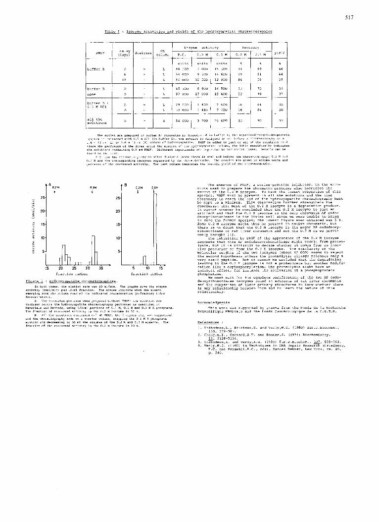
The absence Of PMSF, a serine-protease Inhihltor, in the 501"- tLons used to prepare the chromatin proteins also increases the amount of the 0.2 M lsozyrne. To have the lowest proportion of this species, PMSF must be present in all the solutions and the time necessary to reach the end of the hydroxyapatite chromatography must be kept to a minimum. This observation further strengthens the conclusion that most of the 0.2 M isozyme IS a dearadation Droduct.
0 3 M i
35 5 10 15 fraction number fraction number
Figure 1 : Hydroxyapatite chromatographies.
activity inmol/rnrnl per lo-ml fraction. The arrows indicate when the eluent emerqinq from the column reached the indrcated concentration (refractory index deterrnlnatlonl.
dialyzed before the hydroxyapatite chromatography performed as descrlbed i n Materials and Methods, using 1 2 0 - m l portions of 0.1 M . 0 . 2 M and 0.3 M K phosphate. The fraction of recovered activity ~n the 0 . 2 M isozyme is 5 2 %.
and the chromatography made on a shorter column, sklpplng the 0.1 W K phosphate e l u t i o n and decreasing Lo 60 ml the ~olilrnes of the 0.2 M and 0 . 3 M eluents. The fraction of the recwered activity ~n the 0 . 2 M isoryme IS 10 %.
In both cases, the elution rate was 10 ml/min. The graphs give the enzyme
A . The chromatin p r o t e i n s were prepared without PMSF; the s o l u t i o n was
8. All the solufrons contained 0.S mM PMSF, the dialysis step was suppressed
It Cannot however be concluded that th; 0.2 M iso;yme IS juit an artifact and that the 0.3 M isozyme is the only chromatin AP endo- deoxyribonuclease in the living cell Since we were unable to bring to zero the former species; the lowest figure ever obtained was 5 6 . Some 0.2 M ~sozyme might thus be present in native chromatin, but there is no doubt that the 0.3 M isozyme is the malor AP endodeouy- rihonuclease of rat l i v e r chromatin and not the 0.2 M as we previ- ously thought I l l .
suggests that this AP endodeoxyribonuclease might result from proteo- lvsis. but it 1s difficult to decide whether it comes from an lnac-
The inhibition by PMSF of the appearance of the 0.2 M iso2yme
~. ~
tive precursor or from the 0 . 3 M isozyme. The Similarity of the molecular weights of the two isozymes (about 42 000) Seems to =elect the second hypothesis unless the proteolytic clivage releases only a very small peptide. But It Cannot be excluded that the degradation leading to the 0.2 M lsozyme is not a proteolysis but another modifi- cation like a dephosphorylatron; the proteolysis mrght have an indirect effect, for instance the activation of a phosphoprotein
~ ~. phosphatase.
We must wait for the complete purification of the two AP endo- deoxyribonuclease isozymes found i n extracts of rat llver chromatin and the cornoarison of their nrimarv structures to know whether there ~. ~ ~~ ~ ~~
IS any relationship between them and to learn the nature of this relationship.
Acknowledqments
Scientifique MBdlcale and the Fonds Canc6rologique de la C.G.E.R. This work was supported by grants from the Fonds de la Recherche
References : 1. Thib0deau.L.. Bricteux,S. and Verly,W.G. (1980) Eur.J.Blochem.,
2. Che,M.T., Garrard,W.T. and Bonner,J. ( 1 9 7 4 ) Biochemistry,
3 . Thib0deau.L. and Verly,W.G. ( 1 9 8 0 ) Eur.J.Biochem., 107, 5 5 5 - 5 6 3 . 4. Ver1y.W.G. (1980) ~n Techniques in DNA Repair Research (Friedberg,
E.C. and Hanawalt,P.C., eds). Marcel Dekker, New York, Ch. 20, p . 240.
110. 379-3635,
13, 5128-5134.