the arabidopsis microtubule-associated protein atmap65-1 ... · the arabidopsis...
TRANSCRIPT
The Arabidopsis Microtubule-Associated Protein AtMAP65-1:Molecular Analysis of Its Microtubule Bundling Activity
Andrei P. Smertenko,a Hsin-Yu Chang,a Vera Wagner,b Despina Kaloriti,a Stepan Fenyk,a Seiji Sonobe,c
Clive Lloyd,d Marie-Theres Hauser,b and Patrick J. Husseya,1
a Integrative Cell Biology Laboratory, School of Biological and Biomedical Sciences, University of Durham, South Road,
Durham DH1 3LE, United Kingdomb Institute of Applied Genetics and Cell Biology, BOKU, University of Natural Resources and Applied Life Sciences,
Muthgasse 18 A-1190 Vienna, Austriac Himeji Institute of Technology, Faculty of Science, Hyogo, Japand Department of Cell and Developmental Biology, John Innes Centre, Norwich NR4 7UH, United Kingdom
The 65-kD microtubule-associated protein (MAP65) family is a family of plant microtubule-bundling proteins. Functional
analysis is complicated by the heterogeneity within this family: there are nine MAP65 genes in Arabidopsis thaliana,
AtMAP65-1 to AtMAP65-9. To begin the functional dissection of the Arabidopsis MAP65 proteins, we have concentrated on
a single isoform, AtMAP65-1, and examined its effect on the dynamics of mammalian microtubules. We show that
recombinant AtMAP65-1 does not promote polymerization and does not stabilize microtubules against cold-induced
microtubule depolymerization. However, we show that it does induce microtubule bundling in vitro and that this protein
forms 25-nm cross-bridges between microtubules. We further demonstrate that the microtubule binding region resides in
the C-terminal half of the protein and that Ala409 and Ala420 are essential for the interaction with microtubules. Ala420 is
a conserved amino acid in the AtMAP65 family and is mutated to Val in the cytokinesis-defective mutant pleiade-4 of the
AtMAP65-3/PLEIADE gene. We show that AtMAP65-1 can form dimers and that a region in the N terminus is responsible for
this activity. Neither the microtubule binding region nor the dimerization region alone could induce microtubule bundling,
strongly suggesting that dimerization is necessary to produce the microtubule cross-bridges. In vivo, AtMAP65-1 is
ubiquitously expressed both during the cell cycle and in all plant organs and tissues with the exception of anthers and
petals. Moreover, using an antiserum raised to AtMAP65-1, we show that AtMAP65-1 binds microtubules at specific stages
of the cell cycle.
INTRODUCTION
Most of the plant microtubule-associated proteins (MAPs) dis-
covered so far have structural homologs in other eukaryotes, but
not all animal or fungal MAPs are present in plants (Gardiner and
Marc 2003; Lloyd et al., 2004). Plant microtubules, although
structurally similar to their eukaryotic counterparts, differ in their
organization and dynamics. Plant cells have three unique micro-
tubule organizations: the interphase cortical array (consisting of
parallel rather than radial microtubules), the preprophase band
(which predicts the division plane), and the cytokinetic phrag-
moplast (consisting of two interdigitating sets of antiparallel
microtubules). The radial microtubule array of animal cells is
a result of polymerization from a single centrosome juxtaposed
to the nucleus, whereas plantmicrotubules are polymerized from
multiple dispersed nucleation sites in the cytoplasm (Chan et al.,
2003a). Plant microtubules are also more dynamic (Hush et al.,
1994; Yuan et al., 1994; Moore et al., 1997) and persist in the
treadmilling state longer than their animal counterparts (Shaw
et al., 2003). To understand the molecular basis for these
differences and to model the regulation of plant microtubule
organization and dynamics, more information on the plant
structural and regulatory MAPs is needed.
MAP-65 is the name given to the most abundant group of
MAPs, of electrophoretic molecular weight approximating 65 kD,
in microtubule preparations from tobacco (Nicotiana tabacum
Bright Yellow-2) (Jiang and Sonobe, 1993) and carrot (Daucus
carota) (Chan et al., 1996). Corresponding MAP65 cDNAs have
been cloned (NtMAP65-1, Smertenko et al., 2000; DcMAP65-1,
Chan et al., 2003b), and a gene family of nine members has been
identified in Arabidopsis thaliana (Hussey et al., 2002). Biochemi-
cally purified MAP65 proteins have been found to bind and
bundlemicrotubules in vitro (JiangandSonobe, 1993;Chanet al.,
1999). Antibodies raised against biochemically purified tobacco
MAP65 decorate all microtubules (Jiang and Sonobe, 1993) but
antibodies raised to one isotype recombinant NtMAP65-1 do not
(Smertenko et al., 2000). Anti-NtMAP65-1 recognizes only sub-
sets of interphase microtubules and in particular the anaphase
spindle midzone and at the midline of the cytokinetic phragmo-
plast. The overlapping microtubules at the spindle midzone have
the same polarity as those in the phragmoplast, and it has been
1 To whom correspondence should be addressed. E-mail [email protected]; fax 44-0191-334-1201.The author responsible for distribution of materials integral to thefindings presented in this article in accordance with the policy describedin the Instructions for Authors (www.plantcell.org) is: Patrick J. Hussey([email protected]).Article, publication date, and citation information can be found atwww.plantcell.org/cgi/doi/10.1105/tpc.104.023937.
The Plant Cell, Vol. 16, 2035–2047, August 2004, www.plantcell.orgª 2004 American Society of Plant Biologists
suggested that MAP65 cross-links antiparallel microtubules
(Smertenko et al., 2000). In biochemically purified carrot
MAP65 preparations there are three electrophoretically separa-
ble bands, and only one, the 62-kD band, was found to be
present in elongating cells containing only cortical microtubules,
indicating that this MAP65 is involved in directional cell expan-
sion (Chan et al., 2003b). Furthermore, it has been suggested
that stabilization of the interphase cortical array by MAP65
is necessary for the normal progression of embryogenesis
(Smertenko et al., 2003).
A family of nine AtMAP65 genes with predicted open reading
frames has been identified in the Arabidopsis Genome Database
(Hussey et al., 2002). Cloning the Arabidopsis PLEIADE (PLE)
gene revealed that PLE is synonymous with AtMAP65-3 (Hussey
et al., 2002; Muller et al., 2004). ple mutants were isolated in
genetic screens for defects in root and embryo morphogenesis
(Muller et al., 2002; Sollner et al., 2002; Sorensen et al., 2002). To
date, six ple alleles have been isolated, and all are recessive and
develop short irregular expanded roots with multinucleated cells
and incomplete cell walls. AtMAP65-3/PLE localizes in the
midzone of overlapping microtubules during cell division and
the mutations in ple-1, ple-5, and ple-6 cause C-terminal
truncations of the AtMAP65-3/PLE protein (Muller et al., 2004).
Thus, the ple phenotypes together with the subcellular localiza-
tion ofMAP65-3/PLE support its essential role for the completion
of cytokinesis. Vertebrate PRC1 (Mollinari et al., 2002) and yeast
Ase1p (Schuyler et al., 2003) are homologs of MAP65 (Hussey
et al., 2002), and either downregulation or deletion of these genes
causes the disruption of anaphase/telophase microtubule arrays
and the accumulation of multinucleated cells, respectively.
In this article, we characterize the activities andmechanisms of
action of AtMAP65-1. We have examined its role in microtubule
polymerization, stability, and bundling and also identified the
region responsible for microtubule binding by biochemical and
genetic analyses. In addition, we show that AtMAP65-1 can
dimerize and we identify the dimerization region that strongly
suggests that dimerization is necessary for 25-nm cross-bridge
formation. Also, we note that AtMAP65-1 is ubiquitously ex-
pressed but binds microtubules in a cell cycle–dependant
manner, suggesting tight posttranscriptional control on the
activity of this protein.
RESULTS
AtMAP65-1 Bundles Microtubules but Does Not Promote
Microtubule Polymerization
We have identified an Arabidopsis MAP65, AtMAP65-1
(At5g55230), that encodes a protein that shows 86% similarity
to tobacco NtMAP65-1 (Smertenko et al., 2000). The predicted
open reading frame encodes a 587–amino acid protein of 65.8 kD
molecular mass and a pI of 4.72.We have expressed and purified
recombinant AtMAP65-1 in bacteria and used this protein to
assess its effects on microtubule polymerization and bundling
in vitro.
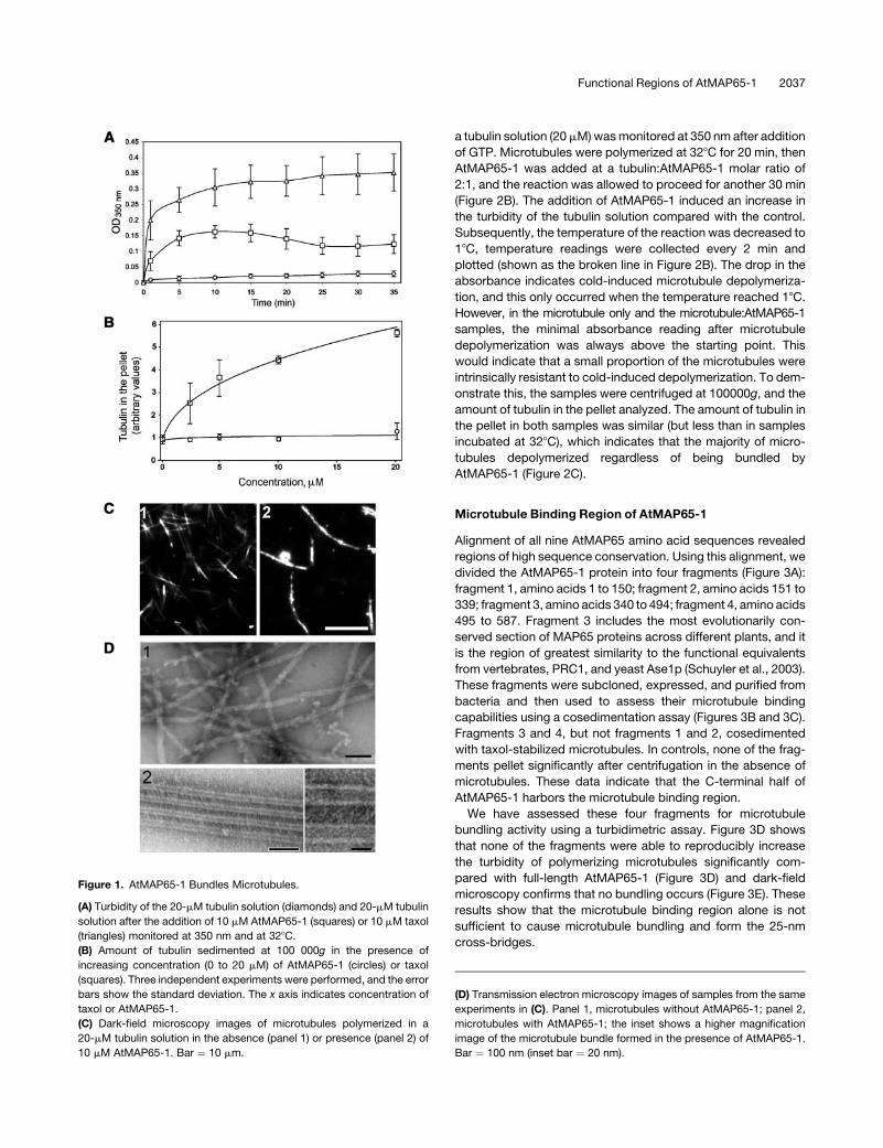
The effect of AtMAP65-1 on microtubule polymerization was
assessed using a turbidimetric assay. AtMAP65-1 was added to
a MAP-free porcine brain tubulin solution (final concentration
20 mM) at the AtMAP65-1 to tubulin dimer molar ratio of 1:2. The
turbidity of the mixture was monitored at 350 nm. AtMAP65-1
induced a dramatic increase in the turbidity of the polymerizing
microtubule mixture compared with control (Figure 1A). These
data indicate two possibilities: AtMAP65-1 could increase the
total amount of microtubule polymer or it could induce bundling
of assembled microtubules. Both processes are not necessarily
mutually exclusive because bundling can also stabilize micro-
tubules resulting in the increase of the total amount of micro-
tubule polymer by preventing dynamic instability. To distinguish
between these two possibilities, we polymerized microtubules
in the presence of increasing concentrations of AtMAP65-1 and
analyzed the amount of tubulin that cosediments with the
AtMAP65-1. If AtMAP65-1 increases the total amount of micro-
tubule polymer, the amount of tubulin in the pellet would
increase proportionally to the point of saturation. However,
the amount of tubulin in the pellet did not change significantly
across the AtMAP65-1 range of 0 to 20 mM (Figure 1B). Taxol,
a microtubule stabilizing agent and capable promoter of
microtubule polymerization, also increases the turbidity of the
tubulin solution (Figure 1A), but in contrast with AtMAP65-1,
taxol increased the total amount of tubulin in the pellet in a
concentration-dependent fashion (Figure 1B). This result
strongly suggests that AtMAP65-1 bundles but does not pro-
mote the polymerization of microtubules. This conclusion was
further confirmed by an analysis using dark-field microscopy
(Figure 1C). Addition of AtMAP65-1 to dynamic tobacco micro-
tubules (shown in panel 1; average length 4.2 6 1.1 mm, n ¼ 89)
caused the formation of long, thick microtubule bundles (panel
2; average length 11.5 6 4.4 mm, n ¼ 62). Examination of these
bundles under the electron microscope showed that they are
composed of parallel microtubules separated by 25-nm cross-
bridges (Figure 1D). These data suggest that AtMAP65-1 does
not promote microtubule polymerization in vitro but bundles
polymerized microtubules via the formation of 25-nm cross-
bridges.
An effect of microtubule bundling can be a reduction in the
depolymerization of microtubules, for example, by inhibiting
catastrophe. To determine whether AtMAP65-1 affected de-
polymerization, microtubules were first polymerized at 328C for
10 min, then AtMAP65-1 protein was added at the tubulin:
AtMAP65-1 molar ratio of 2:1. The mixture was diluted with
microtubule-polymerizing buffer prewarmed to 328C, incubated
for 10 min, and the microtubules were pelleted at 100,000g and
analyzed on SDS-PAGE gels. Taxol at a concentration of 10 mM
was used as a positive control to demonstrate the effect of
a microtubule stabilizing agent on the amount of tubulin polymer
upon isothermal dilution. The results of three independent
experiments are presented in Figure 2A. No significant difference
in the quantity of tubulin in the supernatant with or without
AtMAP65-1 was observed: the total amount of tubulin polymer
decreased fivefold with the decrease in final tubulin concentra-
tion from 20 to 1 mM. By contrast, the amount of tubulin polymer
in the presence of taxol decreased by only 10%.
To assess whether AtMAP65-1 changes the stability of micro-
tubules, we analyzed the effect of AtMAP65-1 on the cold-
induced depolymerization of microtubules. Here, the turbidity of
2036 The Plant Cell
a tubulin solution (20mM)wasmonitored at 350 nm after addition
of GTP. Microtubules were polymerized at 328C for 20 min, then
AtMAP65-1 was added at a tubulin:AtMAP65-1 molar ratio of
2:1, and the reaction was allowed to proceed for another 30 min
(Figure 2B). The addition of AtMAP65-1 induced an increase in
the turbidity of the tubulin solution compared with the control.
Subsequently, the temperature of the reaction was decreased to
18C, temperature readings were collected every 2 min and
plotted (shown as the broken line in Figure 2B). The drop in the
absorbance indicates cold-induced microtubule depolymeriza-
tion, and this only occurred when the temperature reached 18C.
However, in the microtubule only and the microtubule:AtMAP65-1
samples, the minimal absorbance reading after microtubule
depolymerization was always above the starting point. This
would indicate that a small proportion of the microtubules were
intrinsically resistant to cold-induced depolymerization. To dem-
onstrate this, the samples were centrifuged at 100000g, and the
amount of tubulin in the pellet analyzed. The amount of tubulin in
the pellet in both samples was similar (but less than in samples
incubated at 328C), which indicates that the majority of micro-
tubules depolymerized regardless of being bundled by
AtMAP65-1 (Figure 2C).
Microtubule Binding Region of AtMAP65-1
Alignment of all nine AtMAP65 amino acid sequences revealed
regions of high sequence conservation. Using this alignment, we
divided the AtMAP65-1 protein into four fragments (Figure 3A):
fragment 1, amino acids 1 to 150; fragment 2, amino acids 151 to
339; fragment 3, amino acids 340 to 494; fragment 4, amino acids
495 to 587. Fragment 3 includes the most evolutionarily con-
served section of MAP65 proteins across different plants, and it
is the region of greatest similarity to the functional equivalents
from vertebrates, PRC1, and yeast Ase1p (Schuyler et al., 2003).
These fragments were subcloned, expressed, and purified from
bacteria and then used to assess their microtubule binding
capabilities using a cosedimentation assay (Figures 3B and 3C).
Fragments 3 and 4, but not fragments 1 and 2, cosedimented
with taxol-stabilized microtubules. In controls, none of the frag-
ments pellet significantly after centrifugation in the absence of
microtubules. These data indicate that the C-terminal half of
AtMAP65-1 harbors the microtubule binding region.
We have assessed these four fragments for microtubule
bundling activity using a turbidimetric assay. Figure 3D shows
that none of the fragments were able to reproducibly increase
the turbidity of polymerizing microtubules significantly com-
pared with full-length AtMAP65-1 (Figure 3D) and dark-field
microscopy confirms that no bundling occurs (Figure 3E). These
results show that the microtubule binding region alone is not
sufficient to cause microtubule bundling and form the 25-nm
cross-bridges.
Figure 1. AtMAP65-1 Bundles Microtubules.
(A) Turbidity of the 20-mM tubulin solution (diamonds) and 20-mM tubulin
solution after the addition of 10 mM AtMAP65-1 (squares) or 10 mM taxol
(triangles) monitored at 350 nm and at 328C.
(B) Amount of tubulin sedimented at 100 000g in the presence of
increasing concentration (0 to 20 mM) of AtMAP65-1 (circles) or taxol
(squares). Three independent experiments were performed, and the error
bars show the standard deviation. The x axis indicates concentration of
taxol or AtMAP65-1.
(C) Dark-field microscopy images of microtubules polymerized in a
20-mM tubulin solution in the absence (panel 1) or presence (panel 2) of
10 mM AtMAP65-1. Bar ¼ 10 mm.
(D) Transmission electron microscopy images of samples from the same
experiments in (C). Panel 1, microtubules without AtMAP65-1; panel 2,
microtubules with AtMAP65-1; the inset shows a higher magnification
image of the microtubule bundle formed in the presence of AtMAP65-1.
Bar ¼ 100 nm (inset bar ¼ 20 nm).
Functional Regions of AtMAP65-1 2037

Ala420 Is Essential for AtMAP65-1 Interaction
with Microtubules
We have recently shown that the Arabidopsis PLE gene is
synonymous with AtMAP65-3 (Muller et al., 2004). Recessive
ple alleles display irregular expanded roots (Figure 4A) and
enlarged multinucleated cells with incomplete cross-walls (Fig-
ure 4B) characteristic of a defective cytokinesis (Muller et al.,
2002). Whereas ple-1, ple-5, and ple-6 have nonsense muta-
tions, sequencing of the ple-4 alleles revealed a single point
mutation that causes the substitution of the Ala421 to a Val
(Figure 4C). This amino acid is conserved in all nine AtMAP65
proteins and corresponds to Ala420 in fragment 3 of AtMAP65-1
(Figure 4D).
We havemimicked theple4mutation in the AtMAP65-1 protein
by substituting the corresponding conserved Ala420 for the
hydrophobic Val. The mutant protein was unable to bind micro-
tubules (Figure 4E) and did not induce bundling of microtubules
in vitro (data not shown). This hydrophobic substitution is likely to
have caused some conformational change in the protein. To
assess whether Ala420 is solely responsible for microtubule
binding, we have chosen another conserved Ala at position 409
for mutation analysis. This Ala is naturally substituted by a Val in
AtMAP65-8; thus, we substituted it for a charged amino acid, an
Asp. Again the mutated protein did not bind and bundle micro-
tubules. Thus, it is possible that these substitutions induce
conformational changes in the protein. To assess whether the
region harboring the Ala409 and Ala420 was sufficient to affect
microtubule binding, we generated a peptide to a highly con-
served region of 25 amino acids in length corresponding to
residues 403 to 427 in AtMAP65-1 (DQNRYSASRGAHLNLK-
RAEKARILV). This peptide was unable to inhibit microtubule
binding by AtMAP65-1 even at a molar ratio of 100:1, peptide
to AtMAP65-1. These data suggest that the interaction of
AtMAP65-1 with microtubules is complex and depends on
conserved tertiary structural features.
AtMAP65-1 Forms Dimers
Previously it was suggested that the 25- to 30-nm cross-bridges
between microtubules created using a carrot MAP65 enriched
protein preparation were unlikely to be generated by monomeric
MAP65 molecules (Chan et al., 1999). Therefore, we assessed
whether the recombinant AtMAP65-1 could form oligomers.
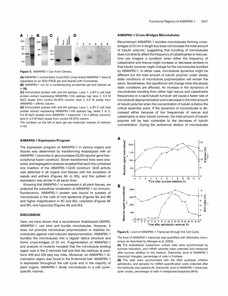
Using two different methods, we show that AtMAP65-1 can
form dimers. Firstly, chemical cross-linking of AtMAP65-1 with
1-ethyl-3-(3-dimethylaminopropyl)-carbodiimide (EDC) produ-
ces a band of ;130 kD molecular mass on one-dimensional
SDS-PAGE (Figure 5A). Secondly, on native acrylamide gel
electrophoresis, recombinant AtMAP65-1 runs at a position
corresponding to 120 to 140 kD (Figure 5B). In both experiments,
the size of the complex indicates an AtMAP65-1 dimer. More-
over, we have immobilized recombinant AtMAP65-1 on a nickel-
nitrilotriacetic acid agarose (Ni-NTA) resin column that was
then loaded with a total cell extract of a tobacco BY-2 cell line
expressing the human influenza hemagglutinin (HA) epitope
tagged tobacco equivalent to AtMAP65-1, NtMAP65-1. Immu-
noblotting of the eluates from control (resin only) and AtMAP65-1
affinity columns with anti-HA antibodies demonstrated that the
HA-epitope–tagged NtMAP65-1 interacted with AtMAP65-1 on
the column (Figure 5C).
AtMAP65-1 Dimerization Region
We have used this column AtMAP65-1 binding method to
determine which fragment in the AtMAP65-1 was capable of
interacting to form the dimer. We prepared affinity columns with
Figure 2. AtMAP65-1 Does Not Affect Microtubule Dynamics.
(A) Amount of tubulin sedimented at 100,000g after dilution of the 20-mM
tubulin mixture. Squares, tubulin only solution; circles, tubulin with 10 mM
AtMAP65-1; triangles, tubulin with 10 mM taxol.
(B) Turbidity of the 20-mM tubulin solution without, and with, the addition
of 10 mM AtMAP65-1 (solid lines) at 328C and after decreasing the
temperature. Temperature is indicated by the broken line. The arrow
indicates the time at which AtMAP65-1 was added.
(C) Coomassie-stained SDS-PAGE gel of microtubule pellets and super-
natants of a 20-mM tubulin solution incubated at 328C for 10 min, a
20-mM tubulin solution with 10 mM AtMAP65-1 incubated at 328C for
10 min and then at 18C for 10 min, and a 20-mM tubulin solution incu-
bated at 328C for 10 min and then at 18C for 10 min. The final tempera-
ture of the reaction mixtures are indicated below the lanes.
2038 The Plant Cell
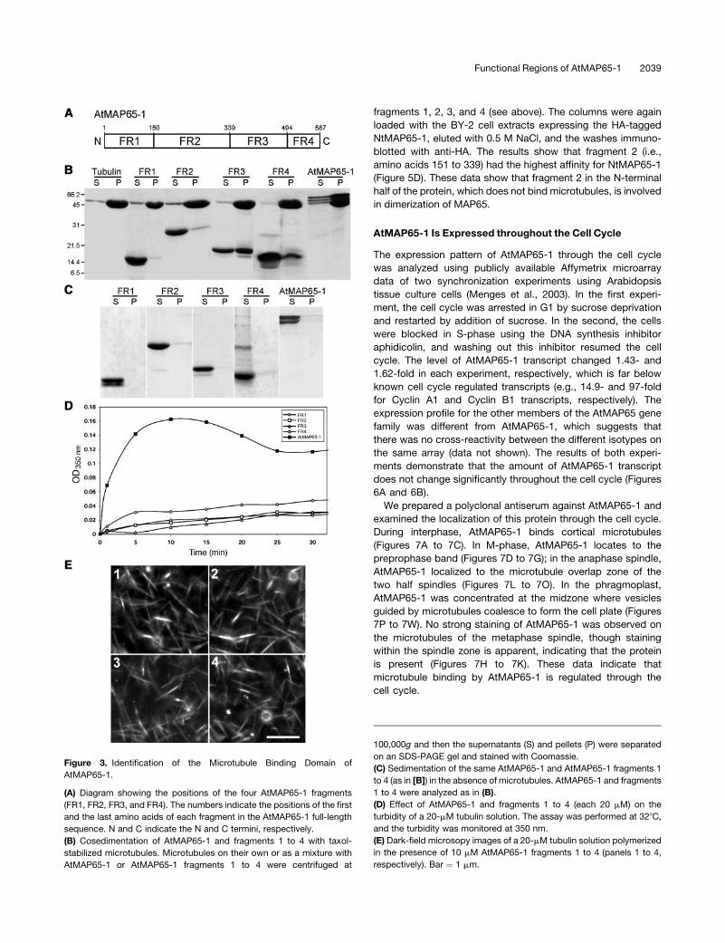
fragments 1, 2, 3, and 4 (see above). The columns were again
loaded with the BY-2 cell extracts expressing the HA-tagged
NtMAP65-1, eluted with 0.5 M NaCl, and the washes immuno-
blotted with anti-HA. The results show that fragment 2 (i.e.,
amino acids 151 to 339) had the highest affinity for NtMAP65-1
(Figure 5D). These data show that fragment 2 in the N-terminal
half of the protein, which does not bind microtubules, is involved
in dimerization of MAP65.
AtMAP65-1 Is Expressed throughout the Cell Cycle
The expression pattern of AtMAP65-1 through the cell cycle
was analyzed using publicly available Affymetrix microarray
data of two synchronization experiments using Arabidopsis
tissue culture cells (Menges et al., 2003). In the first experi-
ment, the cell cycle was arrested in G1 by sucrose deprivation
and restarted by addition of sucrose. In the second, the cells
were blocked in S-phase using the DNA synthesis inhibitor
aphidicolin, and washing out this inhibitor resumed the cell
cycle. The level of AtMAP65-1 transcript changed 1.43- and
1.62-fold in each experiment, respectively, which is far below
known cell cycle regulated transcripts (e.g., 14.9- and 97-fold
for Cyclin A1 and Cyclin B1 transcripts, respectively). The
expression profile for the other members of the AtMAP65 gene
family was different from AtMAP65-1, which suggests that
there was no cross-reactivity between the different isotypes on
the same array (data not shown). The results of both experi-
ments demonstrate that the amount of AtMAP65-1 transcript
does not change significantly throughout the cell cycle (Figures
6A and 6B).
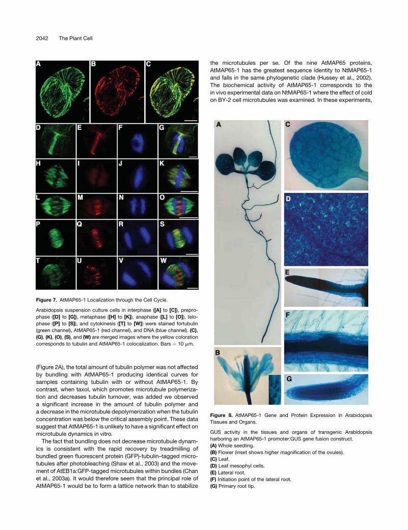
We prepared a polyclonal antiserum against AtMAP65-1 and
examined the localization of this protein through the cell cycle.
During interphase, AtMAP65-1 binds cortical microtubules
(Figures 7A to 7C). In M-phase, AtMAP65-1 locates to the
preprophase band (Figures 7D to 7G); in the anaphase spindle,
AtMAP65-1 localized to the microtubule overlap zone of the
two half spindles (Figures 7L to 7O). In the phragmoplast,
AtMAP65-1 was concentrated at the midzone where vesicles
guided by microtubules coalesce to form the cell plate (Figures
7P to 7W). No strong staining of AtMAP65-1 was observed on
the microtubules of the metaphase spindle, though staining
within the spindle zone is apparent, indicating that the protein
is present (Figures 7H to 7K). These data indicate that
microtubule binding by AtMAP65-1 is regulated through the
cell cycle.
Figure 3. Identification of the Microtubule Binding Domain of
AtMAP65-1.
(A) Diagram showing the positions of the four AtMAP65-1 fragments
(FR1, FR2, FR3, and FR4). The numbers indicate the positions of the first
and the last amino acids of each fragment in the AtMAP65-1 full-length
sequence. N and C indicate the N and C termini, respectively.
(B) Cosedimentation of AtMAP65-1 and fragments 1 to 4 with taxol-
stabilized microtubules. Microtubules on their own or as a mixture with
AtMAP65-1 or AtMAP65-1 fragments 1 to 4 were centrifuged at
100,000g and then the supernatants (S) and pellets (P) were separated
on an SDS-PAGE gel and stained with Coomassie.
(C) Sedimentation of the same AtMAP65-1 and AtMAP65-1 fragments 1
to 4 (as in [B]) in the absence of microtubules. AtMAP65-1 and fragments
1 to 4 were analyzed as in (B).
(D) Effect of AtMAP65-1 and fragments 1 to 4 (each 20 mM) on the
turbidity of a 20-mM tubulin solution. The assay was performed at 328C,
and the turbidity was monitored at 350 nm.
(E) Dark-field microsopy images of a 20-mM tubulin solution polymerized
in the presence of 10 mM AtMAP65-1 fragments 1 to 4 (panels 1 to 4,
respectively). Bar ¼ 1 mm.
Functional Regions of AtMAP65-1 2039
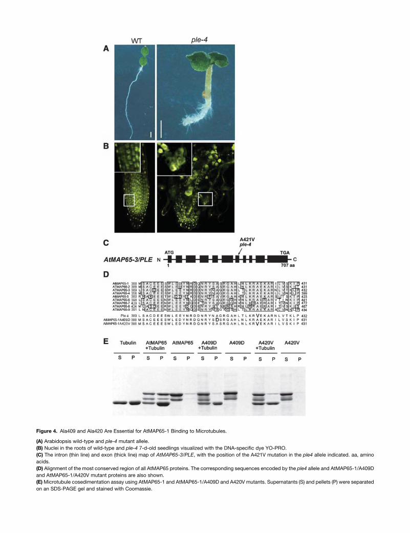
Figure 4. Ala409 and Ala420 Are Essential for AtMAP65-1 Binding to Microtubules.
(A) Arabidopsis wild-type and ple-4 mutant allele.
(B) Nuclei in the roots of wild-type and ple-4 7-d-old seedlings visualized with the DNA-specific dye YO-PRO.
(C) The intron (thin line) and exon (thick line) map of AtMAP65-3/PLE, with the position of the A421V mutation in the ple4 allele indicated. aa, amino
acids.
(D) Alignment of the most conserved region of all AtMAP65 proteins. The corresponding sequences encoded by the ple4 allele and AtMAP65-1/A409D
and AtMAP65-1/A420V mutant proteins are also shown.
(E)Microtubule cosedimentation assay using AtMAP65-1 and AtMAP65-1/A409D and A420V mutants. Supernatants (S) and pellets (P) were separated
on an SDS-PAGE gel and stained with Coomassie.
AtMAP65-1 Expression Program
The expression program of AtMAP65-1 in various organs and
tissues was determined by transforming Arabidopsis with an
AtMAP65-1promoter:b-glucuronidase (GUS) reporter gene tran-
scriptional fusion construct. Seven transformed lines were ana-
lyzed, and segregation analysis revealed that each line contained
one insertion of the AtMAP65-1:GUS construct. GUS activity
was detected in all organs and tissues with the exception of
sepals and anthers (Figures 8A to 8G), and this pattern of
expression was similar in all seven lines.
Knowing that AtMAP65-1 is expressed in all plant tissues, we
analyzed the subcellular localization of AtMAP65-1 by immuno-
fluorescence. AtMAP65-1 protein was bound to subsets of
microtubules in the cells of root epidermis (Figures 9A and 9B
and higher magnification in 9C and 9D), cotyledon (Figures 9E
and 9F), and hypocotyl (Figures 9G and 9H).
DISCUSSION
Here, we have shown that a recombinant Arabidopsis MAP65,
AtMAP65-1, can bind and bundle microtubules. However, it
does not promote microtubule polymerization or stabilize mi-
crotubules against cold-induced depolymerization. AtMAP65-1
bundles the microtubules into a regular lattice structure and
forms cross-bridges of 25 nm. Fragmentation of AtMAP65-1
and analysis of mutants revealed that the microtubule binding
region was in the C-terminal half and that Ala residues at posi-
tions 409 and 420 play key roles. Moreover, an AtMAP65-1 di-
merization region was found in the N-terminal half. AtMAP65-1
is expressed throughout the cell cycle and in the majority of
plant organs. AtMAP65-1 binds microtubules in a cell cycle–
specific manner.
AtMAP65-1 Cross-Bridges Microtubules
Recombinant AtMAP65-1 bundles microtubules forming cross-
bridges of 25 nm in length but does not increase the total amount
of tubulin polymer, suggesting that bundling of microtubules
does not directly affect the frequency of catastrophes or rescues.
One can imagine a condition when either the frequency of
catastrophe and rescue might increase or decrease similarly or
that tubulin turnover might change for the microtubules bundled
by AtMAP65-1. In either case, microtubule dynamics might be
different but the total amount of tubulin polymer under steady
state conditions of microtubule polymerization will remain the
same. Nonetheless, this equilibrium will change when the steady
state conditions are affected. An increase in the dynamics of
microtubules resulting from either high rescue and catastrophe
frequencies or a rapid tubulin turnover will cause a faster rate of
microtubule depolymerization and a decrease in the total amount
of tubulin polymer when the concentration of tubulin is below the
critical assembly point. If the dynamics of microtubules is de-
creased either because of low frequencies of rescue and
catastrophe or slow tubulin turnover, the total amount of tubulin
polymer will be less vulnerable to the decrease of tubulin
concentration. During the isothermal dilution of microtubules
Figure 5. AtMAP65-1 Can Form Dimers.
(A) AtMAP65-1 control (lane 1) and EDC cross-linked AtMAP65-1 (lane 2)
separated on an SDS-PAGE gel and stained with Coomassie.
(B) AtMAP65-1 run on a nondenaturing acrylamide gel and stained as
in (A).
(C) Immunoblot probed with anti-HA epitope. Lane 1, a BY-2 cell total
protein extract expressing NtMAP65-1:HA epitope tag; lane 2, 0.5 M
NaCl eluate from control Ni-NTA column; lane 3, 0.5 M eluate from
AtMAP65-1 affinity column.
(D) Immunoblot probed with anti-HA epitope. Lane 1, a BY-2 cell total
protein extract expressing NtMAP65-1:HA epitope tag; lanes 2 to 5,
0.5 M NaCl eluates from AtMAP65-1 fragments 1 to 4 affinity columns;
lane 6, 0.5 M NaCl eluate from control Ni-NTA column.
The numbers on the left of each gel are molecular masses of markers
in kD.
Figure 6. Level of AtMAP65-1 Transcript through the Cell Cycle.
The level of AtMAP65-1 transcript was quantified with Affymetrix micro-
arrays as described by Menges et al. (2003).
(A) The Arabidopsis suspension culture cells were synchronized by
sucrose starvation, and mRNA samples were collected and measured
after sucrose addition to the medium. Diamonds, level of AtMAP65-1
transcript; triangles, percentage of cells in S-phase.
(B) The cells were synchronized with the DNA synthesis inhibitor
aphidicolin, and samples for mRNA quantification were collected after
the herbicide was washed off. Diamonds, level of AtMAP65-1 transcript;
open circles, percentage of cells in metaphase/anaphase (M/A).
Functional Regions of AtMAP65-1 2041
(Figure 2A), the total amount of tubulin polymer was not affected
by bundling with AtMAP65-1 producing identical curves for
samples containing tubulin with or without AtMAP65-1. By
contrast, when taxol, which promotes microtubule polymeriza-
tion and decreases tubulin turnover, was added we observed
a significant increase in the amount of tubulin polymer and
a decrease in the microtubule depolymerization when the tubulin
concentration was below the critical assembly point. These data
suggest that AtMAP65-1 is unlikely to have a significant effect on
microtubule dynamics in vitro.
The fact that bundling does not decrease microtubule dynam-
ics is consistent with the rapid recovery by treadmilling of
bundled green fluorescent protein (GFP)-tubulin–tagged micro-
tubules after photobleaching (Shaw et al., 2003) and the move-
ment of AtEB1a:GFP-tagged microtubules within bundles (Chan
et al., 2003a). It would therefore seem that the principal role of
AtMAP65-1 would be to form a lattice network than to stabilize
the microtubules per se. Of the nine AtMAP65 proteins,
AtMAP65-1 has the greatest sequence identity to NtMAP65-1
and falls in the same phylogenetic clade (Hussey et al., 2002).
The biochemical activity of AtMAP65-1 corresponds to the
in vivo experimental data on NtMAP65-1 where the effect of cold
on BY-2 cell microtubules was examined. In these experiments,
Figure 7. AtMAP65-1 Localization through the Cell Cycle.
Arabidopsis suspension culture cells in interphase ([A] to [C]), prepro-
phase ([D] to [G]), metaphase ([H] to [K]), anaphase ([L] to [O]), telo-
phase ([P] to [S]), and cytokinesis ([T] to [W]) were stained fortubulin
(green channel), AtMAP65-1 (red channel), and DNA (blue channel). (C),
(G), (K), (O), (S), and (W) are merged images where the yellow coloration
corresponds to tubulin and AtMAP65-1 colocalization. Bars ¼ 10 mm.
Figure 8. AtMAP65-1 Gene and Protein Expression in Arabidopsis
Tissues and Organs.
GUS activity in the tissues and organs of transgenic Arabidopsis
harboring an AtMAP65-1 promoter:GUS gene fusion construct.
(A) Whole seedling.
(B) Flower (inset shows higher magnification of the ovules).
(C) Leaf.
(D) Leaf mesophyl cells.
(E) Lateral root.
(F) Initiation point of the lateral root.
(G) Primary root tip.
2042 The Plant Cell

microtubules in BY-2 cells were depolymerized by cold, and their
reformation was followed as the temperature was increased to
258C (Smertenko et al., 2000). The recovery of the microtubules
occurred independently of MAP65 binding, and MAP65 binding
was only observed after the microtubules were polymerized,
suggesting that NtMAP65-1 was not involved in the promotion of
microtubule polymerization but in the crossbridging of micro-
tubules once formed. These in vivo data from tobacco corre-
spond with the in vitro data described here for AtMAP65-1.
The crossbridging of microtubules by AtMAP65-1 is similar to
that observed using carrot MAP65 enriched preparations (Chan
et al., 1999). The carrot MAP65 preparation contained three
electrophoretically separable bands, minor 60- and 68-kD
bands, and a predominant (85%) 62-kD band (Chan et al.,
1999). These protein bands were subsequently analyzed by
mass spectral analysis: the 68 and 60 kD were shown to
disappear when the carrot suspension culture stopped dividing,
leaving the 62-kD species as the sole detectable MAP65 in
elongating cells containing only cortical microtubules (Chan
et al., 2003b). Peptide sequencing and sequencing the cDNA
(DcMAP65-1) established that carrot MAP62 was most similar to
AtMAP65-1 and NtMAP65-1 than to any other known MAP65
(Chan et al., 2003b). Because MAP-62 was biochemically
purified, it is not knownwhether it is posttranslationally modified.
From the data presented in this study we can conclude that
mixtures of MAP65 isoforms are not required to promote
bundling and that the single unmodified gene product of
AtMAP65-1 is capable of forming the 25-nm cross-bridges.
Identification of Microtubule Binding and Dimerization
Regions of AtMAP65-1
The AtMAP65-1 microtubule binding region is in the C-terminal
half of the protein and two amino acids, Ala420 and Ala409, are
essential structural elements in the microtubule:AtMAP65-1 in-
teraction. The C-terminal half of the protein was divided into
fragments 3 and 4. Fragment 3 harbors sequence that was most
conserved in all nine AtMAP65 genes. Fragment 4 contains the
most divergent sequences across theMAP65 family and is highly
charged (pI of 10.47). Both fragments 3 and 4 bound micro-
tubules, although it cannot be discounted that the high charge of
fragment 4 might be responsible for nonspecific binding. Frag-
ment 3 is not only conserved between plant MAP65 proteins, but
sequence within this fragment also exhibits strong similarity with
mammalian PRC1 and yeast Ase1p especially within a 25–amino
acid region (37.5%; Schuyler et al., 2003). Mutation of a con-
served amino acid within this short sequence, Ala420 Val,
diminished MAP65 microtubule binding. This mutation was
mimicked in AtMAP65-1 based on the sequence of the cytoki-
nesis defective ple-4 allele. Here, we show that changing the
conserved Ala to a more hydrophobic amino acid disturbs the
structure to such an extent that microtubule binding is greatly
reduced.Mutating a second Ala, at residue 409, in the conserved
25–amino acid sequence also diminished microtubule binding.
By synthesizing a peptide covering the whole 25–amino acid
conserved motif, we were able to perform competition studies
where we allowed AtMAP65-1 and the synthetic peptide to
compete for the microtubule binding site. These studies dem-
onstrated that we could not inhibit the AtMAP65-1 microtubule
interaction in this way, which strongly suggests that sequences
other than this 25–amino acid conserved motif, within fragment
3, are structurally important formicrotubule binding. It is possible
that the microtubule binding site depends on several points of
contact and requires a specific tertiary structure as is known for
MAP2/tau.
The amino terminal half of AtMAP65-1 harbors a dimerization
region, and dimerization may be important for 25-nm cross-
bridge formation. The N-terminal half of AtMAP65-1 was divided
into two sections, and the fragment encompassing residues 151
to 339 was found to bind another MAP65. Consideration of the
structure of the cross-bridges between microtubules created by
carrot MAP65s and the size of MAP65 led to the suggestion that
Figure 9. Immunolocalization of AtMAP65-1 in Plant Tissues.
Root epidermal cells stained with anti tubulin (A) and anti AtMAP65-1 (B)
antibodies. A higher magnification of the boxed areas in (A) and (B) is
shown in (C) and (D). Immunostaining of microtubules ([I] and [K]) and
AtMAP65-1 ([J] and [L]) in cotyledons ([I] and [J]) and hypocotyl ([K] and
[L]) epidermal cells.
Functional Regions of AtMAP65-1 2043
MAP65 is unlikely to cross-bridge as a single molecule (Chan
et al., 1999). The fact that neither the microtubule binding region
nor the dimerization region alone were capable of microtubule
bundling strongly suggests that these cross-bridges are formed
by MAP65 dimers, with the C-terminal halves binding adjacent
microtubules and the N-terminal halves being responsible for
MAP65:MAP65 interaction.
AtMAP65-1 Binding to Microtubules Is Cell Cycle Specific
AtMAP65-1 is expressed constitutively through the cell cycle,
but the protein binds only subsets of microtubules in a cell
cycle–dependent manner. The main difference in the localiza-
tion of AtMAP65-1 in cells compared with AtMAP65-3/PLE
(Muller et al., 2004) and its homologs in animals and fungi is that
AtMAP65-1 (like NtMAP65-1; Smertenko et al., 2000) also binds
interphase cortical microtubules (Pellman et al., 1995; Jiang
et al., 1998). However, mammalian PRC1 is capable of binding
and bundling microtubules in interphase as overexpression
causes the disruption of normal microtubule organization and
the appearance of circular filaments around the nuclei (Mollinari
et al., 2002). Bundling of microtubules in interphase in plant
cells has been suggested to be important for the formation of
the interphase cortical array (Shaw et al., 2003). As the cells
enter M-phase, AtMAP65-1 does decorate the preprophase
band but not the metaphase spindle. It appears to bind micro-
tubules once more at the midzone of the anaphase spindle
where the phragmoplast forms. In the phragmoplast it is
present where the microtubules overlap at the midline. Because
the AtMAP65-1 transcript is present throughout the cell cycle,
the microtubule binding activity of AtMAP65-1 must be under
posttranscriptional control.
Motif searches have revealed a cyclin destruction box and sev-
eral phosphorylation sites, including a cyclin-dependent kinase
(CDK) phosphorylation site (Hussey et al., 2002). AtMAP65-1
can be phosphorylated by a protein extract prepared from
3-d-old Arabidopsis tissue culture cells, and the phosphorylation
can be inhibited by roscovitine, suggesting that CDK might
be responsible for this activity. However, phosphorylated
AtMAP65-1 did cosediment with microtubules and did increase
the turbidity of a tubulin solution (data not shown). Interestingly,
a phosphorylation mimic of PRC1 (the mammalian AtMAP65
homolog) where the CDK sites were mutated to Glu was still
capable of binding and bundling microtubules in the same
manner as the nonphosphorylatable protein (Mollinari et al.,
2002). Thus, phosphorylation on its own is not the sole factor that
controls the microtubule:AtMAP65-1 interaction, and unraveling
the signaling pathways that regulate this process will be the next
crucial step in understanding the control of this MAP.
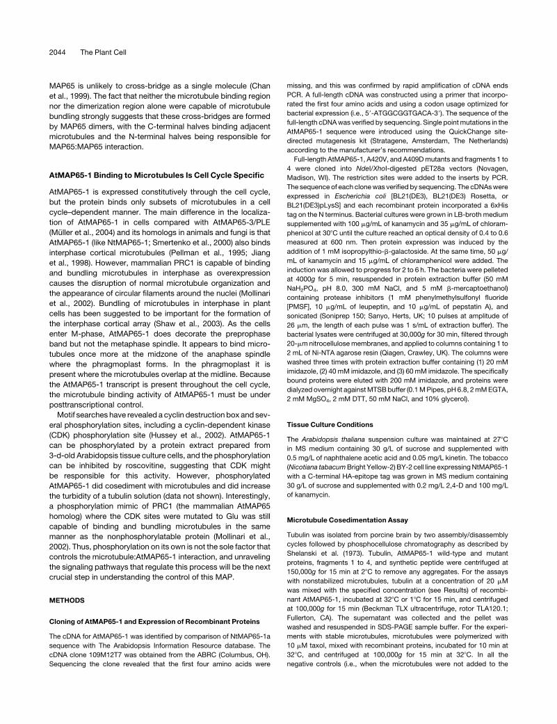
METHODS
Cloning of AtMAP65-1 and Expression of Recombinant Proteins
The cDNA for AtMAP65-1 was identified by comparison of NtMAP65-1a
sequence with The Arabidopsis Information Resource database. The
cDNA clone 109M12T7 was obtained from the ABRC (Columbus, OH).
Sequencing the clone revealed that the first four amino acids were
missing, and this was confirmed by rapid amplification of cDNA ends
PCR. A full-length cDNA was constructed using a primer that incorpo-
rated the first four amino acids and using a codon usage optimized for
bacterial expression (i.e., 59-ATGGCGGTGACA-39). The sequence of the
full-length cDNAwas verified by sequencing. Single pointmutations in the
AtMAP65-1 sequence were introduced using the QuickChange site-
directed mutagenesis kit (Stratagene, Amsterdam, The Netherlands)
according to the manufacturer’s recommendations.
Full-length AtMAP65-1, A420V, and A409Dmutants and fragments 1 to
4 were cloned into NdeI/XhoI-digested pET28a vectors (Novagen,
Madison, WI). The restriction sites were added to the inserts by PCR.
The sequence of each clonewas verified by sequencing. The cDNAswere
expressed in Escherichia coli [BL21(DE3), BL21(DE3) Rosetta, or
BL21(DE3)pLysS] and each recombinant protein incorporated a 6xHis
tag on the N terminus. Bacterial cultures were grown in LB-broth medium
supplemented with 100 mg/mL of kanamycin and 35 mg/mL of chloram-
phenicol at 308C until the culture reached an optical density of 0.4 to 0.6
measured at 600 nm. Then protein expression was induced by the
addition of 1 mM isopropylthio-b-galactoside. At the same time, 50 mg/
mL of kanamycin and 15 mg/mL of chloramphenicol were added. The
induction was allowed to progress for 2 to 6 h. The bacteria were pelleted
at 4000g for 5 min, resuspended in protein extraction buffer (50 mM
NaH2PO4, pH 8.0, 300 mM NaCl, and 5 mM b-mercaptoethanol)
containing protease inhibitors (1 mM phenylmethylsulfonyl fluoride
[PMSF], 10 mg/mL of leupeptin, and 10 mg/mL of pepstatin A), and
sonicated (Soniprep 150; Sanyo, Herts, UK; 10 pulses at amplitude of
26 mm, the length of each pulse was 1 s/mL of extraction buffer). The
bacterial lysates were centrifuged at 30,000g for 30 min, filtered through
20-mmnitrocellulose membranes, and applied to columns containing 1 to
2 mL of Ni-NTA agarose resin (Qiagen, Crawley, UK). The columns were
washed three times with protein extraction buffer containing (1) 20 mM
imidazole, (2) 40 mM imidazole, and (3) 60 mM imidazole. The specifically
bound proteins were eluted with 200 mM imidazole, and proteins were
dialyzed overnight againstMTSBbuffer (0.1MPipes, pH 6.8, 2mMEGTA,
2 mM MgSO4, 2 mM DTT, 50 mM NaCl, and 10% glycerol).
Tissue Culture Conditions
The Arabidopsis thaliana suspension culture was maintained at 278C
in MS medium containing 30 g/L of sucrose and supplemented with
0.5 mg/L of naphthalene acetic acid and 0.05 mg/L kinetin. The tobacco
(Nicotiana tabacumBright Yellow-2) BY-2 cell line expressingNtMAP65-1
with a C-terminal HA-epitope tag was grown in MS medium containing
30 g/L of sucrose and supplemented with 0.2 mg/L 2,4-D and 100 mg/L
of kanamycin.
Microtubule Cosedimentation Assay
Tubulin was isolated from porcine brain by two assembly/disassembly
cycles followed by phosphocellulose chromatography as described by
Shelanski et al. (1973). Tubulin, AtMAP65-1 wild-type and mutant
proteins, fragments 1 to 4, and synthetic peptide were centrifuged at
150,000g for 15 min at 28C to remove any aggregates. For the assays
with nonstabilized microtubules, tubulin at a concentration of 20 mM
was mixed with the specified concentration (see Results) of recombi-
nant AtMAP65-1, incubated at 328C or 18C for 15 min, and centrifuged
at 100,000g for 15 min (Beckman TLX ultracentrifuge, rotor TLA120.1;
Fullerton, CA). The supernatant was collected and the pellet was
washed and resuspended in SDS-PAGE sample buffer. For the experi-
ments with stable microtubules, microtubules were polymerized with
10 mM taxol, mixed with recombinant proteins, incubated for 10 min at
328C, and centrifuged at 100,000g for 15 min at 328C. In all the
negative controls (i.e., when the microtubules were not added to the
2044 The Plant Cell
proteins), the reaction mixture was supplemented with MTSB buffer
containing 10 mM taxol.
Microtubule Polymerization Assay
Pig brain tubulin dimer was used at a final concentration of 20 mM in all
assays. Tubulin solution was stored at�808C. Before each assay, tubulin
was rapidly thawed at 378C, incubated on ice for 5min, and centrifuged at
200,000g for 10 min to pellet polymerized microtubules and tubulin
aggregates. The supernatant was diluted to a 30 mM tubulin stock with
MTSB. The blank was set up with the cuvette containing 100 mL of 30 mM
tubulin inMTSB. ThenGTPwas added up to a final concentration of 1mM
followed by recombinant AtMAP65-1, mutants, or fragments 1 to 4. The
final reaction volumewas adjusted to 150mLwithMTSB, and the turbidity
of the reaction was monitored at 350 nm and at 328C using a Helios b
spectrophotometer equipped with Unicam Peltier temperature control
unit (Thermospectronic, Basingstoke, UK). Each experiment was re-
peated three times and the average (in the case of Figure 1A) or an
example from three series of experiments (in the case of Figure 3D) is
presented.
Isothermal Dilution of Microtubules
To polymerize microtubules, 20 mM tubulin in MTSB was incubated at
328C for 10min. Thereafter, a 50-mL aliquot was kept separately, while the
rest of the solution was diluted with MTSB buffer prewarmed at 328C and
incubated at this temperature for 10min. Alternatively, 10mMAtMAP65-1
or taxol was added to the microtubules before incubation at 328C. The
samples were centrifuged at 100,000g for 10 min, and the amount of
tubulin in the pellet was analyzed by SDS-PAGE electrophoresis.
Microtubule Bundling Assay
The dark-field microscopy analysis was performed with 20 mM tubulin in
MTSB. AtMAP65-1 was added to the tubulin solution at a final concen-
tration of 10 mM and MTSB up to a final volume of 20 mL. The reaction
mixture was incubated for 5 min at room temperature, and 5 mL of the
mixturewas examinedwith anOlympusBX50microscope (Tokyo, Japan)
equipped with UplanAop 1003/1.35 objective, 100-W mercury bulb,
dark-field condenser, and Hamamatsu C4742 black and white CCD
camera (Herts, UK).
For the visualization of cross-bridges, microtubules were polymerized
with recombinant protein and applied to formvar-coated and ionized
copper grids. The gridswere negatively stainedwith 1%aqueous solution
of uranyl acetate and examined using a JEOL transmission electron
microscope (Welwyn Garden City, UK).
Chemical Cross-Linking
Cross-linkingwasperformed using EDC (Doi et al., 1987). EDCwas added
up to the final concentration of 5mM in a 20-mMsolution of AtMAP65-1 in
MTSB and incubated at room temperature for 1 h. The reaction was
stopped by addition of SDS-PAGE sample buffer, and then the mixture
was boiled for 3 min and the sample separated on a 7.5% acrylamide gel.
AtMAP65-1 Promoter GUS Fusions
A fragment of 1612 bp upstream of the translation start codon was
amplified by PCR using the forward primer 59-CAGCAATTCTCCGGA-
GAACT-39 and the reverse primer 59-GCGGAATCAGAAGGTTTCCT-39.
The PCR fragment was cloned into the pGEMT-E vector (Promega,
Southampton, UK) and its sequence verified and then subcloned into the
PDgusBin19 vector (Topping et al., 1991). The final construct was
transformed into Agrobacterium tumefaciens strain C58C3 cells by
electroporation. Arabidopsis plants were transformed using the dipping
method (Clough and Bent, 1998). The positives were selected on half-
strength MS medium containing 0.6% agar, 50 mg/mL of kanamycin,
and 200 mg/mL of augmentin (SmithKline Beecham Pharmaceuticals,
Slough, UK). Positives were transferred to soil and seeds collected. The
T2 generation was used for the histochemical localization of GUS activity.
Plant sampleswere vacuum infiltrated for 30minwith substrate solution
(100 mM sodium phosphate buffer, pH 7.0, 10 mM EDTA, 0.1% Triton
X-100, 0.5 mM potassium ferricyanide, 0.5 mM potassium ferrocyanide,
and 1 mM 5-bromo-4-chloro-3-indolyl glucuronide) and incubated at
378C for 12 h. Plant tissues were cleared using the chloral-lacto-phenol
method (Barthels et al., 1997) and examined using Leica MZ125
(Wetzlar, Germany) and Zeiss Axioscope (Jena, Germany) microscopes
equipped with Photometrix CoolSnap CF color CCD camera (Nippon
Ropper, Chiba-shi, Japan).
Antibodies and Immunostaining
The AtMAP65-1 antiserum was raised in rabbits using the full-length
recombinant AtMAP65-1 protein as immunogen. Recombinant protein
(250 mg) was used for each boost, and four boosts over a period of
3 months were performed. Antiserum was collected 10 d after the final
boost and tested by immunoblotting. The AtMAP65-1 antiserum identi-
fied the AtMAP65-1 recombinant protein, and a single band on a 1-D gel
total protein extract of Arabidopsis tissue culture cells (data not shown).
For immunostaining, Arabidopsis suspension culture cells were sepa-
rated from the tissue culture medium using 100 mesh nylon cloth and
fixed for 30 min at room temperature with 4% paraformaldehyde in 0.1 M
Pipes, pH 6.8, 5mMEGTA, 2mMMgCl2, and 0.4%Triton X-100. The fixa-
tive was washed away with PBS buffer, and cells were treated for 5 min
at room temperature with the solution of 0.8% macerozyme R-10 and
0.2% pectolyase Y-23 in 0.4 M mannitol, 5 mM EGTA, 15 mM Mes, pH
5.0, 1 mM PMSF, 10 mg/mL of leupeptin, and 10 mg/mL of pepstatin A.
Then the cells were washed in PBS buffer and attached to poly-L-Lys–
coated cover slips. The cover slips were then incubated for 30 min in 1%
(w/v) BSA in PBS and incubated for 1 hwith primary antibody. The primary
antibodies used were rabbit anti-AtMAP65-1 diluted 1:500 and mouse
antitubulin DM1A diluted 1:200 (Sigma, Dorset, UK). The specimens were
then washed three times for 10 min in PBS and incubated for 1 h with
secondary antibodies: goat anti-mouse tetramethylrhodamine isothio-
cyanate conjugates and anti-rabbit fluorescein isothiocyanate conju-
gates; both antibodies were diluted 1:200. After washing in the PBS
buffer, the specimens were mounted in Vectashied (Vector Laboratories,
Burlingame, CA) mounting medium and examined with a Zeiss 510
confocal microscope.
For Arabidopsis root immunostaining, the roots were fixed for 1 h in the
same fixative solution as for tissue culture cells, then attached to poly-L-
Lys–coated slides and treated for 10 min with 2% (w/v) Dricelase (Sigma)
in 0.4 M mannitol, 5 mM EGTA, 15 mMMes, pH 5.0, 1 mM PMSF, 10 mg/
mL of leupeptin, 10 mg/mL of pepstatin A, and 10 mg/mL of Na-p-tosyl-L-
lysine chloromethyl ketone. Then, roots were incubated with antibodies,
mounted, and examined as described above with the modification that
the primary antibodies were applied overnight and the secondary anti-
bodies for 3 h.
For immunostaining the hypocotyl and the cotyledons, Arabidopsis
seedlings were fixed for 1 h in the same mixture as for tissue culture cells
but supplemented with 0.2% (v/v) glutaraldehyde. Then cotyledons and
hypocotyl were rapidly immersed into liquid nitrogen, shattered to rupture
the cell walls, and incubatedwith antibodies as described above for roots.
Protein Electrophoresis and Immunoblotting
For the quantification of recombinant proteins in Figures 1B and 2A, the
protein samples were separated on a 7.5% one-dimensional SDS-PAGE.
Functional Regions of AtMAP65-1 2045
The gels were stained with Coomassie Brilliant Blue and scanned using
a flat bed scanner. The amount of protein on the gel was quantified using
NIH image software version 1.62 (National Institutes of Health, Bethesda,
MD). Each experiment was repeated three times. For the quantification
of tubulin polymer in the pellet, local values for the background were
estimated and subtracted from the protein band values. Then the data
from the three replicates were normalized by mean: the intensity for each
band in a particular replicate was divided by the mean intensity for all
bands in the replicate. The datawere then averaged across the replicates.
The final number represents an arbitrary value.
For immunoblotting, proteins were transferred from the gel onto
nitrocellulose membranes and probed with mouse monoclonal anti HA-
epitope diluted 1:100 (Sigma). The secondary anti-rabbit or anti-mouse
horseradish peroxidase conjugates were used at a dilution of 1:2000 and
detected using the enhanced chemiluminescence kit (Amersham Bio-
sciences, Buckinghamshire, UK).
The native acrylamide gel with AtMAP65-1 protein in Figure 5B was
done according to Zabala and Cowan (1992).
Affinity Columns
Recombinant AtMAP65-1 protein or fragments 1 to 4 were dialyzed
against NET buffer (100 mM Tris, pH 7.5, 100 mMNaCl, and 1 mM EDTA)
and bound to the column loaded with Ni-NTA resin (Qiagen). Approxi-
mately 50 mg of total protein were used in each case. About 300 to
500 mg of miniprotoplast (prepared according to the method of Jiang
and Sonobe, 1993) total protein isolated from the BY-2 cell line
expressing NtMAP65-1a with a C-terminal HA-epitope tag was run
through the column. The column was washed with 20 bed volumes of
NET buffer containing 200 mM NaCl, 40 mM imidazole, and 5 mM
b-mercaptoethanol. The interacting proteins were eluted with 0.5 M NaCl
in NET buffer.
Phenotypic, Molecular Genetic, and Microscopic Analysis
For the analysis of the root morphogenesis phenotype, plemutants were
cultivated on vertical nutrient agar plates containing 13 MS salt mixture
and 4.5% sucrose, pH 5.7, in 1% agar. Nuclei were stained with YO-PRO
(Molecular Probes, Eugene, OR) on fixed roots and analyzed with
a confocal scanning laser microscope (Leica TCS SP2) as described
(Muller et al., 2002). Allelism of the ple-4mutant was confirmed by genetic
crosses and sequencing.
Sequence data from this article have been deposited with the EMBL/
GenBank data libraries under accession number NM_124905.
ACKNOWLEDGMENTS
We would like to thank Marylin Vantard (Centre National de la Recher-
che Scientifique, Grenoble) for useful discussions, Margit Menges and
Jim Murray (University of Cambridge) for providing prepublished Affy-
metrix data, S. Muller, I. Kreuzer, and G. Resch for technical assistance,
and Toni Slabas for providing the Arabidopsis suspension culture cells.
We are specially obliged to Farhah Assaad and Wolfgang Lukowitz for
providing the ple-4 allele. We would like to thank the ABRC for the
AtMAP65-1 cDNA clone and the Arabidopsis genome sequencing
project. A.P.S., S.F., C.W.L., and P.J.H. are funded by the Biotechnol-
ogy and Biological Research Council UK. D.K. is supported by the
European Union human potential program (TIPNET HPRN-CT-2002-
00265). H.-Y.C. is funded by an Overseas Research Studentship. This
work was also funded by grants of the FWF Austrian Science Fund to
M.-T.H. (project numbers P14477-B04 and P16410-B12). V.W. is funded
by the DOC fellowship of the Austrian Academy of Sciences.
Received May 5, 2004; accepted June 8, 2004.
REFERENCES
Barthels, N., et al. (1997). Regulatory sequences of Arabidopsis drive
reporter gene expression in nematode feeding structures. Plant Cell 9,
2119–2134.
Chan, J., Calder, G.M., Doonan, J.H., and Lloyd, C.W. (2003a). EB1
reveals mobile microtubule nucleation sites in Arabidopsis. Nat. Cell
Biol. 5, 967–971.
Chan, J., Jensen, C.G., Jensen, L.C.W., Bush, M., and Lloyd, C.W.
(1999). The 65-kDa carrot microtubule-associated protein forms
regularly arranged filamentous cross-bridges between microtubules.
Proc. Natl. Acad. Sci. USA 96, 14931–14936.
Chan, J., Mao, G., Smertenko, A., Hussey, P.J., Naldrett, M., Bottrill,
A., and Lloyd, C.W. (2003b). Identification of a MAP65 isoform
involved in directional expansion of plant cells. FEBS Lett. 534,
161–163.
Chan, J., Rutten, T., and Lloyd, C. (1996). Isolation of microtubule-
associated proteins from carrot cytoskeletons: A 120 kDa map
decorates all four microtubule arrays and the nucleus. Plant J. 10,
251–259.Clough, S.J., and Bent, A.F. (1998). Floral dip: A simplified method
for Agrobacterium-mediated transformation of Arabidopsis thaliana.
Plant J. 16, 735–743.
Doi, Y., Higashida, M., and Kido, S. (1987). Plasma gelsolin binding
sites on the actin sequence. Eur. J. Biochem. 164, 89–94.Gardiner, J., and Marc, J. (2003). Putative microtubule-associated
proteins from the Arabidopsis genome. Protoplasma 222, 61–74.
Hush, J.M., Wadsworth, P., Callaham, D.A., and Hepler, P.K.
(1994). Quantification of microtubule dynamics in living plant cells
using fluorescence redistribution after photobleaching. J. Cell Sci.
107, 775–784.
Hussey, P.J., Hawkins, T.J., Igarashi, H., Kaloriti, D., and
Smertenko, A. (2002). The plant cytoskeleton: Recent advances in
the study of the plant microtubule-associated proteins MAP-65,
MAP-190 and the Xenopus MAP215-like protein, MOR1. Plant Mol.
Biol. 50, 915–924.
Jiang, C.J., and Sonobe, S. (1993). Identification and preliminary
characterization of a 65kDa higher-plant microtubule-associated pro-
tein. J. Cell Sci. 105, 891–901.
Jiang, W., Jimenez, G., Wells, N.J., Hope, T.J., Wahl, G.M., Hunter,
T., and Fukunaga, F. (1998). PRC1: A human mitotic spindle-
associated CDK substrate protein required for cytokinesis. Mol. Cell
2, 877–885.
Lloyd, C.W., Hussey, P.J., and Chan, J. (2004). Microtubules and
microtubule-associated proteins. In The Plant Cytoskeleton in Cell
Differentiation and Development, P.J. Hussey, ed (Oxford: Blackwell
Publishing), pp. 3–31.
Menges, M., Hennig, L., Gruissem, W., and Murrey, J.A.H. (2003).
Genome-wide gene expression in an Arabidopsis cell suspension.
Plant Mol. Biol. 53, 423–442.
Mollinari, C., Kleman, J.P., Jiang, W., Schoehn, G., Hunter, T., and
Margolis, R.L. (2002). PRC1 is a microtubule binding and bundling
protein essential to maintain the mitotic spindle midzone. J. Cell Biol.
157, 1175–1186.
Moore, R.C., Zhang, M., Cassimeris, L., and Cyr, R.J. (1997). In vitro
assembled plant microtubules exhibit a high state of dynamic in-
stability. Cell Motil. Cytoskeleton 38, 278–286.
2046 The Plant Cell
Muller, S., Fuchs, E., Ovecka, M., Wysocka-Diller, J., Benfey, P.N.,
and Hauser, M.-T. (2002). Two new loci, PLEIADE and HYADE,
implicate organ-specific regulation of cytokinesis in Arabidopsis.
Plant Physiol. 130, 312–324.
Muller, S., Smertenko, A., Wagner, V., Heinrich, M., Hussey, P.J.,
and Hauser, M.-T. (2004). The plant microtubule associated protein,
AtMAP65-3/PLE, is essential for cytokinetic phragmoplast function.
Curr. Biol. 14, 412–417.
Pellman, D., Bagget, M., Tu, H., and Fink, G.R. (1995). Two
microtubule-associated proteins required for anaphase spindle move-
ment in Saccharomyces cerevisiae. J. Cell Biol. 13, 1373–1385.
Schuyler, S.C., Liu, J.Y., and Pellman, D.J. (2003). The molecular
function of Ase1p: Evidence for a MAP-dependent midzone-specific
spindle matrix. J. Cell Biol. 160, 517–528.
Shaw, S.L., Kamyar, R., and Ehrhardt, D.W. (2003). Sustained micro-
tubule treadmilling in Arabidopsis cortical arrays. Science 300, 1715–
1718.
Shelanski, M.L., Gaskin, F., and Cantor, C.R. (1973). Microtubule
assembly in absence of added nucleotides. Proc. Natl. Acad. Sci USA
70, 765–768.
Smertenko, A., Saleh, N., Igarashi, H., Mori, H., Hauser-Hahn, I.,
Jiang, C.J., Sonobe, S., Lloyd, C.W., and Hussey, P.J. (2000). A
new class of microtubule-associated proteins in plants. Nat. Cell Biol.
2, 750–753.
Smertenko, A.P., Bozhkov, P.V., Filonova, L.H., von Arnold, S., and
Hussey, P.J. (2003). Re-organisation of the cytoskeleton during
developmental programmed cell death in Picea abies embryos. Plant
J. 33, 813–824.
Sollner, R., Glasser, G., Wanner, G., Somerville, C.R., Jurgens, G.,
and Assaad, F.F. (2002). Cytokinesis-defective mutants of Arabidop-
sis. Plant Physiol. 129, 678–690.
Sorensen, M.B., Mayer, U., Lukowitz, W., Robert, H., Chambrier, P.,
Jurgens, G., Somerville, C., Lepiniec, L., and Berger, F. (2002).
Cellularisation in the endosperm of Arabidopsis thaliana is coupled to
mitosis and shares multiple components with cytokinesis. Develop-
ment 129, 5567–5576.
Topping, J.F., Wei, W.B., and Lindsey, K. (1991). Functional tagging of
regulatoryelements in theplantgenome.Development112,1009–1019.
Yuan, M., Shaw, P.J., Warn, R.M., and Lloyd, C.W. (1994). Dynamic
reorientation of cortical microtubules, from transverse to longitudinal
in living plant cells. Proc. Natl. Acad. Sci. USA 91, 6050–6053.
Zabala, J.C., and Cowan, N.J. (1992). Tubulin dimer formation via the
release of a- and b-tubulin monomers from multimolecular com-
plexes. Cell Motil. Cytoskeleton 23, 222–230.
Functional Regions of AtMAP65-1 2047
DOI 10.1105/tpc.104.023937; originally published online July 23, 2004; 2004;16;2035-2047Plant Cell
Clive Lloyd, Marie-Theres Hauser and Patrick J. HusseyAndrei P. Smertenko, Hsin-Yu Chang, Vera Wagner, Despina Kaloriti, Stepan Fenyk, Seiji Sonobe,
Microtubule Bundling ActivityThe Arabidopsis Microtubule-Associated Protein AtMAP65-1: Molecular Analysis of Its
This information is current as of August 16, 2019
References /content/16/8/2035.full.html#ref-list-1
This article cites 26 articles, 12 of which can be accessed free at:
Permissions https://www.copyright.com/ccc/openurl.do?sid=pd_hw1532298X&issn=1532298X&WT.mc_id=pd_hw1532298X
eTOCs http://www.plantcell.org/cgi/alerts/ctmain
Sign up for eTOCs at:
CiteTrack Alerts http://www.plantcell.org/cgi/alerts/ctmain
Sign up for CiteTrack Alerts at:
Subscription Information http://www.aspb.org/publications/subscriptions.cfm
is available at:Plant Physiology and The Plant CellSubscription Information for
ADVANCING THE SCIENCE OF PLANT BIOLOGY © American Society of Plant Biologists