the arcuate fasciculus and the disconnection theme in...
TRANSCRIPT

c o r t e x 4 4 ( 2 0 0 8 ) 9 5 3 – 9 6 1
ava i lab le at www.sc ienced i rec t . com
journa l homepage : www. e lsev ier . com/ loca te / cor tex
Special issue: Original article
The arcuate fasciculus and the disconnection theme inlanguage and aphasia: History and current state
Marco Catania,* and Marsel Mesulamb
aNatbrainlab, Section of Brain Maturation, King’s College London, Institute of Psychiatry, London, UKbCognitive Neurology and Alzheimer’s Disease Center, Northwestern University, Chicago, IL, USA
a r t i c l e i n f o
Article history:
Received 27 March 2008
Reviewed 11 April 2008
Revised 14 April 2008
Accepted 15 April 2008
Published online 23 May 2008
Keywords:
Arcuate fasciculus
Aphasia
Diffusion tensor imaging (DTI)
Language
Tractography
* Corresponding author. Natbrainlab, SectionE-mail address: [email protected] (M
0010-9452/$ – see front matter ª 2008 Elsevidoi:10.1016/j.cortex.2008.04.002
a b s t r a c t
Few themes have been more central to neurological models of aphasia than the disconnec-
tion paradigm and the role of the arcuate fasciculus. Introduced by luminaries of 19th
Century neurology and resurrected by the charismatic work of Norman Geschwind, the
disconnection theme has triggered spectacular advances of modern understanding of
language and aphasia. But the disconnection paradigm had alternate fortunes, ranging
from irrational exuberance to benign neglect, and its followers have not always shared
the same view on its functional consequences and anatomical correlates. Our goal in
this paper is, first, to survey the 19th Century roots of the connectionist approach to
aphasia and, second, to describe emerging imaging technologies based on diffusion tensor
imaging (DTI) that promise to consolidate and expand the disconnection approach to
language and its disorders.
ª 2008 Elsevier Masson Srl. All rights reserved.
1. Introduction physiological and neuronographic studies both in animals
Language is an exceedingly complex faculty that allows us
to encode, elaborate and communicate thoughts and expe-
riences through the mediation of arbitrary symbols known
as words. The coherent function of the language network
and its interactions with other neurocognitive networks de-
pend on an orderly set of interconnections. Much of current
understanding of language-related pathways is based on
the pioneering work of 19th Century neuroanatomists,
such as Reil, Burdach, Meynert, Wernicke, Dejerine. In the
1960s, in a series of influential papers, Geschwind crystal-
lized those early anatomical findings adding new insights
into brain connectivity as derived from anatomical,
of Brain Maturation PO5. Catani).
er Masson Srl. All rights
and humans (Geschwind, 1965, 1970; Geschwind and Levit-
sky, 1968).
The neuroanatomy of the human brain that Geschwind
relied on was based principally on hand dissection of fixed
specimens and the tracing of degeneration in sections stained
for myelin. Recent developments in magnetic resonance
imaging have introduced new methods, based on diffusion
tensor imaging (DTI) tractography (see also Jones, 2008, this
issue; Catani and Thiebaut de Schotten, 2008, this issue) that
can reconstruct white matter pathways in the living human
brain. The resultant influx of information on human connec-
tional anatomy is likely to modernize the disconnection ap-
proach to behavioural neurology and to reinvigorate models
0, Institute of Psychiatry, De Crespigny Park, SE5 8AF London, UK.
reserved.

c o r t e x 4 4 ( 2 0 0 8 ) 9 5 3 – 9 6 1954
of cognition based on distributed large-scale networks (Catani
and Mesulam, 2008, this issue). An overview of these trends,
and of their historical contexts, with a special focus on the ar-
cuate fasciculus and language, constitutes the subject matter
of this paper.
2. Disconnection accounts of languagedisorders
The term disconnection is generally used to indicate classical
syndromes where lesions to white matter connections lead to
dysfunction of higher cognitive abilities (Catani and ffytche,
2005; Mesulam, 2005). The term became popular in the second
half of the 19th Century following Wernicke’s (1874) descrip-
tion of the disconnection syndrome that was to become the
prototype for all others – conduction aphasia. Wernicke, like
his predecessor Theodore Meynert, conceived the brain as
a mosaic of areas containing ‘memory images’ related to
motor acts (localized in primary motor areas) and sensory ex-
periences (localized in primary visual, somesthestic, auditory,
olfactory and gustatory areas). He also assumed that higher
cognitive functions, in contrast to movements and percep-
tions, are not localized in specific regions but emerge from
associative connections linking areas where images of motor
and sensory memories reside. On the basis of this ‘general
principle’, Wernicke (1874) elaborated the first network model
of language (Fig. 1): ‘.the first frontal gyrus [third frontal circon-
volution according to modern nomenclature], which has motor
function, acts as center for motor imagery; the first temporal gyrus,
which is sensory in nature, may be regarded as the centre of acoustic
images; the fibrae propriae, converging into the insular cortex, form
the mediating arc reflex.’ He argued that ‘aphasia may be caused by
any disruption of this pathway, the clinical picture, however, may
vary considerably and is related to the specific segment of the path-
way involved.’ According to Wernicke, the ‘production of sponta-
neous movement, that is, the consciously formulated word, would be
brought about by the rearousal of the motor image through the asso-
ciated memory image of the sound.’ Spontaneous speech, in his
opinion, resulted from the interaction of distant cortical areas.
Consequently, he interpreted the characteristic paraphasic
speech of patients with conduction aphasia as the expression
of the inability of temporal regions to monitor Broca’s area
Fig. 1 – Carl Wernicke (1848–1905) and his representation
speech output through subinsular connections. Wernicke’s
model was the forerunner of current network models of
cognition. His greatest merit was to anchor his ideas into the
clinical–anatomical correlation method, where he coupled
a careful description of the behavioural disturbances of his
patients to the anatomical findings from post-mortem dissec-
tions. With him aphasiology became a discipline intimately
concerned with the connectional anatomy of the human brain.
In France, the associationist theories were popularized by
Charcot who brought Wernicke’s ideas to his medical trainees
during his ‘lecons du Mardi’ at the Salpetriere (Gelfand, 1999).
However, it was Jules Dejerine who formulated the most ele-
gant contribution of French neurology to the disconnection
paradigm. He beautifully explained the occurrence of reading
difficulties (i.e., pure alexia) in a patient with otherwise nor-
mal writing ability using a pure disconnection mechanism,
which he was able to demonstrate with post-mortem dissec-
tions (Epelbaum et al., 2008, this issue).
Shortly after Wernicke’s description of conduction apha-
sia, Lichtheim (1885) extended the disconnection paradigm
to give a comprehensive account of different aphasic syn-
dromes. He hypothesized that Broca’s and Wernicke’s areas
are interconnected to an hypothetical ‘‘concept center’’ (not
anatomically localized) and added to Wernicke’s nomencla-
ture two other forms of aphasia, i.e., transcortical sensory
and transcortical motor aphasia, that he interpreted as result-
ing from the disconnection of the concept center from the
motor and auditory language centers, respectively (Fig. 2). In
transcortical sensory aphasia, heard words cannot reach the
thought center leading to impairment in understanding
words, in transcortical motor aphasia thoughts cannot be
verbalised due to impaired transfer of inputs from the thought
center to Broca’s area.
Lichtheim translated Wernicke’s ideas into simple and
intuitive diagrams that became standard references for clini-
cians. However, Lichtheim also introduced hypothetical cen-
ters and connections backed by little supportive evidence.
His diagrams served the purpose of fitting a theoretical
framework that best explained clinical empirical observations
without a necessary anatomical correspondence. These dia-
grams promoted a mechanical view of brain function where
connections represented ‘transferring devices’ between stores
of specialized information localized in individual cortical areas.
of the language network from his 1874 MD thesis.

Fig. 2 – Ludwig Lichtheim (1845–1928) and his representation of the language network from his 1885 Brain paper.
c o r t e x 4 4 ( 2 0 0 8 ) 9 5 3 – 9 6 1 955
This approach to brain function generated a wave of criticisms
and the clinico-anatomical correlation method came under
attack by many prominent investigators including John
Hughlings Jackson, Constantin von Monakow, Henry Head,
Karl Lashley and Kurt Goldstein (for a review see Finger, 1994).
In many respects these authors brought forward important
criticisms that are still valid in modern neuroscience. First,
they warned that localization of symptoms and localization
of function were not identical. For example, for John Hugh-
lings Jackson, there was no doubt that verbal fluency is more
likely to be affected by damage to the left hemisphere than
the right hemisphere. Jackson had difficulty, however, with
the belief that observable symptoms specified the locations
of special centers for the affected functions. He argued that
it was entirely possible that some symptoms could be due to
secondary effects of the damage on other regions of the brain,
a distant ‘hodological effect’ according to more recent
terminology (Catani and ffytche, 2005; Catani, 2007). He also
believed that lesions were more useful for finding out what
the remaining unaffected parts of the brain did without the
benefit of the damaged area than what the damaged area
did when it was part of the intact brain (Finger, 1994).
This dialectic between the localizationists and their oppo-
nents lasted for several decades, until the work of Norman
Geschwind in the 1960s. Geschwind brought new credibility
to the localizationist approach by re-interpreting the func-
tional role of connections and specialized cortical areas
according to evidence arising from the new neuroscience of
the 20th Century. He also extended the disconnection para-
digm beyond white matter lesions to lesions of association
cortex. In Geschwind’s (1965) model, even a lesion confined
to association cortex could cause a disconnection syndrome,
little distinction being made between such lesions and those
restricted to white matter tracts (see also Glickstein and
Berlucchi, 2008, this issue). He argued that ‘lesions of association
cortex, if extensive enough, act to disconnect primary receptive or
motor areas from other regions of the cortex in the same or in the
opposite hemisphere.. Thus a ‘disconnexion lesion’ will be a large
lesion either of association cortex or of the white matter leading
from this association cortex’ (Geschwind, 1965).
Based on this broader view, Geschwind reappraised con-
duction aphasia as a disconnection syndrome resulting
either from a lesion of the white matter connections or of
the perisylvian cortex, the latter acting as relay station be-
tween Wernicke’s and Broca’s areas. In Geschwind’s view,
Wernicke’s aphasia could also be conceptualized as a discon-
nection syndrome (Fig. 3). He argued for ‘the importance of the
angular gyrus in acting as a region involved in cross-modal associ-
ations, particularly in cross-association between either vision, or
touch and hearing. If the angular gyrus is important in the process
of associating a heard name to a seen or felt object, it is probably
also important for associations in the reverse direction. A name
passes through Wernicke’s area, then via the angular gyrus
arouses associations in the other parts of the brain’ (Geschwind,
1965). Wernicke’s aphasia could then result either from a le-
sion of Wernicke’s area or of its connections to the angular
gyrus. But Geschwind admitted that his intuitions, pending
experimental anatomical evidence, were to be regarded as
‘speculative’.
With the advent of new information arising from struc-
tural and functional imaging, it appeared that parts of the
Geschwind–Wernicke model represented an over-simplification.
Kempler et al. (1988), for example, showed that lesions to the
arcuate fasciculus were associated with hypometabolism in
Wernicke’s and Broca’s areas only in 50% of the patients,
the remaining showing hypometabolism only in Wernicke’s
area. Furthermore, the Geschwind–Wernicke model predicted
that lesions at any point along the course of the arcuate
fasciculus result in an identical aphasia. Yet, clinically, this
emerged not to be the case with conduction aphasias forming
a heterogeneous group ranging from ‘‘Broca-like’’ to ‘‘Wer-
nicke-like’’ deficits (Levine and Calvanio, 1982). These studies
began to raise questions concerning the validity of existing
neurocognitive formulations of language.
The dilemma that aphasiologists in specific, and behaviou-
ral neurologists in general, had to face stemmed principally
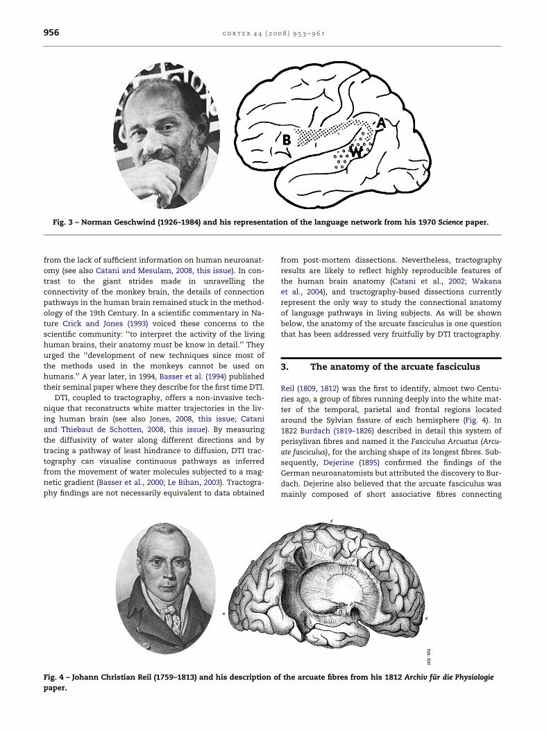
Fig. 3 – Norman Geschwind (1926–1984) and his representation of the language network from his 1970 Science paper.
c o r t e x 4 4 ( 2 0 0 8 ) 9 5 3 – 9 6 1956
from the lack of sufficient information on human neuroanat-
omy (see also Catani and Mesulam, 2008, this issue). In con-
trast to the giant strides made in unravelling the
connectivity of the monkey brain, the details of connection
pathways in the human brain remained stuck in the method-
ology of the 19th Century. In a scientific commentary in Na-
ture Crick and Jones (1993) voiced these concerns to the
scientific community: ‘‘to interpret the activity of the living
human brains, their anatomy must be know in detail.’’ They
urged the ‘‘development of new techniques since most of
the methods used in the monkeys cannot be used on
humans.’’ A year later, in 1994, Basser et al. (1994) published
their seminal paper where they describe for the first time DTI.
DTI, coupled to tractography, offers a non-invasive tech-
nique that reconstructs white matter trajectories in the liv-
ing human brain (see also Jones, 2008, this issue; Catani
and Thiebaut de Schotten, 2008, this issue). By measuring
the diffusivity of water along different directions and by
tracing a pathway of least hindrance to diffusion, DTI trac-
tography can visualise continuous pathways as inferred
from the movement of water molecules subjected to a mag-
netic gradient (Basser et al., 2000; Le Bihan, 2003). Tractogra-
phy findings are not necessarily equivalent to data obtained
Fig. 4 – Johann Christian Reil (1759–1813) and his description o
paper.
from post-mortem dissections. Nevertheless, tractography
results are likely to reflect highly reproducible features of
the human brain anatomy (Catani et al., 2002; Wakana
et al., 2004), and tractography-based dissections currently
represent the only way to study the connectional anatomy
of language pathways in living subjects. As will be shown
below, the anatomy of the arcuate fasciculus is one question
that has been addressed very fruitfully by DTI tractography.
3. The anatomy of the arcuate fasciculus
Reil (1809, 1812) was the first to identify, almost two Centu-
ries ago, a group of fibres running deeply into the white mat-
ter of the temporal, parietal and frontal regions located
around the Sylvian fissure of each hemisphere (Fig. 4). In
1822 Burdach (1819–1826) described in detail this system of
perisylivan fibres and named it the Fasciculus Arcuatus (Arcu-
ate fasciculus), for the arching shape of its longest fibres. Sub-
sequently, Dejerine (1895) confirmed the findings of the
German neuroanatomists but attributed the discovery to Bur-
dach. Dejerine also believed that the arcuate fasciculus was
mainly composed of short associative fibres connecting
f the arcuate fibres from his 1812 Archiv fur die Physiologie

Fig. 5 – Tractography reconstruction of the arcuate
fasciculus. Numbers indicate the cortical projections of the
segments: 1, superior temporal lobe; 2, middle temporal
lobe; 3, inferior frontal and precentral gyrus; 4, middle
frontal and precentral gyrus; 5, supramarginal gyrus; 6,
angular gyrus (mod. from Catani et al., 2005).
c o r t e x 4 4 ( 2 0 0 8 ) 9 5 3 – 9 6 1 957
neighbouring perisylvian cortex. As we have seen above,
Wernicke hypothesized that language relied on the integrity
of a ‘‘psychic reflex arc’’ between temporal and frontal re-
gions. But the arcuate fasciculus was not part of Wernicke’s
original anatomical model (Wernicke, 1874). He thought
that the temporal and frontal language areas were mutually
interconnected by fibres passing through the external cap-
sule and relaying in the cortex of the insula. It was Constan-
tin Von Monakow who first identified the arcuate fasciculus
as the tract connecting Broca’s and Wernicke’s areas,
a view later accepted by Wernicke in 1908 (Geschwind,
1967). Von Monakov’s statement soon became a dogma in
neurology and still today provides the backbone of anatomi-
cal models of language.
Group2 mild lateralization
Group1 extreme leftlateralization (~60%)
A
Females
Group1
(40%)
Group2 (30%)
Group3
(30%)
B
Fig. 6 – Distribution of the pattern of lateralization of the long seg
from Catani et al., 2007).
4. Recent contribution from DTI tractography
Although the existence of the arcuate fasciculus has been
confirmed in several post-mortem studies in humans, these
methods (e.g., blunt dissections, axonal staining of degenerat-
ing axons, etc.) have not shed much light on the detailed
anatomy of the relevant fibres. More powerful methods have
been used to trace homologous axonal pathways in the
monkey but the absence of language in non-human primates
raises doubts on the possibility of translating connectional
anatomy of putative language pathways from animal to man.
Tractography studies are showing that the anatomy of the
arcuate fasciculus is more complex than previously thought
(Fig. 5) (Catani et al., 2005). In addition to the long direct seg-
ment connecting Wernicke’s area with Broca’s area, there is
an indirect pathway consisting of two segments, an anterior
segment linking Broca’s territory with the inferior parietal
lobule and a posterior segment linking the inferior parietal
lobule with Wernicke’s territory. This arrangement not only
supports the more flexible architecture of parallel processing
(Mesulam, 1990), but also is in keeping with some of the
classical neurological models of aphasia, contemporary
models of verbal working memory (Baddeley, 2003) and recent
functional neuroimaging findings (Jung-Beeman, 2005; Sakai,
2005; Stephan et al., 2003). Additional support for the exis-
tence of the three perisylvian segments of the ‘‘arcuate fascic-
ulus’’ comes from human intraoperative electrocorticography
(Matsumoto et al., 2004), functional connectivity (Schmithorst
and Holland, 2007), post-mortem dissections (Lawes et al.,
2008), and experiments in homologous parts of the monkey
brain (Deacon, 1992).
Another unexpected finding derived from the tractography
dissections of the arcuate fasciculus is the extension of its
putative cortical terminations beyond the classical limits of
Broca’s and Wernicke’s areas to include part of the middle
and precentral frontal gyrus and the posterior middle tempo-
ral gyrus, respectively (Catani et al., 2005).
left(~20%)
Group3 bilateral,symmetrical (~20%)
Group1
(85%)
Group2 (10%)
Group3
(5%)
Males
C
ment in the normal population and between genders (mod.
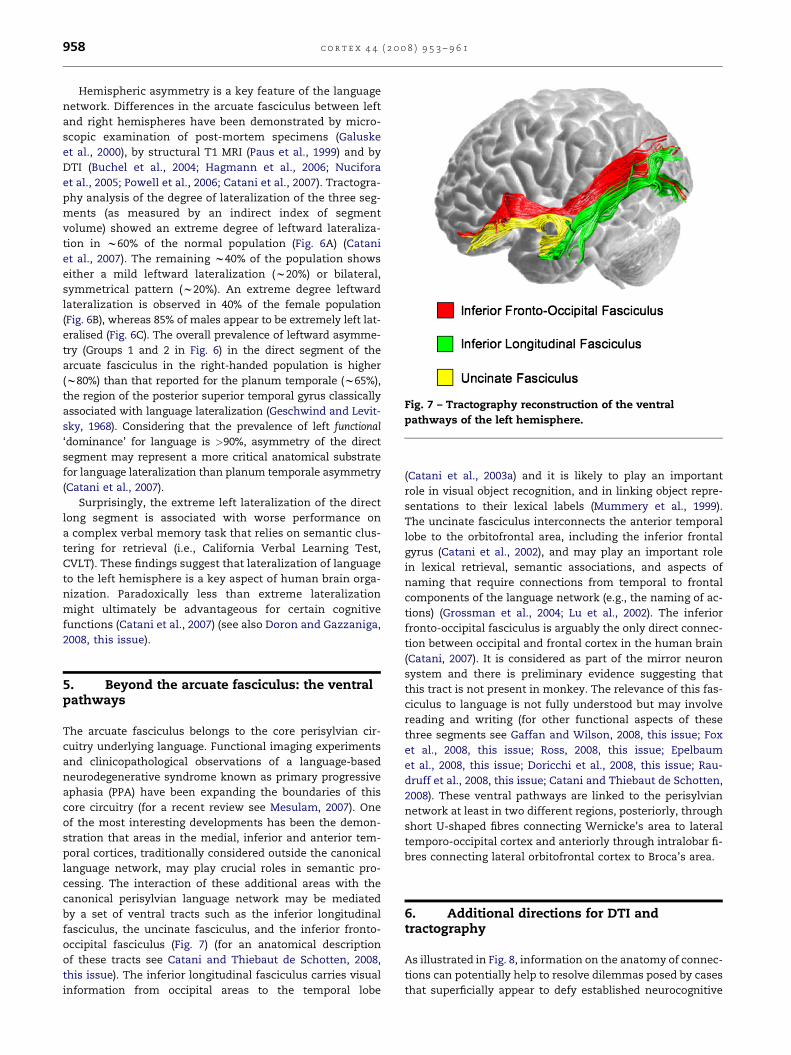
Fig. 7 – Tractography reconstruction of the ventral
pathways of the left hemisphere.
c o r t e x 4 4 ( 2 0 0 8 ) 9 5 3 – 9 6 1958
Hemispheric asymmetry is a key feature of the language
network. Differences in the arcuate fasciculus between left
and right hemispheres have been demonstrated by micro-
scopic examination of post-mortem specimens (Galuske
et al., 2000), by structural T1 MRI (Paus et al., 1999) and by
DTI (Buchel et al., 2004; Hagmann et al., 2006; Nucifora
et al., 2005; Powell et al., 2006; Catani et al., 2007). Tractogra-
phy analysis of the degree of lateralization of the three seg-
ments (as measured by an indirect index of segment
volume) showed an extreme degree of leftward lateraliza-
tion in w60% of the normal population (Fig. 6A) (Catani
et al., 2007). The remaining w40% of the population shows
either a mild leftward lateralization (w20%) or bilateral,
symmetrical pattern (w20%). An extreme degree leftward
lateralization is observed in 40% of the female population
(Fig. 6B), whereas 85% of males appear to be extremely left lat-
eralised (Fig. 6C). The overall prevalence of leftward asymme-
try (Groups 1 and 2 in Fig. 6) in the direct segment of the
arcuate fasciculus in the right-handed population is higher
(w80%) than that reported for the planum temporale (w65%),
the region of the posterior superior temporal gyrus classically
associated with language lateralization (Geschwind and Levit-
sky, 1968). Considering that the prevalence of left functional
‘dominance’ for language is >90%, asymmetry of the direct
segment may represent a more critical anatomical substrate
for language lateralization than planum temporale asymmetry
(Catani et al., 2007).
Surprisingly, the extreme left lateralization of the direct
long segment is associated with worse performance on
a complex verbal memory task that relies on semantic clus-
tering for retrieval (i.e., California Verbal Learning Test,
CVLT). These findings suggest that lateralization of language
to the left hemisphere is a key aspect of human brain orga-
nization. Paradoxically less than extreme lateralization
might ultimately be advantageous for certain cognitive
functions (Catani et al., 2007) (see also Doron and Gazzaniga,
2008, this issue).
5. Beyond the arcuate fasciculus: the ventralpathways
The arcuate fasciculus belongs to the core perisylvian cir-
cuitry underlying language. Functional imaging experiments
and clinicopathological observations of a language-based
neurodegenerative syndrome known as primary progressive
aphasia (PPA) have been expanding the boundaries of this
core circuitry (for a recent review see Mesulam, 2007). One
of the most interesting developments has been the demon-
stration that areas in the medial, inferior and anterior tem-
poral cortices, traditionally considered outside the canonical
language network, may play crucial roles in semantic pro-
cessing. The interaction of these additional areas with the
canonical perisylvian language network may be mediated
by a set of ventral tracts such as the inferior longitudinal
fasciculus, the uncinate fasciculus, and the inferior fronto-
occipital fasciculus (Fig. 7) (for an anatomical description
of these tracts see Catani and Thiebaut de Schotten, 2008,
this issue). The inferior longitudinal fasciculus carries visual
information from occipital areas to the temporal lobe
(Catani et al., 2003a) and it is likely to play an important
role in visual object recognition, and in linking object repre-
sentations to their lexical labels (Mummery et al., 1999).
The uncinate fasciculus interconnects the anterior temporal
lobe to the orbitofrontal area, including the inferior frontal
gyrus (Catani et al., 2002), and may play an important role
in lexical retrieval, semantic associations, and aspects of
naming that require connections from temporal to frontal
components of the language network (e.g., the naming of ac-
tions) (Grossman et al., 2004; Lu et al., 2002). The inferior
fronto-occipital fasciculus is arguably the only direct connec-
tion between occipital and frontal cortex in the human brain
(Catani, 2007). It is considered as part of the mirror neuron
system and there is preliminary evidence suggesting that
this tract is not present in monkey. The relevance of this fas-
ciculus to language is not fully understood but may involve
reading and writing (for other functional aspects of these
three segments see Gaffan and Wilson, 2008, this issue; Fox
et al., 2008, this issue; Ross, 2008, this issue; Epelbaum
et al., 2008, this issue; Doricchi et al., 2008, this issue; Rau-
druff et al., 2008, this issue; Catani and Thiebaut de Schotten,
2008). These ventral pathways are linked to the perisylvian
network at least in two different regions, posteriorly, through
short U-shaped fibres connecting Wernicke’s area to lateral
temporo-occipital cortex and anteriorly through intralobar fi-
bres connecting lateral orbitofrontal cortex to Broca’s area.
6. Additional directions for DTI andtractography
As illustrated in Fig. 8, information on the anatomy of connec-
tions can potentially help to resolve dilemmas posed by cases
that superficially appear to defy established neurocognitive

Fig. 8 – Topological and hodological approaches in clinico-anatomical correlation studies. In the upper row an example of
a lesion overlap study for clinico-anatomical correlation is represented where four patients present with similar neurological
deficits and their respective brain images are overlapped in order to identify a common anatomical substrate. Here we want to
highlight that the conclusion that one may draw from this type of studies depends on the hypothesis that is tested and the
general framework adopted. (A) A strict topological approach considers brain functions as localized in specific cortical regions.
Within this framework the critical area for the same neurological deficit manifested by a group of stroke patients (four in the
example, where each area, from 1 to 4, represents the extension of the lesion for each patient) is located at the cortical region
of maximum lesion overlap (region b in the example). (B) The hodological (network) approach to brain–behaviour correlation
includes a consideration of brain pathways that pass through the damaged area. Within this framework, the neurological
deficit could also be attributed to a disconnection between a and c because all lesions affect the same a to c pathway at
different levels (red circle). Note that A and B represent the same experiment (i.e., same patients and image analysis), however
the conclusions are opposite due to the different approach. (C) Image of the brain of Broca’s aphasic patient showing a lesion
to the inferior frontal cortex. Broca, who worked within a topological framework, considered that his patient’s speech deficit
was the consequence of the cortical lesion in the inferior frontal lobe. (D) Sagittal MRI image (mod. from Dronkers et al., 2007)
of the same brain shown in (C). Clearly the lesion extends into the white matter of the arcuate (red arrows) of the left
hemisphere. If Broca had worked within a hodological framework and performed dissections of his patient’s brain it is
probable that he would have attributed the speech deficit to a lesion of the arcuate fasciculus.
c o r t e x 4 4 ( 2 0 0 8 ) 9 5 3 – 9 6 1 959
models. For example, the site of maximal lesion overlap for
a specific syndrome may extend into axonal pathways that
interconnect a different set of remote areas, raising the possi-
bility that the critical factor is not necessarily the destruction
in the cortical area of overlap but a disconnection of the two
remote areas (Fig. 8).
DTI tractography also has the potential of detecting path-
way changes at early stages of neurodegenerative processes
affecting language function so that the effects of such changes
upon the resultant aphasias can be studied (Catani, 2006). In
primary progressive aphasia, for example, the loss of cortical
neurons is accompanied by axonal degeneration along
specific white matter pathways (Fig. 9) (Catani et al., 2003b;
Borroni et al., 2007). Up to now, morphometric work on PPA
had focused on the relationship of cortical degeneration to
details of the language impairment. An equally interesting de-
velopment would be to use DTI to measure microstructural
changes in specific tracts and to correlate them with the
symptom profile (Catani, 2006).
Individual differences in the asymmetry of the arcuate
fasciculus detected by DTI could conceivably also help to
assess recovery potential in aphasias. It is not unreasonable
to assume that greater symmetry is likely to lead to better
recovery following stroke or neurosurgery. This is an assump-
tion that can be tested experimentally with currently available
methodology.
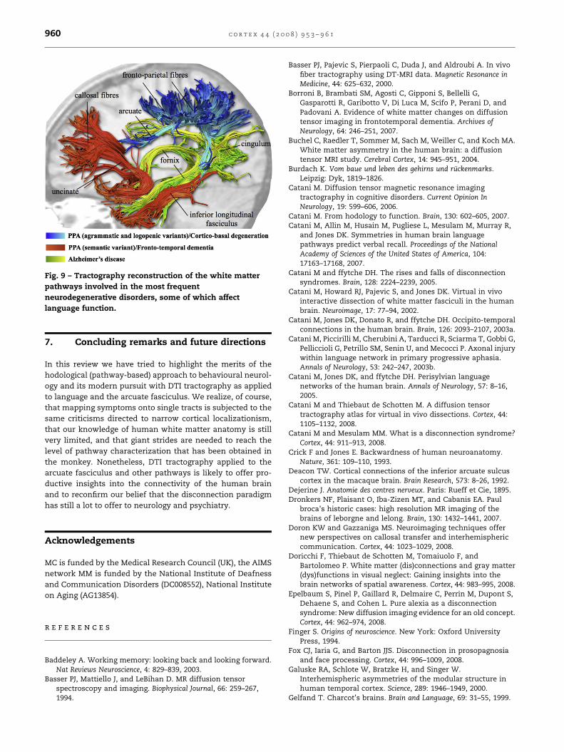
Fig. 9 – Tractography reconstruction of the white matter
pathways involved in the most frequent
neurodegenerative disorders, some of which affect
language function.
c o r t e x 4 4 ( 2 0 0 8 ) 9 5 3 – 9 6 1960
7. Concluding remarks and future directions
In this review we have tried to highlight the merits of the
hodological (pathway-based) approach to behavioural neurol-
ogy and its modern pursuit with DTI tractography as applied
to language and the arcuate fasciculus. We realize, of course,
that mapping symptoms onto single tracts is subjected to the
same criticisms directed to narrow cortical localizationism,
that our knowledge of human white matter anatomy is still
very limited, and that giant strides are needed to reach the
level of pathway characterization that has been obtained in
the monkey. Nonetheless, DTI tractography applied to the
arcuate fasciculus and other pathways is likely to offer pro-
ductive insights into the connectivity of the human brain
and to reconfirm our belief that the disconnection paradigm
has still a lot to offer to neurology and psychiatry.
Acknowledgements
MC is funded by the Medical Research Council (UK), the AIMS
network MM is funded by the National Institute of Deafness
and Communication Disorders (DC008552), National Institute
on Aging (AG13854).
r e f e r e n c e s
Baddeley A. Working memory: looking back and looking forward.Nat Reviews Neuroscience, 4: 829–839, 2003.
Basser PJ, Mattiello J, and LeBihan D. MR diffusion tensorspectroscopy and imaging. Biophysical Journal, 66: 259–267,1994.
Basser PJ, Pajevic S, Pierpaoli C, Duda J, and Aldroubi A. In vivofiber tractography using DT-MRI data. Magnetic Resonance inMedicine, 44: 625–632, 2000.
Borroni B, Brambati SM, Agosti C, Gipponi S, Bellelli G,Gasparotti R, Garibotto V, Di Luca M, Scifo P, Perani D, andPadovani A. Evidence of white matter changes on diffusiontensor imaging in frontotemporal dementia. Archives ofNeurology, 64: 246–251, 2007.
Buchel C, Raedler T, Sommer M, Sach M, Weiller C, and Koch MA.White matter asymmetry in the human brain: a diffusiontensor MRI study. Cerebral Cortex, 14: 945–951, 2004.
Burdach K. Vom baue und leben des gehirns und ruckenmarks.Leipzig: Dyk, 1819–1826.
Catani M. Diffusion tensor magnetic resonance imagingtractography in cognitive disorders. Current Opinion InNeurology, 19: 599–606, 2006.
Catani M. From hodology to function. Brain, 130: 602–605, 2007.Catani M, Allin M, Husain M, Pugliese L, Mesulam M, Murray R,
and Jones DK. Symmetries in human brain languagepathways predict verbal recall. Proceedings of the NationalAcademy of Sciences of the United States of America, 104:17163–17168, 2007.
Catani M and ffytche DH. The rises and falls of disconnectionsyndromes. Brain, 128: 2224–2239, 2005.
Catani M, Howard RJ, Pajevic S, and Jones DK. Virtual in vivointeractive dissection of white matter fasciculi in the humanbrain. Neuroimage, 17: 77–94, 2002.
Catani M, Jones DK, Donato R, and ffytche DH. Occipito-temporalconnections in the human brain. Brain, 126: 2093–2107, 2003a.
Catani M, Piccirilli M, Cherubini A, Tarducci R, Sciarma T, Gobbi G,Pelliccioli G, Petrillo SM, Senin U, and Mecocci P. Axonal injurywithin language network in primary progressive aphasia.Annals of Neurology, 53: 242–247, 2003b.
Catani M, Jones DK, and ffytche DH. Perisylvian languagenetworks of the human brain. Annals of Neurology, 57: 8–16,2005.
Catani M and Thiebaut de Schotten M. A diffusion tensortractography atlas for virtual in vivo dissections. Cortex, 44:1105–1132, 2008.
Catani M and Mesulam MM. What is a disconnection syndrome?Cortex, 44: 911–913, 2008.
Crick F and Jones E. Backwardness of human neuroanatomy.Nature, 361: 109–110, 1993.
Deacon TW. Cortical connections of the inferior arcuate sulcuscortex in the macaque brain. Brain Research, 573: 8–26, 1992.
Dejerine J. Anatomie des centres nerveux. Paris: Rueff et Cie, 1895.Dronkers NF, Plaisant O, Iba-Zizen MT, and Cabanis EA. Paul
broca’s historic cases: high resolution MR imaging of thebrains of leborgne and lelong. Brain, 130: 1432–1441, 2007.
Doron KW and Gazzaniga MS. Neuroimaging techniques offernew perspectives on callosal transfer and interhemisphericcommunication. Cortex, 44: 1023–1029, 2008.
Doricchi F, Thiebaut de Schotten M, Tomaiuolo F, andBartolomeo P. White matter (dis)connections and gray matter(dys)functions in visual neglect: Gaining insights into thebrain networks of spatial awareness. Cortex, 44: 983–995, 2008.
Epelbaum S, Pinel P, Gaillard R, Delmaire C, Perrin M, Dupont S,Dehaene S, and Cohen L. Pure alexia as a disconnectionsyndrome: New diffusion imaging evidence for an old concept.Cortex, 44: 962–974, 2008.
Finger S. Origins of neuroscience. New York: Oxford UniversityPress, 1994.
Fox CJ, Iaria G, and Barton JJS. Disconnection in prosopagnosiaand face processing. Cortex, 44: 996–1009, 2008.
Galuske RA, Schlote W, Bratzke H, and Singer W.Interhemispheric asymmetries of the modular structure inhuman temporal cortex. Science, 289: 1946–1949, 2000.
Gelfand T. Charcot’s brains. Brain and Language, 69: 31–55, 1999.

c o r t e x 4 4 ( 2 0 0 8 ) 9 5 3 – 9 6 1 961
Geschwind N. Disconnexion syndromes in animals and man. I.Brain, 88: 237–294, 1965.
Geschwind N. Wernicke’s contribution to the study of aphasia.Cortex, 3: 449–463, 1967.
Geschwind N. The organization of language and the brain. Science,170: 940–944, 1970.
Geschwind N and Levitsky W. Human brain: left–rightasymmetries in temporal speech region. Science, 161: 186–187,1968.
Grossman M, McMillan C, Moore P, Ding L, Glosser G, Work M, andGee J. What’s in a name: voxel-based morphometric analysesof mri and naming difficulty in Alzheimer’s disease,frontotemporal dementia and corticobasal degeneration.Brain, 127: 628–649, 2004.
Glickstein M and Berlucchi G. Classical disconnection studies ofthe corpus callosum. Cortex, 44: 914–927, 2008.
Gaffan D and Wilson CRE. Medial temporal and prefrontalfunction: Recent behavioural disconnection studies in themacaque monkey. Cortex, 44: 928–935, 2008.
Hagmann P, Cammoun L, Martuzzi R, Maeder P, Clarke S,Thiran JP, and Meuli R. Hand preference and sex shape thearchitecture of language networks. Human Brain Mapping, 27:828–835, 2006.
Jung-Beeman M. Bilateral brain processes for comprehendingnatural language. Trends in Cognitive Sciences, 9: 512–518, 2005.
Jones DK. Studying connections in the living human brain withdiffusion MRI. Cortex, 44: 936–952, 2008.
Kempler D, Metter EJ, Jackson CA, Hanson WR, Riege WH,Mazziotta JC, and Phelps ME. Disconnection and cerebralmetabolism. The case of conduction aphasia. Archives ofNeurology, 45: 275–279, 1988.
Lawes IN, Barrick TR, Murugam V, Spierings N, Evans DR, Song M,and Clark CA. Atlas-based segmentation of white mattertracts of the human brain using diffusion tensor tractographyand comparison with classical dissection. Neuroimage, 39:62–79, 2008.
Le Bihan D. Looking into the functional architecture of the brainwith diffusion mri. Nature Reviews Neuroscience, 4: 469–480, 2003.
Levine D and Calvanio R. Conduction aphasia. Lisse: Swets andZeitlinger, 1982.
Lichtheim L. On aphasia. Brain, 7: 433–484, 1885.Lu LH, Crosson B, Nadeau SE, Heilman KM, Gonzalez-Rothi LJ,
Raymer A, Gilmore RL, Bauer RM, and Roper SN. Category-specific naming deficits for objects and actions: semanticattribute and grammatical role hypotheses. Neuropsychologia,40: 1608–1621, 2002.
Matsumoto R, Nair DR, LaPresto E, Najm I, Bingaman W,Shibasaki H, and Luders HO. Functional connectivity in thehuman language system: a cortico-cortical evoked potentialstudy. Brain, 127: 2316–2330, 2004.
Mesulam MM. Imaging connectivity in the human cerebralcortex: the next frontier? Annals of Neurology, 57: 5–7, 2005.
Mesulam MM. Large-scale neurocognitive networks anddistributed processing for attention, language, and memory.Annals of Neurology, 28: 597–613, 1990.
Mesulam MM. Primary progressive aphasia: a 25-yearretrospective. Alzheimer Disease and Associated Disorders, 21:S8–S11, 2007.
Mummery CJ, Patterson K, Wise RJ, Vandenberghe R, Price CJ, andHodges JR. Disrupted temporal lobe connections in semanticdementia. Brain, 122: 61–73, 1999.
Nucifora PG, Verma R, Melhem ER, Gur RE, and Gur RC. Leftwardasymmetry in relative fiber density of the arcuate fasciculus.Neuroreport, 16: 791–794, 2005.
Paus T, Zijdenbos A, Worsley K, Collins DL, Blumenthal J,Giedd JN, Rapoport JL, and Evans AC. Structural maturation ofneural pathways in children and adolescents: in vivo study.Science, 283: 1908–1911, 1999.
Powell HW, Parker GJ, Alexander DC, Symms MR, Boulby PA,Wheeler-Kingshott CA, Barker GJ, Noppeney U, Koepp MJ, andDuncan JS. Hemispheric asymmetries in language-relatedpathways: a combined functional mri and tractography study.Neuroimage, 32: 388–399, 2006.
Reil JC. Die Sylvische Grube oder das Thal, das gestreifte grobehirnganglium, dessen kapsel und die seitentheile des grobngehirns. Archiv fur die Physiologie, 9: 195–208, 1809.
Reil JC. Die vordere commissur im groben gehirn. Archiv fur diePhysiologie, 11: 89–100, 1812.
Ross ED. Sensory-specific amnesia and hypoemotionality inhumans and monkeys: Gateway for developing a hodology ofmemory. Cortex, 44: 1010–1022, 2008.
Rudrauff D, Mehta S, and Grabowski T. Disconnection’srenaissance takes shape: Formal incorporation in group-levellesion studies. Cortex, 44: 1084–1096, 2008.
Sakai KL. Language acquisition and brain development. Science,310: 815–819, 2005.
Schmithorst VJ and Holland SK. Sex differences in thedevelopment of neuroanatomical functional connectivityunderlying intelligence found using bayesian connectivityanalysis. Neuroimage, 35: 406–419, 2007.
Stephan KE, Marshall JC, Friston KJ, Rowe JB, Ritzl A, Zilles K,and Fink GR. Lateralized cognitive processes andlateralized task control in the human brain. Science, 301:384–386, 2003.
Wakana S, Jiang H, Nagae-Poetscher LM, van Zijl PC, and Mori S.Fiber tract-based atlas of human white matter anatomy.Radiology, 230: 77–87, 2004.
Wernicke C. Der aphasische symptomencomplex. Einpsychologische studie auf anatomischer basis. Breslau: Cohn &Weigert, 1874.