the assessment of femoral shaft morphology in the sagittal
TRANSCRIPT

RESEARCH ARTICLE Open Access
The assessment of femoral shaftmorphology in the sagittal plane inChinese patients with osteoarthritis—aradiographic analysisZhengyuan Bao1,2†, Liang Qiao1,2†, Jianghui Qin1,2, Jiacheng Xu3, Sheng Zhou1,2, Dongyang Chen1,2,Dongquan Shi1,2, Jin Dai1,2, Yao Yao1,2, Qing Jiang1,2* and Zhihong Xu1,2*
Abstract
Background: The purpose of this study was to analyze femoral shaft sagittal parameters in Chinese osteoarthritis(OA) patients undergoing total knee arthroplasty (TKA) and identify whether the parameters in the coronal planecould be predictors of those in the sagittal plane.
Methods: Standard long-standing anteroposterior and femoral lateral radiographs of 50 lower limbs in 50 ChineseOA patients were included. Sagittal femoral bowing angle (sFBA), angle between femoral distal anterior cortex axisand sagittal mechanical axis (DACSMA), angle between femoral distal anterior cortex axis and sagittal distalanatomic axis (DACSDAA), and angle between femoral sagittal mechanical axis and sagittal distal anatomic axis(SMADAA) were measured. Then the relationship between femoral shaft parameters in the sagittal and coronalplanes were identified, including coronal femoral bowing angle (cFBA), valgus angle, hip-knee-ankle angle (HKA),length of femur (LF), femoral offset, femoral neck stem angle (FNS), and mechanical lateral distal femoral angle(mLDFA). A two-sided Pearson correlation coefficient was obtained to identify the correlations between parametersin the coronal and sagittal planes. P values <0.05 were considered statistically significant.
Results: The mean sFBA was 15.08° ± 3.79°, the mean DACSMA was 1.35° ± 2.70°, the mean DACSDAA was −2.66° ± 2.05°, and the mean SMADAA was 4.01° ± 2.55°. No correlation between parameters in the coronal andsagittal planes was found.
Conclusions: In this study, the discreteness of DACSMA, DACSDAA, and SMADAA in Chinese OA patients is highand this may affect the position of femoral prosthesis after TKA using the conventional intramedullary device. Noparameters in the coronal plane are found correlated with those in the sagittal plane.
Trial registration: Researchregistry2337
Keywords: Femoral shaft bowing, Sagittal plane, Radiographic analysis, Knee osteoarthritis, Total knee arthroplasty
* Correspondence: [email protected]; [email protected]†Equal contributors1Department of Sports Medicine and Adult Reconstructive Surgery, DrumTower Hospital, School of Medicine, Nanjing University, 321 ZhongshanRoad, Nanjing, Jiangsu 210008, ChinaFull list of author information is available at the end of the article
© The Author(s). 2017 Open Access This article is distributed under the terms of the Creative Commons Attribution 4.0International License (http://creativecommons.org/licenses/by/4.0/), which permits unrestricted use, distribution, andreproduction in any medium, provided you give appropriate credit to the original author(s) and the source, provide a link tothe Creative Commons license, and indicate if changes were made. The Creative Commons Public Domain Dedication waiver(http://creativecommons.org/publicdomain/zero/1.0/) applies to the data made available in this article, unless otherwise stated.
Bao et al. Journal of Orthopaedic Surgery and Research (2017) 12:127 DOI 10.1186/s13018-017-0626-8

BackgroundAs a major source of lower limb pain and disability, kneeosteoarthritis (OA) generates great impacts on patients’quality of life and brings a heavy burden for public healthsystem [1]. For severe knee OA, total knee arthroplasty(TKA) is the preferred treatment. Postoperative implantalignment is an important factor related to the outcomesof TKA [2]. However, the present conventional intrame-dullary device shows lots of drawbacks in practice. It maycause the coronal malalignment due to the differences offemoral shaft shape. What is more, it ignores the import-ance of good alignment in the sagittal plane. So the asses-sing of femur shaft morphology in both coronal andsagittal planes is important preoperatively.The effect of femoral shaft bowing (FSB) on the position
of implant in TKA has drawn more and more attentionespecially from Asian OA patients. However, the defin-ition for FSB has not been well established. Akamatsu de-fined coronal femoral bowing angle (cFBA) >5° in thecoronal plane as coronal femoral shaft bowing (cFSB) andsagittal femoral bowing angle (sFBA) of >11° in the sagittalplane was defined as sagittal femoral shaft bowing (sFSB)[3]. Previous study also found FSB had racial specificityand Asians were more susceptible [4]. It is related tohigher prevalence and faster progression of knee OA [5].Furthermore, severe FSB may affect the implant positionduring TKA surgery. The conventional intramedullaryfemoral cut system sets femoral coronal mechanical axis(cMA) by referring the intramedullary rod and the valgusangle between cMA and coronal anatomical axis of thefemur. The best outcome of coronal alignment is limitedwithin 3° of cMA [5]. cFBA has been reported to be asso-ciated with valgus angle positively, and if cFBA increases,valgus angle will be larger [6]. So cFBA is also related topostoperative limb and implant alignment [7].Unlike the recognized results in the coronal plane,
there is no unified peri-operative alignment assessmentsystem of femur in the sagittal plane. It has been shownsFBA is associated with the degree of femoral compo-nent flexion [8]. An overly flexional position will limitknee extension and result in posterior insert wear causedby impingement between the polyethylene insert and theintercondylar box in TKA using post-cam mechanism[9]. And an over-extensional position may contribute toa postoperative supracondylar femoral fracture [10]. SocFSB and sFSB are both of important clinical meaning.Considering the negligent assessment of the femoral
morphology in the sagittal plane before TKA inChina, the purpose of this study was as follows: first,to analyze different parameters of femoral shaft in thesagittal plane of Chinese people with knee OA under-going TKA; second, to identify which parameters inthe coronal plane could be predictors of those in thesagittal plane using radiographs.
MethodsPatientsChinese patients with knee OA who underwent TKAfrom May, 2015, to July, 2016, in our surgical team (Xu)were reviewed. The preoperative standard long-standinganteroposterior and femoral lateral radiographs [11] wereexamined in all the patients. When taking long-standinganteroposterior radiographs, two lower limbs were rotatedneutrally with the patellae facing forwards and the beamtube centered at the knee [6]. The key point of taking fem-oral lateral radiographs was to make sure the beam tubewas tilted 15° to aim the midpoint of the patients’ thighdirectly [11]. We excluded limbs which had a prior frac-ture and prior knee or hip arthroplasty, also those withnonstandard films. Totally, 50 patients were enrolled.
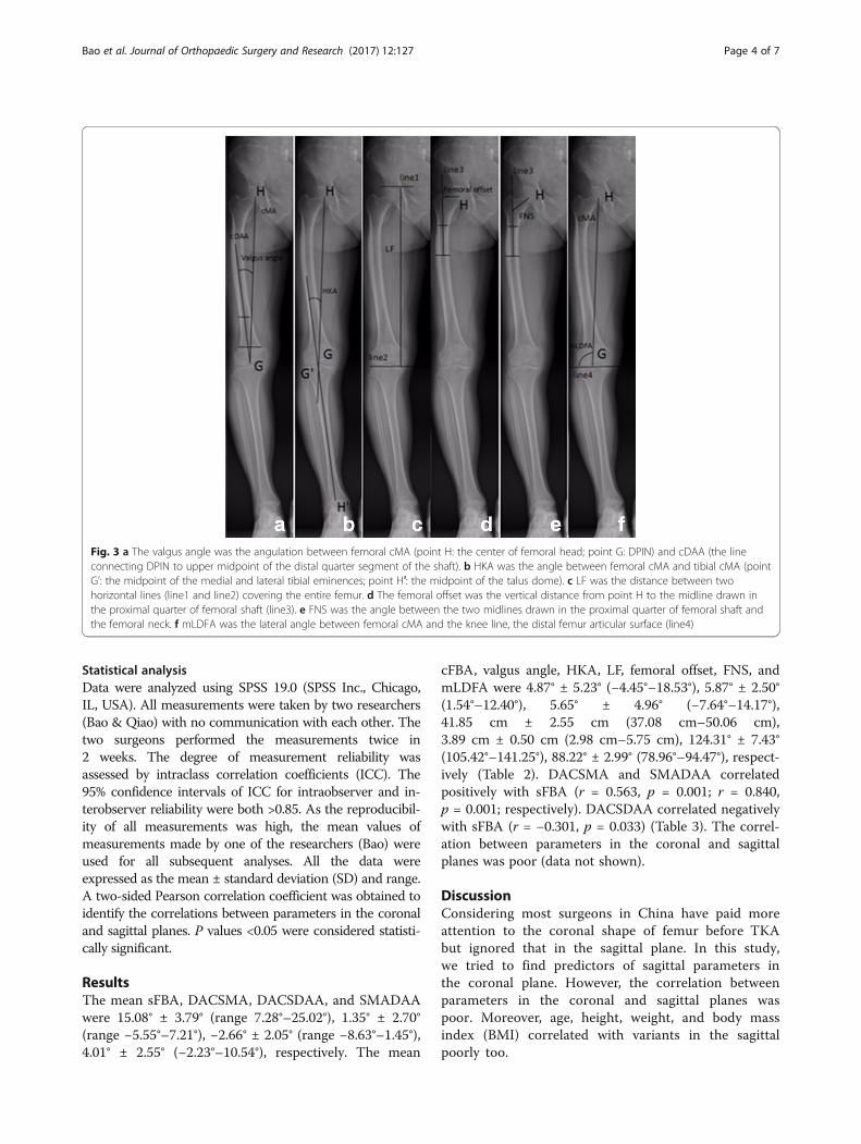
Radiographic assessmentAll radiographic measurements were obtained from truelong-standing anteroposterior and femoral lateral radio-graphs using Picture Archiving Communication System(PACS, FIRSTECH, Hefei, Anhui, China). We only exam-ined the operated limb. In femoral lateral radiographs, thefemoral shaft was divided into four equal parts in the sagit-tal plane [3]. The proximal end of the diaphysis was thelower border of the lesser trochanter and the distal end wasthe junction between the shaft and the condylar region.The angle between the midlines drawn in the proximal anddistal quarter segments was defined as sFBA. Positivevalues meant femoral anterior bowing and negative valuesmeant posterior bowing (Fig. 1a). There was no agreed def-inition of sagittal mechanical axis (sMA) [11–13]. Here wedefined sMA as the line connecting the center of femoralhead and the deepest point of the intercondylar notch(DPIN). In the femoral lateral radiograph, DPIN was theend of Blumensaat’s line [11]. DACSMA was defined as theabbreviation of the angle between femoral distal anteriorcortex axis (DACA) [11] and sMA (Fig. 2a). A positivevalue meant DACA was in flexion to sMA and a negativevalue meant DACA was in extension. DACSDAA was de-fined as the angle between DACA and sagittal distal ana-tomic axis (sDAA) (Fig. 2b); sDAA was the midline drawnin the distal quarter of femoral shaft. If DACA was inflexion to sDAA, this value was positive, otherwise thisvalue was negative. SMADAA was defined as the angle be-tween sDAA and sMA (Fig. 2c). Positive values meantsDAA was in flexion to sMA, otherwise sDAA was in ex-tension to sMA. In long-standing anteroposterior radio-graphs, similar to the partition method in the sagittal plane,the angle between the midlines drawn in the proximal anddistal quarter segments of the femoral shaft was defined ascFBA. Positive values meant femoral lateral bowing andnegative values meant medial bowing (Fig. 1b). The valgusangle was defined as the angulation between femoral cMAand coronal distal anatomic axis (cDAA) (Fig. 3a). Femoral
Bao et al. Journal of Orthopaedic Surgery and Research (2017) 12:127 Page 2 of 7

cMA was a line connecting the center of femoral head toDPIN. Femoral cDAA was the midline drawn in the distalquarter of femoral shaft in the coronal plane. In the pre-operative measurement of the valgus angle, femoral cMAand cDAA shared the same end point, the entry point ofintramedullary rod. Different surgeons prefer different entrypoints and here we chose DPIN as the entry point [11].Then we took the line connecting DPIN to upper midpointof the distal quarter segment of the shaft as cDAA [6]. Hip-knee-ankle angle (HKA) was the angle between femoralcMA and tibial cMA (Fig. 3b). Tibial cMA was a line con-necting the midpoint of the medial and lateral tibial emi-nences and the midpoint of the talus dome. If the knee was
varus, this value was positive, otherwise HKA was negative.The length of femur (LF) was the distance between twohorizontal lines covering the entire femur (Fig. 3c). Thefemoral offset was the vertical distance from the center offemoral head to the midline drawn in the proximal quarterof femoral shaft [14] (Fig. 3d). The femoral neck stemangle (FNS) was the angle between the two midlinesdrawn in the proximal quarter of femoral shaft andthe femoral neck [14](Fig. 3e). mLDFA was the lateralangle between femoral cMA and the knee line, thedistal femur articular surface [13](Fig. 3f ). All param-eters and corresponding definitions are summarizedin Table 1.
Fig. 1 a, b The femoral shaft was divided into four equal parts in both coronal and sagittal planes. The proximal end was the lower border of thelesser trochanter and the distal end was the junction between the shaft and the condylar region. sFBA and cFBA were angles between midlinesdrawn in the proximal and distal quarter segments of the femoral shaft. a Points a, b, c, d were the midpoints of medullary cavity in the sagittalplane. b Points A, B, C, D were the midpoints of medullary cavity in the coronal plane
Fig. 2 a The DACA was the line connecting two points on the anterior cortex at 5 cm (point e) and 10 cm (point f) proximal to L (L was thetangent line of distal femur parallel to the knee line, the knee line was the junction between the shaft and the condylar region). sMA was the lineconnecting DPIN (point g) and the center of femoral head (point h). DACSMA was the angle between sMA and DACA, and L1 was parallel toDACA. b sDAA was the midline drawn in the distal quarter of femoral shaft. DACSDAA was the angle between DACA and sDAA, and L2 wasparallel to DACA. c SMADAA was the angle between sMA and sDAA, and L3 was parallel to sDAA
Bao et al. Journal of Orthopaedic Surgery and Research (2017) 12:127 Page 3 of 7
Statistical analysisData were analyzed using SPSS 19.0 (SPSS Inc., Chicago,IL, USA). All measurements were taken by two researchers(Bao & Qiao) with no communication with each other. Thetwo surgeons performed the measurements twice in2 weeks. The degree of measurement reliability wasassessed by intraclass correlation coefficients (ICC). The95% confidence intervals of ICC for intraobserver and in-terobserver reliability were both >0.85. As the reproducibil-ity of all measurements was high, the mean values ofmeasurements made by one of the researchers (Bao) wereused for all subsequent analyses. All the data wereexpressed as the mean ± standard deviation (SD) and range.A two-sided Pearson correlation coefficient was obtained toidentify the correlations between parameters in the coronaland sagittal planes. P values <0.05 were considered statisti-cally significant.
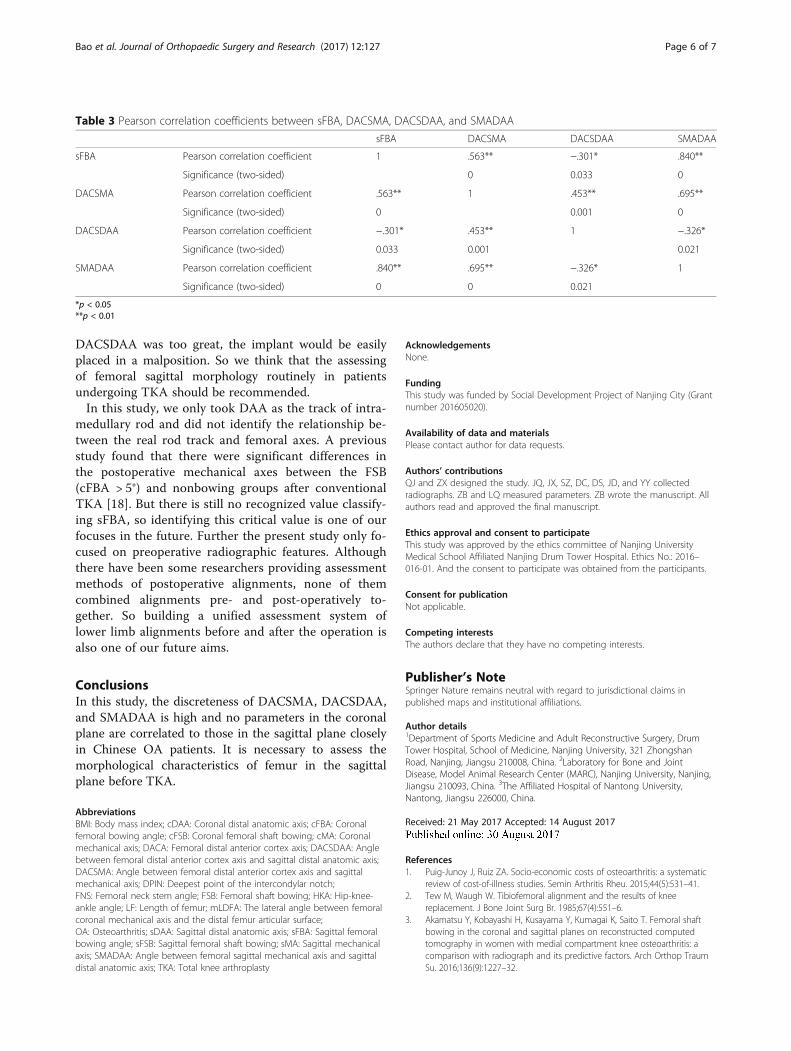
ResultsThe mean sFBA, DACSMA, DACSDAA, and SMADAAwere 15.08° ± 3.79° (range 7.28°–25.02°), 1.35° ± 2.70°(range −5.55°–7.21°), −2.66° ± 2.05° (range −8.63°–1.45°),4.01° ± 2.55° (−2.23°–10.54°), respectively. The mean
cFBA, valgus angle, HKA, LF, femoral offset, FNS, andmLDFA were 4.87° ± 5.23° (−4.45°–18.53°), 5.87° ± 2.50°(1.54°–12.40°), 5.65° ± 4.96° (−7.64°–14.17°),41.85 cm ± 2.55 cm (37.08 cm–50.06 cm),3.89 cm ± 0.50 cm (2.98 cm–5.75 cm), 124.31° ± 7.43°(105.42°–141.25°), 88.22° ± 2.99° (78.96°–94.47°), respect-ively (Table 2). DACSMA and SMADAA correlatedpositively with sFBA (r = 0.563, p = 0.001; r = 0.840,p = 0.001; respectively). DACSDAA correlated negativelywith sFBA (r = −0.301, p = 0.033) (Table 3). The correl-ation between parameters in the coronal and sagittalplanes was poor (data not shown).
DiscussionConsidering most surgeons in China have paid moreattention to the coronal shape of femur before TKAbut ignored that in the sagittal plane. In this study,we tried to find predictors of sagittal parameters inthe coronal plane. However, the correlation betweenparameters in the coronal and sagittal planes waspoor. Moreover, age, height, weight, and body massindex (BMI) correlated with variants in the sagittalpoorly too.
Fig. 3 a The valgus angle was the angulation between femoral cMA (point H: the center of femoral head; point G: DPIN) and cDAA (the lineconnecting DPIN to upper midpoint of the distal quarter segment of the shaft). b HKA was the angle between femoral cMA and tibial cMA (pointG’: the midpoint of the medial and lateral tibial eminences; point H′: the midpoint of the talus dome). c LF was the distance between twohorizontal lines (line1 and line2) covering the entire femur. d The femoral offset was the vertical distance from point H to the midline drawn inthe proximal quarter of femoral shaft (line3). e FNS was the angle between the two midlines drawn in the proximal quarter of femoral shaft andthe femoral neck. f mLDFA was the lateral angle between femoral cMA and the knee line, the distal femur articular surface (line4)
Bao et al. Journal of Orthopaedic Surgery and Research (2017) 12:127 Page 4 of 7

TKA is the mainstream treatment of severe OA andconventional intramedullary device is the most commonfemoral distal cut method during the operation. How-ever, this device identifies femoral cMA indirectly duringthe operation and is restricted to the femoral shape. If
the valgus angle or cFBA is too great, this method can-not ensure cMA and as a result, postoperative align-ments will be in error and several clinical outcomescales will be inferior [7, 15, 16]. Moreover conventionalintramedullary device cannot identify the femoral align-ment in the sagittal plane. Recently, more and more sur-geons have realized the significance of the femoral shapein the sagittal plane. Ko et al. thought sFBA was a riskfactor for femoral implant flexion in conventional intra-medullary TKA and notching in navigated TKA [8].Nakahara et al. promoted an idea that sagittal femoralcutting error could change femoral anteroposterior siz-ing in TKA, for example, downsizing of the femoralcomponent could occur if the distal osteotomy was per-formed in a flexed position [9]. And it is an agreementthat an overly flexion position of femoral componentwill limit knee extension and result in polyethylene postwear caused by impingement between the polyethyleneinsert and the intercondylar box in TKA using post-cammechanism [9]. Moreover, a hyperextension positionmay contribute to a postoperative supracondylar femoralfracture [10]. The alignments in the coronal and sagittalplanes were equally important. Accordingly, our depart-ment senior surgeon Xu invented an extramedullary de-vice and found this instrument could control bothcoronal and sagittal alignments better [17].The present study found that in most limbs sDAA was
in flexion to DACA (44 of 50 limbs) and this explainedwhy the femoral implant was more likely in a flexed pos-ition by conventional intramedullary device [8]. Intrame-dullary method could not ensure sMA and femoralimplant was more likely to be vertical to sDAA, as a re-sult the alignment of prosthesis would be flexed toDACA. On the contrary, sMA was in extension toDACA in most limbs (33 of 50 limbs) and this explainedwhy the femoral implant was more likely in an extendedposition using navigated method [8]. Navigated TKAcould ensure sMA during the operation, and femoralimplant was more likely to be vertical to sMA, then thealignment of prosthesis would be extended to DACA,resulting in anterior notching. Logically, sMA was morelikely to extend to sDAA (45 of 50 limbs), and the meanangle was 4.01° ± 2.55°. This angle may explain the dif-ference of femoral prosthesis position in the sagittalplane using conventional and navigated methods. Wealso found DACSMA and SMADAA were highly corre-lated with sFBA, so in patients with sFSB, it was easierto create anterior notching using a navigated methodand the flexion difference of femoral prosthesis wouldbe larger between conventional and navigated methods.Also it was notable that the discreteness of DACSMA,DACSDAA, and SMADAA was high. Most surgeons ad-justed the femoral component flexion mainly referringto the DACA during TKA. But if DACSMA or
Table 2 Summary of the measured parameters
N Minimum Maximum Mean SD
Age (year) 50 42.00 83.00 69.50 8.42
Weight (kg) 50 45.00 87.00 67.96 10.26
Height (m) 50 1.49 1.80 1.61 0.07
BMI (kg/m2) 50 18.37 35.11 26.39 4.00
cFBA (°) 50 −4.45 18.53 4.87 5.23
Valgus angle (°) 50 1.54 12.40 5.87 2.50
HKA (°) 50 −7.64 14.17 5.65 4.96
LF (cm) 50 37.08 50.06 41.85 2.55
Femoral offset (cm) 50 2.98 5.75 3.89 0.50
FNS (°) 50 105.42 141.25 124.31 7.43
mLDFA (°) 50 78.96 94.47 88.22 2.99
sFBA (°) 50 7.28 25.02 15.08 3.79
DACSMA (°) 50 −5.55 7.21 1.35 2.70
DACSDAA (°) 50 −8.63 1.45 −2.66 2.05
SMADAA (°) 50 −2.23 10.54 4.01 2.55
Table 1 Radiographic parameters and corresponding definitions
Radiographicparameters
Definition
sFBA The angle between the midlines drawn in the proximaland distal quarter segments in the sagittal plane
DACSMA The angle between femoral distal anterior cortexaxis and sagittal mechanical axis
DACSDAA The angle between distal anterior cortex axis andsagittal distal anatomic axis
SMADAA The angle between sagittal distal anatomic axis andsagittal mechanical axis
cFBA The angle between the midlines drawn in theproximal and distal quarter segments of the femoralshaft in the coronal plane
Valgus angle The angle between femoral coronal mechanical axisand coronal distal anatomic axis
HKA The angle between femoral coronal mechanical axisand tibial coronal mechanical axis
LF The distance between two horizontal lines coveringthe entire femur
Femoral offset The vertical distance from the center of femoralhead to the midline drawn in the proximal quarterof femoral shaft
FNS The angle between the two midlines drawn in theproximal quarter of femoral shaft and the femoral neck
mLDFA The lateral angle between femoral coronalmechanical axis and the knee line, the distal femurarticular surface
Bao et al. Journal of Orthopaedic Surgery and Research (2017) 12:127 Page 5 of 7
DACSDAA was too great, the implant would be easilyplaced in a malposition. So we think that the assessingof femoral sagittal morphology routinely in patientsundergoing TKA should be recommended.In this study, we only took DAA as the track of intra-
medullary rod and did not identify the relationship be-tween the real rod track and femoral axes. A previousstudy found that there were significant differences inthe postoperative mechanical axes between the FSB(cFBA > 5°) and nonbowing groups after conventionalTKA [18]. But there is still no recognized value classify-ing sFBA, so identifying this critical value is one of ourfocuses in the future. Further the present study only fo-cused on preoperative radiographic features. Althoughthere have been some researchers providing assessmentmethods of postoperative alignments, none of themcombined alignments pre- and post-operatively to-gether. So building a unified assessment system oflower limb alignments before and after the operation isalso one of our future aims.
ConclusionsIn this study, the discreteness of DACSMA, DACSDAA,and SMADAA is high and no parameters in the coronalplane are correlated to those in the sagittal plane closelyin Chinese OA patients. It is necessary to assess themorphological characteristics of femur in the sagittalplane before TKA.
AbbreviationsBMI: Body mass index; cDAA: Coronal distal anatomic axis; cFBA: Coronalfemoral bowing angle; cFSB: Coronal femoral shaft bowing; cMA: Coronalmechanical axis; DACA: Femoral distal anterior cortex axis; DACSDAA: Anglebetween femoral distal anterior cortex axis and sagittal distal anatomic axis;DACSMA: Angle between femoral distal anterior cortex axis and sagittalmechanical axis; DPIN: Deepest point of the intercondylar notch;FNS: Femoral neck stem angle; FSB: Femoral shaft bowing; HKA: Hip-knee-ankle angle; LF: Length of femur; mLDFA: The lateral angle between femoralcoronal mechanical axis and the distal femur articular surface;OA: Osteoarthritis; sDAA: Sagittal distal anatomic axis; sFBA: Sagittal femoralbowing angle; sFSB: Sagittal femoral shaft bowing; sMA: Sagittal mechanicalaxis; SMADAA: Angle between femoral sagittal mechanical axis and sagittaldistal anatomic axis; TKA: Total knee arthroplasty
AcknowledgementsNone.
FundingThis study was funded by Social Development Project of Nanjing City (Grantnumber 201605020).
Availability of data and materialsPlease contact author for data requests.
Authors’ contributionsQJ and ZX designed the study. JQ, JX, SZ, DC, DS, JD, and YY collectedradiographs. ZB and LQ measured parameters. ZB wrote the manuscript. Allauthors read and approved the final manuscript.
Ethics approval and consent to participateThis study was approved by the ethics committee of Nanjing UniversityMedical School Affiliated Nanjing Drum Tower Hospital. Ethics No.: 2016–016-01. And the consent to participate was obtained from the participants.
Consent for publicationNot applicable.
Competing interestsThe authors declare that they have no competing interests.
Publisher’s NoteSpringer Nature remains neutral with regard to jurisdictional claims inpublished maps and institutional affiliations.
Author details1Department of Sports Medicine and Adult Reconstructive Surgery, DrumTower Hospital, School of Medicine, Nanjing University, 321 ZhongshanRoad, Nanjing, Jiangsu 210008, China. 2Laboratory for Bone and JointDisease, Model Animal Research Center (MARC), Nanjing University, Nanjing,Jiangsu 210093, China. 3The Affiliated Hospital of Nantong University,Nantong, Jiangsu 226000, China.
Received: 21 May 2017 Accepted: 14 August 2017
References1. Puig-Junoy J, Ruiz ZA. Socio-economic costs of osteoarthritis: a systematic
review of cost-of-illness studies. Semin Arthritis Rheu. 2015;44(5):531–41.2. Tew M, Waugh W. Tibiofemoral alignment and the results of knee
replacement. J Bone Joint Surg Br. 1985;67(4):551–6.3. Akamatsu Y, Kobayashi H, Kusayama Y, Kumagai K, Saito T. Femoral shaft
bowing in the coronal and sagittal planes on reconstructed computedtomography in women with medial compartment knee osteoarthritis: acomparison with radiograph and its predictive factors. Arch Orthop TraumSu. 2016;136(9):1227–32.
Table 3 Pearson correlation coefficients between sFBA, DACSMA, DACSDAA, and SMADAA
sFBA DACSMA DACSDAA SMADAA
sFBA Pearson correlation coefficient 1 .563** −.301* .840**
Significance (two-sided) 0 0.033 0
DACSMA Pearson correlation coefficient .563** 1 .453** .695**
Significance (two-sided) 0 0.001 0
DACSDAA Pearson correlation coefficient −.301* .453** 1 −.326*
Significance (two-sided) 0.033 0.001 0.021
SMADAA Pearson correlation coefficient .840** .695** −.326* 1
Significance (two-sided) 0 0 0.021
*p < 0.05**p < 0.01
Bao et al. Journal of Orthopaedic Surgery and Research (2017) 12:127 Page 6 of 7
4. Yau WP, Chiu KY, Tang WM, Ng TP. Coronal bowing of the femur and tibiain Chinese: its incidence and effects on total knee arthroplasty planning. JOrthop Surg (Hong Kong). 2007;15(1):32–6.
5. Matsumoto T, Hashimura M, Takayama K, Ishida K, Kawakami Y, Matsuzaki T, etal. A radiographic analysis of alignment of the lower extremities—initiationand progression of varus-type knee osteoarthritis. Osteoarthr Cartilage. 2015;23(2):217–23.
6. Mullaji AB, Marawar SV, Mittal V. A comparison of coronal plane axialfemoral relationships in Asian patients with varus osteoarthritic knees andhealthy knees. J Arthroplast. 2009;24(6):861–7.
7. Lasam MPG, Lee KJ, Chang CB, Kang YG, Kim TK. Femoral lateral bowingand varus condylar orientation are prevalent and affect axial alignment ofTKA in Koreans. Clin Orthop Relat Res. 2013;471(5):1472–83.
8. Ko JH, Han CD, Shin KH, Nguku L, Yang IH, Lee WS, et al. Femur bowingcould be a risk factor for implant flexion in conventional total kneearthroplasty and notching in navigated total knee arthroplasty. Knee SurgSports Traumatol Arthrosc. 2016;24(8):2476–82.
9. Yehyawi TM, Callaghan JJ, Pedersen DR, O'Rourke MR, Liu SS. Variances insagittal femoral shaft bowing in patients undergoing TKA. Clin Orthop RelatRes. 2007;464:99–104.
10. Lee JH, Wang SI. Risk of anterior femoral notching in navigated total kneearthroplasty. Clin Orthop Surg. 2015;7(2):217–24.
11. Chung BJ, Kang YG, Chang CB, Kim SJ, Kim TK. Differences between sagittalfemoral mechanical and distal reference axes should be considered innavigated TKA. Clin Orthop Relat Res. 2009;467(9):2403–13.
12. Seo J, Kim B, Moon Y, Kim J, Yoon B, Ahn T, et al. Bony landmarks fordetermining the mechanical axis of the femur in the sagittal plane duringtotal knee arthroplasty. Clinics in Orthopedic Surgery. 2009;1(3):128.
13. Kim J, Hong S, Kim J, Lee B, Kim D, Kim K, et al. Femoral shaft bowing in thecoronal plane has more significant effect on the coronal alignment of TKAthan proximal or distal variations of femoral shape. Knee Surg SportsTraumatol Arthrosc. 2015;23(7):1936–42.
14. Pajarinen J, Lindahl J, Savolainen V, Michelsson O, Hirvensalo E. Femoralshaft medialisation and neck-shaft angle in unstable pertrochanteric femoralfractures. Int Orthop. 2004;28(6):347–53.
15. Huang T, Lee C, Lin S, Lee MS, Hsu RW, Shen W. The influence of alignmenton midterm outcome after total knee arthroplasty in patients with markedcoronal femoral bowing. J Arthroplast. 2015;30(9):1531–6.
16. Nakahara H, Matsuda S, Okazaki K, Tashiro Y, Iwamoto Y. Sagittal cuttingerror changes femoral anteroposterior sizing in total knee arthroplasty. ClinOrthop Relat Res. 2012;470(12):3560–5.
17. 徐志宏,徐嘉诚,陈东阳,史冬泉,戴进,徐兴全,蒋青,等, 全膝关节置换术股骨
髓外定位系统的研制及初步临床应用,中华骨科杂志.36(2016) 955–63.18. Huang TW, Hsu WH, Peng KT, Hsu RW. Total knee replacement in patients
with significant femoral bowing in the coronal plane: a comparison ofconventional and computer-assisted surgery in an Asian population. J BoneJoint Surg Br. 2011;93(3):345–50.
• We accept pre-submission inquiries
• Our selector tool helps you to find the most relevant journal
• We provide round the clock customer support
• Convenient online submission
• Thorough peer review
• Inclusion in PubMed and all major indexing services
• Maximum visibility for your research
Submit your manuscript atwww.biomedcentral.com/submit
Submit your next manuscript to BioMed Central and we will help you at every step:
Bao et al. Journal of Orthopaedic Surgery and Research (2017) 12:127 Page 7 of 7