the association between hip fracture and hip osteoarthritis: a case-control study
TRANSCRIPT

RESEARCH ARTICLE Open Access
The association between hip fracture and hiposteoarthritis: A case-control studyJonas Franklin1,2*, Martin Englund2,3, Torvaldur Ingvarsson1,4, Stefan Lohmander2
Abstract
Background: There have been reports both supporting and refuting an inverse relationship between hip fractureand hip osteoarthritis (OA). We explore this relationship using a case-control study design.
Methods: Exclusion criteria were previous hip fracture (same side or contralateral side), age younger than 60 years,foreign nationality, pathological fracture, rheumatoid arthritis and cases were radiographic examinations were notfound in the archives. We studied all subjects with hip fracture that remained after the exclusion process that weretreated at Akureyri University Hospital, Iceland 1990-2008, n = 562 (74% women). Hip fracture cases werecompared with a cohort of subjects with colon radiographs, n = 803 (54% women) to determine expectedpopulation prevalence of hip OA. Presence of radiographic hip OA was defined as a minimum joint space of2.5 mm or less on an anteroposterior radiograph, or Kellgren and Lawrence grade 2 or higher. Possible causes ofsecondary osteoporosis were identified by review of medical records.
Results: The age-adjusted odds ratio (OR) for subjects with hip fracture having radiographic hip OA was 0.30 (95%confidence interval [95% CI] 0.12-0.74) for men and 0.33 (95% CI 0.19-0.58) for women, compared to controls. Theprobability for subjects with hip fracture and hip OA having a secondary cause of osteoporosis was three timeshigher than for subjects with hip fracture without hip OA.
Conclusion: The results of our study support an inverse relationship between hip fractures and hip OA.
BackgroundIt is a common clinical observation that patients withhip fracture very rarely have hip osteoarthritis (OA)[1-3]. This has been examined in several studies, someclaiming that patients with hip fracture have less hipOA than expected [4-6], others that there is no differ-ence between hip fracture patients and the generalpopulation [7,8]. Some studies have claimed that hipOA is only protective against intracapsular fractures[9,10], while one study found that patients with hip OAhave an increased risk for fracture [11]. An increasedbone density in the femoral neck and a reduced densityin the trochanter region in hips with OA, compared tohips without OA, was suggested to increase the risk forextracapsular fractures [12]. Low bone density increasesthe risk for hip fractures [13], and patients with hip OAhave higher bone density in the femoral neck [14]. Aninverse relationship between hip OA and hip fracture
has been suggested, possibly associated with geneticvariation [14,15].The studies done so far have varied greatly in their
methodology and definition of hip OA, some using self-report of hip OA [4,5,8], and others a radiographic defi-nition [6,9-11,16]. The latter studies have in additionused different scoring systems for the definition of hipOA. Some studies have been longitudinal and otherscross-sectional. The cross-sectional studies have variedin the characteristics of the control groups. All thismakes interpretation of the published evidence difficult.The purpose of the present study was to test the
hypothesis that subjects with hip fracture are less likelyto have radiographic hip OA than control subjects with-out hip fracture.
MethodsThe study was approved by the Ethics Committee ofAkureyri University Hospital, where the study wasbased.* Correspondence: [email protected]
1University Hospital, Akureyri, IcelandFull list of author information is available at the end of the article
Franklin et al. BMC Musculoskeletal Disorders 2010, 11:274http://www.biomedcentral.com/1471-2474/11/274
© 2010 Franklin et al; licensee BioMed Central Ltd. This is an Open Access article distributed under the terms of the Creative CommonsAttribution License (http://creativecommons.org/licenses/by/2.0), which permits unrestricted use, distribution, and reproduction inany medium, provided the original work is properly cited.
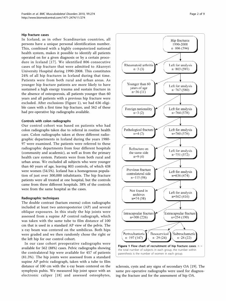
Hip fracture casesIn Iceland, as in other Scandinavian countries, allpersons have a unique personal identification number.This, combined with a highly computerized nationalhealth system, makes it possible to identify all patientsoperated on for a given diagnosis or by a certain proce-dure in Iceland [17]. We identified 806 consecutivecases of hip fracture that were admitted to AkureyriUniversity Hospital during 1990-2008. This constitutes24% of all hip fractures in Iceland during that time.Patients were from both rural and urban areas. Asyounger hip fracture patients are more likely to havesustained a high energy trauma and sustain fracture inthe absence of osteoporosis, all patients younger than 60years and all patients with a previous hip fracture wereexcluded. After exclusions (Figure 1), we had 636 eligi-ble cases with a first time hip fracture, and 562 of thesehad pre-operative hip radiographs available.
Controls with colon radiographsOur control cohort was based on patients who hadcolon radiographs taken due to referral in routine healthcare. Colon radiographs taken at three different radio-graphic departments in Iceland during the years 1980-97 were examined. The patients were referred to theseradiographic departments from four different hospitals(community and academic), as well as from the primaryhealth care system. Patients were from both rural andurban areas. We excluded all subjects who were youngerthan 60 years of age, leaving 803 controls, of which 438were women (54.5%). Iceland has a homogenous popula-tion of just over 300,000 inhabitants. The hip fracturepatients were all treated at one hospital, but the controlscame from three different hospitals. 38% of the controlswere from the same hospital as the cases.
Radiographic techniquesThe double contrast (barium enema) colon radiographsincluded at least two anteroposterior (AP) and severaloblique exposures. In this study the hip joints wereassessed from a supine AP control radiograph, whichwas taken with the same tube to film distance of 100cm that is used in a standard AP view of the pelvis. Thex-ray beam was centered on the umbilicus. Both hipswere graded and we then randomly chose the right orthe left hip for our control cohort.In our case cohort preoperative radiographs were
available for 562 (88%) cases. Pelvic radiographs showingthe contralateral hip were available for 457 of patients(81.3%). The hip joints were assessed from a standardsupine AP pelvic radiograph, taken with a tube to filmdistance of 100 cm with the x-ray beam centered on thesymphysis pubis. We measured hip joint space with anelectronic caliper [18] and assessed osteophytes,
sclerosis, cysts and any signs of secondary OA [19]. Thesame pre-operative radiographs were used for diagnos-ing the fracture and for the assessment of hip OA.
Figure 1 Flow chart of recruitment of hip fracture cases. n =the total number of subjects in each group, the number withinparenthesis is the number of women in each group.
Franklin et al. BMC Musculoskeletal Disorders 2010, 11:274http://www.biomedcentral.com/1471-2474/11/274
Page 2 of 9

Hip fractures were classified as intra- or extracapsular[20]. We measured minimal joint space (MJS) andgraded the radiographic features of OA according toKellgren and Lawrence [19], and individual features ofOA, osteophytes, sclerosis and cysts in both hips.All radiographs of cases were examined by a single
observer (JF). All controls had been previously read by adifferent author (TI) [21]. To calculate inter- andintraobserver reliability [22], we selected 50 radiographsof cases and 50 radiographs of controls, that includedthe full range of radiographic features that were readagain by both these authors. We used Cohen’s kappa tocalculate the intra- and interobserver reliability fordetecting hip OA.For MJS both the intra- and interobserver reliability
was high, but Kellgren and Lawrence grading was not asreliable (Table 1).
Validity of pre-operative radiographsPre-operative radiographs of cases were used since post-operative radiographs are now often taken in the operat-ing theatre which yields radiographs that cannot be usedto evaluate MJS. Presence of a hip fracture (displaced ornot displaced) may affect the measurement of the jointspace in the hip, even if subtle changes in position donot affect MJS in the hip [23]. To evaluate this weselected 50 patients where we had both pre- and post-operative standard pelvic radiographs, with an anatomicreduction of the fracture, and compared MJS measure-ment pre- and post-operatively. All the post-operativeradiographs were taken within 10 days from the frac-ture. The 50 patients that were used for comparison ofpre- and post-operative x-rays were not randomly cho-sen, since we were limited to patients that had a validpostoperative radiograph. We therefore tested if oursample adequately represented our fracture cases. Inde-pendent samples t-test for MJS was not significant (p =0.96) and neither was Kolmogorov-Smirnov Z for OA/Not OA status (p = 0.36), which shows that this sampleof 50 cases adequately represented our fracture cohort.There were 3 patients classified as having OA on a pre-operative x-ray that were not classified as having OA onthe postoperative x-ray. One patient was classified ashaving OA on the postoperative x-ray, that did not have
OA on the preoperative x-ray. We calculated thep-value for pre- and postoperative readings of MJS withpaired t-test (p = 0.18) and used the McNemars test tocompare OA/Not OA status on pre- and postoperativex-rays (p = 0.63). Neither was significant, thereby vali-dating the choice of using pre-operative x-rayexaminations.Unless otherwise stated, hip OA was defined as MJS
of 2.5 mm or less [21,22]. In the hip fracture cohortthere were 6 women and one man that had a total hipreplacement due to OA in the contralateral hip. Thesewere classified as having OA in that hip.
Secondary osteoporosisMedications or diseases that cause secondary osteo-porosis and thus increase the risk for hip fracturemight be more common in the case or control group.To explore the association with risk factors for second-ary osteoporosis in patients with hip fracture we did aprevalence study of possible risk factors for secondaryosteoporosis within the hip fracture cohort using amatched design. This method was chosen as we didnot have access to data on possible causes of second-ary osteoporosis for the control cohort. We definedpatients with hip fracture and MJS 2.5 mm or less aship OA cases and to each of these we assigned tworeference subjects, who had hip fracture, but not hav-ing hip OA (hence both hip OA cases and referencesubjects came from the hip fracture cohort). The refer-ence subjects were matched according to gender, frac-ture type and age ± 1 year. If more than 2 possiblereference subjects were found, we randomly chosewhich to use. We reviewed the medical records of hipOA cases and their matched reference subjects fromthe fracture cohort and registered possible causes ofsecondary osteoporosis that we identified [24,25].
Statistical methodsAge differences between groups were tested with theindependent samples t-test. We age standardized theobserved prevalence of hip OA in both hip fracturecases and controls against the Icelandic population fromthe year 2000 (using 5 year age strata and direct exter-nal standardization) [26]. Intra- and interobserver
Table 1 Intra- and interobserver reliability measured by Cohen’s κ
Radiographicmethod
Definition of diseasepositive
Intra-observer reliability Inter-observer reliability
Controls Fracturedside
Contralateralside
Controls Fracturedside
Contralateralside
Minimum joint space ≤ 2.5 mm 0.94* 0.91 0.89 0.84 0.80 0.82
Kellgren andLawrence
≥ grade 2 0.76* 0.93 0.56 0.67 0.63 0.50
* Previously published results [21]
Franklin et al. BMC Musculoskeletal Disorders 2010, 11:274http://www.biomedcentral.com/1471-2474/11/274
Page 3 of 9

reliability was calculated using Cohen’s Kappa. Pairedt-test and McNemar test were used to compare readingsof pre- and postoperative x-rays and we used indepen-dent samples t-test and Kolmogorov-Smirnov Z to seethat this not randomly selected sample had approxi-mately the same mean MJS and distribution of values asthe entire group of cases. Chi-square was used in calcu-lations for difference in crude rate and binary logisticregression for age-adjusted calculation of odds ratios forhaving OA amongst cases vs. controls. We considered ap-value of less than or equal to 0.05 to be significant,and all tests were 2-tailed. Calculations were done usingSPSS v. 16.0. (SPSS Incorporated 2007).
ResultsCase and control demographics and characteristicsThe male to female ratio was about 1:3 in the fracturecohort, while it was close to one in the control cohort(Table 2). The number of basocervical and subtrochan-teric fractures was small, so these were grouped togetherwith pertrochanteric fractures as extracapsular fractures(Figure 1). There was no difference in mean agebetween the two fracture types, neither for men (p =0.85), nor women (p = 0.48). The mean age of controlswas significantly lower than that of subjects with frac-ture (p < 0.0001).
Prevalence of hip osteoarthritisThere was a significant difference in the crude preva-lence of hip OA between fracture cases and controls formen (p = 0.02), but not for women (p = 0.3). Followinga direct external standardization of the crude prevalence,the differences between our cases and controlsincreased, especially for women where the age differencebetween cases and controls was greater (Table 3).
Age-adjusted association between hip fracture and hiposteoarthritisThere was no significant difference in mean age whencomparing men with or without hip OA (Table 4).
However, women with hip OA were significantly olderthan women without hip OA.The age difference between cases and controls empha-
sized the need to take that into account when assessingthe association between hip fracture and hip OA. Wetherefore did an age-adjusted binary logistic regressionwith hip OA as the dependent variable. All groups of hipfracture patients had a significantly reduced odds ratio ofhip OA (comparing cases with controls) regardless ofgender and classification system of hip OA. The oddsratio for hip OA was significantly reduced in both thefractured hip and the contralateral hip (Figure 2 and 3).For the fractured hip in men the OR was 0.30 (95%CI0.12-0.74) when defining OA by MJS and 0.31 (95% CI0.12-0.76) when defining OA by Kellgren and Lawrence.Corresponding results for the fractured hip in womenwere 0.33 (95%CI 0.19-0.58) and 0.38 (95% CI 0.21-0.69)when defining OA by Kellgren and Lawrence. For thecontralateral hip in men the OR was 0.38 (95%CI 0.16-0.90) when defining OA by MJS and 0.38 (95% CI 0.16-0.91) when defining OA by Kellgren and Lawrence.Corresponding results for the contralateral hip in womenwere 0.55 (95%CI 0.32-0.96) and 0.52 (95% CI 0.29-0.94)when defining OA by Kellgren and Lawrence.
Alternate definition of radiographic OAIn this study we have primarily used MJS 2.5 mm or lessas definition of OA [21,22], but used the Kellgren andLawrence classification as well to enable comparison topreviously published studies. We also tested our datausing MJS 2.0 mm or less as definition of OA. The ORswere lower (meaning a greater difference between casesand controls), although the difference from MJS 2.5 mmor less definition was not statistically significant. UsingMJS 2.0 mm or less as definition the crude prevalenceof OA was 9.3% for male controls, 3.1% for male cases,8.9% for female controls and 3.7% for female cases. Forhip OA in men with hip fracture, we obtained the OR0.18 (95%CI 0.06-0.57) and in women with hip fracturethe OR 0.12 (0.05-0.28).
Intra- vs. extracapsular fracturesWhen defining hip OA by MJS we found no statisticaldifference in the age adjusted estimate of association forhip OA between the two different fracture groups,neither for men (OR = 0.96, 95% CI 0.21-4.5), or forwomen (OR = 1.5, 95% CI 0.75-2.8). When using Kellg-ren and Lawrence definition of OA, we again found nodifference between the fracture types for men (OR =0.96, 95% CI 0.21-4.5), but a significant difference forwomen, with extracapsular more frequently having hipOA than intracapsular fractures (OR = 2.5, 95% CI 1.2-5.2). We then repeated the calculations, using presenceof OA in the contralateral hip. Again, there was no
Table 2 Demographics of cases with hip fracture andcontrols
Cases (hip fractures) Controls
All Intracapsular Extracapsular
Sex
Men 146 (26.0%) 82 (26.6%) 64 (25.2%) 365 (45.5%)
Women 416 (74.0%) 226 (73.4%) 190 (74.8%) 438 (54.5%)
Age
Men 80.3 (8.6) 80.4 (8.3) 80.1 (9.1) 70.9 (7.6)
Women 81.5 (8.1) 81.3 (8.0) 81.8 (8.2) 70.3 (7.1)
Values are number (%) or mean (SD).
Franklin et al. BMC Musculoskeletal Disorders 2010, 11:274http://www.biomedcentral.com/1471-2474/11/274
Page 4 of 9

difference for men (OR = 2.3, 95% CI 0.52-10.4),regardless of classification system for OA. For womenthere was no difference for OA classified by MJS (OR =1.8, 95% CI 0.92-3.5) or by Kellgren and Lawrence (OR= 1.5, 95% CI 0.73-3.2).
Secondary osteoporosisThere were 40 women and seven men who had hip frac-ture and hip OA in the same fractured hip according todefinition by MJS. We reviewed the medical history forall, except one woman whose patient records were not
found. We thus had 46 cases with hip fracture and hipOA matched by age, gender and fracture type to 92reference subjects with hip fracture without hip OA.Patients were defined as having possible secondaryosteoporosis if they had at least one of the conditionslisted in Table 5. Three of 7 men and 15 of 39 womenhad at least one potential cause of secondary osteoporo-sis (Table 5). The probability of having a possible causeof secondary osteoporosis was 3 times higher for thosewith hip fracture and hip OA than for those with hipfracture, but without hip OA.
Table 3 Prevalence of radiographic hip OA in hip fracture cases and controls
N Crude prevalence, % (n) Standardized prevalence‡, %
MJS* K & L† MJS* K & L†
Men
Cases
Fractured side
All 146 4.8% (7) 4.8% (7) 4.1% 4.1%
Intracapsular 82 4.9% (4) 4.9% (4) 6.8% 6.8%
Extracapsular 64 4.7% (3) 4.7% (3) 1.2% 1.2%
Contralateral side
All 121 6.6% (8) 6.6% (8) 4.3% 4.3%
Intracapsular 69 4.3% (3) 4.3% (3) 1.2% 1.2%
Extracapsular 52 9.6% (5) 9.6% (5) 8.1% 8.1%
Controls 365 11.2% (41) 11.2% (41) 11.4% 11.4%
Women
Cases
Fractured side
All 416 9.6% (40) 8.7% (36) 3.9% 5.5%
Intracapsular 226 8.0% (18) 5.3% (12) 3.6% 2.7%
Extracapsular 190 11.6% (22) 12.6% (24) 4.5% 9.4%
Contralateral side
All 336 12.2% (41) 9.5% (32) 9.8% 7.8%
Intracapsular 189 9.5% (18) 7.9% (15) 7.8% 8.0%
Extracapsular 147 15.6% (23) 11.6% (17) 12.0% 7.8%
Controls 438 11.6% (51) 10.5% (46) 13.5% 11.5%
*Osteoarthritis defined as MJS ≤ 2.5 mm. †Osteoarthritis defined as Kellgren and Lawrence grade ≥ 2. ‡ Standardized against the Icelandic population of year2000.
Table 4 Mean age according to hip OA status
Not hip OA Hip OA Difference
Men
Cases (hip fractures) 80.2 (8.6) 80.7 (9.9) 0.5 (p = 0.9)
Controls 70.7 (7.5) 72.7 (8.1) 2.0 (p = 0.1)
Difference 9.5 (p < 0.0001) 8.0 (p = 0.02)
Women
Cases (hip fractures) 81.1 (8.2) 86.2 (5.8) 5.1 (p < 0.001)
Controls 69.8 (6.9) 73.4 (7.4) 3.6 (p < 0.001)
Difference 11.2 (p < 0.0001) 12.7 (p < 0.0001)
Values are mean (SD) or difference in mean age (p).
Franklin et al. BMC Musculoskeletal Disorders 2010, 11:274http://www.biomedcentral.com/1471-2474/11/274
Page 5 of 9

DiscussionThe primary aim of this study was to evaluate the pre-valence of hip OA in patients with hip fracture, comparedto controls having had colon radiography. After adjustingfor age, the odds ratio for hip OA in patients with hip frac-ture was found to be one-third of that in the comparisongroup. The difference in odds ratio between cases andcontrols was slightly less for the contralateral hip inwomen, but nevertheless significant. This suggests that theinverse relationship between hip OA and hip fracture ismainly systemic, i.e. affecting the whole patient, but con-tribution by a local effect in the arthritic hip cannot beexcluded. The nature of the systemic effect is unknown,but a genetic factor may be involved.For men there was no difference in age-adjusted pre-
valence of hip OA between those with intra- and extra-capsular fractures, regardless of classification system andside. For women, there was a significantly greater preva-lence of hip OA in extracapsular fractures only whendefined by Kellgren and Lawrence in the fractured hip.When examining hip OA prevalence defined by MJS orwhen examining the contralateral hip, the differencebetween fracture types was not significant. We thereforeconclude that there is no significant difference in hipOA prevalence when comparing intracapsular and extra-capsular fractures. Our finding of a statistically signifi-cant difference for women in the fractured hip, whenusing Kellgren and Lawrence classification, might be achance finding or perhaps the Kellgren and Lawrenceclassification system is less applicable to women, as wassuggested [27].The probability for subjects with both hip fracture and
hip OA having a possible secondary cause of osteoporo-sis was 3 times higher than for subjects with hip fracturebut without hip OA. This finding supports the possibi-lity that many of the patients with both hip fracture andhip OA had their hip fracture due to secondary osteo-porosis. This could mean that if it had been possible to
Figure 3 Age adjusted odds ratio for having hip OA in thecontralateral hip of hip fracture cases compared to controlswithout hip fracture. Error bars show 95% CI for cases comparedto controls. MJS: Using hip OA defined by minimal joint space. K &L: Using hip OA defined by Kellgren and Lawrence grade.
Table 5 Number (%) of hip OA cases and reference subjects without hip OA in the hip fracture cohort with risk factorsfor secondary osteoporosis
Subjects with Hip OA(hip fracture and hip OA), n = 46
Reference subjects(hip fracture, but not hip OA), n = 92
Chronic obstructive pulmonary disease 8 (17%) 2 (2%)
Polymyalgica rheumatica 3 (7%) 7 (8%)
Steroid use due to other diseases 3 (7%) 0 (0%)
Coeliac disease 0 (0%) 1 (1%)
Gastrectomy (Billroth I) 5 (11%) 3 (3%)
Alcoholism 0 (0%) 1 (1%)
Renal failure 2 (4%) 0 (0%)
Hyperparathyroidism 0 (0%) 0 (0%)
Panhypopituitarism 0 (0%) 0 (0%)
At least one of the above 18 (39%) 12 (13%)
Figure 2 Age adjusted odds ratio for having hip OA in thefractured hip compared to controls without hip fracture. Errorbars show 95% CI for cases compared to controls. MJS: Using hipOA defined by minimal joint space. K & L: Using hip OA defined byKellgren and Lawrence grade.
Franklin et al. BMC Musculoskeletal Disorders 2010, 11:274http://www.biomedcentral.com/1471-2474/11/274
Page 6 of 9

adjust for risk factors for secondary osteoporosis wemight have found that the inverse relationship betweenhip fracture and hip OA is even stronger than reportedin this study. These findings need to be interpreted withsome caution as no gold standard definition of second-ary osteoporosis exists and we retrieved information onsecondary osteoporosis noted in medical records only,as we did not have bone density measures available.Further studies are needed to explore this aspect.Possible confounders not accounted for in the present
study are body mass index (BMI) and occupation, whichhave been shown to be independent risk factors for hipOA [28], and use of medications, such as hormonereplacement therapy and bisphosphonates, that affectosteoporosis. Another limitation is that the controlgroup had colon radiographs but the cases pelvic radio-graphs. A study comparing urograms with pelvic radio-graphs found that joint space width was on average 10%greater on the urograms [29], while another study foundno significant influence of beam angle [23]. If such aneffect exists it could introduce a bias away from thenull. On the other hand our results were also significantwhen using the Kellgren and Lawrence grading system,which is less dependent on joint space.Patients who undergo colon radiography are not a
random sample of the population. Subjects with hipOA, who are seen within health care more often, maymore likely be referred to colon radiography than thebackground population, introducing selection bias awayfrom the null. We do not have information on BMI.Higher BMI is linked to lower hip fracture risk [30], andhigher hip OA risk [28,31]. Obesity is linked to coloncancer [32] and these subjects may more commonlyundergo colon radiography, but we are not aware of anystudies on the BMI of the average patient undergoingcolon radiography. If our controls overall had a higherBMI than the fracture cases, then they might have had ahigher risk for hip OA, compared to the backgroundpopulation introducing bias away from the null.Due to the relatively long period of time that the cases
and controls were sampled there could be a birth cohorteffect. This might be, for example, due to the fact thatBMI has increased steadily between birth cohorts. Themean birth year for our cases was 1918.5 (SD 9.7) andfor our controls the mean birth year was 1922.5 (SD7.8), so a birth cohort effect should affect our cases andcontrols equally.A prerequisite for a hip fracture is a fall, which can be
influenced by age, comorbidity and medications. OAincreases the risk for falls [33,34], which would in con-trast to the above introduce bias towards the null. Thesum and direction of the aforementioned biases and anyunforeseen biases is difficult to ascertain.
We used an AP pelvic radiograph of patients whowere admitted to our hospital because of a hip fracturefor measurement of MJS and assessment of hip OA byKellgren and Lawrence grade. We are not aware of pre-vious publications where MJS measurements of the frac-tured hip have been used. We considered the possibilitythat the traumatized hip would not be representative ofits normal state, perhaps due to bleeding or musclespasm. We therefore validated this method. In 46 out of50 cases the determination of presence of OA accordingto MJS was the same. The fact that in 4 out of 50 caseswe got a different reading is not greater than would beexpected from any re-reading of a radiograph. Hipadduction-abduction has also been shown to result in amean difference of less than 0.2 mm joint space [23],which we suggest would not significantly influence ourinterpretations. To evaluate the whether the relationshipbetween hip OA and hip fracture is systemic or local tothe arthritic hip we graded both the fractured hip andthe contralateral hip.The Kappa values for the fractured hip were in general
acceptable and similar to previously published studies.The Kappa values for the contralateral hip were some-what lower. The films used to evaluate reliability of thereadings were chosen to include the full range of radio-graphic features for the fractured side, but as we usedthe same films for the contralateral side, the spreadbetween different Kellgren and Lawrence grades was noteven and we therefore believe that this difference maybe the result of the inherent flaw of the Kappa methodin such cases [35].Definition of hip OA varies between studies. Recent
studies have criticised the use of Kellgren and Lawrenceclassification [27], while others have shown that mea-surement of joint space width is reliable and reflectsclinical status [36]. We used both joint space width andKellgren and Lawrence classification of hip OA to facili-tate comparison with other studies.Data on possible secondary osteoporosis was only
available for our fractured patients, ideally we wouldhave had data on secondary osteoporosis for both casesand controls. Thus, we were not able to fully evaluate towhat extent secondary osteoporosis influences our con-clusions, but our results indicate that secondary osteo-porosis is overrepresented amongst patients with hipfracture and hip OA.Some studies suggest that an inverse relationship
between OA and hip fracture exists [4-6,16,37], whileand others refute it [7,8]. Most of these studies are ofcase-control design, while the two studies that refutethis relationship are prospective cohort studies. Onemight therefore suggest that the evidence is strongerthat there is no such relationship, even though the
Franklin et al. BMC Musculoskeletal Disorders 2010, 11:274http://www.biomedcentral.com/1471-2474/11/274
Page 7 of 9

studies that support it are greater in number. In fact,one cannot even assume that these studies oppose oneanother, because of the differences in case definitions inthese studies. In cohort studies the exposure is deter-mined at the start of the study. In the case of OA, thebaseline radiographic examination in a cohort studydoes not give an accurate estimate of the prevalence ofradiographic hip OA at the time of fracture, which canbe several years after the baseline examination. Reportson knee OA have also supported an inverse relationshipbetween OA and osteoporosis [38] and a molecularbasis and common pathophysiology was proposed forthe inverse relationship between OA and osteoporosis[39]. A genetic component to both osteoporosis [40]and OA [41], may explain why these conditions seldomcoexist.
ConclusionsTo the best of our knowledge this is the first studybased on radiographically verified hip OA to quantifythe risk of hip OA in patients with hip fracture, andalso the first study to address the possible influence ofsecondary osteoporosis. We found that patients with hipfracture have one-third the risk for having hip OA inthe fractured hip when compared to controls havinghad colon radiography. In the contralateral hip, the riskdecrease was similar for men, and slightly less forwomen.
AcknowledgementsSupported by: Scientific Foundation of Akureyri Central Hospital, TheSwedish Research Council (medicine), Lund University Medical Faculty,Region Skåne, the King Gustaf V 80-year Fund, and The SwedishRheumatism Association.
Author details1University Hospital, Akureyri, Iceland. 2Department of Orthopedics, ClinicalSciences Lund, Lund University, Sweden. 3Clinical Epidemiology Research &Training Unit, Boston University School of Medicine, Boston, MA, USA.4Department of Health Sciences, University of Akureyri, Iceland and Facultyof Medicine, University of Iceland, Reykjavík, Iceland.
Authors’ contributionsJF, TI and SL planned the study. JF read radiographs of hip fractures, TI readradiographs of controls. JF collected data and did the statistical analysis. JFdrafted the manuscript. TI, SL and ME revised the manuscript. All authorstook part in analysing the findings and all authors approved the final versionof the manuscript.
Competing interestsThe authors declare that they have no competing interests.
Received: 21 April 2010 Accepted: 26 November 2010Published: 26 November 2010
References1. Astrom J, Beertema J: Reduced risk of hip fracture in the mothers of
patients with osteoarthritis of the hip. J Bone Joint Surg Br 1992,74:270-271.
2. Biyani A, Simison AJ, Klenerman L: Intertrochanteric fractures of the femurand osteoarthritis of the ipsilateral hip. Acta Orthop Belg 1995, 61:83-91.
3. Dequeker J, Goris P, Uytterhoeven R: Osteoporosis and osteoarthritis(osteoarthrosis). Anthropometric distinctions. JAMA 1983, 249:1448-1451.
4. Cumming RG, Klineberg RJ: Epidemiological study of the relationbetween arthritis of the hip and hip fractures. Annals of the rheumaticdiseases 1993, 52:707-710.
5. Dequeker J, Johnell O: Osteoarthritis protects against femoral neckfracture: the MEDOS study experience. Bone 1993, 14(Suppl 1):S51-56.
6. Foss MV, Byers PD: Bone density, osteoarthrosis of the hip, and fractureof the upper end of the femur. Annals of the rheumatic diseases 1972,31:259-264.
7. Arden NK, Nevitt MC, Lane NE, Gore LR, Hochberg MC, Scott JC,Pressman AR, Cummings SR: Osteoarthritis and risk of falls, rates of boneloss, and osteoporotic fractures. Study of Osteoporotic FracturesResearch Group. Arthritis Rheum 1999, 42:1378-1385.
8. Jones G, Nguyen T, Sambrook PN, Lord SR, Kelly PJ, Eisman JA:Osteoarthritis, bone density, postural stability, and osteoporoticfractures: a population based study. J Rheumatol 1995, 22:921-925.
9. Colhoun EN, Johnson SR, Fairclough JA: Bone scanning for hip fracture inpatients with osteoarthritis: brief report. J Bone Joint Surg Br 1987, 69:848.
10. Wand JS, Hill ID, Reeve J: Coxarthrosis and femoral neck fracture. Clinicalorthopaedics and related research 1992, 88-94.
11. Arden NK, Griffiths GO, Hart DJ, Doyle DV, Spector TD: The associationbetween osteoarthritis and osteoporotic fracture: the Chingford Study.Br J Rheumatol 1996, 35:1299-1304.
12. Wolf O, Strom H, Milbrink J, Larsson S, Mallmin H: Differences in hip bonemineral density may explain the hip fracture pattern in osteoarthritichips. Acta Orthop 2009, 1-6.
13. McClung MR, Geusens P, Miller PD, Zippel H, Bensen WG, Roux C, Adami S,Fogelman I, Diamond T, Eastell R, et al: Effect of risedronate on the risk ofhip fracture in elderly women. Hip Intervention Program Study Group. NEngl J Med 2001, 344:333-340.
14. Antoniades L, MacGregor AJ, Matson M, Spector TD: A cotwin controlstudy of the relationship between hip osteoarthritis and bone mineraldensity. Arthritis Rheum 2000, 43:1450-1455.
15. Styrkarsdottir U, Halldorsson BV, Gretarsdottir S, Gudbjartsson DF,Walters GB, Ingvarsson T, Jonsdottir T, Saemundsdottir J, Center JR,Nguyen TV, et al: Multiple genetic loci for bone mineral density andfractures. N Engl J Med 2008, 358:2355-2365.
16. Weintroub S, Papo J, Ashkenazi M, Tardiman R, Weissman SL, Salama R:Osteoarthritis of the hip and fracture of the proximal end of the femur.Acta orthopaedica Scandinavica 1982, 53:261-264.
17. Ingvarsson T, Hägglund G, Jonsson H Jr, Lohmander LS: Incidence of totalhip replacement for primary osteoarthrosis in Iceland 1982-1996. Actaorthopaedica Scandinavica 1999, 70:229-233.
18. Hilliquin P, Pessis E, Coste section sign J, Mauget D, Azria A, Chevrot A,Menkes CJ, Kahan A: Quantitative assessment of joint space width withan electronic caliper. Osteoarthritis Cartilage 2002, 10:542-546.
19. Kellgren JH, Lawrence JS: Radiological assessment of osteo-arthrosis.Annals of the rheumatic diseases 1957, 16:494-502.
20. Rüedi TP, Buckley RE, Moran CG: AO Principles of Fracture Management. 2edition. AO Publishing; 2007.
21. Ingvarsson T, Hägglund G, Lindberg H, Lohmander LS: Assessment ofprimary hip osteoarthritis: comparison of radiographic methods usingcolon radiographs. Annals of the rheumatic diseases 2000, 59:650-653.
22. Croft P, Cooper C, Wickham C, Coggon D: Defining osteoarthritis of thehip for epidemiologic studies. Am J Epidemiol 1990, 132:514-522.
23. Goker B, Sancak A, Haznedaroglu S, Arac M, Block JA: The effects of minorhip flexion, abduction or adduction and x-ray beam angle on theradiographic joint space width of the hip. Osteoarthritis Cartilage 2005,13:379-386.
24. Kelman A, Lane NE: The management of secondary osteoporosis. Bestpractice & research 2005, 19:1021-1037.
25. Fitzpatrick LA: Secondary causes of osteoporosis. Mayo Clinic proceedings2002, 77:453-468.
26. Curtin LR, Klein RJ: Direct standardization (age-adjusted death rates).Healthy People 2000 Stat Notes 1995, 1-10.
27. Jacobsen S, Sonne-Holm S: Increased body mass index is a predispositionfor treatment by total hip replacement. Int Orthop 2005, 29:229-234.
28. Franklin J, Ingvarsson T, Englund M, Lohmander LS: Sex differences in theassociation between body mass index and total hip or knee joint
Franklin et al. BMC Musculoskeletal Disorders 2010, 11:274http://www.biomedcentral.com/1471-2474/11/274
Page 8 of 9

replacement resulting from osteoarthritis. Annals of the rheumatic diseases2009, 68:536-540.
29. Auleley GR, Duche A, Drape JL, Dougados M, Ravaud P: Measurement ofjoint space width in hip osteoarthritis: influence of joint positioning andradiographic procedure. Rheumatology (Oxford) 2001, 40:414-419.
30. De Laet C, Kanis JA, Oden A, Johanson H, Johnell O, Delmas P, Eisman JA,Kroger H, Fujiwara S, Garnero P, et al: Body mass index as a predictor offracture risk: a meta-analysis. Osteoporos Int 2005, 16:1330-1338.
31. Lohmander LS, Gerhardsson de Verdier M, Rollof J, Nilsson PM, Engstrom G:Incidence of severe knee and hip osteoarthritis in relation to differentmeasures of body mass: a population-based prospective cohort study.Annals of the rheumatic diseases 2009, 68:490-496.
32. Caan BJ, Coates AO, Slattery ML, Potter JD, Quesenberry CP Jr, Edwards SM:Body size and the risk of colon cancer in a large case-control study. Int JObes Relat Metab Disord 1998, 22:178-184.
33. Nevitt MC, Cummings SR, Kidd S, Black D: Risk factors for recurrentnonsyncopal falls. A prospective study. JAMA 1989, 261:2663-2668.
34. Campbell AJ, Borrie MJ, Spears GF: Risk factors for falls in a community-based prospective study of people 70 years and older. J Gerontol 1989,44:M112-117.
35. Byrt T, Bishop J, Carlin JB: Bias, prevalence and kappa. J Clin Epidemiol1993, 46:423-429.
36. Dougados M, Gueguen A, Nguyen M, Berdah L, Lequesne M, Mazieres B,Vignon E: Radiological progression of hip osteoarthritis: definition, riskfactors and correlations with clinical status. Annals of the rheumaticdiseases 1996, 55:356-362.
37. Vestergaard P, Rejnmark L, Mosekilde L: Osteoarthritis and risk of fractures.Calcif Tissue Int 2009, 84:249-256.
38. Hart DJ, Mootoosamy I, Doyle DV, Spector TD: The relationship betweenosteoarthritis and osteoporosis in the general population: the ChingfordStudy. Annals of the rheumatic diseases 1994, 53:158-162.
39. Lories RJ, Peeters J, Bakker A, Tylzanowski P, Derese I, Schrooten J,Thomas JT, Luyten FP: Articular cartilage and biomechanical properties ofthe long bones in Frzb-knockout mice. Arthritis Rheum 2007,56:4095-4103.
40. Spector TD, Keen RW, Arden NK, Morrison NA, Major PJ, Nguyen TV,Kelly PJ, Baker JR, Sambrook PN, Lanchbury JS, et al: Influence of vitamin Dreceptor genotype on bone mineral density in postmenopausal women:a twin study in Britain. BMJ 1995, 310:1357-1360.
41. Ingvarsson T, Stefansson SE, Hallgrimsdottir IB, Frigge ML, Jonsson H Jr,Gulcher J, Jonsson H, Ragnarsson JI, Lohmander LS, Stefansson K: Theinheritance of hip osteoarthritis in Iceland. Arthritis Rheum 2000,43:2785-2792.
Pre-publication historyThe pre-publication history for this paper can be accessed here:http://www.biomedcentral.com/1471-2474/11/274/prepub
doi:10.1186/1471-2474-11-274Cite this article as: Franklin et al.: The association between hip fractureand hip osteoarthritis: A case-control study. BMC MusculoskeletalDisorders 2010 11:274.
Submit your next manuscript to BioMed Centraland take full advantage of:
• Convenient online submission
• Thorough peer review
• No space constraints or color figure charges
• Immediate publication on acceptance
• Inclusion in PubMed, CAS, Scopus and Google Scholar
• Research which is freely available for redistribution
Submit your manuscript at www.biomedcentral.com/submit
Franklin et al. BMC Musculoskeletal Disorders 2010, 11:274http://www.biomedcentral.com/1471-2474/11/274
Page 9 of 9