the authors, some nanostructure, osteopontin, and...
TRANSCRIPT

SC I ENCE ADVANCES | R E S EARCH ART I C L E
APPL I ED PHYS I CS
1Faculty of Dentistry, McGill University, Montreal, Quebec H3A 0C7, Canada. 2Depart-ment of Mining and Materials Engineering, McGill University, Montreal, Quebec H3A0C5, Canada. 3Facility for Electron Microscopy Research, McGill University, Montreal,QuebecH3A0C7, Canada. 4Department of Biomedical Engineering, JohnsHopkinsUni-versity, Baltimore,MD21218, USA. 5Department ofMaterials Science and Engineering,Institute of Glass and Ceramics, Friedrich-Alexander-University Erlangen-Nürnberg,Erlangen 91058, Germany. 6Department of Cellular and Molecular Medicine and De-partment of Innovation in Medical Education, University of Ottawa, Ottawa, OntarioK1H 8M5, Canada. 7DepartamentodeMineralogía yPetrología, UniversidaddeGranada,Granada 18002, Spain. 8Department of Anatomy and Cell Biology, McGill University,Montreal, Quebec H3A 0C7, Canada. 9Interdisciplinary Center for Functional ParticleSystems, Friedrich-Alexander University Erlangen-Nürnberg, Haberstrasse 9a, Erlangen91058,Germany. 10DepartmentofChemical andBiomolecularEngineering, JohnsHopkinsUniversity, Baltimore, MD 21218, USA. 11Program in Molecular Biophysics, Institute forNanobiotechnology, Sidney Kimmel Comprehensive Cancer Center, Johns HopkinsUniversity, Baltimore, MD 21218, USA.*Present address: Department of Chemical and Biomolecular Engineering, JohnsHopkins University, Baltimore, MD 21218, USA.†Corresponding author. Email: [email protected]
Athanasiadou et al., Sci. Adv. 2018;4 : eaar3219 30 March 2018
Copyright © 2018
The Authors, some
rights reserved;
exclusive licensee
American Association
for the Advancement
of Science. No claim to
originalU.S. Government
Works. Distributed
under a Creative
Commons Attribution
NonCommercial
License 4.0 (CC BY-NC).
Dow
Nanostructure, osteopontin, and mechanical propertiesof calcitic avian eggshellDimitra Athanasiadou,1 Wenge Jiang,1 Dina Goldbaum,2 Aroba Saleem,2 Kaustuv Basu,3
Michael S. Pacella,4* Corinna F. Böhm,5 Richard R. Chromik,2 Maxwell T. Hincke,6
Alejandro B. Rodríguez-Navarro,7 Hojatollah Vali,3,8 Stephan E. Wolf,5,9
Jeffrey J. Gray,10,11 Khanh Huy Bui,8 Marc D. McKee1,8†
Avian (and formerly dinosaur) eggshells form a hard, protective biomineralized chamber for embryonic growth—anevolutionary strategy that has existed for hundreds of millions of years. We show in the calcitic chicken eggshell howthe mineral and organic phases organize hierarchically across different length scales and how variation in nanostruc-ture across the shell thicknessmodifies its hardness, elasticmodulus, anddissolutionproperties.We also show that thenanostructure changes during egg incubation, weakening the shell for chick hatching. Nanostructure and increasedhardness were reproduced in synthetic calcite crystals grown in the presence of the prominent eggshell protein os-teopontin. These results demonstrate the contribution of nanostructure to avian eggshell formation,mechanical prop-erties, and dissolution.
n
on July 25, 2018http://advances.sciencemag.org/
loaded from
INTRODUCTIONThe avian (and formerly dinosaur) eggshell is a thin, mineralized (cal-cite) layer that adequately protects the egg content and allows for theextra-uterine development of the chick embryo. Besides its protectivefunction, its partial dissolution and thinning from the inside out duringfertilized egg incubation serve as a source of calciumrequired for calcium-phosphatemineralization of the growing embryonic chick skeleton. Thispartial dissolution of the inner aspect of the shell also facilitates chickhatching/pipping. We hypothesized that these diverse functions of theremarkably designed and evolutionarily persistent avian eggshell likelyresult from regional differences in nanostructure; basic eggshell structurehas been conserved over hundreds of millions of years of evolution.
The eggshell of the domestic chicken (Gallus gallus) is about 95% (byweight) calcium-carbonate mineral in the form of calcite and about3.5% (by weight) organicmaterial/matrix (including water) (1). Amonghundreds of proteins identified by proteomics and various other meansin the eggshell organic matrix (2–5), osteopontin (OPN, the namederived from its initial discovery in bone) is amajor shell matrix protein(6) and amember of a group ofmineral-binding proteins (7) thought tobe prominent in guiding mineralization processes because of their par-ticularly high negative charge and open flexible structure (7, 8). Theseproteins are intrinsically disordered (8) and are thought to have arisenfrom ancestral gene-duplication events. Their high negative charge
(involved in calcium binding and mineral binding) partly derives froman abundance of acidic amino acids (Asp and Glu). In addition, manySer residues are phosphorylated (this is particularly so for OPN), whichimparts additional negative charge to bind ionic calcium and crystallattice calcium (9).
Living organisms produce a wide variety of biominerals for avariety of purposes. Biominerals form hardened structures that oftenhave complex architectures that are hierarchically organized, such asthe human skeleton where calcium-phosphate mineral prevails, withnanocrystals forming within an extensive, fibrillar organic macro-molecular assembly known as the extracellular matrix. On the otherhand, terrestrial and marine organisms typically use calcium-carbonatemineral polymorphs to build functionalized biomineralized structuressuch as seashells, snail shells, and eggshells. These rigid structures arehybrid composite materials where organic-inorganic (protein-mineral)interactions largely improve mechanical properties, such as hardnessand toughness, to adequately provide supportive and/or protectivefunctions to the organism in which they are assembled (10).
In recent years, there has beenmuchwork focusing on the structuralanalysis of various calcareous biominerals at the nanometer length scale.In many cases, closely packed nanosized subunits that are essentiallyperfectly aligned have been observed to form through nonclassical crys-tallization pathways (11). These alternative pathways may give rise tocrystalline material having a nanogranular structure that produces asingle-crystal diffraction pattern as would be obtained for a single,“monolithic” crystal otherwise not having a nanosubstructure. Molluskshells, fish otoliths, coral skeletons, and brachiopods all have been de-scribed as having an internal nanogranular structure (12). Although invitro experiments have evaluated the influence of organic material oncalcium-carbonate polymorph selection (13), little is actually knownabout themechanisms throughwhich biomolecules affect nanogranularstructure in living organisms.
Here, we have investigated, at the nanoscale, the structure of theeggshell from chicken (G. gallus) and OPN incorporation and haveidentified the existence of nanogranular structure in this shell. We haveadditionally correlated this nanostructure with functional properties(hardness, elastic modulus, and dissolution). These findings extend ourknowledge of how incorporated organic constituents can substantiallyenhance mechanical properties and controlled solubility in biostructures
1 of 13

SC I ENCE ADVANCES | R E S EARCH ART I C L E
(14), observations that align with alloy fabrication studies showing thatdual-phase nanostructuring can nearly attain theoretical (“ideal”)strength in synthesized materials (15). Here, we correlate nanostructurewith hardness and elastic modulus in the chicken eggshell. We also de-scribe changes in shells from eggs that have been partially naturally dis-solved after physiologic fertilization and incubation, two processesrequired for chick embryo development. Finally, we report that occludedOPN (an abundant protein in eggshell) can induce nanostructure in syn-thetic (nonbiogenic) calcite, much like what we have observed in thechicken eggshell.
Athanasiadou et al., Sci. Adv. 2018;4 : eaar3219 30 March 2018
RESULTS AND DISCUSSIONNanostructure of avian eggshell (chicken, G. gallus)Here, systematic atomic force microscopy (AFM) analysis of eacheggshell region [shown broadly for orientation purposes at low magni-fication by scanning electron microscopy (SEM) in Fig. 1A] revealedthat the outermost vertical crystal layer (VCL), the central palisadeslayer (PL), and the innermost mammillary layer (ML) all have a finenanostructure varying in size depending on the layer in which it wasobserved (Fig. 1, B to F). Topographic imaging by AFM operated inthe amplitudemode demonstrated an average (±SD) nanostructure size
on July 25, 2018http://advances.sciencem
ag.org/D
ownloaded from
Nan
ostru
ctur
e si
ze (n
m)
G
*
** NS
***
Vertical crystal layer (VCL)
Palisades layer (PL)
Mammillary layer (ML)
BC
E
F
D
A
B C
D E F
200 nm
200 nm
100 μm
Fig. 1. The nanostructure of chicken eggshell (G. gallus). (A) SEMmicrostructure at lowmagnification and regional nomenclature of avian eggshell. (B to F) AFM (top; with ascanning area of 800 nm × 800 nm) and SEM (bottom) images of eggshell nanostructure as observed from the regions indicated in (A). (G) Histogram of nanostructure sizedistribution (Feret diameter) in the VCL (B), upper PL (C),middle PL (D), lower PL (E), andML (F) layers. Significant difference is indicated by brackets (*P<0.05, **P<0.01, and ***P<0.001). No significant difference (NS) (P > 0.05) between bars E and F. Values were compared by a two-tailed Student’s t test.
2 of 13

SC I ENCE ADVANCES | R E S EARCH ART I C L E
http://advaD
ownloaded from
(Feret diameter) of 30 ± 10 nm in the VCL, 33 ± 10 nm in the upper PL,59 ± 27 nm in themiddle PL, 74 ± 36 nm in the lower PL, and 68 ± 26 nmin theML (Fig. 1G). Figure S1 shows the distribution ofmeasured nano-structure size of the eggshell layers as determined from the AFM imagesfrom eggs that were not incubated and after 15 days of egg incubation.In addition, fig. S2 provides the average nanostructure area for alleggshell layers after 15 days of egg incubation. Changes in both nano-structure Feret diameter and area measurements were consistent intheir variation between layers. SEM imaging performed on the samelayers confirmed the variation in nanostructure size across the eggshellthickness (Fig. 1). Within the three sublayers of the PL that we sampled(upper, middle, and lower PL), average nanostructure size (by Feret di-ameter) continuously increased toward the interior of the shell in thislayer, which is the region that forms the main bulk of the shell. In otherbird species, previous AFM examination of all layers in the guinea fowl(Numida meleagris) and goose (Anser anser) eggshell showed a struc-turing described as “rounded nanogranules” (16)—such observationsin these species are consistent with our findings in the chicken eggshell.In that previous study, nanogranular size was in the range of 50 to100 nm, values close to the range of sizes that we have measured inthe chicken shell layers and similar to those observed in other calcium-carbonate biominerals (17, 18).More recently, Rodríguez-Navarro et al.(19) provided evidence for early nanogranular structuring at the firstphase of chicken eggshell deposition where transient, flat, and disk-shaped amorphous calcium carbonate (ACC) forms on the eggshellmembranes. However, there has been no detailed description of com-plete, intact, fully formed chicken eggshell at the nanoscale level to date.
Athanasiadou et al., Sci. Adv. 2018;4 : eaar3219 30 March 2018
Crystallographic features of the chicken eggshell include notableinternal misalignments observed at the nanoscale in the columnar cal-citic crystal units. These internal misalignments presumably occurred,at least in part, from the nonhomogeneous occlusion of abundant or-ganic material (Fig. 2A). This was identified broadly by optical micros-copy under cross-polarized light, where different degrees of lightelimination occur from different microscale regions (Fig. 2B). Thiswas further examined with greater resolution by electron backscatterdiffraction (EBSD), where diffraction deviations within the calcitic co-lumnar units (here pseudocolored, with deviations appearing as slightcolor shade differences) reflect the presence of substantial internal mis-alignments, generated at specific points and extending and propagatingduring calcite column growth (Fig. 2, C to E). Moreover, the 001 polefigure of a columnar calcite unit showed the orientation of c-axis devia-tions to be in the range of several degrees (up to 3° and even higher) (fig.S3A; whereas for a perfect single crystal, all the points in the pole figurewould be concentrated in a single spot). EBSD data also show that thereis an increase in internal misalignment with increasing column widths(fig. S3B), which implies that there is an accumulation of defects duringeggshell calcitic column growth, further supporting the concept of a na-nogranular crystal growth mechanism. Previous EBSD analysis ofchicken eggshell and other eggshell species (16, 20) has also shown thatthe PL consists of elongated, well crystallographically ordered singlecrystals of calcite; however, no evidence of internal misalignments ofthese elongated crystals was described. Thus, these internal misalign-ments were further investigated by two-dimensional x-ray diffraction(2DXRD).This confirms that the calcitic columnarunitswere not perfect
on July 25, 2018nces.sciencem
ag.org/
A B C D
E
G
Inte
nsity
(a.u
.)
F
Inte
nsity
(a.u
.)
γ (°) γ (°)
0.1 mm0.1 mm
Fig. 2. Optical microscopy, EBSD, and 2D XRD of chicken eggshell. (A) Thin eggshell cross section viewed by conventional bright-field light microscopy showing non-homogeneous distribution of organic and inorganic material throughout the eggshell layers. (B) Thin eggshell cross section [same as in (A)] viewed under cross-polarized lightshowingmultiple, closely packed andwell-defined columnar calcite units. (C) Crystal orientationmapobtainedby EBSDof a polished eggshell cross section showing slight internalcrystalline misalignments (up to 4°) within the columns as depicted by different pseudocolor shades [see selected boxed areas in (D) and (E)]. (F) 2D XRD of a cross section of aneggshell showing an elongated single-crystal diffraction spot (arrows) and the associated intensity profile of a 104 calcite reflection as a function of the g angle. Note thewideningof the 104peak due to varying crystallographic orientationwithin a columnar calcite unit. a.u., arbitrary units. (G) 2DXRDof a control powdered Iceland spar calcite crystal showingsingle-crystal diffraction spots (arrows) and the associated intensity profile of a 104 calcite reflection as a function of the g angle.
3 of 13
SC I ENCE ADVANCES | R E S EARCH ART I C L E
Dow
nload
single crystals but rather had a high degree of internal misorientationscharacterized by angular spreading of the diffraction spots (>2.5°) (Fig.2F), producing elongated reflection spots (Fig. 2F, inset). In contrast, gent-ly ground geologic calcite (Iceland spar)—commonly used as a reference,high-quality single-crystal control material for calcite—produced sharp,rounded spots (Fig. 2G, inset) with amuch smaller angular spread (<0.8°)(Fig. 2G). To discard the possibility that these elongated spots wereoverlapping spots obtained from two or more columnar units, we re-cordedmultiple 2D patterns after rotating the eggshell through 0.3° steps.Together, both the EBSD data and the 2D XRD data—which arecomplementary techniques—confirm the presence of deviating crystal-line subdomains within the calcitic columnar units. These findings de-scribing events occurring within the calcitic eggshell columnsemphasize that nanostructure and slight alignment deviations result froma nonclassical crystal growth pathway (11).
Eggshell calcite crystals are known to form initially through the de-position of ACC particles that transform directly into calcite whilepreserving the granular nanostructure of the ACC (19). The newlycrystallized material can adopt the orientation of previously formedcalcite crystals (that is, in the ML) so that the crystallographic orien-tation is propagated as the columnar calcite crystal units develop by
Athanasiadou et al., Sci. Adv. 2018;4 : eaar3219 30 March 2018
epitaxial nucleation (21). Amorphous nanoparticles responsible forthe nanostructural morphology of many mineral formations inmany organisms can occur as an initial precursor phase stabilizedby resident organics, and these can be subsequently crystallized aftercontact with a crystalline substrate (12, 22–24). In the eggshell, theinterplay between organics and mineral precursors likely influencesmineralization events leading to internal misalignments.
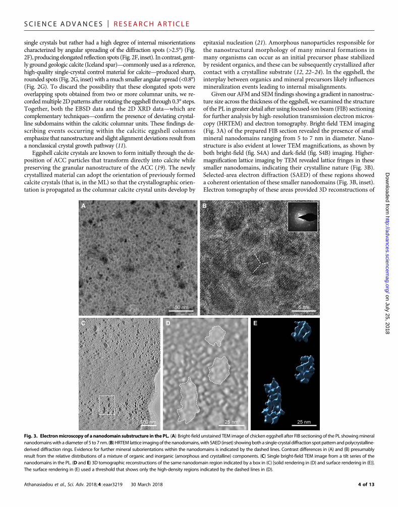
Given our AFMand SEM findings showing a gradient in nanostruc-ture size across the thickness of the eggshell, we examined the structureof the PL in greater detail after using focused-ion beam (FIB) sectioningfor further analysis by high-resolution transmission electron micros-copy (HRTEM) and electron tomography. Bright-field TEM imaging(Fig. 3A) of the prepared FIB section revealed the presence of smallmineral nanodomains ranging from 5 to 7 nm in diameter. Nano-structure is also evident at lower TEM magnifications, as shown byboth bright-field (fig. S4A) and dark-field (fig. S4B) imaging. Higher-magnification lattice imaging by TEM revealed lattice fringes in thesesmaller nanodomains, indicating their crystalline nature (Fig. 3B).Selected-area electron diffraction (SAED) of these regions showeda coherent orientation of these smaller nanodomains (Fig. 3B, inset).Electron tomography of these areas provided 3D reconstructions of
on July 25, 2018http://advances.sciencem
ag.org/ed from
A B
EDC
50 nm 5 nm
100 nm 25 nm 25 nm
Fig. 3. Electronmicroscopy of a nanodomain substructure in the PL. (A) Bright-field unstained TEM image of chicken eggshell after FIB sectioning of the PL showingmineralnanodomainswith adiameter of 5 to 7nm. (B) HRTEM lattice imagingof thenanodomains,with SAED (inset) showingbotha single-crystal diffraction spotpattern andpolycrystalline-derived diffraction rings. Evidence for further mineral suborientations within the nanodomains is indicated by the dashed lines. Contrast differences in (A) and (B) presumablyresult from the relative distributions of a mixture of organic and inorganic (amorphous and crystalline) components. (C) Single bright-field TEM image from a tilt series of thenanodomains in the PL. (D and E) 3D tomographic reconstructions of the same nanodomain region indicated by a box in (C) [solid rendering in (D) and surface rendering in (E)].The surface rendering in (E) used a threshold that shows only the high-density regions indicated by the dashed lines in (D).
4 of 13

SC I ENCE ADVANCES | R E S EARCH ART I C L E
on July 25, 2018http://advances.sciencem
ag.org/D
ownloaded from
this PL nanostructure (Fig. 2, C to E, and movies S1 and S2) whereabundant and homogeneously dispersed 5- to 7-nm nanodomainswere observed to reside within the larger nanostructure initially ob-served topographically by AFM and SEM (Fig. 1C). To confirm thisobservation using an alternative sample-preparation method, awedge-polished section of the PL was prepared and thinned usinga previously described method (18), and similar results were obtained(fig. S4C). Our work is consistent with observations by Lammie et al.(25) who used microfocused, small-angle x-ray scattering to report on“nanotexture” variations averaging ~4.5 nm in the PL, a feature that webelieve we have now directly visualized and characterized in the presentstudy. In the report of Lammie et al., the nanotexture was attributed tovoids presumably occupied by globular organic matrix, whereas here,we identify and visualize actual nanostructured, crystalline mineral in2D and 3D that produces nanostructured texture (and we also presentdata onproteindistribution; seebelow). Both sets of findings are consistentwith the notion that regional variations in organic/protein content (3)[acting as activators or inhibitors ofmineralization (2)], alongwith relativedifferences in the amounts, organization, and location of calcite andACC,all likely contribute to producing the nanostructured texture (19).
In avian eggs, although the biological benefits of eggshell hierarchicalstructure (from the nanoscale to the macroscale) for the developingorganism (chick) are apparent, themechanism of nanostructure forma-tion in the chemically crowded (organic and inorganic), and physicallyconfined, milieu is less clear. As a possibility, the formation of pseudo-periodic structures of a uniform size can be explained by the reaction-diffusionmodel, which is also known as the Turing pattern (26). Turingpatterns are abundant in nature and have remarkable significance inmorphogenesis (27). The basis of the reaction-diffusion pattern forma-tion is in the competition of two or more oscillating processes havingdifferent kinetic constants (for example, short-range activation and long-range inhibition) originating from local instabilities within an initiallyhomogeneous steady-state system (26). In the laying hen, uterine fluidbathing the late developing egg (and then the eggshell) is supersaturatedwith respect to calcite and contains abundant calcium-binding proteinsin ametastable system. In this system, heteronucleation events are likelyto develop as local instabilities, where the competing processes are theCa/P ion supply (activation) and the local ion depletion around thegrowing crystallites (inhibition). Notably, local ion depletion around agrowing calcite crystal or a stabilized ACC particle has been demon-strated in the aqueous state in real time, and moreover, the extent of
Athanasiadou et al., Sci. Adv. 2018;4 : eaar3219 30 March 2018
the depletion zone was proportional to mineral particle size (28). Thesereaction-diffusion kinetics could explain the formation of numeroussmall domains surrounded by a narrow depletion zone or fewer, larger,and more stable domains surrounded by a broader depletion zone, thispossibly being a function of ion supply andOPN concentration (and/orother shell proteins) at different stages of eggshell formation (29).
OPN and the organic matrix compartment inchicken eggshellIn avian eggshell, OPNwas first reported byPines et al. (6)whodetectedstrong OPN gene expression by cells specifically in the shell gland thatwas coincident with eggshell mineralization. In that previous work, im-munohistochemistry and lightmicroscopywere used to show thatOPNwas distributed throughout the shell. Since then, these findings onOPNand eggshell have been confirmed and developed by us and others usinga variety of analytical methods (2, 30, 31). Here, Fig. 4A shows OPNretrieved biochemically from protein extracts of demineralized chickeneggshell. Figure 4B demonstrates OPN localization in situ in eggshellby immunohistochemistry at the light microscope level and ultrastruc-turally by immunogold labeling at the electron microscope level (Fig.4C). Regional variations in the concentration of OPN are apparent[Fig. 4, B (brackets) and C (arrows)]; note that the demineralizationprocedure itself imparts substantial alterations to the localization patternas themineral dissolves and the organicmatrix network partially collapses.Qualitatively, by immunostaining, OPN was most abundant in the VCL/PL and least abundant in theML. Semiquantification of this immunolabel-ing using intensity-profile, linear plot views obtained from immunos-tained eggshell sections using ImageJ (fig. S5A) showed consistentlyhigh concentration of OPN in the outermost VCL layer, followed by asteady decline in OPN content toward the ML layer (fig. S5B). Theseresults are in agreement with quantitative proteomics revealing thateggshellOPN levels are highest during development of the PL (32).Oth-er proteomic data from eggshells obtained at various stages of forma-tion, as retrieved from the shell gland of laying hens, have revealed anextensive protein incorporation profile at all stages of shell formation(3, 4). Proteins (and other organics) that constitute the organicmatrix of the shell interlace extensively, but variably, throughout all re-gions of the mineralized shell. How this cohabitation with calcium-carbonatemineral occurs remains poorly understood—a compartmentalfeature particularly surprising given the highly ordered (albeit withsome misalignment) calcite mineral organization.
Shell membranes
PL
ML
VCL
B C
kDa
52
34
1 2A
►
►200 nm
Fig. 4. OPN in chicken eggshell. (A) Immunoblotting for chicken eggshell OPN. Lane 1, total soluble protein extract from decalcified eggshell (1 M HCl); lane 2, acidic eggshellmatrix proteins, not retained on CM Sephadex (see Methods). (B) Immunohistochemistry (pink) for OPN protein showing its incorporation and distribution throughout the fullthickness of the eggshell (decalcified here). Brackets indicate areas in outermost PLwith concentratedOPN amounts. (C) Immunogold labeling for OPN (arrows) and TEM showingthe association of OPN with a lacy network of organic matrix dispersed throughout the PL of the eggshell. (B) and (C) are from demineralized shell samples, resulting in theartifactual partial collapse of an organic matrix structure.
5 of 13

SC I ENCE ADVANCES | R E S EARCH ART I C L E
We postulated that regional and local variations in OPN content(and likely other regulatory molecules in the shell) may lead to the dif-ferences we observed in nanostructure size that changed from onelayer to another. From this, we also surmised that the sites we observedhaving the 5- to 7-nm nanodomains may contain the highest level ofmineralization-inhibitingmatrix molecules such as OPN. At these sites,the proteins may exert nanodomain control by binding/sequesteringmineral ions and possibly acting to stabilize an ACC precursor phaseandbybinding to crystallographic faces of calcite. It is also likely that acidicorganic matrix molecules fill the internanosubunit volumes that are notcrystallized, all of which may contribute to imaging contrast differencesobserved by TEM that provide visual evidence for nanostructure.
Controlling the nanostructure of synthetic calcite using OPNTo investigate the possibility that OPN might be able to induce nano-structure in synthetic calcium carbonate, calcite crystals were grown
Athanasiadou et al., Sci. Adv. 2018;4 : eaar3219 30 March 2018
in the presence of OPN. After microtoming to expose the interiorof the grown synthetic calcite crystals, control calcite (grown withoutadded protein) showed no internal nanostructure, as observed by AFM(Fig. 5A). The same analysis performed on similarly microtome-cutcalcite showed an internal nanostructure when calcite was grown ineither low (0.9 mM) or high (5.9 mM) OPN concentration (Fig. 5, B andC, respectively). The average (±SD) size (Feret diameter) of the nano-structure induced in the presence of low and high OPN concentrationwas 77 ± 27 nm and 29 ± 7 nm, respectively (fig. S6A). Nanostructuredistribution from the synthetic crystals grown with 0.9 or 5.9 mMOPN is shown in fig. S6 (B and C, respectively). Consistent changesin area measurements for OPN concentration were also observed (fig.S6D). These nanostructural observations, together with the measuredchange in nanostructure size after growth in different OPN concentra-tions, demonstrate not only nanostructure induction but also controlof nanostructure size by OPN (increasing concentration produces
on July 25, 2018http://advances.sciencem
ag.org/D
ownloaded from
DkDa
75 ►
50 ►
25 ►
1 2
02,700 2,800 2,900 3,000 3,100
–2,000
2,000
4,000
6,000
8,000
10,000
12,000
Cou
nts
Wave number (cm–1)
Calcite (no OPN)
0.9 μM OPN
5.9 μM OPN
E
Calcite (no OPN)
Calcite (0.9 µM OPN)
Calcite (5.9 µM OPN)
Acute calcite step Obtuse calcite stepF
A
Control (no OPN)
B
Low OPN
3 µm 3 µm
High OPN
C
3 µm
200 nm
Fig. 5. Nanostructure induced by OPN and protein occlusion within calcite. (A to C) AFM images of the interior of microtome-cut calcite crystals showing no nano-structure in the absence of OPN (A) but visible nanostructure after growth in the presence of 0.9 mM OPN (B) or 5.9 mM OPN (C) (scanning area, 800 nm × 800 nm). Insetsin (A) to (C) show typical SEM images of calcite crystals from which AFM images were obtained after microtoming to expose the interior structure. (D) Immunoblottingafter gel electrophoresis of dissolved crystals showing retrieved OPN and degraded OPN fragments (lane 1, OPN protein alone; lane 2, dissolved crystals growing in thepresence of 5.9 mM). (E) Micro-Raman spectra from grown crystals, demonstrating a C–H protein peak. (F) Computationally simulated (RosettaSurface) conformerdocking of the polyaspartate domain (99DDDDDDDND107) of chicken OPN on the obtuse and acute step of calcite (binding energy, approximately −14 kcal/mol forboth cases). Calcite atoms: Ca, green; C, gray/white; O, red.
6 of 13

SC I ENCE ADVANCES | R E S EARCH ART I C L E
on July 25, 2018http://advances.sciencem
ag.org/D
ownloaded from
smaller nanostructure size). HRTEM lattice imaging of a FIB-cut sec-tion of a synthetic calcite crystal grown with 5.9 mM OPN (fig. S7A)confirmed the existence of a nanostructure, with the nanocrystals hav-ing a significant preferential crystallographic orientation, as shown bySAED (fig. S7B). Notably, the measured nanostructure size from thesynthetic calcite grown at the low OPN concentration was similarto the size found in the inner region of the eggshell PL, whereas thehigher OPN concentration produced a nanostructure size similar tothe outer part of the eggshell in the VCL. TEM of control calcite crys-tals (grown without added OPN) showed no internal nanostructure(fig. S8). Consistent with this, and as previously shown by others, sol-uble acidic organic matrix extracted from nacre induces in vitro the for-mation of nanostructured synthetic calcite crystals (33), and Xu et al.(34) observed a nanocrystalline internal structure and mesoscopic be-havior after growing calcite in the presence of copolymers.
As was performed on eggshell, we examined the structure of thesynthetic crystals in greater detail after FIB sectioning and HRTEMand electron tomography. Bright-field TEM imaging and tilt-serieselectron tomography (fig. S9) showed a homogeneous nanostruc-ture in the crystals having a network of small, high-density mineralnanodomains (movies S3 and S4).
As shown in previous studies (2, 35), the significant alterationsseen by SEM in external calcite rhombohedral morphology was OPNconcentration–dependent (insets in Fig. 5, A toC). Eggshellmatrix con-stituents and uterine fluid extracts and proteins have been previouslyused to modify calcite growth in vitro. SEM observations of calcitecrystals precipitated in the presence of chicken uterine fluid (containingOPN and many other proteins) collected from laying hens at differentstages of eggshell formation showed quasi-spherical or rodlike aggre-gations of calcite microcrystals elongated along the c axis (36). Al-though some effects of OPN have been noted in modifying externalcalcite rhombohedral morphology (2), to date, no one has observedthat an eggshell matrix protein induces internal nanostructure incalcite.
To verify that our nanostructure observations from the interior re-gions of the synthetic calcite crystals grown in the presence ofOPNwereindeed attributable to incorporated protein, we recovered the addedOPN from the interior of the crystals. This was carried out by first re-moving surface-bound protein usingNaOHand then dissolving the calciteto make a protein extract.We submitted this extract to gel electrophoresis,from which we identified recovered OPN by immunoblotting and com-pared it to the state of the originally added protein (Fig. 5D). From the dis-solved calcite, we obtained characteristic OPN peptide bands arising fromoriginally occluded, and then retrieved, OPN. Compared to the state of theOPN at the time of addition to the growth system, the retrieved proteinextract showed no remaining full-length OPN but only the two smallerOPN peptide bands. This perhaps reflects autocatalytic cleavage ofOPN during the occlusion process or site-specific hydrolysis under thebasic conditions that can arise in slow-diffusion systems (such as theone we used) or selective occlusion of OPN fragments.
Further confirmation that OPNwas occluded into the calcite to gen-erate a nanostructure was obtained from micro-Raman spectroscopy,which showed a broad spectral peak between 2850 and 3000 cm−1 forthe two OPN concentrations (Fig. 5E). This peak corresponds to C–Hvibrational stretching (37) observed for proteins, with the peak intensityreflecting the amount of added OPN. Together, these data indicate thatOPN occlusion within calcite is part of a process that contributes to thedevelopment of calcium-carbonate nanostructures, similar to what weobserved in the eggshell.
Athanasiadou et al., Sci. Adv. 2018;4 : eaar3219 30 March 2018
The influence of OPN on inducing nanostructure and modulatingthe dimensions of this nanostructure, and its incorporation into calcite,can be largely attributed to its strong binding to mineral occurringthrough the acidic peptide stretches in its primary amino acid sequence(38). We thus computationally modeled, using RosettaSurface (39),docking of the highly acidic polyaspartate sequence found in chickenOPN (99DDDDDDDND107) to acute and obtuse growth steps of calcite(Fig. 5F). The binding energies to these two calcite surfaces were equal,being approximately−14 kcal/mol. The fact that these two values are thesame indicates that there is no preferred selection between the two steps.This conclusion is supported by the observation of the generallyrounded morphology of the nanostructure, as shown by AFM imaging(Fig. 5, B and C).
Functional properties of the nanostructured eggshellThe hardness and elasticmodulus behavior of the nanostructured layersof chicken eggshell was investigated by observing their nanoindentationcharacteristics. Nanoindentation strikes, across the full cross-sectionalthickness of fractured eggshell, revealed a gradually decreasing hardnessand elastic modulus from the outermost region of the eggshell (theVCL) toward the central region of the PL (Fig. 6, A and B). Furthernanoindenting toward the interior side of the shell showed increasinghardness and elastic modulus values toward the innermost ML (Fig. 6,A andB). In correlating the hardnessmeasurements with nanostructuresize, the highest hardness valueswere obtained from the outermostVCLand upper PL where the smallest nanostructure was demonstrated (Fig.6C). The hardness in nanocrystalline materials, including hardceramics, is known to inversely increase with a decrease in subunitsize (up to a certain critical value), which is a well-known feature of theHall-Petch relationship. Here, the hardness of materials (H ) isdependent on crystal size (d) and is shown by the empirical Hall-Petch equation H = H0 + kHd
−1/2, whereH0 is the hardness of a singlecrystal and kH is a material-specific constant (40). For approximatelythe outermost half of the shell, our results follow the Hall-Petch model,indicating that eggshell nanostructure, in conjunction with its compo-sition and microarchitecture (41), are factors that determines its hard-ness. In addition, high elasticmodulus values were obtained in the outereggshell layers having the smallest observed nanostructure (VCL andupper PL) and the highest amount of protein including OPN. Similar toother mineralized tissues, it is expected that the tension-shear model(Jaeger-Fratzl model) applies in biocomposites, where the mineralcarries the tensile load, whereas the protein transfers the load betweenthe nanomineral units via shear (14). Moving from the middle PL intotheML, the inverse correlation of decreasing hardness and elasticmodu-lus did not continue to hold with increasing nanostructure size; for this,we have no explanation other than there potentially being different com-positions or less structural homogeneity in these regions, as has been de-scribed previously (2).
Measurements at the nanoscale of the mechanical properties andtheir relationship with the size of nanogranular structure complementprevious observations on the influence of the structural organization ofeggshell at the microscale on its mechanical properties (41, 42). In thesestudies, an increase in the size of calcitic columnar crystal units ineggshell laid by older hens correlated with a significant reduction ineggshell breaking strength. The abrupt decrease of eggshell mechanicalproperties after 1 year of laying is a substantial problem for the egg in-dustry, which is actively looking for strategies to extend the laying pe-riod of hens while maintaining eggshell quality. Overall, previous andnew data show that the mechanical properties of eggshell are controlled
7 of 13

SC I ENCE ADVANCES | R E S EARCH ART I C L E
on July 25, 2018http://advances.sciencem
ag.org/D
ownloaded from
by its structural organization over different length scales. Given thiscontext, we assessed by nanoindentation the mechanical properties ofthe synthetic calcite crystals grownwithOPN (5.9 mM). The hardness ofthese calcite crystals increased significantly with the occlusion of OPN,which induced nanostructure (Fig. 5, A to C, and figs. S8 and S9). Theaverage (±SD) hardness value of the calcite grown without OPN was2.02 ± 0.04 GPa, whereas the average (±SD) hardness of the calcitecrystals grown with added OPN was 2.79 ± 0.1 GPa (Fig. 6D).
Bones, teeth, and shells of all types have remarkable mechanicalproperties, as conferred by their composite hybrid structure of min-eral particles/platelets dispersed within an extended organic frame-work commonly called the extracellular matrix. In many cases, thesebiocomposites exhibit levels of hierarchical structure ranging from
Athanasiadou et al., Sci. Adv. 2018;4 : eaar3219 30 March 2018
the nanoscale to the macroscale (14), architectures that typically pro-vide toughness to biomineralized structures (43, 44). In essentiallyall cases, nanometer-length–scale organized structures are importantin providing these mechanical properties (14), a feature that current-ly drives bioinspired materials development (45). The Griffith cri-terion describing crack propagation in materials in terms of lengthscale defines a critical limit below which energy considerations willnot allow a crack to propagate further (46). For biocomposites (wherenanostructure is commonplace), this is considered to ensure opti-mum fracture strength and maximum tolerance of flaws (14). Inthis case, the Griffith criterion applied to intercalated organics be-tween mineral domains at the nanoscale would describe a distribu-tion of stresses across larger volumes; this would provide a toughening
Har
dnes
s (G
Pa)
Position (μm)
A
Control OPN
Har
dnes
s (G
Pa)
**
Elas
tic m
odul
us (G
Pa)
Position (μm)
B
C D
E GF
200 nm
2.5 µm 2.5 µm
y = 11.67x + 1.1652R² = 0.8587; P < 0.02
Har
dnes
s (G
Pa)
Nanostructure size (nm–1/2)
Incubated egg
PL (notincubated)
PL (15 daysincubation)
Nan
ostru
ctur
e si
ze
(nm
) ***
VCL (notincubated)
VCL (15 daysincubation)
Nan
ostru
ctur
e si
ze
(nm
)
NS
ML (notincubated)
ML (15 daysincubation)
Nan
ostru
ctur
e si
ze
(nm
)
***
Fig. 6. Mechanical testing bynanoindentationof eggshell and synthetic calcite crystals and effects of physiologic eggshell dissolution. (A) Hardness distribution acrossthe eggshell layers. (B) Elastic modulus distribution across the eggshell layers. (C) Hall-Petch plot of average hardness versus nanostructure size distribution in the eggshell layers.(D) Hardness values from synthetic calcite crystals grown in the absence (control) and presence of OPN (5.9 mM). Insets show typical images of residual indents on the specimensurface. Significant difference is indicated by a bracket (**P < 0.01). (E to G) AFM images of nanostructured VCL (E), middle PL (F), and ML (G) eggshell layers from a fertilized eggincubated for 15 days (scanning area, 1.2 mm × 1.2 mm). Insets show nanostructure size distribution of the different eggshell layers, comparing eggs that were not incubated toincubated eggs. No significant difference (P > 0.05) is observed between VCLs, whereas a significant difference (***P < 0.001) in size exists between the two groups for the PL andthe ML. Values were compared by a two-paired Student’s t test.
8 of 13

SC I ENCE ADVANCES | R E S EARCH ART I C L E
on July 25, 2018http://advances.sciencem
ag.org/D
ownloaded from
mechanism for function that acts to decrease crack propagation inorganismal biomineralized structures. In nacre, as another exam-ple, Wang et al. (47) reported that cracks deflect and propagate with-in the nanostructured tablets along the location of occluded organics,following an undulating intergranular crack path. Together, these andother studies show that nanostructure and the incorporation of or-ganics (48) lead to enhanced mechanical properties for biominera-lized structures.
Apart from contributing to mechanical properties, we hypothesizedthat the increased surface area afforded by the small mineral nano-domains in the inner layers of the eggshell makes more surface areaavailable for themineral dissolution—a process that provides calcium tothe developing chick embryo primarily for skeletal growth. In principle,such a process would allow retention of overall shell layer structure butwith some thinning and compromised strength, a feature ultimatelynecessary for successful chick pipping to puncture/break the shell dur-ing hatching. To explore this possibility, we examined the shell nanos-tructure of fertilized eggs incubated for 15 days (chick hatchingtypically occurs around 21 days of incubation). AFM of these shellsrevealed that, although the VCL nanostructure remained unaffected[30 ± 7 (SD) nm] as we surmised (Fig. 6E), there was a significant de-crease in nanostructure size (Feret diameters) from this physiologicmineral dissolution in the innermost region of the PL [36 ± 10 (SD) nm](Fig. 6F) and in the ML [36 ± 11 (SD) nm] (Fig. 6G), and changes innanostructure area measurements of the same regions were consist-ent with the changes in diameter values (fig. S2, B to D). At the mi-croscale and macroscale, inner shell dissolution during egg incubationoccurs in the mammillae and at the base plate below the tips of themammillary knobs, the latter dissolution resulting in detachment ofthe shell from the outer eggshell membranes (49). Moreover, at the ul-trastructural and microstructural level, the inner part of the eggshell(ML) consists of much smaller (narrower) calcite crystals comparedto the bulk eggshell material that consists of larger (wider) columnarcalcite units (50); the organization at this level could be a determinantfor dissolution of the eggshell.
Related to this physiologic eggshell dissolution, in terms of the sta-bility of nanocrystals in aqueous environments, where nanostructuresize is very small as in the VCL, crystallite size can be preserved whenthat size correlates with a certain critical value of undersaturationconditions for a particular mineral (51). Under these critical conditions,the dissolution rate of the nanocrystallites can be self-inhibited, in con-trast to the Ostwald-Freundlich scenario, which usually assumes thatsmaller particles are dissolved faster because of their higher solubility.Thus, the smallest nanostructure observed by AFM, appearing in theoutermost region of the VCL, can partly explain eggshell resistanceto dissolution under external aqueous (including environmental)conditions. In addition, occluded pericrystal organic material accu-mulating at nanograin boundaries can reduce their solubility andcontrol the dissolution of biocomposites (52). Consequently, nano-structured biominerals can even remain stable in undersaturatedbiological and other aqueous fluids and remain comparatively resistantto dissolution phenomena (51), which is clearly an evolutionary advan-tage for humid/wet, egg-incubation conditions.
Together, the information we report here on the functional proper-ties of nanostructured eggshell provides insight into biomineralizationmechanisms and eggshell mechanical properties. Moreover, the find-ings also potentially serve to inform rational designs for novel, bio-inspired functional nanomaterials having desirable and tunableunique properties.
Athanasiadou et al., Sci. Adv. 2018;4 : eaar3219 30 March 2018
CONCLUSIONSHere, the nanostructure ofmineralized avian (chicken) eggshellG. gallusis described across the entire thickness of the shell for each of its majorlayers. Nanostructure size is different for each of the layers, presumablyarising from the effects on mineralization of incorporated organicmolecules. Using one such shell-resident biomolecule, the proteinOPN, a synthetic calcite nanostructure can be induced, and its sizecan be controlled by the addition of this inhibitory, mineral-bindingshell protein; higher concentrations of OPN lead to a smaller nano-structure size. In the outer half of the eggshell, decreased nanostructuresize generally correlates with increased shell hardness, an observationreproduced by growing synthetic calcite crystals in the presence ofOPN. In fertilized incubated eggs, partial dissolution of calcitic nano-structure occurs in the inner region of the shell, providing calciumfor the growing chick embryo skeleton and resulting in the shellweakening required for hatching. These findings provide insight intochicken eggshell formation, mechanical function, and dissolution, andthey can be used to inform design concepts for synthetic nanocompo-sites having novel properties.
MATERIALS AND METHODSMaterialsEggshellsThe total number of White Leghorn domestic chicken (G. gallus) eggsstudied for these results was 30 (this includes unfertilized and fertilizedincubated eggs). For each data set collected, at least two to six shell frag-ments fromdifferent eggswere examinedby the respectivemethodologies;all results were reproducible, and we will show typical examples. Eggshellswere prepared according to the method described by Chien et al. (2, 49).Briefly, eggshells were washed with physiologic saline (150 mM sodiumchloride solution) and double-distilledwater and air-dried at room tem-perature. A portion of the air-dried or fixed (and sometimes deminer-alized) shell fragments, removed from the equatorial region of theeggshell, were embedded in LRwhite acrylic resin (London Resin Com-pany) for microtome sectioning and microscopy analyses. In addition,shell cross sections of approximately 100 mmin thickness were cut usinga diamond saw rotating disc (model VC-50 Precision Diamond Saw,LECO).Chemicals and OPNFor crystallization experiments, anhydrous calcium chloride (CaCl2)was purchased fromThermoFisher Scientific, and ammoniumcarbonate[(NH4)2CO3] was purchased from Sigma-Aldrich. Bovine phosphoryl-ated milk OPN (with approximately 24 phosphorylations per molecule)was provided by Arla Foods and was prepared according to the methoddescribed by Sørensen and Petersen (53).
MethodsAtomic force microscopyAFM was conducted on eggshell fragments that were cut with a dia-mond saw and polished across a series of water stones from rough1000 grit to fine 13,000 grit (Lee Valley Company), followed by ultra-sonication andwashing. Height and amplitude images were taken usinga Nanoscope IIIa (Veeco) operating in tapping mode at room tem-perature in air, using a vertical-engage E scanner and NanoScopeversion 5.30 software (Veeco/Bruker-AXS Inc.). V-shaped tappingmode probes (typical tip apex radius of approximately 7 nm) with Sicantilevers having a spring constant k=42N/m (Bruker-AXS Inc.)wereused. To reduce imaging artifacts, the tip force exerted on the surface
9 of 13

SC I ENCE ADVANCES | R E S EARCH ART I C L E
on July 25, 2018http://advances.sciencem
ag.org/D
ownloaded from
was optimized by the amplitude set point being as high as possible.The Feret diameters and area measurements of the units comprisingthe nanostructure observed by AFM were calculated using ImageJsoftware. At least 100 Feret diameters and 100 area measurementsof the nanostructure of each eggshell layer from fertilized incubatedand nonincubated eggs, as well as from each synthetic calcite crystalgrown in the presence of OPN (0.9 and 5.9 mM), were calculated fromAFM images (obtained using amplitude mode) after performing high-pass processing to enhance boundaries.Scanning electron microscopyExamination of the externalmorphology of synthetic calcite crystals grownwith or withoutOPNwas performed using an FEI Inspect F-50 FE-SEM(FEI Company) operating in high-vacuum mode at 5 kV. For eggshellnanostructure observations, sampleswere sputter-coated with an approx-imately 2-nmCr layer using an EMS150T Turbo-Pumped Sputter Coater.FIB sectioning for TEMApproximately 80-nm-thick sections (lamellae) were prepared using adual-beam FIB microscope (FEI Helios 600 NanoLab, FEI) equippedwith a gallium ion source. Samples were mounted on aluminum SEMstubs and coated with a 2-nm platinum layer. For the ion-beam prepa-ration, a rectangular section (2 mm thick) of a Pt protection layer wasdeposited on the area of interest, afterwhich an eggshell slabwith a thick-ness of 2 mmwasmilled by the ion beam, and the sectionwas transferredfor the final thinning onto a copper TEM half-grid using an EasyLiftnanomanipulator. The lift-out section was further milled at 30 kVand 9.4 nA, and final thinning was carried out at 30 kV and 0.77 nAto reach a thickness of 80 to 100 nm.Wedge polishing for TEMElectron-transparent wedge-shaped thin sections of eggshells wereprepared on a MultiPrep precision polishing system (Allied High Tech)using awaning series ofmicrometer-sized diamondpolishing films (from30mmdown to 0.1mm; AlliedHigh Tech) while obeying the rule of the“trinity of damage” [see the study of Hovden et al. (18)]. A non-aqueous lubricant (DP-Lubricant Brown) was used to preventmineraldissolution. Samples were fixed onto a Pyrex-polishing stub (AlliedHigh Tech) with Loctite Super Glue (Henkel AG & Co. KGaA),allowing gentle removal later bymeans of an acetone bath. The convexside of the eggshell was polished at an angle of 10° until all the differentmineral layers were visible at the sample’s edges. The sample was thencarefully removed from the stub, flipped upside down, and again pol-ished under an angle of 2° using a waning series of lapping films. Assoon as the tip of thewedge-shaped sample showed fringes, the samplewas carefully removed from the stub andmounted to an annular mo-lybdenumTEMgrid (PlanoGmbH)withM-Bond 610 epoxy (VPG).Transmission electron microscopyHRTEM was performed to examine crystal lattice fringes on thin(~80 nm) FIB sections of eggshell and of synthetic calcite crystals grownin vitro with (or without) OPN. TEM images using the bright-fieldmode, as well as SAED patterns, were acquired using a FEI Tecnai G2
F20 microscope operating at 200 kV equipped with a Gatan UltraScan4000 charge-coupled device (CCD) camera model 895 and an apertureof either 1 or 270 nm. Images were recorded under a Scherzer defocuscondition of ~67 nm.Optical microscopyTo analyze the eggshell microstructure using a polarized light micro-scope (Nikon LZM 1000), thin eggshell cross sections were preparedby embedding eggshells in epoxy resin (Buehler Epothin), cutting themwith a low-speed diamond saw, mounting them on a glass slide, andpolishing them down to less than 30 mm.
Athanasiadou et al., Sci. Adv. 2018;4 : eaar3219 30 March 2018
Electron backscatter diffractionHigh-resolution EBSD maps of the chicken eggshell microstructurewere obtained from thin, polished eggshell cross sections coated withcarbon. The EBSD maps were collected over 20 hours using an AurigaZeiss scanning electron microscope and a 0.3-mm-step size resolution.All EBSD data were collected and analyzed with the AZtec 2.1 software(Oxford Instruments).2D XRDThe microstructure of the eggshell was analyzed on polished eggshellcross sections (0.4 mm thick) with a Bruker D8 VENTURE x-ray singlecrystal diffractometer equipped with a photon area detector using amolybdenummicrosource. The eggshell cross sections were analyzed intransmissionmode, with the sample oriented perpendicular to the x-raybeam. A series of frames were registered while rotating the sample in φangle within the 3° to 10° angular range using 0.3° steps.Electron tomographyFIB-cut sections approximately 80 nm thick fromeggshell and syntheticcalcite crystals grownwith 5.9 mMOPNwere collected on a copper grid.A series of single–axis tilt images was collected with a Tecnai G2 F20cryo-S/TEM (FEI) operated at an accelerating voltage of 200 kVequippedwith aGatanUltraScan 4000 4k × 4k digital CCD camera sys-tem (model 895). Images were captured at a magnification of 62,000over a tilt range of −40° to +60° for the eggshell samples and −50° to+50° for the synthetic calcite crystals (2° increments in both low tiltsand high tilts on the 80-nm-thick sections). The resulting images hadpixel sizes of 0.19 nm. The images from the tilt series were aligned,filtered, and reconstructed into a tomogram using the IMOD softwarepackage (54). The movies for the raw tilt series and reconstruction werecarried out using IMOD,whereas themovies with 3Dvolumewith solidand surface rendering were generated using UCSF Chimera (version1.10.1).Nanoindentation hardness testingNanoindentation testing was carried out using a Hysitron Ubi III sys-tem with 1D transducer mounted to a piezoelectric scanner capable ofsurface imaging similar to AFM. A Berkovich diamond tip of 50-nmdefect radius was used for indentation and surface imaging. Nano-indentation was performed across the eggshell thickness using a matrixconsisting of five rows of 83 indents spaced at 50 mm in the lateral di-rection and 6 mm in the vertical direction. The peak load was 5 mN,and the loading/unloading function consisted of 5-s loading, 2-s holdtime, and 5-s unloading. All indentation tests were carried out intriplicate on three eggshell sections. For nanoindentation tests on thesynthetic crystals, cold polymerizing epoxy resin was poured over glasscoverslips having attached calcite crystals grown in the absence (con-trol) and presence of OPN (5.9 mM). The glass coverslips were removedafter resin hardening, leaving behind a flat surface with exposed, resin-embedded calcite crystals. The crystal surface was prescanned, andimages (10 mm × 10 mm area) were acquired, which allowed for theidentification of the desired location for indentation. The indenta-tion test was carried out using several partial-unloading steps startingat 0.3 mN peak load, followed by 0.6, 1, 2, and 3 mN to a maximumpeak load of 5 mN. A total number of six loading/unloading cycleswere used to nanoindent 10 synthetic calcite crystals for each condi-tion. After each indentation test, an image was acquired to determinewhere the indentwas placed and indentation features. A correction forthe compliance of the resin was applied using Matlab software, whichwas based on techniques similar to those carried out by Buchheit andVogler (55) and Leggoe (56). Analysis of the load-depth curves ob-tained from indentation was carried out using the Oliver and Pharr
10 of 13

SC I ENCE ADVANCES | R E S EARCH ART I C L E
on July 25, 2018http://advances.sciencem
ag.org/D
ownloaded from
method (57). Figure S10 shows typical nanoindentation displacementcurves for eggshell and synthetic calcite crystals grown inOPN (5.9 mM).Immunodetection of OPN in eggshellEggshell powder (100 g) was demineralized in 1 M HCl and partiallypurified by sequential chromatography on CM Sephadex and DEAE-Sephadex resins (5). Samples containing total extracted OPN [lane 1,soluble acid extract; lane 2, void volume not retained by CM Sephadexin 0.3MNaCl and 25mMNa acetate (pH 4.0)] were separated by SDS–polyacrylamide gel electrophoresis (PAGE) on a 10% gel and trans-ferred to nitrocellulose membrane for immunoblotting, as previouslydescribed (5), with a rabbit antiserum raised to chicken OPN resi-dues 1 to 11.
For light microscopy immunohistochemistry, eggshells demineral-ized in EDTA containing 0.1% glutaraldehyde were embedded in par-affin, and 5-mm-thick sections were immunostained for OPN using arabbit anti-chicken OPN polyclonal antibody (antibody courtesy ofL. C. Gerstenfeld, Boston University). Deparaffinized sections weretreated with 1% bovine testicular hyaluronidase (Sigma-Aldrich) for30 min at 37°C, followed by incubation with anti-OPN antibody di-luted 1:200 in 5% normal goat serum/0.2% bovine serum albumin(BSA) in tris-buffered saline (TBS) with 0.01% Tween 20 (TBS-T)[50 mM tris-HCl, 150 mM NaCl, and 0.01% Tween 20 (pH 7.6)].Sections were washed and incubated with secondary biotinylated goatanti-rabbit immunoglobulin G (Caltag Laboratories, Invitrogen), andthen the VECTASTAIN ABC-AP kit (Vector Laboratories) was ap-plied. Optical micrographs were obtained using a Leitz DMRBE (Leica)and a DXC-950 3-CCD camera (Sony). ImageJ software and line scansacross the eggshell thickness after immunostaining were used tocompare OPN staining intensity in the different shell layers. More spe-cifically, seven staining intensity-profile, linear plot views fromOPN-immunostained eggshell sections that include all eggshell layers(VCL, PL and ML) were used. Each linear plot shows the local stain-ing intensity along a line drawn perpendicular to the eggshell surface.Profile plots were normalized to the background noise level andaveraged.
For immunogold labeling of OPN at the ultrastructural level,eggshell fragments demineralized in EDTA containing 0.1% glutar-aldehyde were embedded in LR white resin and sectioned at 80 nmusing an ultramicrotome, and grid-mounted sections were incu-bated with anti-chicken OPN antibody, as described previously (2).Immunolabeling reactions were visualized by incubation with the pro-tein A–colloidal gold complex (14-nm gold particles; G. Posthuma,University of Utrecht), followed by conventional staining with uranylacetate and lead citrate. TEM of these sections was performed as de-scribed above.Synthetic calcite growth in the presence of OPNCalcite crystals were synthesized by ammonium carbonate diffusioninto a 10 mM CaCl2 solution with (or without) added OPN (0.9 and5.9 mM). Calcite crystallization took place over 2 hours on glass cover-slips in small wells contained within a well sealed desiccator previouslycharged with 1 g of (NH4)2CO3 powder. At the end of each experiment,the glass coverslips were removed from solution, gently rinsed withdistilled water and ethanol, and air-dried for further characterization.All experiments were performed at least in triplicate.
To assess OPN occlusion within the calcite crystals, the OPN-growncalcite crystals were dissolved in 5% acetic acid for 10 min after re-moving any surface-bound protein using 1 M NaOH. SDS-PAGEwasperformedon a 10%gel, andprotein/peptide bandswere transferredto a polyvinylidene difluoride (PVDF) membrane and blocked using a
Athanasiadou et al., Sci. Adv. 2018;4 : eaar3219 30 March 2018
5% BSA solution in TBS-T. The PVDF membrane was probed with arabbit anti-bovine OPN in 5% BSA/TBS-T, followed by visualizationusing enhanced chemiluminescence reagent.
For microtoming to reveal the interior structure by AFM of thecrystals grown with or without OPN, glass coverslips with adherentcrystals were embedded in epoxy resin (Epon, Electron MicroscopySciences) blocks, the glass coverslips were fractured off, and the crystalswere sectioned using an ultramicrotome (Leica).Raman spectroscopyTo investigate the incorporation of OPN inside the calcite crystals, weperformed micro-Raman spectroscopy using a Renishaw inVia Ramanmicroscope (Renishaw) equipped with a holographic spectrometer anda Leica DM2500 M optical microscope (Leica Microsystems GmbH).The excitation source was a 514.5-nm argon laser with a laser spot sizeof approximately 2 mm and an excitation power of 25 mW. The laserwas focused through a 50×objective having a numerical aperture of 0.75on single crystals, as grown on a glass coverslip. Each Raman spectrumwas typically acquired for 10 s, and 10 scans were accumulated for eachmeasurement to minimize noise effects. Several spot analyses weretaken from each selected area to confirm the spectral reproducibility.For the detection of incorporated OPN into the calcite, all crystalswere washed with 1 M NaOH for 2 min to remove surface-boundOPN. The spectra were acquired at room temperature with a spectralresolution of 1 cm−1. Calibration was performed using the 520.5-cm−1
band of a silicon wafer as a standard. Renishaw WiRE 3.4 (Windows-based Raman Environment) software was used for Raman dataacquisition.Computational simulationTo predict the binding energies and geometries of the polyaspartate do-main of chicken OPN on the obtuse and acute steps of the calcite (104)surface, we used the RosettaSurface algorithm (39). The standardalgorithm was modified to account for the asymmetry introduced bystep edges and to model a flexible peptide, as outlined by Pacella et al.(39).We constructed an extended nine–amino acid chickenOPNpeptidehaving the sequence 99DDDDDDDND107 using ideal bond lengths andangles (58).We constructed a calcite (104) slab using unit cell coordinatesfromGraf (59), and obtuse and acute step edgeswere created by removinga layer of atoms along the appropriate edge directions. We calculated ad-sorption energies on both the acute and obtuse step edges using theTalari-2013 energy function (60), which includes a linear combination of termsfor van der Waals energies, hydrogen bonds, electrostatics, and solvationvia an implicit-solvent Gaussian exclusion model. We used energyfunction parameters fromRaiteri et al. (61) for the atoms of the calcite stepedges. For both the acute and obtuse step edges, we generated 20,000 can-didate structures and selected the lowest-scoring structures for energeticanalysis.Statistical analysisStatistical analysis of the samples was performed using a two-tailedStudent’s t test. Measurements were considered statistically significantwhen the P value was less than 0.05.
SUPPLEMENTARY MATERIALSSupplementary material for this article is available at http://advances.sciencemag.org/cgi/content/full/4/3/eaar3219/DC1fig. S1. Distribution of measured eggshell nanostructures by AFM.fig. S2. Eggshell nanostructure area measurements.fig. S3. Internal nanocrystal misalignments in the PL of chicken eggshell.fig. S4. TEM showing eggshell nanostructure.fig. S5. Semiquantification of OPN immunostaining across the eggshell thickness.
11 of 13

SC I ENCE ADVANCES | R E S EARCH ART I C L E
fig. S6. Effect of OPN on nanostructure size in synthetic calcite crystals.fig. S7. OPN induces nanostructure in synthetic calcite crystals.fig. S8. Absence of nanostructure in synthetic control calcite crystal (no added OPN).fig. S9. Electron microscopy of a FIB section showing nanostructure in a synthetic calcitecrystal grown with OPN (5.9 mM).fig. S10. Nanoindentation displacement curves for eggshell and synthetic calcite crystalsgrown in OPN (5.9 mM).movie S1. 3D reconstruction from a tilt series of the upper PL of avian chicken eggshellG. gallus.movie S2. 3D reconstruction of nanodomains found in the upper PL of the eggshell.movie S3. 3D reconstruction from a tilt series of the synthetic calcite crystal grown with 5.9 mMOPN.movie S4. 3D reconstruction of a nanostructured region found in the synthetic calcite crystalgrown with 5.9 mM OPN.
on July 25, 2018http://advances.sciencem
ag.org/D
ownloaded from
REFERENCES AND NOTES1. Y. Nys, M. T. Hincke, J. L. Arias, J. M. Garcia-Ruiz, S. E. Solomon, Avian eggshell
mineralization. Poult. Avian Biol. Rev. 10, 143–166 (1999).2. Y.-C. Chien, M. T. Hincke, H. Vali, M. D. McKee, Ultrastructural matrix–mineral relationships
in avian eggshell, and effects of osteopontin on calcite growth in vitro. J. Struct. Biol. 163,84–99 (2008).
3. K. Mann, B. Maček, J. V. Olsen, Proteomic analysis of the acid-soluble organic matrix ofthe chicken calcified eggshell layer. Proteomics 6, 3801–3810 (2006).
4. K. Mann, J. V. Olsen, B. Maček, F. Gnad, M. Mann, Phosphoproteins of the chickeneggshell calcified layer. Proteomics 7, 106–115 (2007).
5. M. T. Hincke, C. P. Tsang, M. Courtney, V. Hill, R. Narbaitz, Purification andimmunochemistry of a soluble matrix protein of the chicken eggshell (ovocleidin 17).Calcif. Tissue Int. 56, 578–583 (1995).
6. M. Pines, V. Knopov, A. Bar, Involvement of osteopontin in egg shell formation in thelaying chicken. Matrix Biol. 14, 765–771 (1995).
7. L. W. Fisher, D. A. Torchia, B. Fohr, M. F. Young, N. S. Fedarko, Flexible structures ofSIBLING proteins, bone sialoprotein, and osteopontin. Biochem. Biophys. Res. Commun.280, 460–465 (2001).
8. L. Kalmar, D. Homola, G. Varga, P. Tompa, Structural disorder in proteins brings order tocrystal growth in biomineralization. Bone 51, 528–534 (2012).
9. G. K. Hunter, J. O’Young, B. Grohe, M. Karttunen, H. A. Goldberg, The flexiblepolyelectrolyte hypothesis of protein-biomineral interaction. Langmuir 26, 18639–18646(2010).
10. H. A. Lowenstam, S. Weiner, On Biomineralization (Oxford Univ. Press, 1989).11. J. J. De Yoreo, P. U. P. A. Gilbert, N. A. J. M. Sommerdijk, R. L. Penn, S. Whitelam,
D. Joester, H. Zhang, J. D. Rimer, A. Navrotsky, J. F. Banfield, A. F. Wallace, F. M. Michel,F. C. Meldrum, H. Cölfen, P. M. Dove, Crystallization by particle attachment insynthetic, biogenic, and geologic environments. Science 349, aaa6760 (2015).
12. S. E. Wolf, C. F. Böhm, J. Harris, B. Demmert, D. E. Jacob, M. Mondeshki,E. Ruiz-Agudo, C. Rodríguez-Navarro, Nonclassical crystallization in vivo et in vitro (I):Process-structure-property relationships of nanogranular biominerals. J. Struct. Biol.196, 244–259 (2016).
13. G. Falini, S. Albeck, S. Weiner, L. Addadi, Control of aragonite or calcite polymorphism bymollusk shell macromolecules. Science 271, 67–69 (1996).
14. H. Gao, B. Ji, I. L. Jäger, E. Arzt, P. Fratzl, Materials become insensitive to flaws atnanoscale: Lessons from nature. Proc. Natl. Acad. Sci. U.S.A. 100, 5597–5600 (2003).
15. G. Wu, K.-C. Chan, L. Zhu, L. G. Sun, J. Lu, Dual-phase nanostructuring as a route tohigh-strength magnesium alloys. Nature 545, 80–83 (2017).
16. A. Pérez-Huerta, Y. Dauphin, Comparison of the structure, crystallography andcomposition of eggshells of the guinea fowl and graylag goose. Zoology (Jena) 119,52–63 (2016).
17. J. Seto, Y. Ma, S. A. Davis, F. Meldrum, A. Gourrier, Y.-Y. Kim, U. Schilde, M. Sztucki,M. Burghammer, S. Maltsev, C. Jäger, H. Cölfen, Structure-property relationships of abiological mesocrystal in the adult sea urchin spine. Proc. Natl. Acad. Sci. U.S.A. 109,3699–3704 (2012).
18. R. Hovden, S. E. Wolf, M. E. Holtz, F. Marin, D. A. Muller, L. A. Estroff, Nanoscale assemblyprocesses revealed in the nacroprismatic transition zone of Pinna nobilis mollusc shells.Nat. Commun. 6, 1–7 (2015).
19. A. B. Rodríguez-Navarro, P. Marie, Y. Nys, M. T. Hincke, J. Gautron, Amorphous calciumcarbonate controls avian eggshell mineralization: A new paradigm for understandingrapid eggshell calcification. J. Struct. Biol. 190, 291–303 (2015).
20. P. Dalbeck, M. Cusack, Crystallography (electron backscatter diffraction) and chemistry(electron probe microanalysis) of the avian eggshell. Cryst. Growth Des. 6, 2558–2562 (2006).
21. S. Weiner, L. Addadi, Crystallization pathways in biomineralization. Annu. Rev. Mater. Res.41, 21–40 (2011).
Athanasiadou et al., Sci. Adv. 2018;4 : eaar3219 30 March 2018
22. A. Gal, S. Weiner, L. Addadi, A perspective on underlying crystal growth mechanismsin biomineralization: Solution mediated growth versus nanosphere particle accretion.Cryst. Eng. Comm. 17, 2606–2615 (2015).
23. L. B. Gower, Biomimetic model systems for investigating the amorphous precursorpathway and its role in biomineralization. Chem. Rev. 108, 4551–4627 (2008).
24. S. E. G. Wolf, L. B. Gower, Challenges and perspectives of the polymer-inducedliquid-precursor process: The pathway from liquid-condensed mineral precursors tomesocrystalline products in new perspectives on mineral nucleation and growth,in New Perspectives on Mineral Nucleation and Growth, A. E. S. van Driessche,M. Kellermeier, L. G. Benning, D. Gebauer, Eds. (Springer, 2017), pp. 43–75.
25. D. Lammie, M. M. Bain, T. J. Wess, Microfocus X-ray scattering investigations of eggshellnanotexture. J. Synchrotron Radiat. 12, 721–726 (2005).
26. A. M. Turing, The chemical basis of morphogenesis. Philos. Trans. R. Soc. B 237, 37–72(1952).
27. A. D. Economou, A. Ohazama, T. Porntaveetus, P. T. Sharpe, S. Kondo, M. A. Basson,A. Gritli-Linde, M. T. Cobourne, J. B. Green, Periodic stripe formation by a Turingmechanism operating at growth zones in the mammalian palate. Nat. Genet. 44, 348–351(2012).
28. A. Verch, I. E. G. Morrison, R. van de Locht, R. Kröger, In situ electron microscopy studies ofcalcium carbonate precipitation from aqueous solution with and without organicadditives. J. Struct. Biol. 183, 270–277 (2013).
29. T. Bánsági, V. K. Vanag, I. R. Epstein, Tomography of reaction-diffusion microemulsionsreveals three-dimensional turing patterns. Science 331, 1309–1312 (2011).
30. Y.-C. Chien, M. T. Hincke, M. D. McKee, Avian eggshell structure and osteopontin.Cells Tissues Organs 189, 38–43 (2009).
31. M. T. Hincke, Y.-C. Chien, L. C. Gerstenfeld, M. D. McKee, Colloidal-goldimmunocytochemical localization of osteopontin in avian eggshell gland and eggshell.J. Histochem. Cytochem. 56, 467–476 (2008).
32. P. Marie, V. Labas, A. Brionne, G. Harichaux, C. Hennequet-Antier, Y. Nys, J. Gautron,Quantitative proteomics and bioinformatic analysis provide new insight into protein functionduring avian eggshell biomineralization. J. Proteomics 113, 178–193 (2015).
33. Y.-H. Tseng, C. Chevallard, Y. Dauphin, P. Guenoun, CaCO3 nanostructured crystalsinduced by nacreous organic extracts. Cryst. Eng. Comm. 16, 561–569 (2014).
34. A.-W. Xu, M. Antonietti, S.-H. Yu, H. Cölfen, Polymer-mediated mineralization andself-similar mesoscale-organized calcium carbonate with unusual superstructures.Adv. Mater. 20, 1333–1338 (2008).
35. M. N. Hong, K. T. Moreland, J. Chen, H. H. Teng, R. Thalmann, J. J. De Yoreo, Effect ofotoconial proteins fetuin A, osteopontin, and otoconin 90 on the nucleation and growthof calcite. Cryst. Growth Des. 15, 129–136 (2015).
36. M. T. Hincke, Y. Nys, J. Gautron, K. Mann, A. B. Rodriguez-Navarro, M. D. McKee,The eggshell: Structure, composition and mineralization. Front. Biosci. 17, 1266–1280(2012).
37. K. M. Bromley, R. Lakshminarayanan, M. Thompson, S. B. Lokappa, V. A. Gallon, K. R. Cho,S. R. Qiu, J. Moradian-Oldak, Amelogenin processing by MMP-20 prevents proteinocclusion inside calcite crystals. Cryst. Growth Des. 12, 4897–4905 (2012).
38. J. Sodek, B. Ganss, M. D. McKee, Osteopontin. Crit. Rev. Oral Biol. Med. 11, 279–303 (2000).39. M. S. Pacella, D. C. E. Koo, R. A. Thottungal, J. J. Gray, Using the RosettaSurface
algorithm to predict protein structure at mineral surfaces. Method Enzymol. 532, 343–366(2013).
40. J. Musil, D. Rostislav, Novel Nanocomposite Coatings: Advances and Industrial Applications(Pan Stanford, 2013).
41. A. Rodríguez-Navarro, O. Kalin, Y. Nys, J. M. Garcia-Ruiz, Influence of the microstructure onthe shell strength of eggs laid by hens of different ages. Brit. Poult. Sci. 43, 395–403(2002).
42. A. M. H. Ahmed, A. B. Rodriguez-Navarro, M. L. Vidal, J. Gautron, J. M. García-Ruiz, Changesin eggshell mechanical properties, crystallographic texture and in matrix proteinsinduced by moult in hens. Brit. Poult. Sci. 46, 268–279 (2005).
43. R. Menig, M. H. Meyers, M. A. Meyers, K. S. Vecchio, Quasi-static and dynamicmechanical response of Strombus gigas (conch) shells. Mat. Sci. Eng. A 297, 203–211(2001).
44. P. Fratzl, R. Weinkamer, Nature’s hierarchical materials. Prog. Mater. Sci. 52, 1263–1334(2007).
45. F. Nudelman, N. A. J. M. Sommerdijk, Biomineralization as an inspiration for materialschemistry. Angew. Chem. Int. Edit. 51, 6582–6596 (2012).
46. A. A. Griffith, The phenomena of rupture and flow in solids. Philos. Trans. R. Soc. A 221,163–198 (1920).
47. S. Wang, X. Zhu, Q. Li, R. Wang, X. Wang, Damage-tolerance strategies for nacre tablets.J. Struct. Biol. 194, 199–204 (2016).
48. Y.-Y. Kim, J. D. Carloni, B. Demarchi, D. Sparks, D. G. Reid, M. E. Kunitake, C. C. Tang,M. J. Duer, C. L. Freeman, B. Pokroy, K. Penkman, J. H. Harding, L. A. Estroff, S. P. Baker,F. C. Meldrum, Tuning hardness in calcite by incorporation of amino acids. Nat. Mater. 15,903–910 (2016).
12 of 13

SC I ENCE ADVANCES | R E S EARCH ART I C L E
http://advances.scD
ownloaded from
49. Y.-C. Chien, M. T. Hincke, M. D. McKee, Ultrastructure of avian eggshell during resorptionfollowing egg fertilization. J. Struct. Biol. 168, 527–538 (2009).
50. A. B. Rodríguez-Navarro, A. Yebra, Y. Nys, C. Jimenez-Lopez, J. M. Garcia-Ruiz, Analysis ofavian eggshell microstructure using X-ray area detectors. Eur. J. Mineral. 19, 391–398(2007).
51. R. K. Tang, L. Wang, C. A. Orme, T. Bonstein, P. J. Bush, G. H. Nancollas, Dissolution at thenanoscale: Self-preservation of biominerals. Angew. Chem. Int. Ed. 43, 2697–2701 (2004).
52. V. Nelea, Y.-C. Chien, J. Paquette, M. D. McKee, Effects of full-length phosphorylatedosteopontin and constituent acidic peptides and amino acids on calcite dissolution.Cryst. Growth Des. 14, 979–987 (2014).
53. E. S. Sørensen, T. E. Petersen, Purification and characterization of three proteins isolatedfrom the proteose peptone fraction of bovine-milk. J. Dairy Res. 60, 189–197 (1993).
54. J. R. Kremer, D. N. Mastronarde, J. R. McIntosh, Computer visualization of three-dimensional image data using IMOD. J. Struct. Biol. 116, 71–76 (1996).
55. T. E. Buchheit, T. J. Vogler, Measurement of ceramic powders using instrumentedindentation and correlation with their dynamic response. Mech. Mater. 42, 599–614(2010).
56. J. W. Leggoe, Determination of the elastic modulus of microscale ceramic particles viananoindentation. J. Mater. Res. 19, 2437–2447 (2004).
57. W. C. Oliver, G. M. Pharr, An improved technique for determining hardness and elasticmodulus using load and displacement sensing indentation experiments. J. Mater. Res. 7,1564–1583 (1992).
58. R. A. Engh, R. Huber, Accurate bond and angle parameters for X-ray protein-structurerefinement. Acta Crystallogr. A 47, 392–400 (1991).
59. D. L. Graf, Crystallographic tables for the rhombohedral carbonates. Am. Mineral. 46,1283–1316 (1961).
60. M. J. O’Meara, A. Leaver-Fay, M. D. Tyka, A. Stein, K. Houlihan, F. DiMaio, P. Bradley,T. Kortemme, D. Baker, J. Snoeyink, B. Kuhlman, Combined covalent-electrostatic model ofhydrogen bonding improves structure prediction with Rosetta. J. Chem. Theory Comput.11, 609–622 (2015).
61. P. Raiteri, J. D. Gale, D. Quigley, P. M. Rodger, Derivation of an accurate force-field forsimulating the growth of calcium carbonate from aqueous solution: A new model for thecalcite-water interface. J. Phys. Chem. C 114, 5997–6010 (2010).
Acknowledgments: We are grateful to D. Liu, L. Mongeon, L. Malynowsky, Q. Wu, and K. Searsfor assistance with TEM and FIB work. We also thank Y. Nakano for her help with the
Athanasiadou et al., Sci. Adv. 2018;4 : eaar3219 30 March 2018
immunohistochemistry experiments, E. Sørensen and Arla Foods for donating OPNprotein, L. C. Gernstenfeld for the OPN antibody, A. Ryan for providing eggshell samples,and N. Reznikov for her thoughts on Turing patterns. The assistance of A. Gonzalez andJ. Romero (Centro de Instrumentacion Cientifica Universidad de Granada) for the2D XRD and EBSD analyses is acknowledged. We thank A. Saeed for the use of the Matlabcode used for analysis of partial unloading data and compliance corrections. Electronmicroscopy was performed at McGill University’s Facility for Electron Microscopy Research.Funding: This work was supported by a grant from the Canadian Institutes of Health Research(no. MOP-142330) and the Natural Sciences and Engineering Research Council of Canada(NSERC; no. RGPIN-2016-05031) to M.D.M., an NSERC (no. RGPIN-2016-04410) Discovery grantto M.T.H., a Spanish Government grant (CGL2015-64683-P) to A.B.R.-N., an Emmy Noetherresearch grant from the German Research Foundation (no. WO1712/3-1) to S.E.W., and an NSFgrant (NSF BMAT; no. 1507736) to J.J.G. M.D.M. is a member of the Fonds de RechercheQuebec–Sante Network for Oral and Bone Health Research and the McGill Centre for Boneand Periodontal Research. Author contributions: D.A., W.J., and M.D.M. designed the overallexperimental and analytical plan and drafted the initial manuscript. D.A. was involved in allexperimental and analytical work. W.J. contributed to the AFM analysis. D.G., A.S., and R.R.C.contributed to the nanoindentation tests. H.V., K.B., and K.H.B. contributed to the TEM/tomographyanalyses. A.B.R.-N. contributed to the EBSD and 2D XRD data. M.T.H. provided the OPNextraction biochemical data. C.F.B. and S.E.W. prepared the wedge-polished sections. M.S.P.and J.J.G. performed the computational simulations. All authors analyzed and interpreted thedata and participated in the writing and editing of the manuscript. M.D.M. provided overallsupervision of the research. Competing interests: The authors declare that they have nocompeting interests. Data and materials availability: All data needed to evaluate the conclusionsin the paper are present in the paper and/or the Supplementary Materials. Additional datarelated to this paper may be requested from the authors.
Submitted 25 October 2017Accepted 13 February 2018Published 30 March 201810.1126/sciadv.aar3219
Citation: D. Athanasiadou, W. Jiang, D. Goldbaum, A. Saleem, K. Basu, M. S. Pacella, C. F. Böhm,R. R. Chromik, M. T. Hincke, A. B. Rodríguez-Navarro, H. Vali, S. E. Wolf, J. J. Gray, K. H. Bui,M. D. McKee, Nanostructure, osteopontin, and mechanical properties of calcitic avian eggshell.Sci. Adv. 4, eaar3219 (2018).
ien
13 of 13
on July 25, 2018cem
ag.org/

Nanostructure, osteopontin, and mechanical properties of calcitic avian eggshell
Khanh Huy Bui and Marc D. McKeeRichard R. Chromik, Maxwell T. Hincke, Alejandro B. Rodríguez-Navarro, Hojatollah Vali, Stephan E. Wolf, Jeffrey J. Gray, Dimitra Athanasiadou, Wenge Jiang, Dina Goldbaum, Aroba Saleem, Kaustuv Basu, Michael S. Pacella, Corinna F. Böhm,
DOI: 10.1126/sciadv.aar3219 (3), eaar3219.4Sci Adv
ARTICLE TOOLS http://advances.sciencemag.org/content/4/3/eaar3219
MATERIALSSUPPLEMENTARY http://advances.sciencemag.org/content/suppl/2018/03/26/4.3.eaar3219.DC1
REFERENCES
http://advances.sciencemag.org/content/4/3/eaar3219#BIBLThis article cites 58 articles, 7 of which you can access for free
PERMISSIONS http://www.sciencemag.org/help/reprints-and-permissions
Terms of ServiceUse of this article is subject to the
registered trademark of AAAS.is aScience Advances Association for the Advancement of Science. No claim to original U.S. Government Works. The title
York Avenue NW, Washington, DC 20005. 2017 © The Authors, some rights reserved; exclusive licensee American (ISSN 2375-2548) is published by the American Association for the Advancement of Science, 1200 NewScience Advances
on July 25, 2018http://advances.sciencem
ag.org/D
ownloaded from