the bioactive compounds in agricultural products and their
TRANSCRIPT
Louisiana State UniversityLSU Digital Commons
LSU Doctoral Dissertations Graduate School
2015
The Bioactive Compounds in AgriculturalProducts and Their Roles in Health PromotingFunctionsYixiao ShenLouisiana State University and Agricultural and Mechanical College, [email protected]
Follow this and additional works at: https://digitalcommons.lsu.edu/gradschool_dissertations
Part of the Life Sciences Commons
This Dissertation is brought to you for free and open access by the Graduate School at LSU Digital Commons. It has been accepted for inclusion inLSU Doctoral Dissertations by an authorized graduate school editor of LSU Digital Commons. For more information, please [email protected].
Recommended CitationShen, Yixiao, "The Bioactive Compounds in Agricultural Products and Their Roles in Health Promoting Functions" (2015). LSUDoctoral Dissertations. 2791.https://digitalcommons.lsu.edu/gradschool_dissertations/2791

THE BIOACTIVE COMPOUNDS IN AGRICULTURAL PRODUCTS AND THEIR
ROLES IN HEALTH PROMOTING FUNCTIONS
A Dissertation
Submitted to the Graduate Faculty of the
Louisiana State University and
Agricultural and Mechanical College
in partial fulfillment of the
requirements for the degree of
Doctor of Philosophy
in
The School of Nutrition and Food Sciences
by
Yixiao Shen
B.S., Shenyang Agricultural University, 2010
M.S., Shenyang Agricultural University, 2012
December 2015

ii
ACKNOWLEDGEMENTS
This dissertation is a lively description of my whole Ph.D. life which is full of love from
the ones who played an integral role in the completion of this degree. It is with my
deepest gratitude to express my appreciation to those helping me realize my dream.
To Dr. Zhimin Xu, thank you so much for offering me the opportunity to pursue
my doctoral degree under your mentorship. It is really a wonderful experience working
with you because you provide not only a free academic atmosphere for me to think
independently, but also a lot of valuable opportunities to exchange ideas and collaborate
with different people. I am grateful for your trust in me and ungrudgingly pass on your
priceless knowledge and experience to me as much as possible. Thank you for your
patience to watch my gradual growth by advising me every important detail in research
design or development of logic scientific thoughts. When I feel down, every word and
smile you have shown give me the courage to cut away the thistles and thorns and never
say give up. Thank you for holding me up to high standards which make me more
rigorous, independent and progress gradually. I admire the passion and the wisdom you
have for our research area and will always treat you as the perfect role model and the best
advisor.
To Dr. Witoon Prinyawiwatkul, I am so lucky to have you as my co-advisor during
my entire research. Thank you for believing me and try your best to support every
decision I made. Your constant guidance and encouragement make me think bigger and
keep an active attitude. Also, I really appreciate every opportunity that you offer me to
participate in various competitions and trainings. Thank you for keeping your door open
and being willing to give me suggestions on every problem I have met.

iii
To my committee members Dr. Marlene E. Janes and the LSU Graduate School
Dean’s Representative Dr. Jiming Feng, thank you for your time and suggestions
throughout my dissertation. I am grateful to have shared my research with you and for all
your guidance through my graduate program.
To my collaborators in the studies carried out for this dissertation, Dr. Xiumei
Zhang, Dr. Yulin Zhang, Dr. Mustafa Bener, Dr. Hyeung-Rak Kim, Dr. Haiying Zeng, Dr.
Xiaoyan Liu and Dr. Zhanwu Sheng. I would like to acknowledge their collaboration in
teaching me various techniques that I applied in this dissertation. I would also like to
thank my labmates Ya Gao and Yongchao Zhu for their friendship and support throughout
my research.
Finally, my utmost appreciation should go to my beloved parents. To my father
Xiangqun Shen, thank you for the unconditional support in my study at LSU and
encourage me to be active. To my mother Qiyao Gao, thank you for listening to my
soreness and sharing the happiest moment with me. No matter what happens, you always
trust me and show me the contagious optimism.

iv
TABLE OF CONTENTS
ACKNOWLEDGMENTS .................................................................................................... ii
LIST OF TABLES ............................................................................................................. vii
LIST OF FIGURES .......................................................................................................... viii
ABSTRACT ..........................................................................................................................x
CHAPTER 1. LITERATURE REVIEW...............................................................................1
1.1 Lipid Oxidation ...........................................................................................................1
1.1.1 Lipid oxidation food .............................................................................................1
1.1.2 Lipid oxidation and health ...................................................................................3
1.2 Antioxidant ................................................................................................................5
1.2.1 Categories of Antioxidants ..................................................................................6
1.2.1.1 Synthetic antioxidants ....................................................................................6
1.2.1.2 Natural antioxidants .......................................................................................9
1.3 Antioxidant Rich Food ............................................................................................17
1.3.1 Fruits, vegetables and herbs ...............................................................................17
1.3.2 Cereals ................................................................................................................18
1.3.3 Root vegetables ..................................................................................................19
1.4 Evaluation of Antioxidants Activity ........................................................................20
1.4.1 In vitro methods ................................................................................................20
1.4.1.1 Spectrophotometric Assay ..........................................................................20
1.4.1.2 Model System ..............................................................................................24
1.4.2 In vivo methods .................................................................................................25
1.5 References .................................................................................................................27
CHAPTER 2. PHYTOCHEMICALS IN SWEET SORGHUM (DURA) AND THEIR
ANTIOXIDANT CAPABILITIES AGAINST LIPID OXIDATION ................................37
2.1 Introduction ..............................................................................................................37
2.2 Material and Methods ..............................................................................................39
2.2.1 Chemicals and materials .....................................................................................39
2.2.2 Extraction and determination of the antioxidants in sweet sorghum millet .......39
2.2.3 Determination of total phenolic contents of the hydrophilic and lipophilic
extracts.........................................................................................................................40
2.2.4 Determination of antioxidant activities of the hydrophilic and lipophilic extracts
using DPPH method ....................................................................................................41
2.2.5 Preparation of cholesterol - linoleic acid oxidation emulsion ............................41
2.2.6 Extraction and determination of 7-ketocholesterol ............................................42
2.2.7 Extraction and determination of linoleic acid ....................................................42
2.2.8 Data analysis.......................................................................................................43
2.3 Results and Discussion ............................................................................................43
2.3.1 Antioxidant phytochemicals in sweet sorghum millets ......................................43
2.3.2 Evaluation of antioxidant activities of the hydrophilic and lipophilic extracts ..45
2.4 Conclusion ................................................................................................................49
2.5 References ................................................................................................................49

v
CHAPTER 3. COMPARISON OF PHENOLIC PROFILES AND ANTIOXIDANT
POTENTIALS OF THE LEAVES AND SEEDS OF THAI HOLY AND SWEET BASILS
.............................................................................................................................................52
3.1 Introduction ..............................................................................................................52
3.2 Material and Methods ...............................................................................................54
3.2.1 Chemicals and materials .....................................................................................54
3.2.2 Extraction and determination of antioxidants in basil leaves and seeds extracts54
3.2.3 Identification and determination of individual phenolic by using HPLC ..........55
3.2.4 Total phenolic content ........................................................................................55
3.2.5 Scavenging DPPH free radical activity ..............................................................56
3.2.6 Evaluation of anti-lipid-oxidation capability by using cholesterol emulsion
model ...........................................................................................................................56
3.2.7 Data analysis.......................................................................................................56
3.3 Results and Discussion .............................................................................................57
3.3.1 Phenolic profiles of the basil leaves and seeds...................................................57
3.3.2 Total phenolic contents and scavenging DPPH free radical activities of the basil
leaves and seeds extracts .............................................................................................60
3.3.3 Capability of basil leaves and seeds extracts in inhibiting cholesterol oxidation
.............................................................................................................................................62
3.4 Conclusion ................................................................................................................66
3.5 References ................................................................................................................67
CHAPTER 4 BIOACTIVES IN HYDROPHILIC AND LIPOPHILIC BUTTERFLY
PEA (CLITORIA TERNATEA) SEEDS AND PETALS EXTRACTS AND THEIR
CAPABILITIES IN INHIBITING HEP-2 CARCINOMA CELLS PROLIFERATION ...71
4.1 Introduction ..............................................................................................................71
4.2 Material and Methods ...............................................................................................72
4.2.1 Chemicals and materials .....................................................................................72
4.2.2 Extraction of hydrophilic and lipophilic bioactive compounds in butterfly pea
seeds and petals ...........................................................................................................73
4.2.3 Identification and quantification of hydrophilic and lipophilic phytochemicals
and fatty acids ..............................................................................................................74
4.2.4 Determination of capability of inhibiting human carcinoma cell (HEp-2)
proliferation .................................................................................................................76
4.2.5 Data analysis.......................................................................................................77
4.3 Results and Discussion .............................................................................................78
4.3.1 Hydrophilic and lipophilic phytochemicals and fatty acids in butterfly pea seeds
and petals ....................................................................................................................78
4.3.2 Capabilities of hydrophilic and lipophilic butterfly pea seeds and petals extracts
in inhibiting carcinoma cell (HEp-2) proliferation......................................................85
4.4 Conclusion ................................................................................................................89
4.5 References ................................................................................................................89
CHAPTER 5 THE CHANGE OF MICRONUTRIENTS IN SWEET POTATO AND THE
EFFECT OF DIFFERENT EXTRACT FRACTION ON PC-12 CELL PROLIFERATION
AFTER FERMENTATION WITH LACTOBACILLUS ACIDOPHILUS LA-K ...............93
5.1 Introduction ...............................................................................................................93

vi
5.2 Material and Methods ................................................................................................95
5.2.1 Chemicals and materials ......................................................................................95
5.2.2 Fermentation of sweet potato mash and microbiological analyses .....................96
5.2.3 Extraction and determination of hydrophilic phenolic compounds and
lipophilic fatty acids, phytosterols and carotenoids in sweet potato ............................97
5.2.4 The effect of raw and fermented sweet potato extracts on pheochromocytoma
derived cancer cell (PC-12) and normal monkey kidney cell (CV-1) proliferation .....99
5.2.5 Data analysis......................................................................................................101
5.3 Results and Discussion ............................................................................................101
5.3.1 Fermentation characteristics of the sweet potato mash .....................................101
5.3.2 The changes of hydrophilic phenolic acids in sweet potato after fermentation 102
5.3.3 The changes of lipophilic fatty acids, phytosterols and carotenoids in sweet
potato after fermentation ............................................................................................107
5.3.4 The effect of raw and fermented sweet potato extracts on pheochromocytoma
derived cell (PC-12) and normal monkey kidney cell (CV-1) proliferation ..............110
5.4 Conclusion ...............................................................................................................117
5.5 References ...............................................................................................................118
CHAPTER 6. SUMMARY AND CONCLUSIONS ........................................................122
APPENDIX A: THE LETTER OF PERMISSION OF PUBLISHED PAPER
“PHYTOCHEMICALS IN SWEET SORGHUM (DURA) AND THEIR ANTIOXIDANT
CAPABILITIES AGAINST LIPID OXIDATION” ........................................................125
APPENDIX B: THE LETTER OF PERMISSION OF PUBLISHED PAPER
“COMPARISON OF PHENOLIC PROFILES AND ANTIOXIDANT POTENTIALS OF
THE LEAVES AND SEEDS OF THAI HOLY AND SWEET BASILS” ......................126
VITA .................................................................................................................................128

vii
LIST OF TABLES
Table 1.1 Off-flavors produced by various lipid oxidation products products .....................2
Table 1.2 Morphology, limits of synthetic antioxidants .......................................................8
Table 1.3 Hydroxycinnamic acids and ester derivatives .....................................................13
Table 1.4 Sweet potato chemical composition ....................................................................20
Table 1.5 List of in vitro antioxidant evaluation method ....................................................22
Table 2.1Total phenolic content and DPPH free radical scavenging capability
(TEAC) of hydrophilic (HPE) and lipophilic (LPE) extracts from the sweet
sorghum millets ....................................................................................................44
Table 2.2 The concentrations of bioactive components in sweet sorghum millet ..............45
Table 3.1 Phenolic profiles of Thai holy and sweet basil leaves and seeds ........................59
Table 4.1 Bioactive compounds in the hydrophilic extract of butterfly pea seeds .............80
Table 4.2 Hydrophilic bioactive compounds in the hydrophilic extract of butterfly
pea petals .............................................................................................................81
Table 4.3 Lipophilic bioactive compounds in the lipophilic extracts of butterfly pea
seeds and petals ...................................................................................................83
Table 5.1 Change of pH and visible cell of Lactobacillus acidophilus LA-K in
Garnet sweet potato after fermentation ............................................................102
Table 5.2 Phenolic profiles of raw, boiled, hydrolyzed and fermented sweet potato
(n=3) .................................................................................................................104
Table 5.3 Fatty acids, phytosterols and carotenoids in raw, boiled, hydrolyzed and
fermented sweet potato (n=3) ...........................................................................108
viii
LIST OF FIGURES
Figure 1.1 Chemical structures of the synthetic antioxidants ...............................................7
Figure 1.2 The chemical structures of four tocophers and tocotrienals ..............................10
Figure 1.3 The chemical structures of caroteinoids ............................................................11
Figure 1.4 The chemical strcure of hydroxybenzoic acids ..................................................14
Figure 1.5 The basic structure of flavonoid ........................................................................15
Figure 1.6 Different classes of flavonoids and their substitution patterns ..........................16
Figure 1.7 Reduction of Fe3+-TPTZ to Fe2+-TPTZ ..........................................................22
Figure 2.1 The 7-ketocholesterol levels and cholesterol oxidation inhibition rates in blank,
HPE1 (20µg/mL), HPE2 (40µg/mL), LPE1 (20µg/mL) and LPE2 (40µg/mL)
after 24 and 48h oxidation .................................................................................48
Figure 2.2 The retention rates of linoleic acid in blank, HPE1 (20µg/mL), HPE2
(40µg/mL), LPE1 (20µg/mL) and LPE2 (40µg/mL) after 24 and 48h
oxidation .............................................................................................................48
Figure 3.1A typical chromatogram of phenolics in Thai holy basil leaves: 1,
protocatechuic acid; 2, caftaric acid; 3, caffeic acid; 4, chicoric acid; 5,
rosmarinic acid; 6, p-hydroxybenzoic acid .......................................................58
Figure 3.2 Scavenging DPPH free radical activities and total phenolic contents of Thai
holy and sweet basil leaves and seeds (n=3). DPPH or TP content bars with
different letters indicate significant difference (P < 0.05) ...............................61
Figure 3.3 Capabilities of inhibiting 7-ketocholesterol cholesterol oxidation product of
Thai holy and sweet basil leaves and seeds (n=3). Values at the same sampling
time with different letters are statistically different (P< 0.05) .........................63
Figure 4.1Chromatogram of the hydrophilic extract of butterfly pea seeds 1 – Vitamin C; 2
– Gallic acid; 3 – Protocatechuic acid; 4 – Epicatechin; 5 – Caffeic acid; 6 –
Syringic acid; 7 – Sinapic acid; 8 – Hydroxycinnamic acid derivatives; 9 – p-
coumaric acid; 10 – Hydroxycinnamic acid derivatives; 11 – Rutin; 12 –
Ferulic acid; 13 – Rosmarinic acid; 14 – Cinnamic acid; 15 – Kaemferol; 16 –
Apigenin ............................................................................................................79
Figure 4.2 Chromatogram of the hydrophilic extract of butterfly pea petals 1 – Cyanidin-
3-sophoroside; 2 – Delphinidin derivative; 3 – Ternatin A1; 4 – Ternatin B3; 5
– Ternatin D3; 6 – Ellagic acid; 7 – Rutin; 8 - Delphinidin derivative; 9 –
Kaempferol-3- neohesperidoside; 10 – Quercetin-3-(2G-rhamnosylrutinoside);
11 – Ternatin B2; 12 – Ternatin C2; 13 – Ternatin D2 .....................................79

ix
Figure 4.3 Chemical structures of Ternatin A1, B2, B3, C2, D2 and D3 ...........................81
Figure 4.4 The survival rates of Hep-2 cells treated by different concentrations of the
hydrophilic (HBS and HBP) and lipophilic (LBS and LBP) extracts
of butterfly pea seeds and petals........................................................................86
Figure 5.1 Typical chromatograms of hydrolyzed (a) and fermented (b) sweet potato mash
1) chlorogenic acid; 2) caffeic acid; 3) caffeic acid derivative; 4) p-coumaric
acid; 5) ferulic acid; 6) 4,5-dicaffeoylquinic acid; 7) 3,5-dicaffeoylquinic acid;
8) 3,4-dicaffeoylquinic acid; 9) cinnamic acid ................................................103
Figure 5.2 The chromatograms of chlorogenic acid standard fermented Lb.
acidophilus LA-K at 0h (a) and 24h (b) .........................................................107
Figure 5.3 The inhibition of PC-12 cell proliferation by (a) purified hydrophilic fresh
(PHR) and fermented sweet potato (PHF); (b) lipophilic extracts of fresh
(LR) and fermented sweet potato (LF); and the morphology of
normal PC-12 cell (c) and apoptotic PC-12 cell (d) .......................................112
Figure 5.4 The inhibition of PC-12 cell proliferation by hydrophilic extracts of fresh
(HR) and fermented sweet potato (HF) (a) and the morphology
of dead PC-12 cell (b) ...................................................................................115
Figure 5.5 The effect of PHR, PHF, LR and LF on CV-1 cell proliferation .....................116

x
ABSTRACT
Bioactive compounds from different agricultural food products have attracted great
interest from food industries and researchers for their health promoting functions such as
antioxidant, antiaging, anti-inflammatory and anticancer performance. In this study,
hydrophilic and lipophilic fraction of two economical agricultural products sweet sorghum
millet and sweet potato, as well as two herbs, butterfly pea and basil were extracted. The
profiles and contents of phenolics, fatty acids, tocopherols, carotenoids and phytosterols in
these selected agricultural products were determined by chromatography and mass
spectrum methods. Additionally, the anti-lipid-oxidation capability of sweet sorghum
millet and basil, and anti-cancer potential of butterfly pea seed or petal and sweet potato
were evaluated by emulsion models (cholesterol or cholesterol-linoleic acid emulsion) and
cancer cell lines (HEp-2 and PC-12), respectively.
In the study of sweet sorghum millet, nine major hydrophilic phytochemicals were
quantified at levels of 8.9 µg/g for cinnamic acid to 1570.0 µg/g for apigeninidin, and
lipophilic phytochemicals including α- and γ-tocopherol, lutein and β-carotene were
quantified at levels of 7.7, 145.7, 4.8, and 18.8 µg/g, respectively. The total phenolic
content, scavenging DPPH activity and the ability of inhibiting cholesterol oxidation or
stabilizing linoleic acid in hydrophilic extracts of the sweet sorghum millets were
significantly higher than its lipophilic extracts. In Thai holy/sweet basil leaves or seeds,
eight phenolics rosmarinic, caftaric, caffeic, chicoric, p-hydroxybenzoic, p-coumaric,
protocatechuic acid and rutin were identified. The total phenolic content of Thai sweet
basil leaves (TSBL) was significantly higher than Thai holy basil leaves (THBL), Thai
holy (THBS) and sweet basil seed (TSBS). The order of scavenging DPPH free radical

xi
activity and anti-lipid-oxidation ability from high to low was THBL, TSBL, THBS and
TSBS. Butterfly pea seeds contained fifteen major phenolics such as sinapic acid,
epicatechin and hydroxycinnamic acid derivative with concentrations above 0.5 mg/g FW,
while its petals contained a group of ternatins (A1, B2, B3, C2, D2 and D3), flavone
glycosides, delphinidin derivatives and ellagic acid. Both seeds and petals had four
different phytosterols and - and - tocopherol. Linoleic acid is the highest level of fatty
acid in both seeds and petals, while phytanic acid was only found in the petals. The
cellular study demonstrated that hydrophilic butterfly pea seed (HBS) exhibited
significantly higher capability than its petal (HBP) in inhibiting the proliferation of HEp-2
cells. However, the capability of lipophilic extracts of both seed and petal were much
lower than their corresponding hydrophilic extracts. In the sweet potato study, most of the
phenolic compounds, fatty acids, and phytostrols significantly increased, and four more
phenolic acids were found after fermentation of sweet potato due to the enzymatic action
of Lactobacillus acidophilus LA-K compared with raw sweet potato. In the anticancer
potential study, the fermented sweet potato extracts exhibited higher efficiency than raw
extracts in inhibiting the cancer cell PC-12 proliferation. Also, purified hydrophilic
extracts of raw or fermented extracts had greater anticancer potential than their
corresponding lipophilic extracts. However, each type of extracts had little influence on
the normal monkey kidney cell (CV-1) growth. Based on the dissertation research, the
natural agricultural extracts could be used as health promoting ingredients in functional
food or potential therapeutic ingredients for cancer treatment.

1
CHAPTER 1. LITERATURE REVIEW
1.1 Lipid Oxidation
Lipid oxidation involving complicated chain reactions is a major concern in food
products and human health. Autoxidation, photooxidation and enzymatic oxidation are
three typical oxidations occurring in food products based on the lipid substrates,
oxidation agents and environmental factors (Barriuso, Astiasarán, & Ansorena, 2013).
Generally, enzymatic oxidation is catalyzed by lipoxygenases, while, photooxidation is
initiated by active singlet oxygen species formed by the excitation of triplet molecular
oxygen under light exposure or presence of photosensitizers (Choe & Min, 2006).
However, autoxidation undergoes a series of chain reactions involving free radical
initiation, propagation and termination, during which several oxidized compounds such
as peroxides, aldehydes, ketones, epoxides, hydroxy compounds, oligomers and polymers
are produced (Barriuso, Astiasarán, & Ansorena, 2013).
1.1.1 Lipid oxidation and food
Dietary lipids, either naturally existing in raw food materials or manually added during
processing, have important influence on food qualities, especially the flavor and nutrition.
A direct influence of lipid oxidation on high lipid food is the production of flavors and
odors (Eskin et al., 2013). Although lipids play an important role in contributing to the
special aroma characteristics of cooked food, they easily undergo oxidation and
deterioration during processing or storage (Wsowicz et al., 2013). Primary flavor
compounds can be lost, while secondary compounds can be formed and usually cause
rancidity, which is known as characteristic of a variety of pungent, and oily off-odors.
2
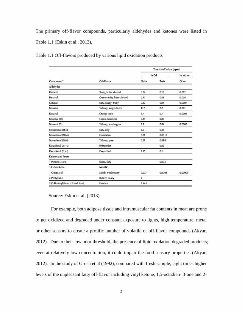
The primary off-flavor compounds, particularly aldehydes and ketones were listed in
Table 1.1 (Eskin et al., 2013).
Table 1.1 Off-flavors produced by various lipid oxidation products
Source: Eskin et al. (2013)
For example, both adipose tissue and intramuscular fat contents in meat are prone
to get oxidized and degraded under constant exposure to lights, high temperature, metal
or other sensors to create a prolific number of volatile or off-flavor compounds (Akyar,
2012). Due to their low odor threshold, the presence of lipid oxidation degraded products;
even at relatively low concentration, it could impair the food sensory properties (Akyar,
2012). In the study of Grosh et al (1992), compared with fresh sample, eight times higher
levels of the unpleasant fatty off-flavor including vinyl ketone, 1,5-octadien- 3-one and 2-

3
nonenal, were generated in butter fat after 42 days of storage. In addition, the increase in
the concentration of carbonyls such as 2-nonenal in butter oil or 1,5-octadien-3-ol in
boiled frozen trout significantly increased, replacing their original aroma with fatty,
tallowy, green off-odors (Grosch et al., 1994). In fried food or aging oil, a group of
volatile compounds including pentane, 2-heptenal, isomers of 2,4-heptadienal, and
isomers of 2,4-decadienal occur. With an increasing storage time, the concentration of
aldehydes and ketones pyridines, sulfides, thiazoles, alcohols, phenols and esters in fried
food or oil were several times higher than the original level (Min, & Schweizer, 1983).
Moreover, trimethylamine, as well as oxidized polyunsaturated fatty acids are responsible
for the off-flavors of products containing fish oil (Van Ba, Touseef, Jeong, & Hwang,
2012). Generally, lipid oxidation not only generates off-flavors of food, but also gives
rise to the loss of essential amino acids, fat-soluble vitamins, and other bioactive
compounds.
1.1.2 Lipid oxidation and health
In human body, free radicals could be generated internally in the normal metabolism or
by reactive oxygen species (ROS) in some external condition such as smoking, pollution,
and radiation. Especially, ROS induces the production of lipid peroxidation products
such as peroxides and aldehydes which then diffuse from their site of generation and
inflict damage at remote locations (Ramana, Srivastava, & Singhal, 2013). Therefore,
initiated by ROS, oxidized lipid products are able to propagate the responses and cause
dysfunction in biological tissue or organ injury (Ramana, Srivastava, & Singhal, 2013).
Generally, the oxidation of fatty acids is one of the most fundamental reactions in
lipid chemistry. Most oxidative degradation of fatty acids occurs in cellular membranes

4
such as mitochondria, microsomes, peroxisomes and plasma membrane (Boveris, &
Navarro, 2008). The onset of lipid peroxidation within biological membranes is
associated with changes in their physicochemical properties and with alteration of
biological function of lipids and proteins (Repetto et al., 2012). As a result, the toxicity
of lipid peroxidation products will further involve in neurotoxicity, hepatotoxicity and
nephrotoxicity (Boveris et al., 2008). Several studies have demonstrated that liver,
kidney, and heart are major target organs for oxidative damage (Eder, 1999). Oxidative
damage in liver is associated with hepatic lipid metabolism, and symptoms of the change
of liver weights and hepatic bile duct lesions were observed in test animals by
administration of oxidized oils and fats in the study of Eder (1999). Similar results
showed that lipid oxidation induced morphological damages and mitochondrial swelling
would further affect the organ structure and malfunction of the bile canaliculi as well as
the absorption and transportation mechanisms of α-tocopherol (Repetto et al., 2012).
Also, cardiac fibrotic lesions and necrosis were observed in heart due to the inflammatory
response from lipid peroxidation and disorder (Repetto et al., 2010). It has been reported
that fatty acid peroxides could accelerate three key phases of atherosclerosis which
includes endothelial injury in initiation, accumulation of plaque in progression and
thrombosis in termination (Singh et al., 2002). With the stimulation of endothelium,
inflammatory response starts to display adhesive molecules for circulating white blood
cells and producing cell type–specific agonists (McIntyre & Hazen, 2010). The vascular
endothelial cells will generate stimuli or plague by the activation of those agonists that
are associated with atherosclerosis, heart attack, Alzheimer’s disease, rheumatic arthritis
and other chronic diseases (McIntyre & Hazen, 2010).

5
In addition, there was a strong relationship between lipid peroxides and DNA
damage or development of cancer in humans (De Bont & van Larebeke, 2004). The
epoxy aldehydes from lipid oxidation are reactive in cross-linking reactions with proteins
and easily attack electron-rich centres in DNA, generating chemically altered bases
known as DNA adducts (Luczaj & Skrzydlewska, 2003). A group of lipid oxidation
derived compounds such as propeno and substituted propano adducts of deoxyguanosine
with malondialdehyde (MDA), acrolein, crotonaldehyde and etheno adducts, leads to
promutagenic lesions, responsible for the mutagenic and carcinogenic effects (Luczaj, &
Skrzydlewska, 2003). The lipid oxidized products will further alter protein properties by
the reaction with lysine amino, cysteine sulfhydryl, and histidine imidazole groups
(Esterbauer, 1996). Thus, the modifications of protein result in neurodegenerative
disorders, kinases activation and nuclear transcription inhibition (Uchida, 2003;
Camandola et al., 2000).
1.2 Antioxidant
Naturally, there is a dynamic balance between pro-oxidants and antioxidants to allow the
body to maintain the normal physiological conditions (Rahal et al., 2014). The
interference of the balance in any direction in the redox potential will lead to deleterious
results for organelle and biological site (Repetto et al., 2012). The oxidative damage that
occurs by an increase in the pro-oxidant over the capacity of the antioxidant is defined as
oxidative stress; while the reductive stress is a result of an increase in the reducing power
(Repetto et al., 2012). However, the protective antioxidant is only available to deal with
physiological rate of free-radical generation. In other words, the external and excessive
free radicals which generated from living environment that cannot be eliminated in time

6
and damage biologically relevant molecules such as proteins, lipid or carbohydrates
(Ashok & Sushil, 2005). Therefore, the antioxidant molecule or enzymatic systems play
important roles against, retard or avoid undesired oxidations in the body defense system
(Vivek & Surendra, 2006).
1.2.1 Categories of antioxidants
1.2.1.1 Synthetic antioxidants
Owing to the low cost and efficient performance, synthetic antioxidants are commonly
used during the storage and distribution of food products. Based on their action mode,
the synthetic antioxidants could be defined as primary antioxidants, which play important
roles in breaking the chain reaction of oxidation by hydrogen donation and generating
more stable radicals; while the secondary antioxidants which slow the oxidation rate by
metal chelation, primary antioxidant regeneration, decomposition of hydroperoxides,
singlet oxygen deactivation, ultraviolet radiation absorption, and oxygen scavenging
(Flora, 2009; Barbut, 2010). Butylated hydroxyanisole (BHA), butylated hydroxytoluene
(BHT), tert-butylhydroquinone (TBHQ), and propyl gallate (PG), dodecyl gallate (DG)
and octyl gallate (OG) are a group of free radical terminators (Figure 1.1).
Butylated hydroxytoluene (BHT) Butylated hydroxyanisole (BHA)
BHA

7
Figure 1.1 Chemical structures of the synthetic antioxidants
Among them, BHA is most effective in preserving food color and flavor as well
as animal fats with short chain fatty acids during baking and frying (Devlieghere,
Vermeiren, & Debevere, 2004). The combination use of BHA and BHT are shown to
have synergistic effects, especially in nut products (Shahidi et al., 1992). TBHQ is an
effective supplement in color improvement and helps to increase oxidative stability in the
process of liquid oil hydrogenation and fried food (Shahidi & Zhong, 2005). Along with
citric acid, PG is commonly used in stabilizing vegetable oil or animal fats and chewing
gum bases (Shahidi & Zhong, 2005). Octyl gallate (OG) and dodecyl gallate (DG) are
the other two gallate antioxidants used as food additives in shortening, baked goods,
candy, chewing gum and dried milk (van der Heijden, Janssen, & Strik, 1986). As for the
chelating agent, EDTA could form complexes with pro-oxidative metal ions and is
mostly used in processed fruits and vegetables, salad dressings, soft drinks, margarine
and canned fish (Rangan & Barceloux, 2009).
However, an excessive dose of synthetic antioxidants implicates carcinogenicity,
liver lesion, haemorrhaging, etc., which are harmful to human health, thus, they are
tert-Butylhydroquinone (TBHQ) Propyl gallate (PG)
Dodecyl gallate (DG) Octyl gallate (OG)

8
strictly limited in food applications (Augustyniak et al., 2010). Even at a normal level
but long term of ingestion, the synthetic antioxidant would assist in modifying acute
toxicity of carcinogenic and mutagenic effect depending on their pattern of metabolism
(Ames, Profet, & Gold, 1990). For example, BHA has been evidenced to cause
hypertrophy of liver, thyroid, adrenals and lungs and cell lysis or alteration of lipid
composition in serum and platelets (Gould, 1995). On the other hand, BHT could inhibit
DNA repair in human lymphocytes and cause introduction of chromosomal and sperm
abnormalities along with the interference with leukemia cell differentiation (Chun et al.,
2006). In addition, a morphological observation showed cell loss and the induction of
cell death by necrosis and apoptosis with the treatment of certain amount of PG (Zurita et
al., 2007). Therefore, each synthetic antioxidant has a limitation for consumption and the
potential common sources are also listed in Table 1.2.
Table 1.2 Morphology, and limits of synthetic antioxidants used in foods
Name Morphology Limit in food Found in Banned in
BHA White waxy
flakes <200mg/kg
Cereal, chewing
gum, potato chip,
vegetable oil
Japan
BHT White crystalline
powder <100mg/kg
Honey, cakes, meat
products None
PG White crystalline
powder <200mg/kg
Spices, sugar, milk
product None
TBHQ Beige colored
powder Not allowed
Cheese, honey,
sea food
Canada, Japan,
European countries
OG
White to creamy
white crystalline
solid
<200mg/kg Oil, meat product,
cereals, honey, None
DG
White to creamy
white crystalline
solid
<200mg/kg Dairy and meat
products None
EDTA White crystalline
powder
Salad dressing, soft
drink, mayonnaise None
Source: Goodman (1980); Morton et al. (2000)

9
1.2.1.2 Natural antioxidants
Different from synthetic antioxidants, most of the natural antioxidants originate from
plant sources such as fruits and vegetables which are much safer than the synthetic ones
(Tayel & El-Tras, 2012). Commonly, vitamins, carotenoids, phenolic acids, and
flavonoids are the primary four types of natural antioxidants.
As one of the most important vitamins, ascorbic acid (vitamin C) is a water
soluble antioxidant and particularly abundant in citrus fruits (Marti, Mena, Canovas,
Micol, & Saura, 2009). It has been proved that the absorption of dietary vitamin C could
be up to 50% at a relatively lower dose of intake (< 39 mg) (Levine et al., 1996). The
participation of vitamin C in numerous oxidation-reduction reactions involves synthesis
of connective tissue of sulfate and collagen and improvement of iron adsorption in
gastrointestinal (GI) tract (Houglum, Brenner, & Chojkier, 1991). Deficiency of vitamin
C results in chronic alcoholism, scurvy, inflammatory bleeding gums, loss of teeth,
arrested skeletal development, dry skin, joint pain and increased susceptibility to the
infections (Hacisevkd, 2009). Vitamin E, a group of tocopherols, is a lipophilic
antioxidant which is abundant in cereal grains, nuts, spinach, oliver or flaxseed oil
(Urquiaga, & Leighton, 2000). It encompasses four types of four tocopherols (α, β, γ, δ)
and four tocotrienols (α, β, γ, δ), among which, α-tocopherol has the biggest poportion in
nature source and the highest biological activity based on fetal resorption assays (Figure
1.2) (Weiser, Riss, and Kormann, 1996). In general, the availability of vitamin E is
relatively low which is about 20-40% of the dietary intake; however, its bioavailability
could be enhanced by taking it in conjunction with dietay fats (Daniel, 1986). The

10
symptoms of vitamin E deficiency appear as neuromuscular abnormalities characterized
by spinocerebellar ataxia and myopathies (Brigelius-Flohé & Traber, 1999).
Figure 1.2 The chemical structures of four tocopherols and tocotrienals Source:
(Brigelius-Flohé & Traber, 1999)
Carotenoids are a group of natural pigments of the polyene type, consisting of a
ubiquitous group of isoprenoid (Figure 1.3) (Fiedor & Burda, 2014). As lipophilic
molecules, β-carotene and lycopene with strict hydrocarbons in their studctures are
located within the inner section of the lipid bilayer, while lutein and zeaxanthin with
attached oxygen atoms are arranged roughly perpendicular to the membrane surface
(Wiśniewska, & Subczyński, 1998; Wiśniewska, & Subczyński, 2006). Also, β and α-
carotene and β-cryptoxanthin perform as provitamin A precursors which have the
capacity to be converted to vitamin A, helping growth and reproductive efficiency,
maintenance of epithelial tissues and immune response (Scott & Rodriquez-Amaya,

11
2000). The major dietary sources of carotenoids include pumpkins, sweet potatoes,
oranges, tomatoes, mangoes, papayas and yellow or red bell peppers, etc. (Xu, 2012).
The intestinal absorption of carotenoids undergoes initially incorporation into mixed lipid
micelles in the lumen, then, followed by the uptake into intestinal mucosa (Deming, &
Erdman, 1999). After incorporation into chylomicrons, the carotenoids will be released
into the lymph (Harrison, 2010). It has been reported that only about 5% of the
carotenoids could be absorbed by the intestine, whereas more than 50% are from micellar
solution (Olson, 1994). However, the deficiency of carotenoids has the consequences of
xerophthalmia, blindness and premature death (Sommer, 2008).
Figure 1.3 The chemical structures of caroteinoids
Source: Fiedor & Burda (2014)
Phenolic acids are referred to the aromatic secondary plant metabolites,
possessing one carboxylic acid group; they are found in fruits, vegetables and product
derivatives (Saxena et al., 2012). Generally, they are subdivided into two major groups:
12
hydroxybenzoic acids and hydroxycinnamic acids (Manach, Scalbert, Morand, Rémésy &
Jiménez, 2004). Hydroxycinnamic acid is a simple ester with glucose or hydroxy
carboxylic acid, consisting of a phenylpropanoid C6-C3 structure, and considered to be
the major subgroup of phenolic acids (Table 1.3) (Teixeira et al., 2013).
Hydroxycinnamic acids mainly consist of p-coumaric, caffeic, ferulic, and sinapic acids
etc., and are important in the biosynthesis of complicated phenolic systems in food and
beverage such as tea leaves, coffee, red wine, and various fruits, vegetables and whole
grains (Teixeira et al., 2013). Hydroxycinnamic acids exist either in free form or in
conjugated forms, including mono- or polyamines, amino acids, peptides and esters as
well as glycosides (Teixeira et al., 2013). The variety of hydroxycinnamic acid in plants
depends on the species of the plants; however, their levels are up to 75% of the totoal
phenolic acids (Terry, 2011). Also, it was found that derivatives of cinnamic acid are
much higher in the outer parts of ripen fruits than in other parts (Ribera et al., 2010). The
association of hydroxycinnamic acids with the reduced risks of cardiovascular disease,
cancer and other chronic diseases could be explained by the ability to scavenge free
radicals and pro-oxidant metals ability as well as as modulation of the specific enzymes
activiy and inhibition of cell proliferation (Spencer et al., 2008; Manach, 2004). The
performance of general antioxidant capability of the hydroxycinnamic acids in some lipid
peroxidation systems can be expressed by the inhibition of malondialdehyde formation
(Laranjinha et al., 1994).
On the other hand, the hydroxybenzoic acids are a group of tannins and lignins
which with either hydroxylations or methoxylations of the aromatic ring (Manach et al.,

13
2004). The benzoic acid derivatives stem from the side chain degradation by lossing an
acetate of the corresponding hydroxycinnamic acid derivatives, or from an intermediate
Table 1.3 Hydroxycinnamic acids and ester derivatives.
R1 R2 R3
p-Coumaric H H H
Caffeic acid OH H H
5-Bromocaffeic acid OH Br H
Methyl caffeate OH H CH3
Ethyl caffeate OH H CH2CH3
Ethyl 5-bromocaffeate OH Br CH2CH3
Propyl caffeate OH H CH2CH2CH3
Butyl caffeate OH H CH2(CH2)2CH3
Hexyl caffeate OH H CH2(CH2)4CH3
3,4,5-Trihydroxycinnamic acid OH OH H
Ethyl 3,4,5-trihydroxycinnamate OH OH CH2CH3
Ferulic acid OCH3 H H
5-Bromoferulic acid OCH3 Br H
Methyl ferulate OCH3 H CH3
Ethyl ferulate OCH3 H CH2CH3
Ethyl 5-bromoferulate OCH3 Br CH2CH3
Propyl ferulate OCH3 H CH2CH2CH3
Butyl ferulate OCH3 H CH2(CH2)2CH3
Hexyl ferulate OCH3 H CH2(CH2)4CH3
Sinapic acid OCH3 OCH3 H
Methyl sinapate OCH3 OCH3 CH3
Ethyl sinapate OCH3 OCH3 CH2CH3
Propyl sinapate OCH3 OCH3 CH2CH2CH3
Butyl sinapate OCH3 OCH3 CH2(CH2)2CH3

14
in the shikimate pathway, involving in a series of enzymatic reactions (Herrmann, 1995).
Its derivatives mainly refer to p-hydroxybenzoic, protocatechuic, salicylic, vannilic,
syringic and gallic acids (Figure 1.4) (Rocha, Monteiro, & Teodoro, 2012). Gallic acid
may be conjugated as its dimer, and trimer, and are able to be esterified to glucose in
hydrolysable, condensed, or derived tannins and quinic acid (Clifford, & Scalbert, 2000).
Protocatechuic acid is considered to be a therapeutic compound which generates potential
antioxidant, antiulcer, antiageing, antihyperlipidemic, antifibrotic and anti-inflammatory
activities (Kakkar & Bais, 2014). The biological properties of p-hydroxybenzoic include
antiviral, antimutagenic, and anti-inflammatory activities, and it has been used as
hypoglycemic or terarogenic agent (Manuja et al., 2013).
Figure 1.4 The chemical structure of hydroxybenzoic acids
Source: Khadem & Marles (2010)
Gallic acid Protocatechuic acid p-Hydroxybenzoic acid
Salicylic acid Vanillic acid Syringic acid

15
Flavonoids are a special class of phenolics in which flavonoid groups have low
toxicity in mammals and are widely distributed in plant kingdom (Manach et al., 2004).
All flavonoids have the common C6-C3-C6 structural skeleton, consisting of two
aromatic rings (A and B) linked through a heterocyclic pyrane ring (C) with one oxygen
atom (Figure 1.5).
Figure 1.5 The basic structure of flavonoid
Primary dietary sources of flavonoides could be subdivided into six subclasses in
the form of flavonols, flavones, isoflavones, flavonones, flavan-3-ol and flavanol which
could be easily found in a variety of fruits and vegetales such as berries, soybeans and
purple onion and cabbage (Figure 1.6) (Beecher, 2003).

16
Figure 1.6 Different classes of flavonoids and their substitution patterns
Source: Beecher (2003)
The role of flavonoids is not only to provide attractive colors to plant but also to
help promote physiological survival by regulating hormones, morphogenesis and
photosynthesis as well as preventing from fungal pathogens and UV-radiation (Harborne
& Williams, 2000). Recently, many studies have shown that flavonoids maintain broad
biological and pharmacological effect on blood vessels, platelets, and lipoproteins and
may exhibit an excellent performace of reducing the risks of coronry heart disease by
modulating cardiac antioxidant defenses (Mazza, 2007). Moreover, the digestion of
flavanoids assists improvement of antioxidant status and displays cholesterol-lowering

17
and distinct hypoglycemic actions (Prior, & Wu, 2012). In addition, flavanoids has the
potential to ameliorate hyperglycemia and enhance insulin sensitivity via activation of
AMP-activated protein kinase (AMPK) (Taikanwa et al., 2010)
1.3 Antioxidant Rich Food
Generally, agricultural products are recognized as important sources of a wide array of
phytochemicals and dietary micronutrients (Yahia, 2010). They are able to generate
favorable effects on the control of various chronic diseases and play a protective role in
health maintenance (Stea et al., 2008). Obtained from diet, antioxidants could neutralize
excessive ROS produced during physiological processes such as increased physical
activity, tissue ischemia, reperfusion, inflammation, and mental stress or depression
(Singh, Shashwat, & Suman, 2004). Therefore, the intake of antioxidant-rich food is
necessary to improve health and quality of life.
1.3.1 Fruits, vegetables and herbs
Berry fruits (blackberries, raspberries, blueberries, cranberries, bilberries, strawberries,
crowberries, cloudberries etc.) with purple, black or red color are abundant in
anthocyanins and flavan-w3-ols hydroxybenzoic or hydroxylcinnamic acid derivatives,
condensed and hydrolyzable tannins, etc. (Howard & Hager, 2007). Those bioactive
compounds have been evidenced to contribute to antioxidant capability in a LDL (Low-
density lipoprotein) oxidation model (Heinonen et al., 1998). Also, in the study of Tsuda
et al. (2003), induced by the high-fat diet, the mice fed up with cyaniding-3-O-glucoside
have lower body weight gain and white and brown adipose tissue weights compared with
control groups. Grapes maintained a significant quantities of anthocyanins, catechin,
gallic acid,and resveratrol and their levels are correlated with anti-lipid-oxidation

18
capabilities in human body (Frankel et al., 1998). Catechin, procyanidins, flavonols,
hydroxycinnamic acid and dihydrochalcones are primary phytochemicals ranging from
0.5-2.7% on dry weight basis in apples (Xu, 2012). In an animal study, the extract of
apple flesh and peels were reported to prevent macro- and microscopic damage and
barrier dysfunction along the gastrointestinal tract (Carrasco-Pozo et al., 2011). Color
peppers commercial available in markets contain carotenoids, vitamin A, C and E, ferulic
acid, sinapic acid, quercetin, luteolins and apigenins (Frank et al., 2001). The presence
of those bioactive compounds enables the color peppers to have capability of preventing
free radical oxidation, cardiovascular disease, cancer and neurological disorders (Xu,
2012). Similarly, the culinary herbs such as basil, ginger, thyme and rosemary are used
to enhance and complement the flavors of various foods (Xu, 2012). Having various
kinds of phenolic acids, including carnosic acid, carnosol, rosmarinic acid, curcumin,
eugenol etc., the culinary herbs showed strong antioxidant potential and medicinal
benefits (Shan et al., 2005).
1.3.2 Cereals
Different from fruits and vegetables, vitamin E (mainly tocopherols and toctrienols) is the
most abundant antioxidant and within the germ of whole-grain cereals (Souci et al., 2000).
For example, rice, containing α- and β-tocopherol, α- and β-tocotrienol, and γ-orazanol
are demonstrated to reduce serum cholesterol level, the risk of tumor formation and
inflammatory action (Tsuji et al., 2003). Similar to other cereal grains, oats have
relatively high amount of vitamin E, while it is also a great source of phenolic acids such
as vanillic and p-hydroxylbenzoic acid (Peterson, 2001). The lipophilic extract of oats
could effectively inhibit oxidation of unsaturated long chain fatty acids and prevent

19
production of toxic cholesterol oxidized products, and is used as the natural food
preservatives (Sun et al., 2006). Additionally, corn is confirmed as a common raw
material for producing edible oil, in which, the concentration of vitamin E could be up to
900 mg/kg (Xu, 2012). Apart from tocopherols and phenolic acids, corn is a rich source
of carotenoids such as α- and β-carotene, β-cryptoxanthin, and zeaxanthin, having a
major function to prevent age-related macular degeneration (AMD) and cataracts and cell
mutation (Johnson, 2000). Soybean is another cereal family in possess of various types
of isoflavones with β-glucoside, daidzin, glycitin and genistin (Xu, 2012). They have
been reported to show the capability of retarding the progression of Alzheimer disease
and osteoporosis, and preventing harmful proliferation of cells (Zhao, et al., 2002).
1.3.3 Root vegetables
As the most common root vegetables, potatoes and sweet potatoes are two staple crops in
tropical and subtropical areas (Mark et al., 2009). Currently, it occupies a dominant place
in the agricultural production of Asia and Africa countries. The nutritional values of
sweet potato are listed in Table 1.4. Specially, carbohydrates of sweet potato could
provide energy in the human diet with a lower intake of lipids (Oke, & Workneh, 2013).
The dietary fiber is believed to reduce the incidence of colon cancer, diabetes, heart
diseases and digestive disturbances (Vimala, Nambisan, & Hariprakash, 2011). The flesh
color of the sweet potato varies from yellow to dark-orange depending upon the levels
and types of carotenoids (Vimala, Nambisan, & Hariprakash, 2011).
Carotenoids are reported to be responsible for the enhancement of immune system
and inhibition of age-related macular degeneration and cataract formation (Byers & Perry
1992). Also, the purple sweet potato becomes more and more popular due to the unique

20
Table 1.4 Sweet potato chemical composition
and abundant anthocyanins which have been reported to suppress the development of
atherosclerotic lesions and oxidative stress in an in vivo study (Miyazaki et al., 2008).
Furthermore, both sweet potatoes and potatoes contain diverse phenolics such as
chlorogenic, gallic, protocatechuic and caffeic acid as well as quercetin 3-O-rutinoside,
kaempferol 3-O-rutinoside and (+)-catechin (Xu, 2012). Lotus root is an herbaceous,
perennial sacred aquatic plant belonging to Nelumbonaceae family (Du et al., 2010). It is
well known from its medical functions such as the treatment of all manners of bleeding
and hematemesis, anti-inflammatory, antipyretic and antianxiety properties and hepatic
protection (Mukherjee et al., 2009). Although carrots and radish are another commonly
consumed root vegetables, their phytochemical profile and antioxidant activity are
relatively lowered than other roots.
1.4 Evaluation of Antioxidants Activity
1.4.1 In vitro methods
1.4.1.1 Spectrophotometric assay
The antioxidant compounds have been well extracted, determined and quantified.
Recently, their antioxidant potential and effectiveness have raised scientists and
21
consumers’ concern. As a result, various in vitro assessments of antioxidant activities
have been developed (Table 1.5). Based on the mechanisms, the antioxidant evaluation
methods could be generally catalogued into hydrogen atom transfer methods (HAT) and
electron transfer methods (ET) (Badarinath et al., 2010). HAT is a ubiquitous reaction
involves the transfer of proton and electron for further measuring the free radical
quenching by hydrogen donation and analyzing the destructive effects of reactive oxygen
species (Mayer, 2011). The ET method aims at determining the ability to transfer one
electron to reduce any compounds such as metals, carbonyls, and radicals (Prior, Wu, &
Schaich, 2005).
DPPH free radical scavenging activity The molecule 1, 1-diphenyl-2-picrylhydrazyl
(a,a-diphenyl-bpicrylhydrazyl; DPPH) is characterized as a relatively stable free radical
with deep violet color by virtue of the delocalization of the spare electron over the
molecule which is not associated with a single atom or covalent bond (Alam, Bristi, &
Rafiquzzaman, 2013). The change in optical density of the DPPH free radical caused by
the donation of hydrogen atom from a potential antioxidant could be determined under
the wavelength of 517 nm (Alam, Bristi, & Rafiquzzaman, 2013).
2,2’-Azinobis-(3-Ethyl-Benzothiazoline-6-Sulphonic Acid)/ Trolox equivalent
antioxidant capacity (ABTS/TEAC) In the ABTS assay, ABTS is incubated with a
peroxidase and a relatively stable radical cation, and ABTS+ is formed, producing a
relatively stable blue-green color, which can be measured at 600nm. Similar to the
DPPH assay, TEAC is based on the ability of molecules to scavenge the stable free
radical of 2,2’- azinobis (3- ethylbenzothiozoline-6-sulfonic acid) and expressed as the
Trolox equivalent (Badarinath et al., 2010).
22
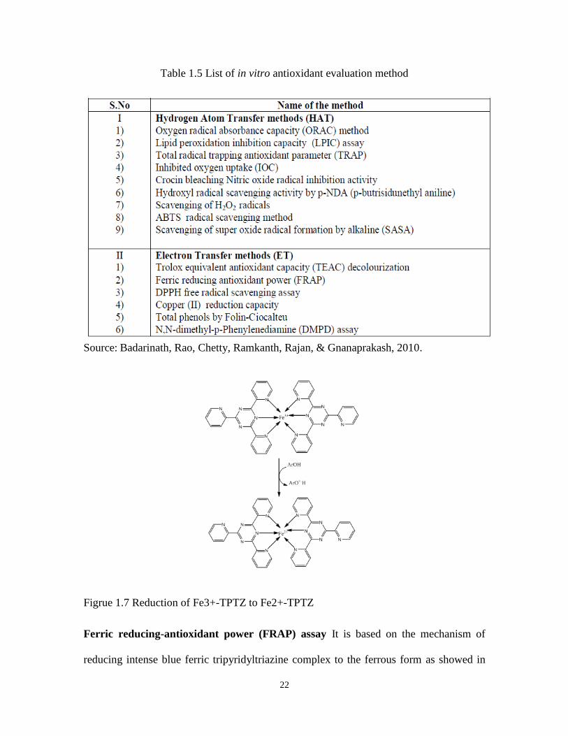
Table 1.5 List of in vitro antioxidant evaluation method
Source: Badarinath, Rao, Chetty, Ramkanth, Rajan, & Gnanaprakash, 2010.
Figrue 1.7 Reduction of Fe3+-TPTZ to Fe2+-TPTZ
Ferric reducing-antioxidant power (FRAP) assay It is based on the mechanism of
reducing intense blue ferric tripyridyltriazine complex to the ferrous form as showed in

23
Figure 1.7 (Gupta, 2015). The reducing power of the target antioxidant could be reflected
by the color change monitored under the wavelength of 593nm.
Cupric ion reducing antioxidant capacity (CUPRAC) CUPRAC method is designed to
demonstrate the ability of scavenging hydroxyl radicals. The chromogenic oxidizing
reagent, bis(neocuproine)copper(II) chloride [Cu(II)-Nc], reacts with polyphenols
[Ar(OH)n] (as shown in the equation below).
It is applicable to both hydrophilic and lipophilic and will not be affected by the sugars
and citric acid commonly present in foods since it has a selective action on antioxidant
compounds (Alam, Bristi, & Rafiquzzaman, 2013).
Folin-Ciocalteu method The reaction involves a chemical reduction of the mixture of
tungsten and molybdenum oxides, in which, the molybdenum centre in the complex
reagent is reduced from Mo (VI) to Mo (V), generating a blue color at 750 nm. The
intensity of color is proportional to the concentrations of phenol compounds, thus, it is
often used for the assessment of total phenolic content (Gupta, 2015).
Oxygen radical absorbing capacity (ORAC) assay Using Trolox as the reference, a
test sample is applied to inhibit the oxidation of B-phycoerythrin initiated by thermal
decomposition of azo compounds such as AAPH in the ORAC assay (Glazer, 1990). The
peroxyl radical reacts with a fluorescent probe to form a non-fluorescent product; thus, it
reflects classical radical chain breaking antioxidant activity by H atom transfer (Ou et al.,
2001).
24
2,2'-Azino-bis(3-ethylbenzothiazoline-6-sulphonic acid (ABTS) Assay: In this method,
the treatment of 2, 2’-azino-bis(3-ethylbenzthiazoline-6-sulphonic acid) with K2S2O8 or
MnO2 provides a bluish-green radical cation (ABTS+) (Re, Pellegrini, Proteggente,
Pannala, Yang, & Rice-Evans, 1999). The ABTS radical cation could be further changed
back to colorless neutral form in the presence of both lipophilic and hydrophilic
compounds such as flavonoids, hydroxycinnamates and carotenoids (Gupta, 2015). Due
to the high reactivity of ABTS radical cation, it could be scavenged by most antioxidants
including phenolics, thiols and vitamin C (Walker & Everette, 2009).
Total radical trapping antioxidant parameter (TRAP) Assay Induced by AAPH (2’-
azobis(2-amidinopropane) hydrochloride), the degree of peroxidation is monitored by
screening of the loss of fluorescence from the protein R-phycoerythrin (R-PE)
(Badarinath et al., 2010). It is especially good for the determination of in-vivo
antioxidant capacity in serum or plasma since it measures nonenzymatic antioxidants
(Huang, Ou, & Prior, 2005).
1.4.1.2 Model system
The existing spectrophotometric system is easy to carry out; however, direct analysis fails
to predict the mechanism of antioxidants in inhibiting the oxidation in the complex food
matrix (Frankel, 1993). Also, the free radicals used in each assay are non- physiological;
therefore, it is difficult to capture the different modes of action of antioxidant. Currently,
a few model systems such as ground meat, fish oil, cholesterol and β-carotene linoleate
model systems have been developed (Rojas and Brewer 2007; Jayaprakasha et al. 2001;
Shen et al., 2013; Zhang et al., 2013).

25
Meat model Since a big amount of iron could be released from denatured myoglobin
and hemoglobin, they are able to catalyze the oxidation rapidly in cooked meats.
Therefore, natural or synthetic antioxidants can be applied in the model for the evaluation
of anti-lipid-oxidation capability through determining the production of malondialdehyde
(MDA) (Rojas and Brewer, 2007).
Fish oil model The fish oil emulsion consists of water, lipid micelles, and other
components which could mimic the biological fluids. Also, the ubiquitous
polyunsaturated fatty acids (PUFAs) such as EPA and DHA would oxidize rapidly and
are critical to indicate antioxidant efficacy. Thus, the results of these emulsion models
could closely reveal the antioxidant power in preventing lipid oxidation in biological
fluids and cells (Zhang et al., 2013).
Cholesterol model Cholesterol is the main components in the cell membrane and blood
serum of mammals and can be oxidized under oxidative stress. The primary oxidized
product of cholesterol is 7-ketocholesterol which is determined to predict the degree of
emulsion oxidation. By comparing the efficiency of the antioxidant in retarding oxidation
in the cholesterol emulsion, the result of the antioxidant potential would be more
convinced (Shen et al., 2013).
1.4.2 In vivo methods
The in vivo studies usually use the test animals such as mice, rats, and rabbits etc. to
evaluate the antioxidant effect of target food extract in specific organs or cells based on
different administration and dose (Alam, Bristi, & Rafiquzzaman, 2013). After a certain
period of time, the animals are sacrificed and blood or tissues are used for the
determination.

26
Reduced glutathione (GSH) estimation GSH is an intra-cellular reductant and helps to
protect cells against free radicals, peroxides and other toxic compounds in catalysis,
metabolism and transport (Sapakal et al., 2008). As the irreplaceable role in transport
system in the kidney, it is an essential indicator to reveal the in vivo antioxidant defense.
The prepared sample supernatant is added with the Ellman’s reagent (5,50-dithiobis-2-
nitrobenzoic acid in phosphate buffer solution), then, the absorbance of the solution is
read at 412 nm against blank after completion of the reaction (Alam, Bristi, &
Rafiquzzaman, 2013).
Superoxide dismutase (SOD) method Similar to GSH, SOD is an enzyme which is
able to remove the free-radicals caused by environmental adversity, and improve stress
tolerance (Kong, Zhao, Liu, He, Tian, & Zhou, 2012). SOD dismutes the superoxide
anion and thereby inhibits the reduction of a small hemeprotein cytochrome-c. Its
antioxidant efficiency is estimated by an increase in absorbance recorded at 420 nm and
expressed as units/mg protein (Alam, Bristi, & Rafiquzzaman, 2013).
Catalase (CAT) method
It is a tetrameric heme-containing enzyme that catalyzes the dismutation of H2O2 into
water and oxygen molecules. It has high specificity for a high level of the presented H2O2
(Sharma, Jha, Dubey, & Pessarakli, 2012). The catalase activity of the hemolysate is
determined by adopting the erythrocyte lysate method at 240 nm. One unit of activity
represents the degradation of 1 mM of H2O2 /min and is expressed as units per milligram
of protein (Alam, Bristi, & Rafiquzzaman, 2013).
Cell culture
A cellular antioxidant activity (CAA) assay was developed to evaluate the antioxidant

27
capability of bioactive compounds from food and plant products (Wolfe & Liu, 2007).
Human hepatocarcinoma Hep-G2 cells are mixed with the redox sensor
dihydrodichlorofluorescein (DCFH2) to oxidized to fluorescent dichlorofluorescein (DCF)
by ROO• radical induced by 2,2'-Azobis(2-amidinopropane) dihydrochloride (AAPH). It
aims at measuring the ability to inhibit the intracellular DCFH2 oxidation determined by
fluorescence at λexc 485 nm and λem 535 nm. The obtained results are expressed in μM
of quercetin equivalents. Therefore, the CAA assay could be used for screening
antioxidant activity in natural product extracts at the cellular level.
1.5 References
Alam, M. N., Bristi, N. J., & Rafiquzzaman, M. (2013). Review on in vivo and in vitro
methods evaluation of antioxidant activity. Saudi Pharmaceutical Journal, 21,
143-152.
Ames, B. N., Profet, M., & Gold, L. S. (1990). Nature's chemicals and synthetic
chemicals: comparative toxicology. Proceedings of the National Academy of
Sciences, 87, 7782-7786.
Angel Catala. (2012). Lipid Peroxidation. (1st ed). Croatia:InTec, (Chapter 1).
Ashok, A. & A. Sushil. (2005). Oxidative stress and antioxidants in male infertility: A
difficult balance. Iranian Journal of Reproductive Medicine, 3, 1-8.
Augustyniak, A., Bartosz, G., Cipak, A., Duburs, G., Horakova, L., Luczaj, W.,
Majekova, M., Odysseos, A. D., Rackova, L., Skrzydlewska, E., Stefek, M.,
Strosova, M., Tirzitis, G., Venskutonis, P. R., Viskupicova, J., Vraka, P. S., &
Zarkovic, N. (2010). Natural and synthetic antioxidants: an updated overview.
Free Radical Research, 44, 1216-1262.
Badarinath, A., Rao, K. M., Chetty, C. M. S., Ramkanth, S., Rajan, T., & Gnanaprakash,
K. (2010). A review on in-vitro antioxidant methods: comparisions, correlations
and considerations. International Journal of PharmTech Research, 2, 1276-1285.
Barbut, S. (2010). Poultry Products Processing: An Industry Guide: CRC Press (pp. 262).
Barriuso, B., Astiasarán, I., & Ansorena, D. (2013). A review of analytical methods
measuring lipid oxidation status in foods: a challenging task. European Food
Research and Technology, 236, 1-15.

28
Beecher, G. R. (2003). Overview of dietary flavonoids: nomenclature, occurrence and
intake. The Journal of Nutrition. 133, 3248-3254.
Boveris, A. & Navarro, A. (2008) Brain mitochondrial dysfunction in aging. Life, 60(5),
308-314.
Brigelius-Flohé, R., & Traber, M. G. (1999). Vitamin E: function and metabolism. The
FASEB Journal, 13, 1145-1155.
Byers, T., & Perry, G. (1992). Dietary carotenes, vitamin C and vitamin E as protective
antioxidants in human cancers. Annual Review of Nutrition,12, 139–159.
Camandola, S., Poli, G., & Mattson, M. (2000) The lipid peroxidation product 4-
hydroxy-2,3-nonenal inhibits constitutive and inducible activity of nuclear factor-
bin neurons. Molecular Brain Research, 85, 53–60.
Carraso-Pozo, C., Speisky, H., Brunser, O., Pastene, E., & Gotteland, M. (2011). Apple
peel polyphenols protect against gastrointestinal mucosa alterations induced by
indomethacin in rates. Journal of Agricultural and Food Chemistry, 59, 6459-
6466.
Choe, E., & Min, D. B. (2006). Chemistry and reactions of reactive oxygen species in
foods. Critical Reviews in Food Science and Nutrition, 46, 1-22.
Chun, J., Lee, J., Ye, L., Exler, J., Eitenmiller, R.R. (2006). Tocopherol and tocotrienol
contents of raw and processed fruits and vegetables in the United States diet.
Journal of Food Composition and Analysis, 19, 196-204.
Clifford, M. N., & Scalbert, A. (2000). Ellagitannins - nature, occurrence and dietary
burden. Journal of the Science of Food and Agriculture ,80,1118-1125.
De Bont, R., & van Larebeke, N. (2004). Endogenous DNA damage in humans: a review
of quantitative data. Mutagenesis, 19, 169-185.
Deming, D.M., & Erdman, J.W., Jr. (1999). Mammalian carotenoid absorption and
metabolism. Pure and Applied Chemistry, 71, 2213–2223.
Daniel, J. W. (1986). Metabolic aspects of antioxidants and preservatives. Xenobiotica,
16, 1073-1078.
Devlieghere, F., Vermeiren, L., & Debevere, J. (2004). New preservation technologies:
Possibilities and limitations. International Dairy Journal, 14, 273-285.
Du, H., Zhao, X., You, J.-S., Park, J.-Y., Kim, S.-H., & Chang, K.-J. (2010). Antioxidant
and hepatic protective effects of lotus root hot water extract with taurine
supplementation in rats fed a high fat diet. Journal of Biomedical Science,17, S39-
S39.

29
Eder, K. (1999). The effect of a dietary oxidized oil on lipid metabolism in rats. Lipids,
34, 717–725.
Eskin, N. A. M., & Shahidi, Fereidoon. Biochemistry of foods. In Karen M. Schaich,
Fereidoon Shahidi, Ying Zhong, and N. A. Michael Eskin (Eds.), Lipid Oxidation
Amsterdam (pp.446-457). Boston : Academic Press.
Esterbauer, H. (1996) Estimation of peroxidative damage. A critical review. Pathologie
Biologie, 44, 25–28.
Frank, C.A., Nelson, R. G., Simonne, E. H., Behe, B. K. & Simonne, A. H. (2001).
Consumer preferences of colour, price, and vitamin C content of bell peppers.
Horticultural Science, 36, 795-800.
Frankel, E. N., Bosanek, C. A., Meyer, A. S., Silliman, K., & Kirk, L. L. (1998).
Commercial grape juices inhibit the in vitro oxidation of human low-density
lipoproteins. Journal of Agricultural and Food Chemistry, 46, 834-838.
Frankel, E. N. (1993). In search of better methods to evaluate natural antioxidants and
oxidative stability in food lipids. Trends in Food Science & Technology, 4, 220-
225.
Fiedor, J., & Burda, K. (2014). Potential Role of Carotenoids as Antioxidants in Human
Health and Disease. Nutrients, 6, 466-488.
Flora, S. J. S. (2009). Structural, chemical and biological aspects of antioxidants for
strategies against metal and metalloid exposure. Oxidative Medicine and Cellular
Longevity, 2, 191-206.
Glazer, A.N. (1990). Phycoerythrin fluorescence-based assay for reactive oxygen species.
Methods in Enzymology,186, 161-8.
Goodman,D.W.S. (1980). Vitamin A metabolism. Federation Proceedings - Fed of Am
Societies for Experimental Biology, 39, 2716-2722.
Gould, G. W. (1995). Biodeterioration of foods and an overview of preservation in the
food and dairy industries. International Biodeterioration & Biodegradation, 36,
267-277.
Grosch, W., Widder, S., & Sen, A. (1992). Changes in the flavour compounds of butterfat
during storage. In M. Rothe, H.P. Kruse (Eds.), Aroma Production and
Application (pp. 147-154). Deutsche Institute für Ernährungschforschung,
Potsdam-Rehbrücke.
Grosch, W., Milo, C., & Widder, S., Identification and quantification of odorants causing
off-flavours. (1994) In H. Maarse & D.G. Van der Hej (Eds.) Trends in Flavour
Research (pp. 409–415). Amsterdam: Elsevier Science.
30
Gupta, D. (2015). Methods for determination of antioxidant capacity: a review.
International journal of pharmaceutical sciences and research, 6, 546-566.
Harborne, J. B., & Williams, C. A. (2000). Advances in flavonoid research since 1992.
Phytochemistry, 55, 481-504.
Hacisevkd, A. (2009). An overview of ascorbic acid biochemistry. Journal of Faculty of
Pharmacy of Ankara, 38, 233–255.
Harrison, E.H. (2010). Mechanisms of Intestinal Absorption of Carotenoids: Insights
from in Vitro Systems. In J.T. Landrum (Eds.) Carotenoids: Physical, Chemical,
and Biological Functions and Properties (pp. 367–381). Boca Raton: CRC Press,
Taylor & Francis.
Heinonen, I. M., Lehtonen, P. J., & Hopia, A. I. (1998). Antioxidant activity of berry and
fruit wines and liquors. Journal of Agricultural and Food Chemistry, 46, 25-31.
Herrmann, K. M. (1995). The shikimate pathway: early steps in the biosynthesis of
aromatic compounds. Plant Cell, 7, 367–381.
Houglum, K.P., Brenner, D.A., & Chojkier. M. (1991). Ascorbic acid stimulation of
collagen biosynthesis independent of hydroxylation. The American Journal of
Clinical Nutrition, 54, 1141–1143.
Howard, L. R., & Hager T. J. (2007). Berry fruit phytochemicals. In Y Zhao (Eds.) Berry
Fruit: Value- Added Products for Health Promotion (pp. 73-104). Boca Raton:
CRC Press.
Huang, D., Ou, B., & Prior, R. L. (2005). The Chemistry behind Antioxidant Capacity
Assays. Journal of Agricultural and Food Chemistry, 53, 1841-1856.
Jayaprakasha, G. K., Singh, R. P., & Sakariah, K. K. (2001). Antioxidant activity of
grape seed (Vitis vinifera) extracts on peroxidation models in vitro. Food
Chemistry, 73, 285-290.
Johnson, E. J. (2000). The role of lutein in disease prevention. Nutrition in Clinical Care,
3, 289-296.
Kakkar, S., & Bais, S. (2014). A Review on Protocatechuic Acid and Its Pharmacological
Potential. ISRN Pharmacology, 2014, 9.
Khadem, S., & Marles, R. J. (2010). Monocyclic phenolic acids; hydroxy- and
polyhydroxybenzoic acids: occurrence and recent bioactivity studies. Molecules,
15, 7985-8005.
Kong, W., Zhao, Y., Liu, F., He, Y., Tian, T., & Zhou, W. (2012). Fast analysis of
superoxide dismutase (SOD) activity in barley leaves using visible and near
infrared spectroscopy. Sensors (Basel), 12, 10871-10880.

31
Laranjinha, J. A. N., Almeida, L. M., & Madeira, V. M. C. (1994). Reactivity of dietary
phenolic acids with peroxyl radicals: antioxidant activity upon low density
lipoprotein peroxidation. Biochemical Pharmacology, 48, 487-494.
Lee, J.-C., Hou, M.-F., Huang, H.-W., Chang, F.-R., Yeh, C.-C., Tang, J.-Y., & Chang,
H.-W. (2013). Marine algal natural products with anti-oxidative, anti-
inflammatory, and anti-cancer properties. Cancer Cell International, 13, 55.
Levine, M., Conry-Cantilena, C., Wang, Y., Welch, R. W., Washko, P. W., Dhariwal, K.
R., Park, J. B., Lazarev, A., Graumlich, J. F., King, J., & Cantilena, L. R. (1996).
Vitamin C pharmacokinetics in healthy volunteers: evidence for a recommended
dietary allowance. Proceedings of the National Academy of Sciences of the United
States of America, 93, 3704-3709.
Luczaj, W., & Skrzydlewska, E. (2003). DNA damage caused by lipid peroxidation
products. Cellular and Molecular Biology Letters, 8, 391-413.
Manach, C., Scalbert, A., Morand, C., Rémésy, C., & Jiménez, L. (2004). Polyphenols:
food sources and bioavailability. The American Journal of Clinical Nutrition, 79,
727-747.
Manuja, R., Sachdeva, S., Jain, A., & Chaudhary, J. (2013). A comprehensive review on
biological activities of p-hydroxy benzoic acid and its derivatives. International
Journal of Pharmaceutical Sciences Review and Research, 22, 109-115.
Mark, L. F., Sagar K. T. and Jung Y. K. (2009). In vitro bioaccessibility of β-carotene in
orange fleshed sweet potato (Ipomoea batatas, Lam.). Journal of Agricultural and
Food Chemistry, 57, 10922-10927.
Marti, N., Mena, P., Canovas, J. A., Micol, V., & Saura, D. (2009). Vitamin C and the
role of citrus juices as functional food. Natural Product Communications, 4, 677-
700.
Mayer, J. M. (2011). Understanding Hydrogen Atom Transfer: From Bond Strengths to
Marcus Theory. Accounts of Chemical Research, 44, 36-46.
Mazza, G. J. (2007). Anthocyanins and heart health. Ann Ist Super Sanità, 43, 369-374.
McIntyre, T. M., & Hazen, S. L. (2010). Lipid oxidation and cardiovascular disease:
introduction to a review series. Circulation Research, 107, 1167-1169.
Min, D. B., & Schweizer, D. Q. (1983). Lipid oxidation in potato chips. Journal of Oil &
Fat Industries, 60,1662-1665.
Miyazaki, K., Makino, K., Iwadate, E., Deguchi, Y., Ishikawa, F. (2008). Anthocyanins
from purple sweet potato Ipomoea batatas cultivar Ayamurasaki suppress the
development of atherosclerotic lesions and both enhancements of oxidative stress

32
and soluble vascular cell adhesion moledcule-1 n apolipoprotein E-deficient mice.
Journal of Agricultural and Food Chemistry, 6, 11485-11492.
Morton, L.W., Caccetta,R.A., Puddey,I.B., & Croft, K.D. (2000). Chemistry and
biological effects of dietary phenolic compounds: Relevance to Cardiovascular
Disease. Clinical and experimental pharmacology and physiology, 27,152-159.
Mukherjee, P., Mukherjee, D., Maji, A., Rai, S., & Heinrich, M. (2009). The sacred lotus
(Nelumbo nucifera)-phytochemical and therapeutic profile. Journal of Pharmacy
and Pharmacology, 61, 407–422.
Oke, M. O. & Workneh, T. S. (2013). A review on sweet potato postharvest processing
and preservation technology. African Journal of Agricultural Research, 8, 4990-
5003.
Olson, J. A. (1994). Absorption, transport and metabolism of carotenoids in humans. In
Pure and Applied Chemistry, 66, 1011.
Ou, B., Hampsch-Woodill, M., Prior, R. L. (2001). Development and validation of an
improved oxygen radical absorbance capacity assay using fluorescein as the
fluorescent probe. Journal of Agricultural and Food Chemistry, 49, 4619-4926.
Perdicaris, S., Vlachogianni, T., & Valavanidis, A. (2013). Bioactive Natural Substances
from Marine Sponges: New Developments and Prospects for Future
Pharmaceuticals. Natural Products Chemistry & Research, 1, 1-8.
Peterson, D. M. (2001). Oat antioxidants. Journal of Cereal Science, 33, 115-129.
Prior, R. L., & Wu, X. (2012). Analysis methods of anthocyanins. In Z. Xu, & L.R.
Howard (Eds.), Analysis of antioxidant-rich phytochemicals (pp.154-155).Iowa: John
Wiley& Sons.Inc.
Prior, R. L., Wu, X., & Schaich, K. (2005). Standardized Methods for the Determination
of Antioxidant Capacity and Phenolics in Foods and Dietary Supplements.
Journal of Agricultural and Food Chemistry, 53, 4290-4302.
Qin, C., Du, Y., Xiao, L., Li, Z., & Gao, X. (2002). Enzymic preparation of water-soluble
chitosan and their antitumor activity. International Journal of Biological
Macromolecules, 31, 111−117.
Rahal, A., Kumar, A., Singh, V., Yadav, B., Tiwari, R., Chakraborty, S., & Dhama, K.
(2014). Oxidative Stress, Prooxidants, and Antioxidants: The Interplay. BioMed
Research International, 2014, 19.
Ramana, K. V., Srivastava, S., & Singhal, S. S. (2013). Lipid Peroxidation Products in
Human Health and Disease. Oxidative Medicine and Cellular Longevity, 2013, 3.

33
Rangan, C., & Barceloux, D. G. (2009). Food Additives and Sensitivities. Disease-A-
Month, 55, 292-311.
Re, R., Pellegrini, N., Proteggente, A., Pannala, A., Yang, M., & Rice-Evans, C. (1999).
Antioxidant activity applying an improved ABTS radical cation decolorization
assay. Free Radical Biology & Medicine, 26, 1231-1237.
Repetto, M., Ossani, G., Monserrat, A., & Boveris, A. (2010) Oxidative damage: The
biochemical mechanism of cellular injury and necrosis in choline deficiency.
Experimental and Molecular Pathology, 88, 143-149.
Ribera, A. E., Reyes-Diaz, M., Alberdi, M., Zuñiga, G. E., & Mora, M. L. (2010).
Antioxidant compounds in skin and pulp of fruits change among genotypes and
maturity stages in highbush blueberry (Vaccinium corymbosum L.) grown in
southern chile. Journal of Soil Science and Plant Nutrition, 10, 509-536.
Rocha, D. L., Monteiro, M.C. & Teodoro, A. J. (2012). Anticancer Properties of
Hydroxycinnamic Acids -A Review. Cancer and Clinical Oncology,1, 109-121.
Rojas, M. C., & Brewer, M. S. (2007). Effect of natural antioxidants on oxidative
stability of cooked, refrigerated beef and pork. Journal of Food Science, 72,
S282-288.
Sapakal, V.D., Shikalgar, T.S., Ghadge, R.V., Adnaik, R.S., Naikwade, N.S., Magdum,
C.S. (2008). In vivo screening of antioxidant profile: a review. Journal of Herbal
Medicine, 2, 1–8.
Saxena, M., Saxena, J., & Pradhan, Alka. (2012). Flavonoids and phenolic acids as
antioxidants in plants and human health. International Journal of Pharmaceutical
Sciences Review & Research,16, 130.
Scott, K. J., & Rodriquez-Amaya, D. (2000). Pro-vitamin A carotenoid conversion
factors: retinol equivalents — fact or fiction? Food Chemistry, 69, 125-127.
Shahidi, F., Janitha, P. K. & Wanasundara, P. D. (1992). Phenolic antioxidants. Critical
Reviews in Food Science and Nutrition, 32, 67-103.
Shahidi, F., & Zhong, Y. (2005). Antioxidants: Regulatory Status. In Bailey's
Industrial Oil and Fat Products (pp.491-503). New York: John Wiley & Sons, Inc.
Shan, B., Cai, Y. Z., Sun, M., & Corke, H. (2005). Antioxidant capacity of 26 spice
extracts and characterization of their phenolic constituents. Journal of
Agricultural and Food Chemistry, 53, 7749-7759.
Sharma, P., Jha, A. B., Dubey, R. S., & Pessarakli, M. (2012). Reactive Oxygen Species,
Oxidative Damage, and Antioxidative Defense Mechanism in Plants under
Stressful Conditions. Journal of Botany, 2012, 26.

34
Shen, Y., Zhang, X., Prinyawiwatkul, W., & Xu, Z. (2013). Phytochemicals in Sweet
Sorghum (Dura) and Their Antioxidant Capabilities against Lipid Oxidation.
Journal of Agricultural and Food Chemistry, 61, 12620-12624.
Singh, R. B., Mengi, S. A., Xu, Y.-J., Arneja, A. S., & Dhalla, N. S. (2002). Pathogenesis
of atherosclerosis: A multifactorial process. Experimental & Clinical Cardiology,
7, 40–53.
Singh, R. P., Shashwat, S., & Suman, K. (2004). Free radicals and oxidative stress in
neurodegenerative diseases: relevance of dietary antioxidants. Journal, Indian
Academy of Clinical Medicine, 5, 218–25
Sommer, A. (2008). Vitamin A Deficiency and Clinical Disease: An Historical Overview.
Journal of Nutrition, 138, 1835-1839.
Souci, S. W., Fachmann, W., & Kraut, H. (2000). Food composition and nutritional tables
(7th ed). Stuttgart : Medpharm Scientific Publishers/CRC Press, (pp.1182).
Spencer, J. P., El Mohsen, M. M. A., Minihane, A., & Mathers, J. C. (2008). Biomarkers
of the intake of dietary polyphenols: strengths, limitations and application in
nutrition research. British Journal of Nutrition, 99, 12-22.
Stea, T. H., Mansoor,M. A.,Wandel,M., Uglem, S., & Frølich,W. (2008). Changes in
predictorsand status of homocysteine in young male adults after a dietary
intervention with vegetables, fruits and bread. European Journal of Clinical
Nutrition, 47, 201−209.
Sun, T., Xu, Z., Godber, J. S., & Prinyawiwatkul, W. (2006). Capabilities of oat extracts
in inhibiting cholesterol and long chain fatty acid oxidation during heating. Cereal
Chemistry, 83, 451-454.
Takiawa, M., Inoi, S., Hoio, F., Tsuda, T. (2010). Dietary anthocyanin-rich bilberry
extract ameliorates hyperglycemia and insulin sensitivity via activation of AM-
activated protein kinase in diabetic mice. The Journal of Nutrition, 140, 527-533.
Tayel, A.A. & El-Tras, W.F. (2012). Plant extracts as potent biopreservatives for
Salmonella typhimurium control and quality enhancement in ground beef. Journal
of Food Safety, 32, 115–121.
Teixeira, J., Gaspar, A., Garrido, E. M., Garrido, J., & Borges, F. (2013).
Hydroxycinnamic Acid Antioxidants: An Electrochemical Overview. BioMed
Research International, 2013, 251754.
Terry, L. A. (2011). Health-promoting Properties of Fruits and Vegetables. Crydon: CPI
Group Ltd. (Chapter 13).
Uchida, K. (2003). 4-Hydroxy-2-nonenal: A product and mediator of oxidative stress.
Progress in Lipid Research, 42, 318–343.
35
Thomas, N. V. & Kim, S.-K. (2011) Potential pharmacological applications of
polyphenolic derivatives from marine brown algae. Environmental Toxicology
and Pharmacology, 32, 325–335.
Tsuda, T., Horio, F., Uchida, K., Aoki, H., Osawa, T. (2003). Dietary cyaniding 3-O-β-D-
glucoside-rich purple corn color prevent obesity and meliorates hyperglycemia in
mice. Journal of Nutrition, 133, 2125-2130.
Tsuji, E., Takahashi, M., Kinoshita, S., Tanaka, M., & Tsuji, K. (2003). Effects of
different contents of gamma-oryzanol in rice bran oil on serum cholesterol levels.
Atherosclerosis Supplments, 4, 278.
Urquiaga, I., & Leighton, F. (2000). Plant Polyphenol Antioxidants and OxidativeStress.
Biological Research 33, 55-64.
Utkina, N. K., Makarchenko, A. E., Shchelokova, O. V., & Virovaya, M. V. (2004).
Antioxidant Activity of Phenolic Metabolites from Marine Sponges. Chemistry of
Natural Compounds, 40, 373-377.
Akyar, I. (2012). Latest Research into Quality Control. Croatia: INTECH. (Chapter 7).
Van der Heijden, C. A., Janssen, P. J., & Strik, J. J. (1986). Toxicology of gallates: a
review and evaluation. Food and Chemical Toxicology, 24, 1067-1070.
Vimala, B., Nambisan, B., & Hariprakash, B. (2011). Retention of carotenoids in orange-
fleshed sweet potato during processing. Journal of Food Science and Technology,
48, 520-524.
Vivek, K.G. and K.S. Surendra, 2006. Plants as natural antioxidants. Natural Product
Radiance, 5, 326-334.
Walker, R. B., & Everette, J. D. (2009). Comparative Reaction Rates of Various
Antioxidants with ABTS Radical Cation. Journal of Agricultural and Food
Chemistry, 57, 1156-1161.
Wan, A., Xu, Q., Sun, Y., & Li, H. (2013). Antioxidant Activity of High Molecular
Weight Chitosan and N,O-Quaternized Chitosans. Journal of Agricultural and
Food Chemistry, 61, 6921-6928.
Weiser, H., Riss, G., and Kormann, A. W. (1996). Biodiscrimination of the eight alpha-
tocopherol stereoisomers results in preferential accumulation of the four 2R forms
in tissues and plasma of rats. Journal of Nutrition, 126, 2539–2549.
Wiśniewska, A. & Subczyński, W.K. (1998). Effects of polar carotenoids on the shape of
the hydrophobic barrier of phospholipid bilayers. Biochimica et Biophysica Acta,
1368, 235–246.

36
Wiśniewska, A.& Subczyński, W.K. (2006). Accumulation of macular xanthophylls in
unsaturated membrane domains. Free Radical Biology & Medicine, 40, 1820–
1826.
Wolfe, K. L., & Liu, R. H. (2007). Cellular Antioxidant Activity (CAA) Assay for
Assessing Antioxidants, Foods, and Dietary Supplements. Journal of Agricultural
and Food Chemistry, 55, 8896-8907.
Wsowicz, E., Gramza, A., Hêoe1, Marzanna., Jeleñ, H. H., Korczak, J., Malecka, M.,
Mildner-Szkudlarz,S., Rudziñska, Magdalena., Samotyja, U., Zawirska-
Wojtasiak, R. 2013. Lipid oxidation in food. Polish Journal of Food and Nutrition
Sciences, 13, 87–100.
Xia, W., Liu, P., Zhang, J., & Chen, J. (2011). Biological activities of chitosan and
chitooligosaccharides. Food Hydrocolloids, 25, 170− 179.
Xu, Z. (2012). Important antioxidant phytochemicals in agricultural food products. In Z.
Xu, & L.R. Howard (Eds.), Analysis of antioxidant-rich phytochemicals. (pp.1-
16). Iowa: John Wiley& Sons. Inc.
Yahia, E. M. (2010). The contribution of fruits and vegetable consumption to human
health. In L. A. De la Rosa, E. Álvarez-Parilla, & G. A. González-Aguilar (Eds.), Fruit
and vegetable phytochemicals (pp. 3−51). Iowa: Wiley-Blackwell.
Zhang, X., Shen, Y., Prinyawiwatkul, W., King, J. M., & Xu, Z. (2013). Comparison of
the activities of hydrophilic anthocyanins and lipophilic tocols in black rice bran
against lipid oxidation. Food Chemistry, 141, 111-116.
Zhao, L., Chen, Q., & Brinton, R. D. (2002). Neuroprotective and neurotrophic efficacy
of phytoestrogens in cultured hippocampal neurons. Experimental Biology and
Medicine, 227, 509-519.
Zurita, J. L., Jos, A., del Peso, A., Salguero, M., Lopez-Artiguez, M., & Repetto, G.
(2007). Ecotoxicological effects of the antioxidant additive propyl gallate in five
aquatic systems. Water Research, 41, 2599-2611.

37
CHAPTER 2. PHYTOCHEMICALS IN SWEET SORGHUM (DURA) AND
THEIR ANTIOXIDANT CAPABILITIES AGAINST LIPID OXIDATION
2.1 Introduction
Sweet sorghum is widely cultivated around the world and utilized for food or animal feed
(Afify et al., 2012). In the US, sorghum is mainly used for animal feed after the stalks
are removed during post-harvest processing. Recently, sorghum millets have been found
as an economical biofuel material for producing ethanol (Vasilakoglou et al., 2011). This
potential application in bioenergy has raised the value of sorghum and led to its
increasing production in the US annually. Although the color and texture of sorghum
millets is not as desirable as other cereals such as wheat and oats for food applications, it
has been reported to possess a variety of antioxidant phytochemicals including
polyphenols, anthocyanins, carotenoids and tocopherols (Dykes & Rooney, 2006). Daily
consumption of these antioxidants could help reduce the risk of developing chronic
diseases associated with the oxidation of cholesterol and fatty acids (Parr, & Bolwell,
2003). For hydrophilic soluble antioxidants, they are usually extracted by water or
alcohols and leave the major sorghum biomass intact for biofuel production. Therefore,
the hydrophilic antioxidants extraction could be a pre-treatment in the biofuel production
of sorghum. As the extract has great potential in health promoting application, the
treatment can increase the economic value of sorghum used as a biofuel material.
The profiles and levels of phytochemicals in sorghum millets are dependent on
the varieties and growth environments of sorghums. In this study, antioxidant
This chapter previously appeared as Shen, Y., Zhang, X., Prinyawiwatkul, W., & Xu, Z.,
Phytochemicals in Sweet Sorghum (Dura) and Their Antioxidant Capabilities against
Lipid Oxidation. (2013). Journal of Agricultural and Food Chemistry, 61, 12620-12624. I
is reprinted by permission of copyright (2013) American Chemical Society- see the
permission letter for proper acknowledgment phrase.

38
phytochemicals and their antioxidant activity in the sweet sorghums harvested in
Louisiana were identified and evaluated. The results would be helpful in utilizing
Louisiana sorghum millets as a potential health promoting source and biofuel material at
the meantime. To identify the major phytochemicals responsible for the activity and
health benefits of sorghum millets, the hydrophilic and lipophilic phytochemicals in
millets were extracted and evaluated individually. In addition to using common
antioxidant activity assay methods, the antioxidant capabilities of both extracts were
evaluated by using an emulsion model which consisted of cholesterol and linoleic acid at
the same level as found in the human blood serum.
Cholesterol and fatty acids are the primary components in the cell membrane and
blood serum of mammals and can be oxidized under oxidative stress. The oxidation of
cholesterol and fatty acids results in generation of lipid oxidation products which are
toxic to the endothelial cells of blood vessels and cell membranes. For example, 7-
ketocholesterol, the primary cholesterol oxidation product could display pro-apoptotic,
pro-inflammatory activities and cause degenerative diseases or lipid metabolism disorders
(Biasi et al., 2009; Osada et al., 2012; Otaegui-Arrazola et al., 2010). The fatty acids,
especially unsaturated fatty acids likely undergo lipid peroxidation and yield short chain
inflammatory mediators, such as aldehydes and hydroperoxides which are associated with
cellular damage, arthritis, cerebral arteries and other coronary diseases (Romero et al.,
1998). Thus, inhibition of cholesterol or fatty acid oxidation in the serum is crucial to
maintain normal lipid metabolism and integrity or permeability of cell membranes in the
body. However, evidence of the function of antioxidant phytochemicals present in sweet
sorghum millets in inhibiting cholesterol and fatty acid oxidation has not been

39
documented. The performance of antioxidants in sorghum millets in the cholesterol and
linoleic acid emulsion model could closely reflect their antioxidant activity in the human
blood serum.
2.2 Material and Methods
2.2.1 Chemicals and materials
HPLC grade methanol, hexane, acetic acid and sodium phosphate dibasic (anhydrous)
were purchased from Fisher Chemicals (Fair Lawn, NJ, USA). Sodium phosphate
monobasic (anhydrous) was obtained from Amresco (Solon, OH, USA). Isopropanol and
sodium bicarbonate were obtained from Mallinckrodt Co. (Paris, KY, USA). Ethyl
acetate and hydrochloride acid were purchased from EM Science (Gibbstown, NJ, USA).
Tween 20, 2,2-diphenyl-l-picrylhydrazyl (DPPH), Trolox, Folin-Ciocalteau reagent, 2,2’-
azobis(2-methylpropionamidine) dihydrochloride (AAPH), cholesterol, 7-ketocholesterol,
linoleic acid, heptadecanoid acid (C17:0), boron trichloride in methabol (BCl3-methanol),
α-tocopherol, -tocopherol, apigeninidin-chloride and all the phenolic standards (ferulic
acid, p-coumaric acid, cinnamic acid, catechin, gallic acid, syringic acid, kaempferol and
quercetin ) were provided by Sigma-Aldrich (St. Louis, MO, USA). Sweet sorghum
millets (Dura) were obtained from the Sugar Research Station, Louisiana State
University Agricultural Center (St. Gabriel, LA, USA).
2.2.2 Extraction and determination of the antioxidants in sweet sorghum millets
The extraction method was described in the study of Jang and Xu (2009). The sweet
sorghum millets were ground using a kitchen blender. The ground powder was screened
by using a 1 mm brass sieve. Ten grams of the sifted powder sample was extracted using
50 mL of acidified methanol (pH 1) twice at 60C. The methanol layers were combined

40
and evaporated by a vacuum centrifuge evaporator (Labconco, Kansas City, MO, USA)
to obtain the hydrophilic extract (HPE). The same procedure was applied to extract
lipophilic compounds in the millet powder by using hexane solvent instead of methanol.
Both hydrophilic and lipophilic extracts were weighed (dry weight basis) and used to
prepare the stock solution (10 mg/mL) in methanol or hexane, respectively.
Anthocyanins and other phenolic compounds in the hydrophilic extract (HPE)
were determined by the HPLC method described by Jang and Xu (2009). Carotenoids in
the HPE were determined by the method as described by Sun et al (2007). Tocopherols in
the lipophilic extract (LPE) were measured by using a normal phase HPLC system with a
fluorescence detector. The HPLC analysis conditions were as described by Jang and Xu
(2009).
2.2.3 Determination of total phenolic contents of the hydrophilic and lipophilic
extracts
The total phenolic content method was described in the study of Jang and Xu (2009).
Ten-fold diluted Folin-Ciocalteau reagent (0.75 mL) was reacted with 0.1 mL of the
extract stock solution. After 5 min, 0.75 mL of sodium bicarbonate (60 g/L) was added
to the mixture and incubated at 25°C for 90 min. The absorbance was recorded by a UV-
visible SpectraMax Plus384 spectrophotometer (Molecular Devices, Sunnyvale, CA,
USA) at 750 nm. Catechin (0.05, 0.1, 0.2 mg/mL) was used to plot a standard curve.
The total phenolic content of the extract was expressed as µg catechin equivalent /gram
of sweet sorghum millets.

41
2.2.4 Determination of antioxidant activities of the hydrophilic and lipophilic
extracts using DPPH method
The DPPH method was carried out using a modification of the procedure by Yue and Xu
(2008). DPPH solution (0.1mM, 1.8 mL) was mixed with 0.2 mL of the hydrophilic
extract stock solution or the lipophilic extract stock solution which was first evaporated to
dryness and then re-dissolved by 0.2 mL of methanol. The reaction was carried out at
25°C for 30 min in the dark. The absorbance of the mixture at 515 nm was monitored at
zero time (Ab0) and after 30 min incubation (Ab30), respectively. The inhibition rate was
calculated as:
Inhibition rate (%) = [(Ab0 – Ab30) / Ab0] × 100
Different concentrations of Trolox (0.05, 0.10, 0.20 and 0.50 mM) versus their
corresponding inhibition rates were plotted to obtain a standard curve. The inhibition rate
of the testing sample was calculated and converted to μmol Trolox equivalent /gram of
sweet sorghum millets.
2.2.5 Preparation of cholesterol - linoleic acid oxidation emulsion
The cholesterol - linoleic acid oxidation emulsion consisted of cholesterol (1000 µg/mL),
linoleic acid (500 µg/mL), AAPH (300 µg/mL), and Tween 20 (10 µL/mL) in phosphate
buffer solution (PBS, pH 7.2). The emulsion was homogenized by a microfluidizer
materials processor (M-110P, Microfluidics, Newton, MA, USA). The hydrophilic or
lipophilic extract stock solution (40 or 80 µL) was added to a 40-mL vial and dried. Then,
it was mixed and homogenized with 20 mL of the emulsion, resulting in the antioxidant-
containing emulsion having either 20 or 40 µg/mL of the extract. The emulsion without
the extract served as a blank group. Each treatment and the blank group had three

42
replications. All the vials were incubated in a 37C water bath and continually agitated
by a multiple magnetic stirrer (Multistirrer, VELP Company, Italy) for 48h.
2.2.6 Extraction and determination of 7-ketocholesterol
One milliliter of the emulsion sample was collected from each vial at different time (0, 24
and 48 h) and mixed with 2 mL of hexane. The solution was vortexed and centrifuged at
5000 x g for 15 min. The upper hexane layer was separated and transferred to a clean test
tube with 0.1 g of anhydrous NaSO4. The primary oxidation cholesterol product, 7-
ketocholesterol, was analyzed by an HPLC with normal phase column (id 250× 4.60 mm
5 micron, Supelco, Bellefonte, PA) and a UV detector. The mobile phase composition
and gradient program were described in the studies by Xu et al (2005) and Zhang et al
(2013). The inhibition rate of the extract for preventing cholesterol oxidation at each
sampling time was calculated using the following equation:
Inhibition rate (%) = [(Cb – Ct) / Cb] × 100
where Cb was the concentration of 7-ketocholesterol in the blank at a sampling time; Ct
was the concentration of 7-ketocholesterol in the treatment group at the same sampling
time.
2.2.7 Extraction and determination of linoleic acid
One milliliter of the emulsion sample was collected and mixed with 2 mL of hexane
containing internal standard C17:0 (100 µg/mL). The hexane supernatant was separated
by centrifugation and transferred to a clean test tube. The hexane was evaporated by
nitrogen flow to obtained dried sample. Two milliliters of BCl3-methonal was added to
the dried sample for esterification at 60°C for 30 min. Then, 1 mL of water and 1mL of
hexane were mixed with the reaction solution and vortexed. After centrifugation, the
43
upper hexane layer was mixed with anhydrous NaSO4 and transferred to a GC vial. The
operating condition of GC analysis was as described by Zhang et al (2013). The retention
rate of linoleic acid in the emulsion was calculated as follows:
Retention rate (%) = (Ct / C0) × 100
where C0 was the concentration of linoleic acid at 0 h; Ct was the concentration of
linoleic acid at 24 or 48 h in the same emulsion.
2.2.8 Data analysis
The concentrations of bioactive components, total phenolic contents, and antioxidant
activities of each extract were expressed as the mean and standard error from three
independent extractions and analysis. The inhibition rate of cholesterol and retention rate
of linoleic acid were expressed as the mean ± standard error of three replications as well.
All calculations were done using Microsoft Excel (Redmond, WA, US) and one-way
ANOVA was used to evaluate significant differences between treatment means using the
statistical analysis software (SAS, 9.1.3, Cary, NY, US).
2.3 Results and Discussion
2.3.1 Antioxidant phytochemicals in sweet sorghum millets
In addition to its protein, starch, and other macronutrients related to the body energy
supply, sorghum millet offers a rich source of antioxidant phenolics with potential health
benefits of preventing various chronic diseases. However, the profile and levels of
phenolics in a crop usually varies by crop variety and growth environment (Afify et al.,
2012). In this study, sweet sorghum (Dura) samples were harvested in Louisiana where
annual average climate temperatures are much higher than in most traditional sorghum
growing areas. The local environmental factors such as the higher average temperature

44
(25˚C) or the average rainfall (5-6 inches/month) during growing season (May to October)
may result in the antioxidant profiles and levels of Dura sorghum millets that differ from
those harvested in cold and dry areas. In our study, the total phenolic content of HPE was
768.9 ± 46.7 µg of catechin equivalent/g of sorghum millets which was eight times higher
than that of LPE (97.6 ± 8.2 µg of catechin equivalent/g of sorghum millets) (Table 2.1).
The dominant phenolic sorghum millets was apigeninidin at a level of 1.57 mg/g which
is lower than that reported in black (4.0-9.8 mg/g) and red (3.3 mg/g) sorghum bran by
Awika and Rooney (2004). The grain bran usually contains higher level of antioxidant
phenolics than its kernels.
Table 2.1 Total phenolic content and DPPH free radical scavenging capability (TEAC) of
hydrophilic (HPE) and lipophilic (LPE) extracts from the sweet sorghum millets
HPE LPE
Total phenolic content (µg of catechin equiv/g) 768.9 ± 46.7 97.6 ± 8.2
TEAC (µmol of trolox equiv/g) 6.5 ± 0.1 0.8 ± 0.1
Other phenolics such as ferulic, p-coumaric, cinnamic, gallic and syringic acid, catechin,
kaempferol and quercetin were identified and quantified in the sorghum millet sample
(Table 2.2). The level of catechin was 144.9 ± 3.7 µg/g and much higher than that in the
millets reported (6.2-84.7 g/g) by Bröhan et al (2011). Also, kaempferol, gallic and
ferulic acid were the major phenolics in the sweet sorghum millets at levels of 133.7 ±
6.7, 130.6 ± 8.1 and 107.6 ± 10.1 µg/g, respectively. In a previous study, the levels of
gallic and ferulic acid ranged from 13.2-46.0 and 8.9-95.7 µg/g, respectively, in different
sorghum varieties (Dykes & Rooney, 2006). The differences in phenolic composition
and concentration of the sweet sorghum from other studies may be due to differences in

45
the light intensity, relative humidity, an average temperature of growing environment as
well as the genetic background of the sorghum species. The combination of these
phenolics has been reported to have pharmacological potential in preventing cell
inflammatory and cardiovascular diseases (Calderon-Montano et al., 2011; Kalender et al.,
2012). Although the carotenoids in fruits and vegetables and their health benefits in
preventing retina-aging and prostate and breast cancer are widely reported, their levels
and profile in sweet sorghum millets have not been well documented. Concentration of
lutein and β-carotene in the sorghum millets was of 4.8 ± 0.2 and 18.8 ± 1.1µg/g,
respectively (Table 2.2).
Table 2.2 The concentrations of bioactive components in sweet sorghum millets
Name Concentration (µg/g)
Ferulic acid
p-couramic acid
Cinnamic acid
Catechin
Gallic acid
Syringic acid
Kaempferol
Quercetin
Apigeninidin
107.6 ±10.1
17.9 ± 1.4
8.9 ± 0.5
144.9 ± 3.7
130.6 ± 8.1
38.8 ± 4.0
133.7 ± 6.7
22.1 ± 0.9
1570.0 ± 9.3
Lutein
-carotene
4.8 ± 0.2
18.8 ± 1.1
α-tocopherol
-tocopherol
7.7 ± 0.7
145.7 ±12.7
2.3.2 Evaluation of antioxidant activities of the hydrophilic and lipophilic extracts
In the DPPH assay, the antioxidant activity of HPE was 6.5 ± 0.1 µmol of Trolox
equivalent/g of sorghum millets, while it was only 0.8 ± 0.1 µmol of Trolox
equivalent/gram of sorghum millets for LPE (Table 2.1). Although the DPPH assay is a
common method for evaluating the antioxidant activity of phenolics in plant extracts, the

46
absorbance at 515 nm for measuring DPPH change is prone to the interference by other
components, such as anthocyanins in the extract which has absorbance in that wavelength
range as well. Thus, the DPPH method is not an ideal assay for determining the
antioxidant activity of anthocyanins-rich extract. As the antioxidant activity determined
by DPPH assay is the activity of quenching free radicals or H-donor capability of the
antioxidant, it may not be actually associated with the required antioxidant function in the
body. In fact, not only the activity of an antioxidant but also the properties of the
surrounding media and lipid substrates could affect the performance of antioxidant
capacity.
In our study, cholesterol and fatty acids, which were the key protecting targets of
a health promoting antioxidant, were used as the substrates in the antioxidant activity
evaluation model. Compared with DPPH assay, the activity obtained in this model is
much correlated to the capability of the antioxidant in stabilizing lipids in a biological
system and preventing hyperlipidemia stress. In this study, an in vitro cholesterol-linoleic
acid emulsion model which simulated the blood serum environment was used to
determine antioxidant activity of the sorghum millet extracts. Cholesterol is an important
component of LDL and HDL in the serum. Cholesterol and linoleic acid are also the
major lipids of the cell membrane. However, they are vulnerable to oxidation under
oxidative stress or attack by free radicals. During oxidation, the generation and
accumulation of cholesterol oxidation product 7-ketocholesterol, could lead to plague
formation in the blood vessel wall and development of coronary atherosclerosis (Tian et
al., 2011). Also, linoleic acid could be oxidized to produce cytotoxic short chain
aliphatic compounds (Spiteller, 1998). In the cholesterol-linoleic acid emulsion, an

47
increase in 7-ketocholesterol and decrease in linoleic acid were measured to assess the
degree of cholesterol and fatty acid oxidation in the system, individually.
After 24 h oxidation, 7-ketocholesterol in the blank increased to 2.85 µg/mL from
undetectable levels (Figure 2.1). Meanwhile, the treatments with 20 and 40 µg/mL of
LPE had 7-ketocholesterol at 1.86 and 1.48 µg/mL, respectively. For the two HPE
treatments, the level of 7-ketocholesterol was below 1 µg/mL. After 48 h oxidation, HPE
showed higher antioxidant activity with an inhibition rate of 88.5% for HPE1 (20 µg/mL)
and 92.2% for HPE2 (40 µg/mL), while it was 45.2 and 65.4% for LPE1 (20 µg/mL) and
LPE2 (40 µg/mL), respectively (Figure 2.1). For linoleic acid oxidation, only 33.6% of
linoleic acid was retained in the blank after 24 h oxidation. The retention rate from low
to high was in the order of LPE1 40.1%, LPE2 45.7%, HPE1 61.7%, and HPE2 83.3%
(Figure 2.2). After 48 h oxidation, the retention rate of linoleic acid in the blank was
only 15.5%. HPE2 still exhibited the highest retention rate (70.4%) compared to HPE1
(52.8%), LPE1 (22.0%), and LPE2 (33.6%). Although both HPE and LPE exhibited
inhibiting activity against cholesterol and linoleic acid oxidation in a dose-dependent
property, HPE had higher antioxidant activity than LPE in the emulsion model. The
abundant apigeninidin and other phenolics could be responsible for the high antioxidant
activity of HPE. Most of the hydrophilic phenolics possess at least two hydroxyl groups
that could greatly contribute to the protection of lipid substrates from oxidation. The
results (Figure 2.1) are agreement with the study of Zhang et al (2013). In that study,
hydrophilic anthocyanins extract had a higher capability in inhibiting cholesterol
oxidation than tocols extract. Since apigeninidin has multiple cyclic rings and its
molecular structure is similar to that of cholesterol, it may have better accessibility than

48
Figure 2.1 The 7-ketocholesterol levels and cholesterol oxidation inhibition rates in blank,
HPE1 (20µg/mL), HPE2 (40µg/mL), LPE1 (20µg/mL) and LPE2 (40µg/mL) after 24 and
48h oxidation.
*Different letters represent significant differences among the five treatments at P< 0.05.
Figure 2.2 The retention rates of linoleic acid in blank, HPE1 (20µg/mL), HPE2
(40µg/mL), LPE1 (20µg/mL) and LPE2 (40µg/mL) after 24 and 48h oxidation.
*Different letters represent significant difference among the five treatments at P < 0.05.

49
other long carbon chain antioxidants in contacting and protecting cholesterol against
oxidation. Although LPE had a high level of γ-tocopherols, the lower level and diversity
of other phenolics in LPE were responsible for the lower overall capability in stabilizing
lipid. In general, HPE played an important role in preventing lipid oxidation in the
emulsion system. In other words, the antioxidant capacity of the sweet sorghum millets
is mainly contributed by the hydrophilic phenolic antioxidants.
2.4 Conclusion
In this study, the levels and profiles of hydrophilic and lipophilic antioxidant
phytochemicals in sweet sorghum millets were evaluated. The dominant hydrophilic and
lipophilic antioxidant was apigeninidin and -tocopherol, respectively. The hydrophilic
antioxidants showed higher antioxidant activity than lipophilic antioxidants in both the
DPPH assay and cholesterol-linoleic acid oxidation model. Therefore, the health benefits
of sweet sorghum may be mainly attributed by its hydrophilic antioxidant phytochemicals.
Thus, with its high biomass and diverse antioxidants, sweet sorghum (Dura) could be a
valuable biomaterial for both biofuel and health promoting applications.
2.5 References
Afify, A. E.-M. M. R., El-Beltagi, H. S., El-Salam, S. M. A., & Omran, A. A. (2012).
Biochemical changes in phenols, flavonoids, tannins, vitamin E, β-carotene and
antioxidant activity during soaking of three white sorghum varieties. Asian Pacific
Journal of Tropical Biomedicine, 2, 203-209.
Awika, J. M., & Rooney, L. W. (2004). Sorghum phytochemicals and their potential
impact on human health. Phytochemistry, 65, 1199-1221.
Biasi, F., Mascia, C., Astegiano, M., Chiarpotto, E., Nano, M., Vizio, B., Leonarduzzi, G.,
& Poli, G. (2009). Pro-oxidant and proapoptotic effects of cholesterol oxidation
products on human colonic epithelial cells: A potential mechanism of
inflammatory bowel disease progression. Free Radical Biology & Medicine, 47,
1731-1741.
50
Bröhan, M., Jerkovic, V., Wilmotte, R., & Collin, S. (2011). Catechins and derived
procyanidins in red and white sorghum: their contributions to antioxidant activity.
Journal of the Institute of Brewing,117, 600-607.
Calderon-Montano, J. M., Burgos-Moron, E., Perez-Guerrero, C., & Lopez-Lazaro, M.
(2011). A review on the dietary flavonoid kaempferol. Mini Reviews in Medicinal
Chemistry, 11, 298- 344.
Dykes, L., & Rooney, L. W. (2006). Sorghum and millet phenols and antioxidants.
Journal of Cereal Science, 44, 236-251.
Hadbaoui, Z., Djeridane, A., Yousfi, M., Saidi, M., & Nadjemi, B. (2010). Fatty acid,
tocopherol composition and the antioxidant activity of the lipid extract from the
sorghum grains growing in Algeria. Mediterranean Journal of Nutrition and
Metabolism, 3, 215-220.
Jang, S., & Xu, Z. (2009). Lipophilic and hydrophilic antioxidants and their antioxidant
activities in purple rice bran. Journal of Agricultural and Food Chemistry, 57,
858-862.
Kalender, Y., Kaya, S., Durak, D., Uzun, F. G., & Demir, F. (2012). Protective effects of
catechin and quercetin on antioxidant status, lipid peroxidation and testis-
histoarchitecture induced by chlorpyrifos in male rats. Environmental Toxicology
and Pharmacology, 33, 141-148.
Osada, K. (2002). Cholesterol oxidation products: other biological effects. In F.
Guardiola, P. C Dutta, R. Codony, & G. P. Savage (Eds.), Cholesterol and
phytosterol oxidation products: analysis, occurrence, and biological effect (pp.
278-318). Champaign, AOCS Press.
Otaegui-Arrazola, A., Menendez-Carreno, M., Ansorena, D., & Astiasaran, I. (2010).
Oxysterols: A world to explore. Food and Chemical Toxicology, 48, 3289-3303.
Parr, A. J., & Bolwell, G. P. (2000). Phenols in the plant and in man. The potential for
possible nutritional enhancement of the diet by modifying the phenols content or
profile. Journal of the Science of Food and Agriculture, 80, 985-1012.
Romero, F. J., Bosch-Morell, F., Romero, M. J., Jareño, E. J., Romero, B., Marín, N., &
Romá, J. (1998). Lipid peroxidation products and antioxidants in human disease.
Environmental Health Perspectives, 106, 1229-1234.
Spiteller, G. (1998). Linoleic acid peroxidation-the dominant lipid peroxidation process
in low density lipoprotein-and its relationship to chronic diseases. Chemistry and
Physics of Lipids, 95, 105-162.
Sun, T., Xu, Z., Wu, C. T., Janes, M., Prinyawiwatkul W., & No, H. K. (2007)
Antioxidant activities of different colored sweet bell peppers (Capsicum annuum
L.). Journal of Food Science, 72, S98-102.

51
Tian, L., Wang, H., Abdallah, A. M., Prinyawiwatkul, W., & Xu, Z. (2011). Red and
White Wines Inhibit Cholesterol Oxidation Induced by Free Radicals. Journal of
Agricultural and Food Chemistry, 59, 6453-6458.
Vasilakoglou, I., Dhima, K., Karagiannidis, N., & Gatsis, T. (2011). Sweet sorghum
productivity for biofuels under increased soil salinity and reduced irrigation. Field
Crops Research, 120, 38-46.
Xu, Z., Zhang, T., Prinyawiwatkul, W., & Godber, J. S. (2005). Capabilities of different
cooking oils in prevention of cholesterol oxidation during heating. Journal of the
American Oil Chemists' Society, 82, 243-248.
Yue, X., & Xu, Z. (2008). Changes of anthocyanins, anthocyanidins, and antioxidant
activity in bilberry extract during dry heating. Journal of Food Science, 73, 494-
499.
Zhang, X., Shen, Y., Prinyawiwatkul, W., King, J. M., & Xu, Z. (2013). Comparison of
the activities of hydrophilic anthocyanins and lipophilic tocols in black rice bran
against lipid oxidation. Food Chemistry, 141, 111-116.

52
3.1 Introduction
Basil, originating from India and other regions of Asia, is well known for its distinct
aroma which was mainly contributed by 1,8-cineole, methyl cinnamate, methyl chavicol,
and linalool (Lee et al., 2005; Klimánková et al., 2008). Among nearly 150 varieties,
Thai holy basil (Ocimum sanctum Linn.) and Thai sweet basil (Ocimum basilicum
var.thyrsiflora) leaves were commonly used as flavorful additives in Thai cuisine such as
stir-fried dishes and spicy soups (Juntachote et al., 2007). On the other hand, the basil
seed extract could be used in preparing edible films, or emulsifying solutions because of
abundant polysaccharides (Khazaei et al., 2014).
Recently, the aromatic compositions of basils have been widely studied, while it
is also noted for its antioxidant values. The determination of phytochemical phenolics
present in basils is important in studying the relationship with some health promoting
functions, including protection of cells from damage inflicted by free radicals,
improvement of immune system, and anti-inflammatory properties (Zhang et al., 2013).
For example, it has been reported that basil leaves were promising immunostimulants
which could increase the serum protein and globulin in the blood of the fish; this was
considered as a sign of improvement in both specific immune response and non specific
immune responses (Nahak & Sahu, 2014). However, the current information of
This chapter previously appeared as Shen, Y., Prinyawiwatkul, W., Lotrakul, P., & Xu, Z.
Comparison of phenolic profiles and antioxidant potentials of the leaves and seeds of
Thai holy and sweet basils. (2015). International Journal of Food Science & Technology,
50, 1651-1657). It is reprinted by permission of copyright (2015) Copyright © 1999-2015
John Wiley & Sons, Inc- see the permission letter for proper acknowledgment phrases.
CHAPTER 3. COMPARISON OF PHENOLIC PROFILES AND ANTIOXIDANTPOTENTIALS OF THE LEAVES AND SEEDS OF THAI HOLY AND SWEET
BASILS

53
antioxidant capability of basil leaves or seeds was relatively general and limited in the
results of total phenolic content, reducing power, DPPH free radical scavenging activity,
superoxide anion and hydrogen peroxide assays (Yeşiloğlu & Şit, 2012). There was no
study on comparing phenolic profiles and the lipid or cholesterol oxidation inhibition
capabilities of both seeds and leaves of basils. The information is very important to
understand the antioxidation and health promoting function mechanisms of basils.
It is well known that reactive oxygen species (ROS) and free radicals could be
produced by the potential environmental pollutions or unhealthy living habits such as
smoking or consumption of lipid-oxidized food (Lushchak, 2011). Thus, the latent
adverse compounds in environment and oxidized food products could act as
inflammatory mediators, invade the body circulation system and accelerate
atherothrombotic and other cardiovascular diseases (Sun et al., 2005). Generally, various
synthetic and natural antioxidants have been widely used to inhibit lipid oxidation in food
products and assist to prevent the development of pathological events associated with
lipid oxidation in body (Sgherri et al., 2010). However, the long-term consumption of
synthetic antioxidants in food products may cause potential health hazards (Lobo et al.,
2010). Thus, except the flavor enhancing characteristic, basil leaves may be an edible
plant with existing ideal antioxidant properties to extend food shelf life and reduce the
risks of lipid-oxidation-related diseases in human body (Shimizu et al., 2013; Abo et al.,
2008).
To our knowledge, this is the first study reporting the anti-cholesterol-oxidation
efficiency of the selected basils in an emulsion model. Also, the phenolic profile and
antioxidant potential of basil seeds and leaves were evaluated and compared. In general,

54
the results obtained from this study revealed the abundant phenolics and effective anti-
lipid-oxidation capability of common Thai basils. The information could be useful for
utilizing the basil or its extract as a food preservative or potential health promoting
ingredient in food products.
3.2 Materials and Methods
3.2.1 Chemicals and materials
HPLC grade acetonitrile, methanol, hexane, acetic acid and sodium phosphate dibasic
(anhydrous) were purchased from Fisher Chemicals (Fair Lawn, NJ, USA). Sodium
phosphate monobasic (anhydrous), isopropanol and sodium bicarbonate were obtained
from Amresco (Solon, OH, USA) and Mallinckrodt Co. (Paris, KY, USA), respectively.
Tween 20, 2,2-diphenyl-l-picrylhydrazyl (DPPH), 2,2-azobis(2-methylpropionamidine)
dihydrochloride (AAPH), Trolox, Folin-Ciocalteau reagent, cholesterol, 7-ketocholesterol
and the phenolic standards (rosmarinic, caftaric, chicoric, protocatechuic, caffeic, p-
coumaric and p-hydroxybenzoic acid, rutin, and catechin) were purchased from Sigma-
Aldrich (St. Louis, MO, USA). Thai holy and sweet basil leaves and seeds were
harvested from a local horticultural garden during summer season (July) (Baton Rouge,
LA, USA).
3.2.2 Extraction and determination of antioxidants in basil leaves and seeds extracts
Fifty grams of Thai holy basil (Ocimum sanctum Linn.) or Thai sweet basil (Ocimum
basilicum var. thyrsiflora) leaves and seeds were freeze dried and then ground. Five
grams of the ground sample was then mixed with 10 mL of methanol and incubated in a

55
60℃ water bath for 30 min prior to extraction. After centrifuged for 20 min, the
supernatant of the extraction mixture was collected. The extraction was repeated twice.
Both of the supernatants were combined and evaporated to dryness. Each dried extract
was prepared as a stock solution of 100 mg/mL in methanol.
3.2.3 Identification and determination of individual phenolic by using HPLC
One milliliter of the diluted basil leaves or seeds extract stock solution (20 mg/mL) was
transferred to an HPLC vial. The determination was carried out by using an high
performance liquid chromatography system (2690, Waters, Torrance, USA) with a C18
column (id 250×4.60 mm 5micron, Phenomenex, Torrance, USA) and a photodiode array
detector. The HPLC running condition was based on the study of Du et al (2014). The
phenolics were identified and quantified by comparing the retention times and spectrums
and calibration curves obtained from their corresponding standards, respectively.
3.2.4 Total phenolic content
Folin- Ciocalteau method was used for the measurement of TP content based on the
description in the study of Du et al (2014). The Folin reagent (0.75 mL) was diluted ten
times with distilled water and mixed with 0.1 mL of the diluted stock solution (5 mg/mL).
Then, 0.75 mL of sodium bicarbonate (60 g/L) was added to the mixture and incubated at
25 °C for 90 min. The absorbance was measured at 750 nm by an UV-visible
SpectraMax Plus384 spectrophotometer (Molecular Devices, Sunnyvale, CA). The TP
content of each extract was expressed as µg catechin equivalent (CE)/g dry weight (DW)
based on the calibration curve of catechin.

56
3.2.5 Scavenging DPPH free radical activity
The scavenging DPPH free radical ability was determined by using the procedure with
minor modification in the study of Yue & Xu (2008). DPPH solution (0.1 mM, 1.8 mL)
was mixed with 0.2 mL of each diluted basil extract stock solution (5 mg/mL) and
incubated in dark for 30 min at 25 °C. The absorbance before (Ab0) and after (Ab30) the
incubation were recorded at 515 nm, respectively. The scavenging DPPH free radical
ability was expressed as µmol Trolox equivelant (TE)/g dry weight (DW) based on the
calibration curve of Trolox.
3.2.6 Evaluation of anti-lipid-oxidation capability by using cholesterol emulsion
model
The cholesterol emulsion which contained 1% cholesterol, 0.1% of Tween 20 and 2% of
AAPH was homogenized by using a microfluidizer materials processor (M-110P,
Microfluidics, Newton, MA, USA). The Thai holy basil or sweet basil leave and seed
extract (250 µg/mL of the emulsion) was mixed with 20 mL and prepared as the
treatment group, respectively, while the emulsion without any extract was used as a
control group. Each of the emulsions was placed in a 40 mL vial and incubated in a 37°C
water bath and continually agitated by a multiple magnetic stirrer (Multistirrer, VELP
Company, Italy) for 144h. The major cholesterol oxidation product 7-ketocholesterol in
the emulsion was determined every 48h to assess the degree of cholesterol oxidation by
the HPLC method described in the study of Zhang et al (2013).
3.2.7 Data analysis
The levels of phenolics, total phenolic contents, scavenging free radical activities and the
anti-lipid-oxidation capability were expressed as the mean ± standard error from three
independent determinations. All the data was calculated by using Microsoft Excel

57
(Redmond, WA), and one-way analysis of variance ANOVA (SAS, 9.1.3, Cary, NY) was
used to evaluate significant differences at p<0.05.
3.3 Results and Discussion
3.3.1 Phenolic profiles of the basil leaves and seeds
In this study, a total of eight primary phenolics including rosmarinic, caftaric, chicoric,
protocatechuic, caffeic, p-coumaric and p-hydroxybenzoic acid and rutin were identified
and determined in the Thai holy and sweet basil leaves and seeds (Figure 3.1). Chicoric
acid was the most dominant phenolic in both THBL and TSBL (Table 3.1). Compared
with ten commercial basil varieties with chicoric acid levels (µg/g DW) ranging from
64.8 ± 0.3 to 162.4 ± 0.1 in the study of Lee & Scagel (2010), THBL and TSBL
contained much higher concentrations of 439.85 ± 5.28 and 224.17 ± 0.34, respectively
(Table 3.1). Although rosmarinic acid has been consistently noted as the primary
phenolic acid in most basil varieties, it was about 1.5 and 2 times lower than chicoric acid
in THBL and TSBL, respectively (Table 3.1) (Lee & Scagel, 2010; Hakkim et al., 2007).
Also, the following caftaric acid (µg/g DW) at the level of 202.18 ± 0.66 for THBL and
59.00 ± 2.31 for TSBL was more abundant than the ten selected basil samples ranging
from 22.8 to 3.5 µg/g DW in the study of Lee & Scagel (2010). The abundant phenolic
profile of the basils in this study might be due to the optimal growing condition where
average temperature in summer was 25 to 32C with sufficient sunlight. Shiga et al.
(2009) stated that it may be possible to increase the phenolic content of sweet basil by
manipulating light conditions.
However, caffeic acid was only identified in Thai holy basils. It has been
reported that caftaric acid was the secondary metabolites of caffeic acid in plants (Crozier

58
et al., 2006). The higher amount of caftaric acid and lower amount of caffeic acid in
basil leaves could be explained that most caftaric acid was derived from caffeic acid
Figure 3.1 A typical chromatogram of phenolics in Thai holy basil leaves: 1,
protocatechuic acid; 2, caftaric acid; 3, caffeic acid; 4, chicoric acid; 5, rosmarinic acid; 6,
p-hydroxybenzoic acid.
during growth by phytochemical and microbial conversion (Wu et al., 2007). Thus,
relatively high level of caftaric acid was found in THBL, while its caffeic acid was much
lower than that of THBS (Table 3.1). Based on various in vivo or in vitro studies, the
phenolics, chicoric, rosmarinic, caftaric and caffeic acids have been confirmed to provide
various health promoting functions (Khair-ul-Bariyah et al., 2012). For example,chicoric
acid was reported to prevent hyperglycemic and obesity through attenuating hepatic
steatosis, while, rosmarinic, caffeic and caftaric acid were responsible for anti-
inflammatory, antitumor and antimutagenicity functions, respectively (Zhang et al., 2011;
Xiao et al., 2013; Boonyarikpunchai et al., 2014). Additionally, compared with other
studies on the phenolic profile of different basil varieties, protocatechuic and p-
hydroxybenzoic acids were only detected in Thai sweet and holy basil leaves (Kwee &

59
Niemeyer, 2011). The protocatechuic acid, which has been evidenced to offer
antiatherosclerotic, hyperlipidemic and cardiac protective functions, was approximately
twice higher in TSBL than in THBL (Table 3.1) (Borate, Suralkar, Birje, Malusare, &
Bangale, 2011). Thus, as a culinary seasoning, the basil leaves could not only enhance
food flavors but also potentially provide health benefits.
Table 3.1 Phenolic profiles of Thai holy and sweet basil leaves and seeds
*Mean values with different letters are significantly different (p<0.05).
*N.D. not detected.
For basil seeds, the information of their phenolic profiles is very limited.
Generally, basil seeds were processed into essential oil products or prepared as the
thickening and stabilizing ingredient in food system because of the abundant
polysaccharides (Rafe et al., 2012). In this study, THBS had caffeic acid which was the
dominant phenolic and at a level above 200 µg/g DW. The following phenolics were
Phenolics
(µg/g DW)
Thai Holy Basil Thai Sweet Basil
Seeds Leaves Seeds Leaves
Rosmarinic Acid 17.00 ± 0.27c 303.83 ± 4.55a N.D. 116.18 ± 4.11b
Caftaric Acid N.D. 202.18 ± 0.66a N.D. 59.00 ± 2.31b
Caffeic Acid 200.52 ± 8.80a 70.22 ± 2.79b N.D. N.D.
Chicoric Acid 27.57 ± 1.25c 439.85 ± 5.28a N.D. 224.17 ± 0.34b
p-Hydroxybenzoic Acid 32.52 ± 2.20b 52.09 ± 0.64a 22.59 ± 5.27c 26.06 ± 0.60c
p-Coumaric Acid 59.32 ± 2.18a N.D. N.D. N.D.
Protocatechuic Acid 47.70 ± 2.49c 81.14 ± 0.66b N.D. 152.64 ± 1.80a
Rutin 24.66 ± 2.25a N.D. 26.26 ± 5.08a N.D.
Overall Level 409.29 1149.31 48.85 578.05

60
chicoric, p-coumaric, protocatechuic, and p-hydroxybenzoic acid with the concentrations
ranging from 27.57 ± 1.25 to 59.32 ± 2.18 µg/g DW (Table 3.1), while the level of
rosmarinic acid and rutin was below 25 µg/g DW. However, TSBS only contained p-
hydroxybenzoic acid and rutin with the concentration of 22.59 ± 5.27 and 26.26 ± 5.08
µg/g DW, respectively. The overall levels of phenolics in THBL, TSBL, THBS and
TSBS were 1149.31, 578.05, 409.29 and 48.85 µg/g DW, respectively. THBL had the
greatest diversity and highest overall level of phenolics.
3.3.2 Total phenolic contents and scavenging DPPH free radical activities of the
basil leaves and seeds extracts
Basil variety had a statistically significant effect on the TP content of leaves which was
based on the reducing power contributed by the compounds in the sample. For example,
the TP content (µg CE/g DW) was significantly higher in TSBL (304.01 ± 12.46) than
THBL (212.05 ± 4.90) while it was below 10 (µg CE/g DW) in THBS and TSBS. In a
previous study, TP content of the selected basil was only 20.25 ± 0.85 µg CE/g DW
which was fifteen and ten times lower than that of TSBL and THBL, respectively (Kim et
al., 2011). Even compared with some related herb species which TP content ranged from
7.78 ± 0.31 to 108.28 ± 7.11 µg CE/g DW, the TP contents of TSBL and THBL was still
much higher (Kim et al., 2011). However, the TP contents of the basil extracts were not
consistent with their overall phenolic levels. For instance, TSBL had significantly higher
TP content than THBL. However, the overall phenolic level in TSBL was twice lower
than that of THBL (Table 3.1 & Figure 3.2). The disparity of TP content and overall
phenolic level in the basil extracts may be due to some interference such as reducing

61
sugar in TP content assays (Table 3.1 & Figure 3.2). Except phenolics, other non-
antioxidant compounds such as reducing sugar, or aromatic amines in a test sample could
also exhibit reducing power and contribute to the total phenolic content (Dai & Mumper,
2010). As Thai sweet basil has significantly higher sugar content than Thai holy basil,
the higher TP content in TSBL may be due to its high reducing sugars responsible for the
sweet taste of the basil (Luthria et al., 2006).
Figure 3.2 Scavenging DPPH free radical activities and total phenolic contents of Thai
holy and sweet basil leaves and seeds (n=3). DPPH or TP content bars with different
letters indicate significant difference (P < 0.05).
The DPPH assay reflects the scavenging metastable free radicals capability
(DPPH) which could incorporate with the hydrogen radicals from potential antioxidants
(Kwee & Niemeyer, 2011). In this study, the free radical quenching activity of each basil
extract was correspondingly in the same order with the overall phenolic level rather than
TP content (Table 3.1 & Figure 3.2). In general, the basil leaves had greater performance

62
than the seeds in scavenging DPPH free radicals since most phytochemical synthesis
occurs in the leaves during plant growth (Rosa et al., 2009). THBL dominated in the
DPPH assay with the free radical scavenging activity of 51.56 ± 0.35 µmol TE/g DW
(Figure 3.1). It was at the similar level as Genovese basil (54.0 µmol TE/g DW), but
significantly higher than Dark Opal or Sweet Thai basil in the study of Nguyen et al
(2010). The following TSBL was 37% lower than THBL in scavenging DPPH free
radicals activity, however, it was approximately three and five times higher than THBS
and TSBS, respectively (Figure 3.2). The free radical quenching ability of THBL and
THBS were approximately twice and one and a half times higher than the white and red
holy basil in the study of Wangcharoen & Morask (2007), respectively.
3.3.3 Capability of basil leaves and seeds extracts in inhibiting cholesterol oxidation
Apart from the DPPH and TP content assays, a cholesterol emulsion model was
established to provide complementary insight into the antioxidant capacity of basil leaves
and seeds. The level of 7-ketocholesterol in the emulsion was determined by a normal
phase HPLC method to predict the degree of cholesterol oxidation and antioxidant
efficiency of each extract. At the beginning of 48h, the four treatments and control group
generated a similar level of 7-ketocholesterol (approximately 0.5 µg/mL) (Figure 3.3).
Among all the four treatments, TSBL and THBL exhibited the greatest anti-lipid-
oxidation capabilities. Their concentrations of 7-ketocholesterol were below 1.0 µg/mL
after 96h without significant difference (Figure 3.3). However, it was 7.18 ± 0.71 µg/mL
after 96h in the control group which was twice higher than that in THBS and TSBS

63
(Figure 3.3). After 144h oxidation, TSBL and THBL contained much lower levels of 7-
ketocholesterol at 4.5 ± 0.3 and 2.7 ± 0.3 µg/mL, respectively.
Figure 3.3 Capabilities of inhibiting 7-ketocholesterol cholesterol oxidation product of
Thai holy and sweet basil leaves and seeds (n=3). Values at the same sampling time with
different letters are statistically different (P< 0.05).
It is well known that the free radicals are ubiquitous and could initiate and
accelerate oxidative stress which further causes food deterioration or excessive free
radical injury in human bodies (Lobo et al., 2010). In order to simulate the oxidative
environment, AAPH was used in this study to generate free radicals through interacting
with carbon-centered radicals and molecular oxygen (Yokozawa et al., 2000). Then, the
yielded peroxyl radicals from the reaction could attack other lipid molecules to form lipid

64
hydroperoxide and new radicals repeatedly and result in the damage in physical or
chemical alterations, cellular damage and a diverse array of pathological changes
(Yokozawa et al., 2000). In this study, the presence of basil leaves extracts in the
cholesterol emulsion demonstrated effective inhibition capability against the cholesterol
oxidation and production of 7-ketocholesterol. It was in accordance with the results
obtained in DPPH assay (Figure 3.2) and the overall phenolic contents (Table 3.1). The
capability of inhibiting cholesterol oxidation was correspondingly in the same order with
the overall phenolic level of each basil extract rather than its TP content. The abundant
phenolics in basil leaves were responsible for inactivating the catalytic cations or
scavenging free-radical chain reactions.
Compared with the seeds, the basil leaves contained at least five phenolics
including rosmarinic, caftaric, chicoric, p-ydroxybenzoic and protocatechuic with O-
dihydroxy structure in the B ring which is regarded as the key site to contribute to the
great antioxidative potential (Yokozawa et al., 2000). Similarly, although the phenolic
diversity in Thai holy basil seeds was similar to its leaves, the relatively low
concentrations and the lack of important chicoric and caftaric acids which are associated
with anti-inflammatory effects made the seeds not as competitive as the leaves (Lee &
Scagel, 2009; Casanova et al., 2014). Thus, the combination of multiple phenolics and
higher levels of certain phenolics with great lipid oxidation suppression capability could
provide synergistic and efficient antioxidant effect.
Additionally, based on the concentration of 7-ketocholesterol, either Thai holy
basil leaves or seeds had better performance in inhibiting cholesterol oxidation than Thai
sweet basil. For instance, THBS had twice lower level of 7-ketocholesterol than TSBS at

65
144h which may result from the poor diversity and low overall level of the phenolics in
TSBS. On the other hand, despite both types of basil leaves having 7-ketocholesterol
below 5.0 µg/mL, the oxidation product in THBL was twice lower than that in TSBL.
Except for protochatchuic acid, other phenolic acids determined in the Thai holy basil
leaves were several folds higher than those in Thai sweet basil leaves, especially the top
two important phenolics, chicoric and rosmarinic acid (Table 3.1). For example,
rosmarinic acid contains a caffeoyl group which has been reported to contribute to the
potent superoxide anion scavenging activity by an electron spin resonance (ESR) method
(Yagi et al., 2002). Also, rosmarinic acid was evidenced to spontaneously penetrate
membranes to inhibit lipid peroxidation and prevent the alteration of lipid membranes by
oxidative stress and reduce the atherogenic index in an in vitro study (Fadel et al., 2011).
In fact, the number, orientation, and distance of active caffeoyl group have important
influence on the molecular antioxidant activity. Therefore, the diaxial conformations of
the two caffeoyl residues in chicoric acid could maximize their interactions in scavenging
free radicals and prevent lipid oxidation according to the study of LeBlanc et al (2012).
Additionally, based on a study of high-fat-diet-mice, chicoric acid was found to
lower serum lipid parameters and attenuate hepatic inflammation as well as exhibited
hypoglycemic activity on type-2 diabetic rats (Xiao et al., 2013). Thus, the enhanced
antioxidant performance of Thai holy basil leaves than those of Thai sweet basil leaves
might be due to the abundant levels and profile of the phenolics. The result obtained
from the cholesterol emulsion model could not only be helpful to understand the anti-
lipid-oxidation capabilities of Thai holy and sweet basils, but also provide useful

66
information linking their antioxidant capability with maintaining the body healthy
antioxidative status and preventing various lipid-oxidation related diseases.
3.4 Conclusions
In general, the phenolic profiles of Thai holy and sweet basil seeds and leaves were
determined in this study. This is the first study to investigate the anti-lipid-oxidation
capabilities of Thai holy and sweet basil seeds and leaves by using a cholesterol emulsion
model. In this study, rosmarinic, caftaric, chioric, p-hydroxybenzoic and protoatechuic
acid were detected in both THBL and TSBL. Caffeic acid was only found in Thai holy
basil. Additionally, THBS had seven primary phenolics including rosmarinic, caffeic,
chicoric, p-hydroybenzoic, p-coumaric, protocatechuic acid and rutin, while only p-
hydroybenzoic and rutin were found in TSBS. In DPPH assay, THBL dominated in
scavenging free radical activity and was followed by TSBL, THBS and TSBS. The order
of TP content from high to low TSBL, THBL, THBS and TSBS was not consistent with
their order in anti-lipid-oxidation and scavenging free radicals activities. These activities
were consistent with their overall levels and diversities of phenolics in the basil extracts.
Thai holy basil leaves exhibited greater anti-lipid-oxidation performance than its seeds or
Thai sweet basil leaves and seeds. Therefore, Thai holy basil leaves could be processed
as an antioxidant ingredient to be applied in various food products. In our further study,
animal and cell system will be applied to verify the health promoting function of the Thai
holy basil leaves.

67
3.5 References
Abo, K. A., Fred-Jaiyesimi, A. A., & Jaiyesimi, A. E. A. (2008). Ethnobotanical studies
of medicinal plants used in the management of diabetes mellitus in South Western
Nigeria. Journal of Ethnopharmacology, 115, 67-71.
Boonyarikpunchai, W., Sukrong, S., & Towiwat, P. (2014). Antinociceptive and anti-
inflammatory effects of rosmarinic acid isolated from Thunbergia laurifolia Lindl.
Pharmacology Biochemistry and Behavior, 124, 67-73.
Borate, A. R., Suralkar, A. A., Birje, S. S., Malusare, P. V., & Bangale, P. A. (2011).
Antihyperlipidemic effect of protocatechuic acid in fructose induced
hyperlipidemia in rats. International Journal of Pharmacy and Biological
Sciences, 2, 456.
Casanova, L. M., da Silva, D., Sola-Penna, M., Camargo, L. M., Celestrini Dde, M.,
Tinoco, L. W., & Costa, S. S. (2014). Identification of chicoric acid as a
hypoglycemic agent from Ocimum gratissimum leaf extract in a biomonitoring in
vivo study. Fitoterapia, 93, 132-141.
Crozier, A., Jaganath, I.B., & Clifford, M.N. (2006). Phenols, polyphenols and tannins:
and overview. In A. Crozier, M.N. Clifford, & H. Ashihara, (Eds.), Plant
secondary metabolites: occurrence, structure and role in the human diet. (pp.12-
15). Wiley-Blackwell Publishing. Ltd.
Dai, J. and R. J. Mumper (2010). Plant phenolics: extraction, analysis and their
antioxidant and anticancer properties. Molecules, 15, 7313-7352.
Du, L., Shen, Y., Zhang, X., Prinyawiwatkul, W., & Xu, Z. (2014). Antioxidant-rich
phytochemicals in miracle berry (Synsepalum dulcificum) and antioxidant activity
of its extracts. Food Chemistry, 153, 279–284.
Fadel, O., El Kirat, K., & Morandat, S. (2011). The natural antioxidant rosmarinic acid
spontaneously penetrates membranes to inhibit lipid peroxidation in situ.
Biochimica et Biophysica Acta (BBA) - Biomembranes, 1808, 2973-2980.
Hakkim, F. L., Shankar, C. G., & Girija, S. (2007). Chemical composition and
antioxidant property of holy basil (Ocimum sanctum L.) leaves, stems, and
inflorescence and their in vitro callus cultures. Journal of Agricultural and Food
Chemistry, 55, 9109-9117.
Juntachote, T., Berghofer, E., Siebenhandl, S., & Bauer, F. (2007). Antioxidative effect
of added dried Holy basil and its ethanolic extracts on susceptibility of cooked
ground pork to lipid oxidation. Food Chemistry, 100, 129-135.

68
Khair-ul-Bariyah, S., Ahmed D. & Ikram, M. (2012). Ocimum Basilicum: A Review on
Phytochemical and Pharmacological Studies. Pakistan Journal of Chemistry, 2,
78-85.
Khazaei, N., Esmaiili, M., Djomeh, Z. E., Ghasemlou, M., & Jouki, M. (2014).
Characterization of new biodegradable edible film made from basil seed (Ocimum
basilicum L.) gum. Carbohydrate Polymers, 102, 199-206.
Kim, I. S., Yang, M. R., Lee, O. H., & Kang, S. N. (2011). Antioxidant Activities of Hot
Water Extracts from Various Spices. International Journal of Molecular Sciences,
12, 4120-4131.
Klimánková, E., Holadová, K., Hajšlová, J., Čajka, T., Poustka, J., & Koudela, M. (2008).
Aroma profiles of five basil (Ocimum basilicum L.) cultivars grown under
conventional and organic conditions. Food Chemistry, 107, 464-472.
Kwee, E. M. & Niemeyer, E. D. (2011). Variations in phenolic composition and
antioxidant properties among 15 basil (Ocimum basilicum L.) cultivars. Food
Chemistry 128, 1044-1050.
LeBlanc, L. M., Pare, A. F., Jean-Francois, J., Hebert, M. J., Surette, M. E., & Touaibia,
M. (2012). Synthesis and antiradical/antioxidant activities of caffeic acid
phenethyl ester and its related propionic, acetic, and benzoic acid analogues.
Molecules, 17, 14637-14650.
Lee, J. and Scagel, C. F. (2010). Chicoric acid levels in commercial basil (Ocimum
basilicum) and Echinacea purpurea products. Journal of Functional Foods, 2, 77-
84.
Lee, J., & Scagel, C. F. (2009). Chicoric acid found in basil (Ocimum basilicum L.)
leaves. Food Chemistry, 115, 650-656.
Lee, S.J., Umano, K., Shibamoto, T., & Lee, K.G. (2005). Identification of volatile
components in basil (Ocimum basilicum L.) and thyme leaves (Thymus vulgaris
L.) and their antioxidant properties. Food Chemistry, 91, 131-137.
Lobo, V., Patil, A., Phatak, A., & Chandra, N. (2010). Free radicals, antioxidants and
functional foods: Impact on human health. Pharmacogn Reviews, 4, 118-126.
Lushchak, V. I. (2011). Environmentally induced oxidative stress in aquatic animals.
Aquatic Toxicology, 101, 13-30.
Luthria, D. L., Mukhopadhyay, S., & Krizek, D. T. (2006). Content of total phenolics and
phenolic acids in tomato (Lycopersicon esculentum Mill.) fruits as influenced by
cultivar and solar UV radiation. Journal of Food Composition and Analysis, 19,
771-777.

69
Nahak, G., & Sahu, R. K. (2014). Immunomodulatory activity of aqueous leaf extract of
Ocimum Basilicum Linn in clarias batrachus. International Journal of Pharmacy
& Pharmaceutical Sciences, 6, 433.
Nguyen, P. M., Kwee, E. M., & Niemeyer, E. D. (2010). Potassium rate alters the
antioxidant capacity and phenolic concentration of basil (Ocimum basilicum L.)
leaves. Food Chemistry, 123, 1235-1241.
Rafe, A., Razavi, S. M. A., & Khan, S. (2012). Rheological and structural properties of β-
lactoglobulin and basil seed gum mixture: Effect of heating rate. Food Research
International, 49, 32-38.
Rosa, L. A. de la, Alvarez-Parrilla, E., & González-Aguilar, G.A. (2009). Synthesis and
Metabolism of Phenolic Compounds. In M.E. Saltveit, (Eds.), Fruit and vegetable
phytochemicals: chemistry, nutritional value, and stability. (pp.53-58). Wiley-
Blackwell Publishing Ltd.
Sgherri, C., Cecconami, S., Pinzino, C., Navari-Izzo, F., & Izzo, R. (2010). Levels of
antioxidants and nutraceuticals in basil grown in hydroponics and soil. Food
Chemistry, 123, 416-422.
Shiga, T., Shoji, K., Shimada, H., Hashida, S., Goto, F., & Yoshihara, T. (2009). Effect
of light quality on rosmarinic acid content and antioxidant activity of sweet basil,
Ocimum basilicum L. Plant Biotechnology, 26, 255–259.
Shimizu, T., Torres, M. P., Chakraborty, S., Souchek, J. J., Rachagani, S., Kaur, S.,
Macha, M., Ganti, A. K., Hauke, R. J., & Batra, S. K. (2013). Holy Basil leaf
extract decreases tumorigenicity and metastasis of aggressive human pancreatic
cancer cells in vitro and in vivo: Potential role in therapy. Cancer Letters, 336,
270-280.
Sun, Q., Wang, A., Jin, X., Natanzon, A., Duquaine, D., Brook, R. D., Aguinaldo, J. G.,
Fayad, Z. A., Fuster, V., Lippmann, M., Chen, L. C., & Rajagopalan, S. (2005).
Long-term air pollution exposure and acceleration of atherosclerosis and vascular
inflammation in an animal model. Journal of the American Medical Association,
294, 3003-3010.
Wangcharoen, W & Morasuk, W. (2007). Antioxidant capacity and phenolic content of
holy basil. Songklanakarin Jounal of Science Technology, 29, 1407-1415.
Wu, C. H., Murthy, H. N., Hahn, E. J., & Paek, K. Y. (2007). Improved production of
caffeic acid derivatives in suspension cultures of Echinacea purpurea by medium
replenishment strategy. Archives of Pharmacal Research, 30, 945-949.
Xiao, H., Xie, G., Wang, J., Wang, X., Wu, W., & Liu, X. (2013). Chicoric acid prevents
obesity by attenuating hepatic steatosis, inflammation and oxidative stress in
high-fat diet-fed mice. Food Research International, 54, 345-353.

70
Yagi, A., Kabash, A., Okamura, N., Haraguchi, H., Moustafa, S. M., & Khalifa, T. I.
(2002). Antioxidant, free radical scavenging and anti-inflammatory effects of
aloesin derivatives in aloe vera. Planta Medica, 68, 957-960.
Yeşiloğlu, Y. & Şit, L. (2012). Antioxidant properties of various solvent extracts from
purple basil. Spectrochimica Acta Part A: Molecular and Biomolecular
Spectroscopy, 95, 100-106.
Yokozawa, T., Cho, E. J., Hara, Y., & Kitani, K. (2000). Antioxidative activity of green
tea treated with radical initiator 2, 2'-azobis(2-amidinopropane) dihydrochloride.
Journal of Agricultural and Food Chemistry, 48, 5068-5073.
Yue, X., & Xu, Z. (2008). Changes of anthocyanins, anthocyanidins, and antioxidant
activity in bilberry extract during dry heating. Journal of Food Science, 73, 494-
499.
Zhang, X., Ishida, R., Yuhara, Y., Kamiya, T., Hatano, T., Okamoto, G., & Arimoto-
Kobayashi, S. (2011). Anti-genotoxic activity of Vitis coignetiae Pulliat towards
heterocyclic amines and isolation and identification of caftaric acid as an
antimutagenic component from the juice. Mutation Research, 723, 182-189.
Zhang, X., Shen, Y., Prinyawiwatkul, W., King, J. M., & Xu, Z. (2013). Comparison of
the activities of hydrophilic anthocyanins and lipophilic tocols in black rice bran
against lipid oxidation. Food Chemistry, 141, 111-116.

71
CHAPTER 4 BIOACTIVES IN HYDROPHILIC AND LIPOPHILIC
BUTTERFLY PEA (CLITORIA TERNATEA) SEEDS AND PETALS EXTRACTS
AND THEIR CAPABILITIES IN INHIBITING HEP-2 CARCINOMA CELLS
PROLIFERATION
4.1 Introduction
Butterfly pea (Clitoria ternatea), a member of Fabaceae family and Papilionaceae sub-
family, is a perennial leguminous twiner (Al-Asmari et al., 2014). Approximately 60
butterfly pea species are distributed within the tropical belt while a few species can be
found in temperate areas (Al-Asmari et al., 2014). The abundant dietary anthocyanins in
butterfly pea petal make it as popular health prompting tea and natural blue colorant in a
variety of foods (Mukherjee et al., 2008). Also, triterpenoids, flavonol glycosides,
alkaloids were reported in butterfly pea leaves, while, pentacyclic triterpenoids, taraxerol
and taraxerone were identified in the roots (Singh & Tiwari, 2010). Since butterfly pea is
both rainfall and drought tolerant and self-pollinated, it is easy to harvest a large quantity
of its seeds (Morris, 2009). However, information of the composition of the seeds,
especially its health promoting constitutes such as phenolics, tocols, and phytosterols was
limited. Thus, the hydrophilic and lipophilic phytochemicals in the butterfly pea seeds
and petals were identified and quantified in this study.
Except for the antioxidant and antibacterial function in food application, the
pharmacognostic performance of anti-diabetes, nootropic, anxiolytic, anti-convulsant,
sedative, antipyretic, anti-inflammatory and analgesic in different parts of butterfly pea
72
also have been investigated (Jain & Shukla, 2011). However, the anti-cancer potential of
butterfly pea petal and seed has not been well documented. Since laryngeal carcinoma
accounts for 25% of head and neck carcinoma, it becomes the second most common
respiratory cancer following lung cancer (Mirunalini et al., 2011). The limitless
replication of cancer cells and multiple interactions with their microenvironments
increase the difficulties in cancer treatments (Pienta et al., 2008). Primary clinical
treatments for laryngeal cancer include surgery, chemotherapy and radiotherapy, however,
they would induce adverse side effects or even result in resistance to these therapies
(Agostinis et al., 2011). Therefore, attention has recently been devoted to plant-derived
compounds which have pharmacological functions on tumor with minimum side
symptoms (Veerabadran et al., 2013). In order to evaluate the anticancer capabilities of
butterfly pea petal and seed, the effect of hydrophilic and lipophilic extracts on inhibition
of laryngeal cancer cell (HEp-2) proliferation was studied. Therefore, the results of this
study not only provide specific phytochemical profile in both butterfly pea petal and seed,
but also are very helpful to understand the connections between the bioactive compounds
and anti-cancer potential.
4.2 Materials and Methods
4.2.1 Chemicals and materials
HPLC grade acetonitrile, acetic acid, methanol, and hexane were purchased from Fisher
Chemicals (Fair Lawn, NJ, USA). Acetone was purchased from Macron (Charlotte, NC,

73
USA). Ethyl acetate was purchased from EM Science (Gibbstown, NJ, USA).
trimethylsilyl imidazole (TMSIM), BCl3-methanol and phenolics, fatty acids and
tocopherol standards were purchased from Sigma-Aldrich (St. Louis, MO, USA). Fresh
butterfly pea seeds and petals (Clitoria ternatea) were obtained from a local garden in
Baton Rouge, LA, USA. The human carcinoma HEp-2 cell line was purchased from
American Type Culture Collection (ATCC, Manassas, VA, USA). Other reagents and
culture media including fetal bovine serum (FBS), antibiotic (penicillin–streptomycin),
Cell Titer Blue, dimethyl sulfoxide (DMSO), and phosphate buffered saline (PBS) were
purchased from Invitrogen (Grand Island, NY, USA).
4.2.2 Extraction of hydrophilic and lipophilic bioactive compounds in butterfly pea
seeds and petals
The freeze dried butterfly pea seeds and petals were ground by a coffee blender
(Hamilton Beach, Southern Pines, NC, USA). The hydrophilic compounds of petals or
seeds (20 g) was obtained by the extraction of using 50 mL of methanol at 60°C for 20
min. After centrifugation, the methanol layer was transferred to a clean tube. Then, the
solid residues was extracted two more times at the same condition and procedure. The
dried hydrophilic extract was obtained by evaporating the methanol using a vacuum
centrifuge evaporator (Labconco, Kansas City, MO, USA). The dried extracts were
combined and prepared for a stock solution (50 mg/mL in methanol). As for the
lipophilic extract of butterfly pea petals or seeds, 50 mL of a mixture of ethyl acetate and

74
hexane (50:50; v:v) was used to carry out the extraction at the same procedure used for
obtaining the hydrophilic extract. After the solvent extract was dried, a stock solution of
the lipophilic extract (50 mg/mL in hexane) was prepared as well.
4.2.3 Identification and quantification of hydrophilic and lipophilic phytochemicals
and fatty acids
Phenolics Phenolic profiles of the hydrophilic extracts were determined by a reversed
phase HPLC (2690, Waters, Torrance, USA) coupled with C18 column (id 250×4.60 mm,
5 micron, Phenomenex, Torrance, USA), a diode array detector and the operation
condition as same as in the study of Du et al. (2014). The concentrations of major
phenolics were calculated based on their corresponding standard curves. Both of the
extracts were also subject to an LC-MS analysis to identify the compounds without their
reference standards available. The LC-MS consisted of a UPLC apparatus (Thermo
Scientific Dionex UltiMate 3000, Waltham, MA, USA) and a MS (Q Exactive™ Plus
Hybrid Quadrupole-Orbitrap™) which had an electrospray ionization source (ESI) and
was operated in the positive mode with a full MS scan from 150 to 2000 m/z. The LC-
MS separation was carried out using a reversed phase column (Acclaim® Mixed-Mode
WAX-1,150 × 2.1 mm, 5 µm). The mobile phase consisted of A (1% formic acid
solution) and B (acetonitrile) at a constant flow rate of 0.2 mL/min with a gradient
program of 0 - 70% B at 0.0 - 8.0 min, 70 - 100% B at 8.0 - 10.0 min, 100 - 0% B at 10 -
15 min. The MS parameters were set as: electric potential of the ESI source, 3.0 kV;

75
capillary temperature, 300 °C; heater temperature, 200 °C. The concentrations of
ternatins and delphinidin derivative were calculated by the standard curve of cyanidin
chloride in molar concentration and then converted to mg/g of sample based on its
molecular weight.
Tocopherols A normal phase HPLC (1100 series, Agilent, Santa Clara, CA, USA) with
Supelcosil LC-Si column (id 250× 4.60 mm 5micron, Supelco, Bellefonte, PA, USA) was
used for the determination of tocopherols. The condition of the HPLC analysis was the
same as the method described in Jang and Xu (2009).
Fatty Acids each of the lipophilic extracts was reacted with 2 mL of BCl3-methanol to
carry out methylation after 200 µg of internal standard (C17:0) was added. The
methylation reaction mixture was incubated at 60°C for 30 min. Then, 1 mL of water
and 1 mL of hexane were added to the reaction solution and vortexed. After
centrifugation, the upper hexane layer was transferred to a clean test tube and mixed with
anhydrous NaSO4 to remove any moisture before it was transferred to a GC vial. The
fatty acids were determined by a GC equipped with FID detector with a Supelco SP2380
(30 m x 0.25 mm) column (Bellefonte, PA, USA). The GC condition was as same as that
in the study of Yue et al (2008).
Phytosterols The determination of phytosterols was based on the study of Xu and
Godber (1999) with minor revision. After the lipophilic extract was mixed with 20 µg
cholesterol internal standard, it was dried and incubated with 200 µL of TMS and 50 uL

76
of acetonitrile at 65 ˚C for 30 min. The derived products were extracted with 200 uL of
hexane followed by a GC-MS analysis. A Varian CP-3800 GC (Valnut Creek, CA, USA)
coupled with a Saturn 2200 mass spectrometer and a DB-5 column (L60 m × i.d. 0.25
mm and 0.25 µm thin coating film) (Supelco, Bellefonate, PA, USA) was used in the
analysis. The initial oven temperature was 200˚C, then, ramped to 280 ˚C at an rate of
10oC/min and held at the final temperature for 62 min. Helium was the carrier gas at a
constant flow rate of 1.5 mL/min. The injection port temperature and a split ratio were
set at 280˚C and 1:50, respectively. The peak area ratios of different levels (5, 10, 20, 50
and 100 µg) of campesterol, stigmasterol, and β-sitosterol to 20 µg of cholesterol internal
standard were used to set up a standard curve for quantifying the phytosterols.
4.2.4 Determination of capability of inhibiting human carcinoma cell (HEp-2)
proliferation
The human carcinoma HEp-2 cell line was used to assess the anticancer capability of
hydrophilic extracts of butterfly pea seed (HBS) and petal (HBP) and lipophilic extracts
of butterfly pea seed (LBS) and petal (LBP). The cell line was cultured in
Dulbecco'smodified eagle's media (DMEM), supplemented with 10% fetal bovine serum
(FBS) and 1% antibiotic (penicillin–streptomycin) and grown in a 5% CO2 atmosphere
with 95% humidity at 37 ˚C for 24h. Then, the cells were harvested, counted (3 × 10⁴
cells/ml), and transferred into a 96-well plate. The working solution of each extract was
prepared by dissolving HBS, HBP, LBS, or LBP with 0.2 % DMSO in PBS culture

77
media. The HEp-2 cells were incubated with a series of concentrations of HBS or HBP
working solution (1.0, 0.5, 0.25, 0.12 and 0.06 mg/mL) and LBS or LBP working
solution (12.0, 9.0, 6.0, 3.0 and 1.5 mg/mL) for 96 h at 37°C for a dose dependent study.
The cells only mixed with the media and 0.2% DMSO were used as the control. For
measure the cell viability, the media was discarded and replaced with 100 µL fresh media
containing 20% Cell Titer Blue. After the cells were stained for 4 h, fluorescence
intensity of the media was read at excitation/emission wavelengths of 570/615 nm using a
FluoStar Optima micro-plate reader (BMG, Germany). The anticancer capability of the
extract at each concentration treatment was expressed by a survival rate which was the
percentage of the viable cells in the treatment versus the control.
4.2.5 Data analysis
The determination of hydrophilic or lipophilic phytochemicals and fatty acids in each
extract was repeated in triplicate and expressed as means ± standard deviation. The
significant differences between the components in two lipophilic extracts were
determined by one-way ANOVA at P< 0.05 (SAS, 9.1.3, Cary, NY, USA). The
determination of anticancer capability of each treatment concentration or control was
repeated five times and analyzed by GraphPad Prism (version 6.0, GraphPad Software
Inc., USA). The differences between the treatments and control were analyzed by two-
way ANOVA at P < 0.05.

78
4.3 Results and Discussions
4.3.1 Hydrophilic and lipophilic phytochemicals and fatty acids in butterfly pea
seeds and petals
The yields of the hydrophilic extracts, HBS and HBP, were 3.66 and 4.05 %, respectively.
The chromatograms of the hydrophilic phytochemicals in HBS and HBP are shown in
Figure 4.1 and 4.2, respectively. In addition abundant ascorbic acid (1.32 ± 0.02 mg/g
FW), sinapic acid was the dominant phenolic among the fifteen major hydrophilic
phenolics at a concentration of 1.01 ± 0.07 mg/g FW followed by epicatechin and gallic
acid in the seeds (Table 4.1). Compared with the contents in rapeseeds (0.09 - 0.59 mg/g
FW) and camelina seeds (0.39 mg/g FW) (Nićiforović & Abramovič, 2014), the sinapic
acid content in butterfly pea seeds was much higher than each of them (Table 4.1). It was
reported that sinapic acid could assist to suppress the expression of proinflammatory
mediators via NF-κB inactivation in regulating inflammatory status and immune response
(Yun et al., 2008). Epicatechin, a proanthocyanidin, in the butterfly pea seeds which was
reported to exhibit immunoregulatory, anti-hypertension effects as well was ten times
higher (0.56 mg/g FW, Table 4.1) than that reported in the garden pea seeds (Pisum
sativum) (0.05 mg/g) (Ferraro et al., 2014; Litterio et al., 2015). Protocatechuic, p-
coumaric, rutin and two hydroxycinnamic acid derivatives in the butterfly pea seeds were
all above 0.30 mg/g FW, while kaempherol, apigenin, caffeic, syringic, ferulic,
rosmarinic and cinamic acid were in a range of 0.04 to 0.22 mg/g FW (Table 4.1).

79
Compared with the rutin content in edible Amaranthus seeds which was between 0.0711
and 0.0775 mg/g DW) (Li et al., 2015), the content in butterfly pea seeds was much
higher and reached 0.31 mg/g FW (Table 4.1).
Figure 4.1 Chromatogram of the hydrophilic extract of butterfly pea seeds
1 – Vitamin C; 2 – Gallic acid; 3 – Protocatechuic acid; 4 – Epicatechin; 5 – Caffeic acid;
6 – Syringic acid; 7 – Sinapic acid; 8 – Hydroxycinnamic acid derivatives; 9 – p-
coumaric acid; 10 – Hydroxycinnamic acid derivatives; 11 – Rutin; 12 – Ferulic acid; 13
– Rosmarinic acid; 14 – Cinnamic acid; 15 – Kaemferol; 16 – Apigenin.
Figure 4.2 Chromatogram of the hydrophilic extract of butterfly pea petals
1 – Cyanidin-3-sophoroside; 2 – Delphinidin derivative; 3 – Ternatin A1; 4 – Ternatin B3;
5 – Ternatin D3; 6 – Ellagic acid; 7 – Rutin; 8 - Delphinidin derivative; 9 – Kaempferol-
3- neohesperidoside; 10 – Quercetin-3-(2G-rhamnosylrutinoside); 11 – Ternatin B2; 12 –
Ternatin C2; 13 – Ternatin D2.

80
Although mung bean, radish, broccoli and sunflower seeds contain gallic, protocatechuic,
caffeic, p-coumaric, ferulic, chlorogenic, sinapic acid, quercetin and kaempferol (Pająk et
al., 2014), each of their levels was significantly lower than that in the butterfly pea seeds.
Table 4.1 Bioactive compounds in the hydrophilic extract of butterfly pea seeds
Peak No. Compounds Concentration (mg/g FW)
1 Ascorbic acid 1.32 ± 0.02
2 Gallic acid 0.42 ± 0.00
3 Protocatechuic acid 0.34 ± 0.01
4 Epicatechin 0.56 ± 0.03
5 Caffeic acid 0.22 ± 0.01
6 Syringic acid 0.14 ± 0.01
7 Sinapic acid 1.01 ± 0.07
8 Hydroxycinnamic acid derivative 1 0.57 ± 0.19
9 p-Coumaric acid 0.30 ± 0.01
10 Hydroxycinnamic acid derivative 2 0.44 ± 0.02
11 Rutin 0.31 ± 0.01
12 Ferulic acid 0.15 ± 0.00
13 Rosmarinic acid 0.05 ± 0.00
14 Cinamic acid 0.08 ± 0.00
15 Kaempherol 0.04 ± 0.00
16 Apigenin 0.09 ± 0.00
The unique phytochemicals in butterfly pea petals are ternatins, a group of polyacylated
delphinidin derivatives (Sasaki et al., 2013). It was reported that Ternatin A1-A3, B1-B4,
C1-C5 and D1-D3 consist of delphinidin 3, 3’, 5’-triglucoside attached with malonic acid,
glucose, p-coumaric acid or caffeic acid (Kazuma et al., 2003). In this study, ternatin C2
and D2 in the butterfly pea petals were observed at higher concentrations of 1.81 ± 0.09
and 1.45 ± 0.07 mg/g FW, respectively. The concentrations of ternatin A1, B3, D3 and

81
B2 were in a range of 0.32 to 0.51 mg/g FW (Table 4.2). The chemical structures of the
six types of ternatins are elucidated in Figure 4.3. Two delphinidin derivatives and
Figure 4.3 Chemical structures of Ternatin A1, B2, B3, C2, D2 and D3
Table 4.2 Hydrophilic bioactive compounds in the hydrophilic extract of butterfly pea
petals
Peak No. Compounds Concentration (mg/g FW)
1 Cyanidin-3-sophoroside 0.31 ± 0.02
2 Delphinidin derivative 0.28 ± 0.01
3 Ternatin A1 0.51 ± 0.03
4 Ternatin B3 0.50 ± 0.03
5 Ternatin D3 0.54 ± 0.01
6 Ellagic acid 0.21 ± 0.01
7 Rutin 0.89 ± 0.04
8 Delphinidin derivative 2.13 ± 0.16
9 Kaempferol 3-neohesperidoside 1.76 ± 0.05
10 Quercetin 3-(2G-rhamnosylrutinoside) 0.37 ± 0.01
11 Ternatin B2 0.32 ± 0.01
12 Ternatin C2 1.81 ± 0.09
13 Ternatin D2 1.45 ± 0.07

82
cyanidin-3-sophoroside were also identified and responsible for the blue color of the
petals together with ternatins. In general, kaempferol 3-neohesperidoside, quercetin 3-
(2G-rhamnosylrutinoside) and rutin were the major flavanol glycoside compounds in the
petals, while ellagic acid was the only phenolic acid identified in the butterfly pea petals
(Table 4.2).
The yields of the lipophilic extracts, LBS and LBP, were 5.28 and 0.80 %,
respectively. Lipophilic phytosterols are chemically characterized as triterpenes and
considered to be the structural components of plant cell membranes (Moreau, Whitaker,
& Hicks, 2002). Similar to the function of cholesterol in animal cells, free phytosterols
serve to stabilize phospholipid bilayers in the plant cells (Moreau, Whitaker, & Hicks,
2002). Although several studies investigated phytosterols in Clitoria Ternatea species,
most of them only focus on leaves, roots or petals (Kapoor & Purohit, 2013). In this
study, β-sitosterol (40.17 ± 3.73 mg/100g FW) in the butterfly pea seeds extract LBS was
significantly higher than that of the petals extract LBP (6.77 ± 0.19 mg/100g FW) (Table
4.3). It was reported that the level in butterfly pea roots and shoots was between 6 and 9
mg/100g (Kapoor & Purohit, 2013). Also, the seeds extract LBS contained campesterol
at a level of 8.07 ± 0.22 mg/100g FW which was several times higher than that of the
petals extract LBP (1.24 ± 0.02 mg/100g FW) (Table 4.3) and some fruit or vegetable
seed such as pepper seeds (4.23 - 5.41 mg/100g FW) and tomato seeds (1.08 - 6.56
mg/100g FW) (Silva et al., 2013; Gupta et al., 2015). However, the levels of stigmasterol

83
in butterfly pea seeds and petals were similar and 7.95 ± 0.63 and 6.70 ± 0.83 mg/100g
FW, respectively (Table 4.3) and approximately five and eight times higher than in
berryfruit (0.50 - 1.60 mg/100g FW) and pepper seeds (0.63 - 0.93 mg/100g FW) (Silva
et al., 2013; Salvador et al., 2015). Phytosterols were confirmed to possess
hypocholesterolimic function and reduce the risks of benign prostatic hyperplasia,
cardiovascular diseases, colon and breast cancer development, as well as immunological
effects in macrophages (Hamedi et al., 2014).
Table 4.3 Lipophilic bioactive compounds in the lipophilic extracts of butterfly pea
seeds and petals
*N.D. not detected
**Concentrations in each row with different letters are statistically different at p< 0.05.
As for the tocol contents, the butterfly pea seeds had abundant γ -tocopherol (5.44
± 0.30 mg/100g FW) compared with the grape seeds (14.1 - 30.2 mg/kg ) and Jatropha
Compounds Seeds Petals
Fatty Acid
(mg/g FW)
Palmitic acid (C16:0) 3.61 ± 0.13b 2.13 ± 0.18a
Stearic acid (C18:0) 2.85 ± 0.15b 1.99 ± 0.16a
Petroselenic acid (C18:1n6c) 1.55 ± 0.10b 1.01 ± 0.04a
Linoleic acid (C18:2n6c) 8.73 ± 0.61b 4.72 ± 0.51a
Arachidic acid (C20:0) 0.46 ± 0.03b 0.36 ± 0.01a
Behenic acid (C22:0) 0.41 ± 0.02b 0.30 ± 0.03a
Phytanic acid N.D. 0.81 ± 0.06a
Phytosterol
(mg/100g FW)
Campesterol 8.07 ± 0.22b 1.24 ± 0.02a
Stigmasterol 7.95 ± 0.63a 6.70 ± 0.83a
β-Sitosterol 40.17 ± 3.73b 6.77 ± 0.19a
Sitostanol 5.10 ± 0.05b 1.20 ± 0.03a
Tocols
(mg/100g FW)
α-Tocopherol 0.17 ± 0.06a 0.20 ± 0.01a
γ-Tocopherol 5.44 ± 0.30b 0.24 ± 0.02a

84
curcas seeds (33.9 mg/kg) reported in the studies of Sabir et al (2012) and Corzo-
Valladares et al (2012), respectively. However, -tocopherol in the butterfly pea petals
was twenty times lower than that in the seeds (Table 4.3). The levels of α-tocopherol in
butterfly pea seeds and petals were similar and 0.17 ± 0.06 and 0.20 ± 0.06 mg/100g FW,
respectively (Table 4.3). These tocols have been evidenced for protecting cell membrane
against reactive lipid radicals and prevention of atherosclerosis and carcinogenesis (Yang
et al., 2013).
For the fatty acids profile, both of the butterfly pea seeds and petals had palmitic
acid (C16:0), stearic acid (C18:0), petroselenic acid (C18:1), linoleic acid (C18:2) and
arachidic acid (C20:0) and behenic acid (C22:0) (Table 4.3). Among them, linoleic acid
was the most abundant fatty acid and had 8.73 ± 0.61 and 4.72 ± 0.51 mg/g FW in the
butterfly pea seeds and petals, respectively. It is well known that linoleic acid is an
essential fatty acid and required for assisting normal biological activities in the brain and
heart (Blanchard et al., 2013). Besides, palmitic, stearic and petroselenic acids were all
above 1.0 mg/g in the seeds and petals (Table 4.3). Different from the results of a
previous study (Mukherjee et al., 2008), arachidic and behenic acids were first time
observed in the butterfly pea in this study (Table 4.3). Furthermore, phytanic acid was
found in the butterfly pea petals and might be derived from a microbial breakdown of
chlorophyll to release phytol followed by further oxidation to phytanic acid (Jansen &

85
Wanders, 2006). In the study of Jansen & Wanders (2006), phytanic acid was involved
in several mechanisms for regulating, triglycerides/cholesterol status in skeletal muscles.
4.3.2 Capabilities of hydrophilic and lipophilic butterfly pea seeds and petals
extracts in inhibiting carcinoma cell (HEp-2) proliferation
Different concentrations of the four extracts HBS, HBP, LBS and LBP in media were
prepared and used to treat HEp-2 cells to carry out a dose dependent study for each of
them. HBS was the most effective extract against the survival rate of HEp-2 cells which
rapidly decreased from 100.0 to 7.2% at the level of HBS ranging from 0 to 0.25 mg/mL
(Figure 4.4). However, the survival rates of HEp-2 in HBP treatment still remained 90.6
- 100.0 % at the same concentration range (Figure 4.4). As the concentration of HBP
increased from 0.25 to 0.50 mg/mL, the survival rate of HEp-2 then rapidly reduced to
17.2 % (Figure 4.4). Both HBP and HBS could inhibit 95% of the HEp-2 cell
proliferation after the concentration was increased to 1.0 mg/mL (Figure 4.4). On the
other hand, the inhibition effect was observed in LBS and LBP treatment when their
concentrations had to be increased to 1.5 mg/mL. There was no significant difference
between the two treatments with the concentration lower than 6 mg/mL (Figure 4.4). At
a concentration of 9 mg/mL, LBS exhibited approximately 2.5 times higher capability
than LBP in inhibiting HEp-2 cell growth. Compared with the lipophilic extracts of
seeds and petals, the hydrophilic extracts had much higher capability in inhibiting
carcinoma proliferation (Figure 4.4).

86
0 . 0 0 . 2 0 . 4 0 . 6 0 . 8 1 . 0
0
2 0
4 0
6 0
8 0
1 0 0
C o n c e n t r a t i o n o f t h e e x t r a c t s ( m g / m L )
H B S
H B P
Su
rv
iv
al
ra
te
(%
)
0 5 1 0 1 5
0
2 0
4 0
6 0
8 0
1 0 0
C o n c e n t r a t i o n o f t h e e x t r a c t s ( m g / m L )
L B S
L B P
Su
rv
iv
al
ra
te
(%
)
Figure 4.4 The survival rates of Hep-2 cells treated by different concentrations of the
hydrophilic (HBS and HBP) and lipophilic (LBS and LBP) extracts of butterfly pea seeds
and petals
Generally, Krebs Cycle is the primary metabolic pathway for providing ATP for
normal cell growth with the help of mitochondria (Wen, Zhu, & Huang, 2013). Different
from normal cells, cancer cells inevitably rely on metabolic reprogramming and undergo
glycolytic pathway to realize rapid energy generation and macromolecular synthesis
because of mitochondria dysfunction (Suh et al., 2013). The high glycolytic fluxes in
cancer cells further induces apoptosis in neighborhood normal cells, blocks immune
system and induces tissue invasion by tumors (Suh et al., 2013). Thus, the inhibition of
glycolysis is a biochemical basis for designing therapeutic strategies to preferentially
inhibit cancer cells with minimal residual systemic toxicity. Since the butterfly pea seeds
contained abundant phenolics, they might act individually or synergistically to block the
key enzyme in glycolytic metabolism in cancer cells and inhibit cancer cells proliferation.
For example, the anti-cancer proliferation mechanism was based on the non-covalent
interactions between cellular proteins and phenolic compounds (Aslan, Guler & Adem,

87
2015). Phenolic compounds such as rutin, quercetin , kaempferol, catechin, p-coumaric,
sinapic, ferulic, syringic, caffeic and gallic acid which were determined in butterfly pea
seed have been proved to affect important control point enzyme (pyruvate kinase
isoenzyme M2) or attack glucose transporters (GLUT) in glycolytic pathway and regulate
cancer cell production (Aslan, Guler & Adem, 2015). Thus, it may explain the reason
that HBS exhibited the highest efficiency among the four types of extracts in inhibiting
HEp-2 cell proliferation. Furthermore, various anthocyanins such as ternatins and
cyanidin glycoside could be the primary bioactive compounds in HBP for contributing to
its anticancer capability. This finding was in agreement with the results obtained in the
studies of Wang and Stoner (2008) that anthocyanin extracted from black raspberries
could counteract cancer cell motility through disruption of an essential mediator
cyclooxygenase-2 (COX-2) in tumorigenesis. However, Dai et al. (2009) suggested that
anthocyanin extract alone could less contribute to anticancer ability but may act
additively or synergistically with other active components in inhibition of cancer cell
growth which explain the possible reason that HBS had greater anti-cancer potential than
HBP.
Tocopherols and phytosterols were the primary bioactive compounds in LBP and
LBS. As reported by Yu et al. (2009), the anticancer actions of γ-tocopherol involved in
death receptor 5 (DR5) protein upregulation, which could further stimulate tumor

88
necrosis and restrict its proliferation. Among the four determined phytosterols, β-
sitosterol has been evidenced as the most effective one in inhibiting the growth of cancer
cells via activation of certain enzymes which in turn induce cellular apoptosis (Bradford
& Awad, 2007). Woyengo et al. (2009) suggested that β-sitosterol and campesterol could
alleviate cancer development by reducing the production of carcinogens in biological
metabolism. The levels of β-sitosterol and campesterol in LBS was approximately seven
and eight times higher than those in LBP, respectively. Thus, it may be the reason why
LBS had better performance than LBP in inhibiting HEp-2 cell growth in the
concentrations ranging from 5 to 12 mg/mL.
As discussed above, certain phenolic compounds, tocopherols and phytosterols
could demonstrate inhibitory efficiency by targeting specific enzymes or interfering
metabolic pathway of cancer cells without affecting other non-tumorigenic counterparts
and cells. Similar results were discovered that normal human epidermal keratinocytes
and astrocytes were able to survive when exposed to flavonoids or green tea leaves rich
in polyphenols, while they elicited the death of tumor cells (Hsu et al., 2003; Das et al.,
2010). Therefore, the butterfly pea seed or petal extracts, especially the seeds hydrophilic
extract, could possibly be an efficient supplement which has anticancer function without
the adverse side effects as chemotherapeutic drugs and therapy.

89
4.4 Conclusion
The concentration and profile of phenolics, tocopherols, phytosterols and fatty acids in
butterfly pea seeds and petals were determined. HEp-2 carcinoma cell line was used to
evaluate the anticancer capability of butterfly pea seeds and petals extracts (HBP, HBS,
LBP and LBS). The results indicated HBS was the most efficient in inhibiting the
viability of HEp-2 cells. Both of HBS and HBP exhibited greater performance than LBS
and LBP in inhibiting the cells proliferation. Therefore, the hydrophilic and lipophilic
butterfly pea seeds and petals extracts possess health promoting function and could be
used as a functional food ingredient or potential cancer therapy supplement.
4.5 References
Agostinis, P., Berg, K., Cengel, K. A., Foster, T. H., Girotti, A. W., Gollnick, S. O., Hahn,
S. M., Hamblin, M. R., Juzeniene, A., Kessel, D., Korbelik, M., Moan, J., Mroz,
P., Nowis, D., Piette, J., Wilson, B. C., & Golab, J. (2011). Photodynamic therapy
of cancer: An update. CA: A Cancer Journal for Clinicians, 61, 250-281.
Al-Asmari, A. K., Al-Elaiwi, A. M., Athar, M. T., Tariq, M., Al Eid, A., & Al-Asmary, S.
M. (2014). A review of hepatoprotective plants used in saudi traditional medicine.
Evidence-based complementary and alternative medicine, 2014, 22.
Aslan, E., Guler, C., & Adem, S. (2015). In vitro effects of some flavonoids and phenolic
acids on human pyruvate kinase isoenzyme M2. Journal of Enzyme Inhibition and
Medicinal Chemistry, 23, 1-4.
Blanchard, H., Pédrono, F., Boulier-Monthéan, N., Catheline, D., Rioux, V., & Legrand,
P. (2013). Comparative effects of well-balanced diets enriched in α-linolenic or
linoleic acids on LC-PUFA metabolism in rat tissues. Prostaglandins,
Leukotrienes and Essential Fatty Acids (PLEFA), 88, 383-389.
Bradford, P. G., & Awad, A. B. (2007). Phytosterols as anticancer compounds.
Molecular Nutrition & Food Research, 51, 161-170.

90
Corzo-Valladares, P. A., Fernández-Martínez, J. M., & Velasco, L. (2012).
Tocochromanol content and composition in Jatropha curcas seeds. Industrial
Crops and Products, 36, 304-307.
Dai, J., Gupte, A., Gates, L., & Mumper, R. J. (2009). A comprehensive study of
anthocyanin-containing extracts from selected blackberry cultivars: extraction
methods, stability, anticancer properties and mechanisms. Food and Chemical
Toxicology, 47, 837-847.
Das, A., Banik, N. L., & Ray, S. K. (2010). Flavonoids Activated Caspases for Apoptosis
in Human Glioblastoma T98G and U87MG Cells But Not in Human Normal
Astrocytes. Cancer, 116, 164-176.
Du, L., Shen, Y., Zhang, X., Prinyawiwatkul, W., & Xu, Z. (2014). Antioxidant-rich
phytochemicals in miracle berry (Synsepalum dulcificum) and antioxidant activity
of its extracts. Food Chemistry, 153, 279-284.
Ferraro, K., Jin, A. L., Nguyen, T.-D., Reinecke, D. M., Ozga, J. A., & Ro, D.-K. (2014).
Characterization of proanthocyanidin metabolism in pea (Pisum sativum) seeds.
BMC Plant Biology, 14, 238.
Gupta, V. K., Tuohy, M. G., O'Donovan, A., & Lohani, M. (2015). Bioactive compounds
from diverse plant, microbial, and marine sources. In Biotechnology of Bioactive
Compounds: Sources and Applications (pp.14-15). New York, John Wiley &
Sons.
Hamedi, A., Ghanbari, A., Saeidi, V., Razavipour, R., & Azari, H. (2014). Effects of β-
sitosterol oral administration on the proliferation and differentiation of neural
stem cells. Journal of Functional Foods, 8, 252-258.
Hsu, S., Bollag, W. B., Lewis, J., Huang, Q., Singh, B., Sharawy, M., Yamamoto, T., &
Schuster, G. (2003). Green Tea Polyphenols Induce Differentiation and
Proliferation in Epidermal Keratinocytes. Journal of Pharmacology and
Experimental Therapeutics, 306, 29-34.
Jain, R. A., & Shukla, S. H. (2011). Pharmacognostic Evaluation and Phytochemical
Studies on Stem of Clitoria ternatea linn. Pharmacognosy Journal, 3, 62-66.
Jang, S., & Xu, Z. (2009). Lipophilic and hydrophilic antioxidants and their antioxidant
activities in purple rice bran. Journal of Agricultural and Food Chemistry, 57,
858-862.
Jansen, G. A., & Wanders, R. J. A. (2006). Alpha-Oxidation. Biochimica et Biophysica
Acta (BBA) - Molecular Cell Research, 1763, 1403-1412.
Kapoor, B.B.S. & Purohit, V. (2013). Sterol contents from some fabaceous medicinal
plants of Rajasthan desert. Indian Journal of Pharmaceutical and Biological
Research, 1,13-15.

91
Kazuma, K., Noda, N., & Suzuki, M. (2003). Flavonoid composition related to petal
color in different lines of Clitoria ternatea. Phytochemistry, 64, 1133-1139.
Litterio, M. C., Vazquez Prieto, M. A., Adamo, A. M., Elesgaray, R., Oteiza, P. I.,
Galleano, M., & Fraga, C. G. (2015). (−)-Epicatechin reduces blood pressure
increase in high-fructose-fed rats: effects on the determinants of nitric oxide
bioavailability. The Journal of Nutritional Biochemistry, 26, 745-751.
Mirunalini, S., Arulmozhi, V., & Shahira, R. (2011). Effect of diosgenin - a plant steroid
on lipid peroxidation and antioxidant status in human laryngeal carcinoma cells
(Hep2). International Journal of Pharmacy & Pharmaceutical Sciences, 3, 94-
100.
Moreau, R. A., Whitaker, B. D., & Hicks, K. B. (2002). Phytosterols, phytostanols, and
their conjugates in foods: structural diversity, quantitative analysis, and health-
promoting uses. Progress in Lipid Research, 41, 457-500.
Morris, J. B. (2009). Characterization of butterfly pea (Clitoria ternatea L.) accessions for
morphology, phenology, reproduction and potential nutraceutical, pharmaceutical
trait utilization. Genetic Resources and Crop Evolution, 56, 421-427.
Mukherjee, P. K., Kumar, V., Kumar, N. S., & Heinrich, M. (2008). The Ayurvedic
medicine Clitoria ternatea—From traditional use to scientific assessment. Journal
of Ethnopharmacology, 120, 291-301.
Nićiforović, N., & Abramovič, H. (2014). Sinapic Acid and Its Derivatives: Natural
Sources and Bioactivity. Comprehensive Reviews in Food Science and Food
Safety, 13, 34-51.
Pająk, P., Socha, R., Gałkowska, D., Rożnowski, J., & Fortuna, T. (2014). Phenolic
profile and antioxidant activity in selected seeds and sprouts. Food Chemistry,
143, 300-306.
Pienta, K. J., McGregor, N., Axelrod, R., & Axelrod, D. E. (2008). Ecological Therapy
for Cancer: Defining Tumors Using an Ecosystem Paradigm Suggests New
Opportunities for Novel Cancer Treatments. Translational Oncology, 1, 158-164.
Ponnusamy, S., Gnanaraj, W. E., Antonisamy, J. M., Selvakumar, V., & Nelson, J.
(2010). The effect of leaves extracts of Clitoria ternatea Linn against the fish
pathogens. Asian Pacific Journal of Tropical Medicine, 3, 723-726.
Sabir, A., Unver, A., & Kara, Z. (2012). The fatty acid and tocopherol constituents of the
seed oil extracted from 21 grape varieties (Vitis spp.). Journal of the Science of
Food and Agriculture, 92, 1982-1987.
Salvador, Â. C., Rocha, S. M., & Silvestre, A. J. D. (2015). Lipophilic phytochemicals
from elderberries (Sambucus nigra L.): Influence of ripening, cultivar and season.
Industrial Crops and Products, 71, 15-23.

92
Sasaki, N., Matsuba, Y., Abe, Y., Okamura, M., Momose, M., Umemoto, N., Nakayama,
M., Itoh, Y., & Ozeki, Y. (2013). Recent advances in understanding the
anthocyanin modification steps in carnation flowers. Scientia Horticulturae, 163,
37-45.
Silva, L. R., Azevedo, J., Pereira, M. J., Valentão, P., & Andrade, P. B. (2013). Chemical
assessment and antioxidant capacity of pepper (Capsicum annuum L.) seeds.
Food and Chemical Toxicology, 53, 240-248.
Singh, J., & Tiwari, K. N. (2010). High-frequency in vitro multiplication system for
commercial propagation of pharmaceutically important Clitoria ternatea L.—A
valuable medicinal plant. Industrial Crops and Products, 32, 534-538.
Suh D.H., Kim M.K., Kim, H.S., Chung, H.H., & Song Y.S. (2013). Cancer-specific
Therapeutic Potential of Resveratrol: Metabolic Approach against Hallmarks of
Cancer. Future Medicinal Chemistry, 5, 53-67.
Veerabadran, U., Venkatraman, A., Souprayane, A., Narayanasamy, M., Perumal, D.,
Elumalai, S., Sivalingam, S., Devaraj, V., & Perumal, A. (2013). Evaluation of
antioxidant potential of leaves of Leonotis nepetifolia and its inhibitory effect on
MCF7 and HEp2 cancer cell lines. Asian Pacific Journal of Tropical Disease, 3,
103-110.
Wang, L.-S., & Stoner, G. D. (2008). Anthocyanins and their role in cancer prevention.
Cancer Letters, 269, 281-290.
Wen, S., Zhu, D., & Huang, P. (2013). Targeting cancer cell mitochondria as a
therapeutic approach. Future Medicinal Chemistry, 5, 53-67.
Woyengo, T. A., Ramprasath, V. R., & Jones, P. J. (2009). Anticancer effects of
phytosterols. European Journal of Clinical Nutrition, 63, 813-820.
Xu, Z., & Godber, J. S. (1999). Purification and identification of components of gamma-
oryzanol in rice bran Oil. Journal of Agricultural and Food Chemistry, 47, 2724-
2728.
Yang, C. S., Li, G., Yang, Z., Guan, F., Chen, A., & Ju, J. (2013). Cancer prevention by
tocopherols and tea polyphenols. Cancer Letters, 334, 79-85.
Yue, X., Xu, Z., Prinyawiwatkul, W., Losso, J. N., King, J. M., & Godber, J. S. (2008).
Comparison of soybean oils, gum, and defatted soy flour extract in stabilizing
menhaden oil during heating. Journal of Food Science, 73, C19-23.
Yun, K. J., Koh, D. J., Kim, S. H., Park, S. J., Ryu, J. H., Kim, D. G., Lee, J. Y., & Lee,
K. T. (2008). Anti-inflammatory effects of sinapic acid through the suppression of
inducible nitric oxide synthase, cyclooxygase-2, and proinflammatory cytokines
expressions via nuclear factor-kappaB inactivation. Journal of Agricultural and
Food Chemistry, 56, 10265-10272.

93
CHAPTER 5 THE CHANGE OF MICRONUTRIENTS IN SWEET POTATO AND
THE EFFECT OF DIFFERENT EXTRACT FRACTION ON PC-12 CELL
PROLIFERATION AFTER FERMENTATION WITH LACTOBACILLUS
ACIDOPHILUS LA-K
5.1 Introduction
Sweet potato is the root of Ipomoea batatas (L.) Lam. (Convolvulaceae) originated from
the Central America, an important industrial crop, and a good source of useful dietary
fiber and vitamins (Kim et al., 2013). It has been considered to be one of the most
promising economic crops due to its versatility, high production yield and strong
resistance for various environmental conditions (Wu et al., 2015). As sweet potato can
provide necessary nutrients in daily food supplies, it has been concerned by more and
more people. In order to obtain the maximum absorption of the bioactive compounds,
sweet potato is usually boiled, steamed, baked or roasted before consumption. However,
the negative correlation between thermal processing and the impairment of the functional
compounds of sweet potato has been widely reported (Tang, Cai & Xu, 2015). Currently,
fermentation becomes a popular processing method which provides the preservation and
organoleptic properties for the food such as cheese, vinegar, wine and fermented soybean
with desirable sensory, aromas and textures (Kwon, Nyakudya & Jeong, 2014). Sweet
potato has been fermented for the production of bioethanol; however, characterization
and bioactivities of different fractions of sweet potato extracts have not been well
documented (Masiero, Peretti, Trierweiler, & Trierweiler, 2014).

94
Generally, most bioactive compounds are in a bound form in the sweet potato
flesh and they would be gradually metabolized in intestine which contains probiotic
floras helping the releasing and biotransformation of those health benefit compounds via
fermentation (Kubow et al., 2016). However, a number of people with digestion
problems such as small intestinal bowel infection or a colonic infection are not able to
metabolize and absorb the nutrients in sweet potato (Gibson, Varney, Malakar, & Muir,
2015). Therefore, the pre-digestion is a desirable processing method which would
improve bioavailability of nutrients and exert new bioactive compounds derived from the
sweet potato substrates during fermentation (Duangjitcharoen et al., 2008).
Lactobacillus is one of the normal inhabitants in the human gastrointestinal (GI)
tract and the dominant probiotic floras in the colon (Ren et al., 2014). It plays an
important role in inducing host cells to generate antimicrobial peptides, improving
gastrointestinal barrier function and competing with pathogens for epithelial adherence
(Morrow, Gogineni, & Malesker, 2012). Currently, Lactobacillus was mainly used in
dairy products for health beneficial effects including the prevention of diarrhea,
stimulation of immune system and anti-inflammation of intestinal disorders (Ren et al.,
2014). In fact, Lactobacillus could also convert some carbohydrates to organic acids and
exhibit cinnamoyl esterase and decarboxylase activities for releasing phenolic
compounds by taking advantages of food matrix (Barthelmebs, Diviés, & Cavin, 2001).
As an economic plant, sweet potato is an ideal substrate to provide necessary nutrients to

95
support the growth of Lactobacillus which, in turn, has the potential to release conjugated
bioactive compounds and produce secondary metabolites with more beneficial nutrition
values. In order to evaluate the nutrition value of the fermented sweet potato, the change
of phenolics, fatty acids, carotenoids and phytosterols in sweet potato after fermentation
and the effect of different extract fractions on inhibiting pheochromocytoma derived
cancer cell (PC-12) and normal monkey kidney cell (CV-1) proliferation were
determined in this study. Therefore, the fermented sweet potato by efficient pre-digesting
with Lactobacillus would be an economic and promising functional food product.
5.2 Materials and Methods
5.2.1 Chemicals and materials
HPLC grade acetonitrile, acetic acid and hexane were purchased from Fisher Chemicals
(Fair Lawn, NJ, USA). Acetone and ethyl acetate were purchased from Macron
(Charlotte, NC, USA) and EM Science (Gibbstown, NJ, USA), respectively. Standards
of phenolic acids, fatty acids, phytosterols, carotenoids and cholesterol as well as the
derivatization reagents trimethylsilyl imidazole (TMSIM) and BCl3-methanol were
purchased from Sigma-Aldrich (St. Louis, MO, USA). Raw Garnet sweet potato was
obtained from Whole Food Market in Baton Rouge, LA, US. Frozen cultures of
Lactobacillus acidophilus LA-K (Lb. acidophilus LA-K) (Chr. Hansen’s Laboratory,
Milwaukee, WI) were stored at -35°C before use. The pheochromocytoma derived
cancer cell (PC-12) and normal monkey kidney cell (CV-1) cell lines were purchased

96
from ATCC® (Manassas, VA, USA). Fetal bovine serum (FBS), dimethyl sulfoxide
(DMSO), phosphate buffered saline (PBS), Cell Titer Blue and antibiotic (penicillin–
streptomycin) were ordered from Invitrogen (Grand Island, NY, US).
5.2.2 Fermentation of sweet potato mash and microbiological analyses
Five Garnet sweet potatoes were cleaned, peeled and cut into 1 cm cubes and ground by a
kitchen blender with sterilized water at the ratio of 2:1 (wt/vol). A series of 250 mL pre-
autoclaved flasks each containing 30g of the sweet potato mash were used for different
treatments. Three treatments including boiling, hydrolysis and fermentation were used in
this study, while the raw sweet potato mash served as the control group. For the boiling
group, samples were boiled for 15 min with magnetic stirrer stirring during heating on a
hot plate (PC-351 Corning, Corning, NY, USA). For the hydrolysis group, 15 mL
hydrochloric acid solution (pH 2) was mixed with sweet potato mash (30 g) in the flask
and the mixture was incubated at 37 °C for 30 min. Triplications of each treatment were
carried out. The fermentation of the sweet potato mash was described as follows: two
milliliters of freshly thawed Lb. acidophilus LA-K culture was suspended in 20 mL of
sterile phosphate buffer (10 mM, pH 7.0) to obtain a working solution of 10% (vol/vol).
Three flasks each containing 30 g sweet potato mash were aseptically inoculated with Lb.
acidophilus LA-K working solution at 10% (wt/vol) and incubated at 37 ℃ for 24 h. The
moisture content of each treatment was recorded for further standardizing the
concentrations of the bioactive compounds. The pH of the fermented sweet potato mash

97
was measured using a pH meter (Orion 3 star Benchtop; Thermo Orion, Beverly, MA,
USA). The colonies (CFU/mL) of Lb. acidophilus LA-K were counted before and after
incubation by the standard plate method with Lactobacilli MRS broth after a 24 h
inoculation at 37˚C.
5.2.3 Extraction and determination of hydrophilic phenolic compounds and
lipophilic fatty acids, phytosterols and carotenoids in sweet potato
Twenty grams of the sweet potato mash prepared from different treatments (boiling,
hydrolysis and fermentation) and control (raw mash) was extracted with 20 mL of
methanol three times at 60°C for 20 min. The methanol layer was combined after being
centrifuged and transferred to a clean tube which was then subjected to solvent
evaporation by a vacuum centrifuge evaporator (Labconco, Kansas City, MO, USA).
The obtained extracts were used for the determination of hydrophilic phenolic
compounds in control (HR), boiled (HB), hydrolyzed (HH) and fermented sweet potato
mash (HF), respectively. While the lipophilic fraction of control (LR), boiled (LB),
hydrolyzed (LH) and fermented sweet potato mash (LF) used for fatty acid, carotenoids
and phytosterol determination was extracted with a mixture of hexane and ethyl acetate
(50:50; v:v) and followed the same procedure as hydrophilic extraction. A working
solution (10 mg/mL) of each hydrophilic and lipophilic extract was prepared by being re-
dissolved with methanol and hexane, respectively.

98
For the cellular study, the hydrophilic and lipophilic extracts from raw and
fermented sweet potato mash were extracted with the same procedure as described above
but with a larger amount. While, in order to avoid the influence of carbohydrates on cell
growth, the sweet potato mash samples (20 g) were purified by being extracted with
acetone (20 mL) three times and the supernatant was collected and combined after
centrifugation. Then, 50 mL of ethyl acetate was added to the solution and the upper
layer was collected after completely mixed with a vortex. The ethyl acetate extract of
raw (PHR) and fermented sweet potato mash (PHF) was further dried and weighed. All
types of the extracts were stored in a -80 ºC before use.
Phenolics Phenolic profile of Garnet sweet potato was determined by a HPLC (2690,
Waters, Torrance, USA) and a diode array detector. Separations were carried out using a
reversed phase column (C18, id 250×4.60 mm, 5 µm, Phenomenex, Torrance, USA)
based on the method of Du et al. (2014). The hydrophilic extract (2 mL) was filtered by
0.45 µm microporous film before injection. The concentration of each compound was
calculated based on their standards, while the caffeic acid derivative was obtained by
using caffeic acid standard.
Fatty Acid The lipophilic extract (2 mL) was mixed with 200 µg of internal standard
(C17:0) and 2 mL of BCl3-methanol. After incubation at 60°C for 30 min, 1 mL of
hexane was added to the reaction solution and vortexed. The upper hexane layer was
collected and transferred to a clean test tube with 0.1 g anhydrous NaSO4 at the bottom to
99
remove the potential moisture and transferred to a GC vial. GC (hp 5890, Hewlett-
Packard, Wilmington, DE, USA) equipped with FID detector and Supelco SP2380 (30 m
× 0.25 mm) column (Bellefonte, PA, USA) was employed to determine the fatty acid
profile. The GC program was the same as described in the study of Yue et al. (2008).
Phytosterol The determination of phytosterols was based on the study of Xu and Godber
(1999) with slight modification. Two milliliter of the diluted lipophilic extract was
transferred to a clean test tube and dried again. Then, 200 µL of TMS and 50 µL of
acetonitrile were mixed with the extract at 65 ˚C for 30 min for derivatization. After
extraction with 200 µL of hexane, 2 µL of the solution was injected. A Varian CP-3800
GC (Valnut Creek, CA, USA) interfaced with a Saturn 2200 mass spectrometer and a
DB-5 column (L60 m × i.d. 0.25 mm and df 0.25 µm thin coating film) (Supelco,
Bellefonate, PA, USA) was used for analysis. The oven temperature increased from
200˚C to 280 ˚C at the rate of 10 ˚C /min and was held at the final temperature for 62 min.
Helium was the carrier gas at a constant flow rate of 1.5 mL/min with a split mode (1: 50).
The injection port temperature was set at 280˚C.
5.2.4 The effect of raw and fermented sweet potato extracts on pheochromocytoma
derived cancer cell (PC-12) and normal monkey kidney cell (CV-1) proliferation
The PC-12 cell line derived from a pheochromocytoma of the rat adrenal medulla and
normal monkey kidney cell CV-1 cell line were used for the antiproliferative study. The
cells were maintained in a CO2 incubator with 5% CO2 and 95% humidity and

100
supplemented with Dulbecco's modified Eagle's medium (DMEM) and 10% fetal bovine
serum (FBS) and 1 % antibiotic (penicillin–streptomycin). The PC-12 cells or CV-1
were plated in a 96-well plate at a density of 3.0 × 10³ cells/well after incubation for 48 h.
The dried extracts of raw sweet potato (HR, LR, and PHR) and fermented sweet potato
(HF, LF, and PHF) were dissolved with 0.2 % DMSO in PBS culture media as the test
sample. Then, the PC-12 cell or CV-1 was exposed to a series of concentrations of sweet
potato mash extracts (0.5, 0.2, 0.1, 0.05, and 0.02 mg/mL for PHR, PHF, LR and LF;
100.0, 50.0, 20.0, 10.0 and 5.0 mg/mL for HR and HF) and then incubated for 24 h at
37 °C. The cells mixed with only medium and DMSO were evaluated as blank. After
incubation, the media was discarded and the cells were stained with 100 µL fresh media
containing 20% Cell Titer Blue. The fluorescence intensity indicating the cell viability
was obtained by a FluoStar Optima micro-plate reader (BMG, Germany) at
excitation/emission wavelengths of 570/615 nm and reflected the number of survived
cells. The percentage of survived cells was inversely proportional to the cell proliferation
inhibition ability of different sweet potato extracts.
5.5 Data analysis
The determination of bioactive components of phenolics, fatty acids, phytosterols and
carotenoids in the extracts was in triplicate and expressed as means ± standard deviation
by using Microsoft Excel (Redmond, WA). The significant differences among treatments
were conducted by one-way ANOVA at P< 0.05 (SAS, 9.1.3, Cary, NY, US). The

101
results from cellular study were obtained from five repetitions for each treatment and
analyzed by GraphPad Prism (Version 6.0, GraphPad Software Inc., USA). Results were
considered statistically significant at p < 0.05 between treatments or concentrations by
two-way ANOVA.
5.3 Results and Discussions
5.3.1 Fermentation characteristics of the sweet potato mash
As shown in Table 5.1, the pH dropped from 6.20 to 3.45 in the sweet potato mash after
fermentation. This was due to the degradation of granular starch and the conversion of
glucose via enzymatic pathway of Lb. acidophilus LA-K (Masiero, Peretti, Trierweiler &
Trierweiler, 2014). Then, the bacteria would take advantage of glucose to synthesize
lactic or acetic acids and release CO2 which were responsible for lowering the pH of the
sweet potato mash (Cui, Liu, Li, & Song, 2011). On the other hand, the sweet potato had
protein and carbohydrates which are important elements for providing sufficient source
of nitrogen and carbon for the bacteria. As the results showed, the viable cell counts of
Lb. acidophilus LA-K in the fermented sweet potato increased from 3.80 ± 0.01 × 10⁶
to
7.48 ± 0.67 × 10⁸ CFU/mL after a 24 h incubation at 37˚C. Thus, it indicated that the
sweet potato mash is a great substrate for the growth and biotransformation performance
of Lb. acidophilus LA-K.

102
Table 5.1 Change of pH and visible cell of Lactobacillus acidophilus LA-K in Garnet
sweet potato after fermentation
Mean values with the different letters are significantly different (p<0.05)
5.3.2 The changes of hydrophilic phenolic acids in sweet potato after fermentation
Generally, the phenolic acids found in sweet potato are mainly chlorogenic acid and its
related derivatives. Raw Garnet sweet potato contained five phenolic acids including
chlorogenic, caffeic, 4,5-dicaffeoylquinic, 3,5-dicaffeoylquinic and 3,4-dicaffeoylquinic
acids. Their chromatograms are shown in Figure 5.1 and the concentrations listed in
Table 5.2 showed that 3,4-dicaffeoylquinic acid (29.12 ± 1.68 mg/100g DW) was the
dominant phenolic acid, while the other phenolic acid was in a range of 2.39 mg/100g
DW for caffeic acid to 5.00 mg/100g DW for 4,5-dicaffeoylquinic acid.
With the involvement of fiber, starch, carbohydrate or protein, most
phytochemicals in sweet potato are trapped in the cores of starch and protein, or in
bounded and conjugated forms with low bioavailability (Khoddami, Wilkes, & Roberts,
2013). In order to release and absorb those health-benefiting compounds, further
degradation or digestion are needed which would cause extra burdens for gastrointestinal
system in human body, especially for those who suffer from small bowel infection or
colonic infection (Gibson, Varney, Malakar, & Muir, 2015). Generally, boiling is
Compounds 0h 24h
pH Value 6.20 ± 0.15a 3.45 ± 0.02b
Viable cell (cfu/mL) 3.80 ± 0.01 × 10⁶ a 7.48 ± 0.67 × 10
8 b

103
believed to disrupt the amylose-amylopectin structure of the starch complex, which could
release some trapped phytochemicals (Bahado-Singh et al., 2011). As shown in Table
5.2, a new caffeic acid derivative appeared and the other phenolic acids were
significantly higher than those in raw sweet potato except 3,4-dicaffeoylquinic acid. The
increase of the phenolic content in boiled samples could be attributed to the release of
bound phenolics and inactivation of polyphenoloxidase by the heat treatment (Truong et
al., 2007).
Figure 5.1 Typical chromatograms of hydrolyzed (a) and fermented (b) sweet potato
mash 1) chlorogenic acid; 2) caffeic acid; 3) caffeic acid derivative; 4) p-coumaric acid;
5) ferulic acid; 6) 4,5-dicaffeoylquinic acid; 7) 3,5-dicaffeoylquinic acid; 8) 3,4-
dicaffeoylquinic acid; 9) cinnamic acid

104
Table 5.2 Phenolic profiles of raw, boiled, hydrolyzed and fermented sweet potato (n=3)
*N.D. not detected.
*Values with different lower case letters in a column and capital letters in a row are
significantly different at p<0.05.
Compared with raw sweet potato, three more phenolic compounds caffeic derivative, p-
coumaric and cinnamic acid were found after acid hydrolysis. The concentration of
chlorogenic acid (16.85 ± 0.04 mg/100g DW) in hydrolyzed samples was approximately
twice and four times higher than that in raw and boiled sample, respectively.
Additionally, hydrolysis helped release twice and four times of 4,5-dicaffeoylquinic and
3,5-dicaffeoylquinic acids than raw sweet potato and significantly higher than boiling
treatment. The observed increased phenolic acids could be due to the acid solution that
assisted the release of phenolic molecules which have ester and ether linked with cell
wall polymers and binding to fibers (Xu, 2012).
Phenolics
(mg/100g DW)
Raw
Sweet Potato
Boiled
Sweet Potato
Hydrolyzed
Sweet Potato
Fermented
Sweet Potato
1 Chlorogenic acid 4.26 ± 0.05 bD 7.05 ± 0.26 cB 16.85 ± 0.04 bA 5.74 ± 1.25 eC
2 Caffeic acid 2.39 ± 0.02 dC 3.16 ± 0.55 eB 3.77 ± 0.66 dB 10.22 ± 0.34 dA
3 Caffeic acid
derivative N.D. 0.98 ± 0.02 fC 1.78 ± 0.25 eB 4.77 ± 1.21 eA
4 p-Coumaric acid N.D. N.D. 3.52 ± 0.10dB 26.42 ± 2.01 bA
5 Ferulic acid N.D. N.D. N.D. 19.10 ± 1.87 c
6 4,5-
Dicaffeoylquinic
acid
5.00 ± 0.96 bC 8.29 ± 0.63 bB 10.95 ± 1.43 cA 3.17 ± 0.02 fD
7 3,5-
Dicaffeoylquinic
acid
3.14 ± 0.41 cD 6.08 ± 0.28 dC 11.36 ± 1.16 cB 23.34 ± 0.75 bA
8 3,4-
Dicaffeoylquinic
acid
29.12 ± 1.68 aB 21.13 ± 0.21 aD 23.34 ± 0.40 aC 31.90 ± 1.77 aA
9 Cinnamic acid
Total
N.D.
43.91
N.D.
46.69
1.23 ± 0.18 fB
72.80
2.70 ± 0.51 gA
127.36

105
Though boiling and hydrolysis were effective in releasing phenolic compounds
from sweet potato mash, both types of processing methods easily cause degradation of
other potential phytochemicals and would be the major limitations applied in food system.
However, the bio-processing method such as fermentation, is able to improve food
inherent functional value and bioavailability in human body with the bioconversion of
microorganisms. As a common probiotic in intestine, Lactobacillus acidophilus has been
evidenced to help balancing gut microbiota, inhibiting pathogenic infection, lowering
blood cholesterol and reducing the risks of colon cancer (Huang et al., 2014). In this
study, after fermentation with Lb. acidophilus LA-K, the concentrations of most phenolic
acids in Garnet sweet potato significantly increased (Table 5.2). For example, the caffeic
acid (10.22 ± 0.34 g/100g DW) in the fermented sample was about four times of that in
raw sample (2.39 ± 0.02 g/100g DW) and three times higher than boiled (3.16 ± 0.55
g/100g DW) or hydrolyzed sample (3.77 ± 0.66 g/100g DW) (Table 5.2). However,
compared with boiled (7.05 ± 0.20 g/100g DW) and hydrolyzed samples (16.85 ± 0.04
g/100g DW), chlorogenic acid decreased to 5.74 ± 1.25 g/100g DW in fermented sample
which corresponded to the accumulation of caffeic acid in approximately equivalent
concentration. In order to prove the relationship of chlorogenic acid and caffeic acid, the
chlorogenic acid standard was incubated with Lb. acidophilus LA-K at MSR media for
24 h and the result demonstrated that Lb. acidophilus LA-K was responsible for the
biotransformation of chlorogenic acid to caffeic acid (Figure 5.2). Similar result has been
reported in the study of Bel-Rhlid et al., (2013), in which, L. johnsonii NCC 533 could
convert chlorogenic acid to caffeic acid in green coffee extract. The biotransformation
might be due to Lb. species that contain chlorogenate esterase and hydroxycinnamate

106
decarboxylase which exhibited high affinity and catalytic efficiency toward aromatic
compounds such as chlorogenic acids (Bel-Rhlid et al., 2013). Generally, ferulic and p-
coumaric acid are hydroxycinnamic acids and most of them are in bound form as ester-
linked in the cell wall polysaccharides, organic acids, lipids or protein of plants
(Szwajgier, 2010). Among the four treatments, hydrolyzed and fermented samples
contained p-coumaric acid at levels of 3.52 ± 0.10 and 26.42 ± 2.01 mg/100g DW,
respectively, while the cinnamic acid (mg/100g DW) was 1.23 ± 0.18 for hydrolyzed and
2.70 ± 0.51 for fermented samples, respectively (Table 5.2). However, they were not
detected in raw and boiled samples (Table 5.2). Ferulic acid was only found in the
fermented sample with a concentration of 19.10 ± 1.87 mg/100g DW (Table 5.2). It has
been reported that Lactobacillus species secrets active extracellular cellulolytic enzyme
such as feruloyl esterases, thus, it could de-esterify dietary fiber and release free ferulic,
p-coumaric and cinnamic acid from plant cell wall (Esteban-Torres et al., 2013). Also,
the presence of ferulic acid and increase of p-coumaric acid in fermented sweet potato
mash could result from their ferulate form via biotransformation of Lb. acidophilus LA-K
during fermentation (Esteban-Torres et al., 2013). In the study of Snook et al. (1994),
hexadecyl, octadecyl, and eicosyl p-coumarates as well as hexadecyl (Z)- and (E)-
ferulates were determined in sweet potato root latex, and they were potential substrates
for Lb. acidophilus LA-K to generate free ferulic acid or p-coumaric acid from methyl
ferulate or methyl p-coumarate (Szwajgier, 2010). Numerous clinic studies have
demonstrated free caffeic acid could be more easily absorbed in the stomach and small
intestine than chlorogenic acid (Olthof et al., 2001). Also, ferulic acid and p-coumaric
could easily get access to blood plasma (Olthof et al., 2001). Therefore, the releasing of
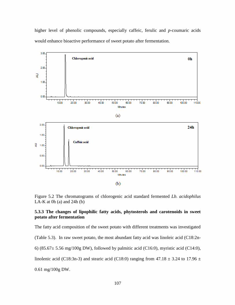
107
higher level of phenolic compounds, especially caffeic, ferulic and p-coumaric acids
would enhance bioactive performance of sweet potato after fermentation.
Figure 5.2 The chromatograms of chlorogenic acid standard fermented Lb. acidophilus
LA-K at 0h (a) and 24h (b)
5.3.3 The changes of lipophilic fatty acids, phytosterols and carotenoids in sweet
potato after fermentation
The fatty acid composition of the sweet potato with different treatments was investigated
(Table 5.3). In raw sweet potato, the most abundant fatty acid was linoleic acid (C18:2n-
6) (85.67± 5.56 mg/100g DW), followed by palmitic acid (C16:0), myristic acid (C14:0),
linolenic acid (C18:3n-3) and stearic acid (C18:0) ranging from 47.18 ± 3.24 to 17.96 ±
0.61 mg/100g DW.
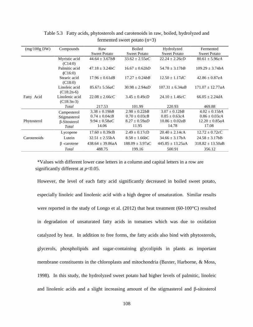
108
Table 5.3 Fatty acids, phytosterols and carotenoids in raw, boiled, hydrolyzed and
fermented sweet potato (n=3)
*Values with different lower case letters in a column and capital letters in a row are
significantly different at p<0.05.
However, the level of each fatty acid significantly decreased in boiled sweet potato,
especially linoleic and linolenic acid with a high degree of unsaturation. Similar results
were reported in the study of Longo et al. (2012) that heat treatment (60-100°C) resulted
in degradation of unsaturated fatty acids in tomatoes which was due to oxidation
catalyzed by heat. In addition to free forms, the fatty acids also bind with phytosterols,
glycerols, phospholipids and sugar-containing glycolipids in plants as important
membrane constituents in the chloroplasts and mitochondria (Baxter, Harborne, & Moss,
1998). In this study, the hydrolyzed sweet potato had higher levels of palmitic, linoleic
and linolenic acids and a slight increasing amount of the stigmasterol and β-sitosterol
(mg/100g DW) Compounds Raw
Sweet Potato
Boiled
Sweet Potato
Hydrolyzed
Sweet Potato
Fermented
Sweet Potato
Fatty Acid
Myristic acid
(C14:0)
44.64 ± 3.67bB 33.62 ± 2.55aC 22.24 ± 2.26cD 80.61 ± 5.96cA
Palmitic acid
(C16:0)
47.18 ± 3.24bC 16.67 ± 0.62bD 54.78 ± 3.17bB 109.29 ± 3.74bA
Stearic acid
(C18:0)
17.96 ± 0.61dB 17.27 ± 0.24bB 12.50 ± 1.17dC 42.86 ± 0.87eA
Linoleic acid
(C18:2n-6)
85.67± 5.56aC 30.98 ± 2.94aD 107.31 ± 6.34aB 171.07 ± 12.77aA
Linolenic acid
(C18:3n-3)
22.08 ± 2.66cC 3.45 ± 0.49cD 24.10 ± 1.46cC 66.05 ± 2.24dA
Total 217.53 101.99 220.93 469.88
Phytosterol
Campesterol
Stigmasterol
β-Sitosterol
Total
3.38 ± 0.19bB
0.74 ± 0.04cB
9.94 ± 0.58aC
14.06
2.98 ± 0.22bB
0.70 ± 0.03cB
8.27 ± 0.59aD
11.95
3.07 ± 0.12bB
0.85 ± 0.63cA
10.86 ± 0.02aB
14.78
4.02 ± 0.15bA
0.86 ± 0.03cA
12.20 ± 0.85aA
17.08
Lycopene 17.60 ± 0.39cB 2.49 ± 0.17cD 20.40 ± 2.14cA 12.72 ± 0.72cC
Carotenoids Lutein 32.51 ± 2.55bA 8.58 ± 1.66bC 34.66 ± 3.17bA 24.58 ± 3.17bB
β -carotene 438.64 ± 39.86aA 188.09 ± 3.97aC 445.85 ± 13.25aA 318.82 ± 13.50aB
Total 488.75 199.16 500.91 356.12

109
compared with the raw sweet potato (Table 5.3). Therefore, the increment might be
obtained from release of free fatty acids trapped in the starch granules or the breakdown
of bindings from phytosterol esters by acid hydrolysis. After fermented with Lb.
acidophilus LA-K, the linolenic acid (171.07 ± 12.77 mg/100g DW) was approximately
three times higher and the other determined fatty acids (ranged from 42.86 ± 0.87 to
109.29 ± 3.74 mg/100g DW) were approximately twice higher than each of them in raw
sweet potato. The increase of the fatty acids might result from enzymatic action based
de-esterification of bound fatty acids and the contribution of Lb. acidophilus LA-K which
also produce palmitic, myristic and stearic acid during growth (Johnsson et al., 1995).
Since the fatty acids, especially unsaturated ones help support brain cells growth,
neurological development and cognitive function, the accumulation of the fatty acid
would enhance the nutritional value of the fermented sweet potato (Panickar & Bhathena,
2010). Also, raw, boiled and hydrolyzed sweet potato had little difference in campesterol,
stigmasterol and β-sitosterol contents, while the three phytosterols exhibited increasing
levels after fermentation (Table 5.3). Thus, they would help improve the nutrition value
of fermented sweet potato as the phytosterols were recommended for consumption to
inhibit intestinal cholesterol absorptions and to control cholesterol concentrations in
plasma due to the similar structure of cholesterol but with different side chains (Ishida,
2014). Among the three determined carotenoids, β-carotene was the dominant (438.64 ±
39.86 mg/100g DW) which was followed by lutein (32.51 ± 2.55 mg/100g DW) and

110
lycopene (17.60 ± 0.39 mg/100g DW) in raw sweet potato, while each of the carotenoids
slightly increase in the hydrolyzed samples. However, different degree of reduction of
carotenoids profile was observed in boiling and fermentation treatments which were due
to the long time heat or light exposure (Table 5.3).
On the other hand, the Lactobacillus acidophilus is a probiotic bacteria species
which could help reducing overgrowth of pathogens, stimulating an immune response,
improving the blood lipid metabolism or potentially killing cancer cells in human
gastrointestinal tract (Sanders & Klaenhammer, 2001). Therefore, the fermented sweet
potato would be considered an ideal food system to enhance the bioavailability of the
bioactive compounds of sweet potato, but also deliver Lactobacillus acidophilus for its
probiotic performance. As a result, the hydrophilic and lipophilic raw and fermented
sweet potato extracts were selected and compared in the following cellular study.
5.3.4 The effect of raw and fermented sweet potato extracts on pheochromocytoma
derived cell (PC-12) and normal monkey kidney cell (CV-1) proliferation
The anticancer potentials of hydrophilic, purified hydrophilic and lipophilic extracts of
raw sweet potato versus fermented sweet potato were evaluated by the inhibition
efficiency on cancer cell PC-12 growth. Figure 5.3a demonstrated that purified
hydrophilic extracts of raw (PHR) and fermented (PHF) sweet potato caused dose-
dependent inhibition of PC-12 cell proliferation after 24 h incubation. As the extract
concentration increased to 0.2 mg/mL, the viable cancer cells dropped to 9.5% in PHF,

111
while there was 63.2% remained in PHR (Figure 5.3a). The highest inhibitory rate was
achieved at a concentration of 0.5 mg/mL, at which level, only 30.1% and 2.1 % of the
cells survived in purified hydrophilic extracts of raw (PHR) and fermented (PHF) sweet
potato, respectively (Figure 5.3a). Thus, the greater performance of PHF was due to the
higher levels and abundant free-form phenolic compounds than those of PHR. For
example, there were four more phenolics including caffeic acid derivative, p-coumaric,
ferulic and cinnamic acids in fermented sweet potato than raw sweet potato (Table 5.2).
Also, compared with raw sweet potato, the concentration of caffeic acid or 3,5-
dicaffeoylquinic acid in fermented sweet potato was approximately four and seven times
higher (Table 5.2). Similar result was found in the study of Rocha et al. (2012), who
reported that caffeic acid, 3,4-, 3,5- and 4,5- dicaffeoylquinic acid as well as ferulic and
p-coumaric acid could inhibit colon cancer cell lines including RKO, HT-29 and Caco-2.
There was no significant difference between lipophilic extract of raw (LR) and
fermented (LF) sweet potato in inhibiting PC-12 cell growth below 0.1 mg/mL (Figure
5.3b). As the extract concentration increased to 0.2 mg/mL, the inhibition efficiency of
LF was significantly higher than LR (Figure 5.3b). However, with the same
concentration of 0.5 mg/mL, LF and LR contained 44.6 and 53.4 % of viable cancer cells
which was much higher than that of PHF (2.1%) and PHR (30.1%) (Figure 5.3a & 5.3b).

112
Figure 5.3 The inhibition of PC-12 cell proliferation by (a) purified hydrophilic raw
(PHR) and fermented sweet potato (PHF); (b) lipophilic extracts of raw (LR) and
fermented sweet potato (LF); and the morphology of normal PC-12 cell (c) and apoptotic
PC-12 cell (d)
As reported in the study of Upadhyay et al. (2007), carotenoids could deregulate the
cancer cell proliferation rate by interfering different phases of the cell cycle, therefore, it
was possible that the abundant carotenoids (488.75 mg/100g DW for LR and 356.12
mg/100g DW for LF) in the lipophilic extracts that mainly contributed to the capability in
inhibiting the cancer cell proliferation. Although phytosterols also play an important role
in suppressing the growth of the cancer cell, its total content was only 14.06 and 17.08
mg/100g DW in LR and LF, respectively, which may have limited its anti-cancer
efficiency (Table 5.3). In addition, the fatty acids concentrations rose dramatically in

113
fermented sweet potato, however, they were not the primary factors that were responsible
for the anticancer performance of the lipophilic extracts. Thus, the LR and LF exhibited
relatively weaker anti-cancer potential than PHR and PHF.
The interaction of lipophilic and hydrophilic bioactive compounds with the cancer
cell is based on different accessibility and inhibition pathway. Generally, lipophilic
compounds could easily pass across the lipid bilayers of biomembranes by simple
diffusion or special membrane proteins for transferring such no-polar solutes (Alberts et
al., 2002). Distinctive from that of lipophilic substances, most hydrophilic compounds
have difficulties in penetrating the cell membrane. However, at physiologic pH, the
multiple hydroxyl groups in small molecule phenolics could interact with the polar head
groups of phospholipids at the membrane surface via the formation of hydrogen bonds
(Manach et al., 2004). It would then increase pH and the penetration of the phenolics
would be improved by deprotonation of the hydroxyl groups and variations in membrane
structure and fluidity (Castelli et al., 1999). Compared with the control cancer cell shown
in Figure 5.3c, the speeded up apoptotic progress of cancer cells was induced by
lipophilic or purified hydrophilic sweet potato extracts which resulted in the presence of
cell shrinkage, apoptotic bodies, and margination of the nucleus indicated by arrows
(Figure 5.3d). However, the anti-cancer potential of the hydrophilic phytochemicals
would be affected since hydrophilic extracts of sweet potato contained a large amount of

114
carbohydrates but much fewer equivalents of phenolic compounds as purified hydrophilic
extracts. Thus, it would have side influence on cancer cell growth.
As shown in Figure 5.4b, both HR and HF helped to remain the growth of the cancer
cell at a relatively low concentration (<1 mg/mL). Similar result was demonstrated in the
study of Masur et al. (2011) that with progressive tumorigenesis, the promotion of cell
proliferation, progression of cell cycle, anti-apoptotic signaling and angiogenesis in
tumor cell lines from several organs were observed at a diabetogenic glucose
concentration (11 mM) compared to physiological one (5.5 mM). At the same
concentration of 0.5 mg/mL with lipophilic (LR and LF) and purified hydrophilic extracts
(PHR and PHF), HR and HF exhibited much weaker inhibitory efficiency (Figure 5.4a).
However, the viability of the cancer cell rapidly reduced to 21.11 % in HF and 35.42 %
in HR as their concentration increased to 50 mg/mL and finally exhibited 100%
inhibitory efficiency in both HR and HF at the concentrations as high as 100 mg/mL
(Figure 5.4a). The significant reduction of the survival rate at high concentrations could
be explained by osmotic pressure of the high carbohydrate concentration which caused
the aggregation and morphological variation of the cells as marked with arrow in Figure
5.4b.

115
Figure 5.4 The inhibition of PC-12 cell proliferation by hydrophilic extracts of fresh (HR)
and fermented sweet potato (HF) (a) and the morphology of dead PC-12 cell (b)
On the other hand, the priority of the applying plant-derived bioactive extracts as
alternative cancer remedies is to effectively reduce the incidence of cancers but with low
toxicities and low side effects compared with other cancer treatments such as
chemotherapeutic agents or radiation. In this study, the monkey kidney cell lines (CV-1)
were used for assessing the influence of different sweet potato extracts on the growth of
normal cells. Figure 5.5 elucidated that little inhibition effect was observed in CV-1 cells
treated with the same concentrations of the extract as in PC-12 cell lines. For example,
relatively low concentration (0.02 mg/mL) of PHR and PHF slightly assisted the CV-1
growth. Even at the maximum concentration of 0.5 mg/mL, only 22.37-24.74% or 16.75-
19.75% of CV-1 cells were inhibited in purified hydrophilic extracts and lipophilic
extracts, respectively (Figure 5.5).

116
Figure 5.5 The effect of PHR, PHF, LR and LF on CV-1 cell proliferation
Bars with different letters are significantly different (p < 0.05)
The high inhibitory efficiency of the plant derived extracts on cancer cell but
weak influence on normal cells could be explained by their different energy metabolisms.
Generally, normal cells with competent mitochondria undergo Krebs Cycle metabolic
pathway and generate the majority of ATP for cell growth (Wen, Zhu, & Huang, 2013).
However, most cancer cells have dysfunction in mitochondrial, and rely more on the
glycolytic pathway in the cytosol to generate the metabolic intermediates and ATP (Wen,
Zhu, & Huang, 2013). Thus, the key enzymes in glycolytic pathway have been
considered as potential therapeutic targets for inhibition of cancer cell proliferation. Due
to the non-covalent interactions with proteins, phenolic compounds have been reported as
the inhibitors of key enzymes in glycolytic pathway, such as glucosidases, xanthine
oxidase and PKM2 activities which further perturb proliferation of the cancer cell (Aslan,
Guler, & Adem, 2015). As for the lipophilic phytosterols and carotenoids, they could

117
interrupt balance between cancer cell proliferation and apoptosis by exerting interference
with protein phosphatase 2A (PP2A) in sphingomyelin cycle and block the cell cycle at
G0/G1 phase in prostate cancer, hepatocyte, and breast cancer cell lines (Awad & Fink,
2000; van Breemen & Pajkovic, 2008). Therefore, phytochemicals could be used as
either mono-treatments or in association with classical chemotherapeutic drugs,
increasing the therapeutic efficacy on cancer cells and lowering the toxic side effects on
normal cells.
5.4 Conclusion
In summary, this study demonstrated that fermentation of Garnet sweet potato with
probiotic Lb. acidophilus LA-K could significantly increase the content of most
hydrophilic phenolic acids, and lipophilic fatty acids or phytosterols via enzymatic action
based bio-releasing or bioconversion of the microorganisms. However, it slightly
reduced the carotenoids level. Also, the fermented sweet potato extracts with a higher
level and abundant phytochemicals exhibited higher efficiency than raw extracts in
inhibiting the pheochromocytoma derived cancer cell (PC-12) proliferation. The purified
hydrophilic extracts of raw or fermented extracts had greater anticancer potential than
their corresponding lipophilic extracts. However, there were minimal effects on the
normal monkey kidney cell (CV-1) growth because those phytochemicals targeted on
deactivating the key enzymes of glycolytic pathway in cancer cells energy metabolism

118
but has less influence on the Krebs Cycle pathway for normal cells. Therefore,
fermentation with Lb. acidophilus LA-K is a valuable biotechnological approach for
sweet potato nutritional value enrichment, and bioavailability improvement, and the
fermented extracts could be further applied as promising therapeutic ingredients for
cancer treatment.
5.5 References
Alberts, B., Johnson, A., Lewis, J., Raff, M., Roberts, K., & Walter, P. (2002). Molecular
Biology of the Cell. (4th ed.). New York: Garland Science, (Chapter 11).
Aslan, E., Guler, C., & Adem, S. (2015). In vitro effects of some flavonoids and phenolic
acids on human pyruvate kinase isoenzyme M2. Journal of Enzyme Inhibition and
Medicinal Chemistry, 23, 1-4.
Awad, A. B., & Fink, C. S. (2000). Phytosterols as anticancer dietary components:
evidence and mechanism of action. Journal of Nutrition, 130, 2127-2130.
Bahado-Singh, P. S., Riley, C. K., Wheatley, A. O., & Lowe, H. I. C. (2011).
Relationship between Processing Method and the Glycemic Indices of Ten Sweet
Potato (Ipomoea batatas) Cultivars Commonly Consumed in Jamaica. Journal of
Nutrition and Metabolism, 2011, 6.
Barthelmebs, L., Diviés, C., & Cavin, J. (2001). Molecular characterization of the
phenolic acid metabolism in the lactic acid bacteria Lactobacillus plantarum.
Dairy Science and Technology, 81, 161-171.
Baxter, H., Harborne, J. B., & Moss, G. P. (1998). Phytochemical Dictionary: A
Handbook of Bioactive Compounds from Plants. (2nd ed.). Taylor & Francis,
(Chapter 6).
Bel-Rhlid, R., Thapa, D., Kraehenbuehl, K., Hansen, C. E., & Fischer, L. (2013).
Biotransformation of caffeoyl quinic acids from green coffee extracts by
Lactobacillus johnsonii NCC 533. AMB Express, 3, 28.
Castelli, F., Uccella, N., Trombetta, D., & Saija, A. (1999). Differences between
Coumaric and Cinnamic Acids in Membrane Permeation As Evidenced by Time-
Dependent Calorimetry. Journal of Agricultural and Food Chemistry, 47, 991-
995.
119
Cui, L., Liu, C.-q., Li, D.-j., & Song, J.-f. (2011). Effect of Processing on Taste Quality
and Health-Relevant Functionality of Sweet Potato Tips. Agricultural Sciences in
China, 10, 456-462.
Du, L., Shen, Y., Zhang, X., Prinyawiwatkul, W., & Xu, Z. (2014). Antioxidant-rich
377 phytochemicals in miracle berry (Synsepalum dulcificum) and antioxidant activity of
its extracts. Food Chemistry, 153, 279-284.
Duangjitcharoen, Y., Kantachote, D., Ongsakul, M., Poosaran, N., and Chaiyasut, C.
(2008). Selection of probiotic lactic acid bacteria isolated from fermented plant
beverages. Pakistan Journal of Biological Science, 11, 652-655.
Esteban-Torres, M., Reveron, I., Mancheno, J. M., de Las Rivas, B., & Munoz, R. (2013).
Characterization of a feruloyl esterase from Lactobacillus plantarum. Applied and
Environmental Microbiology, 79, 5130-5136.
Gibson, P. R., Varney, J., Malakar, S., & Muir, J. G. (2015). Food Components and
Irritable Bowel Syndrome. Gastroenterology, 148, 1158-1174.
Huang, H.-Y., Hsieh, H.-Y., King, V. A.-E., Chi, L.-L., & Tsen, J.-H. (2014). To pre-
challenge lactic acid bacteria with simulated gastrointestinal conditions is a
suitable approach to studying potential probiotic properties. Journal of
Microbiological Methods, 107, 138-146.
Ishida, N. (2014). A method for simultaneous analysis of phytosterols and phytosterol
esters in tobacco leaves using non aqueous reversed phase chromatography and
atmospheric pressure chemical ionization mass spectrometry detector. Journal of
Chromatography A, 1340, 99-108.
Johnsson, T., Nikkila, P., Toivonen, L., Rosenqvist, H., & Laakso, S. (1995). Cellular
Fatty Acid profiles of lactobacillus and lactococcus strains in relation to the oleic
Acid content of the cultivation medium. Applied and Environmental Microbiology,
61, 4497-4499.
Khoddami, A., Wilkes, M. A., & Roberts, T. H. (2013). Techniques for analysis of plant
phenolic compounds. Molecules, 18, 2328-2375.
Kim, Y. H., Jeong, J. C., Lee, H. S., & Kwak, S. S. (2013). Comparative characterization
of sweetpotato antioxidant genes from expressed sequence tags of dehydration-
treated fibrous roots under different abiotic stress conditions. Molecular Biology
Reports, 40, 2887-2896.
Kubow, S., Iskandar, M. M., Sabally, K., Azadi, B., Sadeghi Ekbatan, S., Kumarathasan,
P., Das, D. D., Prakash, S., Burgos, G., & zum Felde, T. (2016).
Biotransformation of anthocyanins from two purple-fleshed sweet potato
accessions in a dynamic gastrointestinal system. Food Chemistry, 192, 171-177.

120
Kwon, D. Y., Nyakudya, E., & Jeong, Y. S. (2014). Fermentation: Food Products. In N.
K. V. Alfen (Eds.), Encyclopedia of Agriculture and Food Systems (pp. 113-123).
Oxford: Academic Press.
Longo, C., Leo, L., & Leone, A. (2012). Carotenoids, Fatty Acid Composition and Heat
Stability of Supercritical Carbon Dioxide-Extracted-Oleoresins. International
Journal of Molecular Sciences, 13, 4233.
Manach, C., Scalbert, A., Morand, C., Rémésy, C., & Jiménez, L. (2004). Polyphenols:
food sources and bioavailability. The American Journal of Clinical Nutrition, 79,
727-747.
Masiero, S. S., Peretti, A., Trierweiler, L. F., & Trierweiler, J. O. (2014). Simultaneous
cold hydrolysis and fermentation of fresh sweet potato. Biomass and Bioenergy,
70, 174-183.
Masur, K., Vetter, C., Hinz, A., Tomas, N., Henrich, H., Niggemann, B., & Zanker, K. S.
(2011). Diabetogenic glucose and insulin concentrations modulate transcriptome
and protein levels involved in tumour cell migration, adhesion and proliferation.
British Journal of Cancer, 104, 345-352.
Morrow, L. E., Gogineni, V., & Malesker, M. A. (2012). Probiotic, prebiotic, and
synbiotic use in critically ill patients. Current Opinion in Critical Care, 18, 186-
191.
Olthof, M. R., Hollman, P. C., & Katan, M. B. (2001). Chlorogenic acid and caffeic acid
are absorbed in humans. Journal of Nutrition, 131, 66-71.
Panickar, K.S., & Bhathena, S.J. (2010). Control of Fatty Acid Intake and the Role of
Essential Fatty Acids in Cognitive Function and Neurological Disorders. In:
Montmayeur JP, le Coutre J (Eds.), Fat Detection: Taste, Texture, and Post
Ingestive Effects. (pp. 470-474). Boca Raton: CRC Press.
Ren, D., Li, C., Qin, Y., Yin, R., Du, S., Ye, F., Liu, C., Liu, H., Wang, M., Li, Y., Sun,
Y., Li, X., Tian, M., & Jin, N. (2014). In vitro evaluation of the probiotic and
functional potential of Lactobacillus strains isolated from fermented food and
human intestine. Anaerobe, 30, 1-10.
Rocha, L. D., Monteiro, M.C., & Teodoro, A. J. (2012). Anticancer properties of
hydroxycinnamic acids-a review. Cancer and Clinical Oncology, 1, 109-121.
Sanders, M. E., & Klaenhammer, T. R. (2001). Invited review: the scientific basis of
Lactobacillus acidophilus NCFM functionality as a probiotic. Journal of Dairy
Science, 84, 319-331.
Snook, M. E., Data, E. S., & Kays, S. J. (1994). Characterization and quantitation of
hexadecyl, octadecyl, and eicosyl esters of p-coumaric acid in the vine and root

121
latex of sweetpotato [Ipomoea batatas (L.) Lam.]. Journal of Agricultural and
Food Chemistry, 42, 2589-2595.
Szwajgier, D. (2010). Biotransformation of ferulic acid by Lactobacillus acidophilus K1
and selected Bifidobacterium strains. Acta Scientiarum Polonorum. Technologia
Alimentaria, 9, 45-59.
Tang, Y., Cai, W., & Xu, B. 2015. Profiles of phenolics, carotenoids and antioxidative
capacities of thermal processed white, yellow, orange and purple sweet potatoes
grown in Guilin, China. Food Science and Human Wellness. (In press).
Truong, V. D., McFeeters, R. F., Thompson, R. T., Dean, L. L., & Shofran, B. (2007).
Phenolic acid content and composition in leaves and roots of common
commercial sweetpotato (Ipomea batatas L.) cultivars in the United States.
Journal of Food Science, 72, C343-349.
Upadhyaya, K. R., Radha, K. S., & Madhyastha, H. K. (2007). Cell cycle regulation and
induction of apoptosis by beta-carotene in U937 and HL-60 leukemia cells.
Journal of biochemistry and molecular biology, 40, 1009-1015.
van Breemen, R. B., & Pajkovic, N. (2008). Multitargeted therapy of cancer by lycopene.
Cancer Letters, 269, 339-351.
Wen, S., Zhu, D., & Huang, P. (2013). Targeting cancer cell mitochondria as a
therapeutic approach. Future Medicinal Chemistry, 5, 53-67.
Wu, Q., Qu, H., Jia, J., Kuang, C., Wen, Y., Yan, H., & Gui, Z. (2015). Characterization,
antioxidant and antitumor activities of polysaccharides from purple sweet potato.
Carbohydrate Polymers, 132, 31-40.
Xu, Z., & Godber, J. S. (1999). Purification 478 and identification of components of
gamma oryzanol in rice bran Oil. Journal of Agricultural and Food Chemistry, 47,
2724-2728.
Xu, Z. (2012). Analysis methods of phenolic acids. In Z. Xu, & L.R. Howard (Eds.),
Analysis of antioxidant-rich phytochemicals. (pp.69-74). Iowa: John Wiley& Sons.
Inc.
Yue, X., Xu, Z., Prinyawiwatkul, W., Losso, J. N., King, J. M., & Godber, J. S. (2008).
Comparison of soybean oils, gum, and defatted soy flour extract in stabilizing
menhaden oil during heating. Journal of Food Science, 73, C19-23.

122
CHAPTER 6 SUMMARY AND CONCLUSIONS
Chapter 2 reported the levels and profiles of hydrophilic and lipophilic antioxidant
phytochemicals in sweet sorghum millets. The dominant hydrophilic and lipophilic
antioxidants were apigeninidin and γ-tocopherol, respectively. The hydrophilic
antioxidants showed higher antioxidant activity than lipophilic antioxidants in both the
DPPH assay and the cholesterol-linoleic acid oxidation model. Therefore, the health
benefits of sweet sorghum may be mainly attributed by its hydrophilic antioxidant
phytochemicals. Thus, sweet sorghum (Dura) could be a valuable biomaterial for health
promoting applications.
Chapter 3 investigated the phenolic profiles of Thai holy and sweet basil seeds
and leaves. This is the first study to investigate the anti-lipid-oxidation capabilities of
Thai holy and sweet basil seeds and leaves by using a cholesterol emulsion model. In
this study, rosmarinic, caftaric, chioric, p-hydroxybenzoic and protoatechuic acid were
detected in both THBL and TSBL. Caffeic acid was only found in Thai holy basil.
Additionally, THBS had seven primary phenolics including rosmarinic, caffeic, chicoric,
p-hydroybenzoic, p-coumaric, protocatechuic acid and rutin, while only p-hydroybenzoic
and rutin were found in TSBS. Thai holy basil leaves exhibited greater anti-lipid-
oxidation performance than its seeds or Thai sweet basil leaves and seeds. Therefore,
Thai holy basil leaves could be processed as an antioxidant ingredient to be applied in

123
various food products. In our future study, animal and cell system will be applied to
verify the health promoting function of the Thai holy basil leaves.
Chapter 4 demonstrated the concentration and profile of phenolics, tocopherols,
phytosterols and fatty acids in butterfly pea seeds and petals. HEp-2 carcinoma cell line
was used to evaluate the anticancer capability of butterfly pea seeds and petals extracts
(HBP, HBS, LBP and LBS). The results indicated HBS was the most efficient in
inhibiting the viability of HEp-2 cells. Both HBS and HBP exhibited greater
performance than LBS and LBP in inhibiting the cells proliferation. Therefore, the
hydrophilic and lipophilic butterfly pea seeds and petals extracts possess health
promoting function and could be used as a functional food ingredient or potential cancer
therapy supplement.
Chapter 5 summarized that fermentation of Garnet sweet potato with probiotic Lb.
acidophilus LA-K can significantly increase the content of most hydrophilic phenolic
acids, and lipophilic fatty acids or phytosterols via enzymatic action based bio-releasing
or bioconversion of the microorganisms. However, it slightly reduced the carotenoids
level. Also, the fermented sweet potato extracts with a higher level and abundant
phytochemicals exhibited higher efficiency than raw extracts in inhibiting the
pheochromocytoma derived cancer cell (PC-12) proliferation. The purified hydrophilic
extracts of raw or fermented extracts had greater anticancer potential than their
corresponding lipophilic extracts. However, these extracts had minimal impact on the

124
normal monkey kidney cell (CV-1) growth because they targeted on deactivating the key
enzymes of glycolytic pathway in cancer cells energy metabolism but has less influence
on the Krebs Cycle pathway for normal cells. Therefore, fermentation with Lb.
acidophilus LA-K is a valuable biotechnological approach for sweet potato nutritional
value enrichment, and bioavailability improvement, and the fermented extracts could be
further applied as promising therapeutic ingredients for cancer treatment.

125
APPENDIX A: THE LETTER OF PERMISSION OF PUBLISHED PAPER “PHYTOCHEMICALS IN SWEET SORGHUM (DURA) AND THEIR ANTIOXIDANT
CAPABILITIES AGAINST LIPID OXIDATION”

126
APPENDIX B: THE LETTER OF PERMISSION OF PUBLISHED PAPER “COMPARISON OF PHENOLIC PROFILES AND ANTIOXIDANT POTENTIALS OF
THE LEAVES AND SEEDS OF THAI HOLY AND SWEET BASILS”

201B AFS buildingSchool of Nutrition and Food SciLouisiana State UniversityBATON ROUGE, LA 70803United StatesAttn: Witoon Prinyawiwatkul
Total 0.00 USD
Would you like to purchase the full text of this article? If so, please continue on to thecontent ordering system located here: Purchase PDF
If you click on the buttons below or close this window, you will not be able to return to thecontent ordering system.
ORDER MORE CLOSE WINDOW
Copyright © 2015 Copyright Clearance Center, Inc. All Rights Reserved. Privacy statement. Terms and Conditions. Comments? We would like to hear from you. Email us at [email protected]
127

128
VITA
Yixiao Shen was born in May, 1987, Shenyang, China. In June 2010, she graduated from
Shenyang Agricultural University with a Bachelor of Food Quality and Safety. After receiving
her bachelor’s degree, she was recommended for Master study without examination majoring in
Food Engineering at Shenyang Agricultural University in September, 2010. She joined the Ph.D.
program in the School of Nutrition and Food Sciences at Louisiana State University in 2011
under the mentorship of Dr. Zhimin Xu and Dr. Witoon Prinyawiwatkul. She will receive degree
of Doctor of Philosophy in Food Science during the December 2015 commencement ceremony.